Mechanistic similarities between oxidation of hydroethidine by Fremy’s salt and superoxide:...
11
Original Contribution Mechanistic similarities between oxidation of hydroethidine by Fremy_s salt and superoxide: Stopped-flow optical and EPR studies Jacek Zielonka, Hongtao Zhao, Yingkai Xu, B. Kalyanaraman * Department of Biophysics and Free Radical Research Center, Medical College of Wisconsin, 8701 Watertown Plank Road, P.O. Box 26509, Milwaukee, WI 53226, USA Received 18 February 2005; revised 29 April 2005; accepted 3 May 2005 Abstract We have previously shown that superoxide radical anion (O 2 S ) reacts with hydroethidine (HE) to form a product that is distinctly different from ethidium (E + ) (Zhao et al., Free Radic. Biol. Med. 34:1359; 2003). The structure of this product was recently determined as the 2-hydroxyethidium cation (2-OH-E + ) (Zhao et al., Proc. Natl. Acad. Sci. USA 102:5727; 2005). In this study, using HPLC and mass spectrometry techniques, we show that 2-OH-E + is formed from the reaction between HE and nitrosodisulfonate radical dianion (NDS) or Fremy_s salt. The reaction kinetics and mechanism were determined using steady-state and time-resolved optical and EPR techniques. Within the first 50 ms, an intermediate was detected. Another intermediate absorbing strongly at 460 nm and weakly at 670 nm was detected within a second. The structure of this species was assigned to an imino quinone derivative of HE. The stoichiometry of the reaction indicates that two molecules of NDS were needed to oxidize a molecule of HE. We postulate that the first step of the reaction involves the hydrogen atom abstraction from HE to form an aminyl radical that reacts with another molecule of NDS to form an adduct that decomposes to an imino quinone derivative of HE. A similar mechanism has been proposed for the reaction between HE and O 2 S . The reaction between HE and the Fremy_s salt should provide a facile route for the synthesis of 2-OH-E + , a diagnostic marker product of the HE/O 2 S reaction. D 2005 Elsevier Inc. All rights reserved. Keywords: Hydroethidine; Fremy_s salt; Nitrosodisulfonate radical dianion; Superoxide radical anion; 2-Hydroxyethidium; Fluorescent probes; Stopped flow; HPLC Introduction It is now generally believed that reactive oxygen species (ROS) are not only deleterious by-products of selected enzymatic and nonenzymatic processes, but are also important cell signaling intermediates [1,2]. Superoxide radical anion (O 2 S ), the primary one-electron reduction product of molecular oxygen, triggers the formation of other ROS, such as H 2 O 2 , hydroxyl radical ( S OH), and peroxyl radicals (ROO S ). Thus, it is often difficult to attribute a particular signal transduction mechanism to a specific ROS [3]. This situation is further complicated by the fact that most extracellular ROS scavengers are not cell permeable. One of the factors that hampers our understanding of ROS in cell signaling and signal transduction mechanisms is the lack of specific cell-permeable diagnostic probes for O 2 S . It is essential that these probes are sensitive enough to detect low levels of ROS in cells without affecting the cellular function [4,5]. In this regard, the fluorescence technique is promising due to the availability of cell-permeable non- fluorescent probes that are trapped inside the cells by esterase-dependent hydrolysis, leading to the formation of characteristic fluorescent products during intracellular oxi- dative stress. Recently, 5-ethyl-5,6-dihydro-6-phenyl-3,8-diaminophe- nanthridine (hydroethidine, HE, also known as dihydro- 0891-5849/$ - see front matter D 2005 Elsevier Inc. All rights reserved. doi:10.1016/j.freeradbiomed.2005.05.001 Abbreviations: HE, hydroethidine; NDS, nitrosodisulfonate radical dianion; Fs, Fremy_s salt; XO, xanthine oxidase; DTPA, diethylenetriamine pentaacetic acid; E + , ethidium cation; 2-OH-E + , 2-hydroxyethidium cation; ROS, reactive oxygen species; DMSO, dimethyl sulfoxide; TFA, trifluoro- acetic acid. * Corresponding author. Fax: +1 414 456 6512. E-mail address: [email protected] (B. Kalyanaraman). Free Radical Biology & Medicine 39 (2005) 853 – 863 www.elsevier.com/locate/freeradbiomed
Transcript of Mechanistic similarities between oxidation of hydroethidine by Fremy’s salt and superoxide:...
www.elsevier.com/locate/freeradbiomed
Free Radical Biology &
Original Contribution
Mechanistic similarities between oxidation of hydroethidine by Fremy_ssalt and superoxide: Stopped-flow optical and EPR studies
Jacek Zielonka, Hongtao Zhao, Yingkai Xu, B. Kalyanaraman*
Department of Biophysics and Free Radical Research Center, Medical College of Wisconsin, 8701 Watertown Plank Road,
P.O. Box 26509, Milwaukee, WI 53226, USA
Received 18 February 2005; revised 29 April 2005; accepted 3 May 2005
Abstract
We have previously shown that superoxide radical anion (O2S�) reacts with hydroethidine (HE) to form a product that is distinctly
different from ethidium (E+) (Zhao et al., Free Radic. Biol. Med. 34:1359; 2003). The structure of this product was recently determined as the
2-hydroxyethidium cation (2-OH-E+) (Zhao et al., Proc. Natl. Acad. Sci. USA 102:5727; 2005). In this study, using HPLC and mass
spectrometry techniques, we show that 2-OH-E+ is formed from the reaction between HE and nitrosodisulfonate radical dianion (NDS) or
Fremy_s salt. The reaction kinetics and mechanism were determined using steady-state and time-resolved optical and EPR techniques. Within
the first 50 ms, an intermediate was detected. Another intermediate absorbing strongly at 460 nm and weakly at 670 nm was detected within a
second. The structure of this species was assigned to an imino quinone derivative of HE. The stoichiometry of the reaction indicates that two
molecules of NDS were needed to oxidize a molecule of HE. We postulate that the first step of the reaction involves the hydrogen atom
abstraction from HE to form an aminyl radical that reacts with another molecule of NDS to form an adduct that decomposes to an imino
quinone derivative of HE. A similar mechanism has been proposed for the reaction between HE and O2S�. The reaction between HE and the
Fremy_s salt should provide a facile route for the synthesis of 2-OH-E+, a diagnostic marker product of the HE/O2S� reaction.
D 2005 Elsevier Inc. All rights reserved.
Keywords: Hydroethidine; Fremy_s salt; Nitrosodisulfonate radical dianion; Superoxide radical anion; 2-Hydroxyethidium; Fluorescent probes; Stopped flow;
HPLC
Introduction
It is now generally believed that reactive oxygen species
(ROS) are not only deleterious by-products of selected
enzymatic and nonenzymatic processes, but are also
important cell signaling intermediates [1,2]. Superoxide
radical anion (O2S�), the primary one-electron reduction
product of molecular oxygen, triggers the formation of other
ROS, such as H2O2, hydroxyl radical (SOH), and peroxyl
0891-5849/$ - see front matter D 2005 Elsevier Inc. All rights reserved.
doi:10.1016/j.freeradbiomed.2005.05.001
Abbreviations: HE, hydroethidine; NDS, nitrosodisulfonate radical
dianion; Fs, Fremy_s salt; XO, xanthine oxidase; DTPA, diethylenetriamine
pentaacetic acid; E+, ethidium cation; 2-OH-E+, 2-hydroxyethidium cation;
ROS, reactive oxygen species; DMSO, dimethyl sulfoxide; TFA, trifluoro-
acetic acid.
* Corresponding author. Fax: +1 414 456 6512.
E-mail address: [email protected] (B. Kalyanaraman).
radicals (ROOS). Thus, it is often difficult to attribute a
particular signal transduction mechanism to a specific ROS
[3]. This situation is further complicated by the fact that
most extracellular ROS scavengers are not cell permeable.
One of the factors that hampers our understanding of ROS
in cell signaling and signal transduction mechanisms is the
lack of specific cell-permeable diagnostic probes for O2S�. It
is essential that these probes are sensitive enough to detect
low levels of ROS in cells without affecting the cellular
function [4,5]. In this regard, the fluorescence technique is
promising due to the availability of cell-permeable non-
fluorescent probes that are trapped inside the cells by
esterase-dependent hydrolysis, leading to the formation of
characteristic fluorescent products during intracellular oxi-
dative stress.
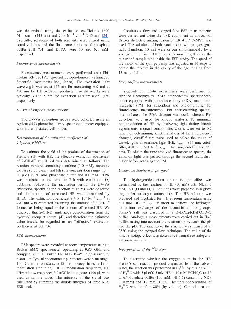
Recently, 5-ethyl-5,6-dihydro-6-phenyl-3,8-diaminophe-
nanthridine (hydroethidine, HE, also known as dihydro-
Medicine 39 (2005) 853 – 863
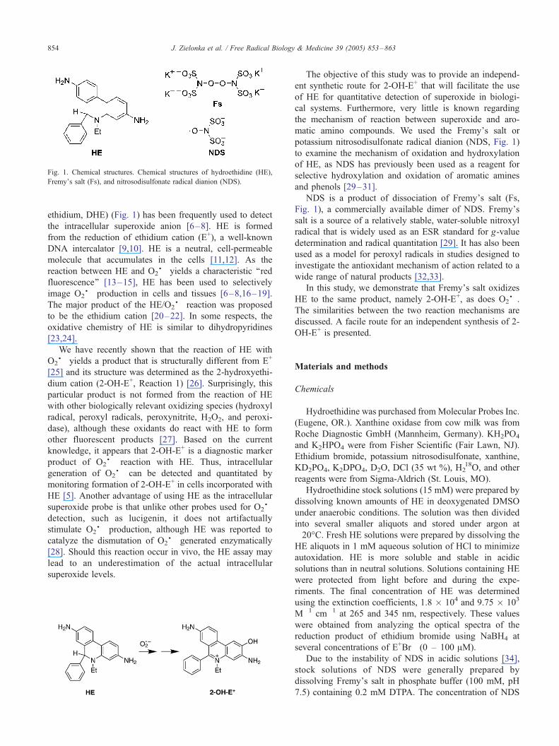
Fig. 1. Chemical structures. Chemical structures of hydroethidine (HE),
Fremy_s salt (Fs), and nitrosodisulfonate radical dianion (NDS).
J. Zielonka et al. / Free Radical Biology & Medicine 39 (2005) 853–863854
ethidium, DHE) (Fig. 1) has been frequently used to detect
the intracellular superoxide anion [6–8]. HE is formed
from the reduction of ethidium cation (E+), a well-known
DNA intercalator [9,10]. HE is a neutral, cell-permeable
molecule that accumulates in the cells [11,12]. As the
reaction between HE and O2S� yields a characteristic ‘‘red
fluorescence’’ [13–15], HE has been used to selectively
image O2S� production in cells and tissues [6–8,16–19].
The major product of the HE/O2S� reaction was proposed
to be the ethidium cation [20–22]. In some respects, the
oxidative chemistry of HE is similar to dihydropyridines
[23,24].
We have recently shown that the reaction of HE with
O2S� yields a product that is structurally different from E+
[25] and its structure was determined as the 2-hydroxyethi-
dium cation (2-OH-E+, Reaction 1) [26]. Surprisingly, this
particular product is not formed from the reaction of HE
with other biologically relevant oxidizing species (hydroxyl
radical, peroxyl radicals, peroxynitrite, H2O2, and peroxi-
dase), although these oxidants do react with HE to form
other fluorescent products [27]. Based on the current
knowledge, it appears that 2-OH-E+ is a diagnostic marker
product of O2S� reaction with HE. Thus, intracellular
generation of O2S� can be detected and quantitated by
monitoring formation of 2-OH-E+ in cells incorporated with
HE [5]. Another advantage of using HE as the intracellular
superoxide probe is that unlike other probes used for O2S�
detection, such as lucigenin, it does not artifactually
stimulate O2S� production, although HE was reported to
catalyze the dismutation of O2S� generated enzymatically
[28]. Should this reaction occur in vivo, the HE assay may
lead to an underestimation of the actual intracellular
superoxide levels.
The objective of this study was to provide an independ-
ent synthetic route for 2-OH-E+ that will facilitate the use
of HE for quantitative detection of superoxide in biologi-
cal systems. Furthermore, very little is known regarding
the mechanism of reaction between superoxide and aro-
matic amino compounds. We used the Fremy_s salt or
potassium nitrosodisulfonate radical dianion (NDS, Fig. 1)
to examine the mechanism of oxidation and hydroxylation
of HE, as NDS has previously been used as a reagent for
selective hydroxylation and oxidation of aromatic amines
and phenols [29–31].
NDS is a product of dissociation of Fremy_s salt (Fs,
Fig. 1), a commercially available dimer of NDS. Fremy_ssalt is a source of a relatively stable, water-soluble nitroxyl
radical that is widely used as an ESR standard for g-value
determination and radical quantitation [29]. It has also been
used as a model for peroxyl radicals in studies designed to
investigate the antioxidant mechanism of action related to a
wide range of natural products [32,33].
In this study, we demonstrate that Fremy_s salt oxidizesHE to the same product, namely 2-OH-E+, as does O2
S�.
The similarities between the two reaction mechanisms are
discussed. A facile route for an independent synthesis of 2-
OH-E+ is presented.
Materials and methods
Chemicals
Hydroethidine was purchased fromMolecular Probes Inc.
(Eugene, OR.). Xanthine oxidase from cow milk was from
Roche Diagnostic GmbH (Mannheim, Germany). KH2PO4
and K2HPO4 were from Fisher Scientific (Fair Lawn, NJ).
Ethidium bromide, potassium nitrosodisulfonate, xanthine,
KD2PO4, K2DPO4, D2O, DCl (35 wt %), H218O, and other
reagents were from Sigma-Aldrich (St. Louis, MO).
Hydroethidine stock solutions (15 mM) were prepared by
dissolving known amounts of HE in deoxygenated DMSO
under anaerobic conditions. The solution was then divided
into several smaller aliquots and stored under argon at
�20-C. Fresh HE solutions were prepared by dissolving the
HE aliquots in 1 mM aqueous solution of HCl to minimize
autoxidation. HE is more soluble and stable in acidic
solutions than in neutral solutions. Solutions containing HE
were protected from light before and during the expe-
riments. The final concentration of HE was determined
using the extinction coefficients, 1.8 � 104 and 9.75 � 103
M�1 cm�1 at 265 and 345 nm, respectively. These values
were obtained from analyzing the optical spectra of the
reduction product of ethidium bromide using NaBH4 at
several concentrations of E+Br� (0 – 100 AM).
Due to the instability of NDS in acidic solutions [34],
stock solutions of NDS were generally prepared by
dissolving Fremy_s salt in phosphate buffer (100 mM, pH
7.5) containing 0.2 mM DTPA. The concentration of NDS
J. Zielonka et al. / Free Radical Biology & Medicine 39 (2005) 853–863 855
was determined using the extinction coefficients 1690
M�1 cm�1 (248 nm) and 20.8 M�1 cm�1 (545 nm) [34].
Typically, solutions of both reactants were mixed using
equal volumes and the final concentrations of phosphate
buffer (pH 7.4) and DTPA were 50 and 0.1 mM,
respectively.
Fluorescence measurements
Fluorescence measurements were performed on a Shi-
madzu RF-5301PC spectrofluorophotometer (Shimadzu
Scientific Instruments Inc., Japan). The excitation light
wavelength was set at 356 nm for monitoring HE and at
470 nm for HE oxidation products. The slit widths were
typically 3 and 5 nm for excitation and emission light,
respectively.
UV-Vis absorption measurements
The UV-Vis absorption spectra were collected using an
Agilent 8453 photodiode array spectrophotometer equipped
with a thermostatted cell holder.
Determination of the extinction coefficient of
2-hydroxyethidium
To estimate the yield of the product of the reaction of
Fremy_s salt with HE, the effective extinction coefficient
of 2-OH-E+ at pH 7.4 was determined as follows: The
reaction mixture containing xanthine (1.0 mM), xanthine
oxidase (0.05 U/ml), and HE (the concentration range: 10 –
60 AM) in 50 mM phosphate buffer and 0.1 mM DTPA
was incubated in the dark for 2 h with continuous O2
bubbling. Following the incubation period, the UV-Vis
absorption spectra of the reaction mixtures were collected
and the amount of unreacted HE was determined by
HPLC. The extinction coefficient 9.4 � 103 M�1 cm�1 at
470 nm was estimated assuming the amount of 2-OH-E+
formed as being equal to the amount of reacted HE. We
observed that 2-OH-E+ undergoes deprotonation from the
hydroxyl group at neutral pH, and therefore the estimated
value should be regarded as an ‘‘effective’’ extinction
coefficient at pH 7.4.
ESR measurements
ESR spectra were recorded at room temperature using a
Bruker EMX spectrometer operating at 9.85 GHz and
equipped with a Bruker ER 4119HS-WI high-sensitivity
resonator. Typical spectrometer parameters were scan range,
100 G; time constant, 5.12 ms; sweep time, 5.12 s;
modulation amplitude, 1.0 G; modulation frequency, 100
kHz; microwave power, 5.0 mW.Micropipettes (100 Al) wereused as sample tubes. The intensity of the signal was
calculated by summing the double integrals of three NDS
ESR peaks.
Continuous flow and stopped-flow ESR measurements
were carried out using the ESR equipment as above, but
Bruker dielectric mixing resonator ER 4117 D-MVT was
used. The solutions of both reactants in two syringes (gas-
tight Hamilton, 10 ml) were driven simultaneously by a
syringe pump via PEEK tubes (0.7 mm i.d.), through the
mixer and sample tube inside the ESR cavity. The speed of
the motor of the syringe pump was adjusted in 10 steps to
obtain the mixture in the cavity of the age ranging from
15 ms to 1.5 s.
Stopped-flow measurements
Stopped-flow kinetic experiments were performed on
Applied Photophysics 18MX stopped-flow spectrophoto-
meter equipped with photodiode array (PDA) and photo-
multiplier (PM) for absorption and photomultiplier for
fluorescence measurements. For characterizing spectral
intermediates, the PDA detector was used, whereas PM
detectors were used for kinetic analysis. To minimize
photooxidation of HE by analyzing light during kinetic
experiments, monochromator slits widths were set to 0.2
mm. For determining kinetic analysis of the fluorescence
changes, cutoff filters were used to select the range of
wavelengths of emission light (HE, kexc = 356 nm; cutoff
filter, 400 nm; 2-OH-E+: kexc = 470 nm; cutoff filter, 550
nm). To obtain the time-resolved fluorescence spectra, the
emission light was passed through the second monochro-
mator before reaching the PM.
Deuterium kinetic isotope effect
The hydrogen/deuterium kinetic isotope effect was
determined by the reaction of HE (50 AM) with NDS (5
mM) in H2O and D2O. Solutions were prepared in a glove
bag under an argon atmosphere. The HE solution was
prepared and incubated for 1 h at room temperature using
a 1 mM DCl in D2O in order to achieve the hydrogen/
deuterium exchange of the aromatic amino groups.
Fremy_s salt was dissolved in a K2DPO4/KD2PO4/D2O
buffer. Analogous measurements were carried out in H2O
buffer, taking into account the relationship between the pH
and the pD. The kinetics of the reaction was measured at
25-C using the stopped-flow technique. The value of the
kinetic isotope effect was determined from three independ-
ent measurements.
Incorporation of the 18O atom
To determine whether the oxygen atom in the HE/
Fremy_s salt reaction product originated from the solvent
water, the reaction was performed in H218O by mixing 40 Al
of H218O with 5 Al of 0.5 mM HE in 10 mM HCl/H2O and 5
Al of phosphate buffer (100 mM, pH 7.5) containing NDS
(1.0 mM) and 0.2 mM DTPA. The final concentration of
H218O was therefore 80% (by volume). Control measure-
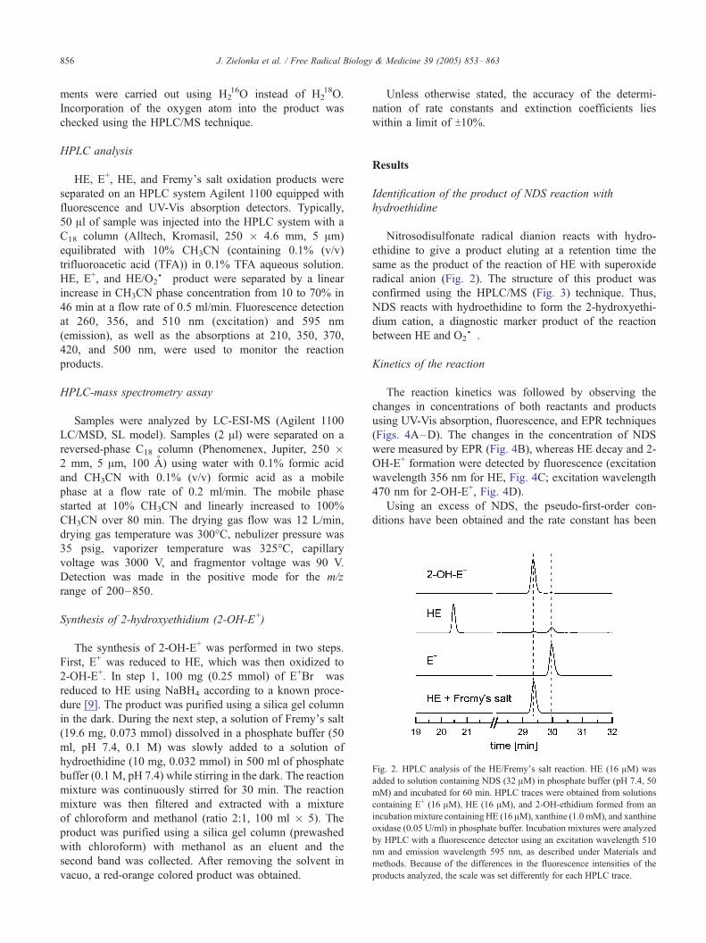
Fig. 2. HPLC analysis of the HE/Fremy_s salt reaction. HE (16 AM) was
added to solution containing NDS (32 AM) in phosphate buffer (pH 7.4, 50
mM) and incubated for 60 min. HPLC traces were obtained from solutions
containing E+ (16 AM), HE (16 AM), and 2-OH-ethidium formed from an
incubation mixture containing HE (16 AM), xanthine (1.0 mM), and xanthine
oxidase (0.05 U/ml) in phosphate buffer. Incubation mixtures were analyzed
by HPLC with a fluorescence detector using an excitation wavelength 510
nm and emission wavelength 595 nm, as described under Materials and
methods. Because of the differences in the fluorescence intensities of the
products analyzed, the scale was set differently for each HPLC trace.
J. Zielonka et al. / Free Radical Biology & Medicine 39 (2005) 853–863856
ments were carried out using H216O instead of H2
18O.
Incorporation of the oxygen atom into the product was
checked using the HPLC/MS technique.
HPLC analysis
HE, E+, HE, and Fremy_s salt oxidation products were
separated on an HPLC system Agilent 1100 equipped with
fluorescence and UV-Vis absorption detectors. Typically,
50 Al of sample was injected into the HPLC system with a
C18 column (Alltech, Kromasil, 250 � 4.6 mm, 5 Am)
equilibrated with 10% CH3CN (containing 0.1% (v/v)
trifluoroacetic acid (TFA)) in 0.1% TFA aqueous solution.
HE, E+, and HE/O2S� product were separated by a linear
increase in CH3CN phase concentration from 10 to 70% in
46 min at a flow rate of 0.5 ml/min. Fluorescence detection
at 260, 356, and 510 nm (excitation) and 595 nm
(emission), as well as the absorptions at 210, 350, 370,
420, and 500 nm, were used to monitor the reaction
products.
HPLC-mass spectrometry assay
Samples were analyzed by LC-ESI-MS (Agilent 1100
LC/MSD, SL model). Samples (2 Al) were separated on a
reversed-phase C18 column (Phenomenex, Jupiter, 250 �2 mm, 5 Am, 100 A) using water with 0.1% formic acid
and CH3CN with 0.1% (v/v) formic acid as a mobile
phase at a flow rate of 0.2 ml/min. The mobile phase
started at 10% CH3CN and linearly increased to 100%
CH3CN over 80 min. The drying gas flow was 12 L/min,
drying gas temperature was 300-C, nebulizer pressure was
35 psig, vaporizer temperature was 325-C, capillary
voltage was 3000 V, and fragmentor voltage was 90 V.
Detection was made in the positive mode for the m/z
range of 200–850.
Synthesis of 2-hydroxyethidium (2-OH-E+)
The synthesis of 2-OH-E+ was performed in two steps.
First, E+ was reduced to HE, which was then oxidized to
2-OH-E+. In step 1, 100 mg (0.25 mmol) of E+Br� was
reduced to HE using NaBH4 according to a known proce-
dure [9]. The product was purified using a silica gel column
in the dark. During the next step, a solution of Fremy_s salt(19.6 mg, 0.073 mmol) dissolved in a phosphate buffer (50
ml, pH 7.4, 0.1 M) was slowly added to a solution of
hydroethidine (10 mg, 0.032 mmol) in 500 ml of phosphate
buffer (0.1 M, pH 7.4) while stirring in the dark. The reaction
mixture was continuously stirred for 30 min. The reaction
mixture was then filtered and extracted with a mixture
of chloroform and methanol (ratio 2:1, 100 ml � 5). The
product was purified using a silica gel column (prewashed
with chloroform) with methanol as an eluent and the
second band was collected. After removing the solvent in
vacuo, a red-orange colored product was obtained.
Unless otherwise stated, the accuracy of the determi-
nation of rate constants and extinction coefficients lies
within a limit of T10%.
Results
Identification of the product of NDS reaction with
hydroethidine
Nitrosodisulfonate radical dianion reacts with hydro-
ethidine to give a product eluting at a retention time the
same as the product of the reaction of HE with superoxide
radical anion (Fig. 2). The structure of this product was
confirmed using the HPLC/MS (Fig. 3) technique. Thus,
NDS reacts with hydroethidine to form the 2-hydroxyethi-
dium cation, a diagnostic marker product of the reaction
between HE and O2S�.
Kinetics of the reaction
The reaction kinetics was followed by observing the
changes in concentrations of both reactants and products
using UV-Vis absorption, fluorescence, and EPR techniques
(Figs. 4A–D). The changes in the concentration of NDS
were measured by EPR (Fig. 4B), whereas HE decay and 2-
OH-E+ formation were detected by fluorescence (excitation
wavelength 356 nm for HE, Fig. 4C; excitation wavelength
470 nm for 2-OH-E+, Fig. 4D).
Using an excess of NDS, the pseudo-first-order con-
ditions have been obtained and the rate constant has been
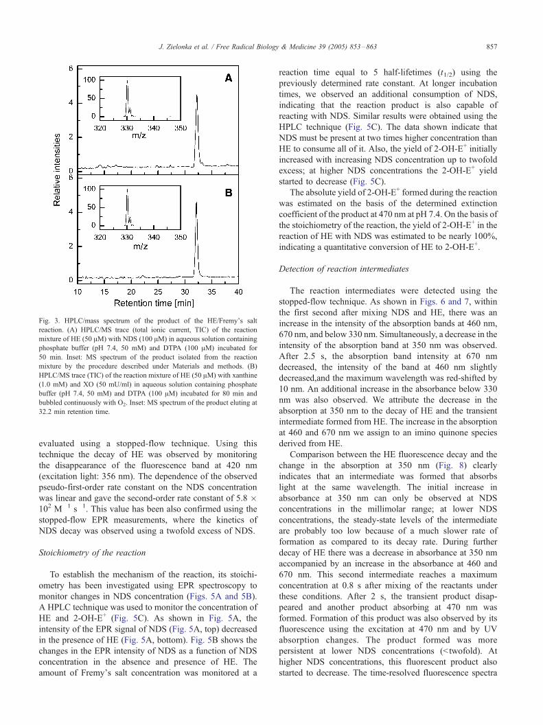
Fig. 3. HPLC/mass spectrum of the product of the HE/Fremy_s salt
reaction. (A) HPLC/MS trace (total ionic current, TIC) of the reaction
mixture of HE (50 AM) with NDS (100 AM) in aqueous solution containing
phosphate buffer (pH 7.4, 50 mM) and DTPA (100 AM) incubated for
50 min. Inset: MS spectrum of the product isolated from the reaction
mixture by the procedure described under Materials and methods. (B)
HPLC/MS trace (TIC) of the reaction mixture of HE (50 AM) with xanthine
(1.0 mM) and XO (50 mU/ml) in aqueous solution containing phosphate
buffer (pH 7.4, 50 mM) and DTPA (100 AM) incubated for 80 min and
bubbled continuously with O2. Inset: MS spectrum of the product eluting at
32.2 min retention time.
J. Zielonka et al. / Free Radical Biology & Medicine 39 (2005) 853–863 857
evaluated using a stopped-flow technique. Using this
technique the decay of HE was observed by monitoring
the disappearance of the fluorescence band at 420 nm
(excitation light: 356 nm). The dependence of the observed
pseudo-first-order rate constant on the NDS concentration
was linear and gave the second-order rate constant of 5.8 �102 M�1 s�1. This value has been also confirmed using the
stopped-flow EPR measurements, where the kinetics of
NDS decay was observed using a twofold excess of NDS.
Stoichiometry of the reaction
To establish the mechanism of the reaction, its stoichi-
ometry has been investigated using EPR spectroscopy to
monitor changes in NDS concentration (Figs. 5A and 5B).
A HPLC technique was used to monitor the concentration of
HE and 2-OH-E+ (Fig. 5C). As shown in Fig. 5A, the
intensity of the EPR signal of NDS (Fig. 5A, top) decreased
in the presence of HE (Fig. 5A, bottom). Fig. 5B shows the
changes in the EPR intensity of NDS as a function of NDS
concentration in the absence and presence of HE. The
amount of Fremy_s salt concentration was monitored at a
reaction time equal to 5 half-lifetimes (t1/2) using the
previously determined rate constant. At longer incubation
times, we observed an additional consumption of NDS,
indicating that the reaction product is also capable of
reacting with NDS. Similar results were obtained using the
HPLC technique (Fig. 5C). The data shown indicate that
NDS must be present at two times higher concentration than
HE to consume all of it. Also, the yield of 2-OH-E+ initially
increased with increasing NDS concentration up to twofold
excess; at higher NDS concentrations the 2-OH-E+ yield
started to decrease (Fig. 5C).
The absolute yield of 2-OH-E+ formed during the reaction
was estimated on the basis of the determined extinction
coefficient of the product at 470 nm at pH 7.4. On the basis of
the stoichiometry of the reaction, the yield of 2-OH-E+ in the
reaction of HE with NDS was estimated to be nearly 100%,
indicating a quantitative conversion of HE to 2-OH-E+.
Detection of reaction intermediates
The reaction intermediates were detected using the
stopped-flow technique. As shown in Figs. 6 and 7, within
the first second after mixing NDS and HE, there was an
increase in the intensity of the absorption bands at 460 nm,
670 nm, and below 330 nm. Simultaneously, a decrease in the
intensity of the absorption band at 350 nm was observed.
After 2.5 s, the absorption band intensity at 670 nm
decreased, the intensity of the band at 460 nm slightly
decreased,and the maximum wavelength was red-shifted by
10 nm. An additional increase in the absorbance below 330
nm was also observed. We attribute the decrease in the
absorption at 350 nm to the decay of HE and the transient
intermediate formed from HE. The increase in the absorption
at 460 and 670 nm we assign to an imino quinone species
derived from HE.
Comparison between the HE fluorescence decay and the
change in the absorption at 350 nm (Fig. 8) clearly
indicates that an intermediate was formed that absorbs
light at the same wavelength. The initial increase in
absorbance at 350 nm can only be observed at NDS
concentrations in the millimolar range; at lower NDS
concentrations, the steady-state levels of the intermediate
are probably too low because of a much slower rate of
formation as compared to its decay rate. During further
decay of HE there was a decrease in absorbance at 350 nm
accompanied by an increase in the absorbance at 460 and
670 nm. This second intermediate reaches a maximum
concentration at 0.8 s after mixing of the reactants under
these conditions. After 2 s, the transient product disap-
peared and another product absorbing at 470 nm was
formed. Formation of this product was also observed by its
fluorescence using the excitation at 470 nm and by UV
absorption changes. The product formed was more
persistent at lower NDS concentrations (<twofold). At
higher NDS concentrations, this fluorescent product also
started to decrease. The time-resolved fluorescence spectra
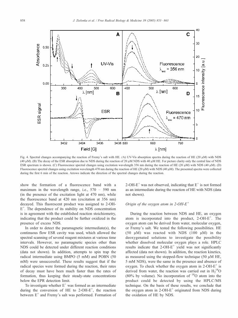
Fig. 4. Spectral changes accompanying the reaction of Fremy_s salt with HE. (A) UV-Vis absorption spectra during the reaction of HE (20 AM) with NDS
(40 AM). (B) The decay of the ESR absorption due to NDS during the reaction of 20 AM NDS with 40 AM HE. For picture clarity only the central line of NDS
ESR spectrum is shown. (C) Fluorescence spectral changes using excitation wavelength 356 nm during the reaction of HE (20 AM) with NDS (40 AM). (D)
Fluorescence spectral changes using excitation wavelength 470 nm during the reaction of HE (20 AM) with NDS (40 AM). The presented spectra were collected
during the first 6 min of the reaction. Arrows indicate the direction of the spectral changes during the reaction.
J. Zielonka et al. / Free Radical Biology & Medicine 39 (2005) 853–863858
show the formation of a fluorescence band with a
maximum in the wavelength range, i.e., 570 – 590 nm
(in the presence of the excitation light at 470 nm), while
the fluorescence band at 420 nm (excitation at 356 nm)
decayed. This fluorescent product was assigned to 2-OH-
E+. The dependence of its stability on NDS concentration
is in agreement with the established reaction stoichiometry,
indicating that the product could be further oxidized in the
presence of excess NDS.
In order to detect the paramagnetic intermediate(s), the
continuous flow ESR cavity was used, which allowed the
spectral scanning of several reagent mixtures at various time
intervals. However, no paramagnetic species other than
NDS could be detected under different reaction conditions
(data not shown). In addition, attempts to spin trap the
radical intermediate using BMPO (5 mM) and POBN (50
mM) were unsuccessful. These results suggest that if the
radical species were formed during the reaction, their rates
of decay must have been much faster than the rates of
formation, thus keeping their steady-state concentrations
below the EPR detection limit.
To investigate whether E+ was formed as an intermediate
during the conversion of HE to 2-OH-E+, the reaction
between E+ and Fremy_s salt was performed. Formation of
2-OH-E+ was not observed, indicating that E+ is not formed
as an intermediate during the reaction of HE with NDS (data
not shown).
Origin of the oxygen atom in 2-OH-E+
During the reaction between NDS and HE, an oxygen
atom is incorporated into the product, 2-OH-E+. The
oxygen atom can be derived from water, molecular oxygen,
or Fremy_s salt. We tested the following possibilities. HE
(50 AM) was reacted with NDS (100 AM) in the
deoxygenated solutions to investigate the possibility
whether dissolved molecular oxygen plays a role. HPLC
results indicate that 2-OH-E+ yield was not significantly
affected (data not shown). In addition, the reaction kinetics,
as measured using the stopped-flow technique (50 AM HE,
5 mM NDS), were the same in the presence and absence of
oxygen. To check whether the oxygen atom in 2-OH-E+ is
derived from water, the reaction was carried out in H218O
(80% by volume). No incorporation of 18O atom into the
product could be detected by using the HPLC/MS
technique. On the basis of these results, we conclude that
the oxygen atom in 2-OH-E+ originated from NDS during
the oxidation of HE by NDS.
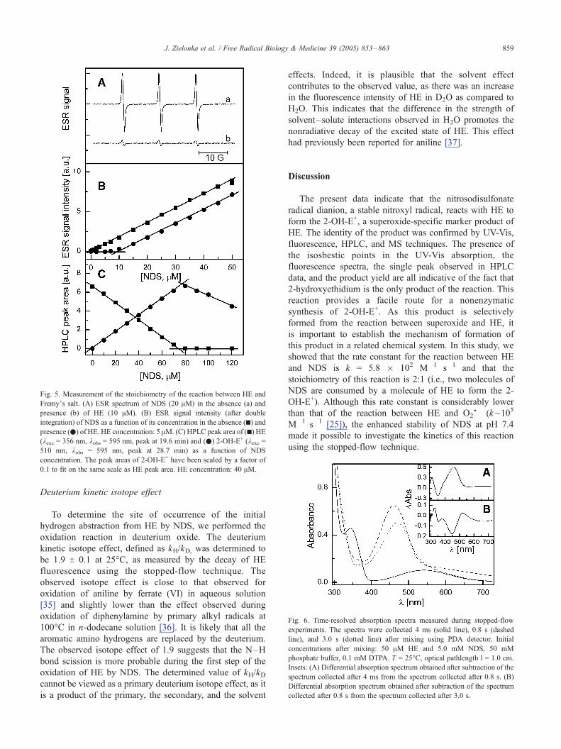
Fig. 5. Measurement of the stoichiometry of the reaction between HE and
Fremy_s salt. (A) ESR spectrum of NDS (20 AM) in the absence (a) and
presence (b) of HE (10 AM). (B) ESR signal intensity (after double
integration) of NDS as a function of its concentration in the absence (h) andpresence (.) of HE. HE concentration: 5 AM. (C) HPLC peak area of (h) HE(kexc = 356 nm, kobs = 595 nm, peak at 19.6 min) and (.) 2-OH-E+ (kexc =510 nm, kobs = 595 nm, peak at 28.7 min) as a function of NDS
concentration. The peak areas of 2-OH-E+ have been scaled by a factor of
0.1 to fit on the same scale as HE peak area. HE concentration: 40 AM.
Fig. 6. Time-resolved absorption spectra measured during stopped-flow
experiments. The spectra were collected 4 ms (solid line), 0.8 s (dashed
line), and 3.0 s (dotted line) after mixing using PDA detector. Initial
concentrations after mixing: 50 AM HE and 5.0 mM NDS, 50 mM
phosphate buffer, 0.1 mM DTPA. T = 25-C, optical pathlength l = 1.0 cm.
Insets: (A) Differential absorption spectrum obtained after subtraction of the
spectrum collected after 4 ms from the spectrum collected after 0.8 s. (B)
Differential absorption spectrum obtained after subtraction of the spectrum
collected after 0.8 s from the spectrum collected after 3.0 s.
J. Zielonka et al. / Free Radical Biology & Medicine 39 (2005) 853–863 859
Deuterium kinetic isotope effect
To determine the site of occurrence of the initial
hydrogen abstraction from HE by NDS, we performed the
oxidation reaction in deuterium oxide. The deuterium
kinetic isotope effect, defined as kH/kD, was determined to
be 1.9 T 0.1 at 25-C, as measured by the decay of HE
fluorescence using the stopped-flow technique. The
observed isotope effect is close to that observed for
oxidation of aniline by ferrate (VI) in aqueous solution
[35] and slightly lower than the effect observed during
oxidation of diphenylamine by primary alkyl radicals at
100-C in n-dodecane solution [36]. It is likely that all the
aromatic amino hydrogens are replaced by the deuterium.
The observed isotope effect of 1.9 suggests that the N–H
bond scission is more probable during the first step of the
oxidation of HE by NDS. The determined value of kH/kDcannot be viewed as a primary deuterium isotope effect, as it
is a product of the primary, the secondary, and the solvent
effects. Indeed, it is plausible that the solvent effect
contributes to the observed value, as there was an increase
in the fluorescence intensity of HE in D2O as compared to
H2O. This indicates that the difference in the strength of
solvent–solute interactions observed in H2O promotes the
nonradiative decay of the excited state of HE. This effect
had previously been reported for aniline [37].
Discussion
The present data indicate that the nitrosodisulfonate
radical dianion, a stable nitroxyl radical, reacts with HE to
form the 2-OH-E+, a superoxide-specific marker product of
HE. The identity of the product was confirmed by UV-Vis,
fluorescence, HPLC, and MS techniques. The presence of
the isosbestic points in the UV-Vis absorption, the
fluorescence spectra, the single peak observed in HPLC
data, and the product yield are all indicative of the fact that
2-hydroxyethidium is the only product of the reaction. This
reaction provides a facile route for a nonenzymatic
synthesis of 2-OH-E+. As this product is selectively
formed from the reaction between superoxide and HE, it
is important to establish the mechanism of formation of
this product in a related chemical system. In this study, we
showed that the rate constant for the reaction between HE
and NDS is k = 5.8 � 102 M�1 s�1 and that the
stoichiometry of this reaction is 2:1 (i.e., two molecules of
NDS are consumed by a molecule of HE to form the 2-
OH-E+). Although this rate constant is considerably lower
than that of the reaction between HE and O2S� (k~105
M�1 s�1 [25]), the enhanced stability of NDS at pH 7.4
made it possible to investigate the kinetics of this reaction
using the stopped-flow technique.
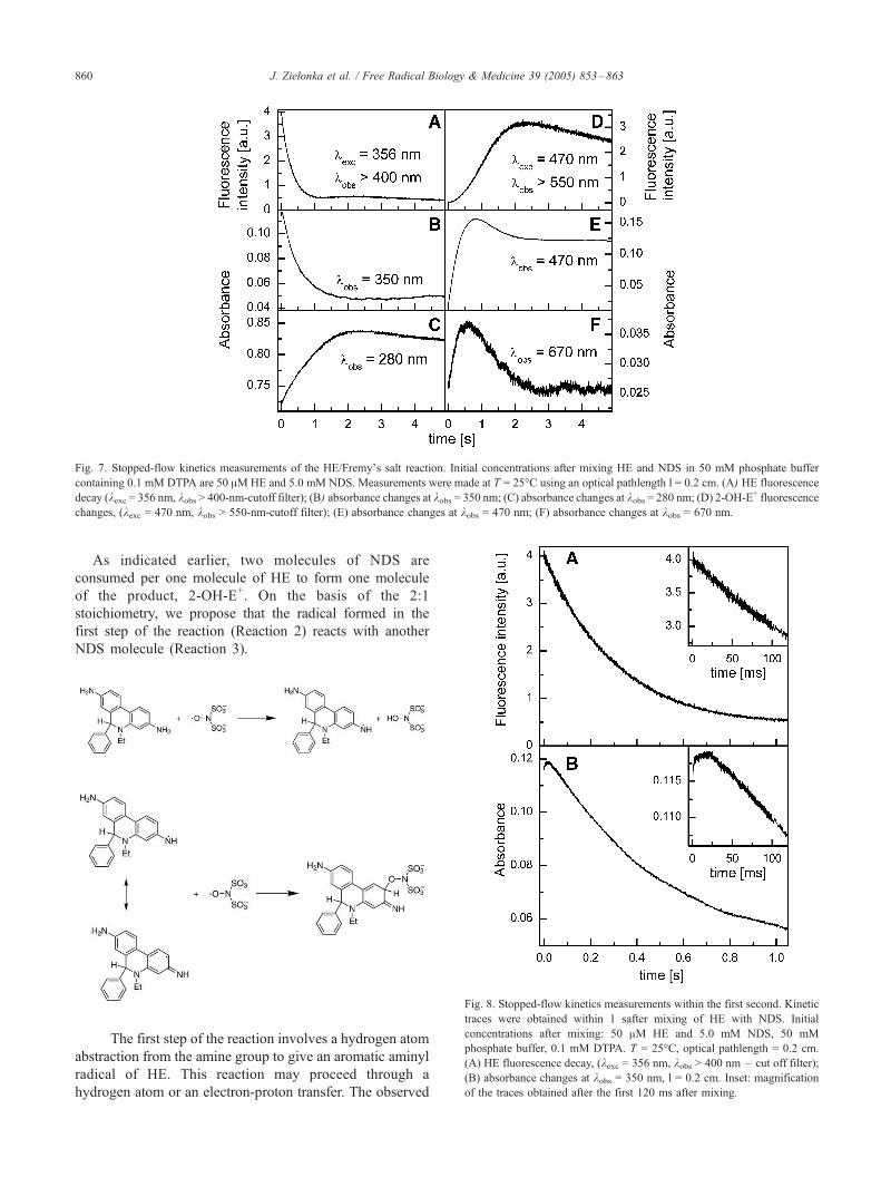
Fig. 7. Stopped-flow kinetics measurements of the HE/Fremy_s salt reaction. Initial concentrations after mixing HE and NDS in 50 mM phosphate buffer
containing 0.1 mM DTPA are 50 AMHE and 5.0 mM NDS. Measurements were made at T = 25-C using an optical pathlength l = 0.2 cm. (A) HE fluorescence
decay (kexc = 356 nm, kobs > 400-nm-cutoff filter); (B) absorbance changes at kobs = 350 nm; (C) absorbance changes at kobs = 280 nm; (D) 2-OH-E+ fluorescence
changes, (kexc = 470 nm, kobs > 550-nm-cutoff filter); (E) absorbance changes at kobs = 470 nm; (F) absorbance changes at kobs = 670 nm.
J. Zielonka et al. / Free Radical Biology & Medicine 39 (2005) 853–863860
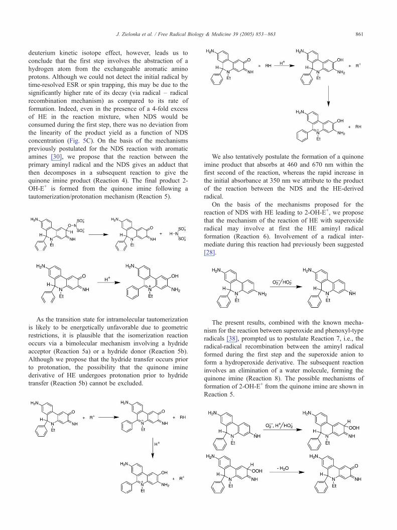
As indicated earlier, two molecules of NDS are
consumed per one molecule of HE to form one molecule
of the product, 2-OH-E+. On the basis of the 2:1
stoichiometry, we propose that the radical formed in the
first step of the reaction (Reaction 2) reacts with another
NDS molecule (Reaction 3).
Fig. 8. Stopped-flow kinetics measurements within the first second. Kinetic
traces were obtained within 1 safter mixing of HE with NDS. Initial
concentrations after mixing: 50 AM HE and 5.0 mM NDS, 50 mM
phosphate buffer, 0.1 mM DTPA. T = 25-C, optical pathlength = 0.2 cm.
(A) HE fluorescence decay, (kexc = 356 nm, kobs > 400 nm – cut off filter);
(B) absorbance changes at kobs = 350 nm, l = 0.2 cm. Inset: magnification
of the traces obtained after the first 120 ms after mixing.
The first step of the reaction involves a hydrogen atom
abstraction from the amine group to give an aromatic aminyl
radical of HE. This reaction may proceed through a
hydrogen atom or an electron-proton transfer. The observed
J. Zielonka et al. / Free Radical Biology & Medicine 39 (2005) 853–863 861
deuterium kinetic isotope effect, however, leads us to
conclude that the first step involves the abstraction of a
hydrogen atom from the exchangeable aromatic amino
protons. Although we could not detect the initial radical by
time-resolved ESR or spin trapping, this may be due to the
significantly higher rate of its decay (via radical – radical
recombination mechanism) as compared to its rate of
formation. Indeed, even in the presence of a 4-fold excess
of HE in the reaction mixture, when NDS would be
consumed during the first step, there was no deviation from
the linearity of the product yield as a function of NDS
concentration (Fig. 5C). On the basis of the mechanisms
previously postulated for the NDS reaction with aromatic
amines [30], we propose that the reaction between the
primary aminyl radical and the NDS gives an adduct that
then decomposes in a subsequent reaction to give the
quinone imine product (Reaction 4). The final product 2-
OH-E+ is formed from the quinone imine following a
tautomerization/protonation mechanism (Reaction 5).
As the transition state for intramolecular tautomerization
is likely to be energetically unfavorable due to geometric
restrictions, it is plausible that the isomerization reaction
occurs via a bimolecular mechanism involving a hydride
acceptor (Reaction 5a) or a hydride donor (Reaction 5b).
Although we propose that the hydride transfer occurs prior
to protonation, the possibility that the quinone imine
derivative of HE undergoes protonation prior to hydride
transfer (Reaction 5b) cannot be excluded.
We also tentatively postulate the formation of a quinone
imine product that absorbs at 460 and 670 nm within the
first second of the reaction, whereas the rapid increase in
the initial absorbance at 350 nm we attribute to the product
of the reaction between the NDS and the HE-derived
radical.
On the basis of the mechanisms proposed for the
reaction of NDS with HE leading to 2-OH-E+, we propose
that the mechanism of the reaction of HE with superoxide
radical may involve at first the HE aminyl radical
formation (Reaction 6). Involvement of a radical inter-
mediate during this reaction had previously been suggested
[28].
The present results, combined with the known mecha-
nism for the reaction between superoxide and phenoxyl-type
radicals [38], prompted us to postulate Reaction 7, i.e., the
radical-radical recombination between the aminyl radical
formed during the first step and the superoxide anion to
form a hydroperoxide derivative. The subsequent reaction
involves an elimination of a water molecule, forming the
quinone imine (Reaction 8). The possible mechanisms of
formation of 2-OH-E+ from the quinone imine are shown in
Reaction 5.
J. Zielonka et al. / Free Radical Biology & Medicine 39 (2005) 853–863862
As other ROS also may react with HE to form an
aminyl radical, the specific formation of 2-OH-E+ in the
case of O2S� is dependent on the secondary reaction
between the superoxide and the aminyl radical to form the
2-quinone-3-imine derivative of HE. The formation of the
aminyl radical in the first step of the reaction may explain
why HE does not by itself induce O2S� formation. The
aromatic aminyl radicals do not typically reduce oxygen to
superoxide [39].
In conclusion, our study indicates that 2-hydroxyethi-
dium, a specific marker product formed from the intra-
cellular superoxide/HE reaction, also can be independently
synthesized by the oxidation of HE using Fremy_s salt. We
propose that the conversion of HE to 2-OH-E+ involves the
intermediate formation of an aminyl radical and the quinone
imine species.
Acknowledgments
This work was supported by NIH Grants 5RO1HL067244,
2RO1NS39958, and 5PO1HL68769-01.
References
[1] Thannickal, V. J.; Fanburg, B. L. Reactive oxygen species in cell
signaling. Am. J. Physiol. 279:L1005–L1028; 2000.
[2] Droge, W. Free radicals in the physiological control of cell function.
Physiol. Rev. 82:47–95; 2002.
[3] Halliwell, B.; Gutteridge, J. M. C. Free Radicals in Biology and
Medicine, third ed. Oxford: Oxford Univ. Press; 1999.
[4] Tarpey, M. M.; Wink, D. A.; Grisham, M. B. Methods for detection
of reactive metabolites of oxygen and nitrogen: in vitro and in vivo
considerations. Am. J. Physiol. Regul. Integr. Comp. Physiol.
286:R431–R444; 2004.
[5] Fridovich, I. Editorial commentary on ‘‘Superoxide reacts with
hydroethidine but forms a fluorescent product that is distinctly
different from ethidium: potential implications in intracellular
fluorescence detection of superoxide’’ by H. Zhao et al. Free Radic.
Biol. Med. 34:1357–1358; 2003.
[6] Hwang, J.; Saha, A.; Boo, Y. C.; Sorescu, G. P.; McNally, J. S.;
Holland, S. M.; Dikalov, S.; Giddens, D. P.; Griendling, K. K.;
Harrison, D. G.; Jo, H. Oscillatory shear stress stimulates endothelial
production of O2� from p47phox-dependent NAD(P)H oxidases,
leading to monocyte adhesion. J. Biol. Chem. 278:47291–47298;
2003.
[7] Han, D.; Antunes, F.; Canali, R.; Rettori, D.; Cadenas, E. Voltage-
dependent anion channels control the release of the superoxide anion
from mitochondria to cytosol. J. Biol. Chem. 278:5557–5563; 2003.
[8] Budd, S. L.; Castilho, R. F.; Nicholls, D. G. Mitochondrial
membrane potential and hydroethidine-monitored superoxide gene-
ration in cultured cerebellar granule cells. FEBS Lett. 415:21–24;
1997.
[9] Thomas, G.; Roques, B. Proton magnetic resonance studies of
ethidium bromide and its sodium borohydride reduced derivative.
FEBS Lett. 26:169–175; 1972
[10] Olmsted III, J.; Kearns, D. R. Mechanism of ethidium bromide
fluorescence enhancement on binding to nucleic acids. Biochemistry
16:3647–3654; 1977.
[11] Bucana, C.; Saiki, I.; Nayar, R. Uptake and accumulation of the vital
dye hydroethidine in neoplastic cells. J. Histochem. Cytochem.
34:1109–1115; 1986.
[12] Saiki, I.; Bucana, C. D.; Tsao, J. Y.; Fidler, I. J. Quantitative
fluorescent microassay for identification of antiproliferative com-
pounds. J. Natl. Cancer Inst. 77:1235–1240; 1986.
[13] Rothe, G.; Valet, G. Flow cytometric analysis of respiratory burst
activity in phagocytes with hydroethidine and 2V,7V-dichlorofluo-rescin. J. Leukoc. Biol. 47:440–448; 1990.
[14] Carter, W. O.; Narayanan, P. K.; Robinson, J. P. Intracellular
hydrogen peroxide and superoxide anion detection in endothelial
cells. J. Leukoc. Biol. 55:253–258; 1994.
[15] Bindokas, V. P.; Jordan, J.; Lee, C. C.; Miller, R. J. Superoxide
production in rat hippocampal neurons: selective imaging with
hydroethidine. J. Neurosci. 16:1324–1336; 1996.
[16] Endl, E.; Steinbach, P.; Hofstadter, F. Flow cytometric analysis of cell
suspensions exposed to shock waves in the presence of the radical
sensitive dye hydroethidine. Ultrasound Med. Biol. 21:569–577;
1995.
[17] Dussmann, H.; Kogel, D.; Rehm, M.; Prehn, J. H. M. Mitochondrial
membrane permeabilization and superoxide production during
apoptosis. J. Biol. Chem. 278:12645–12649; 2003.
[18] Bindokas, V. P.; Kuznetsov, A.; Sreenan, S.; Polonsky, K. S.; Roe,
M. W.; Philipson, L. H. Visualizing superoxide production in normal
and diabetic rat islets of langerhans. J. Biol. Chem. 278:9796–9801;
2003.
[19] Peterson, S. L.; Morrow, D.; Liu, S.; Liu, K. J. Hydroethidine
detection of superoxide production during the lithium-pilocarpine
model of status epilepticus. Epilepsy Res. 49:226–238; 2002.
[20] Kobzik, L.; Godleski, J. J.; Brain, J. D. Oxidative metabolism in the
alveolar macrophage: analysis by flow cytometry. J. Leukoc. Biol.
47:295–303; 1990.
[21] Al-Mehdi, A. B.; Shuman, H.; Fisher, A. B. Intracellular generation
of reactive oxygen species during nonhypoxic lung ischemia. Am. J.
Physiol. 272:L294–L300; 1997.
[22] Narayanan, P. K.; Goodwin, E. H.; Lehnert, B. E. a Particles initiate
biological production of superoxide anions and hydrogen peroxide in
human cells. Cancer Res. 57:3963–3971; 1997.
[23] Zielonka, J.; Marcinek, A.; Adamus, J.; Gebicki, J. Direct observa-
tion of NADH radical cation generated in reactions with one-electron
oxidants. J. Phys. Chem. A 107:9860–9864; 2003.
[24] Gebicki, J.; Marcinek, A.; Zielonka, J. Transient species in the
stepwise interconversion of NADH and NAD+. Acc. Chem. Res.
37:379–386; 2004.
[25] Zhao, H.; Kalivendi, S.; Zhang, H.; Joseph, J.; Nithipatikom, K.;
Vasquez-Vivar, J.; Kalyanaraman, B. Superoxide reacts with hydro-
ethidine but forms a fluorescent product that is distinctly different
from ethidium: potential implications in intracellular fluorescence
detection of superoxide. Free Radic. Biol. Med. 34:1359–1368;
2003.
[26] Zhao, H.; Joseph, J.; Fales, H. M.; Sokoloski, E. A.; Levine, R. L.;
Vasques-Vivar, J.; Kalyanaraman, B. Detection and characterization
of the product of hydroethidine and intracellular superoxide by
HPLC and limitations of fluorescence. Proc. Natl. Acad. Sci. USA
102:5727–5732; 2005.
[27] Zhao, H. Detection of superoxide by ESR-spin trapping and hydro-
ethidine fluorescence. Ph.D. dissertation submitted to the Medical
College of Wisconsin; 2004.
[28] Benov, L.; Sztejnberg, L.; Fridovich, I. Critical evaluation of the use
of hydroethidine as a measure of superoxide anion radical. Free
Radic. Biol. Med. 25:826–831; 1998.
[29] Weil, J. A.; Bolton, J. R.; Wertz, J. E. Electron paramagnetic
resonance: elementary theory and practical applications. New York:
Wiley-Interscience; 1994.
[30] Zimmer, H.; Lankin, D. C.; Horgan, S. W. Oxidations with potassium
nitrosodisulfonate (Fremy_s radical). The Teuber reaction. Chem.
Rev. 71:229–246; 1971.
[31] Palmisano, G.; Danielli, B.; Lesma, G.; Trupiano, F. Oxidation
J. Zielonka et al. / Free Radical Biology & Medicine 39 (2005) 853–863 863
of h-anilinoacrylate alkaloids vincadifformine and tabersonine by
Fremy_s salt. A Mechanistic insight into the rearrangement
of Aspidosperma to Hunteria alkaloids. J. Org. Chem. 53:
1056–1064; 1988.
[32] Liu, Z.-L.; Han, Z.-X.; Chen, P.; Liu, Y.-C. Stopped-flow ESR study
on the reactivity of vitamin E, vitamin C and its lipophilic derivatives
towards Fremy_s salt in micellar systems. Chem. Phys. Lipids
56:73–80; 1990.
[33] Holler, T. P.; Hopkins, P. B. Ovothiols as free-radical scavengers and
the mechanism of ovothiol-promoted NAD(P)H-O2 oxidoreductase
activity. Biochemistry 29:1953–1961; 1990.
[34] Murib, J. H.; Ritter, D. M. Decomposition of nitrosyl disulfonate ion.
I. Products and mechanism of color fading in acid solution. J. Am.
Chem. Soc. 74:3394–3398; 1952.
[35] Huang, H.; Sommerfeld, D.; Donn, B. C.; Lloyd, C. R.; Eyring,
E. M. Ferrate(VI) oxidation of aniline. J. Chem. Soc., Dalton
Trans.,1301–1305; 2001.
[36] Burton, A.; Ingold, K. U.; Walton, J. C. Absolute rate constants for
the reactions of primary alkyl radicals with aromatic amines. J. Org.
Chem. 61:3778–3782; 1996.
[37] Tobita, S.; Ida, K.; Shiobara, S. Water-induced fluorescence quench-
ing of aniline and its derivatives in aqueous solution. Res. Chem.
Intermed. 27:205–218; 2001.
[38] Winterbourn, C. C.; Kettle, A. J. Radical-radical reactions of
superoxide: a potential route to toxicity. Biochem. Biophys. Res.
Commun. 305:729–736; 2003.
[39] Alfassi, Z. B., ed. The Chemistry of N-Centered Radicals. New York:
Wiley; 1998.