Mechanism of IS< i> 200/IS< i> 605 Family DNA Transposases: Activation and Transposon-Directed...
22
Mechanism of IS200/IS605 Family DNA Transposases: Activation and Transposon-Directed Target Site Selection Orsolya Barabas 1 , Donald R. Ronning 1,*,# , Catherine Guynet 2,* , Alison Burgess Hickman 1 , Bao Ton-Hoang 2 , Michael Chandler 2 , and Fred Dyda 1,¶ 1 Laboratory of Molecular Biology, National Institute of Diabetes, Digestive, and Kidney Diseases, National Institutes of Health, Bethesda, MD 20892 USA 2 Laboratoire de Microbiologie et Génétique Moléculaires Centre National de la Recherche Scientifique, 118 Route de Narbonne, 31062, Toulouse Cedex, France SUMMARY The smallest known DNA transposases are those from the IS200/IS605 family. Here we show how the interplay of protein and DNA activates TnpA, the Helicobacter pylori IS608 transposase, for catalysis. First, transposon end binding causes a conformational change that aligns catalytically important protein residues within the active site. Subsequent precise cleavage at the left and right ends, the steps that liberate the transposon from its donor site, does not involve a site-specific DNA binding domain. Rather, cleavage site recognition occurs by complementary base pairing with a TnpA-bound subterminal transposon DNA segment. Thus, the enzyme active site is constructed from elements of both protein and DNA, reminiscent of the interdependence of protein and RNA in the ribosome. Our structural results explain why the transposon ends are asymmetric and how the transposon selects a target site for integration, and allow us to propose a molecular model for the entire transposition reaction. INTRODUCTION DNA transposition is a ubiquitous phenomenon occurring in all kingdoms of life during which discrete segments of DNA called transposons move from one genomic location to another. In both eukaryotes and prokaryotes, DNA transposition has been a significant part of evolution. Many eukaryotic genomes are littered with transposons or their inactive remnants (Lander et al., 2001), primarily scattered between genes. In bacteria, transposable elements can carry antibiotic resistance genes and, when combined with conjugation, are major drivers of broad genome remodeling and the emergence of antibiotic resistant strains. The discovery and engineering of DNA transposons active in vertebrate cells (Miskey et al., 2005) has led to their use in identifying oncogenes and tumor suppressors and characterizing genes of unknown function, through their ability to disrupt genes or regulatory regions. They also have exciting potential as gene delivery systems for gene therapy applications. ¶ To whom correspondence should be addressed: E-mail: [email protected] Phone: 301-402-4496 FAX: 301-496-0201. * These authors made equal contributions to the work # Present address: Department of Chemistry, University of Toledo, Toledo OH 43606 Publisher's Disclaimer: This is a PDF file of an unedited manuscript that has been accepted for publication. As a service to our customers we are providing this early version of the manuscript. The manuscript will undergo copyediting, typesetting, and review of the resulting proof before it is published in its final citable form. Please note that during the production process errors may be discovered which could affect the content, and all legal disclaimers that apply to the journal pertain. NIH Public Access Author Manuscript Cell. Author manuscript; available in PMC 2009 May 11. Published in final edited form as: Cell. 2008 January 25; 132(2): 208–220. doi:10.1016/j.cell.2007.12.029. NIH-PA Author Manuscript NIH-PA Author Manuscript NIH-PA Author Manuscript
-
Upload
independent -
Category
Documents
-
view
0 -
download
0
Transcript of Mechanism of IS< i> 200/IS< i> 605 Family DNA Transposases: Activation and Transposon-Directed...

Mechanism of IS200/IS605 Family DNA Transposases: Activationand Transposon-Directed Target Site Selection
Orsolya Barabas1, Donald R. Ronning1,*,#, Catherine Guynet2,*, Alison Burgess Hickman1,Bao Ton-Hoang2, Michael Chandler2, and Fred Dyda1,
1 Laboratory of Molecular Biology, National Institute of Diabetes, Digestive, and Kidney Diseases, NationalInstitutes of Health, Bethesda, MD 20892 USA
2 Laboratoire de Microbiologie et Génétique Moléculaires Centre National de la Recherche Scientifique, 118Route de Narbonne, 31062, Toulouse Cedex, France
SUMMARYThe smallest known DNA transposases are those from the IS200/IS605 family. Here we show howthe interplay of protein and DNA activates TnpA, the Helicobacter pylori IS608 transposase, forcatalysis. First, transposon end binding causes a conformational change that aligns catalyticallyimportant protein residues within the active site. Subsequent precise cleavage at the left and rightends, the steps that liberate the transposon from its donor site, does not involve a site-specific DNAbinding domain. Rather, cleavage site recognition occurs by complementary base pairing with aTnpA-bound subterminal transposon DNA segment. Thus, the enzyme active site is constructed fromelements of both protein and DNA, reminiscent of the interdependence of protein and RNA in theribosome. Our structural results explain why the transposon ends are asymmetric and how thetransposon selects a target site for integration, and allow us to propose a molecular model for theentire transposition reaction.
INTRODUCTIONDNA transposition is a ubiquitous phenomenon occurring in all kingdoms of life during whichdiscrete segments of DNA called transposons move from one genomic location to another. Inboth eukaryotes and prokaryotes, DNA transposition has been a significant part of evolution.Many eukaryotic genomes are littered with transposons or their inactive remnants (Lander etal., 2001), primarily scattered between genes. In bacteria, transposable elements can carryantibiotic resistance genes and, when combined with conjugation, are major drivers of broadgenome remodeling and the emergence of antibiotic resistant strains. The discovery andengineering of DNA transposons active in vertebrate cells (Miskey et al., 2005) has led to theiruse in identifying oncogenes and tumor suppressors and characterizing genes of unknownfunction, through their ability to disrupt genes or regulatory regions. They also have excitingpotential as gene delivery systems for gene therapy applications.
¶ To whom correspondence should be addressed: E-mail: [email protected] Phone: 301-402-4496 FAX: 301-496-0201.*These authors made equal contributions to the work#Present address: Department of Chemistry, University of Toledo, Toledo OH 43606Publisher's Disclaimer: This is a PDF file of an unedited manuscript that has been accepted for publication. As a service to our customerswe are providing this early version of the manuscript. The manuscript will undergo copyediting, typesetting, and review of the resultingproof before it is published in its final citable form. Please note that during the production process errors may be discovered which couldaffect the content, and all legal disclaimers that apply to the journal pertain.
NIH Public AccessAuthor ManuscriptCell. Author manuscript; available in PMC 2009 May 11.
Published in final edited form as:Cell. 2008 January 25; 132(2): 208–220. doi:10.1016/j.cell.2007.12.029.
NIH
-PA Author Manuscript
NIH
-PA Author Manuscript
NIH
-PA Author Manuscript

A variety of structurally and mechanistically distinct transposase enzymes have evolved tocarry out transposition by several different pathways (Curcio & Derbyshire, 2003). In all cases,these enzymes possess a nuclease activity that allows them to cleave DNA in order to excisetransposon DNA and subsequently splice it into a new location. Depending on the system(Dyda et al., 1994; Grindley et al., 2006), different types of nucleophiles can be used bytransposases to cleave DNA by attacking a phosphorus atom of a backbone phosphate group:water, generally activated by enzyme-bound metal ions; a hydroxyl group at the 5′ or 3′ endof a DNA strand; or a hydroxyl-group bearing amino acid in the active site of the transposaseitself, such as serine or tyrosine. When a catalytic serine or tyrosine is used, the enzymebecomes attached to DNA through a covalent phosphotyrosine or phosphoserine bond.
One group of transposases that use catalytic tyrosines are the Y1 transposases (Ronning et al.,2005). These are members of a vast superfamily of nucleases characterized by a conservedHis-hydrophobic-His (HUH) motif (Koonin & Ilyina, 1993) that provides two ligands to adivalent metal ion cofactor. HUH nucleases always cut a DNA strand with a polarity resultingin a 5′ phosphotyrosine linkage and a free 3′ OH group. In contrast to many HUH superfamilymembers which are monomeric and have two catalytic tyrosines, Y1 transposases have onlyone catalytic tyrosine and form obligatory dimers.
We have recently determined the structure of a Y1 transposase from the insertion sequenceIS608 (Ronning et al., 2005), originally identified in Helicobacter pylori (Kersulyte et al.,2002), a bacterium that causes gastric inflammation leading to ulcers and occasionally tostomach cancer. Insertion sequences (IS) are the simplest autonomous transposable elements.While they tend to be short (< 2.5 kb) and carry only those genes needed for transposition, ifplaced flanking a DNA segment, many are able to mobilize the intervening genes (Mahillon& Chandler, 1998). In addition to dispersing antibiotic resistance genes, IS transposition canindirectly lead to antibiotic-resistant bacterial strains. For example, a metronidazole-resistantstrain of H. pylori has arisen because the nitroreductase gene needed for pro-drug activationhas been disrupted by an IS605-related transposition event (Debets-Ossenkopp et al., 1999).
ISs can be classified into groups or families based on the general features of their DNAsequences and associated transposases (www-is.biotoul.fr). A particularly interesting familyconsists of the IS200/IS605 elements (Kersulyte et al., 2002) which do not have invertedsequences at their ends characteristic of many prokaryotic and eukaryotic transposons. Rather,imperfect palindromic (IP) sequences are located close to the transposon ends (Figure 1A). Inthe case of one family member IS608, the left end (LE) and right end (RE) have almost identicalIP sequences that form DNA stem loop structures (Ronning et al., 2005). Intriguingly, theseIPs are asymmetrically located: the IP at RE (IPRE) is closer to the RE cleavage site at thetransposon end than the IP at LE (IPLE) is to the LE cleavage site, and the nucleotide sequencesbetween the IPs and the cleavage sites at the two ends are unrelated (Figure 1A). AmongIS608 copies from different H. pylori strains, the sequence between IPRE and the RE cleavagesite is completely conserved whereas it is variable between IPLE and the LE cleavage site.
Another curious feature of IS200/IS605 family members is that, unlike many DNA transposonswhich integrate essentially randomly, they always insert just 3′ of a specific four or fivenucleotide (nt) sequence. The form of IS608 that is inserted is an excised circular intermediate(also known as a transposon junction; Figure 1B) in which the RE is linked directly to the LE(Guynet et al., in press). Upon excision, IS608 precisely reseals the gap left behind in the donorDNA, and insertion occurs without target site duplications (Ton-Hoang et al., 2005). Thus,these seamless reactions proceed without loss or gain of nucleotides or the need for host cellDNA repair factors.
Barabas et al. Page 2
Cell. Author manuscript; available in PMC 2009 May 11.
NIH
-PA Author Manuscript
NIH
-PA Author Manuscript
NIH
-PA Author Manuscript

All IS608 cleavage and rejoining steps are carried out by a single 155-amino acid transposase,TnpA (Guynet et al., in press). In the TnpA dimer, there are two active sites, assembled intrans, in which the tyrosine nucleophile of one monomer is near the HUH motif of the other.TnpA recognizes and cleaves only the top strand of each transposon end (Ton-Hoang et al.,2005), resulting in yet another level of asymmetry: due to the conserved polarity of cleavage,after nucleophilic attack by the active site tyrosine on the phosphodiester backbone of DNAat LE, TnpA is covalently linked to the transposon 5′ end whereas cleavage at RE results inthe attachment of TnpA to flanking donor DNA (Figure 1B).
We have previously shown how TnpA recognizes and pairs its transposon ends (Ronning etal., 2005), a necessary prelude to the cycle of coordinated DNA strand cleavage and rejoiningsteps that constitute transposition. Here, based on five different crystal structures of complexesbetween TnpA and various IS608 LE and RE sequences, and associated biochemicalexperiments, we describe the entire transposition cycle - activation of TnpA upon end binding,recognition of the cleavage sites at the very edges of the transposon, and target site selection– and the absolute dependence of these steps on the inherent asymmetry of the transposon ends.
RESULTSTnpA complexes with the LE26 DNA hairpin
We have previously described the crystal structures of TnpA alone and complexed with a 22-mer hairpin (RE22) representing IPRE (Figure 2A; Ronning et al., 2005). While these wereinformative, in both structures, the catalytic tyrosine, Y127, was tucked away from the HUHmotif and hydrogen-bonded to S110 where it could not act as a nucleophile. As there are severalcrystal structures of HUH nucleases with fully assembled active sites (Guasch et al., 2003;Larkin et al., 2005), it was clear that a conformational change was required. The other puzzlingaspect of active site assembly was how the catalytically required metal ion would bind. In otherHUH nuclease crystal structures, the metal cofactor (Mg2+ or Mn2+) is coordinated by threeprotein ligands: the two HUH histidines and either a third histidine or a glutamate, dependingon the nuclease. Neither of our structures suggested an obvious candidate for the third metalion ligand.
To shed light on these issues, we crystallized TnpA with a series of longer DNA substratesthat contained the IPs and extended towards the transposon ends. (In our base numberingconvention [Figure 1], the cleavage site at each transposon end is between base−1 and base+1.Base numbers increase with distance from the cleavage site with negative numbers on the 5′side of each cleavage site. Nucleotides (nt) flanking LE are italicized and the last four nt onRE are in bold.) We first co-crystallized and solved the 2.1 Å resolution structure of TnpAcomplexed with a 26-mer hairpin DNA (LE26) representing IPLE with a 4 nt 5′ extension (TableS1, Figure 2B). In the TnpA/LE26 complex, IPLE binds precisely as seen for IPRE. The onlydifference between IPLE and IPRE, an extra T in the LE hairpin loop, is turned toward thesolvent and does not contact TnpA. However, as a consequence of adding four nt in the directionof the transposon end, there has been a major conformational change affecting both TnpAmonomers in which helix αD carrying Y127 has swung across the face of the β-sheet to adopta completely new location (Movie S1, Figure 2). The pivot point for the movement is aroundtwo residues, G117 and G118, in the loop immediately preceding helix αD (Figure S1). As aresult of the helix movement, the nucleophilic -OH group of Y127 shifts 12 Å to a positionnow consistent with active site arrangements of other HUH nucleases.
The crucial element of LE26 that appears to drive the conformational change in helix αD isbase A+18 in the 4 nt 5′ extension. In the TnpA/LE26 complex, the previously observedhydrogen bond between Y127 and S110 has been disrupted, and the space that had beenoccupied by Y127 in the TnpA/RE22 structure is now occupied by A+18 (Figure 2). The new
Barabas et al. Page 3
Cell. Author manuscript; available in PMC 2009 May 11.
NIH
-PA Author Manuscript
NIH
-PA Author Manuscript
NIH
-PA Author Manuscript

hydrogen bond between A+18 and S110 is the only additional base-specific interaction thatTnpA makes with LE26 when compared with RE22. The ability of A+18 to evict helix αDfrom its previous location is due in part to the dearth of interactions between helix αD and therest of TnpA. In the TnpA/RE22 complex, helix αD is held in place by only one hydrogen bondand some scattered van der Waals contacts between hydrophobic groups. As a result of theintrusion of base A+18 in the TnpA/LE26 complex, the hydroxyl group of Y127 is no longerhydrogen-bonded to anything and thus is ready to attack the scissile phosphate.
Another residue of interest located on helix αD is Q131 which is highly conserved amongIS200 transposases and whose mutation to alanine dramatically reduces transposition in vivo(Figure S2). As a consequence of the conformational change in helix αD, Q131 is now closeto the HUH histidines where it appears appropriately placed to complete the divalent metal ionbinding site. To test this, we soaked preformed TnpA/LE26 crystals overnight in buffercontaining 5 mM MnCl2 (Table S1). The anomalous difference Fourier electron densitycalculated from data measured on these crystals in the neighborhood of the active site indeedshows Mn2+ coordinated by H64, H66, and Q131 (Figure S2).
We also compared the metal ion binding affinity of TnpA/RE22 and TnpA/LE26 complexesusing isothermal titration calorimetry. The experiments were performed with Mn2+ as ourprevious experience with an HUH nuclease suggested that Mn2+ might bind with higher affinitythan Mg2+(Hickman et al., 2002). As shown in Figure S2, TnpA/RE22 has no detectableaffinity for Mn2+ whereas the divalent metal ion is clearly bound by TnpA/LE26. Consistentwith the role of Q131 as the crucial third metal ion ligand, we cannot detect Mn2+ binding bythe TnpA(Q131A)/LE26 complex. Taken together, these data indicate that the 4 nt 5′ extensionof LE26 relative to RE22 causes a rearrangement in TnpA leading to active site assembly inwhich Y127 and the third metal ion ligand are simultaneously transported to where they areneeded for catalysis.
Cleavage activity of TnpA is dependent on the 4 nt 5′ extension of the IPsOur observation that a short 5′ extension of IPLE causes a conformational change in TnpA thatappears to assemble the active site led us to ask if, in the LE26-bound form, TnpA wascompetent for catalysis. We have previously shown that TnpA cleaves 70- and 80-mer ssDNARE and LE substrates which span the IPs and the cleavage sites (Ronning et al., 2005; Ton-Hoang et al., 2005). To investigate the effect of adding the four nt to IP sequences, we modifiedthe cleavage reaction by providing the IPs and the cleavage sites on separate oligonucleotides.TnpA was first bound to oligonucleotides containing the IP sequences, with or without the 4nt 5′ extensions, and substrates that spanned the cleavage sites from bases −4 to +4 were thenadded. Upon cleavage, TnpA forms a phosphotyrosine link to the four nt on the 5′ end of thebreak (Ronning et al., 2005; Ton-Hoang et al., 2005), and the resulting covalent complexescan be detected by SDS-PAGE on the basis of the increase in the molecular weight of TnpA.
As shown in Figure 5A, on both LE (lanes 5 and 6) and RE (lanes 3 and 4), formation ofcovalent complexes between TnpA and the cleavage substrates is dependent on the 4 nt 5′extension. This is consistent with the crucial role of the additional nucleotides in inducingactive site assembly. Furthermore, cleavage is dependent on the “correct” combination of LEand RE oligonucleotides: when IPLE with the 4 nt 5′ extension was mixed with the RE cleavagesubstrate (lane 9), or IPRE with the 4 nt 5′ extension was mixed with the LE cleavage substrate(lane 11), covalent complexes were not detected. This indicates that TnpA is able todifferentiate between the LE and RE cleavage sites in a manner that depends on the particularIP bound.
Barabas et al. Page 4
Cell. Author manuscript; available in PMC 2009 May 11.
NIH
-PA Author Manuscript
NIH
-PA Author Manuscript
NIH
-PA Author Manuscript
RE31 and RE35 complexesTo address how the transposon ends reach the TnpA active sites, we determined the 2.9 Åresolution structure of TnpA co-crystallized with a RE 31-mer (RE31) that contains IPRE andthe 9 nt in the 3′ direction reaching the end of the transposon (Table S1). In marked contrastto the effect seen on LE where extending the IP sequence towards the LE cleavage site leadsto TnpA activation, in the TnpA/RE31 complex, each TnpA monomer is in the inactiveconformation. Although IPRE is bound as seen in the TnpA/RE22 complex, the nucleotides 3′of IPRE protrude out into solvent and become completely disordered after a few residues (FigureS4).
We therefore extended RE31 by adding 4 nt in the 5′ direction. Co-crystals of TnpA bound toRE35 diffracted to 2.4 Å resolution (Table S1), and the structure revealed two importantdifferences when compared to that of TnpA/RE31 (Figure 3A, B; Figure S4). First, the activesite has been assembled: helix αD has undergone the conformational change seen in the TnpA/LE26 complex and Mn2+ that was included in the crystallization buffer is bound. Secondly,all RE35 nucleotides are ordered and the conformation is such that its 3′ end, the last nt of RE,is now located in the active site.
In the TnpA/RE35 complex, RE35 resembles a distorted letter “C” in which IPRE is bound onone side of TnpA, and the 4 nt 5′ extension and the 9 nt that lead to the cleavage site both curlaround the protein surface into the active site of the same monomer on the opposite face (Figure3C). Furthermore, there are specific base pairing interactions between the 4 nt 5′ extension ofthe IPRE and 3 nt at the 3′ end of the transposon that direct the terminal 3′ OH group, whoseoxygen would be part of the scissile phosphate, into the active site (Figure 3B). The resultingarrangement of active site elements resembles the co-crystal structure of TraI, an HUH protein,bound to ssDNA that spans the F plasmid nic site in which the scissile phosphate was capturedin the active site (Larkin et al., 2005).
A second important role of the 4 nt 5′ extension of IPRE is now clear: the last four nt of thetransposon RE, TCAA, that immediately precede the cleavage site are held in place by a setof Watson-Crick base pairs with the 5′ extension of IPRE (Figure 3C). The base pairs (A−1:T−32, C−3:G−35, and T−4:A−34) do not match up linearly in the transposon sequence and theresulting twisted structure was unexpected. One base of the 5′ extension, A−33, recapitulatesthe role of A+18 in the TnpA/LE26 complex in that it has displaced Y127 during the transitionfrom the inactive to the active conformation, and is now tucked into the pocket previouslyoccupied by Y127.
Another surprising aspect of the way in which the 3′ end of the transposon is brought into theactive site is that two base pairs, C−3:G−35 and T−4:A−34, are, in fact, part of base triplets(Figures 3C, S4). G−9, G−35, and C−3 form a G−G·C triplet which is stacked on the A−A·Ttriplet formed by A−10, A−34, and T−4. Although base triples have been frequently observedin RNA structures, to our knowledge, this is the first time DNA base triplets have been observedbound to a protein in a biologically relevant context. An interesting question is whether REfolds by itself to achieve the observed configuration or it is induced by TnpA.
The LE ternary complexAttempts to co-crystallize TnpA with longer LE sequences that extend 5′ from IPLE towardsthe LE cleavage site have thus far been unsuccessful, due to the poor biophysical properties ofthese complexes. However, the observation that IS608 always integrates precisely 3′ ofTTAC led us to explore the possibility that this sequence might somehow be recognized byTnpA. Although short TTAC-containing oligonucleotides do not bind to TnpA on their own,
Barabas et al. Page 5
Cell. Author manuscript; available in PMC 2009 May 11.
NIH
-PA Author Manuscript
NIH
-PA Author Manuscript
NIH
-PA Author Manuscript
they do so in the presence of LE26 (data not shown), and we obtained crystals of TnpA boundto LE26 and the 6-mer TATTAC (denoted D6) that diffracted to 1.9 Å resolution (Table S1).
In the ternary LE complex, we observe the TTAC bases of D6 in the same position andorientation as the TCAA bases at the very end of RE35 in the TnpA/RE35 complex. TheTTAC tetranucleotide is located in the active site where it is held in place by the 4 nt 5′ extensionof IPLE (Figure 4). In this case, base pairs occur between A+16:T−3, A+17:T−4, and G+19:C−1 (Figure 4B). These orient the scissile phosphate (whose position can be inferred from theterminal 3′ OH group of D6) for nucleophilic attack by Y127. The overall DNA conformationis very similar to that seen in TnpA/RE35 except that the nucleotides between the cleavage siteand LE26 are missing; the analogous regions of the TnpA/LE26/D6 and TnpA/RE35complexes are virtually superimposable.
The observation that the LE ternary complex structurally echoes the TnpA/RE35 complex isexplained by the conserved polarity of the cleavage reactions. Although the interactions aresimilar on both ends, the flanking TTAC sequence recognized by the 4 nt 5′ extension ofIPLE differs from the transposon 3′ end TCAA sequence that is recognized by the IPRE 5′extension. The key is that the compensating changes to retain Watson-Crick base pairing arepresent in the 5′ extensions of the IPs.
In the TnpA/LE26/D6 complex, helix αD appears in yet another inactive position away fromthe active site. We suspect this is a crystallization artifact as the activated position of helixαD is blocked by a crystal lattice contact and we know that, in solution, helix αD is able toadopt the activated conformation in LE ternary complexes as TnpA pre-bound to LE26 cancleave an 8-mer that spans the cleavage site (Figure 5A). The previously seen inactiveconformation (Ronning et al., 2005) is also unavailable as base A+18 of LE26 occupies thepocket where Y127 sits in uncomplexed TnpA and in the TnpA/RE22 and TnpA/RE31complexes. Finally, the relevance of the observed position of helix αD in the TnpA/LE26/D6complex is suspect as any extension of D6 in the 3′ direction (i.e., uncleaved LE) wouldimmediately be in steric conflict with helix αD. This variability in the position of helix αD addsto the body of data that suggests it is a mobile structural element that tends to wander (FigureS5).
We note that the TnpA/LE26/D6 structure represents not only how TnpA recognizes its LEcleavage site but also how a TTAC-containing target site is captured. Since the circulartransposon intermediate that can be integrated into target DNA, a transposon junction (Guynetet al., in press), contains the LE26 sequence but not the LE TTAC flank (Figure 1B), TnpAbound to the transposon junction is capable of binding (and subsequently cleaving) a TTAC-containing target. Therefore, recognition of TTAC of the target and of the LE flank is identical.
Changing cleavage specificityThe recognition of the integration target DNA sequence by protein-bound transposon DNA,rather than by TnpA directly, raises the obvious possibility that TnpA-catalyzed insertionsmight readily be re-targeted to other tetranucleotide sequences. This seems possible inprinciple, as three of the four nt in the 5′ extensions of the IPs do not appear to play any rolebeyond recognizing TTAC on LE or TCAA on RE. Thus, their replacement should not impedetransposition.
To test the potential for re-directing, we changed either two or three of the nucleotides in the5′ extension of IPLE, and introduced the compensating base-pairing partners in 8-mer cleavagesubstrates (which represent both the LE cleavage site and the target site). In the variant 5′extensions, A+18 was retained as its role is to displace Y127 from its inactive location and itis not involved in DNA recognition. As shown in Figure 5B for two variant target sites, the
Barabas et al. Page 6
Cell. Author manuscript; available in PMC 2009 May 11.
NIH
-PA Author Manuscript
NIH
-PA Author Manuscript
NIH
-PA Author Manuscript

cleavage specificity of TnpA can indeed be effectively changed, and we are in the process ofexamining transposon re-targeting in vivo.
Substrate cleavage and junction formation by active site mutants of TnpAAll our crystal structures of TnpA show the dimer in a trans active site configuration in whichY127 of one monomer is close to the HUH motif of the other. To confirm that this shared activesite arrangement is used during catalysis, we performed several types of in vitro assays whichrecapitulate biochemical steps of IS608 transposition (Guynet et al., in press) using mixturesof single active site mutant versions of TnpA. We reasoned that, if the shared activearrangement is indeed used, mixing an inactive Y127F mutant with an inactive H64A mutantshould result in measurable activity as any heterodimers would have one active site whichcombines the H64A and Y127F mutations yet the other would have the HUH motif and Y127intact (Figure 6A).
Figure 6A shows the results of RE cleavage experiments in which a 56-mer RE substrate thatincludes six flanking nt is incubated with wild-type TnpA, single active site point mutants, ormixed active site mutants. As expected (Ronning et al., 2005), no covalent products areobserved with the H64A, H66A, or Y127F mutants (lanes 3-5). However, when the Y127Aand H64A mutants are mixed, a covalent complex between TnpA and the 6-mer cleavageproduct can be detected (lanes 6 and 7), suggesting that a catalytically competent trans activesite has assembled. The 6-mer product can also be directly detected after digestion withproteinase K. As shown in Figure 6B, in control experiments with the 56-mer RE cleavagesubstrate, wild-type TnpA generates the 6-mer product whereas the Y127F and H64A mutantsdo not (lanes 1 in first three panels). However, mixing these mutants results in the cleavage ofthe RE substrate to generate the 6-mer (fourth panel, lane 1), consistent with a trans active siteconfiguration.
One of the hallmarks of IS608 transposition is that, upon transposon excision, the donorbackbone is precisely sealed (Figure 1B). To determine whether mixed mutant dimers cangenerate sealed donor backbones, the 56-mer RE cleavage substrate with six flanking nt wasmixed with a 40-mer LE backbone (i.e., 40 nt of flanking sequence ending with TTAC-3′) inthe presence of LE26. The LE donor substrate is necessarily pre-cleaved since, in the mixedmutant dimers, only one active site is functional; LE26 is also required because the 5′ extensionof its IP is needed to localize the TTAC of the LE donor substrate in the active site. As shownin Figure 6B, wild-type TnpA covalently joins the 40-mer LE donor substrate to the 6-merproduct of RE cleavage to produce a 46-mer sealed donor backbone (panel 1, lane 4), but nosuch product is observed with the mixed mutant dimers (panel 4, lane 4).
Another reaction that occurs during IS608 transposition is the formation of a circularintermediate (Figure 1B) in which RE is linked directly to LE (Ton-Hoang et al., 2005). Totest whether mixed mutant dimers can form such transposon junctions, we mixed a pre-cleaved45-mer RE substrate that ends in TCAA-3′, representing the product of RE cleavage on thetransposon side, with a 100-mer LE cleavage substrate that contains 60 nt of LE flanked by 40nt (Figure 6C). Wild-type TnpA (lane 7) produces 105-mer junctions by catalyzing the attackof the RE 3′ OH of TCAA on the LE 5′ phosphotyrosine linkage that is formed upon LEcleavage. While mixed mutant dimers can cleave LE (Figure 6D, lane 3), junctions cannot bedetected (Figure 6C, lane 9).
These observations show that, while mixed mutant dimers can cleave both LE and RE, a resultconsistent with a trans active site arrangement for cleavage, they cannot form sealed donorbackbones or transposon junctions. This suggests that the mixed mutant dimers are deficientin the resolution steps of transposition, as they are unable to resolve phosphotyrosineintermediates to form the appropriately joined products.
Barabas et al. Page 7
Cell. Author manuscript; available in PMC 2009 May 11.
NIH
-PA Author Manuscript
NIH
-PA Author Manuscript
NIH
-PA Author Manuscript
DISCUSSIONThe overwhelming body of evidence indicates that IS608 transposes using ssDNA (Kersulyteet al., 2002; Ton-Hoang et al., 2005; Ronning et al. 2005), and we have recently shown thatssDNA LE and RE oligonucleotides are readily cleaved, form transposon junctions and sealeddonor backbones, and integrate pre-formed transposon junctions into target DNA in thepresence of TnpA (Guynet et al., in press). With our structural results demonstrating how TnpAis activated and how it recognizes the transposon ends and its cleavage sites, together with theobservation that helix αD is a mobile structural unit, we propose the following model forIS608 transposition (Figure 7, Movie S2):
Transposition starts with TnpA locating and pairing the transposon ends by binding to thehairpins formed by the top strand LE and RE IPs (Figure 7A). IPLE and IPRE differ by onlyone base in the hairpin loop, and our structures of the TnpA dimer bound to two identical DNAmolecules most likely reliably reflect features of TnpA bound to one IPLE and one IPRE.Binding the transposon IPs induces the conformational change seen in the TnpA/LE26 complexduring which helix αD moves into the activated position thereby assembling the active site.
Divalent metal ion binding to the assembled active site localizes and polarizes the scissilephosphate, preparing it for nucleophilic attack by Y127 (Hickman et al., 2002; Larkin et al.,2005). On LE, upon cleavage, Y127 becomes covalently linked to the 5′ end of the transposonwhile the 3′ end of flanking donor remains bound through base-base interactions between itsTTAC and the four nt just 5′ of IPLE (as in the TnpA/LE26/D6 complex). We have no evidencethat the 15 transposon nt between this 5′ extension and the 5′ end of the transposon interactwith TnpA, consistent with their variability among different IS608 isolates (Berg et al., 2002).
On RE, cleavage results in a 5′ phosphotyrosine linkage between Y127 and the flanking donorDNA while the 3′ end of the transposon stays bound through base-pairing interactions betweenthe terminal TCAA tetranucleotide and the four nt 5′ extension of IPRE (as in the TnpA/RE35complex). The entire RE (represented by RE35) is ordered and its DNA conformation isstabilized by internal hydrogen bonds, consistent with its complete sequence conservation.
The subsequent formation of the transposition intermediates, a transposon junction and thesealed donor backbone, would arise straightforwardly if the two αD helices now trade places,pivoting on G117 and G118 and bringing the covalently linked DNA strands with them (Figure7B, Movie S2). This model requires the 3′ OH groups to remain in their “active sites of origin”to act as the nucleophiles to resolve the swapped phosphotyrosine linkages. This immobilityseems likely as our structures of both LE and the RE complexes suggest that 3′ ends wouldremain bound as long as the IPs with their four nt 5′ extensions are present.
By this mechanism, in one active site of the dimer, a sealed donor backbone would be generatedby nucleophilic attack by the stationary LE donor flanking 3′ OH (TTAC-OH) on the swappedRE donor flank 5′ phosphotyrosine linkage. In the other active site, attack by the resident RE3′ OH (TCAA-OH) on the 5′ phosphotyrosine linkage of the swapped transposon LE wouldresult in a transposon junction. This proposed reaction scheme is supported by the threepreviously unexplainable aspects of asymmetry seen at the transposon ends: the tolerance ofsequence variability at LE but not at RE; the cleavage polarities that dictate that onephosphotyrosine linkage joins TnpA to the transposon end whereas the other covalentlyattaches TnpA to flanking DNA; and, finally, the spacing differences between the cleavagesites and the IPs. On RE, upon cleavage TnpA becomes attached to the RE flank which is partof the donor plasmid and presumably can move freely. It is therefore easy to imagine that thetraveling helix αD can bring covalently attached flanking DNA to the adjacent active site.However, on LE, it is the transposon 5′ end that is attached to helix αD. Thus, LE must havea sufficiently long and flexible spacer between the 4 nt IPLE extension and the 5′ end of the
Barabas et al. Page 8
Cell. Author manuscript; available in PMC 2009 May 11.
NIH
-PA Author Manuscript
NIH
-PA Author Manuscript
NIH
-PA Author Manuscript

transposon that can act as a tether with some slack so helix αD can move from one active siteto the other. The slack is necessary because LE cannot move as a unit as it is anchored to theTnpA dimer by the IPLE hairpin.
The notion of a switch from the trans to a cis active site arrangement is supported by the crystalstructure (PDB ID 2fyx) of a closely related transposase from Deinococcus radiodurans, whichwas captured in the inactive configuration but with a cis helix αD arrangement. Although wehave no structural evidence that the cis configuration occurs during IS608 transposition, ourbiochemical data showing that mixed mutant dimers are defective in the resolution stepsprovides strong support that both transposon junction and sealed backbone formation occurswith helix αD in cis: in this configuration, mixed mutant dimers have no functional active sites.As shown in Movie S2, the switch of helices from one active site to the other can be easilymodeled as long as the RE flank and the nucleotides between IPLE and the LE cleavage siteare free to move.
For the next step, integration, we do not know if the excised transposon junction intermediateremains bound to TnpA or is released and rebound. If, during recombination, the junction isreleased upon formation, it seems likely that TnpA resets to the trans configuration beforetransposon junction recapture, as we have only observed uncomplexed TnpA in that state. Ifthe transposon junction stays bound, the cleavage steps required for integration may start withTnpA in the cis configuration and switch to trans for resolution. From the point of view of theoverall mechanism, the outcomes of these two possibilities are equivalent.
We propose that integration of a transposon junction intermediate into a TTAC-containingtarget is a mechanistic replay of excision. After transposon junction formation, the sealed donorbackbone dissociates from the complex and is replaced by a TTAC-containing target sequence(Figure 7C). At one active site, the target DNA would bind through interactions between itsTTAC tetranucleotide and the four nt 5′ extension of IPLE. Upon cleavage, the TTAC targetflank would stay bound to the complex and the 5′ end would be covalently attached to Y127.At the other active site, the transposon junction would be cleaved, leaving TCAA (the RE 3′end of IS608) bound to the active site and the LE (5′ end of IS608) linked through a 5′phosphotyrosine linkage to Y127. A switch in the active site arrangements leads directly to theresolution steps in which the top strand of IS608 is inserted into a new target site: the stationary3′ OH of the TTAC target flank attacks the swapped 5′ phosphotyrosine link at LE while theRE TCAA 3′ OH attacks the swapped 5′ phosphotyrosine linkage of the target flank that wascreated during target cleavage (Figure 7D).
One of the most baffling questions about IS608 transposition was how its transposase, a 155residue, single-domain protein, could carry out all of the required steps. Part of the answer isthat TnpA is sneakier than we thought: it uses bound transposon DNA to recognize its cleavagesites both on donor and target DNA obviating the need for an additional DNA binding domain.Furthermore, TnpA works only on one DNA strand and does not have to deal with thecomplementary strand (Kersulyte et al., 2002; Ton-Hoang et al., 2005; Ronning et al. 2005;Guynet et al., in press).
If TnpA acts solely on ssDNA, how do its substrates arise? ssDNA formation might bepromoted by plasmid supercoiling combined with the propensity of the IPs to form stem loops;if this occurs, the cleavage sites might, with some frequency, dissociate from theircomplementary strands. On the other hand, IS608 might excise in vivo only when DNAbecomes single stranded during a normal cellular process such as lagging strand synthesis. Iftransposition occurs only during DNA replication, excised TnpA-bound sealed transposoncircles might readily find a suitable TTAC-containing ssDNA target. Restricting transpositionto one stage in the cell cycle would also ensure that the reaction is substrate-limited, thus
Barabas et al. Page 9
Cell. Author manuscript; available in PMC 2009 May 11.
NIH
-PA Author Manuscript
NIH
-PA Author Manuscript
NIH
-PA Author Manuscript

preventing wanton and possibly destructive movement. Since ssDNA is also generated duringconjugative DNA transfer, conjugating plasmids might be preferred targets for IS608 with theadded advantage of immediate horizontal transfer.
The importance of ssDNA substrates for IS200 transposition is illustrated by recent work onDeinococcus radiodurans which can survive severe ionizing radiation damage using amechanism called “extended synthesis-dependent strand annealing” to reconstitute itsshattered chromosomes (Zahradka et al., 2006). During recovery, as much as 15% of newlysynthesized DNA is single-stranded and, remarkably, the transposition frequency of ISDra2,an IS200/IS605 family member, increases 500-fold (Mennecier et al., 2006), suggesting thatssDNA is normally rate-limiting.
One of our most surprising results is that IS608 uses DNA sequences in the transposon itselfto recognize its cleavage sites. This mode of recognition suggests that TnpA may be re-directedto new target sites, opening up an unanticipated avenue of transposon targeting with an arrayof genomic and biotechnological applications.
The interdependence of protein and DNA in creating suitable substrates is reminiscent of theintertwining of protein and RNA in ribosomes (Noller, 2005). This "self-recognition" has alsobeen reported in other RNA-dependent systems. For example, the structural basis of thematchmaking functions of mitochondrial RNA binding proteins (MRPs) has recently beenreported (Schumacher et al., 2006).
There is a striking parallel between the IS608 transposition pathway and the mobility of groupII introns, particularly with that of the bacterial L1.LtrB system (Belfort et al., 2005; Lambowitz& Zimmerly, 2004). For example, the mobility of RNA introns is dependent on an intron-encoded multifunctional protein which is needed to bind the RNA. Upon splicing, exonsbordering the intron are joined to each other, reminiscent of the resealing of donor DNA endsby TnpA. The lariat intermediate formed during intron splicing is similar to the circularIS608 transposon junction intermediate, both of which are covalently sealed. Perhaps the mostsignificant parallel involves targeting, as these introns select their target, or “retrohoming” site,by base-pairing interactions between segments of the lariat intermediate and the target DNAstrand. This is the basis for the development of re-targeted introns (Karlberg et al., 2001) thatcan be used to disrupt chromosomal genes. In intron mobility, the key catalytic component isthe RNA itself, which catalyzes both the splicing and reverse splicing events. Because DNAhas no known self-cleavage and strand transfer activity, IS608 has to rely on the assistance ofTnpA to carry out the necessary cleavage and joining reactions. Our work demonstrates that atargeting mechanism previously thought to be the property of RNA-based mobile systems alsooccurs in the context of a mobile DNA element.
From a structural and functional perspective, the closest characterized relatives of TnpA areIS91-related transposases (Garcillán-Barcia et al., 2001). These are widespread in bacteria,have eukaryotic homologs (Kapitonov & Jerka, 2001), and have been implicated in themovement of so-called Common Regions (Stokes et al., 1997; Toleman et al., 2006) whichmay be responsible for a large fraction of the spread of antibiotic resistance genes. WhileIS91-like elements transpose by a mechanism that is likely different from IS608, they alsocontain subterminal IPs and insert just 3′ of short conserved sequences. It will be interestingto determine if their mode of target recognition resembles that shown here for IS608. If so, thepotential for their re-targeting could provide new possibilities toward the site-specificmodification of eukaryotic genomes.
Barabas et al. Page 10
Cell. Author manuscript; available in PMC 2009 May 11.
NIH
-PA Author Manuscript
NIH
-PA Author Manuscript
NIH
-PA Author Manuscript

EXPERIMENTAL PROCEDURESProtein Purification and Crystallization
TnpA was purified as previously described (Ronning et al., 2005). DNA was synthesized onan Applied Biosciences 394 DNA/RNA synthesizer or purchased from IDT (Coralville, IA).Sequences used for crystallization are shown in Figures 2-4. Oligonucleotides containinginverted repeat sequences were resuspended in TE, heated to 95°C for 15 min, and then rapidlycooled on ice and placed at −20°C until use. All crystals were obtained by vapor diffusion inhanging drops by mixing complexes 1:0.8-1.0 (v/v) with well solutions as described below.TnpA/LE26 crystals were frozen in paratone, and the rest were flash frozen in liquid propane.
TnpA/LE26—TnpA at 8 mg/ml was mixed with LE26 at a final protein:DNA molar ratio of1:1.1, and dialyzed against 20 mM Tris pH 7.5, 0.3 M sodium malonate, 0.2 mM TCEP, and2 mM EDTA. Well solutions consisted of 0.2 M sodium formate and 15-20% PEG 3350.
TnpA/RE31—TnpA at 5 mg/ml was mixed with RE31 at a molar ratio of 1:1.1 and dialyzedagainst 20 mM Tris pH 7.5, 0.2 M sodium malonate, 10 mM MnCl2, and 0.2 mM TCEP. Wellsolutions consisted of 15-20% PEG 3350 and 0.1 M MES pH 6.5. Crystals were cryoprotectedusing well solution supplemented with 10% glycerol.
TnpA/RE35—The complex was prepared as for TnpA/RE31, and the well solution consistedof 20-25% PEG 3350, 0.1 M MES pH 5.5, and 0.1 M ammonium acetate. Crystals were frozenafter stepwise replacement of water for 10% glycerol in the mother liquor.
TnpA/LE26/D6—TnpA (5 mg/ml) was mixed with LE26 and D6 at a 1:1.1:1.3 molar ratio,dialyzed against 20 mM Tris pH 7.5, 0.2 M MgCl2, 0.2 mM TCEP, and mixed with wellsolutions consisting of 6-10% PEG 600 and 50 mM sodium citrate pH 5.0. Crystals werecryoprotected by transfer into 10% PEG 600, 0.1 M MgCl2, 25 mM sodium citrate,10 mM TrispH 7.5, and 15% glycerol.
Data Collection and Structure DeterminationData on the TnpA/LE26 and TnpA/RE31 crystals were collected on beamline 22-ID at APSon a MAR300 CCD detector. All other diffraction data were collected at 95 K on a rotatinganode source equipped with multilayer focusing optics using Cu Kα radiation and an Raxis IVimage plate detector. Data were integrated and scaled with Denzo and Scalepack (Otwinowskiand Minor, 1997; Table S1). The structures were solved with molecular replacement byAMoRe (Navaza 2001), providing clear solutions in all cases. Search models and solutionstatistics are in Table S1, as are refinement statistics. For refinement, manual model buildingwith program O (Jones, et al., 1991) was alternated with cartesian simulated annealing,positional and restrained B-factor refinement cycles using CNS 1.1 (Brünger et al. 1998). TnpAresidues 133-155 could not be traced in the RE31, RE35, and LE26/D6 complexes. In the LE26and LE26/Mn2+ complex structures, all protein residues could be included in one monomer.Both LE26 complexes showed weak electron densities and high temperature factors forresidues 132-141 between helices αD and αE, suggesting high mobility. All molecular figuresand animations were made with Pymol (DeLano, 2002). Coordinates have been deposited inthe Protein Data Bank with the accession codes xxxx (TnpA/LE26), xxxx (TnpA/LE26+Mn2+), xxxx (TnpA/RE31), xxxx (TnpA/RE35), and xxxx (TnpA/LE26/D6).
Isothermal Titration Calorimetry (ITC)Complexes of TnpA and TnpA/Q131A prebound to RE22 or LE26 (TnpA concentration of0.035 - 0.038 mM) were prepared by dialysis against 10 mM HEPES pH 7.5, 0.2M NaCl, 5
Barabas et al. Page 11
Cell. Author manuscript; available in PMC 2009 May 11.
NIH
-PA Author Manuscript
NIH
-PA Author Manuscript
NIH
-PA Author Manuscript

mM DTT, 5 mM MgCl2, and 10% glycerol. Mn2+ binding was measured by ITC as described(Hickman et al., 2002) except that aliquots of 2 μl MnCl2 (5 mM in dialysis buffer) were used.
Covalent Complex Formation (Figure 5)Wild type or the Y127F mutant TnpA at 1 mg/ml was mixed with hairpin oligos and cleavagesubstrates at a final protein:DNA molar ratio of 1:1.1:1.1. Protein/hairpin mixtures weredialyzed overnight against 20 mM Tris pH 7.5, 0.2 M sodium malonate, 20 mM MgCl2, priorto addition of the substrate and incubation for 1 h at 20 °C. Samples were heat-denatured inSDS-containing sample buffers and analyzed on 4-12% SDS-PAGE gels. TnpA and TnpAattached to the 4-mer product of cleavage were detected by Coomassie staining.
Purification of Mixed TnpA Mutants and Activity AssaysEach point mutant (Y127F, H64A, H66A) was introduced into pBS107, expressed in E. coliRosetta (DE3) cells (Novagen), and separately purified as previously described (Ton-Hoanget al., 2005). To form mixed mutant dimers, equimolar amounts of either Y127F and H64A orY127F and H66A were incubated together on ice for 20 min. RE cleavage reactions (Figure6A) were performed by 30 min incubation at 37°C of 28 nM of a 3′-32P-labelled RE 70-mercleavage substrate (see Supplemental Material for sequence) with 10μM of total protein in afinal volume of 10 μl in 20 mM HEPES pH 7.5, 160 mM NaCl, 5 mM MgCl2, 10 mM DTT,20 μg/ml BSA, 0.5 μg of poly-dIdC, and 20% glycerol. Reaction products were separated ona 16% SDS-PAGE gel and subsequently analysed by phosphorimaging. Similar reactionconditions (Guynet et al., in press) were used for RE cleavage and donor backbone formation(Figure 6B), transposon junction formation (Figure 6C), and LE cleavage (Figure 6D). AllDNA sequences are listed in Supplemental Material. Products were separated on an 8%sequencing gel and analyzed by phosphorimaging.
Supplementary MaterialRefer to Web version on PubMed Central for supplementary material.
ACKNOWLEDGMENTSThis work was supported in part by the Intramural Program of the National Institute of Diabetes, Digestive, and KidneyDiseases of the NIH. The Chandler lab was supported by CNRS (France) and EU contract LSHM-CT-2005-019023.C.G. received a training grant from the French MENRT. Data were also collected at the SER-CAT 22-ID beamlineat the Advanced Photon Source, Argonne National Laboratory. Use of the APS was supported by the U.S. Departmentof Energy, Basic Energy Sciences, Office of Science, under Contract No. W-31-109-Eng-38. We thank Dr. Wei Yangfor insightful comments.
REFERENCESBelfort, M.; Derbyshire, V.; Parker, MM.; Cousineau, B.; Lambowitz, AM. Mobile Introns: Pathways
and Proteins in Mobile DNA II. Craig, NL., et al., editors. ASM Press; Washington, D.C.: 2002.Brünger AT, Adams PD, Clore GM, Delano WL, Gros P, Grossekunstleve RW, Jiang JS, Kuszewski J,
Nilges M, Pannu NS, et al. Crystallography and Nmr System - a New Software Suite ForMacromolecular Structure Determination. Acta Crystallogr. D 1998;54:905–921. [PubMed: 9757107]
Curcio MJ, Derbyshire KM. The outs and ins of transposition: From Mu to kangaroo. Nature Rev. Mol.Cell Biol 2003;4:1–13.
Debets-Ossenkopp YJ, Pot RGJ, van Westerloo DJ, Goodwin A, Vandenbroucke-Grauls CMJE, BergDE, Hoffman PS, Kusters JG. Insertion of mini-IS605 and deletion of adjacent sequences in thenitroreductase (rdxA) gene cause metronidazole resistance in Helicobacter pylor NCTC11637.Antimicrob. Agents Chemother 1999;43:2657–2662. [PubMed: 10543743]
DeLano, WL. The PyMol Molecular Graphics System. 2002. at www.pymol.org
Barabas et al. Page 12
Cell. Author manuscript; available in PMC 2009 May 11.
NIH
-PA Author Manuscript
NIH
-PA Author Manuscript
NIH
-PA Author Manuscript

Dyda F, Hickman AB, Jenkins TM, Engelman A, Craigie R, Davies DR. Crystal structure of the catalyticdomain of HIV-1 integrase: similarity to other polynucleotidyl transferases. Science 1994;266:1981–1986. [PubMed: 7801124]
Garcillán-Barcia M, Bernales I, Mendiola MV, de la Cruz F. Single-stranded DNA intermediates in IS91rolling-circle transposition. Mol. Microbiol 2001;39:494–501. [PubMed: 11136468]
Grindley NDF, Whiteson KL, Rice PA. Mechanisms of site-specific recombination. Annu. Rev. Biochem2006;75:567–605. [PubMed: 16756503]
Guasch A, Lucas M, Moncalian G, Cabezas M, Perez-Luque R, Gomis-Ruth FX, de la Cruz F, Coll M.Recognition and processing of the origin of transfer DNA by conjugative relaxase TrwC. Nat. Struct.Biol 2003;10:1002–1010. [PubMed: 14625590]
Guynet C, Hickman AB, Barabas O, Dyda F, Chandler M, Ton-Hoang B. Single-stranded transpositionof IS608: In vitro reconstitution of a new transposition mechanism. Mol. Cell. in press
Hickman AB, Ronning DR, Kotin RM, Dyda F. Structural unity among viral origin binding proteins:crystal structure of the nuclease domain of adeno-associated virus. Rep. Mol. Cell 2002;10:327–337.
Jones TA, Zou JY, Cowan SW, Kjeldgaard M. Improved methods for building protein models in electrondensity maps and the location of errors in these models. Acta Crystallogr. A 1991;47:110–119.[PubMed: 2025413]
Kapitonov VV, Jurka J. Rolling-circle transposons in eukaryotes. Proc. Natl. Acad. Sci. USA2001;98:8714–8719. [PubMed: 11447285]
Karberg M, Guo H, Zhong J, Coon R, Perutka J, Lambowitz AM. Group II introns as controllable genetargeting vectors for genetic manipulation of bacteria. Nature Biotech 2001;19:1162–1167.
Kersulyte D, Velapatino B, Dailide G, Mukhopadhyay AK, Ito Y, Cahuayme L, Parkinson AJ, GilmanRH, Berg DE. Transposable element ISHp608 of Helicobacter pylori: nonrandom geographicdistribution, functional organization, and insertion specificity. J. Bacteriol 2002;184:992–1002.[PubMed: 11807059]
Koonin EV, Ilyina TV. Computer-assisted dissection of rolling circle DNA replication. Biosystems1993;30:241–268. [PubMed: 8374079]
Lander ES, Linton LM, Birren B, Nusbaum C, Zody MC, Baldwin J, Devon K, Dewar K, Doyle M,FitzHugh W, et al. Initial sequencing and analysis of the human genome. Nature 2001;409:860–921.[PubMed: 11237011]
Lambowitz AM, Zimmerly S. Mobile Group II Introns. Annu. Rev. Genet 2004;38:1–35. [PubMed:15568970]
Larkin C, Datta S, Harley MJ, Anderson BJ, Ebie A, Hargreaves V, Schildbach JF. Inter- andintramolecular determinants of the specificity of single-stranded DNA binding and cleavage by theF factor relaxase. Struct 2005;13:1533–1544.
Lee HH, Yoon JY, Kim HS, Kang JY, Kim KH, Kim DJ, Ha JY, Mikami B, Yoon HJ, Suh SW. Crystalstructure of a metal ion-bound IS200 transposase. J. Biol. Chem 2006;281:4261–4266. [PubMed:16340015]
Mahillon J, Chandler M. Insertion sequences. Microbiol. Mol. Biol. Rev 1998;62:725–774. [PubMed:9729608]
Mennecier S, Servant P, Coste G, Bailone A, Sommer S. Mutagenesis via IS transposition in Deinococcusradiodurans. Mol. Microbiol 2006;59:317–325. [PubMed: 16359337]
Miskey C, Izsvák Z, Kawakami K, Ivics Z. DNA transposons in vertebrate functional genomics. Cell.Mol. Life Sci 2005;62:629–641. [PubMed: 15770416]
Navaza J. Implementation of molecular replacement in AMoRe. Acta Crystallogr 2001;D57:1367–1372.Noller HF. Reading the ribosome. Science 2005;309:1508–1514. [PubMed: 16141058]Otwinowski, Z.; Minor, W. Processing of X-ray diffraction data collected in oscillation mode. Vol. 276.
Academic Press; New York: 1997.Ronning DR, Guynet C, Ton-Hoang B, Perez ZN, Ghirlando R, Chandler M, Dyda F. Active site sharing
and subterminal hairpin recognition in a new class of DNA transposases. Mol. Cell 2005;20:143–154. [PubMed: 16209952]
Barabas et al. Page 13
Cell. Author manuscript; available in PMC 2009 May 11.
NIH
-PA Author Manuscript
NIH
-PA Author Manuscript
NIH
-PA Author Manuscript

Schumacher MA, Karamooz E, Zíková A, Trantírek L, Lukes J. Crystal structures of T. bruncei MRP1/MRP2 guide-RNA binding complex reveal RNA matchmaking mechanism. Cell 2006;126:701–711.[PubMed: 16923390]
Stokes HW, O'Gorman DB, Recchia GD, Parsekhian M, Hall RM. Structure and function of 59-baseelement recombination sites associated with mobile gene cassettes. Mol. Microbiol 1997;26:731–745. [PubMed: 9427403]
Toleman MA, Bennett PM, Walsh TR. ISCR elements: Novel gene-capturing systems of the 21st century?Microbiol. Mol. Biol. Rev 2006;70:296–316. [PubMed: 16760305]
Ton-Hoang B, Guynet C, Ronning DR, Cointin-Marty B, Dyda F, Chandler M. Transposition of ISHp608,member of an unusual family of bacterial insertion sequences. EMBO J 2005;24:3325–3338.[PubMed: 16163392]
Zahradka K, Slade D, Bailone A, Sommer S, Averbeck D, Petranovic M, Lindner AB, Radman M.Reassembly of shattered chromosomes in Deinococcus radiodurans. Nature 2006;443:569–573.[PubMed: 17006450]
Barabas et al. Page 14
Cell. Author manuscript; available in PMC 2009 May 11.
NIH
-PA Author Manuscript
NIH
-PA Author Manuscript
NIH
-PA Author Manuscript

Figure 1. IS608 Transposition(A) Alignment (adapted from Kersulyte et al. [2002]) of the termini and flanking sequences ofIS608 elements from Helicobacter pylori. The strain is indicated on the left; all the workreported here was done with IS608 from PeCan2A (top line). Flanking DNA is in black, basesof the left end (LE) are shown in red and orange, and bases of the right end (RE) are shown inshades of blue. Boxed sequences underlined with inverted arrows delineate the ImperfectPalindromes (IP) at each end. The bases are numbered such that, at each end, the cleavage siteis between base−1 and base+1 where bases 5′ of the cleavage site are negative, and those 3′are positive. (B) Model of single-stranded transposition from Guynet et al. (in press).
Barabas et al. Page 15
Cell. Author manuscript; available in PMC 2009 May 11.
NIH
-PA Author Manuscript
NIH
-PA Author Manuscript
NIH
-PA Author Manuscript

Intermediates in the reaction are a circular transposon junction and a precisely sealed donorbackbone.
Barabas et al. Page 16
Cell. Author manuscript; available in PMC 2009 May 11.
NIH
-PA Author Manuscript
NIH
-PA Author Manuscript
NIH
-PA Author Manuscript

Figure 2. Conformational Change Induced in TnpA by a Four Nucleotide Extension of theImperfect Palindrome(A) Active site of TnpA bound to the RE22 hairpin (shown in blue). The two monomers of theTnpA dimer are colored green and orange. In this inactive conformation, the nucleophilic Y127of one monomer is hydrogen-bonded to Ser110 of the other. (B) The same region of TnpAwhen bound to LE26 (shown in red and yellow) in which the IP is extended by 4 nt in the 5′direction. Base A+18 has displaced Y127. Note that in the hairpin regions, LE and RE differonly by one T at the tip. The conformational change also necessarily shifts the residues thatfollow helix αD; we are only able to trace these in one of the monomers in the TnpA/LE26structure.
Barabas et al. Page 17
Cell. Author manuscript; available in PMC 2009 May 11.
NIH
-PA Author Manuscript
NIH
-PA Author Manuscript
NIH
-PA Author Manuscript

Figure 3. The IS608 Right End is Directed into the TnpA Active Site by Internal DNA Interactions,not by Protein-DNA Recognition(A) Overall view of theTnpA/RE35 structure. (B) Close-up of the active site showing the basepairs between the four bases at the RE of IS608 (TCAA) and bases 5′ of IPRE (GAAT) frombase G−35 to T−32, in light blue). The grey sphere is bound Mn2+. (C) Two base triplets (top)are central to the RE35 conformation (shown in two orthogonal views in the middle and bottompanels).
Barabas et al. Page 18
Cell. Author manuscript; available in PMC 2009 May 11.
NIH
-PA Author Manuscript
NIH
-PA Author Manuscript
NIH
-PA Author Manuscript
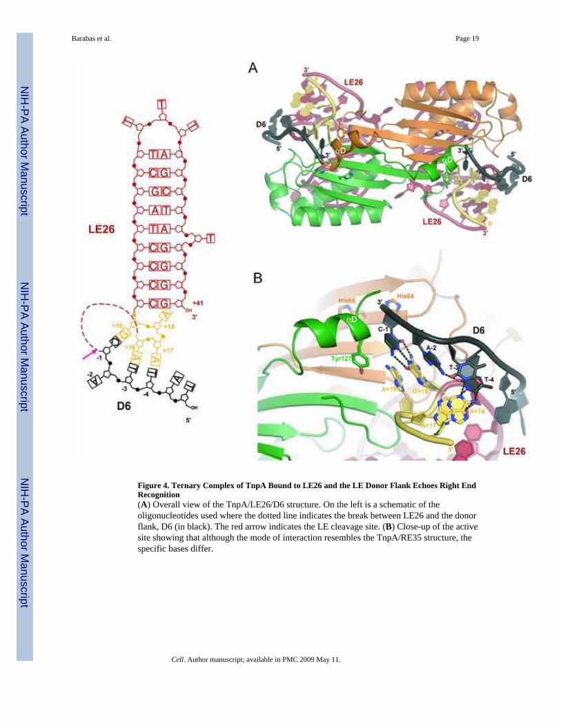
Figure 4. Ternary Complex of TnpA Bound to LE26 and the LE Donor Flank Echoes Right EndRecognition(A) Overall view of the TnpA/LE26/D6 structure. On the left is a schematic of theoligonucleotides used where the dotted line indicates the break between LE26 and the donorflank, D6 (in black). The red arrow indicates the LE cleavage site. (B) Close-up of the activesite showing that although the mode of interaction resembles the TnpA/RE35 structure, thespecific bases differ.
Barabas et al. Page 19
Cell. Author manuscript; available in PMC 2009 May 11.
NIH
-PA Author Manuscript
NIH
-PA Author Manuscript
NIH
-PA Author Manuscript

Figure 5. Transposon End Cleavage Can Be Re-Directed(A) SDS-PAGE gel showing the results of cleavage assays. Upon cleavage of an 8-mer thatspans the LE cleavage site, TnpA (green oval) becomes covalently attached to four nt and canbe resolved from unmodified TnpA. TnpA cleaves LE and RE cleavage substrates (cs) onlywhen the appropriate 5′ extension is added to IP sequences. No covalent complex is formedwith the Y127F active site mutant. (B) Modification of cleavage specificity. Shown are theresults of LE cleavage assays using LE26 and LEcs, and two variants.
Barabas et al. Page 20
Cell. Author manuscript; available in PMC 2009 May 11.
NIH
-PA Author Manuscript
NIH
-PA Author Manuscript
NIH
-PA Author Manuscript

Figure 6. Mixed Mutant Dimers Suggest Transposon End Cleavage is in Trans and Resolution isin Cis(A) Results of RE cleavage assays using mixed active site point mutants. The radiolabeledoligonucleotide is detected. (B) RE cleavage and donor backbone formation by mixed activesite mutants was assessed by mixing a RE cleavage substrate (56-mer) with a LE flank (40-mer). The product of RE cleavage is a 6-mer, and the 46-mer is the sealed donor backbone thatforms between the LE flank and the RE cleavage product. (C) Transposon junction formationby mixed active site mutants was assessed by mixing a LE cleavage substrate (100-mer whichis cleaved to yield a 60-mer transposon LE) with a precleaved transposon RE (45-mer). Thetransposon junction is formed by the WT protein (lane 7) but not by mixed active site mutants(lane 9). (D) LE cleavage is catalyzed by mixed active site mutants (lane 3), but not by thesingle point mutants (lanes 4 and 5).
Barabas et al. Page 21
Cell. Author manuscript; available in PMC 2009 May 11.
NIH
-PA Author Manuscript
NIH
-PA Author Manuscript
NIH
-PA Author Manuscript

Figure 7. Model for IS608 TranspositionUpon LE and RE binding (A), Y127 of each monomer cleaves the transposon ends and becomescovalently attached to the 5′ side of the gap (B). Movement of αD helices from trans to cis andresolution of the phosphotyrosine intermediates results in a transposon junction and a sealeddonor backbone (C). For integration (D), the donor backbone is replaced by target DNAcontaining TTAC which is recognized by an element of LE DNA. Cleavage (E), movement ofthe αD helices, and resolution of the phosphotyrosine intermediates (F) results in transposoninsertion into target DNA immediately 3′ of TTAC.
Barabas et al. Page 22
Cell. Author manuscript; available in PMC 2009 May 11.
NIH
-PA Author Manuscript
NIH
-PA Author Manuscript
NIH
-PA Author Manuscript