Maxillary canine–first premolar bilateral transposition in a Class III patient: A case report
11
Case Report Maxillary canine–first premolar bilateral transposition in a Class III patient: A case report Maciej Iancu Potrubacz a ; Michele Tepedino b ; Claudio Chimenti c ABSTRACT Tooth transposition is a rare dental anomaly that often represents a challenge for the clinician. The case of a girl with skeletal Class III malocclusion and concomitant maxillary canine–first premolar bilateral transposition, followed from 7 to 17 years of age, is presented. After a first phase of treatment aimed at resolving the Class III malocclusion, the transposition was maintained and the case finalized with a multibracket appliance. (Angle Orthod. 0000;00:000–000.) KEY WORDS: Ectopic tooth eruption; Transposition; Mx.C.P1; Interceptive orthodontics; Angle Class III INTRODUCTION Dental transposition is the positional interchange of two adjacent teeth, or the development or eruption of a tooth in a position normally occupied by a non- adjacent tooth. 1 The prevalence of this anomaly varies according to the sample studied, but it remains under 1% in most of the reports provided by the literature. 2–4 Dental transposition can affect the maxillary or mandibular arch, but it has never been reported in both jaws simultaneously. Maxillary transpositions are more frequent, while the prevalence of mandibular transposition, which affects only the canine and lateral incisor, has been reported to be around 0.03%. 5 Dental transposition has never been reported in the de- ciduous dentition. Dental transpositions are observed unilaterally more frequently than bilaterally (12:1) and affect mainly the left side (2:1). 6 When the anomaly is bilateral, the same teeth are affected on both sides; asymmetrical transposition is a very rare phenomenon. 7 Many authors found no gender predilection, 6 while others reported a higher frequency in females. 1,8,9 Peck and Peck 8 classified maxillary transposition into five categories, ordered by incidence: N Canine–first premolar (Mx.C.P1); N Canine–lateral incisor (Mx.C.I2); N Canine to first molar site (Mx.C to M1); N Lateral incisor–central incisor (Mx.I2.I1); and N Canine to central incisor site (Mx.C to I1). Dental transposition can be also classified into complete or incomplete types. In complete trans- position, both the crown and the entire roots of the involved teeth are found in their transposed position, while in incomplete transpositions only the crown is transposed, but the root apex remains in its relative normal position. Mx.C.P1 is the most frequent type of dental trans- position, accounting for nearly 71% of all cases, followed by Mx.C.I2 transposition, representing 20% of cases. 8 Regarding the etiology of this anomaly, many theories have been proposed, including positional interchange of tooth buds, 1,9 altered eruption paths, 10 the presence of retained primary teeth, 11 and trauma. 12 Recent evidence suggests that dental transposition represents a multifactorial condition in which both genetic and environmental factors seem to be in- volved, and the relationships are complex. 13 The Mx.C.P1 transposition has been determined to be influenced by genetic factors within a multifactorial inheritance model; 1 the findings of frequent association with other dental anomalies, common bilateral occur- rence, familial occurrence, and difference in male to a Adjunct Professor, Department of Biotechnological and Applied Clinical Sciences, University of L’Aquila, L’Aquila, Italy. b PhD student, Department of Biotechnological and Applied Clinical Sciences, University of L’Aquila, L’Aquila, Italy. c Professor, Department of Biotechnological and Applied Clinical Sciences, University of L’Aquila, L’Aquila, Italy. Corresponding author: Dr Michele Tepedino, Department of Biotechnological and Applied Clinical Sciences, University of L’Aquila, Viale S. Salvatore, Edificio Delta 6, 67100 L’Aquila, Italy (e-mail: [email protected]) Accepted: July 2015. Submitted: April 2015. Published Online: August 17, 2015 G 0000 by The EH Angle Education and Research Foundation, Inc. DOI: 10.2319/060215-371.1 1 Angle Orthodontist, Vol 00, No 0, 0000
Transcript of Maxillary canine–first premolar bilateral transposition in a Class III patient: A case report

Case Report
Maxillary canine–first premolar bilateral transposition in a Class III patient:
A case report
Maciej Iancu Potrubacza; Michele Tepedinob; Claudio Chimentic
ABSTRACTTooth transposition is a rare dental anomaly that often represents a challenge for the clinician. Thecase of a girl with skeletal Class III malocclusion and concomitant maxillary canine–first premolarbilateral transposition, followed from 7 to 17 years of age, is presented. After a first phase oftreatment aimed at resolving the Class III malocclusion, the transposition was maintained and thecase finalized with a multibracket appliance. (Angle Orthod. 0000;00:000–000.)
KEY WORDS: Ectopic tooth eruption; Transposition; Mx.C.P1; Interceptive orthodontics; AngleClass III
INTRODUCTION
Dental transposition is the positional interchange oftwo adjacent teeth, or the development or eruption ofa tooth in a position normally occupied by a non-adjacent tooth.1 The prevalence of this anomaly variesaccording to the sample studied, but it remains under1% in most of the reports provided by the literature.2–4
Dental transposition can affect the maxillary ormandibular arch, but it has never been reported inboth jaws simultaneously. Maxillary transpositions aremore frequent, while the prevalence of mandibulartransposition, which affects only the canine and lateralincisor, has been reported to be around 0.03%.5 Dentaltransposition has never been reported in the de-ciduous dentition.
Dental transpositions are observed unilaterally morefrequently than bilaterally (12:1) and affect mainly theleft side (2:1).6 When the anomaly is bilateral, thesame teeth are affected on both sides; asymmetricaltransposition is a very rare phenomenon.7 Many
authors found no gender predilection,6 while othersreported a higher frequency in females.1,8,9
Peck and Peck8 classified maxillary transpositioninto five categories, ordered by incidence:
N Canine–first premolar (Mx.C.P1);
N Canine–lateral incisor (Mx.C.I2);
N Canine to first molar site (Mx.C to M1);
N Lateral incisor–central incisor (Mx.I2.I1); and
N Canine to central incisor site (Mx.C to I1).
Dental transposition can be also classified intocomplete or incomplete types. In complete trans-position, both the crown and the entire roots of theinvolved teeth are found in their transposed position,while in incomplete transpositions only the crown istransposed, but the root apex remains in its relativenormal position.
Mx.C.P1 is the most frequent type of dental trans-position, accounting for nearly 71% of all cases,followed by Mx.C.I2 transposition, representing 20%of cases.8
Regarding the etiology of this anomaly, manytheories have been proposed, including positionalinterchange of tooth buds,1,9 altered eruption paths,10
the presence of retained primary teeth,11 and trauma.12
Recent evidence suggests that dental transpositionrepresents a multifactorial condition in which bothgenetic and environmental factors seem to be in-volved, and the relationships are complex.13 TheMx.C.P1 transposition has been determined to beinfluenced by genetic factors within a multifactorialinheritance model;1 the findings of frequent associationwith other dental anomalies, common bilateral occur-rence, familial occurrence, and difference in male to
a Adjunct Professor, Department of Biotechnological andApplied Clinical Sciences, University of L’Aquila, L’Aquila, Italy.
b PhD student, Department of Biotechnological and AppliedClinical Sciences, University of L’Aquila, L’Aquila, Italy.
c Professor, Department of Biotechnological and AppliedClinical Sciences, University of L’Aquila, L’Aquila, Italy.
Corresponding author: Dr Michele Tepedino, Department ofBiotechnological and Applied Clinical Sciences, University ofL’Aquila, Viale S. Salvatore, Edificio Delta 6, 67100 L’Aquila,Italy(e-mail: [email protected])
Accepted: July 2015. Submitted: April 2015.Published Online: August 17, 2015G 0000 by The EH Angle Education and Research Foundation,Inc.
DOI: 10.2319/060215-371.1 1 Angle Orthodontist, Vol 00, No 0, 0000

female prevalence support this hypothesis. The otherfour types of transposition seems to be mainly relatedto environmental factors; very little evidence suggestsa possible genetic influence in some cases of Mx.C.I2transpositions.8
In this case report, a maxillary bilateral canine–firstpremolar (Mx.C.P1) transposition in a Class III patientis presented. Since the treatment of this case wasarticulated into two phases, each phase will be shownand discussed separately.
CASE REPORT
First Phase of Treatment
Diagnosis. The patient was a 7-year-old female witha chief complaint of anterior crossbite. Facial photo-graphs revealed a prognathic profile, a poor zygomaticprojection, and a facial asymmetry (Figure 1). Thelateral cephalogram analysis showed a Class III, low-angle skeletal pattern with a maxillary retrusion(Figure 2; Table 1). Dental casts and intraoral photo-graph analysis revealed a transverse maxillary
deficiency with an anterior crossbite, while clinical
examination revealed a functional mandibular lateral
deviation and a lower tongue posture (Figures 1 and 3).
The jaw relation was recorded in the most retruded
mandibular position, confirming the presence of a true
skeletal Class III malocclusion. Evaluating the pano-
ramic radiograph, a slightly altered position of the
permanent tooth buds was found, which led us to
consider a possible initial Mx.C.P1 bilateral transposi-
tion (Figure 4).
Treatment objectives. The treatment objectives
were to establish a correct transverse skeletal re-
lationship, to protract the maxilla to solve the anterior
crossbite, and to check the occlusion to eliminate
the functional mandibular lateral deviation. After that,
there was the need to monitor the eruption of
permanent teeth.Treatment alternatives. An alternative to the treat-
ment proposed would have been to await the end ofgrowth and a full permanent dentition and to treat thepatient in only one phase, perhaps even taking into
Figure 1. Pretreatment facial and intraoral photographs.
2 POTRUBACZ, TEPEDINO, CHIMENTI
Angle Orthodontist, Vol 00, No 0, 0000

consideration a combined surgical-orthodontic treat-ment. According to the literature, the debate about one-phase or two-phase treatment is still open. Since it isdifficult to predict mandibular growth, many authorssuggest that a one-phase treatment can save thepatient time and money while assuring the same qualityof treatment results. On the other hand, an earlytreatment can prevent gingival recession, improveocclusal function, provide more favorable conditionsfor future growth, prevent excessive dental compensa-tion, simplify the second phase of treatment, andprovide more pleasant facial esthetic, thus improvingthe psychosocial development of the patient.14–16
However, considering the presence of a functionalmandibular latero-deviation and of a severe anteriorcrossbite with deep bite, there was an indication forearly intervention.
Treatment progress. The treatment began with
a bonded rapid maxillary expander (RME) with acrylic
pads, provided with hooks for a face mask (Figure 5A).
The RME was activated twice a day for 7 days; then
the screw was blocked with composite.
When a correct palatal expansion was achieved,the patient started wearing the face mask with16-oz elastics, with a 25u downward angulation. After6 months, when a complete correction of the anteriorcrossbite was achieved, the face mask and RME wereremoved (Figure 5B). Then the patient was instructedto wear a removable bionator III appliance at night forretention purposes and also for tongue posture re-habilitation (Figure 5C).
The bionator III appliance was discontinued afterabout 3 years; then the patient was put under a periodiccontrol protocol.
Treatment results. At the end of the first phase oftreatment, a good transversal proportion betweenupper and lower jaws was achieved, the anteriorcrossbite and the deep-bite were resolved, and themolar relationship was overcorrected. A better profileand improved facial esthetics were obtained, along withthe absence of mandibular lateral deviation (Figure 6).
Table 1. Cephalometric Analysisa
Norm
Measurement Mean SD Pretreatment Posttreatment Difference
SNA, u 82.0 3.5 81.6 84.3 2.7
SNB, u 80.0 3.0 83.6 84.1 0.5
ANB, u 2.0 2.4 22.0 0.2 2.2
Wits appraisal, mm 0.0 1.0 27.7 22.9 4.8
FMA, u 26.0 5.0 19.6 17.0 22.6
U1-APo, mm 6.0 2.2 21.7 2.5 4.2
L1-Apo, mm 2.0 2.3 1.5 20.4 21.9
U1-PP, u 110.0 5.0 89.3 112.3 23.0
IMPA, u 95.0 7.0 84.4 81.6 22.8
a SD indicates standard deviation.
Figure 2. Pretreatment lateral cephalogram and tracing.
MAXILLARY TRANSPOSITION 3
Angle Orthodontist, Vol 00, No 0, 0000

Figure 3. Pretreatment dental casts.
Figure 4. Pretreatment panoramic radiograph.
4 POTRUBACZ, TEPEDINO, CHIMENTI
Angle Orthodontist, Vol 00, No 0, 0000
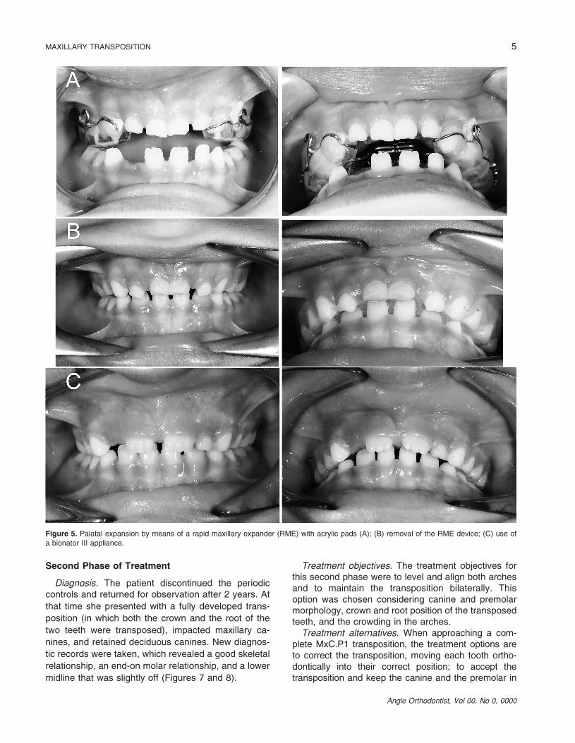
Second Phase of Treatment
Diagnosis. The patient discontinued the periodiccontrols and returned for observation after 2 years. At
that time she presented with a fully developed trans-
position (in which both the crown and the root of the
two teeth were transposed), impacted maxillary ca-
nines, and retained deciduous canines. New diagnos-
tic records were taken, which revealed a good skeletal
relationship, an end-on molar relationship, and a lower
midline that was slightly off (Figures 7 and 8).
Treatment objectives. The treatment objectives forthis second phase were to level and align both archesand to maintain the transposition bilaterally. Thisoption was chosen considering canine and premolarmorphology, crown and root position of the transposedteeth, and the crowding in the arches.
Treatment alternatives. When approaching a com-plete MxC.P1 transposition, the treatment options areto correct the transposition, moving each tooth ortho-dontically into their correct position; to accept thetransposition and keep the canine and the premolar in
Figure 5. Palatal expansion by means of a rapid maxillary expander (RME) with acrylic pads (A); (B) removal of the RME device; (C) use of
a bionator III appliance.
MAXILLARY TRANSPOSITION 5
Angle Orthodontist, Vol 00, No 0, 0000

their transposed positions; or to extract one of theteeth.17 Since there was no space deficiency or need fora sagittal correction, extractions were not consideredsuitable for this case.
An alternative would have been to correct thetransposition.18 While making this decision, severalpros and cons were taken into account. From an
occlusal and functional point of view, having the canineand the premolar in their correct positions is the bestoption.6 However, moving two teeth into the alveolarprocess to switch their positions is difficult, risky, andtime consuming.6,17,19 In fact, the risk of root resorptionand periodontal recession is quite high,6,17 since inmost of the cases there isn’t enough space in the
Figure 6. Photographs taken at the end of the first phase of treatment.
Figure 7. Intraoral photographs at the beginning of the second phase of treatment.
6 POTRUBACZ, TEPEDINO, CHIMENTI
Angle Orthodontist, Vol 00, No 0, 0000

alveolar process to contain two teeth moving inopposite directions, and this must be balanced withthe possible benefits of such treatment. Keeping thetransposition is an option widely approved by manyauthors6,8,17,20,21 and was chosen for this case becauseof the fewer contraindications and side effects asso-ciated with this choice, the minor treatment time, andthe simpler and more predictable mechanics.
Treatment progress. The treatment started with theextraction of the retained deciduous maxillary canines,the bonding of the upper arch with a multibracketstraightwire appliance with MBT prescription, and theinsertion of a 0.014-inch nickel-titanium (NiTi) wire.When was possible to put a 0.016-inch NiTi wire in theupper arch, the lower arch was bonded (Figure 9A).Open coil springs were applied between the upper
Figure 8. Panoramic radiograph showing a complete Mx.C.P1 bilateral transposition.
Figure 9. (A, B, C) Intraoral photographs showing the progress of the multibracket appliance treatment.
MAXILLARY TRANSPOSITION 7
Angle Orthodontist, Vol 00, No 0, 0000

second molar and first premolar to mesialize the latterteeth. When enough space for the canines wasobtained, they were bonded and included in theappliance. Tooth 23 erupted spontaneously, whiletooth 13 required a surgical exposure of the crown(Figure 9B). For the canines brackets with 0u of torquewere used, while for teeth 14 and 24 brackets with 27uof torque were maintained to eliminate the interferenceof the palatal cusps (Figure 9C). The archwire sequenceused was 0.014-inch NiTi, 0.016-inch NiTi, 0.017 3
0.025-inch NiTi, 0.019 3 0.025-inch stainless steel (SS),and 0.019 3 0.025-inch SS posted with tie-backs forspace closure in the upper arch and 0.016-inch NiTi,0.017 3 0.025-inch NiTi, and 0.019 3 0.025-inch SS inthe lower arch. Class II elastics were used in the lastphase of treatment. Total treatment time was 29 months.
Treatment results. A Class I molar relationship withcorrect overjet and overbite was obtained, along witha pleasant facial esthetic. A satisfying result wasachieved, from both a dental and a facial point of view(Figures 10 through 13). A coronal reshaping of thepalatal cusp of the upper first premolar was necessary
to avoid occlusal interferences and to achieve goodcanine function. The patient was fully satisfied with hersmile esthetics. At the debonding, the patient was 15years old, so it was decided, in accord with herparents, to delay prosthetics restoration to the end ofgrowth, when the patient would be able to decide toproceed by herself.
DISCUSSION
Having the possibility of following a patient from theearly stage of development offers many opportunitiesto intercept and correct some types of pathology at theright moment. In this case, an early interventionallowed us to reestablish a harmonic dentoskeletalpattern and form the basis for correct dentofacialdevelopment, as can be seen from the outcome of thistreatment. However, regarding the transposition, theinterceptive treatment was insufficient in correcting theeruptive path of the upper canines and premolars,since even in the earlier stage of development thetooth buds were in a completely transposed position.
Figure 10. Posttreatment facial and intraoral photographs.
8 POTRUBACZ, TEPEDINO, CHIMENTI
Angle Orthodontist, Vol 00, No 0, 0000

For this reason, a complete Mx.C.P1 in the permanentdentition during the second phase of treatment had tobe managed. Among the different treatment strategiesavailable to us, we decided to keep the transposition.Despite an easier mechanics and a reduced treatment
time, this choice presents some challenges. In fact, inorder to achieve an esthetic and functional result, it isimportant to control the torque of the canine to movethe root palatally to hide the root prominence and thetorque of the premolar to move the root buccally to
Figure 11. Posttreatment panoramic radiographs, lateral cephalogram, and tracings.
Figure 12. Pre- and posttreatment superimposition.
MAXILLARY TRANSPOSITION 9
Angle Orthodontist, Vol 00, No 0, 0000
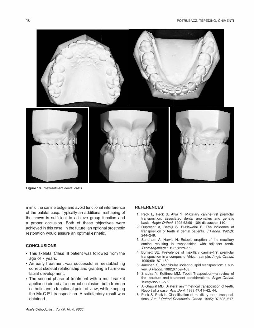
mimic the canine bulge and avoid functional interference
of the palatal cusp. Typically an additional reshaping of
the crown is sufficient to achieve group function and
a proper occlusion. Both of these objectives were
achieved in this case. In the future, an optional prosthetic
restoration would assure an optimal esthetic.
CONCLUSIONS
N This skeletal Class III patient was followed from theage of 7 years.
N An early treatment was successful in reestablishingcorrect skeletal relationship and granting a harmonicfacial development.
N The second phase of treatment with a multibracketappliance aimed at a correct occlusion, both from anesthetic and a functional point of view, while keepingthe Mx.C.P1 transposition. A satisfactory result wasobtained.
REFERENCES
1. Peck L, Peck S, Attia Y. Maxillary canine-first premolartransposition, associated dental anomalies and geneticbasis. Angle Orthod. 1993;63:99–109; discussion 110.
2. Ruprecht A, Batniji S, El-Neweihi E. The incidence oftransposition of teeth in dental patients. J Pedod. 1985;9:244–249.
3. Sandham A, Harvie H. Ectopic eruption of the maxillarycanine resulting in transposition with adjacent teeth.Tandlaegebladet. 1985;89:9–11.
4. Burnett SE. Prevalence of maxillary canine-first premolartransposition in a composite African sample. Angle Orthod.1999;69:187–189.
5. Jarvinen S. Mandibular incisor-cuspid transposition: a sur-vey. J Pedod. 1982;6:159–163.
6. Shapira Y, Kuftinec MM. Tooth Trasposition—a review ofthe literature and treatment considerations. Angle Orthod.1989;59:271–276.
7. Al-Shawaf MD. Bilateral asymmetrical transposition of teeth.Report of a case. Ann Dent. 1988;47:41–42, 44.
8. Peck S, Peck L. Classification of maxillary tooth transposi-tions. Am J Orthod Dentofacial Orthop. 1995;107:505–517.
Figure 13. Posttreatment dental casts.
10 POTRUBACZ, TEPEDINO, CHIMENTI
Angle Orthodontist, Vol 00, No 0, 0000

9. Peck S, Peck L, Kataja M. Mandibular lateral incisor-canine
transposition, concomitant dental anomalies, and genetic
control. Angle Orthod. 1998;68:455–466.10. Gholston LR, Williams PR. Bilateral transposition of maxil-
lary canines and lateral incisors: a rare condition. ASDC
J Dent Child. 1984;51:58–63.11. Laptook T, Silling G. Canine transposition—approaches to
treatment. J Am Dent Assoc. 1983;107:746–748.12. Dayal PK, Shodhan KH, Dave CJ. Transposition of canine
with traumatic etiology. J Indian Dent Assoc. 1983;55:283–285.
13. Ely NJ, Sherriff M, Cobourne MT. Dental transposition as
a disorder of genetic origin. Eur J Orthod. 2006;28:145–151.14. Ngan P. Early timely treatment of Class III malocclusion.
Semin Orthod. 2005;11:140–145.15. Ngan P. Early treatment of Class III malocclusion: is it worth
the burden? Am J Orthod Dentofacial Orthop. 2006;129
(4 suppl):S82–S85.
16. De Toffol L, Pavoni C, Baccetti T, Franchi L, Cozza P.Orthopedic treatment outcomes in Class III malocclusion.A systematic review. Angle Orthod. 2008;78:561–573.
17. Ciarlantini R, Melsen B. Maxillary tooth transposition: correct oraccept? Am J Orthod Dentofacial Orthop. 2007;132:385–394.
18. Nishimura K, Nakao K, Aoki T, Fuyamada M, Saito K, GotoS. Orthodontic correction of a transposed maxillary canineand first premolar in the permanent dentition. Am J OrthodDentofacial Orthop. 2012;142:524–533.
19. Silva Camara Mattos B, Carlos Mesquita Carvalho J, MatusitaM, Pereira Pinheiro Alves AP. Tooth transposition—a literaturereview and a clinical case. Braz J Oral Sci. 2006;5:953–957.
20. Weeks EC, Power SM. The presentations and managementof transposed teeth. Br Dent J. 1996;181:421–424.
21. Di Palma E, Di Giuseppe B, Tepedino M, Chimenti C.Orthodontic management of bilateral maxillary canine-firstpremolar transposition and bilateral agenesis of maxillarylateral incisors: a case report. Dent Press J Orthod. 2015;20:100–109.
MAXILLARY TRANSPOSITION 11
Angle Orthodontist, Vol 00, No 0, 0000