Maternal high fat diet during pregnancy and lactation alters hepatic expression of insulin like...
12
BioMed Central Page 1 of 12 (page number not for citation purposes) BMC Genomics Open Access Research article Maternal high fat diet during pregnancy and lactation alters hepatic expression of insulin like growth factor-2 and key microRNAs in the adult offspring Junlong Zhang* 1 , Fang Zhang 1 , Xavier Didelot 2 , Kimberley D Bruce 3 , Felino R Cagampang 3 , Manu Vatish 1,4 , Mark Hanson 3 , Hendrik Lehnert 1,5 , Antonio Ceriello 6 and Christopher D Byrne 3 Address: 1 Clinical Science Research Institute, Warwick Medical School, Clinical Sciences Building, University Hospital - Walsgrave Campus, Clifford Bridge Road, Coventry, CV2 2DX, UK, 2 Department of Statistics, University of Warwick, Coventry, CV4 7AL, UK, 3 Institute of Developmental Sciences, Developmental Origins of Health and Disease Division, University of Southampton Medical School, Southampton General Hospital, Southampton, SO16 0YD, UK, 4 Albert Einstein College of Medicine, 1301 Morris Park Avenue, Bronx, New York, NY 10461, USA, 5 1st Medical Department, University of Lübeck Medical School Ratzeburger Allee 160, 23538 Luebeck, Germany and 6 Chair of Endocrinology, University of Udine, Italy Email: Junlong Zhang* - [email protected]; Fang Zhang - [email protected]; Xavier Didelot - [email protected]; Kimberley D Bruce - [email protected]; Felino R Cagampang - [email protected]; Manu Vatish - [email protected]; Mark Hanson - [email protected]; Hendrik Lehnert - [email protected]; Antonio Ceriello - [email protected]; Christopher D Byrne - [email protected] * Corresponding author Abstract Background: miRNAs play important roles in the regulation of gene functions. Maternal dietary modifications during pregnancy and gestation have long-term effects on the offspring, but it is not known whether a maternal high fat (HF) diet during pregnancy and lactation alters expression of key miRNAs in the offspring. Results: We studied the effects of maternal HF diet on the adult offspring by feeding mice with either a HF or a chow diet prior to conception, during pregnancy and lactation, and all offspring were weaned onto the same chow diet until adulthood. Maternal HF fed offspring had markedly increased hepatic mRNA levels of peroxisome proliferator activated receptor-alpha (ppar-alpha) and carnitine palmitoyl transferase-1a (cpt-1a) as well as insulin like growth factor-2 (Igf2). A HF diet induced up-regulation of ppar-alpha and cpt-1a expression in the wild type but not in Igf2 knock out mice. Furthermore, hepatic expression of let-7c was also reduced in maternal HF fed offspring. Among 579 miRNAs measured with microarray, ~23 miRNA levels were reduced by ~1.5-4.9-fold. Reduced expression of miR-709 (a highly expressed miRNA), miR-122, miR-192, miR-194, miR- 26a, let-7a, let7b and let-7c, miR-494 and miR-483* (reduced by ~4.9 fold) was validated by qPCR. We found that methyl-CpG binding protein 2 was the common predicted target for miR-709, miR- let7s, miR-122, miR-194 and miR-26a using our own purpose-built computer program. Conclusion: Maternal HF feeding during pregnancy and lactation induced co-ordinated and long- lasting changes in expression of Igf2, fat metabolic genes and several important miRNAs in the offspring. Published: 16 October 2009 BMC Genomics 2009, 10:478 doi:10.1186/1471-2164-10-478 Received: 13 February 2009 Accepted: 16 October 2009 This article is available from: http://www.biomedcentral.com/1471-2164/10/478 © 2009 Zhang et al; licensee BioMed Central Ltd. This is an Open Access article distributed under the terms of the Creative Commons Attribution License (http://creativecommons.org/licenses/by/2.0 ), which permits unrestricted use, distribution, and reproduction in any medium, provided the original work is properly cited.
-
Upload
independent -
Category
Documents
-
view
3 -
download
0
Transcript of Maternal high fat diet during pregnancy and lactation alters hepatic expression of insulin like...
BioMed CentralBMC Genomics
ss
Open AcceResearch articleMaternal high fat diet during pregnancy and lactation alters hepatic expression of insulin like growth factor-2 and key microRNAs in the adult offspringJunlong Zhang*1, Fang Zhang1, Xavier Didelot2, Kimberley D Bruce3, Felino R Cagampang3, Manu Vatish1,4, Mark Hanson3, Hendrik Lehnert1,5, Antonio Ceriello6 and Christopher D Byrne3Address: 1Clinical Science Research Institute, Warwick Medical School, Clinical Sciences Building, University Hospital - Walsgrave Campus, Clifford Bridge Road, Coventry, CV2 2DX, UK, 2Department of Statistics, University of Warwick, Coventry, CV4 7AL, UK, 3Institute of Developmental Sciences, Developmental Origins of Health and Disease Division, University of Southampton Medical School, Southampton General Hospital, Southampton, SO16 0YD, UK, 4Albert Einstein College of Medicine, 1301 Morris Park Avenue, Bronx, New York, NY 10461, USA, 51st Medical Department, University of Lübeck Medical School Ratzeburger Allee 160, 23538 Luebeck, Germany and 6Chair of Endocrinology, University of Udine, Italy
Email: Junlong Zhang* - [email protected]; Fang Zhang - [email protected]; Xavier Didelot - [email protected]; Kimberley D Bruce - [email protected]; Felino R Cagampang - [email protected]; Manu Vatish - [email protected]; Mark Hanson - [email protected]; Hendrik Lehnert - [email protected]; Antonio Ceriello - [email protected]; Christopher D Byrne - [email protected]
* Corresponding author
AbstractBackground: miRNAs play important roles in the regulation of gene functions. Maternal dietarymodifications during pregnancy and gestation have long-term effects on the offspring, but it is notknown whether a maternal high fat (HF) diet during pregnancy and lactation alters expression ofkey miRNAs in the offspring.
Results: We studied the effects of maternal HF diet on the adult offspring by feeding mice witheither a HF or a chow diet prior to conception, during pregnancy and lactation, and all offspringwere weaned onto the same chow diet until adulthood. Maternal HF fed offspring had markedlyincreased hepatic mRNA levels of peroxisome proliferator activated receptor-alpha (ppar-alpha)and carnitine palmitoyl transferase-1a (cpt-1a) as well as insulin like growth factor-2 (Igf2). A HFdiet induced up-regulation of ppar-alpha and cpt-1a expression in the wild type but not in Igf2 knockout mice. Furthermore, hepatic expression of let-7c was also reduced in maternal HF fed offspring.Among 579 miRNAs measured with microarray, ~23 miRNA levels were reduced by ~1.5-4.9-fold.Reduced expression of miR-709 (a highly expressed miRNA), miR-122, miR-192, miR-194, miR-26a, let-7a, let7b and let-7c, miR-494 and miR-483* (reduced by ~4.9 fold) was validated by qPCR.We found that methyl-CpG binding protein 2 was the common predicted target for miR-709, miR-let7s, miR-122, miR-194 and miR-26a using our own purpose-built computer program.
Conclusion: Maternal HF feeding during pregnancy and lactation induced co-ordinated and long-lasting changes in expression of Igf2, fat metabolic genes and several important miRNAs in theoffspring.
Published: 16 October 2009
BMC Genomics 2009, 10:478 doi:10.1186/1471-2164-10-478
Received: 13 February 2009Accepted: 16 October 2009
This article is available from: http://www.biomedcentral.com/1471-2164/10/478
© 2009 Zhang et al; licensee BioMed Central Ltd. This is an Open Access article distributed under the terms of the Creative Commons Attribution License (http://creativecommons.org/licenses/by/2.0), which permits unrestricted use, distribution, and reproduction in any medium, provided the original work is properly cited.
Page 1 of 12(page number not for citation purposes)
BMC Genomics 2009, 10:478 http://www.biomedcentral.com/1471-2164/10/478
BackgroundmiRNAs are small (~21 nt) non-coding RNAs that wereoriginally discovered to regulate development in C. ele-gans [1-3]. A significant number of miRNAs are conservedacross different species [4-7]. miRNAs regulate gene func-tions mainly through degradation of their cognatemRNAs by perfect matches with the mRNA molecules; orvia inhibition of protein translation through base pairingof ~7 nucleotides (called "seed sequence") betweenmiRNA and the 3'-untranslated region (3'-UTR) of the tar-get mRNA molecules [8]. Expression of miRNAs may beregulated by transcription factors (e.g. myogenin andmyoD regulate expression of a number of miRNAs [9]),and transcription factors per se may also be regulated bymiRNAs (e.g. miR-1 promotes myogenesis by targetinghistone deacetylase 4, a transcriptional repressor of mus-cle gene expression) [10]. A single miRNA can repress theproduction of hundreds of proteins, but this repression isrelatively mild [11]. On the other hand one mRNA can betargeted by several miRNAs, which have additive effects inregulation of protein synthesis [12]. For example, SMAD-1 gene has two predicted binding sites for miR-26a [12],and greater suppression effects on protein translationhave been observed in mRNAs containing multiple bind-ing sites for a miRNA [13].
miRNAs are involved in the regulation of almost allimportant biological processes including development[14], differentiation, cell proliferation, cell cycle regula-tion [15,16] and energy metabolism [17], including fatmetabolism and glucose homeostasis [18,19]. For exam-ple, miR-375 suppresses glucose-induced insulin secre-tion in pancreatic β-cells [20], thus demonstrating anessential role in plasma glucose homeostasis. Knockingdown of endogenous miR-122, a miRNA abundantlyexpressed in the liver, reduces plasma cholesterol concen-trations in mice [21], with parallel up-regulation of 363mRNA transcripts and down-regulation of 305 mRNAtranscripts in the liver [21]. MiR-143 stimulates humanadipocyte differentiation [22]. Analysis of global profilesof miRNA expression in skeletal muscle with microarrayshows that expression of 4 miRNAs (miR-29a, miR-29b,miR-29c and miR-150) are up-regulated [23], whereasexpression of 11 miRNAs (miR-379, miR-127, miR299-5p, miR-434-3p, miR-335, miR130a, miR-19b, miR-451,miR-148a, miR-199a and miR-152) are down-regulatedin skeletal muscle of type 2 diabetic rats [23].
The prevalence of obesity is increasing markedly in indus-trialised countries [24-28], and high fat, high protein, lowcarbohydrate diets including proprietary diets such as theAtkins diet are widely consumed [29-31]. The prevalenceof obesity in women of reproductive age continues to rise[32], and it is likely that many women of reproductive agealso consume a low carbohydrate, high fat and high pro-
tein diet during pregnancy and lactation. However theeffects of increased maternal dietary consumption of fatduring pregnancy and weaning on the long term health ofthe offspring are not fully characterized.
Many studies have indicated long-term consequences ofmaternal dietary modifications (e.g. caloric or proteinrestrictions) during pregnancy and lactation on the devel-opment of insulin resistance and risk of cardiovasculardisease in the offspring [33-37]. We have previouslyshown in mice that adult offspring of dams fed a low car-bohydrate, high fat and high protein diet during preg-nancy and lactation but weaned onto a chow diet havereduced hepatic triglyceride levels in association withincreased protein levels of key genes regulating fatty acidoxidation including carnitine palmitoyltransferase-1a(CPT-1a) and peroxisome proliferator-activated receptor-alpha (PPARα) predominantly in the female offspring[33]. Pups born to dams on a high fat (HF) diet duringgestation and lactation have increased percentage of bodyfat, plasma glucose, free fatty acids, insulin and choles-terol levels, liver weight and lipid concentrations at wean-ing or in adulthood [38,39].
Fetal growth is regulated by insulin-like growth factor 2(IGF2) [40]. Recent data suggest that IGF2 may regulatefat metabolism. For example, body weight is affected byseveral polymorphisms in the Igf2 gene [41,42], and lowcirculating IGF2 concentrations are associated with weightgain and obesity [43]. In contrast, high circulating IGF2levels associated with the Apal polymorphism of Igf2 areassociated with low body weight in middle aged men[44]. Mice overexpressing Igf2 have increased fatty acidoxidation [45]. Maternal dietary protein restrictionreduces hepatic expression of Igf2 in the male fetal off-spring. However, whether maternal HF feeding alters off-spring Igf2 expression has not been documented.
Following our previous studies on maternal high fat, highprotein and low carbohydrate diet[33], we used a modi-fied diet to investigate whether maternal HF feeding dur-ing pregnancy and lactation altered mRNA levels of ppar-α and cpt-1a and whether changes in ppar-α and cpt-1awere related to changes in Igf2 expression. We also ana-lyzed global miRNA expression profile in the liver todetermine which miRNAs were altered in the offspringborn to dams fed a HF diet during pregnancy and lacta-tion.
ResultsMaternal HF fed offspring had increased mRNA levels of ppar-α, cpt-1a and Igf2 in the liverWe have previously shown that maternal high fat, highprotein and low carbohydrate diet fed offspring hadincreased protein levels of PPARα and CPT-1a levels in the
Page 2 of 12(page number not for citation purposes)
BMC Genomics 2009, 10:478 http://www.biomedcentral.com/1471-2164/10/478
liver, in association with reduced hepatic lipid levels,despite having no significant changes in body weight,plasma glucose and lipid profile [33]. In this study, amodified HF diet was fed to dams, in which the percent-age of fat was increased by more than 2-fold with a smallincrease in protein levels compared to the chow diet (seemethods section). Consistently, no significant differencein body weight, fasting plasma triglyceride, total choles-terol and glucose levels were observed between maternalHF fed offspring weaned on a chow diet (HF/C) and con-trol mice (C/C, data not shown). mRNA levels of ppar-αand cpt-1a in the HF/C mice were increased by ~1.6 and~3.7-fold respectively compared to C/C mice (p < 0.05and 0.01 for ppar-α and cpt-1a respectively, Table 1).
As the maternal HF diet was implemented prior to con-ception and continued throughout pregnancy and lacta-tion, we investigated whether expression of Igf2, animprinted gene encoding a growth factor expressed duringearly development [40] was altered in the maternal HF fedoffspring. The mRNA level of Igf2 was increased by ~2.7-fold in maternal HF diet fed offspring compared to thecontrol animals (p < 0.01, Table 1).
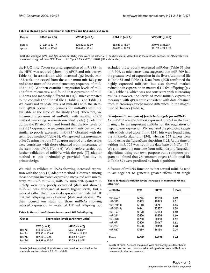
To determine whether increased expression of ppar-α andcpt-1a was related to increased expression of Igf2 in thematernal HF fed offspring, we measured mRNA levels ofppar-α and cpt-1a in Igf2 KO mice. A HF diet modestlyincreased hepatic expression of ppar-α and cpt-1a in theWT mice (p < 0.05 and 0.01 for ppar-α and cpt-1a respec-tively, Table 2), but the HF diet had no effects on ppar-αand cpt-1a expression in the KO mice (p = 0.98 and 1.0 forppar-α and cpt-1a respectively, Table 2), suggesting thatexpression of Igf2 was required for the HF diet inducedup-regulation of expression of ppar-α and cpt-1a.
Hepatic expression of let-7c was reduced in maternal HF offspringLet-7 was originally discovered due to its regulation ofdevelopmental timing in C. elegans, through binding tothe 3'-UTR region of Lin-41 [46,47]. Levels of let-7c andother members of let-7 including let-7a, let-7b and let-7dwere reduced by 2-2.5-fold in maternal HF fed offspringcompared to the control animals (p < 0.01 for let-7a, let-7b and let-7d, Table 3).
Having observed reduced expression of let-7s in maternalHF fed offspring, we measured the global miRNA expres-sion profile with microarrays.
Expression of ~5.7% of miRNAs was altered in the maternal HF fed offspringA cut-off threshold of 1.5-fold change [48] in miRNAs wasused to determine whether altered miRNAs levels werelikely to be significant. Of 579 miRNAs measured, expres-sion of 10 miRNAs (~1.7%) was increased by ~1.5-2-fold(average increase was ~1.64-fold, Table 4), whereasexpression of 23 miRNAs (~3.97%) were reduced by 1.51- 4.93 fold (average reduction of 2.16-fold, Table 5), withmiR-483* showing the biggest reduction (by ~4.9-fold,Table 5). In contrast, expression of most miRNAsremained unchanged (Additional file 1: Table S1).
Among those miRNAs showing reduced expression, aver-age levels of expression were 90310 arbitrary units (Table5), whereas in those showing increased expression, theaverage levels of the 10 miRNAs were 16083 arbitraryunits (Table 4), which was 5.6-fold lower than those miR-NAs showing reduced expression.
We validated microarray data with the stem-loop RT-PCRmethod [49] using purchased miRNA primers (ABI). 5miRNAs (let-7c, miR-483*, miR-22, miR-29a and miR-30c) were measured as these miRNAs showed differentmagnitude of reduced expression in the HF offspring(Table 6). Expression of miR-483*, let-7c and miR-29ameasured with qPCR were consistent with valuesobtained from microarray data, with minor differences inthe magnitude of changes in expression (Table 6). How-ever, a discrepancy in levels of miR-30c and miR-22between qPCR and microarray was observed (Table 6).Levels of miR-30c between maternal HF and chow fed off-spring were similar when measured with microarrays, butsignificantly different when measured with qPCR. A ~2.9-fold reduction was obtained with microarray analyseswhereas a ~42% increase occurred in levels of miR-22 inmaternal HF offspring measured with qPCR (Table 6). Wealso noted that levels of miR-483* were very low whenmeasured with qPCR, consistent with poorly expressedIgf2 mRNA levels. However, data from microarray sug-gested that miR-483* was abundantly expressed, whichwas not consistent with qPCR data (Table 6).
We examined the miR-483* genomic DNA locationbecause miR-483* showed the greatest reduction inexpression in maternal HF fed offspring, and found thatmiR-483* was encoded in an intron of Igf2. As intronicmiRNAs may share common promoters as their hostgenes, many intronic miRNAs show significantly corre-lated expression profiles with their host genes[50,51].Thus, we would expect that the levels of intronic miRNA(e.g. miR-483*) are increased with the host gene (Igf2) in
Table 1: Effects of maternal HF feeding on hepatic mRNA levels in adult offspring
Genes C/C (n = 7) HF/C (n = 7)
ppar-α 61873 ± 7638 97445 ± 11712*cpt-1a 34193 ± 4420 126777 ± 23720**Igf2 520 ± 70 1404 ± 266**
Levels of mRNA expression (arbitrary units) were measured with real time qPCR as described in the method section. Mean ± S.E. * p < 0.05 and ** p < 0.01.
Page 3 of 12(page number not for citation purposes)
BMC Genomics 2009, 10:478 http://www.biomedcentral.com/1471-2164/10/478
the HF/C mice. To our surprise, expression of miR-483* inthe HF/C was reduced (shown by qPCR and microarray,Table 4a) in association with increased Igf2 levels. Mir-483 is also processed from the same mmu-mir-483 geneand share most of the complementary sequence of miR-483* [52]. We then examined expression levels of miR-483 from microarray, and found that expression of miR-483 was not markedly different in HF/C mice comparedto the controls (Additional file 1: Table S1 and Table 6).We could not validate levels of miR-483 with the stem-loop qPCR because the primers for miR-483 were notavailable at the time of the study (ABI). Therefore, wemeasured expression of miR-483 with another qPCRmethod involving reverse-transcribed poly(T) adaptorduring the RT step [53], and found that the low levels ofmiR-483 expression were consistent with microarray data,similar to poorly expressed miR-483* obtained with thestem-loop method (Table 6). We repeated measurementsof let-7c using the poly (T) adaptor method and the resultswere consistent with those obtained from microarray orthe stem-loop qPCR (Table 6). We therefore carried outfurther validation of miRNAs with the poly (T) adaptormethod as this methodology provided flexibility inprimer design.
We tried to validate miRNAs showing increased expres-sion with the poly (T) adaptor method. However, amongthose showing increased expression measured with micro-array, miR-667, miR-207, miR-197, miR-770-3p and miR-369-3p were very poorly expressed (data not shown).miR-328 was expressed at much higher levels, but areduced rather than increased expression in maternal HFdiet fed offspring was observed (data not shown). Wethen focused our study on those miRNAs showingreduced expression in maternal HF fed offspring but
excluded those poorly expressed miRNAs (Table 5) plusmiR-709, as microarray data suggested that miR-709 hadthe greatest level of expression in the liver (Additional file1: Table S1 and Table 6). Data from qPCR confirmed thehighly expressed miR-709, but also showed markedreduction in expression in maternal HF fed offspring (p <0.01, Table 6), which was not consistent with microarrayresults. However, the levels of most miRNA expressionmeasured with qPCR were consistent with data obtainedfrom microarrays except minor differences in the magni-tude of changes (Table 6).
Bioinformatic analysis of predicted targets for miRNAsAs miR-709 was the highest expressed miRNA in the liver,it might be an important miRNA for the regulation ofhepatic gene expression. We analysed the predicted targetswith widely used algorithms. 1241 hits were found usingthe miRanda algorithm [54], whereas 353 targets werefound using the TargetScan algorithm [12]. At the time ofwriting, miR-709 was not in the data base of PicTar [55].We compared the outcome from miRanda and TargetSanalgorithms using our own purpose-built computer pro-gram and found that 28 common targets (Additional file2: Table S2) were predicted by both algorithms.
A feature of miRNA function is that several miRNAs tendto act together to generate greater effects than single
Table 2: Hepatic gene expression in wild type and Igf2 knock out mice
Genes KO-C (n = 5) WT-C (n = 6) KO-HF (n = 6) WT-HF (n = 6)
ppar-α 210.34 ± 33.17 220.32 ± 40.99 283.88 ± 15.97 370.91 ± 31.35*cpt-1a 266.71 ± 17.41 236.68 ± 50.41 266.05 ± 36.59 391.26 ± 21.61**
Both the wild type (WT) and Igf2 knock out (KO) mice were fed either a HF or chow diet as described in the methods section. mRNA levels were measured using real time PCR. Mean ± S.E. * p < 0.05 and ** p < 0.01 (HF v chow diet).
Table 3: Hepatic let-7s levels in maternal HF fed offspring.
Genes Expression levels (arbitrary units)
C/C (n = 7) HF/C (n = 7)let-7c 118.10 ± 9.71 60.31 ± 6.80**let-7a 278.62 ± 15.64 107.64 ± 11.82*let-7b 107.10 ± 5.45 45.82 ± 4.28**let-7d 164.68 ± 15.50 80.29 ± 8.10**
Levels (arbitrary units) of let-7s were measured as described in the methods section. Mean ± S.E. ** p < 0.01.
Table 4: Hepatic miRNA levels increased in maternal HF fed offspring
miRNAs C/C HF/C ↑ Fold
miR-503* 12765 19148 1.50miR-379 13463 20313 1.51miR-770-3p 17118 26761 1.56miR-369-3p 14441 22857 1.58miR-197 20128 32193 1.60miR-21* 12420 19874 1.60miR-328 18750 30348 1.62miR-471 12420 20167 1.62miR-207 21638 38458 1.78miR-667 17689 36156 2.04
Mean 16083 26628 1.61
Levels of miRNAs were measured with microarrays as described in the method section. Relative values of signals for each miRNAs are presented in the two columns.
Page 4 of 12(page number not for citation purposes)

BMC Genomics 2009, 10:478 http://www.biomedcentral.com/1471-2164/10/478
miRNA [13]. We therefore undertook bioinformaticsanalysis to investigate whether it was possible to identifycommon targets for those validated miRNAs that showedreduced expression in the maternal HF fed offspring usingour own purpose-built computer program. We found thatZSWIM3 (zinc finger, SWIM domain containing 3), a pro-tein whose function was yet to be characterised [56], wastargeted by 5 miRNAs namely miR-122, miR-192, miR-194, miR-709 and miR-483*. 14 genes were targeted by 3different miRNAs ' [see Additional file 22: Table S3]' and10 genes (including citrate synthase and Igf-1 receptor)were targeted by miR-122 and miR-494 ' [see Additionalfile 2: Table S4]'. These results suggested that functions ofspecific genes might be co-ordinately regulated by a smallnumber of miRNAs.
DiscussionMaternal HF fed offspring mice have: 1) increased hepaticexpression of key genes including those regulating fetalgrowth (such as Igf2) and fat metabolism (such as ppar-αand cpt-1a); 2) altered expression of a small percentage(~5.7%) of important miRNAs. Among the miRNAsshowing reduced expression, let-7c regulates developmen-tal timing [46,47] and miR-122 regulates fat oxidation[21,57]. Thus, these data suggest co-ordinated changes inkey metabolic genes and miRNAs that regulate early fetalgrowth and fat metabolism in offspring of dams fed HFdiet.
As both offspring from HF- and chow-fed dams areweaned onto the same chow diet and maintained on
Table 5: Hepatic miRNAs levels reduced in maternal HF fed offspring
miRNAs C/C HF/C ↓ Fold
miR-410 18072 11997 1.51miR-804 19831 13138 1.51miR-323-5p 17402 11500 1.51let-7c 108628 71343 1.52miR-302a* 16365 10647 1.54miR-711 25823 16057 1.61miR-26a 95209 58142 1.64miR-122 324798 192912 1.68miR-216b 16179 9463 1.71miR-294* 17402 10168 1.71miR-185 27121 15378 1.76miR-192 80361 44865 1.79miR-29a 30002 16331 1.84miR-194 92178 49817 1.85miR-145 28108 14841 1.89miR-126-3p 41868 20020 2.09miR-762 76284 32872 2.32miR-16 109631 43004 2.55miR-1224 69200 24241 2.85miR-22 130448 44304 2.94miR-30c-2* 67031 20460 3.28miR-494 298733 82842 3.61miR-483* 366458 74387 4.93
Mean 90310 38640 2.16
Levels of miRNAs were measured with microarrays as described in the method section. Relative values of signals for each miRNAs were presented in the two columns.
Table 6: Validation of microarray data with stem-loop real-time qPCR
Genes qPCR (arbitrary units) Microarray ↓ Fold (HF/C v C/C)
C/C (n = 7) HF/C (n = 7) C/C HF/C qPCR Microarray
Stem-loop real time PCRmiR-30c 1572 ± 670 1168 ± 230* 19436 18861 1.35 1.03miR-22 309 ± 163 440 ± 179* 130448 44304 0.70 2.94miR-29a 370 ± 117 276 ± 109* 30002 16331 1.34 1.84Let-7c 330 ± 31 227 ± 19* 108628 71343 1.45 1.52miR-483* 1.9 ± 0.2 0.3 ± 0.1*** 366458 74387 6.89 4.93Poly dT adaptor qPCR methodmiR-709 1304 ± 118 768 ± 129** 1387121 1258883 1.70 1.10miR-122 948 ± 61 473 ± 40*** 324798 192912 2.00 1.68miR-192 321 ± 24 121 ± 10*** 80361 44865 2.64 1.79miR-194 124 ± 11 81 ± 7** 92178 49817 1.53 1.85miR-26a 157 ± 17 58 ± 10*** 95209 58142 2.72 1.64Let-7c 118 ± 10 60 ± 7*** 108628 71343 1.96 1.52let-7a 279 ± 16 108 ± 12*** 60650 43004 2.59 1.41let-7b 107 ± 5 46 ± 4*** 83730 63907 2.34 1.31let-7d 165 ± 16 80 ± 8*** 91702 72642 2.05 1.26miR-494 12 ± 2 5.5 ± 0.8*** 298733 82842 2.18 3.61miR-483 1.3 ± 0.2 1.0 ± 0.2 17689 22100 0.78 0.80
Levels of miRNAs were measured either with purchased primers for specific miRNAs (stem-loop real time PCR) or poly dT adaptor as described in the method section. Mean ± S.E. * p < 0.05 and *** p < 0.001 (HF/C v C/C). ** p < 0.01, and ***p < 0.001 (HF/C v C/C).
Page 5 of 12(page number not for citation purposes)
BMC Genomics 2009, 10:478 http://www.biomedcentral.com/1471-2164/10/478
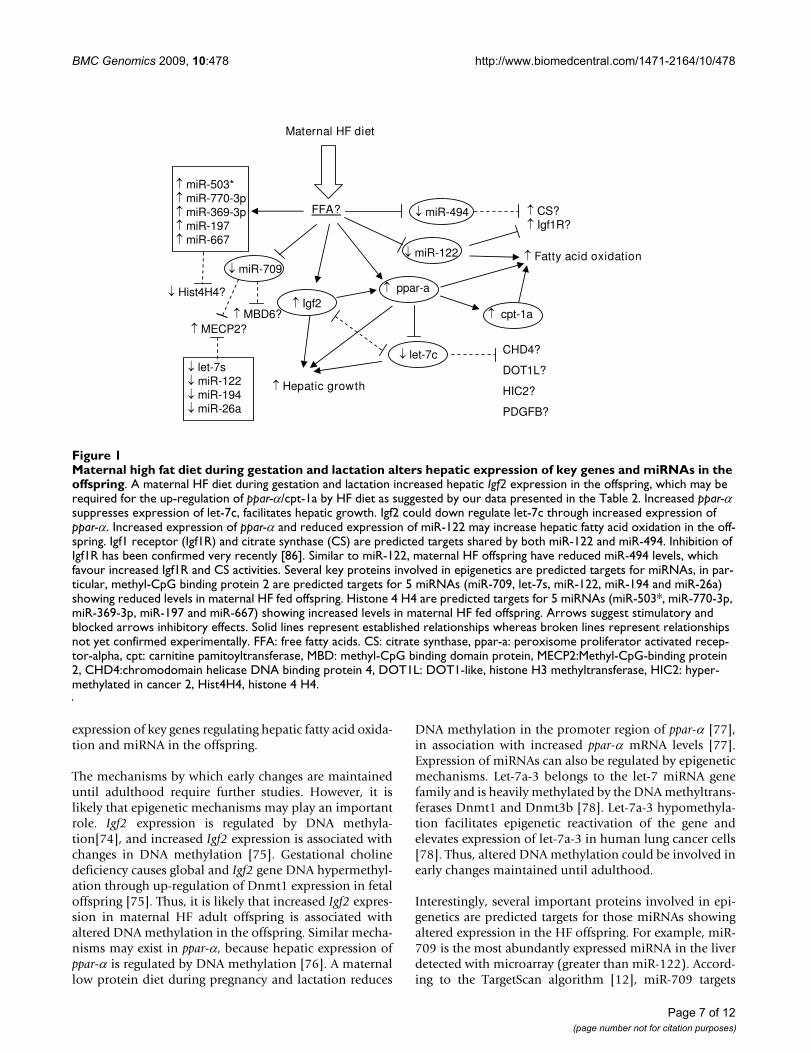
chow until adulthood, changes in expression of key meta-bolic genes and miRNAs in adult offspring are likely tooccur prior to weaning. IGF2 is a growth factor highlyexpressed during early development [58]. Offspring Igf2gene expression can be altered by maternal dietary modi-fications during early development. For example, a mater-nal low protein diet restricted only to the preimplantationperiod reduces hepatic Igf2 mRNA in fetal rats [59] andmaternal dietary calorie restriction increases Igf2 mRNAlevels in the liver and skeletal muscle in fetal sheep [60].Here we further show that hepatic mRNA levels of Igf2 areelevated in the adult mouse offspring born to dams fed aHF diet, suggesting that maternal HF feeding increases off-spring hepatic Igf2 expression prior to weaning. This issupported by our observation that hepatic Igf2 levels infetal offspring from dams fed a HF diet are increased(unpublished data). Similarly, it is likely that alteredexpression of hepatic ppar-α and cpt-1a and miRNAs inmaternal HF fed adult offspring might have also occurredprior to weaning.
GrowthIGF2 is an early growth factor expressed at the two-cellstage in the mouse embryo[61], and mice deficient inIGF2 have reduced birth weight [58]. In contrast, excessIGF2 in transgenic mice promotes fetal overgrowth result-ing in increased birth weight [62]. Our data showing thatHF/C offspring have increased Igf2 expression and a trendtoward increased liver weight [~16% increase, comparedwith the control mice (p = 0.44)], suggest that liver growthprior to weaning may be increased in maternal HF fed off-spring (Fig 1). This finding is consistent with an earlyobservation in rats showing that maternal HF feeding dur-ing gestation increases offspring liver weight, in associa-tion with increased body weight and percentage body fatat weaning[38]. In our study, the body weight of maternalHF fed offspring at weaning is ~19.6% (p < 0.05) greaterthan control mice (data not shown), consistent withincreased hepatic Igf2 expression. However, we observedno significant difference in body weights between mater-nal HF and chow fed adult offspring, when both sets ofoffspring were fed the same chow diet from weaning,which could be due to that circulating IGF2 is markedlydecreased after birth in rodents [63,64], suggesting thatIGF2 is unlikely to play a major role in post weaninggrowth.
PPARα promotes hepatic proliferation through inhibitionof let-7c [65,66]. Let-7c plays a critical role in the regula-tion of growth[67]. Overexpression of let-7c decreases c-myc and miR-17, suppressing the growth of hepatocytes[66]. Consistently, we have observed that mRNA levels ofppar-α (and protein [33]) are elevated whereas levels oflet-7c reduced in maternal HF fed offspring (Fig. 1), sug-gesting a co-ordinated regulation of mRNA and miRNAexpression in favour of promoting hepatic growth.
It is uncertain whether let-7c is regulated by IGF2. Ourdata showing increased Igf2 expression in association withreduced let-7c expression, is consistent with the negativeregulation of let-7c by PPAR-α as discussed above. In Igf2KO mice, expression of let-7c levels are increased by ~31%(p = 0.004). These data suggest a negative correlationbetween Igf2 and let-7c expression (Fig. 1). Further studiesare required to determine whether let-7c expression isdirectly regulated by IGF2. Taking together, our data sug-gest that maternal HF feeding has induced co-ordinatedchanges in expression of early growth factor (e.g. Igf2),transcription factor (e.g. ppar-α) and miRNA (e.g. let-7c)to promote hepatic growth.
Fat metabolismPPAR-α is a master transcription factor regulating hepaticfatty acid oxidation [68-73]. We have shown previouslythat maternal high fat high protein and low carbohydratediet-fed offspring have increased protein levels of PPAR-αand CPT-1a in association with reduced hepatic lipid lev-els [33]. Here we further show that mRNA levels of ppar-αand cpt-1a are increased in the maternal HF fed offspring,suggesting that maternal HF feeding increases expressionof ppar-α and cpt-1a mRNA and protein levels.
IGF2 may also regulate fat metabolism as low circulatingIGF2 concentrations are associated with weight gain andobesity [43], whereas high circulating IGF2 levels are asso-ciated with low body weight in middle aged men [44].Mice overexpressing Igf2 have increased fatty acid oxida-tion [45]. Our data show that maternal HF offspring haveincreased Igf2 expression with parallel increased ppar-α,whereas a HF induced increase in ppar-α expression is sup-pressed in the Igf2 KO mice. These data suggest that Igf2might regulate fat metabolism through regulation of ppar-α expression, and that up-regulation of ppar-α in maternalHF diet fed offspring is mediated, at least in part throughincreased expression of Igf2.
miR-122 is abundantly expressed in the liver and regulatesfat metabolism [21], as knocking down miR-122 increaseshepatic fatty-acid oxidation [21,57]. Hepatic expression ofmiR-122 is reduced in the maternal HF adult offspring(Fig. 1), which is consistent with increased expression ofPPARα and CPT-1a, two key molecules regulating hepaticfatty acid oxidation (Fig. 1). Maternal HF fed adult off-spring have reduced hepatic lipid levels when weanedonto a chow diet and maintained on the chow diet untiladulthood[33]. It is likely that increased capacity of fatoxidation (due to reduced miR-122 and increased PPARαand CPT-1a) prior to weaning are maintained until adult-hood. This continuing increased fatty acid oxidationcapacity leads to reduced hepatic lipid levels when HF off-spring mice are weaned onto a chow diet. Taking together,our data suggest that maternal HF feeding increases
Page 6 of 12(page number not for citation purposes)
BMC Genomics 2009, 10:478 http://www.biomedcentral.com/1471-2164/10/478
expression of key genes regulating hepatic fatty acid oxida-tion and miRNA in the offspring.
The mechanisms by which early changes are maintaineduntil adulthood require further studies. However, it islikely that epigenetic mechanisms may play an importantrole. Igf2 expression is regulated by DNA methyla-tion[74], and increased Igf2 expression is associated withchanges in DNA methylation [75]. Gestational cholinedeficiency causes global and Igf2 gene DNA hypermethyl-ation through up-regulation of Dnmt1 expression in fetaloffspring [75]. Thus, it is likely that increased Igf2 expres-sion in maternal HF adult offspring is associated withaltered DNA methylation in the offspring. Similar mecha-nisms may exist in ppar-α, because hepatic expression ofppar-α is regulated by DNA methylation [76]. A maternallow protein diet during pregnancy and lactation reduces
DNA methylation in the promoter region of ppar-α [77],in association with increased ppar-α mRNA levels [77].Expression of miRNAs can also be regulated by epigeneticmechanisms. Let-7a-3 belongs to the let-7 miRNA genefamily and is heavily methylated by the DNA methyltrans-ferases Dnmt1 and Dnmt3b [78]. Let-7a-3 hypomethyla-tion facilitates epigenetic reactivation of the gene andelevates expression of let-7a-3 in human lung cancer cells[78]. Thus, altered DNA methylation could be involved inearly changes maintained until adulthood.
Interestingly, several important proteins involved in epi-genetics are predicted targets for those miRNAs showingaltered expression in the HF offspring. For example, miR-709 is the most abundantly expressed miRNA in the liverdetected with microarray (greater than miR-122). Accord-ing to the TargetScan algorithm [12], miR-709 targets
Maternal high fat diet during gestation and lactation alters hepatic expression of key genes and miRNAs in the offspringFigure 1Maternal high fat diet during gestation and lactation alters hepatic expression of key genes and miRNAs in the offspring. A maternal HF diet during gestation and lactation increased hepatic Igf2 expression in the offspring, which may be required for the up-regulation of ppar-α/cpt-1a by HF diet as suggested by our data presented in the Table 2. Increased ppar-α suppresses expression of let-7c, facilitates hepatic growth. Igf2 could down regulate let-7c through increased expression of ppar-α. Increased expression of ppar-α and reduced expression of miR-122 may increase hepatic fatty acid oxidation in the off-spring. Igf1 receptor (Igf1R) and citrate synthase (CS) are predicted targets shared by both miR-122 and miR-494. Inhibition of Igf1R has been confirmed very recently [86]. Similar to miR-122, maternal HF offspring have reduced miR-494 levels, which favour increased Igf1R and CS activities. Several key proteins involved in epigenetics are predicted targets for miRNAs, in par-ticular, methyl-CpG binding protein 2 are predicted targets for 5 miRNAs (miR-709, let-7s, miR-122, miR-194 and miR-26a) showing reduced levels in maternal HF fed offspring. Histone 4 H4 are predicted targets for 5 miRNAs (miR-503*, miR-770-3p, miR-369-3p, miR-197 and miR-667) showing increased levels in maternal HF fed offspring. Arrows suggest stimulatory and blocked arrows inhibitory effects. Solid lines represent established relationships whereas broken lines represent relationships not yet confirmed experimentally. FFA: free fatty acids. CS: citrate synthase, ppar-a: peroxisome proliferator activated recep-tor-alpha, cpt: carnitine pamitoyltransferase, MBD: methyl-CpG binding domain protein, MECP2:Methyl-CpG-binding protein 2, CHD4:chromodomain helicase DNA binding protein 4, DOT1L: DOT1-like, histone H3 methyltransferase, HIC2: hyper-methylated in cancer 2, Hist4H4, histone 4 H4.
let-7c
ppar-aIgf2
Maternal HF diet
miR-122
cpt-1a
FFA?
Fatty acid oxidation
Hepatic growth
CHD4?
DOT1L?
HIC2?
PDGFB?
miR-494 CS?Igf1R?
miR-709
MECP2?
miR-503*miR-770-3pmiR-369-3pmiR-197miR-667
Hist4H4?
MBD6?
let-7smiR-122miR-194miR-26a
Page 7 of 12(page number not for citation purposes)
BMC Genomics 2009, 10:478 http://www.biomedcentral.com/1471-2164/10/478
include methyl-CpG binding domain protein 6 andmethyl CpG binding protein2 (MECP2, Fig. 1). Amongpredicted targets of let-7c are proteins including hyper-methylated in cancer 2 (HIC2), chromodomain helicase4, DOT1-like histone H3 methyltransferase (Fig. 1).
A feature of miRNA function is that several miRNAs tendto act together, and a relatively small set of miRNAsaccount for most of the differences in miRNA profilesbetween cell lineages and tissues [52]. For example, it hasbeen shown that expression of 4 miRNAs (miR-29a, miR-29b, miR-29c and miR-150) is up-regulated [23], whereasexpression of 11 miRNAs (miR-379, miR-127, miR299-5p, miR-434-3p, miR-335, miR130a, miR-19b, miR-451,miR-148a, miR-199a and miR-152) is down-regulated inskeletal muscle of type 2 diabetic rats [23]. Here we showthat levels of only ~5.7% of miRNAs are altered in thematernal HF fed mouse offspring, whereas levels of theremaining miRNAs are unchanged. These data suggestthat 1) these 23 miRNAs are likely to be expressed duringearly development and play active roles in the regulationof metabolism and fetal growth; and 2) if these miRNAshave common targeted transcripts, they are likely to havegreater effects than a single miRNA in suppressing proteinsynthesis [13].
However, it remains a challenge to identify common tar-gets shared by several miRNAs, because several hundredsor even over 1000 predicted targets may arise from onesingle miRNA using current algorithms. Experimentally, itis impractical to knock down each of the miRNAs. In thisstudy, we have written a computer program that allows usto analyse quickly common targets shared by several miR-NAs. For example, we have undertaken analysis of com-mon targets among 11 miRNAs and found that themaximum number of shared targets is 5 miRNAs and nocommon targets are found among 6 different miRNAs.MECP2 is a common predicted target for 5 miRNAsincluding two abundantly expressed miRNAs (miR-709and miR-122, Fig. 1). MeCP2 is required to maintain CpGstatus of genomic DNA[79,80]. Maternal nutrient restric-tion decreases MeCP2 levels in the brain in offspringrats[81]. Among those miRNAs showing increased expres-sion in the HF fed offspring, histone 4 H4 is a commontarget for 5 different miRNAs (miR-503*, miR-770-3p,miR-369-3p, miR-197 and miR-667, Fig. 1).
Finally, despite the observation that offspring born todams fed a HF diet during pregnancy and lactation andfed a chow diet from weaning have no significant changesin phenotype compared to the control animals, markedchanges in expression of important genes such as Igf2,ppar-α and cpt-1a and a class of miRNAs have occurred.Such altered expression of metabolic genes and miRNAsare likely to affect the homeostatic responses of such off-spring to dietary challenges in later life.
ConclusionA maternal HF diet prior to conception, during pregnancyand lactation induces coordinated and long-lastingchanges in expression of Igf2 and key fat metabolic genesand miRNAs in the offspring, which may have long-termeffects on their health.
MethodsAnimalAll procedures in this study were carried out in accordancewith the UK Animal Scientific Procedures Act of 1986 andapproved by a local ethics committee. Female C57 BL6Jblack mice were maintained under controlled conditions(room temperature at 22 ± 2°C; 12 hr light/dark cycle)and randomly assigned to either a HF (22.6% fat, 23%protein and 48.6% carbohydrate, W/W) or standard chowdiet (10% fat, 18% protein and 68.8% carbohydrate, W/W [82,83], RM1 - special diet services) diet. They were pro-vided with water ad-libitum. Dams were fed either the HFor chow diet 4 weeks prior to conception, during preg-nancy (day 1 of pregnancy indicated by presence of copu-lation plug) and lactation. Litter size were standardised to6 pups. All offspring were weaned at 3 weeks of age andfed the same chow diet for 12 weeks. At 15 weeks of age,female mice (n = 7 per group from different litters) weresacrificed and liver samples were quickly removed, snapfrozen in liquid nitrogen, and stored at -80°C for furtheranalysis.
Igf2 KO miceFemale B6CBF1 mice were bred with male Igf2-knock (+/-) out mice [40]. Offspring of either WT or Igf2 KO mice(females only) were determined with genotyping usingPCR as previously described [84]. Female animals (KOand WT) were housed individually under controlled con-ditions. At two months of age, female mice (both WT andKO, from different dams) were age-matched, divided intotwo groups, and were fed ad-libitum either a HF or chowdiets for 6 months. Mice were sacrificed at 8 months ofage, liver samples were quickly removed, snap frozen inliquid nitrogen, and stored at -80°C for further analysis.
Preparation of total RNAsFor preparation of miRNA containing total RNAs, ~100mg of liver tissue was homogenised in lyses buffer pro-vided with the mirVana™ miRNA Isolation Kit (P/N: 1560,Ambion, Austin, TX) and total RNA were prepared accord-ing to the manufacturer's protocol. Purified total RNA waseluted in 100 μl of elution buffer. Concentrations of totalRNAs were measured using a Nanodrop (ND-1000, Nan-oDrop products, Bancroft Building, Wilmington, USA).The RNA integrity was analysed using an Agilent Bioana-lyser 2100 (Agilent Technologies UK Limited, Cheshire,UK) with the RNA integrity number > 8.0, and the ratio ofOD260/280 = ~2.0.
Page 8 of 12(page number not for citation purposes)
BMC Genomics 2009, 10:478 http://www.biomedcentral.com/1471-2164/10/478
MiRNA microarrayThis work was carried out at Febit Biomed Gmbh (Febitbiomed gmbh, Heidelberg, Germany). Each sample fromthe control (n = 7) or HF (n = 7) offspring were pooled formicroarray analysis (two-class experiment [48]). Eacharray contains the reverse complements of all majormature miRNAs and the mature* sequences published inthe Sanger miRBase release (version 10.1, December2007, see http://microrna.sanger.ac.uk/sequences/index.shtml) for mice. Each miRNA contains 10 replicatesto increase the statistical confidence. For each array 2 μgof total RNA were labelled according to the manufac-turer's instructions (miRVana labelling kit from Ambion).After labelling, samples were dried in a speed-vac and re-suspended in febit's proprietary miRNA HybridizationBuffer (18 μl per array). Samples were loaded onto a chip,and overnight (16 hours) hybridization was undertakenat 42°C, using argon pressure to move the samples withinthe arrays. After the hybridization, the array was washedwith the 'febit miRNA standard (external incubation)'hybridization profile and a standard detection using theappropriate filter set. Data was normalised using the soft-ware "R" with the "VSN" package and are presented inAdditional file 1: Table S1.
Measurement of mRNA expression using real time PCRDuring cDNA synthesis, ~200-500 ng of total RNA wasused in a 20 μl cDNA synthesis reaction. Total RNA wasdenatured at 70°C for 5 min and chilled in ice. Then thereaction was added with random hexamers (2.5 ng/μl).The reactions were undertaken at 42°C for 60 min and thereaction was stopped by denaturing at 95°C for 5 min.
For PCR reactions, allsamples from 4 groups of animalswere measured in one single 96-well plate, with each reac-tion undertaken in triplicates. Equal volume of cDNA (0.5μl/reaction) was added to Sensimix Lowref SYBR greenqPCR reagent (Quantace Ltd, London, UK) with gene spe-cific primers (1.0 μM) ' [see Additional file 2: Table S5]'.PCR reactions for all samples including no temperate con-trols were run on a 7500 Fast Realtime PCR System(Applied Biosystems, Warrington UK, which was alsoused for all miRNA analysis described below). The reac-tion conditions were 95°C for 15 min (hotstart) at 95°Cfor 15 sec, 60°C for 30 sec and 72°C for 30 sec. Resultswere analyzed using 7500 System SDS software (v1.4).Expression levels were calculated by normalisation to astandard curve using the total amount of RNA as adenominator and expressed as arbitrary units.
Measurement of miRNAs using real time RT-PCRTwo qPCR methods were used in the validation of micro-array: the stem-loop RT-PCR method [49], using miRNAspecific primers purchased from Applied Biosystems andpolyadenylated and reverse-transcribed with a poly(T)
adapter into cDNAs for real-time PCR using sequencecomplementary to the poly(T) adapters during RT reac-tions [53].
The stem-loop RT PCR (Taqman based technology) wasperformed according to the manufacturer's protocol(Applied Biosystems, Foster City, CA, USA). ~100 ng totalRNA was added to each reverse transcription reaction (RT)for each miRNA. Three replicates were done for eachmiRNA from RT to PCR and the results were averaged.
For poly(T) adaptor RT-PCR, ~100 ng total RNA wasadded to a reaction containing 2.5 units E. Coli Poly Apolymerase (New England Biolabs Ltd. Herts. UK), 0.75mM rATP and 1 × Pol A polymerase buffer containing 250mM NaCl, 50 mM Tris-HCl, 10 mM MgCl2. The reaction(10 μl) was incubated at 37°C for 30 min for extension ofthe poly A tail. The reaction was heated to 60°C for 5 min,cooling to 4°C and added with an Oligo dT adaptor (0.5μl of 5 μM) with RT buffer, dNTP mix and MMLV andH2O to a total 20 μl reaction volume (Applied Biosystems,according to the manufacturers' conditions) and incu-bated at 42°C for 60 min for cDNA synthesis. The cDNAsynthesis reaction was stopped by heating at 95°C for 5min.
Real time PCR was undertaken with Sensimix LowrefSYBR green qPCR reagent (Quantace Ltd, London, UK) intriplicates. 0.5 μl of cDNA was added to PCR reaction con-taining 1 × PCR reagent mix and universal primer (0.25μM), miRNA specific primer (0.5 μM, designed based onmiRNA sequences released (Release 12.0 Sept 2008) bythe Sanger Institute [85]. The reaction conditions were95°C for 15 min (hotstart), and 95°C for 15 sec, 60-62°Cfor 60 sec (optimised according to each specific miRNAprimers) and a total of 40 cycles.
Computer analysis of target predictions for miRNAsPrediction of targets for a single miRNA was undertakenusing three algorithsms: TargetScan [12], miRanda [54]and PicTar [55]. To identify groups of miRNAs havingcommon predicted targets, we have written a computerprogram which can be used in conjunction with any of thethree algorithms. As the list of targets generated by Target-Scan or miRanda are more comprehensive than thosefrom PicTar, we based our analysis of common targets onTargetScan and miRanda.
Statistical analysisData from real time PCR were presented as mean ± SE.Skewed data were transformed before statistical analysis.A student t-test was used to compare results between twogroups, and a p value < 0.05 was considered to be signifi-cant.
Page 9 of 12(page number not for citation purposes)
BMC Genomics 2009, 10:478 http://www.biomedcentral.com/1471-2164/10/478
Authors' contributionsJZ and CB conceived and designed the study. JZ and FZundertook experiments. JZ conceived the idea and XDwrote the programme for analysis of common targets pre-diction. JZ analysed the data and wrote the manuscript.KB and FC conducted the animal studies and prepared theliver samples. KB, FC, AC, CB, HL, MH and MV contrib-uted to the discussion of data. All authors read andapproved the final manuscript.
Additional material
AcknowledgementsThe maternal high fat animal model for this project was funded by a BBSRC grant awarded to CDB, the publication cost was partly funded by a BBSRC grant awarded to FC, Igf2 KO mice were provided by University of War-wick animal unit. Molecular analysis was mainly funded by Research Develop Fund of Warwick University awarded to JZ. MAH is supported by British Heart Foundation.
References1. Lee RC, Feinbaum RL, Ambros V: The C. elegans heterochronic
gene lin-4 encodes small RNAs with antisense complemen-tarity to lin-14. Cell 1993, 75(5):843-854.
2. Wightman B, Ha I, Ruvkun G: Posttranscriptional regulation ofthe heterochronic gene lin-14 by lin-4 mediates temporalpattern formation in C. elegans. Cell 1993, 75(5):855-862.
3. Olsen PH, Ambros V: The lin-4 regulatory RNA controls devel-opmental timing in Caenorhabditis elegans by blocking LIN-14 protein synthesis after the initiation of translation. Dev Biol1999, 216(2):671-680.
4. Lagos-Quintana M, Rauhut R, Lendeckel W, Tuschl T: Identificationof novel genes coding for small expressed RNAs. Science 2001,294(5543):853-858.
5. Lagos-Quintana M, Rauhut R, Meyer J, Borkhardt A, Tuschl T: NewmicroRNAs from mouse and human. RNA 2003, 9(2):175-179.
6. Lim LP, Lau NC, Weinstein EG, Abdelhakim A, Yekta S, RhoadesMW, Burge CB, Bartel DP: The microRNAs of Caenorhabditiselegans. Genes Dev 2003, 17(8):991-1008.
7. Pasquinelli AE, Reinhart BJ, Slack F, Martindale MQ, Kuroda MI, MallerB, Hayward DC, Ball EE, Degnan B, Muller P, et al.: Conservation ofthe sequence and temporal expression of let-7 hetero-chronic regulatory RNA. Nature 2000, 408(6808):86-89.
8. Zamore PD, Haley B: Ribo-gnome: the big world of small RNAs.Science 2005, 309(5740):1519-1524.
9. Rao PK, Kumar RM, Farkhondeh M, Baskerville S, Lodish HF: Myo-genic factors that regulate expression of muscle-specificmicroRNAs. Proc Natl Acad Sci USA 2006, 103(23):8721-8726.
10. Chen JF, Mandel EM, Thomson JM, Wu Q, Callis TE, Hammond SM,Conlon FL, Wang DZ: The role of microRNA-1 and microRNA-133 in skeletal muscle proliferation and differentiation. NatGenet 2006, 38(2):228-233.
11. Selbach M, Schwanhausser B, Thierfelder N, Fang Z, Khanin R, Rajew-sky N: Widespread changes in protein synthesis induced bymicroRNAs. Nature 2008, 455(7209):58-63.
12. Lewis BP, Shih IH, Jones-Rhoades MW, Bartel DP, Burge CB: Predic-tion of mammalian microRNA targets. Cell 2003,115(7):787-798.
13. Doench JG, Petersen CP, Sharp PA: siRNAs can function as miR-NAs. Genes Dev 2003, 17(4):438-442.
14. Wienholds E, Plasterk RHA: MicroRNA function in animal devel-opment. FEBS Letters 2005, 579(26):5911-5922.
15. Liu W, Mao SY, Zhu WY: Impact of tiny miRNAs on cancers.World J Gastroenterol 2007, 13(4):497-502.
16. Oakley EJ, Van Zant G: Unraveling the complex regulation ofstem cells: implications for aging and cancer. Leukemia 2007,21(4):612-621.
17. Boehm M, Slack FJ: MicroRNA control of lifespan and metabo-lism. Cell Cycle 2006, 5(8):837-840.
18. Gauthier BR, Wollheim CB: MicroRNAs: 'ribo-regulators' of glu-cose homeostasis. Nat Med 2006, 12(1):36-38.
19. Krutzfeldt J, Stoffel M: MicroRNAs: A new class of regulatorygenes affecting metabolism. Cell Metab 2006, 4(1):9-12.
20. Poy MN, Eliasson L, Krutzfeldt J, Kuwajima S, Ma X, Macdonald PE,Pfeffer S, Tuschl T, Rajewsky N, Rorsman P, et al.: A pancreaticislet-specific microRNA regulates insulin secretion. Nature2004, 432(7014):226-230.
21. Krutzfeldt J, Rajewsky N, Braich R, Rajeev KG, Tuschl T, ManoharanM, Stoffel M: Silencing of microRNAs in vivo with 'antagomirs'.Nature 2005, 438(7068):685-689.
22. Esau C, Kang X, Peralta E, Hanson E, Marcusson EG, Ravichandran LV,Sun Y, Koo S, Perera RJ, Jain R, et al.: MicroRNA-143 RegulatesAdipocyte Differentiation. J Biol Chem 2004,279(50):52361-52365.
23. He A, Zhu L, Gupta N, Chang Y, Fang F: Over-expression of miR-29, highly upregulated in diabetic rats, leads to insulin resist-ance in 3T3-L1 adipocytes. Mol Endocrinol 2007, 21(11):2785-94.
24. Mokdad AH, Ford ES, Bowman BA, Nelson DE, Engelgau MM, VinicorF, Marks JS: Diabetes trends in the U.S.: 1990-1998. DiabetesCare 2000, 23(9):1278-1283.
25. Flegal KM, Carroll MD, Kuczmarski RJ, Johnson CL: Overweightand obesity in the United States: prevalence and trends,1960-1994. Int J Obes Relat Metab Disord 1998, 22(1):39-47.
26. Wild S, Roglic G, Green A, Sicree R, King H: Global Prevalence ofDiabetes: Estimates for the year 2000 and projections for2030. Diabetes Care 2004, 27(5):1047-1053.
27. Kuskowska-Wolk A, Bergström R: Trends in body mass indexand prevalence of obesity in Swedish men 1980-89. J EpidemiolCommunity Health 1993, 47(2):103-108.
28. Seidell JC: Time trends in obesity: an epidemiological perspec-tive. Horm Metab Res 1997, 29(4):155-158.
29. Foster GD, Wyatt HR, Hill JO, McGuckin BG, Brill C, Mohammed BS,Szapary PO, Rader DJ, Edman JS, Klein S: A randomized trial of alow-carbohydrate diet for obesity. N Engl J Med 2003,348(21):2082-2090.
30. Atkins RC: Dr Atkins New Diet Revolution. Vermillion, London;2003.
31. Shai I, Schwarzfuchs D, Henkin Y, Shahar DR, Witkow S, GreenbergI, Golan R, Fraser D, Bolotin A, Vardi H, et al.: Weight loss with alow-carbohydrate, Mediterranean, or low-fat diet. N Engl JMed 2008, 359(3):229-241.
32. Nelson SM, Fleming R: Obesity and reproduction: impact andinterventions. Curr Opin Obstet Gynecol 2007, 19(4):384-389.
33. Zhang J, Wang C, Terroni PL, Cagampang FRA, Hanson M, Byrne CD:High-unsaturated-fat, high-protein, and low-carbohydratediet during pregnancy and lactation modulates hepatic lipidmetabolism in female adult offspring. Am J Physiol Regul IntegrComp Physiol 2005, 288(1):R112-118.
Additional file 1Table S1. Hepatic miRNA expression profile in adult mice. The data set provided analysed data obtained from microarray on either HF/C or C/C using pooled total RNA samples.Click here for file[http://www.biomedcentral.com/content/supplementary/1471-2164-10-478-S1.XLS]
Additional file 2Table S2, S3, S4 and S5. S2: The table listed 28 common predicted tar-gets shared by both TargetScan and miRbase algorithm. S3: The table listed 14 common predicted targets shared by 3 different miRNAs. S4: The table listed 11 common targets predicted by both miRNA-122a and miR-494. S5: The table listed DNA sequences for measurement of 3 mRNA transcripts.Click here for file[http://www.biomedcentral.com/content/supplementary/1471-2164-10-478-S2.DOC]
Page 10 of 12(page number not for citation purposes)
http://www.ncbi.nlm.nih.gov/entrez/query.fcgi?cmd=Retrieve&db=PubMed&dopt=Abstract&list_uids=8252621
http://www.ncbi.nlm.nih.gov/entrez/query.fcgi?cmd=Retrieve&db=PubMed&dopt=Abstract&list_uids=8252621
http://www.ncbi.nlm.nih.gov/entrez/query.fcgi?cmd=Retrieve&db=PubMed&dopt=Abstract&list_uids=8252621
http://www.ncbi.nlm.nih.gov/entrez/query.fcgi?cmd=Retrieve&db=PubMed&dopt=Abstract&list_uids=8252622
http://www.ncbi.nlm.nih.gov/entrez/query.fcgi?cmd=Retrieve&db=PubMed&dopt=Abstract&list_uids=8252622
http://www.ncbi.nlm.nih.gov/entrez/query.fcgi?cmd=Retrieve&db=PubMed&dopt=Abstract&list_uids=8252622
http://www.ncbi.nlm.nih.gov/entrez/query.fcgi?cmd=Retrieve&db=PubMed&dopt=Abstract&list_uids=9481598
http://www.ncbi.nlm.nih.gov/entrez/query.fcgi?cmd=Retrieve&db=PubMed&dopt=Abstract&list_uids=9481598
http://www.ncbi.nlm.nih.gov/entrez/query.fcgi?cmd=Retrieve&db=PubMed&dopt=Abstract&list_uids=9481598
http://www.ncbi.nlm.nih.gov/entrez/query.fcgi?cmd=Retrieve&db=PubMed&dopt=Abstract&list_uids=8326266
http://www.ncbi.nlm.nih.gov/entrez/query.fcgi?cmd=Retrieve&db=PubMed&dopt=Abstract&list_uids=8326266
http://www.ncbi.nlm.nih.gov/entrez/query.fcgi?cmd=Retrieve&db=PubMed&dopt=Abstract&list_uids=9178022
BMC Genomics 2009, 10:478 http://www.biomedcentral.com/1471-2164/10/478
34. Zhang J, Lewis RM, Wang C, Hales N, Byrne CD: Maternal dietaryiron restriction modulates hepatic lipid metabolism in thefetuses. Am J Physiol Regul Integr Comp Physiol 2005,288(1):R104-111.
35. Ozanne SE, Olsen GS, Hansen LL, Tingey KJ, Nave BT, Wang CL, Har-til K, Petry CJ, Buckley AJ, Mosthaf-Seedorf L: Early growth restric-tion leads to down regulation of protein kinase C zeta andinsulin resistance in skeletal muscle. J Endocrinol 2003,177(2):235-241.
36. Khan IY, Dekou V, Douglas G , Jensen R, Hanson MA, Poston L,Taylor PD: A high-fat diet during rat pregnancy or sucklinginduces cardiovascular dysfunction in adult offspring. Am JPhysiol Regul Integr Comp Physiol 2005, 288:R127-R133.
37. Trottier G, Koski KG, Brun T, Toufexis DJ, Richard D, Walker C-D:Increased Fat Intake during Lactation Modifies Hypotha-lamic-Pituitary-Adrenal Responsiveness in Developing RatPups: A Possible Role for Leptin. Endocrinology 1998,139(9):3704-3711.
38. Guo F, Jen KL: High-fat feeding during pregnancy and lactationaffects offspring metabolism in rats. Physiol Behav 1995,57(4):681-686.
39. Srinivasan M, Katewa SD, Palaniyappan A, Pandya JD, Patel MS:Maternal high-fat diet consumption results in fetal malpro-gramming predisposing to the onset of metabolic syndrome-like phenotype in adulthood. Am J Physiol Endocrinol Metab 2006,291(4):E792-799.
40. DeChiara TM, Efstratiadis A, Robertson EJ: A growth-deficiencyphenotype in heterozygous mice carrying an insulin-likegrowth factor II gene disrupted by targeting. Nature 1990,345(6270):78-80.
41. Gaunt TR, Cooper JA, Miller GJ, Day IN, O'Dell SD: Positive asso-ciations between single nucleotide polymorphisms in theIGF2 gene region and body mass index in adult males. HumMol Genet 2001, 10(14):1491-1501.
42. Gu D, O'Dell SD, Chen XH, Miller GJ, Day IN: Evidence of multi-ple causal sites affecting weight in the IGF2-INS-TH regionof human chromosome 11. Hum Genet 2002, 110(2):173-181.
43. Sandhu MS, Gibson JM, Heald AH, Dunger DB, Wareham NJ: Lowcirculating IGF-II concentrations predict weight gain andobesity in humans. Diabetes 2003, 52(6):1403-1408.
44. O'Dell SD, Miller GJ, Cooper JA, Hindmarsh PC, Pringle PJ, Ford H,Humphries SE, Day IN: Apal polymorphism in insulin-likegrowth factor II (IGF2) gene and weight in middle-agedmales. Int J Obes Relat Metab Disord 1997, 21(9):822-825.
45. Da Costa TH, Williamson DH, Ward A, Bates P, Fisher R, RichardsonL, Hill DJ, Robinson IC, Graham CF: High plasma insulin-likegrowth factor-II and low lipid content in transgenic mice:measurements of lipid metabolism. J Endocrinol 1994,143(3):433-439.
46. Reinhart BJ, Slack FJ, Basson M, Pasquinelli AE, Bettinger JC, RougvieAE, Horvitz HR, Ruvkun G: The 21-nucleotide let-7 RNA regu-lates developmental timing in Caenorhabditis elegans.Nature 2000, 403(6772):901-906.
47. Slack FJ, Basson M, Liu Z, Ambros V, Horvitz HR, Ruvkun G: The lin-41 RBCC gene acts in the C. elegans heterochronic pathwaybetween the let-7 regulatory RNA and the LIN-29 transcrip-tion factor. Mol Cell 2000, 5(4):659-669.
48. Liu CG, Calin GA, Volinia S, Croce CM: MicroRNA expressionprofiling using microarrays. Nat Protoc 2008, 3(4):563-578.
49. Chen C, Ridzon DA, Broomer AJ, Zhou Z, Lee DH, Nguyen JT, Bar-bisin M, Xu NL, Mahuvakar VR, Andersen MR, et al.: Real-timequantification of microRNAs by stem-loop RT-PCR. Nuc AciRes 2005, 33(20):e179.
50. Baskerville S, Bartel DP: Microarray profiling of microRNAsreveals frequent coexpression with neighboring miRNAs andhost genes. RNA 2005, 11(3):241-247.
51. Rodriguez A, Griffiths-Jones S, Ashurst JL, Bradley A: Identificationof Mammalian microRNA Host Genes and TranscriptionUnits. Genome Res 2004, 14(10a):1902-1910.
52. Landgraf P, Rusu M, Sheridan R, Sewer A, Iovino N, Aravin A, PfefferS, Rice A, Kamphorst AO, Landthaler M, et al.: A mammalianmicroRNA expression atlas based on small RNA librarysequencing. Cell 2007, 129(7):1401-1414.
53. Shi R, Chiang VL: Facile means for quantifying microRNAexpression by real-time PCR. BioTechniques 2005,39(4):519-525.
54. John B, Enright AJ, Aravin A, Tuschl T, Sander C, Marks DS: HumanMicroRNA targets. PLoS Biol 2004, 2(11):e363.
55. Krek A, Grun D, Poy MN, Wolf R, Rosenberg L, Epstein EJ, MacMe-namin P, da Piedade I, Gunsalus KC, Stoffel M, et al.: CombinatorialmicroRNA target predictions. Nat Genet 2005, 37(5):495-500.
56. Gerhard DS, Wagner L, Feingold EA, Shenmen CM, Grouse LH,Schuler G, Klein SL, Old S, Rasooly R, Good P, et al.: The status,quality, and expansion of the NIH full-length cDNA project:the Mammalian Gene Collection (MGC). Genome Res 2004,14(10B):2121-2127.
57. Esau C, Davis S, Murray SF, Yu XX, Pandey SK, Pear M, Watts L,Booten SL, Graham M, McKay R, et al.: miR-122 regulation of lipidmetabolism revealed by in vivo antisense targeting. CellMetab 2006, 3(2):87-98.
58. Constancia M, Hemberger M, Hughes J, Dean W, Ferguson-Smith A,Fundele R, Stewart F, Kelsey G, Fowden A, Sibley C, et al.: Placental-specific IGF-II is a major modulator of placental and fetalgrowth. Nature 2002, 417(6892):945-948.
59. Kwong WY, Miller DJ, Ursell E, Wild AE, Wilkins AP, Osmond C,Anthony FW, Fleming TP: Imprinted gene expression in the ratembryo-fetal axis is altered in response to periconceptionalmaternal low protein diet. Reproduction (Cambridge, England)2006, 132(2):265-277.
60. Brameld JM, Mostyn A, Dandrea J, Stephenson TJ, Dawson JM, But-tery PJ, Symonds ME: Maternal nutrition alters the expressionof insulin-like growth factors in fetal sheep liver and skeletalmuscle. J Endocrinol 2000, 167(3):429-437.
61. Rappolee DA, Sturm KS, Behrendtsen O, Schultz GA, Pedersen RA,Werb Z: Insulin-like growth factor II acts through an endog-enous growth pathway regulated by imprinting in earlymouse embryos. Gen Dev 1992, 6(6):939-952.
62. Ludwig T, Eggenschwiler J, Fisher P, D'Ercole AJ, Davenport ML,Efstratiadis A: Mouse mutants lacking the type 2 IGF receptor(IGF2R) are rescued from perinatal lethality in Igf2 and Igf1rnull backgrounds. Devl Biol 1996, 177(2):517-535.
63. Adams SO, Nissley SP, Handwerger S, Rechler MM: Developmentalpatterns of insulin-like growth factor-I and -II synthesis andregulation in rat fibroblasts. Nature 1983, 302(5904):150-153.
64. Holly JM: The IGF-II enigma. Growth Horm IGF Res 1998,8(3):183-184.
65. Gonzalez FJ, Shah YM: PPARalpha: mechanism of species differ-ences and hepatocarcinogenesis of peroxisome prolifera-tors. Toxicology 2008, 246(1):2-8.
66. Shah YM, Morimura K, Yang Q, Tanabe T, Takagi M, Gonzalez FJ:Peroxisome proliferator-activated receptor alpha regulatesa microRNA-mediated signaling cascade responsible forhepatocellular proliferation. Mol Cell Biol 2007,27(12):4238-4247.
67. Grosshans H, Johnson T, Reinert KL, Gerstein M, Slack FJ: The tem-poral patterning microRNA let-7 regulates several tran-scription factors at the larval to adult transition in C.elegans. Dev Cell 2005, 8(3):321-330.
68. Tugwood JD, Issemann I, Anderson RG, Bundell KR, McPheat WL,Green S: The mouse peroxisome proliferator activatedreceptor recognizes a response element in the 5' flankingsequence of the rat acyl CoA oxidase gene. Embo J 1992,11:433-439.
69. Zhang B, Marcus S, Sajjadi F, Alvares K, Reddy J, Subramani S,Rachubinski R, Capone J: Identification of a Peroxisome Prolif-erator-Responsive Element Upstream of the Gene EncodingRat Peroxisomal Enoyl-CoA Hydratase/3-Hydroxyacyl-CoADehydrogenase. PNAS 1992, 89(16):7541-7545.
70. Gulick T, Cresci S, Caira T, Moore D, Kelly D: The PeroxisomeProliferator-Activated Receptor Regulates MitochondrialFatty Acid Oxidative Enzyme Gene Expression. PNAS 1994,91(23):11012-11016.
71. Rodriguez JC, Gil Gomez G, Hegardt FG, Haro D: Peroxisome pro-liferator-activated receptor mediates induction of the mito-chondrial 3-hydroxy-3-methylglutaryl-CoA synthase gene byfatty acids. J Biol Chem 1994, 269(29):18767-18772.
72. Muerhoff A, Griffin K, Johnson E: The peroxisome proliferator-activated receptor mediates the induction of CYP4A6, acytochrome P450 fatty acid omega-hydroxylase, by clofibricacid. J Biol Chem 1992, 267(27):19051-19053.
Page 11 of 12(page number not for citation purposes)
http://www.ncbi.nlm.nih.gov/entrez/query.fcgi?cmd=Retrieve&db=PubMed&dopt=Abstract&list_uids=9724021
http://www.ncbi.nlm.nih.gov/entrez/query.fcgi?cmd=Retrieve&db=PubMed&dopt=Abstract&list_uids=9724021
http://www.ncbi.nlm.nih.gov/entrez/query.fcgi?cmd=Retrieve&db=PubMed&dopt=Abstract&list_uids=9724021
http://www.ncbi.nlm.nih.gov/entrez/query.fcgi?cmd=Retrieve&db=PubMed&dopt=Abstract&list_uids=7777603
http://www.ncbi.nlm.nih.gov/entrez/query.fcgi?cmd=Retrieve&db=PubMed&dopt=Abstract&list_uids=7777603
http://www.ncbi.nlm.nih.gov/entrez/query.fcgi?cmd=Retrieve&db=PubMed&dopt=Abstract&list_uids=2330056
http://www.ncbi.nlm.nih.gov/entrez/query.fcgi?cmd=Retrieve&db=PubMed&dopt=Abstract&list_uids=2330056
http://www.ncbi.nlm.nih.gov/entrez/query.fcgi?cmd=Retrieve&db=PubMed&dopt=Abstract&list_uids=2330056
http://www.ncbi.nlm.nih.gov/entrez/query.fcgi?cmd=Retrieve&db=PubMed&dopt=Abstract&list_uids=9376897
http://www.ncbi.nlm.nih.gov/entrez/query.fcgi?cmd=Retrieve&db=PubMed&dopt=Abstract&list_uids=9376897
http://www.ncbi.nlm.nih.gov/entrez/query.fcgi?cmd=Retrieve&db=PubMed&dopt=Abstract&list_uids=9376897
http://www.ncbi.nlm.nih.gov/entrez/query.fcgi?cmd=Retrieve&db=PubMed&dopt=Abstract&list_uids=7836887
http://www.ncbi.nlm.nih.gov/entrez/query.fcgi?cmd=Retrieve&db=PubMed&dopt=Abstract&list_uids=7836887
http://www.ncbi.nlm.nih.gov/entrez/query.fcgi?cmd=Retrieve&db=PubMed&dopt=Abstract&list_uids=7836887
http://www.ncbi.nlm.nih.gov/entrez/query.fcgi?cmd=Retrieve&db=PubMed&dopt=Abstract&list_uids=6338399
http://www.ncbi.nlm.nih.gov/entrez/query.fcgi?cmd=Retrieve&db=PubMed&dopt=Abstract&list_uids=6338399
http://www.ncbi.nlm.nih.gov/entrez/query.fcgi?cmd=Retrieve&db=PubMed&dopt=Abstract&list_uids=6338399
http://www.ncbi.nlm.nih.gov/entrez/query.fcgi?cmd=Retrieve&db=PubMed&dopt=Abstract&list_uids=1537328
http://www.ncbi.nlm.nih.gov/entrez/query.fcgi?cmd=Retrieve&db=PubMed&dopt=Abstract&list_uids=1537328
http://www.ncbi.nlm.nih.gov/entrez/query.fcgi?cmd=Retrieve&db=PubMed&dopt=Abstract&list_uids=1537328
http://www.ncbi.nlm.nih.gov/entrez/query.fcgi?cmd=Retrieve&db=PubMed&dopt=Abstract&list_uids=1502166
http://www.ncbi.nlm.nih.gov/entrez/query.fcgi?cmd=Retrieve&db=PubMed&dopt=Abstract&list_uids=1502166
http://www.ncbi.nlm.nih.gov/entrez/query.fcgi?cmd=Retrieve&db=PubMed&dopt=Abstract&list_uids=1502166
http://www.ncbi.nlm.nih.gov/entrez/query.fcgi?cmd=Retrieve&db=PubMed&dopt=Abstract&list_uids=7971999
http://www.ncbi.nlm.nih.gov/entrez/query.fcgi?cmd=Retrieve&db=PubMed&dopt=Abstract&list_uids=7971999
http://www.ncbi.nlm.nih.gov/entrez/query.fcgi?cmd=Retrieve&db=PubMed&dopt=Abstract&list_uids=7971999
http://www.ncbi.nlm.nih.gov/entrez/query.fcgi?cmd=Retrieve&db=PubMed&dopt=Abstract&list_uids=7913466
http://www.ncbi.nlm.nih.gov/entrez/query.fcgi?cmd=Retrieve&db=PubMed&dopt=Abstract&list_uids=7913466
http://www.ncbi.nlm.nih.gov/entrez/query.fcgi?cmd=Retrieve&db=PubMed&dopt=Abstract&list_uids=7913466
http://www.ncbi.nlm.nih.gov/entrez/query.fcgi?cmd=Retrieve&db=PubMed&dopt=Abstract&list_uids=1326542
BMC Genomics 2009, 10:478 http://www.biomedcentral.com/1471-2164/10/478
Publish with BioMed Central and every scientist can read your work free of charge
"BioMed Central will be the most significant development for disseminating the results of biomedical research in our lifetime."
Sir Paul Nurse, Cancer Research UK
Your research papers will be:
available free of charge to the entire biomedical community
peer reviewed and published immediately upon acceptance
cited in PubMed and archived on PubMed Central
yours — you keep the copyright
Submit your manuscript here:http://www.biomedcentral.com/info/publishing_adv.asp
BioMedcentral
73. Aldridge TC, Tugwood JD, Green S: Identification and character-ization of DNA elements implicated in the regulation ofCYP4A1 transcription. Biochem J 1995, 306(Pt 2):473-479.
74. Hu JF, Nguyen PH, Pham NV, Vu TH, Hoffman AR: Modulation ofIgf2 genomic imprinting in mice induced by 5-azacytidine, aninhibitor of DNA methylation. Mol Endocrinol 1997,11(13):1891-1898.
75. Kovacheva VP, Mellott TJ, Davison JM, Wagner N, Lopez-Coviella I,Schnitzler AC, Blusztajn JK: Gestational choline deficiencycauses global and Igf2 gene DNA hypermethylation by up-regulation of Dnmt1 expression. J Biol Chem 2007,282(43):31777-31788.
76. Burdge GC, Slater-Jefferies J, Torrens C, Phillips ES, Hanson MA, Lil-lycrop KA: Dietary protein restriction of pregnant rats in theF0 generation induces altered methylation of hepatic genepromoters in the adult male offspring in the F1 and F2 gen-erations. Br J Nutr 2007, 97(3):435-439.
77. Lillycrop KA, Phillips ES, Jackson AA, Hanson MA, Burdge GC: Die-tary Protein Restriction of Pregnant Rats Induces and FolicAcid Supplementation Prevents Epigenetic Modification ofHepatic Gene Expression in the Offspring. J Nutr 2005,135(6):1382-1386.
78. Brueckner B, Stresemann C, Kuner R, Mund C, Musch T, Meister M,Sultmann H, Lyko F: The human let-7a-3 locus contains an epi-genetically regulated microRNA gene with oncogenic func-tion. Cancer Res 2007, 67(4):1419-1423.
79. Lewis JD, Meehan RR, Henzel WJ, Maurer-Fogy I, Jeppesen P, Klein F,Bird A: Purification, sequence, and cellular localization of anovel chromosomal protein that binds to methylated DNA.Cell 1992, 69(6):905-914.
80. Kimura H, Shiota K: Methyl-CpG-binding protein, MeCP2, is atarget molecule for maintenance DNA methyltransferase,Dnmt1. J Biol Chem 2003, 278(7):4806-4812.
81. Ke X, Lei Q, James SJ, Kelleher SL, Melnyk S, Jernigan S, Yu X, WangL, Callaway CW, Gill G, et al.: Uteroplacental insufficiencyaffects epigenetic determinants of chromatin structure inbrains of neonatal and juvenile IUGR rats. Physiol Genom 2006,25(1):16-28.
82. Gardner DS, Jackson AA, Langley-Evans SC: Maintenance ofMaternal Diet-Induced Hypertension in the Rat Is Depend-ent on Glucocorticoids. Hypertension 1997, 30(6):1525-1530.
83. Zhang J, Byrne CD: Differential hepatic lobar gene expressionin offspring exposed to altered maternal dietary proteinintake. Am J Physiol Gastrointest Liver Physiol 2000, 278(1):G128-136.
84. Christofori G, Naik P, Hanahan D: Deregulation of bothimprinted and expressed alleles of the insulin-like growthfactor 2 gene during beta-cell tumorigenesis. Nat Genet 1995,10(2):196-201.
85. Griffiths-Jones S: The microRNA Registry. Nucl Acids Res 2004,32(90001):D109-111.
86. Bai S, Nasser MW, Wang B, Hsu SH, Datta J, Kutay H, Yadav A,Nuovo G, Kumar P, Ghoshal K: MicroRNA-122 inhibits tumori-genic properties of hepatocellular carcinoma cells and sensi-tizes these cells to Sorafenib. J Biol Chem 2009 in press.
Page 12 of 12(page number not for citation purposes)
http://www.ncbi.nlm.nih.gov/entrez/query.fcgi?cmd=Retrieve&db=PubMed&dopt=Abstract&list_uids=7887901
http://www.ncbi.nlm.nih.gov/entrez/query.fcgi?cmd=Retrieve&db=PubMed&dopt=Abstract&list_uids=7887901
http://www.ncbi.nlm.nih.gov/entrez/query.fcgi?cmd=Retrieve&db=PubMed&dopt=Abstract&list_uids=7887901
http://www.ncbi.nlm.nih.gov/entrez/query.fcgi?cmd=Retrieve&db=PubMed&dopt=Abstract&list_uids=9415394
http://www.ncbi.nlm.nih.gov/entrez/query.fcgi?cmd=Retrieve&db=PubMed&dopt=Abstract&list_uids=9415394
http://www.ncbi.nlm.nih.gov/entrez/query.fcgi?cmd=Retrieve&db=PubMed&dopt=Abstract&list_uids=9415394
http://www.ncbi.nlm.nih.gov/entrez/query.fcgi?cmd=Retrieve&db=PubMed&dopt=Abstract&list_uids=1606614
http://www.ncbi.nlm.nih.gov/entrez/query.fcgi?cmd=Retrieve&db=PubMed&dopt=Abstract&list_uids=1606614
http://www.ncbi.nlm.nih.gov/entrez/query.fcgi?cmd=Retrieve&db=PubMed&dopt=Abstract&list_uids=9403577
http://www.ncbi.nlm.nih.gov/entrez/query.fcgi?cmd=Retrieve&db=PubMed&dopt=Abstract&list_uids=9403577
http://www.ncbi.nlm.nih.gov/entrez/query.fcgi?cmd=Retrieve&db=PubMed&dopt=Abstract&list_uids=9403577
http://www.ncbi.nlm.nih.gov/entrez/query.fcgi?cmd=Retrieve&db=PubMed&dopt=Abstract&list_uids=7663515
http://www.ncbi.nlm.nih.gov/entrez/query.fcgi?cmd=Retrieve&db=PubMed&dopt=Abstract&list_uids=7663515