Marburg virus, a filovirus: méssenger RNAs, gene order, and regulatory elements of the replication...
19
virus Research, 24 (1992) 1-19 0 1992 Elsevier Science Publishers B.V. All rights reserved 01681702/92/$05.00 VIRUS 00752 Marburg virus, a filovirus: messenger RNAs, gene order, and regulatory elements of the replication cycle * Heinz Feldmann ‘, Elke Miihlberger ‘, Anke Randolf ‘, Christiane Will ‘, Michael P. Kiley 2Ta, Anthony Sanchez 2 and Hans-Dieter Klenk ’ i Institut fuer Virologie, Philipps-Uniuersitaet, Marburg (F.R.G.) and 2 Special Pathogens Branch, Division of Wal and Rickettsial Diseases, Centers for Infectious Diseases, Centers for Disease Control, Georgia, GA 30333 (U.S.A.) (Received 22 October 1991; revision received 12 December 1991; accepted 13 December 1991) Summary The genome of Marburg virus (MBG), a filovirus, is 19.1 kb in length and thus the largest one found with negative-strand RNA viruses. The gene order - 3’ untranslated region-NP-VP35-VP40-GP-VP30-VP24-L-5’ untranslated re- gion-resembles that of other non-segmented negative-strand (NNS) RNA viruses. Six species of polyadenylated subgenomic RNAs, isolated from MBG-infected cells, are complementary to the negative-strand RNA genome. They can be translated in vitro into the known structural proteins NP, GP (non-glycosylated form), VP40, VP35, VP30 and VP24. At the gene boundaries conserved transcrip- tional start (3’-NNCUNCNUNUAAUU-5’) and stop signals (3’-UAAUUCUU- UUU-5’) are located containing the highly conserved pentamer 3’-UAAUU-5’. Comparison with other NNS RNA viruses shows conservation primarily in the termination signals, whereas the start signals are more variable. The intergenic regions vary in length and nucleotide composition. All genes have relatively long 3’ and 5’ end non-coding regions. The putative 3’ and 5’ leader RNA sequences of Correspondence to: H. Feldmann, Institut fuer Virologie, Philipps-Universitaet, Robert Koch-Strasse 17, Marburg, F.R.G. a Present address: Research and Development Program, Government Service Division, Salk Institute, Swiftwater, PA 18370, U.S.A. * Presented in part at the Eighth International Conference on Negative Strand Viruses, Charleston, SC, U.S.A., 15-20 September 1991.
-
Upload
independent -
Category
Documents
-
view
0 -
download
0
Transcript of Marburg virus, a filovirus: méssenger RNAs, gene order, and regulatory elements of the replication...

virus Research, 24 (1992) 1-19
0 1992 Elsevier Science Publishers B.V. All rights reserved 01681702/92/$05.00
VIRUS 00752
Marburg virus, a filovirus: messenger RNAs, gene order, and regulatory elements
of the replication cycle *
Heinz Feldmann ‘, Elke Miihlberger ‘, Anke Randolf ‘, Christiane Will ‘, Michael P. Kiley 2Ta, Anthony Sanchez 2 and Hans-Dieter Klenk ’
i Institut fuer Virologie, Philipps-Uniuersitaet, Marburg (F.R.G.) and 2 Special Pathogens Branch, Division of Wal and Rickettsial Diseases, Centers for Infectious Diseases, Centers for Disease Control,
Georgia, GA 30333 (U.S.A.)
(Received 22 October 1991; revision received 12 December 1991; accepted 13 December 1991)
Summary
The genome of Marburg virus (MBG), a filovirus, is 19.1 kb in length and thus the largest one found with negative-strand RNA viruses. The gene order - 3’ untranslated region-NP-VP35-VP40-GP-VP30-VP24-L-5’ untranslated re- gion-resembles that of other non-segmented negative-strand (NNS) RNA viruses. Six species of polyadenylated subgenomic RNAs, isolated from MBG-infected cells, are complementary to the negative-strand RNA genome. They can be translated in vitro into the known structural proteins NP, GP (non-glycosylated form), VP40, VP35, VP30 and VP24. At the gene boundaries conserved transcrip- tional start (3’-NNCUNCNUNUAAUU-5’) and stop signals (3’-UAAUUCUU- UUU-5’) are located containing the highly conserved pentamer 3’-UAAUU-5’. Comparison with other NNS RNA viruses shows conservation primarily in the termination signals, whereas the start signals are more variable. The intergenic regions vary in length and nucleotide composition. All genes have relatively long 3’ and 5’ end non-coding regions. The putative 3’ and 5’ leader RNA sequences of
Correspondence to: H. Feldmann, Institut fuer Virologie, Philipps-Universitaet, Robert Koch-Strasse 17,
Marburg, F.R.G.
a Present address: Research and Development Program, Government Service Division, Salk Institute,
Swiftwater, PA 18370, U.S.A.
* Presented in part at the Eighth International Conference on Negative Strand Viruses, Charleston,
SC, U.S.A., 15-20 September 1991.
2
the MBG genome resemble those of other NNS RNA viruses in length, conserva- tion at the 3’ and 5’ ends, and in being complementary at their extremities. The data support the concept of a common taxonomic order Mononegavirales compris- ing the Filoviridae, Paramyxoviridae, and Rhabdoviridae families.
Marburg virus; Filovirus; Subgenomic RNAs; Gene order; Regulatory elements in transcription and replication; Evolutionary relationship to paramyxoviruses and rhabdoviruses
Introduction
Marburg virus (MBG) is a non-segmented negative-strand (NNS) RNA virus and a member of the family Filoviridae (Riley et al., 1982), the youngest family in the proposed order Mononegavirales (ICTV, 1991) besides the Paramyxoviridae and Rhabdoviridae families. The related Ebola virus (EBO) (subtypes Zaire and Sudan) and a recently isolated Ebola-like filovirus (Reston (RES)) (CDC, 1989; Jahrling et al., 1990) are the only other two members of this relatively new virus family. MBG and EBO are highly pathogenic for human and non-human primates and cause a severe haemorrhagic disease with mortality rates of 35% for MBG (Martini and Siegert, 1971) and up to 88% for EBO (subtype Zaire) (Johnson et al., 1977). According to the evidence available to date, RES is infectious to humans but does not cause serious human disease. Filoviruses have at least seven structural proteins with similar electrophoretic mobility patterns and presumed identical functions for the different viruses. These proteins are: a RNA-dependent RNA polymerase (L protein), a single, glycosylated integral membrane protein (GP), a nucleoprotein (NP), and four viral structural proteins VP40, VP35, VP30 and VP24. VP30 might be a second minor nucleoprotein, VP35 is thought to be the P protein of filoviruses, and VP40 and VP24 are membrane-associated proteins (Elliott et al., 1985; Kiley et al., 1988; Sanchez et al., 1989; Feldmann et al., 1991).
The work on MBG and EBO has been greatly impeded in the past by the high pathogenicity of these agents. Knowledge about the molecular structure of the genome, the transcription and replication strategy, the structure of the proteins, and the evolutionary relationship to other viruses is necessary to better understand the biology of these human pathogens. This basic information would create a starting point for the development of better diagnostic tools and of vaccines or antiviral drugs which are not available today. The isolation of RES clearly demonstrated that filoviruses with variable pathogenic potential may be circulating in many parts of the world. The highly pathogenic members of this family represent a potential risk for fatal outbreaks of haemorrhagic diseases everywhere.
In this report we present the gene order of MBG, strain Musoke, and evidence for the presence of at least seven open reading frames (ORFs). We further demonstrate the presence of polyadenylated, MBG subgenomic RNAs comple- mentary to the negative-strand genomic RNA, and regulatory elements for tran-
3
scription and replication (transcription start and termination signals of the genes, intergenic sequences, 3’ and 5’ untranslated regions of the genes, and 3’ and 5’ leader sequences). The results are discussed concerning the relationship to NNS RNA viruses.
Materials and Methods
Virus strain and cell line
The Musoke strain of MBG isolated 1980 in Kenya (Smith et al., 1982) was used for cloning and sequencing. The virus was grown in E6 cells, a cloned Vero cell line (ATCC CRL 15861.
Isolation of virus and genomic RNA
The propagation of MBG in E6 cells and the isolation of genomic RNA were performed as described previously (Feldmann et al., 1991, Miihlberger et al., 1992).
Subgenomic RNA (mRNA) isolation and purification
Viral subgenomic RNA was obtained from E6 cells infected with MBG, strain Musoke, at m.o.i.s of l-10 PFU/cell. Following an adsorption period of 30 min the inoculum was removed and the infected cells were incubated at 37°C in Dulbecco medium containing 2% fetal calf serum. Seventeen hours postinfection actinomycin D (in water) was added to a final concentration of 10 pg/ml and mRNA was harvested 6 h thereafter. The mRNAs were radiolabeled in vivo by adding [3H]uridine to a concentration of 30 pCi/ml 1 h after addition of actinomycin D with continued incubation for 5 h. The procedure for the isolation and purification was performed as described previously (Sanchez and Kiley, 1987). Polyadenylated (poly(Al+) RNA was selected from total cell RNA by oligo(dT)- cellulose affinity chromatography.
Analysis of RNA and hybridization
RNA was analyzed using electrophoresis in 1.5% acid-urea-agarose gels (Ro- sen et al., 1975). Gels used for fluorography were soaked in 10% glacial acetic acid for fixation, treated with ENLIGHTNING autoradiography enhancer (NEN), dried, and exposed to X-ray film. For hybridization, gels were stained for 30 min in 50 mM NaOH containing 2 pg/ml ethidium bromide, washed with 25 mM phosphate buffer (pH 6.51, and photographed. The gels were then blotted onto nylon membranes (Amersham Buchler) by capillary action (Northern blotting) using 10 x SSC, air-dried, and exposed to UV light for 3 min for fixation. [ 32P]yATP-labeled RNA (kinase-reaction) (Maniatis et al., 1989) was hybridized to the membranes under stringent conditions (50% formamide, 42°C).

4
In vitro translation of subgenomic RNA
The purified polyadenylated subgenomic RNAs were translated in vitro in the presence of [ 35S]methionine using a commercial rabbit reticulocyte-lysate system as recommended by the manufacturer (Promega). The radiolabeled translation products were immunoprecipitated using guinea pig antisera raised against total virion proteins and human reconvalescence sera as described previously (Sanchez and Riley, 1987). The proteins were analyzed on 12.5% SDS-PAGE, soaked 30 min in 30% methanol and 10% glacial acetic acid for fixation, treated 30 min with ENLIGHTNING autoradiography enhancer (NEN), dried, and exposed to X-ray films.
cDNA synthesis, cloning, and sequence analysis
cDNA was synthesized from genomic RNA and mRNA using RNA-PCR (polymerase chain reaction) and the method of Gubler and Hoffman (Gubler and Hoffman, 1983; Feldmann et al., 1988; Miihlberger et al., 1992). cDNA was ligated into the pGEM3Zf( + > transcription vector for amplification in E. coli. RNA and DNA sequence determination was done as described elsewhere (Daniels et al., 1983, Schuy et al., 1986, Sanger et al., 1977; Maxam and Gilbert, 1980).
Construction of MBG genes for in vitro transcription and translation
The coding regions of the MBG genes were synthesized from genomic RNA using RNA-PCR. Briefly, the putative AUG initiation codons for eukaryotic ribosomes (A--AUG) (Kozak, 1986) for all genes were determined. The synthetic oligonucleotides for the reverse transcriptase reaction were synthesized comple- mentary to the genomic RNA sequences in a manner that these AUG initiation codons were located inside of the molecules. The second oligonucleotides used for amplification in RNA-PCR were synthesized in genomic RNA sense from differ- ent regions at the 5’ ends of the genes (5’ untranslated regions) downstream of the translation termination codons. Each pair of oligonucleotides were synthesized with either a Sal1 (NP, GP) or a BamHI recognition site (VP40, VP35, VP30, VP24) at their 5’ ends. All components used for RNA-PCR were ordered from Perkin Elmer Cetus, and the reactions were performed in a modified version of the protocol recommended by the manufacturer as described previously (Miihlberger et al., 1992). The PCR products were cut with the appropriate restriction enzymes (SalI, BamHI) and ligated into the Sal1 or BamHI-site of the plasmid pGEM3Zf( + > (Promega). Recombinant plasmid DNA was purified by precipita- tion with polyethylene glycol (Maniatis et al., 1989) and linearized with an appropriate restriction enzyme for in vitro transcription. Uncapped run-off RNA transcripts were produced by using either bacteriophage T7 or SP6 DNA-depen- dent RNA polymerase (Promega protocol). The transcripts were in vitro translated using a rabbit-reticulocyte-lysate system and analyzed as described above (Materi- als and Methods: In vitro translation of subgenomic RNA).

5
Computer analyses
The computer-assisted analyses were performed using the program of HUSAR (Heidelberg Unix Sequence Analysis Resources), Release 2.0 (German Cancer Research Center Heidelberg and Center for Molecular Biology, University of Heidelberg, 1990).
Results and Discussion
The Musoke strain of MBG was used to determine the first complete sequence of a filovirus genome. The sequence of the 3’ end of the genome (3’ untranslated region and NP gene, nucleotide position l-3000) and the sequence of the 5’ end of the genome (L gene and 5’ untranslated region, nucleotide position 11282-190991 can be obtained elsewhere (Sanchez et al., 1992; Miihlberger et al., 1992). The nucleotide sequences of the remaining genes and their deduced amino acid sequences will be published in separate communications. cDNA synthesis was performed using RNA-PCR as well as the method of Gubler and Hoffman (1983) with random hexamers, oligo(dT),,_,,, and specific oligonucleotides as primer molecules for the reverse transcriptase reaction. The genome is 19099 nucleotides in length and very rich in adenosine and uridine residues (A/U-content 61.75%; C/G-content 38.25%). It is larger than those of other negative-strand RNA viruses sequenced so far (e.g., the genome of an influenza A virus has been determined as 13588 nucleotides, that of a bunyavirus as 12278 nucleotides, that of a rhabdovirus (vesicular stomatitis virus (VSV), Indiana serotype) as 11162 nucleotides, and that of a paramyxovirus (Sendai virus) as 15385 nucleotides). A schematical illustration of the filovirus genome is shown in Fig. 3A.
Subgenomic RhLAs (mRNAs)
The analyses of the MBG genome (gene order, transcriptional signals, inter- genie sequences, and ORFs) indicate that the genome is transcribed into seven monocistronic mRNAs schematically illustrated in Fig. 3A. To demonstrate the synthesis of MBG-subgenomic RNAs in infected cells, newly synthesized RNA was in vivo labeled with [3H]uridine in the presence of actinomycin D (10 pg/ml). RNA was isolated, purified and electrophoresed in acid-urea-agarose gels (Fig. lA, B). Selection for polyadenylated RNA was performed by ohgo(cellulose chromatography of total infected cell RNA (Fig. lB>. Fig. 1 demonstrates the presence of six subgenomic RNA species in total cell and poly(A)+ selected RNA. No significant newly synthesized and [3Hluridine-labeled RNA was isolated from actinomycin D-treated uninfected cells (Fig. lB, lane 3). Northern blot-hybridiza- tion (Fig. lA, right panel) with [32P]yATP-labeled genomic RNA showed the complementarity of the subgenomic, polyadenylated RNA species to the genomic negative-strand RNA demonstrating their (+)-sense nature and that they are transcripts of the viral genome. The subgenomic RNA species comigrated in denaturing agarose gels between the 18s and 28s ribosomal RNAs (Fig. lA, B).

6
A 6
123 123 12 3 4
28S- ‘2 28S- 18S-
345
A2
6 18S- q j ,I=
-6 I- I-
Fig. 1. Marburg virus subgenomic RNAs. E6 cells were infected with MBG at m.o.i.s of l-10 PFU/cell.
Actinomycin D was added 17 h postinfection at a concentration of 10 pg/ml for 6 h. Cell RNA was
purified as described above. NP, nucleoprotein; GP, glycoprotein; VP, viral structural protein 40, 35, 30
and 24. Bands numbered l-6 indicate MBG subgenomic RNAs. 18s and 28s indicate ribosomal RNA.
(A) Gel analysis and Northern blot hybridization. Actinomycin D-resistant RNA was electrophoresed in
a 1.5% acid-urea-agarose gel, stained with ethidium bromide (left panel), blotted onto a nylon
membrane, fixed by UV-light and hybridized with [32P]yATP-labeled genomic RNA (right panel). Lane
1, actinomycin D-resistant uninfected cell RNA; lane 2, MBG genomic RNA (vRNA); lane 3,
actinomycin D-resistant MBG-infected cell RNA. (B) Fluorogram of in vivo [3H]uridine-labeled
MBG-infected cell RNA in the presence of actinomycin D. For in vivo labeling of cell RNA [“Hluridine
was added 1 h after addition of actinomycin D. RNA was electrophoresed in 1.5% acid-urea-agarose.
Lane 1, total MBG-infected cell RNA, lane 2, flowthrough of oligo(dT) cellulose chromatography of
MBG-infected cell RNA, lane 3, total uninfected cell RNA; lane 4, poly(A)+ selected MBG-infected
cell RNA. (0 In vitro translation of MBG subgenomic RNAs. In vitro translation was performed in the
presence of [35S]methionine using a rabbit reticulocyte-lysate system (Promega). The translation
products were immunoprecipitated and analyzed on 12.5% SDS-PAGE. Lane 1, total uninfected cell
RNA; lane 2, flowthrough of oligo(dT) cellulose chromatography of MBG-infected cell RNA; lanes 3
and 4, total MBG-infected cell RNA; lane 5, poly(A)’ selected MBG-infected cell RNA.
However, the bands from the poly(A)+ RNA in the area of the 2% and 18s ribosomal RNA differed from those of total cell RNA, most likely due to the large quantities of unlabeled ribosomal RNA in total RNA preparations (Fig. lB>.
The subgenomic RNA species were translated in vitro using a rabbit reticulo- cyte-lysate system. The translation products were immunoprecipitated and ana- lyzed on SDS-PAGE. In the in vitro assay, both total cell RNA and poly(A)+ RNA produced five major proteins which comigrated with the MBG structural proteins NP, VP40, VP35 VP30 and VP24 (Fig. 10. A weaker but prominent translation product was synthesized and immunoprecipitated which migrated below the NP and may represent the unglycosylated form of the viral GP (Fig. lC, lanes 3, 4 and 5). A mRNA specific for the L protein could not be detected so far probably due to the low amount present in infected cells (Riley et al., 1988).
Gene order
To determine the linear gene arrangement of the MBG genome the predicted ORFs of the different genes were synthesized by PCR-technique and ligated into
A 6
12345678 12345 67
12345678
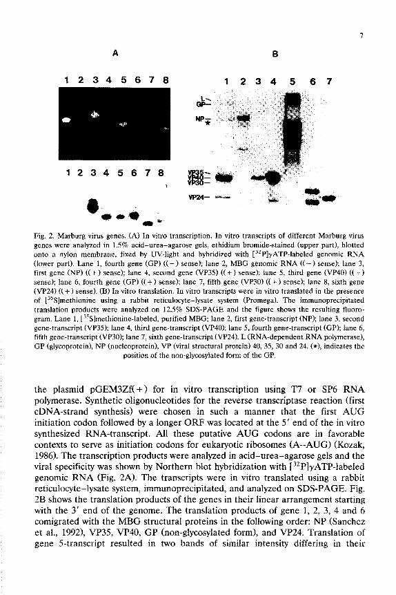
Fig. 2. Marburg virus genes. (A) In vitro transcription. In vitro transcripts of different Marburg virus genes were analyzed in 1.5% acid-urea-agarose gels, ethidium bromide-stained (upper part), blotted onto a nylon membrane, fixed by UV-light and hybridized with [32P]yATP-labeled genomic RNA (lower part). Lane 1, fourth gene (GP) (( - ) sense); lane 2, MBG genomic RNA ((- 1 sense); lane 3, first gene (NP) (( + ) sense); lane 4, second gene (VP35) (( + ) sense); lane 5, third gene (VP40) (f + ) sense); lane 6, fourth gene (GP) ((+f sense); lane 7, fifth gene (VP301 ((+) sense); lane 8, sixth gene (VP24) (( +) sense). (B) In vitro translation. In vitro transcripts were in vitro translated in the presence of [3sS]methionine using a rabbit reticulocyte-lysate system (Promega). The immunoprecipitated translation products were analyzed on 12.5% SDS-PAGE and the figure shows the resulting fluoro- gram. Lane 1, [3sS]methionine-labeled, purified MBG; lane 2, first gene-transcript (NP); lane 3, second gene-transcript (VP35); lane 4, third gene-transcript (VP40); lane 5, fourth gene-transcript (GP); lane 6. fifth gene-transcript (VP30); lane 7, sixth gene-transcript (VP24). L (RNA-dependent RNA polymerase). GP (glycoprotein), NP (nucleoprotein), VP (viral structural protein) 40,35,30 and 24. (*I, indicates the
position of the non-glycosylated form of the GP.
the plasmid pGEM3Zf(+) for in vitro polymerase. Synthetic oligonucleotides for cDNA-strand synthesis) were chosen in initiation eodon followed by a longer ORF
transcription using 117 or SP6 RNA the reverse transcriptase reaction (first such a manner that the first AUG
was located at the 5’ end of the in vitro synthesized RNA-transcript. All these putative AUG codons are in favorable contexts to serve as initiation codons for eukaryotic ribosomes (A--AUG) (Kozak, 1986). The transcription products were analyzed in acid-urea-agarose gels and the viral specificity was shown by Northern blot hybridization with [32P]~ATP-labeled genomic RNA (Fig. 2A). The transcripts were in vitro translated using a rabbit reticulocyte-lysate system, immunoprecipitated, and analyzed on SDS-PAGE. Fig. 2B shows the translation products of the genes in their linear arrangement starting with the 3’ end of the genome. The translation products of gene 1, 2, 3, 4 and 6 comigrated with the MBG structural proteins in the following order: NP (Sanchez et al., 1992), VP35 VP40, GP (non-glycosy~ated form), and VP24. Translation of gene 5transcript resulted in two bands of similar intensity differing in their

8
electrophoretic mobility patterns to all known viral structural proteins (Fig. 2B, lane 6). However, both products could be immunoprecipitated with anti-MBG antisera. The appearance of the two bands is due to a second, downstream located, in frame AUG codon which is also in a favorable context for initiation of protein synthesis. The in vitro translation of gene 4 resulted in three bands which is also due to the use of downstream located, in frame AUG codons. The larger one comigrates with the non-glycosylated form of the GP (Fig. 2B, lane 5). A transla- tion product of the construct with the ORF of the large gene at the 5’ end of the viral genome (data not shown) could not be detected. However, the length of the ORF, the location at the 5’ end of the genome, and the sequence comparison with other L proteins of NNS RNA viruses (Miihlberger et al., 1992) clearly demon- strate that this gene encodes the RNA-dependent RNA polymerase. Our results indicate the following 3’-5’ linear gene order of the MBG genome, strain Musoke: NP, VP35, VP40, GP, VP30, VP24 and L protein (Fig. 3A).
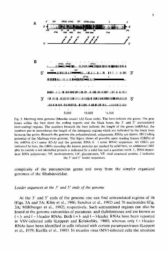
Fig. 3B shows the coding potential of the MBG genome with the three possible frames of the viral complementary RNA (mRNA sense) and the genomic RNA sequences. Seven major open reading frames (ORF) are present in the mRNA sequence encoding the seven known viral structural proteins. Within the GP gene a second ORF is located able to encode a protein of about 15 kDa (Fig. 3B, upper part). The ORF analysis of the genomic RNA sequence shows only short frames in an ambisense organization which could encode proteins of a molecular weight less than lo-12 kDa (Fig. 3B, lower part), but not any of the known viral structural proteins. However, besides the seven known structural proteins no other structural or non-structural protein according to the above-mentioned predicted sizes has been detected so far. Thus, the evaluation of the ORF analysis of the MBG genome shows no evidence for an ambisense coding strategy as found for Bun- yaviridae and Arenaviridae.
A comparison of gene orders of different NNS RNA viruses is shown in Fig. 4. All known NNS RNA viruses share a similar linear organization of their genomes in the order: 3’ untranslated region-core protein genes-envelope protein genes- RNA-dependent RNA polymerase (L) gene-5’ untranslated region. The genomes can be divided into two conserved parts at their 3’ and 5’ ends encoding core proteins and the L protein, respectively, and a variable part in the middle that encodes the envelope proteins. The genome of the vesiculovirus genus of the Rhabdoviridae is the least complex one consisting of five linear arrayed genes and the genome of the pneumovirus genus of the Paramyxoviridae is the most complex one encoding ten linear arrayed genes. The pneumovirus genome is also unique among the NNS RNA viruses in that two additional genes (1C and 1B) are located upstream of the N gene which encode two non-structural proteins of unknown function (Fig. 4). The MBG genome consists of seven genes in linear order which suggests an evolutionary position between vesiculovirus and pneumovirus genera at the extreme ends of the complexity scale and puts the filoviruses more closely to the paramyxo- and morbillivirus genera of the Paramyxoviridae. However, the presence of two additional genes (VP30 and VP24, Figs. 3A and 4) between the envelope protein genes and the L gene indicates a progression towards the greater
9
B
5’
3’
VP35 VP40 Vi24 I
3’
5’
Fig. 3. Marburg virus genome (Musoke strain). (A) Gene order. The bars indicate the genes. The gray
boxes within the bars show the coding regions and the black boxes the 3’ and 5’ untranslated
(non-coding) regions. The numbers beneath the bars indicate the length of the genes (mRNAs), the
numbers put in parentheses the length of the intergenic regions which are indicated by the black lines
between the genes. Beneath the genome the polyadenylated, subgenomic RNAs are shown. (B) Coding
potential of the Marburg virus genome. The figure shows all possible open reading frames (ORFs) of
the mRNA ((+I sense RNA) and the genomic RNA (( -) sense RNA) sequences. All ORFs are
indicated by bars, the ORFs encoding the known proteins are marked by solid bars, an additional ORF
able to encode a not identified protein is indicated by a solid bar and a question mark. L, RNA-depen-
dent RNA polymerase; NP, nucleoprotein; GP, glycoprotein, VP, viral structural protein. I indicates the 3’ and 5’ leader sequences.
complexity of the pneumovirus genomes of the Rhabdoviridae.
genus and away from the simpler organized
Leader sequences at the 3’ and 5’ ends of the genome
At the 3’ and 5’ ends of the genome one can find untranslated regions of 46 (Figs. 3A and 5A; Kiley et al., 1986; Sanchez et al., 1992) and 76 nucleotides (Fig. 3A; Miihlberger et al., 19921, respectively. Such untranslated regions can also be found at the genome extremities of paramyxo- and rhabdoviruses and are known as ( + >- and ( - )-leader RNAs. Both ( + >- and ( -)-leader RNAs have been reported in VSV-infected cells (Leppert and Kolakofsky, 19801, whereas only (+)-leader RNAs have been identified in cells infected with certain paramyxoviruses (Leppert et al., 1979; Kurilla et al., 1985). In measles virus (MV)-infected cells the situation

Fig. 4. Comparison of the gene orders of non-segmented negative-strand RNA viruses of the families
Paramyxoviridae, Rhabdoviridae and Filoviridae. 3’ and 5’ leader sequences are indicated by solid
lines. RSV, human respiratory syncytial virus; MBG, Marburg virus; Mumps, mumps virus; PF3, human
parainfluenza 3 virus; MV, measles virus; RAB, rabies virus; VW, vesicular stomatitis virus; 1C and lB,
genes of unknown function; N, nucleoprotein gene; NP, nucleoprotein gene; P, phospoprotein gene;
VP35, viral structural protein gene, possibly P equivalent; M, non-glycosylated membrane protein gene;
VP40, viral structural protein gene, non-glycosylated membrane protein?; lA, glycosylated membrane
protein gene; G, H, HN and GP, glycoprotein genes; F, fusion protein gene; SH, small hydrophobic
protein gene; 22K, non-glycosylated membrane protein gene; VP30, viral structural protein gene.
unknown function; VP24, viral structural protein gene, unknown function; ?, pseudogene in the G-L intergenic region; L, RNA-dependent RNA polymerase gene.
is even more extreme as neither (+ >- nor (-)-leader RNAs have been detected (Billeter et al., 1984; Crowley et al., 1988).
In the VSV system, leader RNAs, the initial product of viral transcription, are an essential element in transcription and replication (Banerjee, 1987). Within the first 14 nucleotides of the nascent leader RNAs a nucleation site for encapsidation
by the nucleocapsid protein is located (Blumberg et al., 1983). The viral poly- merase (L and NS protein) contacts the genomic RNA template in the middle part of the leader RNA (Keene et al., 1981). With paramyxoviruses the situation is less clear, since leader RNAs may not be essential operating elements for all of these viruses (Blumberg et al., 1991).
Leader RNAs of all NNS RNA viruses are similar in length ranging from 46 to 58 nucleotides for the (+)-leader RNAs (Fig. 51 and from 41 to 155 for the (-)-leader RNAs (data not shown). The putative MBG 3’ leader RNAs show a high content of adenine and uridine residues, with twice as much uridine as adenine (genomic (- > RNA sense). This feature is common among all NNS RNA viruses. Detailed computer-assisted comparisons demonstrate that the putative 3’ leader RNA of the EBO genome (subtype Zaire) (Kiley et al., 1986; Sanchez et al., 1989) and the 3’ leader RNA of the MBG genome show an extremely high degree of homology as already mentioned by Kiley et al. (1986). Especially a run of 12 common identical nucleotides (3’-CUGUGUGUUUUU-5 ‘; ( - ) sense> at the very

11
(A)
(B)
(0
I ----“c”o”ouoovo”vo”~e~e~~e~~c~-e~c*~~~”~~~~~”~~------------------------------ 16 MBC 1 ---ccc”o”o~oopo”~co~-“c~~c~--“~-~ce~~~~e~e~e~e~~~“~----------------------- 50 EBO 1 ----------g”O”-““DO-~~=“~~~~=~~~=~~~~~~~~~==~~~~~=“~=“~--~-~=””~~------------ 53 NW 1 ----------~oo~-“~oo--oco----cooo--o”oooAcAorcc-co--~“AC~~--”-AC”“CMll~~~OCC”- 55 SEN 1 ----------voou-vpoopcucuocuuoollcurccuno------------ 55 PF.3 1 ----------voov-“vooo-ocAAcccluuccmocuo~~=~”=~~~“~~------------ 56 MV 1 ----------uocv-“cpo”~“=~““~~“~~~~~~~~~~~~=~~~~”~=“~“““~-------------------- 50 VS1 1 “ocoM”oc”vcou-ceacuuocuuuuuuGucuoococro=~------------------- 58 KAH
. ..* . . . . . l . . . . . . . . . . . . . UOG” ““G”” UC”” * “A”“” c ” ” “” , O\‘(E\h, 5
Fig, 5. Comparison of 3’ leader RNAs of non-segmented negative-strand RNA viruses. The 3’ leader
RNA sequences of members of the three families Filoviridae, Paramyxoviridae and Rhabdoviridae,
were analyzed for homologies using the program of HUSAR. To obtain a nucleotide in the consensus
sequence it has to be present in more than 50% of the sequences involved in the alignment. (A)
Filoviridae: MBG, Marburg virus, Musoke strain; EBO, Ebola virus, subtype Zaire. (B) Paramyxoviri-
dae: NDV, Newcastle disease virus; SEN, Sendai virus; PF3, human parainfluenza 3 virus; MV, measles
virus). (C) Rhabdoviridae: VSV, vesicular stomatitis virus; RAB, rabies virus. (D) Filoviridae. Paramyx-
oviridae, and Rhabdoviridae. * , identical nucleotides; 0, nucleotides in
sequences involved in the alignment. more than 50% of the
3’ end and a box of 5 nucleotides (3’-UAAAA-5’; (-) sense) in the middle of the
leader RNAs are striking (Fig. 5A). These boxes could serve as functional elements for filovirus transcription and replication.
Fig. 5 shows a detailed and continued computer assisted comparison of 3’ leader sequences within the three families of NNS RNA viruses as well as among the families. The highest degree of homology can be found for the filovirus family with 62% homology (Fig. 5A). Paramyxovirus leaders show an overall homology of 28.5% with an eight base run of identity at the 3’ ends and more variable 5’ ends (Fig. 5B). Rhabdovirus leader RNAs have an overall homology of 46.5% without

12
clusters of identity (Fig. 50. Comparison of leaders among the three families demonstrates a region of conservation at the 3’ ends that contains uridine residues repeated at every second or third position (Fig. 5D, underlined). Blumberg et al. (1983) suggested that these uridine residues (adenine residues in the synthesized
leader RNA) serve as the encapsidation signal for VSV. In addition, a smaller conserved region is located in the middle of the leaders (Fig. 5D). These two boxes, similar with those found conserved for filoviruses, could serve as functional
domains involved in transcription and/or replication (e.g. encapsidation signal, entry signal for the polymerase complex) of NNS RNA viruses. These data indicate that the transcription and/or replication mechanisms of the different NNS RNA
viruses share at least some similarities. The existence of leader RNAs implies the operation of leader-termination
signals as has been indirectly shown for VSV. The location of VSV leader-termina- tion sequences is unknown, but it seems resonable that they are located near the cutoff point at the start of the N mRNA (Blumberg and Kolakofsky, 1983). The absence of leader RNAs in some paramyxovirus-infected cells suggests that such signals are weak or absent in paramyxoviruses. The 5’ end of the putative MBG 3’
leader shows a sequence (3’-UAGUAUAUUUAU-5 ‘; ( - > sense; Fig. 5A) which is similar to the transcription termination signal of the MBG genes (3’-UAAUUCU- UUUU-5’; (-> sense; Fig. 6A) and could serve as a leader-termination signal. However, neither (+)- nor (-)-leader RNAs have yet been detected in filovirus-
infected cells.
Complementarity qf the genome extremities
The first 13 nucleotides at the 3’ end and the last 13 nucleotides of the 5’ end of
the MBG genome are highly complementary (one missmatch, position 2 and 19098) (Miihlberger et al., 1992). These parts are followed by regions with lower complementarity (data not shown). Complementarity of lo-20 nucleotides at the genome extremities are a common feature throughout NNS RNA virus genomes (e.g., Sendai virus (SEN), human parainfluenza 3 virus (PF3), measles virus (MV), respiratory syncytial virus (RSV), vesicular stomatitis virus (VSV), and rabies virus CRAB)) (Tordo et al., 1988). Complementarity at the genome extremities could lead to a panhandle structure which might influence the stability of the genome. However, in vivo the encapsidation of the nascent RNA as soon as it is synthesized would prevent the formation of such a structure.
The synthesis of the antigenome is an important intermediate product for the replication cycle of NNS RNA viruses and serves as the template for the synthesis of progeny negative-strand RNA anticomplementary to the parental template RNA. In general, it is thought that the polymerase complex of NNS RNA viruses enters the encapsidated genome and antigenome at their 3’ ends (Emerson, 1982) and that the encapsidation signal of the nascent RNA is located near its 5’ ends (Blumberg et al., 1983). The fact, that only the very extremities of the 3’ and 5’ leader RNAs of the genomes are complementary suggests a single, identical encapsidation site on the genome and antigenome and an identical entry signal for

13
the viral polymerase complex for both the transcription as well as the replication mode. The decrease of complementarity towards the non-complementary and different 5’ ends of both leader RNAs is in line with this concept.
As already mentioned, leader termination has to be implied for the existence of leader RNAs. Leader termination signals might be essential elements for the synthesis of (+)-leader RNAs and their function in the transcription process (VSV), but not essentially for (-)-leader RNAs. Termination at the 5’ end of (-)-leaders could prevent the synthesis of negative-strand progeny RNAs and consequently virus reproduction. The presence of only weak termination signals at the 5’ ends of (-)-leaders or even their absence could be an important additional regulatory element besides the concentration of nucleocapsid structure units (encapsidation) for the synthesis of the negative-strand RNA during the replication process. The 5’ end of the putative MBG (-l-leader shows no region with homology to the transcription termination signals of the MBG genes as is found at the 5’ end of the MBG ( + j-leader (Figs. 5A and 6A; Miihlberger et al., 1992). The absence of such a termination signal at the 5’ end of the (-)-leader would allow read-throughs to produce progeny negative-strand RNA genomes. Whether the possible termination signal at the 5’ end of the MBG (+ )-leader serves as a leader termination signal during transcription and replication is unknown.
Transcription start and stop signals
At the gene boundaries of all NNS RNA virus genes conserved transcription start and stop (termination/polyadenylation1 signals are located. Such signals are also present at the boundaries of each MBG gene (Fig. 6A). The transcription start signals of the seven MBG genes show minor variations, five nucleotides out of fourteen are not conserved. The transcription stop signals of all genes are identical with the exception of the VP40 (C at position 2 instead of an A; (-) sense). An interesting finding is the highly conserved pentamer 3’-UAAUU-5’ ((-1 sense) located at the 5’ end of every transcription start and the 3’ end of every transcription stop signal. A highly conserved region like this pentamer is not found in any other NNS RNA virus transcription start or stop signal. The function of this region is unknown, but it could serve as a signal for the viral polymerase complex for promoting transcription initiation. It is unknown what effect the single base variation in the VP40 stop signal has on the poladenylation of the VP40 transcript or the transcription of the downstream located GP gene. However, this pentamer by itself is not only present in the transcription start and stop signals but is also found at some locations inside the genes. This finding suggests that the pentamer may be functional in connection with upstream or downstream surrounded semi- conserved sequences as shown for the transcription signals.
A comparison based on the consensus sequences of transcription start and stop signals of several NNS RNA viruses is shown in Fig. 7. Within the transcription start signals no conservation, only minor similarities, can be found among these different but related families (Fig. 7A). However, within the transcription stop signals conservation is obvious (Fig. 7B), primarily in the 5’ end of the signals that

14
(4
GENEB SEQUENCE (3’ - 5’1
NP - VP35 - _ (+a_“_“-“_“_” _ _
VP35 - VP40 _ _ 0-A _ _ _ _ _ _ _
“PI0 - GP - _ G-h_” _ - - _ _ _
GP - VP30 - - C-“-A-(r,92 - - -
VP30 - VP*, - - GENE OVERLAP - -
VP24 - L _ _ &d+” - - _ _ - _
((7
Fig. 6. Potential regulatory sequences of the Marburg virus (Musoke strain) genome. (A) Transcription
start and stop (termination/polyadenylation) signals. The putative transcription start and stop signals of the MBG genes and the consensus start and stop sequences are shown in the figure. The highly
conserved pentamer at the 5’ end of all transcription start and the 3’ end of all transcription stop
signals is underlined. N, any nucleotide. (B) Intergenic regions. The putative intergenic seqences of the
MBG genome are listed. x, any nucleotide. (C) 3’ and 5’ non-coding regions of the genes. The lengths
(number of nucleotides) of the 3’ and 5’ non-coding regions of the MBG genes are given. NP,
nucleoprotein gene; GP, glycoprotein gene; L, RNA-dependent RNA polymerase gene; VP40, VP35:
VP30 and VP24, viral structural protein 38K, 32K, 28K and 24K, respectively (Kiley et al., 1988).
contain four to seven uridines and is assumed to be the site where stuttering of the viral polymerase occurs in the polyadenylation of transcripts. These data again indicate similarities in the transcription mechanisms of NNS RNA viruses and suggest that the transcription termination event might be more critical to the RNA-dependent RNA polymerases than the transcription start.
Intergenic sequences
Between the gene-end boundaries of NNS RNA viruses intergenic sequences are located which are not present in the mRNA sequences. Such intergenic regions

1.5
NBQ
P13
lmv
888
8v 88v
VBV
BA8
(B)
PV3 3, ~-~-~-~-~-~-~-~-~-~-0_0 _ - - _ 3,
NDV 3, _ _ - ~-~-I)-~-V-lpIJ-~-V1_ll - _ _ 3,
Bill 3, _ _ _ A-~-~-C-~-p~-plJ _ - - - 3,
HV 3, _ _ _ ~-~-=-=-pI)-p~ _ _ - _ _ 3,
RBV 3’ ~-~-~-).-~-*-~-~-~-~-II_D - - - - 3’
VBV 3, _ _ - ~-~-~-~-~-~-~-“-~-~_~ - _ 3,
RA8 3, - _ _ _ ~-*-C-~ .pV-t)-pI)_lJ _ _ 3,
cON8LWBUB 3’ - - _ ~-lJ-~:-~-V_lJ-~l-~-lJ _ _ _ _ 3,
Fig. 7. Comparison of transcription signals of non-segmented negative-strand RNA viruses. (A)
Transcription start signals. (B) Transcription stop (termination/polyadenylation) signals. The transcrip-
tion start and stop signal consensus sequences of all genes of eight different NNS RNA viruses of the
three families Filoviridae, Paramyxoviridae and Rhabdoviridae were analyzed using the program of HUSAR. To obtain a nucleotide in the consensus sequences it has to be present in more than 50% of
the sequences involved in the alignment. MBG, Marburg virus; PF3, human parainfluenza 3 virus;
NDV, Newcastle disease virus; SEN, Sendai virus; MV, measles virus; RSV, respiratory syncytial virus;
VSV, vesicular stomatitis virus; RAB, (rabies virus); x, any nucleotide.
can also be found in the MBG genome. Besides the exact 5’ end of the NP mRNA (Sanchez et al., 1992), the intergenic regions between the NP and VP35 genes, the VP35 and VP40 genes, and the VP30 and VP24 genes have been identified by determination of the exact 5’ and 3’ ends of the corresponding mRNAs (data not shown), whereas the other gene boundaries have been deduced from the transcrip- tion start and stop signals. The intergenic regions of the MBG genome are variable in length as well as in nucleotide composition (Figs. 3A and 6B). This corresponds to the variability of the intergenic regions found in some paramyxoviruses (Newcas- tle disease virus (NDV), mumps virus, RSV) and rhabdoviruses (RAB), whereas it is in contrast to the conservation of intergenic regions in other paramyxoviruses (SEN, PF3, MV) and rhabdoviruses (VSV). Interesting is the presence of two overlapping genes (VP30 and VP24). Overlapping genes have also been found with Ebola virus (NP and VP35, GP and the downstream located gene) (Sanchez, unpublished data) and RSV (22K and L gene) (Collins et al., 19871, but not with any of the other NNS RNA virus genes sequenced so far. The function of the intergenic regions is not known but they could be involved in the regulation of

16
transcription by presenting additional signals for the polymerases or by forming secondary structures interacting with the transcriptional machinery.
Untranslated regions at the 3’ and 5’ gene ends
Besides the transcription start and stop signals and the intergenic regions, there are additional untranslated regions of variable lengths at the 3’ and 5’ ends of each gene. These regions, which are relatively long compared to those of other NNS RNA viruses, have an average of 153 nucleotides at the 3’ end and of 510 nucleotides at the 5’ end (Fig. 60. Long untranslated regions have also been found at the 3’ end of the M mRNA and the 5’ end of the F mRNA of MV (Barrett et al., 19911, but not with other mRNAs of NNS RNA viruses. Such regions are not unique, however, to NNS RNA viruses. In picornaviruses, for example, even much longer 5’ non-coding regions have been found which allow the RNA to initiate protein synthesis in the absence of the usual 5’ 7-methylguanosine capping group (Trono et al., 1988). The function of these long non-coding regions is not known. In addition to the intergenic regions and the transcription start and stop signals, they might be involved in the regulation of transcription. The correct AUG start codons, opening the ORFs encoding the known viral structural proteins (Fig. 3B), are not always those that are located most closely to the transcription start signals nor are they always in the proposed favorable context to serve as an initiation codon for eukaryotic ribosomes (A--AUG) (Kozak, 1986). The long 5’ non-coding regions of the mRNAs, however, could be involved in the regulation of translation by directing downstream located AUG start codons to the eukaryotic ribosomes to initiate protein synthesis at the correct AUG start codons.
Conclusions
Data discussed in this report demonstrate the relationship of filoviruses with other NNS RNA viruses. The unique organization and gene structure supports the classification of MBG (EBO and RES) in the family Filoviridae, separate from the Paramyxoviridae and Rhabdoviridae. Our comparative sequence analyses of lim- ited but important parts of the genome, however, clearly support the concept of the taxonomic order Mononegavirales comprising the three related families of NNS RNA viruses (ICTV, 1991). The gene order of the MBG genome suggests an evolutionary position between the simpler organized genomes of the vesiculoviruses and the more complex organized genomes of pneumoviruses. However, MBG appears more closely related to the pneumoviruses if one considers: (1) the presence of two genes between the GP and the L gene (Figs. 3 and 4); (2) an overlap of the VP30 and VP24 genes (Figs. 3A and 6B); and (31 variable lengths of the intergenic regions (Figs. 3A and 6B). From the data available so far, it is not yet clear how closely filoviruses resemble in their transcription and replication patterns VSV and paramyxoviruses and to what extent they follow their own specific strategies. All three families are alike, however, in that their genomes are

17
structured and replicated in a basically similar fashion. Conserved sequences in the L proteins of NNS RNA viruses (Tordo et al., 1988; Barik et al., 1990; Stec et al., 1991; Miihlberger et al., 19921, conservation in the leader RNA sequences (Fig. 5D) and the transcription signals (Figs. 6A and 71, and complementarity at the very extremities of the genomes (Miihlberger et al., 1992) are further indications that these viruses employ comparable mechanisms in the transcription and replication and share a common evolutionary lineage.
Acknowledgements
We greatly thank F. Mahner for excellent technical assistance and R. Ebendt for his assistance with the computer work. These studies were supported by grants from the Deutsche Forschungsgemeinschaft (Forschergruppe Kl 238/1-l and SFB 286).
References
Banejee, A.K. (1987) Transcription and replication of rhabdoviruses. Microbial. Rev. 51, 66-87.
Barik, S.E., Rud, W., Luk, D., Banerjee, A.K. and Yong Kang, C. (1990) Nucleotide sequence analysis
of the L gene of vesicular stomatitis virus (New Jersey serotype): identification of conserved
domains in L proteins of nonsegmented negative-strand RNA viruses. Virology 175, 332-337.
Barrett, T., Subbarao, SM., Belsham, G.J. and Mahy, B.W.J. (1991) The molecular biology of the
morbillivirus. In: Kingsbury, D.W. (Ed.), Paramyxoviruses, Plenum, New York. pp. 83-102.
Billeter, M.A., Baczko, K., Schmid, A. and ter Meulen, V. (1984) Cloning of DNA corresponding to
four different measles virus genomic regions. Virology 132, 147-159.
Blumberg, B.M., Chan, J. and Udem, S.A. (1991) Function of paramyxovirus 3’ and 5’ end sequences.
In: Kingsbury, D.W., (Ed.), Paramyxoviruses, Plenum, New York. pp. 235-248.
Blumberg, B.M., Giorgio, C. and Kolakofsky, D. (1983) N protein of vesicular stomatitis virus selectively
encapsidates leader RNA in vitro. Cell 32, 559-567.
Blumberg, B.M. and Kolakofsky, D. (1983) An analytical review of defective infections of vesicular
stomatitis virus. J. Gen. Virol. 64, 1839-1847.
CDC (1989) Ebola virus infection in imported primates - Virginia, 1989. Morbidity and Mortality
Weekly Report 38, 831-838.
Collins, P.L., Olmsted, R.A., Spriggs, M.K., Johnson, P.R. and Buckler-White, A.J. (1987) Gene overlap
and site-specific attenuation of transcription of the viral polymerase L gene of human respiratory
syncytial virus. Proc. Natl. Acad. Sci. U.S.A. 84, 5134-5138.
Crowley, J.C., Dowling, P.C., Menonna, J., Silverman, J.I., Schuback, D., Cook, S.D. and Blumberg,
B.M. (1988) Sequence variability and function of measles virus 3’ and 5’ end and intercistronic
regions. Virology 164, 498-506.
Daniels, R.S., Douglas, A.R., Skehel, J.J. and Wiley, D.C. (1983) Analysis of the antigenicity of
influenza virus hemagglutinin at the pH optimum for virus mediated membrane fusion. J. Gen. Virol. 64, 1657-1662.
Elliott, L.H., Kiley, M.P. and McCormick, J.B. (1985) Descriptive analysis of Ebola virus proteins.
Virology 163, 169-176.
Emerson, S.U. (1982) Reconstitution studies detect a single polymerase entry site on the vesicular
stomatitis virus genome. Cell 31, 635-642.
Feldmann, H., Kretzschmar, E., Klingeborn, B., Rott, R., Klenk, H.-D. and Garten, W. (1988) The
structure of serotype HlO hemagglutunin of influenza A virus: comparison of an apathogenic avian and a mammalian strain pathogenic for mink. Virology 165, 428-437.

18
Feldmann, H., Will, C., Schikore, M., Slenczka, W. and Klenk, H.-D. (1991) Glycosylation and
oligomerization of the spike protein of Marburg virus. Virology 182, 353-356.
Gubler, U. and Hoffman, B.J., (1983) A simple and very efficient method for generating cDNA
libraries. Gene 25, 263-269.
ICTV (1991) The order Mononegavirales. Paramyxovirus Study Group of the Vertebrate Subcommit-
tee. Virology Division News. Archives of Virology 117, 137-140.
Jahrling, P.B., Geisbert, T.W., Dalgard, D.W., Johnson, E.D., Ksiazek, T.G., Hall, WC. and Peters,
C.J. (1990) Preliminary report: Isolation of Ebola virus from monkeys imported to U.S.A. Lancet
335, 502-505.
Johnson, K.M., Lang, J.V., Webb, P.A. and Murphy, F.A. (1977) Isolation and partial characterization
of a new virus causing acute haemorrhagic fever in Zaire. Lancet 1, 569-571.
Keene, J.D., Thornton, B.J. and Emerson, S.U. (1981) Sequence-specific contacts between the RNA
polymerase of vesicular stomatitis virus and the leader RNA gene. Proc. Natl. Acad. Sci. U.S.A. 78, 6191-6195.
Kiley, M.P., Bowen, E.T.W., Eddy, G.A., Isaicson, M., Johnson, K.M., McCormick, J.B., Murphy, F.A..
Pattyn, S.R., Peters, D., Prozesky. O.W., Regnety, O.R.L., Simpson, D.I.H., Slenczka, W., Sureau,
P., van der Green, G., Webb, P.A. and Wulff. H. (1982) Filoviridae: V taxonomic home for Marburg
and Ebola viruses? Intervirology 18. 24-32.
Kiley, M.P., Cox, N.J., Elliott, L.H., Sanchez, A., DeFries, R., Buchmeier, M.J., Richman, D.D. and
McCormick, J.B. (1988) Physicochemical properties of Marburg virus: Evidence for three distinct
virus strains and their relationship to Ebola virus. J. Gen. Virol. 69, 1957-1967.
Kiley, M.P., Wilusz, J., McCormick, J.B. and Keene, J.D. (1986) Conservation of the 3’ terminal
nucleotide sequence of Ebola and Marburg virus. Virology 149, 251-254.
Kozak, M. (1986) Point mutations define a sequence flanking the AUG initiator codon that modulates
translation by eukaryotic ribosomes. Cell 44. 283-292.
Kurilla, M.G., Stone, H.O. and Keene, J.D. (1985) RNA sequence and transcriptional properties of the
3’ end of the Newcastle disease virus genome. Virology 145, 203-212.
Leppert, M. and Kolakofsky, D. (1980) Effect of defective interfering particles on plus- and minus-strand
leader RNAs in vesicular stomatitis virus infected cells. J. Viral. 35, 704-709.
Leppert, M., Rittenhouse, L., Perrault, J., Summers, D.F. and Kolakofsky, D. (1979) Plus and minus
strand leader RNAs in negative strand virus-infected cells. Cell 18, 7355747.
Maniatis, T., Fritsch, E.F. and Sambrook, J. (1989) Molecular Cloning: A Laboratory Manual. Cold
Spring Harbor Laboratory, Cold Spring Harbor, New York.
Martini, G. and Siegert, R. (Eds.) (1971) Marburg Virus Disease, Springer, New York.
Maxam, A. and Gilbert, W. (1980) Sequencing end-labeled DNA with base-specific chemical cleavages,
In L. Grossman and K. Moldave (eds.). Methods in Enzymology. Vol. 65 pp. 4999560, Academic
New York.
Miihlberger, E., Sanchez, A., Randolf, A., Will, C., Kiley, M.P., Klenk, H.-D. and Feldmann, H. (1992)
The nucleotide sequence of the L gene of Marburg virus, a filovirus: Homologies with paramyx-
oviruses and rhabdoviruses. Virology 187. in press,
Rosen, J.M., Woo, S.L.C., Holder, J.W., Means, A.T. and O’Malley, B. (1975) Preparation and
preliminary characterization of purified ovalbumin messenger RNA from the hen oviduct. Biochem- istry 14, 69-78.
Sanchez, A. and Kiley, M.P. (1987) Identification and analysis of Ebola virus messenger RNA. Virology
157, 414-420.
Sanchez, A., Kiley, M.P., Halloway, B.P., McCormick. J.B. and Auperin, D.D. (1989) The nucleoprotein
gene of Ebola virus: cloning, sequencing and in vitro expression, Virology 170, 81-91.
Sanchez, A., Kiley, M.P., Klenk, H.-D. and Feldmann, H. (1992) Sequence analysis of the Marburg
virus nucleoprotein gene: comparison to Ebola virus and other nonsegmented negative-strand RNA viruses. J. Gen. Virol., in press.
Sanger, F., Nicklen, S. and Coulson, A.R. (1977) DNA sequencing with chain termination inhibitors.
Proc. Natl. Acad. Sci. U.S.A. 74, 5463-5467.
Schuy, W., Will, C., Kuroda, K., Scholtissek, C., Garten, W. and Klenk, H.-D. (1986) Mutations
blocking the transport of the influenza virus hemagglutinin between the rough endoplasmic
reticulum and the Golgi apparatus. EMBO J. 5, 2831-2836.
19
Smith, D.H., Johnson, B.K., Isaacson, M., Swanapoel, R., Johnson, K.M., Kiley, M.P., Bagshawe, A.,
Siongok, T. and Keruga, W.K. (1982) Marburg-virus disease in Kenya. Lancet 1, 816-820.
Stec, D.S., Hill, III, M.G. and Collins, P.L. (1991) Sequence analysis of the polymerase L gene of
human respiratory syncytial virus and predicted phylogeny of nonsegmented negative-strand viruses.
Virology 183, 273-287.
Tordo, N., Poch, O., Ermine, A., Keith, G. and Rougeon, F. (1988) Completion of the rabies virus
genome sequence determination: highly conserved domains among L (polymerase) proteins of
unsegmented negative-strand RNA viruses. Virology 165, 565-576.
Trono, D., Pelletier, J., Sonnenberg, N. and Baltimore, D. (1988) Translation in mammalian cells of a
gene linked to the poliovirus 5’ noncoding region. Science 241, 445-448.