Mapping of a conformational epitope on the cashew allergen Ana o 2: A discontinuous large subunit...
9
Molecular Immunology 47 (2010) 1808–1816 Contents lists available at ScienceDirect Molecular Immunology journal homepage: www.elsevier.com/locate/molimm Mapping of a conformational epitope on the cashew allergen Ana o 2: A discontinuous large subunit epitope dependent upon homologous or heterologous small subunit association Lixin Xia a,1 , LeAnna N. Willison a , Lauren Porter a , Jason M. Robotham a , Suzanne S. Teuber b , Shridhar K. Sathe c , Kenneth H. Roux a,∗ a Department of Biological Science and Institute of Molecular Biophysics, Florida State University, Tallahassee, FL, USA b Department of Internal Medicine, School of Medicine, University of California, Davis, CA, USA c Department of Nutrition, Food and Exercise Sciences, Florida State University, Tallahassee, FL, USA article info Article history: Received 23 December 2009 Received in revised form 19 January 2010 Accepted 22 January 2010 Available online 1 April 2010 Keywords: Food allergy Tree nut Legumin 11S globulin Cashew Conformational epitope Epitope map Electron microscopy abstract The 11S globulins are members of the cupin protein superfamily and represent an important class of tree nut allergens for which a number of linear epitopes have been mapped. However, specific conformational epitopes for these allergens have yet to be described. We have recently reported a cashew Ana o 2 conformational epitope defined by murine mAb 2B5 and competitively inhibited by a subset of patient IgE antibodies. The 2B5 epitope appears to reside on the large (acidic) subunit, is dependent upon small (basic) subunit association for expression, and is highly susceptible to denaturation. Here we fine map the epitope using a combination of recombinant chimeric cashew Ana o 2-soybean Gly m 6 chimeras, deletion and point mutations, molecular modeling, and electron microscopy of 2B5-Ana o 2 immune complexes. Key residues appear confined to a 24 amino acid segment near the N-terminus of the large subunit peptide, a portion of which makes direct contact with the small subunit. These data provide an explanation for both the small subunit dependence and the structurally labile nature of the epitope. © 2010 Elsevier Ltd. All rights reserved. 1. Introduction Tree nuts are major etiological agents of food allergy affecting 0.2% children and 0.4% adults in the USA (Sampson, 2004). Allergy to cashew ranks second among the tree nut allergies (Sicherer et al., 2003) and has been reported to cause allergic responses in sen- sitive individuals exceeding those observed for peanut (Clark et al., 2007). The major classes of tree nut allergens include 7S globulins (vicilins), 11S globulins (legumins), and 2S albumins, all of which are classified as food storage proteins (Roux et al., 2003; Sathe et al., 2005). The native and recombinant cashew homologues of each of these proteins have been characterized and are designated Ana Abbreviations: mAb, monoclonal antibody; TBS, tris-buffered saline; aa, amino acid. Funded in part by USDA NRI CREES, CSREES grant #2006-35503-17229 (KHR, SKS). ∗ Corresponding author at: Department of Biological Science, 319 Stadium Dr., Florida State University, Tallahassee, FL 32306-4295, USA. Tel.: +1 850 644 5037; fax: +1 850 645 8447. E-mail address: [email protected] (K.H. Roux). 1 Current address: State Key Laboratory of Respiratory Disease for Allergy at Shen- zhen University, College of Medicine, Shenzhen University, 518060, PR China. o 1, 2 and 3, respectively (Teuber et al., 2002; Wang et al., 2002, 2003; Robotham et al., 2005). Of the cashew allergens, Ana o 2, the 11S globulin, is the best characterized (Wang et al., 2003; Barre et al., 2007; Robotham et al., 2009). Ana o 2, like other 11S globulins, is synthesized as a large propro- tein in the developing seed and is posttranslationally cleaved into large (acidic, 257 aa) and small (basic, 171 aa) subunits that remain associated via disulfide bonds (Staswick et al., 1984; Wang et al., 2003). The proproteins typically form trimers which, upon matu- ration to the cleaved form, dimerize face-to-face to form hexamers (Adachi et al., 2003; Shewry et al., 2004). The atomic structures of the native 11S globulin of soybean and, more recently, of peanut and almond have been reported and share considerable structural homology (Adachi et al., 2003; Jin et al., 2009a,b). We have previously described a series of linear epitopes recog- nized by patients with severe allergic reactions upon cashew nut ingestion (Wang et al., 2003). Homology modeling of cashew 11S globulins revealed that the linear epitopes are dispersed over the large and small subunits and display a range of exposure to solvent (Barre et al., 2007; Robotham et al., 2009). Screening for the presence of specific conformational epitopes, using patients’ serum IgE, and their subsequent characterization is technically challenging compared to screening for linear epitopes. 0161-5890/$ – see front matter © 2010 Elsevier Ltd. All rights reserved. doi:10.1016/j.molimm.2010.01.018
-
Upload
independent -
Category
Documents
-
view
1 -
download
0
Transcript of Mapping of a conformational epitope on the cashew allergen Ana o 2: A discontinuous large subunit...

Mdh
LSa
b
c
a
ARRAA
KFTL1CCEE
1
0tas2(aao
a
S
Ff
z
0d
Molecular Immunology 47 (2010) 1808–1816
Contents lists available at ScienceDirect
Molecular Immunology
journa l homepage: www.e lsev ier .com/ locate /mol imm
apping of a conformational epitope on the cashew allergen Ana o 2: Aiscontinuous large subunit epitope dependent upon homologous oreterologous small subunit association�
ixin Xiaa,1, LeAnna N. Willisona, Lauren Portera, Jason M. Robothama,uzanne S. Teuberb, Shridhar K. Sathec, Kenneth H. Rouxa,∗
Department of Biological Science and Institute of Molecular Biophysics, Florida State University, Tallahassee, FL, USADepartment of Internal Medicine, School of Medicine, University of California, Davis, CA, USADepartment of Nutrition, Food and Exercise Sciences, Florida State University, Tallahassee, FL, USA
r t i c l e i n f o
rticle history:eceived 23 December 2009eceived in revised form 19 January 2010ccepted 22 January 2010vailable online 1 April 2010
eywords:
a b s t r a c t
The 11S globulins are members of the cupin protein superfamily and represent an important class of treenut allergens for which a number of linear epitopes have been mapped. However, specific conformationalepitopes for these allergens have yet to be described. We have recently reported a cashew Ana o 2conformational epitope defined by murine mAb 2B5 and competitively inhibited by a subset of patientIgE antibodies. The 2B5 epitope appears to reside on the large (acidic) subunit, is dependent upon small(basic) subunit association for expression, and is highly susceptible to denaturation. Here we fine map
ood allergyree nutegumin1S globulinashew
the epitope using a combination of recombinant chimeric cashew Ana o 2-soybean Gly m 6 chimeras,deletion and point mutations, molecular modeling, and electron microscopy of 2B5-Ana o 2 immunecomplexes. Key residues appear confined to a 24 amino acid segment near the N-terminus of the largesubunit peptide, a portion of which makes direct contact with the small subunit. These data provide anexplanation for both the small subunit dependence and the structurally labile nature of the epitope.
onformational epitopepitope maplectron microscopy
. Introduction
Tree nuts are major etiological agents of food allergy affecting.2% children and 0.4% adults in the USA (Sampson, 2004). Allergyo cashew ranks second among the tree nut allergies (Sicherer etl., 2003) and has been reported to cause allergic responses in sen-itive individuals exceeding those observed for peanut (Clark et al.,007). The major classes of tree nut allergens include 7S globulins
vicilins), 11S globulins (legumins), and 2S albumins, all of whichre classified as food storage proteins (Roux et al., 2003; Sathe etl., 2005). The native and recombinant cashew homologues of eachf these proteins have been characterized and are designated AnaAbbreviations: mAb, monoclonal antibody; TBS, tris-buffered saline; aa, aminocid.� Funded in part by USDA NRI CREES, CSREES grant #2006-35503-17229 (KHR,KS).∗ Corresponding author at: Department of Biological Science, 319 Stadium Dr.,
lorida State University, Tallahassee, FL 32306-4295, USA. Tel.: +1 850 644 5037;ax: +1 850 645 8447.
E-mail address: [email protected] (K.H. Roux).1 Current address: State Key Laboratory of Respiratory Disease for Allergy at Shen-
hen University, College of Medicine, Shenzhen University, 518060, PR China.
161-5890/$ – see front matter © 2010 Elsevier Ltd. All rights reserved.oi:10.1016/j.molimm.2010.01.018
© 2010 Elsevier Ltd. All rights reserved.
o 1, 2 and 3, respectively (Teuber et al., 2002; Wang et al., 2002,2003; Robotham et al., 2005). Of the cashew allergens, Ana o 2, the11S globulin, is the best characterized (Wang et al., 2003; Barre etal., 2007; Robotham et al., 2009).
Ana o 2, like other 11S globulins, is synthesized as a large propro-tein in the developing seed and is posttranslationally cleaved intolarge (acidic, 257 aa) and small (basic, 171 aa) subunits that remainassociated via disulfide bonds (Staswick et al., 1984; Wang et al.,2003). The proproteins typically form trimers which, upon matu-ration to the cleaved form, dimerize face-to-face to form hexamers(Adachi et al., 2003; Shewry et al., 2004). The atomic structures ofthe native 11S globulin of soybean and, more recently, of peanutand almond have been reported and share considerable structuralhomology (Adachi et al., 2003; Jin et al., 2009a,b).
We have previously described a series of linear epitopes recog-nized by patients with severe allergic reactions upon cashew nutingestion (Wang et al., 2003). Homology modeling of cashew 11Sglobulins revealed that the linear epitopes are dispersed over the
large and small subunits and display a range of exposure to solvent(Barre et al., 2007; Robotham et al., 2009).Screening for the presence of specific conformational epitopes,using patients’ serum IgE, and their subsequent characterization istechnically challenging compared to screening for linear epitopes.

unol
Ormwrafw(amutmiittp2
ttttrlr
2
2
FdBi1Sr(Ar
2
caeSmvvcp(
2a
0m
L. Xia et al. / Molecular Imm
ne approach is to screen mouse monoclonal antibodies, (mAbs)aised against the allergen, for the ability to recognize confor-ational epitopes and then to determine if patient IgE competesith the mAb for binding to the identified epitopes. We previously
eported the characterization of one such mAb (2B5) which definesn epitope on the cashew 11S globulin large subunit that is con-ormationally dependent upon the association of the large subunitith the small subunit of the cashew allergen for its expression
Venkatachalam et al., 2008; Robotham et al., 2010). Interestingly,ssociation with the small subunit of the soybean homologue, Gly
6, can also foster 2B5 epitope expression on the cashew large sub-nit (2B5 does not bind Gly m 6). This epitope is expressed in bothhe recombinant cashew proprotein and the enzymatically cleaved
ature protein. The 2B5 epitope is highly susceptible to loss ofmmunoreactivity upon protein denaturation, further attesting tots conformational nature. In addition, mAb 2B5 partially inhibitshe binding of human IgE to Ana o 2 and patient’s IgE fully inhibitshe binding of 2B5 to Ana o 2 indicating the likelihood of at leastartial overlap of one or more epitopes recognized by IgE with theB5 epitope.
In the present study, we have attempted to fine map the 2B5 epi-ope by first probing a series of cashew–soybean chimeric proteinso locate the target region necessary for epitope expression, andhen, using molecular modeling and alanine mutagenesis to iden-ify residues in the epitope. Our results showed that the specificesidues residing in an area spanning amino acids (aa) 20–43 of thearge subunit, a region of mixed secondary structure, are primarilyesponsible for 2B5 binding.
. Methods
.1. Antibody production
Ana o 2-reactive mAbs were generated in the Hybridoma Coreacility at Florida State University according to standard proce-ures as previously described (Venkatachalam et al., 2008). Briefly,ALB/c mice were immunized with 25 �g of whole cashew extract
n RIBI adjuvant (Corixa Inc., Hamilton, MT) and boosted with5–20 �g of antigen in the RIBI adjuvant system 3 weeks later.pleen cells were fused with NS-2 myeloma cells and screened foreactivity to whole cashew protein extract and reactivity to nativen) and recombinant (r) Ana o 2 by ELISA. A total of 20 monospecificna 2-binding mAbs were obtained, one of which was identified asecognizing a conformational epitope (Robotham et al., 2010).
.2. Cloning and expression of chimeric molecules
In addition to rAna o 2 and Gly m 6, 28 Ana o 2/Gly m 6himeric and truncated genes were constructed by blunt end lig-tion of phosphorylated PCR fragments as described in Robothamt al. (2010). Point mutations were made using the QuikChangeite-Directed Mutagenesis Kit (Stratagene, USA) as directed by theanufacturer. The various constructs were ligated into a modified
ersion of the maltose-binding protein (MBP) fusion expressionector pMAL-c2 (New England BioLabs Inc., Beverly, Mass) and theloning, expression, and purification of the chimeric-MBP fusionroteins were carried out as previously described for rAna o 2Wang et al., 2003; Robotham et al., 2010).
.3. Dot blot probing of chimeric molecules with monoclonal
ntibodyTwo micrograms of the purified proteins were dotted onto.45 �m nitrocellulose membranes (Shleicher & Schuell). Theembranes were blocked by Tris-buffered saline (TBS)-T (20 mM
ogy 47 (2010) 1808–1816 1809
Tris, 137 mM NaCl, 0.2% Tween 20, pH 7.6) containing 5% (w/v) non-fat dry milk for 1 h at room temperature and then reacted with mAb2B5 at 4 ◦C overnight. The membranes were then washed threetimes by TBS-T and incubated at 4 ◦C overnight with horseradishperoxidase-labeled goat-anti-human IgE (Biosource Intl., Camar-illo, CA) diluted 1:2000 (v/v) in TBS-T. Membranes were visualizedby a 5-min incubation in ECL+ (Amersham Pharmacia) and subse-quent exposure to Kodak XAR film (Kodak Molecular Imaging, NewHaven, CT, USA). Recombinant Ana o 2 was used as a positive con-trol and the Ana o 2 large subunit (ALG) and small subunit (ASM)were used as negative controls as previously described (Robothamet al., 2010).
2.4. Molecular modeling
Molecular modeling was performed using PyMOL(http://www.pymol.org) by decorating the atomic structurescorresponding to homologous segments and residues of Ana o 2using the soybean glycinin homohexamer (PDB ID: 1OD5) A3B4(Adachi et al., 2003) and the soybean proglycinin (PDB ID: 1fxz)A1aB1b homotrimer (Adachi et al., 2001) structures as templates.Solvent accessible surface areas were calculated using VADAR(http://redpoll.pharmacy.ualberta.ca/vadar).
2.5. Electron microscopy
Ana o 2 was purified from an aqueous cashew nut proteinextract as previously described (Sathe et al., 1997) and subjectedalone or in complex with mAb 2B5 (1:1 molar ratio) to nega-tive stain electron microscopy using previously described methods(Roux, 1989, 1996). Briefly, soluble proteins and complexes wereallowed to spontaneously adhere to carbon membranes and weresubsequently stained with 1% (w/v) uranyl formate. Electron micro-graphs were recorded at 100,000× magnification at 100 kV on aJOEL JEM 1200 electron microscope.
3. Results
3.1. Chimeric constructs
Various chimeras composed of cashew 11S globulin Ana o 2 sub-stituted with homologous segments of soybean 11S globulin Glym 6 (Fig. 1A) were iteratively produced, expressed, purified, andprobed with mAb 2B5 in dot blot assays (Fig. 2) in an effort to definethe minimal Ana o 2 segment(s) required for 2B5-reactivity. Allrecombinant proteins included fusion partners (MBP) as attemptsto cleave and remove the fusion partner typically resulted in insol-uble aggregation. For simplicity, the numbering of the aa residuesin both constructs begins at 1 and the descriptions of the chimerasare presented in the text with reference to the Ana o 2 numberedresidues (a codon deletion at position 3 of Gly m 6 shifts its num-bering by one with respect to that of Ana o 2, Fig. 1).
3.2. Defining the 2B5-reactive segment
Dot blot probing of rAna o 2 gave a strong positive signal (+++)whereas none was detected with rGly m 6 (−) (Figs. 1A and 2A).The location of the 2B5 epitope was initially narrowed to rAna o 2segment 1–87 by demonstrating a similarly strong signal when thissegment was associated with Gly m 6 87–289 of the large subunitand either the Ana o 2 (C1, +++) or Gly m 6 small subunit (C2, +++).
The “opposite” construct, Gly m 6 1–87/Ana o 2 88–257/Ana o 2 SM(C3,−), was non-reactive as was the N-terminal truncated constructAna o 2 88–257/Ana o 2 SM (C4, −).Several constructs were produced in an attempt to define theN-terminal boundary of the 2B5-reactive segment(s). The results

1810 L. Xia et al. / Molecular Immunology 47 (2010) 1808–1816
Fig. 1. Chimera constructs, mAb 2B5-reactivity, and sequence comparison. (A) Diagram and aa sequences of constructs (C1–C17) showing Ana o 2 (Ao2, red font) and Gly m 6(Gm6, green font) segments. Closed rectangles: segments of the large subunit. Dotted rectangle: the small subunit. The degree of dot blot reactivity with mAb 2B5 (DOT: − to+++) shown to the left of each construct. (B) Alignment of Ana o 2 aa 1–87 (red) and Gly m 6 aa 1–86 (green). Boxed segment: 2B5-reactive sequence. Black font: amino acidsb b inteA (Ala ME activer
dmswAC
2thogna6NnbN
2Atf
3
ppKha
uried within the Gly m 6 homohexamer crystal structure (i.e., not accessible for mAsterisk (*) denotes aa identity, colon (:) indicates aa similarity. (C) Alanine mutantsach alanine substitution represents a different construct. Boxed segment: 2B5-reeader is referred to the web version of the article.)
emonstrated that the chimeras could remain reactive upon Gly6 substitutions for Ana o 2 residues 1–19 (C5, +++) but not for
ubstitution of the first 20 (C6, −) or 30 (C7, −) aa. A weak signalas produced upon deletion of the N-terminal 15 aa (C8, +) ofna o 2 but not for deletions of the N-terminal 16 or 19 aa (C9, −;10, −).
We also sought to determine if a C-terminal boundary of theB5 epitope could be defined. We previously demonstrated thathe entire Ana o 2 small subunit could be replaced by the Gly m 6omologue (Robotham et al., 2010) and thus focused our attentionn the C-terminus of the Ana o 2 large subunit. A Gly m 6 homolo-ous substitution for the Ana o 2 segment aa 88–257 (C2, +++) didot diminish the 2B5 binding signal, as indicated above, nor did ana 44–257 substitution (C11, +++). However, an aa 31–257 Gly msubstitution eliminated binding (C12, −) as did a less intrusive-terminal substitution for Ana o 2 aa 39–257 (C13, −). A similarlyegative signal was observed for a construct (C14, −) which com-ined the C-terminal 39–257 (as in C13, −) and the above described-terminal 1–19 substitution (C5, +++).
Taken together, these data indicate that full expression of theB5 epitope requires an intact 11S globulin structure displaying thena o 2 sequence within the E20-R43 segment. Some N-terminal
runcation of the Ana o 2 sequence (S1-R15, C8, +) is also toleratedor epitope display.
.3. Identification of key amino acids and molecular modeling
In examining the primary sequence of the Ana o 2 aa 20–43
eptide in comparison to the Gly m 6 counterpart, we noted twoositions with oppositely charged residues (Ana o 2 E20/Gly m 619 and Ana o 2 E40/Gly m 6 K39). Since such differences couldave profound effects on epitope expression, especially since theyre surface exposed, we generated constructs in which one or bothraction). Amino acids in lowercase are not present in the Gly m 6 crystal structures.ut) shown in blue font with 2B5-reactivity (− to +++) below each mutated residue.sequence. (For interpretation of the references to color in this figure legend, the
residues were switched. An E20K substitution (C15, −) preventedbinding of 2B5 whereas the E40K substitution (C16, ++) had rela-tively little effect. Not surprisingly, no binding was observed whenboth substitutions were made (E20K and E40K, C17, −). E20 there-fore appears to be a critical aa for epitope recognition whereas E40has little if any effect. With respect to E20, these data are consis-tent with the observation described above in which the N-terminal19 aa of Ana o 2 could be substituted with the homologous Glym 6 segment (see C5, +++) but substitution for Ana o 2 aa 1–20greatly inhibited binding (C6, −). Note that E20 sits in an extendedsequence which is otherwise identical between Ana o 2 and Gly m6 (18ALE/KPDNR24).
An atomic model of Gly m 6 is presented in Fig. 3. In the toppanel, two of the visible monomeric subunits on one of the IA(solvent-exposed) faces are indicated in different shades of gray.The third is subdivided into the small subunit (blue), the 2B5-reactive segment homologue of the large subunit (red, aa 20–43using the Ana o 2 homologous numbering), and the remainder ofthe large subunit (yellow, aa 1–19 segment and 44–257) (for inter-pretation of the references to color in this sentence, the readeris referred to the web version of the article). The bottom panelshows a side view wherein the second trimer layer is shown inwhite.
To further fine map the 2B5 epitope, surface-exposed residueswithin the modeled Ana o 2 E20-R43 segment (Fig. 4A–F) werescreened by alanine mutagenesis (Figs. 1C and 2B). The resultsshow that of the 11 residues screened, three completely abolished2B5-reactivity (D22A, R24A, and D36A), six greatly reduced binding
(E20A, V25A, E26A, E28A, H39A and R43A), and two had little to noeffect (N38A and E40A) (Fig. 1C). Fig. 4A and B shows a surface mapwherein the critical residues are colored magenta, the less criticalresidues light pink and the unmutated, less exposed residues green(for interpretation of the references to color in this sentence, the
L. Xia et al. / Molecular Immunology 47 (2010) 1808–1816 1811
FcA
ro
rcsmPsCteET(
altmmrsc(Eaie
Fig. 3. Atomic model of 11S globulin homohexamer showing relative positions ofsubunits and 2B5 epitope. “Top” view showing the solvent-exposed IA face (top) and“side view” (bottom) showing the face-to-face (IE-IE) stacked trimer conformation.Two of the monomeric subunits in the top view are in shades of gray and the thirdmonomer is subdivided into the small subunit (blue) and the large subunit (yellow)
ig. 2. Dot blot analysis of mAb 2B5-reactivity with (A) Ana o 2 (Ao2), Gly m 6 (Gm6),himeric constructs (C1–C17) and (B) alanine point mutants. ALG, large subunit ofna o 2; ASM, small subunit of Ana o 2.
eader is referred to the web version of the article). For simplicity,nly one trimer layer is shown.
An analysis of the �-carbon backbone structure of the 2B5-eactive segment homologue reveals that it contains two �-strandsonnected by a �-turn along with segments of random coil and amall portion (single turn) of an �-helix (Fig. 4C and D, red seg-ent). We noted that several residues in the 2B5 segment (W36,
42, F43) appear to be in direct contact with residues of the smallubunit (Fig. 4F). In addition, a conserved disulfide bond between44 of the �-helix and C11 provide additional stability. Mapping ofhe aa involved in 2B5 expression shows that most of the surface-xposed residues are critical for binding (Fig. 4E). Only N38A and40A near the C-terminus were permissive of alanine mutations.he inability of the E40K substitution (C16 ++), described aboveFig. 1A), to block binding is consistent with this pattern.
Although the Ana o 2 segment identified by the chimeranalyses and the specific residues identified by mutagenesisikely represent the core of the epitope, it is possible that addi-ional key contact or otherwise structurally important residues
ay lie in adjacent regions. To investigate this possibility, weutated several surface-exposed aa in topologically adjacent
egions of the large and small subunits through alanine sub-titutions as shown on the homology model (Fig. 4G). Eightonstructs, representing 12 alanine substitutions, were screenedR15A, H178-R181AAAA, D212-N213AA, R214A, N346A, E352A,
355A, and Q357A). Interestingly, none of the mutations hadn effect on 2B5 epitope expression (data not shown) suggest-ng that the above identified core segment constitutes the entirepitope.including the amino acids of glycinin corresponding to the 2B5 epitope-bearingsegment (aa 20–43 in red) of Ana o 2. (For interpretation of the references to colorin this figure legend, the reader is referred to the web version of the article.)
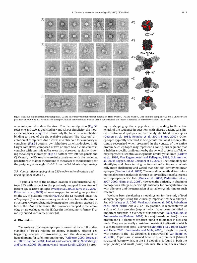
3.4. Electron microscopy of mAb 2B5-Ana o 2 immune complexes
The location of the epitope on the surface of the subunits of thenAna o 2 hexameric structure suggest that the Fab arms of 2B5would bind such that the pseudo-two-fold axis of the bound Fabswould protrude from the planar surface of the IA (solvent accessi-ble) face at an angle projecting about 30◦ away from the 3-fold axisof symmetry, assuming a flush contact between the two surfaces.To test this hypothesis, we performed negative stain transmissionelectron microscopy (EM) on nAna o 2 and nAna o 2-2B5 immunecomplexes and analyzed the resulting images. In the absence ofmAb, nAna o 2 assumed two predominant orientations with respectto the carbon membrane support. One motif showed a slightly tri-angular configuration typically with a central stain-filled void (toprow, Fig. 5A). We interpreted this configuration as representingmolecules being viewed “face on” where the planar faces of the
molecules are parallel to the carbon support and the 3-fold axis ofsymmetry is at 90◦ to the carbon as depicted in Fig. 5D. Theother predominant molecular configuration was square in pro-file, often having some stain appearing to divide the square into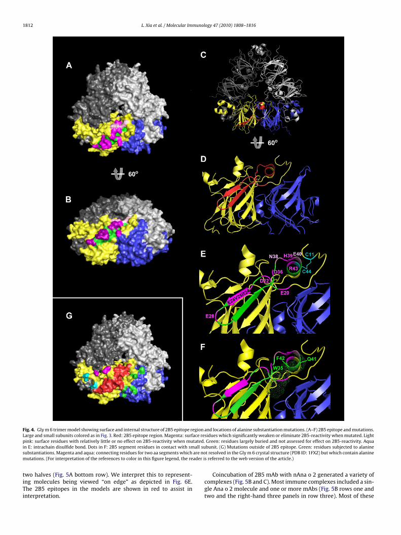
1812 L. Xia et al. / Molecular Immunology 47 (2010) 1808–1816
Fig. 4. Gly m 6 trimer model showing surface and internal structure of 2B5 epitope region and locations of alanine substantiation mutations. (A–F) 2B5 epitope and mutations.Large and small subunits colored as in Fig. 3. Red: 2B5 epitope region. Magenta: surface residues which significantly weaken or eliminate 2B5-reactivity when mutated. Lightp tated.i all sus are nom ader i
tiTi
ink: surface residues with relatively little or no effect on 2B5-reactivity when mun E: intrachain disulfide bond. Dots in F: 2B5 segment residues in contact with smubstantiations. Magenta and aqua: connecting residues for two aa segments whichutations. (For interpretation of the references to color in this figure legend, the re
wo halves (Fig. 5A bottom row). We interpret this to represent-ng molecules being viewed “on edge” as depicted in Fig. 6E.he 2B5 epitopes in the models are shown in red to assist innterpretation.
Green: residues largely buried and not assessed for effect on 2B5-reactivity. Aquabunit. (G) Mutations outside of 2B5 epitope. Green: residues subjected to alaninet resolved in the Gly m 6 crystal structure (PDB ID: 1FXZ) but which contain alanine
s referred to the web version of the article.)
Coincubation of 2B5 mAb with nAna o 2 generated a variety ofcomplexes (Fig. 5B and C). Most immune complexes included a sin-gle Ana o 2 molecule and one or more mAbs (Fig. 5B rows one andtwo and the right-hand three panels in row three). Most of these
L. Xia et al. / Molecular Immunology 47 (2010) 1808–1816 1813
F odelsp his fig
wrebecLciCpt
3l
tpRoosfem
4
sthaa
ig. 5. Negative stain electron micrographs (A–C) and interpretive homohexamer matches = 2B5 epitope. Bar = 50 nm. (For interpretation of the references to color in t
ere interpreted to show the Ana o 2 in the on edge view (Fig. 5Bows one and two as depicted in F and G). For simplicity, the mod-led complexes in Fig. 5F–H show only the Fab arms of antibodiesinding to three of the six available epitopes. The “face on” ori-ntation of complexed Ana o 2 was also observed for a minority ofomplexes (Fig. 5B bottom row, right three panels as depicted in H).arger complexes composed of two or more Ana o 2 molecules inomplex with multiple mAbs were also observed, typically show-ng the allergens “on edge” (Fig. 5B bottom row, left two panels and). Overall, the EM results were fully consistent with the modelingredictions in that the mAb bound to the IA face of the hexamer nearhe periphery at an angle of ∼30◦ from the 3-fold axis of symmetry.
.5. Comparative mapping of the 2B5 conformational epitope andinear epitopes on Ana o 2
To gain a sense of the relative location of conformational epi-ope 2B5 with respect to the previously mapped linear Ana o 2atient IgE-reactive epitopes (Wang et al., 2003; Barre et al., 2007;obotham et al., 2009), all were mapped to homologous positionsn the Gly m 6 atomic model (Fig. 6). Of the 12 mapped linear Ana2 epitopes (3 others were on segments not resolved in the atomic
tructure), 4 were substantially mapped to the solvent-exposed IAace of the nAna o 2 hexamer; the remainder mapped to the lateraldge or are occluded on the IE face (in the hexameric form) (4) orostly buried within the trimer (4).
. Discussion
The analysis of allergen epitopes is essential for a full under-
tanding of issues relating to allergy induction, effector cellriggering, allergen cross-reactivity, and the development ofypoallergens for safer immunotherapy (Aalberse, 2000; Lorenz etl., 2001; Bannon, 2004; Linhart and Valenta, 2005; Niederbergernd Valenta, 2006; Untersmayr and Jensen-Jarolim, 2006). By prob-(D–H) of nAna o 2 (A) and nAna o 2-2B5 immune complexes (B and C). Red surfaceure legend, the reader is referred to the web version of the article.)
ing overlapping synthetic peptides, corresponding to the entirelength of the sequence in question, with allergic patient sera, lin-ear (continuous) epitopes can be readily identified on allergens(Geysen et al., 1984; Reineke et al., 2001; Frank, 2002). Otherepitopes, typically described as being conformational, are only effi-ciently recognized when presented in the context of the nativeprotein. Such epitopes may represent a contiguous segment thatis held in a specific configuration by the general protein scaffold ormay represent discontinuous segments similarly stabilized (Barlowet al., 1986; Van Regenmortel and Pellequer, 1994; Schramm etal., 2001; Roggen, 2006; Gershoni et al., 2007). The technology foridentifying and characterizing conformational epitopes is techni-cally more challenging and varied than that for identifying linearepitopes (Gershoni et al., 2007). The most direct method for confor-mational epitope analysis is through co-crystallization of allergenswith epitope-specific Fab (Mirza et al., 2000; Padavattan et al.,2007, 2009; Niemi et al., 2008). However, the difficulty in obtaininghomogenous allergen-specific IgE antibody for co-crystallizationwith allergens and the generation of suitable crystals hinders suchresearch.
We have been developing a model system for the study of foodallergen epitopes using the clinically important cashew allergen,Ana o 2 (Wang et al., 2003; Venkatachalam et al., 2008; Robothamet al., 2009, 2010). Ana o 2, an 11S globulin, is representative ofa class of plant proteins (cupins) which have been identified asimportant allergens in a variety of nuts and seeds (Roux et al., 2003;Breiteneder and Radauer, 2004). As a major seed (nutrient) storageprotein, the 11S globulins are often found in abundance in nuts andseeds. They are generally considered resistant to digestion whichis a characteristic of class I allergens (Metcalfe et al., 1996; Taylor
and Hefle, 2001; Breiteneder and Mills, 2005), though this point,with respect to the 11S globulins, is controversial (van Boxtel etal., 2008). Cupins are so named because of a prominent �-barrelstructural feature which, in the 11S globulins, is found in both thelarge (acidic) and small (basic) subunits. Thus far, linear epitope
1814 L. Xia et al. / Molecular Immunol
Fig. 6. Surface model showing the location of the 2B5 epitope (red) in relation tothose of the previously reported linear Ana o 2 epitopes (other variously coloredrmtw
m22(e
iffmowtstmofap
egions) (Wang et al., 2003). Segments in the large and small subunits of the selectedonomer that are not identified as containing epitopes are in yellow. (For interpre-
ation of the references to color in this figure legend, the reader is referred to theeb version of the article.)
aps have been generated for 11S globulins of cashew (Wang et al.,003), walnut (Robotham et al., 2009), hazelnut (Robotham et al.,009), peanut (Rabjohn et al., 1999; Rouge et al., 2009), and soybeanZeece et al., 1999; Helm et al., 2000), however, no conformationalpitope maps have been published for these allergens.
Our Ana o 2 conformational mapping stems from the fortuitousdentification of a mouse IgG mAb (2B5) which recognized a con-ormational epitope. This mAb competes with a subset of IgE Absrom cashew-allergic patients for binding to Ana o 2. The confor-
ational nature of the epitope was demonstrated by its expressionn the cashew 11S large subunit of Ana o 2 only when associatedith a small subunit (Robotham et al., 2010). Interestingly, in addi-
ion to being expressed when associated with the Ana o 2 smallubunit, the 2B5 epitope is fully expressed when associated withhe homologous small subunit from the soybean 11S globulin, Gly
6, an allergen in its own right but not one that is recognized byur cashew-allergic patients or by mAb 2B5. Additional evidenceor the conformational nature of the 2B5 epitope was provided byssays demonstrating the loss of 2B5 binding following a variety ofrotein denaturation treatments (Robotham et al., 2010).
ogy 47 (2010) 1808–1816
In the present study, we generated a set of chimeric proteinsin which segments of the cashew Ana o 2 were substituted withhomologous segments of soybean Gly m 6, and assessed them forbinding with mAb 2B5 in dot blot assays. The data from thesechimeras, as well as from N-terminal deletion mutants of rAna o2, narrowed the epitope to a 24 aa segment (E20-R43) (Fig. 1).Analysis of the homologous Gly m 6 crystal structure showed thatthis segment is comprised of two �-strands and a connecting loopof the large subunit �-barrel, N-terminal and C-extended strands,and a short helical segment (Fig. 4D). Alanine scanning mutagenesisof 11 surface-exposed residues, identified by molecular modeling,showed that 9 were critical for expression (Figs. 1C and 4A, B and E).The 9 key residues were scattered throughout the 24 aa segmentwith the exception of the C-terminal �-strand, which is largelyburied. Similar mutations in surrounding surface-exposed residuesand unmodeled loops, including residues in the adjacent small sub-unit, did not adversely influence 2B5 binding (Fig. 4G). Though it isstill formally possible that some contact may be made with topo-logically neighboring residues, it appears likely that the identifiedsegment is largely responsible for binding to mAb 2B5. The solventaccessible surface of the segment (∼890 excluding the two non-contact residues) is in line with that of a typical protein epitope(∼750) (Sheriff et al., 1987).
Having identified the likely position of the 2B5 epitope on rAnao 2, we performed negative stain EM on purified nAna o 2 with andwithout 2B5 mAb. This technique has been applied to a wide varietyof proteins and complexes (Roux, 1996) but, to our knowledge, notto the analysis of allergens. The uncomplexed images were easilyinterpretable based on correspondence with the atomic structureof previously crystallized native 11S globulins (Adachi et al., 2003;Huber et al., 2009; Jin et al., 2009b). The “face on” configurationshowed a rounded triangular configuration with a central stain-filled hole. The “on edge” view showed a squared off shape withhints of two protein layers in some images. The immune complexestypically showed one or more 2B5 mAbs binding obliquely to theouter edge of the planar surface as would be predicted from a flushinteraction between Fab and the 2B5 peptide segment as modeled(Fig. 5). The multivalency of hexameric Ana o 2, as visualized in theEMs, and of the 11S globulins in general would favor extensive Igcross-linking during both the induction and effector phases of anallergic response (Untersmayr and Jensen-Jarolim, 2006).
Both linear (Niemi et al., 2008; Padavattan et al., 2009) and con-formational (Mirza et al., 2000; Padavattan et al., 2007) allergenepitopes have thus far been identified by X-ray crystallography. Inboth cases of linear epitope recognition (birch pollen Bet v 1 andhoney bee venom Api m 2), murine IgG Fabs bound to prominentprotruding epitopes. In contrast, the two conformational epitopeswere situated on relatively flat surfaces (grass pollen Phl p 2and bovine �-lactoglobulin) and were recognized by patient IgEFabs. The epitope recognized by our murine IgG mAb 2B5 moreclosely resembles those of the human IgE Abs in that it binds anon-protruding surface. Also, the 2B5 epitope is technically discon-tinuous (conformational) even though it resides within a relativelyshort (24 aa) peptide. For comparison, the Phl p 2 IgE Fab makescontact with residues in four strands of a �-sheet, essentially bind-ing the sheet “face on”. In contrast 2B5 appears to make contactwith a single �-strand (R24-E26) of the outer rim of the large sub-unit �-barrel, as well as some additional residues on a connectingloop, extended strands, and a helical component (Fig. 4E).
Two interesting features of the 2B5 epitope, namely the absolutedependence upon small subunit association for expression and its
susceptibility to destruction by a variety of denaturants (Robothamet al., 2010), can be explained by its structural features. Whereasthe two �-strands of the 2B5 segment (R24-E26 and G30-E33)and the connecting loop probably remain intact when expressedin the absence of the small subunit, other portions of the peptide
unol
miao(ctbe
bcacmpashs
t(secc(lehptJa
t(riiwmo
gtnatpmfpoemta
R
A
A
L. Xia et al. / Molecular Imm
ake direct contact with the small subunit and likely misfold ints absence. Residues W35, Q41 and F42 of the 2B5 peptide (W35nd F42 are conserved between Ana o 2 and Gly m 6) and othersutside of the peptide make direct contact with the small subunitFig. 4F). This region may also be susceptible to denaturation thoughomplete disordering of the non-�-strand regions of the 2B5 pep-ide region is unlikely since it should be at least partially stabilizedy the adjacent C44–C11 disulfide bond (Fig. 4E). Reduction wouldliminate this stabilizing feature.
Our observation that 2B5 mAb reacted similarly with the recom-inant pro and mature 11S globulin forms is predicted from aomparison of the two crystallized Gly m 6 homologues (Adachi etl., 2001, 2003). The peptide cleavage of the pro form, with its con-omitant peptide rearrangement that facilitates dimerization uponaturation, occurs on the IE face of the molecule. In contrast, a com-
arison of the crystal structures of the 2B5 homologous segmentsnd neighboring peptide regions, which are located on the oppo-ite (IA) face of the proglycinin trimer, with the mature glycininexamer reveal that they are structurally quite similar (data nothown).
The known Ana o 2 linear epitopes (Wang et al., 2003) mapo both exposed and occluded surfaces and to internal sitesRobotham et al., 2009). With respect to those on the exposedurfaces, the four IA-situated and one of the edge-situated lin-ar epitope peptides make direct surface contact with the 2B5onformational epitope peptide segment forming a large nearlyontiguous patch on the monomeric solvent-exposed surfaceFig. 6). It is worth noting that even though the mapped Ana o 2inear epitopes do not overlap with the 2B5 epitope segment, linearpitopes have mapped to segments homologous to the C-terminalalf of this region (i.e., the region of the 2B5 epitope topologicallyroximal to the 3-fold axis of symmetry, near the central hole in therimer/hexamer) for peanut Ara h 3 (Rabjohn et al., 1999), walnutug r 4 (Robotham et al., 2009), and hazelnut Cor a 9 (Robotham etl., 2009) (see Fig. 1 in Robotham et al., 2009).
There is a general consensus that the entire surface of a pro-ein may be targeted by antibodies, especially if unglycosylatedBerzofsky, 1985; Van Regenmortel, 1996). If so, then perhaps theegion of the Ana o 2 solvent-exposed monomer surface not show-ng mapped epitopes (i.e., the region to the right of the 2B5 epitopen Fig. 6, top image) is shielded by segments of the 11S globulin that
ere not resolved in the atomic structure of Gly m 6 used in ourodeling and/or they may contain other, as yet, unidentified linear
r conformational epitopes.In summary, we have used a molecular genetic approach to
enerate chimeras composed of cashew Ana o 2, which expresseshe labile 2B5 conformational epitope, in combination with aon-target (soybean) 11S globulin homologue, Gly m 6, as wells Ana o 2 deletion and point mutation constructs, to fine maphe 2B5 discontinuous epitope. The position of the epitope, asredicted by molecular modeling, was confirmed by electronicroscopy of mAb 2B5-nAna o 2 immune complexes. The sur-
ace location and structural characteristics of the 2B5 epitoperovide likely explanations for its unique attributes. The usef murine IgG mAbs to probe for and identify conformationalpitopes on allergens is a useful approach but further assess-ent using patient IgE directly will be needed to fully define
he nature of conformational epitopes on this and other tree nutllergens.
eferences
alberse, R., 2000. Structural biology of allergens. J. Allergy Clin. Immunol. 106,228–238.
dachi, M., Kanamori, J., Masuda, T., Yagasaki, K., Kitamura, K., Mikami, B., Utsumi, S.,2003. Crystal structure of soybean 11S globulin: glycinin A3B4 homohexamer.Proc. Natl. Acad. Sci. U.S.A. 100, 7395–7400.
ogy 47 (2010) 1808–1816 1815
Adachi, M., Takenaka, Y., Gidamis, A.B., Mikami, B., Utsumi, S., 2001. Crystal structureof soybean proglycinin A1aB1b homotrimer. J. Mol. Biol. 305, 291–305.
Bannon, G.A., 2004. What makes a food protein an allergen? Curr. Allergy AsthmaRep. 4, 43–46.
Barlow, D.J., Edwards, M.S., Thornton, J.M., 1986. Continuous and discontinuousprotein antigenic determinants. Nature 322, 747–748.
Barre, A., Jacquet, G., Sordet, C., Culerrier, R., Rouge, P., 2007. Homology modellingand conformational analysis of IgE-binding epitopes of Ara h 3 and other leguminallergens with a cupin fold from tree nuts. Mol. Immunol. 44, 3243–3255.
Berzofsky, J.A., 1985. Intrinsic and extrinsic factors in protein antigenic structure.Science 229, 932–940.
Breiteneder, H., Mills, E.N.C., 2005. Molecular properties of food allergens. J. AllergyClin. Immunol. 115, 14–23.
Breiteneder, H., Radauer, C., 2004. A classification of plant food allergens. J. AllergyClin. Immunol. 113, 821–830.
Clark, A.T., Anagnostou, K., Ewan, P.W., 2007. Cashew nut causes more severereactions than peanut: case-matched comparison in 141 children. Allergy 62,913–916.
Frank, R., 2002. The SPOT-synthesis technique: synthetic peptide arrays on mem-brane supports—principles and applications. J. Immunol. Methods 267, 13–26.
Gershoni, J.M., Roitburd-Berman, A., Siman-Tov, D.D., Tarnovitski Freund, N., Weiss,Y., 2007. Epitope mapping: the first step in developing epitope-based vaccines.BioDrugs 21, 145–156.
Geysen, H.M., Meloen, R.H., Barteling, S.J., 1984. Use of peptide synthesis to probeviral antigens for epitopes to a resolution of a single amino acid. Proc. Natl. Acad.Sci. U.S.A. 81, 3998–4002.
Helm, R.M., Cockrell, G., Connaughton, C., Sampson, H.A., Bannon, G.A., Beilinson,V., Nielsen, N.C., Burks, A.W., 2000. A soybean G2 glycinin allergen. 2. Epitopemapping and three-dimensional modeling. Int. Arch. Allergy Immunol. 123,213–219.
Huber, D., Rudolf, J., Ansari, P., Galler, B., Führer, M., Hasenhindl, C., Baumgartner, S.,2009. Effectiveness of natural and synthetic blocking reagents and their applica-tion for detecting food allergens in enzyme-linked immunosorbent assays. Anal.Bioanal. Chem. 394, 539–548.
Jin, T., Albillos, S.M., Guo, F., Howard, A., Fu, T.J., Kothary, M.H., Zhang, Y.Z., 2009a.Crystal structure of prunin-1, a major component of the almond (Prunus dulcis)allergen amandin. J. Agric. Food Chem. 57, 8643–8651.
Jin, T., Guo, F., Chen, Y.W., Howard, A., Zhang, Y.Z., 2009b. Crystal structure of Ara h3, a major allergen in peanut. Mol. Immunol. 46, 1796–1804.
Linhart, B., Valenta, R., 2005. Molecular design of allergy vaccines. Curr. Opin.Immunol. 17, 646–655.
Lorenz, A., Haustein, D., Vieths, S., 2001. IgE-binding properties of recombinant foodallergens. Internet Symp. Food Allergens 3, 1–36.
Metcalfe, D., Astwood, J., Townsend, R., Sampson, H., Taylor, S., Fuchs, R., 1996.Assessment of the allergenic potential of foods derived from genetically engi-neered crop plants. Crit. Rev. Food Sci. Nutr. 36, S165–S186.
Mirza, O., Henriksen, A., Ipsen, H., Larsen, J.N., Wissenbach, M., Spangfort, M.D.,Gajhede, M., 2000. Dominant epitopes and allergic cross-reactivity: complexformation between a Fab fragment of a monoclonal murine IgG antibody andthe major allergen from birch pollen Bet v 1. Immunology 165, 331–338.
Niederberger, V., Valenta, R., 2006. Molecular approaches for new vaccines againstallergy. Expert Rev. Vaccines 5, 103–110.
Niemi, M., Janis, J., Jylha, S., Kallio, J.M., Hakulinen, N., Laukkanen, M.L., Takkinen,K., Rouvinen, J., 2008. Characterization and crystallization of a recombinantIgE Fab fragment in complex with the bovine beta-lactoglobulin allergen. ActaCrystallogr. Sect. F: Struct. Biol. Cryst. Commun. 64, 25–28.
Padavattan, S., Flicker, S., Schirmer, T., Madritsch, C., Randow, S., Reese, G., Vieths, S.,Lupinek, C., Ebner, C., Valenta, R., Markovic-Housley, Z., 2009. High-affinity IgErecognition of a conformational epitope of the major respiratory allergen Phl p2as revealed by X-ray crystallography. J. Immunol. 182, 2141–2151.
Padavattan, S., Schirmer, T., Schmidt, M., Akdis, C., Valenta, R., Mittermann, I., Solda-tova, L., Slater, J., Mueller, U., Markovic-Housley, Z., 2007. Identification of a B-cellepitope of hyaluronidase, a major bee venom allergen, from its crystal structurein complex with a specific Fab. J. Biol. 368, 742–752.
Rabjohn, P., Helm, E.M., Stanley, J.S., West, C.M., Sampson, H.A., Burks, A.W., Bannon,G.A., 1999. Molecular cloning and epitope analysis of the peanut allergen Ara h3. J. Clin. Invest. 103, 535–542.
Reineke, U., Volkmer-Engert, R., Schneider-Mergener, J., 2001. Applications of pep-tide arrays prepared by the SPOT-technology. Curr. Opin. Biotechnol. 12, 59–64.
Robotham, J.M., Hoffman, G.G., Teuber, S.S., Beyer, K., Sampson, H.A., Sathe,S.K., Roux, K.H., 2009. Linear IgE-epitope mapping and comparative struc-tural homology modeling of hazelnut and English walnut 11S globulins. Mol.Immunol. 46, 2975–2984.
Robotham, J.M., Wang, F., Seamon, V., Teuber, S.S., Sathe, S.K., Sampson, H.A., Beyer,K., Seavy, M., Roux, K.H., 2005. Ana o 3, an important cashew nut (Anacardiumoccidentale L.) allergen of the 2S albumin family. J. Allergy Clin. Immunol. 115,1284–1290.
Robotham, J.M., Xia, L., Willison, L.N., Teuber, S.S., Sathe, S.K., Roux, K.H., 2010. Char-acterization of a cashew allergen, 11S globulin (Ana o 2), conformational epitope.Mol. Immunol., in press, doi:10.1016/j.molimm.2009.12.009.
Roggen, E.L., 2006. Recent developments with B-cell epitope identification for pre-dictive studies. J. Immunotoxicol. 3, 137–149.
Rouge, P., Culerrier, R., Sabatier, V., Granier, C., Rance, F., Barre, A., 2009. Mappingand conformational analysis of IgE-binding epitopic regions on the molecularsurface of the major Ara h 3 legumin allergen of peanut (Arachis hypogaea). Mol.Immunol. 46, 1067–1075.
1 munol
R
R
R
SS
S
S
S
S
S
S
Wang, F., Robotham, J.M., Teuber, S.S., Tawde, P., Sathe, S.K., Roux, K.H., 2002. Ana o
816 L. Xia et al. / Molecular Im
oux, K.H., 1989. Immunoelectron microscopy of idiotype–anti-idiotype complexes.Methods Enzymol. 178, 130–144.
oux, K.H., 1996. Negative stain immunoelectron microscopic analysis of smallmacromolecules of immunologic significance. Methods 10, 247–256.
oux, K.H., Teuber, S.S., Sathe, S.K., 2003. Tree nut allergens. Internat. Arch. AllergyImmunol. 131, 234–244.
ampson, H.A., 2004. Update on food allergy. J. Allergy Clin. Immunol. 113, 805–819.athe, S.K., Kshirsagar, H.H., Roux, K.H., 2005. Advances in seed protein research: a
perspective on seed allergens. J. Food Sci. 70, 93–120.athe, S.K., Sze-Tao, K.W.C., Wolf, W.J., Hamaker, B.R., 1997. Biochemical characteri-
zation and in vitro digestibility of the major globulin in cashew nut (Anacardiumoccidentale). J. Agric. Food Chem. 45, 2854–2860.
chramm, G., Bufe, A., Petersen, A., Haas, H., Merget, R., Schlaak, M., Becker, W.M.,2001. Discontinuous IgE-binding epitopes contain multiple continuous epitoperegions; results of an epitope mapping on recombinant Hol 5, a major allergenfrom velvet grass pollen. Clin. Exp. Allergy 31, 331–341.
heriff, S., Silverton, E.W., Padlan, E.A., Cohen, G.H., Smith-Gill, S.J., Finzel, B.C., Davies,D.R., 1987. Three-dimensional structure of an antibody-antigen complex. Proc.Natl. Acad. Sci. U.S.A. 84, 8075–8079.
hewry, P.R., Jenkins, J.A., Beaudoin, F., Mills, E.N., 2004. The classification, functionsand evolutionary relationships of plant proteins in relation to food allergies. In:Mills, E.N., Shewry, P.R. (Eds.), Plant Food Allergens. Blackwell Publishing Ltd.,Malden, MA, pp. 24–41.
icherer, S.H., Munoz-Furlong, A., Sampson, H.A., 2003. Prevalence of peanut and
tree nut allergy in the United States determined by means of random digitdial telephone survey: a 5-year follow-up study. J. Allergy Clin. Immunol. 112,1203–1207.taswick, P.E., Hermodson, M.A., Nielsen, N.C., 1984. Identification of the crystineswhich link the acidic and basic components of the glycinin subunits. J. Biol.Chem. 259, 13431–13435.
ogy 47 (2010) 1808–1816
Taylor, S.L., Hefle, S.L., 2001. Will genetically modified foods be allergenic? J. AllergyClin. Immunol. 107, 765–771.
Teuber, S.S., Sathe, S.K., Peterson, W.R., Roux, K.H., 2002. Characterization of thesoluble allergenic proteins of cashew nut (Anacardium occidentale L.). Agric. FoodChem. 50, 6543–6549.
Untersmayr, E., Jensen-Jarolim, E., 2006. Mechanisms of type I food allergy. Pharma-col. Ther. 112, 787–798.
van Boxtel, E.L., van den Broek, L.A.M., Koppelman, S.J., Gruppen, H., 2008. Leguminallergens from peanuts and soybeans: effects of denaturation and aggregationon allergenicity. Mol. Nutr. Food Res. 52, 674–682.
Van Regenmortel, M.H., Pellequer, J.L., 1994. Predicting antigenic determinants inproteins: looking for unidimensional solutions to a three-dimensional problem?Pept. Res. 7, 224–228.
Van Regenmortel, M.H.V., 1996. Mapping epitope structure and activity: from one-dimensional prediction to four-dimensional description of antigenic specificity.Methods 9, 465–472.
Venkatachalam, M., Monaghan, E.K., Kshirsagar, H.H., Robotham, J.M., O’Donnell,S.E., Gerber, M.S., Roux, K.H., Sathe, S.K., 2008. Effects of processing on antigenicstability of cashew nut (Anacardium occidentale L.) proteins. J. Agric. Food Chem.56, 8998–9005.
Wang, F., Robotham, J.M., Teuber, S.S., Sathe, S.K., Roux, K.H., 2003. Ana o 2, a majorcashew nut (Anacardium occidentale L.) allergen of the legumin family. Int. Arch.Allergy Immunol. 132, 27–39.
1, a cashew (Anacardium occidental) allergen of the vicilin seed storage proteinfamily. J. Allergy Clin. Immunol. 110, 160–166.
Zeece, M.G., Beardslee, T.A., Markwell, J.J.P., Sarath, G., 1999. Identification of anIgE-binding region in soybean acidic glycinin G1. Food Agric. Immunol. 11,83–90.