MAPLE deposition of Mn(III) metalloporphyrin thin films: Structural, topographical and...
23
Accepted Manuscript Title: MAPLE DEPOSITION OF Mn(III) METALLOPORPHYRIN THIN FILMS: STRUCTURAL; TOPOGRAPHICAL AND ELECTROCHEMICAL INVESTIGATIONS Authors: R. Cristescu, C. Popescu, A.C. Popescu, S. Grigorescu, I.N. Mihailescu, A.A. Ciucu, S. Iordache, A. Andronie, I. Stamatin, E. Fagadar-Cosma, D.B. Chrisey PII: S0169-4332(10)01814-3 DOI: doi:10.1016/j.apsusc.2010.12.085 Reference: APSUSC 21151 To appear in: APSUSC Received date: 12-6-2010 Revised date: 14-12-2010 Accepted date: 15-12-2010 Please cite this article as: R. Cristescu, C. Popescu, A.C. Popescu, S. Grigorescu, I.N. Mihailescu, A.A. Ciucu, S. Iordache, A. Andronie, I. Stamatin, E. Fagadar-Cosma, D.B. Chrisey, MAPLE DEPOSITION OF Mn(III) METALLOPORPHYRIN THIN FILMS: STRUCTURAL; TOPOGRAPHICAL AND ELECTROCHEMICAL INVESTIGATIONS, Applied Surface Science (2010), doi:10.1016/j.apsusc.2010.12.085 This is a PDF file of an unedited manuscript that has been accepted for publication. As a service to our customers we are providing this early version of the manuscript. The manuscript will undergo copyediting, typesetting, and review of the resulting proof before it is published in its final form. Please note that during the production process errors may be discovered which could affect the content, and all legal disclaimers that apply to the journal pertain.
-
Upload
independent -
Category
Documents
-
view
3 -
download
0
Transcript of MAPLE deposition of Mn(III) metalloporphyrin thin films: Structural, topographical and...

Accepted Manuscript
Title: MAPLE DEPOSITION OF Mn(III)METALLOPORPHYRIN THIN FILMS: STRUCTURAL;TOPOGRAPHICAL AND ELECTROCHEMICALINVESTIGATIONS
Authors: R. Cristescu, C. Popescu, A.C. Popescu, S.Grigorescu, I.N. Mihailescu, A.A. Ciucu, S. Iordache, A.Andronie, I. Stamatin, E. Fagadar-Cosma, D.B. Chrisey
PII: S0169-4332(10)01814-3DOI: doi:10.1016/j.apsusc.2010.12.085Reference: APSUSC 21151
To appear in: APSUSC
Received date: 12-6-2010Revised date: 14-12-2010Accepted date: 15-12-2010
Please cite this article as: R. Cristescu, C. Popescu, A.C. Popescu, S.Grigorescu, I.N. Mihailescu, A.A. Ciucu, S. Iordache, A. Andronie, I.Stamatin, E. Fagadar-Cosma, D.B. Chrisey, MAPLE DEPOSITION OF Mn(III)METALLOPORPHYRIN THIN FILMS: STRUCTURAL; TOPOGRAPHICALAND ELECTROCHEMICAL INVESTIGATIONS, Applied Surface Science (2010),doi:10.1016/j.apsusc.2010.12.085
This is a PDF file of an unedited manuscript that has been accepted for publication.As a service to our customers we are providing this early version of the manuscript.The manuscript will undergo copyediting, typesetting, and review of the resulting proofbefore it is published in its final form. Please note that during the production processerrors may be discovered which could affect the content, and all legal disclaimers thatapply to the journal pertain.

Page 1 of 22
Accep
ted
Man
uscr
ipt
We deposited (5,10,15,20-tetraphenyl)porphinato manganese (III) chloride (MnTPP)Cl)
thin films by MAPLE.
Globular structures with average diameters decreasing with laser fluence.
Surface enhanced Raman effect was noticed on MAPLE-deposited thin films at 300
mJ/cm2.
(MnTPP)Cl-coated Au-SPE is appropriate as a single mediator for dopamine sensing in
the specific case of gold screen-printed electrodes.
*Research Highlights

Page 2 of 22
Accep
ted
Man
uscr
ipt
REVISED III MANUSCRIPT: APSUSC_D-10-01835
1
MAPLE DEPOSITION OF Mn(III) METALLOPORPHYRIN THIN FILMS:
STRUCTURAL, TOPOGRAPHICAL AND ELECTROCHEMICAL
INVESTIGATIONS
R. Cristescu1, C. Popescu
1, A.C. Popescu
1, S. Grigorescu
1, I.N. Mihailescu
1, A.A. Ciucu
2,
S. Iordache3, A. Andronie
3, I. Stamatin
3, E. Fagadar-Cosma
4, and D.B. Chrisey
5
1National Institute for Lasers, Plasma & Radiation Physics, Lasers Department, P.O.
Box MG-36, Bucharest-Magurele, Romania, [email protected]
2University of Bucharest, Faculty of Chemistry, Bucharest, Romania
3University of Bucharest, 3Nano-SAE Research Center, PO Box MG-38, Bucharest-
Magurele, Romania
4Institute of Chemistry Timisoara of Romanian Academy, Department of Organic
Chemistry, 300223, Timisoara, Romania
5Rensselaer Polytechnic Institute, Department of Materials Science & Engineering, Troy,
12180-3590, NY USA
Abstract
We report the deposition by MAPLE of metallized nanostructured (5,10,15,20-
*Corresponding Author: Tel: +40-21-4574491; Fax: +40-21-4574243; E-mail:

Page 3 of 22
Accep
ted
Man
uscr
ipt
REVISED III MANUSCRIPT: APSUSC_D-10-01835
2
tetraphenyl)porphinato manganese (III) chloride thin films onto gold screen-printed
electrodes, or <111> Si substrates. The deposited nanostructures were characterized by
atomic force microscopy and exhibited globular structures with average diameters
decreasing with laser fluence. Raman spectroscopy showed that no major decomposition
appeared. We have investigated the Mn(III)-metalloporphyrin thin films by cyclic
voltammetry in order to evaluate the potential bio/chemosensing activity on dopamine
neurotransmitter analyte. We have found that the manganese(III)-porphyrin is appropriate
as a single mediator for dopamine sensing in the specific case of gold screen-printed
electrodes.
Keywords: Metalloporphyrins; Dopamine; Thin films; Matrix assisted pulsed laser
evaporation

Page 4 of 22
Accep
ted
Man
uscr
ipt
REVISED III MANUSCRIPT: APSUSC_D-10-01835
3
1. Introduction
Porphyrins have been used in many applications ranging from fuel cells to drug
development and chemical/biological sensors. The interaction of porphyrins with many
compounds has been studied including, but not limited to: metals (atoms) [1-2], small
molecules [3], amines [4], amino acids and amino acid derivatives [5], alcohols [6],
carbohydrates [7], quinones [8], proteins [9], DNA [10], nucleobases, and nucleic acids
[11], surfactants [12], volatile organic compounds [13], and a variety of other cyclic
and/or aromatic compounds [14]. The metalloporphyrin nanostructures with unoccupied
orbitals in metals having symmetries as eg(d(π))dxz and dyz were designed for new
efficient catalysts mimicking natural enzymatic systems [15], and electrochemical [16]
and biological sensors [17].
(5,10,15,20-tetraphenyl)porphinato manganese (III) chloride, (MnTPP)Cl and its
derivatives, generally named Mn(III)-metalloporphyrins, are intensively used in current
research as catalysts in oxidative cleavage of plasmid bluescript [18] and as ionophores in
novel potentiometric, piezoelectric, or fluorimetric sensor devices for the detection of
hydrazine [19], thiocyanate [20], dioxins [21], and salicylate [22]. Each Mn(III)
porphyrin complex might be oxidized or reduced to derivatives containing either Mn(II),
Mn(IV) or Mn(V) valence states depending upon the axial ligands on the macrocycle,
type of solution, environmental conditions. The increasing demand for catalysts in
oxidation reactions under mild conditions is the motivation for research concerning the
photoactivity of TiO2 anatase [23] impregnated with Mn-porphyrins.
The major pitfall faced for these applications is the selection of the porphyrin structure
and its deposition on the sensor substrate. Traditional solvent-based deposition
Page 5 of 22
Accep
ted
Man
uscr
ipt
REVISED III MANUSCRIPT: APSUSC_D-10-01835
4
techniques (e.g., drop-casting, spin-casting, Langmuir–Blodgett, etc.) require a solution
of the material in a solvent to physically coat the surface of the substrate. Such techniques
limit the substrate choices to materials that do not dissolve in solvent. The uneven and
unpredictable wetting, distribution, and evaporation of the solvent molecules result in
non-uniform coatings. To avoid these difficulties, the necessity for accurate deposition of
the sensitive element onto support/substrate has emerged. A proven solution to similar
problems is to use matrix assisted pulsed laser evaporation (MAPLE) [24]. The MAPLE
technique has demonstrated it's capability for obtaining high quality thin films and
structures in many solvent-material combinations that have applications in optical data
storage, optical communications, gene therapy, medical implants, microfluidic
biosensors, and biochips, etc. [25-37]. In this work, we demonstrate that MAPLE is
capable of growing uniform thin films of (MnTPP)Cl. The evaluation of the response to
dopamine was performed on gold screen-printed electrodes.
2. Experimental
2.1. Materials
The synthesis methods of (MnTPP)Cl (Fig. 1) and its full physico-chemical
characterization were described [38-42]. (MnTPP)Cl is soluble and stable (within a wide
range of pH (6 - 13.5)) in acetonitrile, N,N-dimethylformamide, dimethylsulfoxide,
dichloromethane, dichloroethane, THF, and chloroform. In order to get a suitable
MAPLE target, (MnTPP)Cl was solvated into a 1% solution with chloroform.
2.2. Experimental conditions
A copper target holder was filled with ~5 ml (MnTPP)Cl dissolved in chloroform and
frozen by immersing in liquid nitrogen. The frozen target was mounted on a refrigerated
Page 6 of 22
Accep
ted
Man
uscr
ipt
REVISED III MANUSCRIPT: APSUSC_D-10-01835
5
assembly which was cooled down to 173 K. MAPLE depositions of (MnTPP)Cl were
performed using a pulsed excimer KrF* laser ( = 248 nm, tFWHM = 25 ns, pulse
repetition rate = 10 Hz, laser fluence = 200–500 mJ/cm2). The threshold fluence for
MAPLE deposition of (MnTPP)Cl was about 200 mJ/cm2. The incident angle of the laser
beam was 45º and the target–substrate distance was set at 4 cm. The laser spot size was ~
8 mm2 and the beam was scanned over the entire surface of the 3 cm diameter target
rotating at 5 Hz for 10,000 pulses. During deposition the residual pressure inside chamber
was stabilized at a value between ~ 9.3 - 11.5 Pa. The evaporated material was collected
on <111> Si wafers or gold screen-printed electrodes (Au-SPE) and kept at room
temperature for post-deposition analyses. All the Si substrates have been cleaned prior to
deposition by immersion in an Elma Transsonic T 310 ultrasonic bath filled with ethanol
and then dried in air under UV exposure from a VL-115 MVilber Lourmat UV lamp.
2.3 Characterization methods
(MnTPP)Cl thin films were characterized by Raman spectroscopy, Atomic Force
Microscopy (AFM) and cyclic voltammetry. Raman spectra of thin films were recorded
by Jasco NRS 3100 with dual laser beams, 532 nm and 785 nm, respectively. AFM phase
contrast micrographs were made with an Integrated Platform SPM-NTegra model Prima
in tapping mode. Cyclic voltammetry tests were performed with a Voltalb 40 system
(Radiometer Analytical) adapted for screen-printed electrodes (SPE). SPE, model DS—
220-AT (Drop Sense) is designed with 3 separate electrodes: i. auxiliary electrode
(counter), ii. working electrode (4 mm diameter) and iii. silver reference electrode.
Dopamine (Sigma Aldrich) was dissolved in ultrapure water (18 MOhm) to a
concentration of 10-2
M and used for cyclic voltammetry. Both oxidation and reduction
Page 7 of 22
Accep
ted
Man
uscr
ipt
REVISED III MANUSCRIPT: APSUSC_D-10-01835
6
potentials were recorded within the range (−0.8-1.2) V. Cyclic voltammetry is currently
used for the monitoring of the redox reaction between analytes (dopamine) and sensing
elements (MnTPP)Cl). In a cyclic voltammetry experiment, the working electrode
potential is ramped linearly versus time and the current density is directly related to the
electron transfer that takes place within the oxidation-reduction reactions. For each
progress of oxidation or reduction reaction, both voltage and associated current density
values were assigned.
3. Results and discussion
3.1. AFM investigations
AFM micrograph of (MnTPP)Cl thin films obtained by MAPLE at the fluence of 200 (a),
300 (b), and 500 mJ/cm2 (c) are given in Fig. 2. Globular structures with average
diameters that decrease for higher fluence values were noticed. Both a uniform surface
morphology (62.07 nm average RMS) and a preferential orientation were observed in the
case of 200 mJ/cm2 laser fluence. At 300 mJ/cm
2, both alignment and globular/columnar
tendency were observed. The average diameter was 100 nm with a RMS of 139.48 nm.
At 500 mJ/cm2, the globular structures stack into a more columnar shape. In this case, a
RMS value of 80.58 nm was inferred.
3.2. Raman spectroscopy
Typical Raman spectra of (MnTPP)Cl drop cast (a-(MnTPP)Cl symbol) and MAPLE-
deposited thin films on a Si substrate at 200 mJ/cm2 (b-(MnTPP)Cl-200-Si symbol), 300
mJ/cm2 (c-(MnTPP)Cl-300-Si symbol), 500 mJ/cm
2 (d-(MnTPP)Cl-500-Si symbol) and
Au-SPE at 300 mJ/cm2 (e-(MnTPP)Cl-300-SPE-Au symbol) are presented in Fig. 3.
Raman spectra for porphyrins and metalloporphyrins are complicated because of their

Page 8 of 22
Accep
ted
Man
uscr
ipt
REVISED III MANUSCRIPT: APSUSC_D-10-01835
7
complex structures. For pristine porphyrins, the vibration reference data calculated by
DFT-SQM (density functional theory−scaled quantum mechanical) analysis [43] have a
complex spectrum. For metalloporphyrins the vibration spectra depend on nature of
metallic ion core. It was identified that the wavenumber positions are sensitive to both
central metallic ion and type of constituents grafted on the phenyl rings [44-48]. Among
the metalloporphyrins, the most studied have a metallic core: Ni, Co, Mg, Zn, Cu where
bivalent and zero-valent oxidation states are dominant. The wavenumber position of
vibration bands within the high-frequency region (1,600-900 cm-1
) are sensitive to the
core size, axial ligands, and electron density of the central metal ion. In this respect,
(MnTPP)Cl has a similar behavior. The assignment of Raman bands for the (MnTPP)Cl
complex are briefly discussed on the basis of band shifts of (MnTPP)Cl thin films vs.
drop cast at higher wavenumbers (1,550-1,600) cm-1
. A first observation is the role of the
substrate on which porphyrins are deposited. In drop cast form, (MnTPP)Cl shows a
Raman spectrum with less resolved bands. Deposited by MAPLE on electronic grade Si
substrates and Au-SPE, the Raman spectra show an enhanced resolution and
amplification of the band intensities. Within the fluence range of 200-500 mJ/cm2,
Raman spectra were very similar to drop cast (MnTPP)Cl; he only difference being in
their morphology and topography of the films (Fig. 2).
In Table I we give the bands for the different laser fluences (200, 300 and 500 mJ/cm2)
and substrates (Si, and Au-SPE) in comparison with the pristine (MnTPP)Cl (Fig. 3)
indexing different carbons: a-carbon in the pyrrole ring which connects to the phenyl-
mesocarbon (m) and b-the carbon in the pyrrole rings. Cb-Cb symmetric (1,570 cm-1
) and
asymmetric (1,583 cm-1
) stretching vibrations attributed to Ca-Cm are dominant in the

Page 9 of 22
Accep
ted
Man
uscr
ipt
REVISED III MANUSCRIPT: APSUSC_D-10-01835
8
upper wavenumber region. The porphyrin ligand band centered at about 1,495 cm-1
is
appointed to phenyl ring vibration. It has a little shift to 1,501-1,503 cm-1
in complexes
that confirmed the fact that the metal ion has a small effect on the phenyls at meso-
positions.
The surface enhanced Raman effect is clearly observed for the band centered at 1,444 cm-
1 which can be attributed to phenyl vibration. These bands were not observed in either
drop cast (MnTPP)Cl nor thin films deposited at 500 mJ/cm2, because of the layer
thickness. The porphyrin ligands were assigned to both 1,371 and 1,343 cm-1
. They were
usually associated with symmetric stretch pyrrole half-ring vibrations (C-N). The bands
group 1,272 and 1,236 cm-1
was assigned to Cm-Phenyl. The band of 1,016 cm-1
belonging to porphyrin ligands was ascribed to both vibration of pyrrole breathing and
phenyl stretching that do not shift in Mn complexes. The bands within the range 1,000-
450 cm-1
were assigned to rotations, wagging and out of plane vibrations of TPP structure
[46-48]. The bands centered at 438, 436 and 328 cm-1
, were associated to Mn-N
vibrations.
These results imply that no major decomposition or rearrangement occurred. In case of
MAPLE-deposited thin films at 200 mJ/cm2, the Raman investigations evidenced that the
spectra were very close to the drop cast. The typical thickness of one film deposited at
200 mJ/cm2 is about 2 μm. When increasing the laser fluence (over 300 mJ/cm
2), the
conformational modifications started to appear as is evident from the changing
vibrational groups (symmetric/antisymmetric groups). The films deposited at 300 and
500 mJ/cm2 have 7 and 8 μm thickness, respectively. Under these conditions we decided
to continue our further investigations with a 300 mJ/cm2 fluence.
Page 10 of 22
Accep
ted
Man
uscr
ipt
REVISED III MANUSCRIPT: APSUSC_D-10-01835
9
None of the features of the recorded Raman spectra indicated residual solvent in the
MAPLE deposited films.
3.3. Cyclic voltammetry
The voltammetric behavior of dopamine was compared for Au-SPE, and Au-SPE with
(MnTPP)Cl deposited on the electrode by MAPLE and is shown in Fig. 4. The response
of dopamine at Au-SPE electrode displayed a well-defined oxidation peak at 104 mV and
a larger reduction peak centered around 50 mV (Fig. 4, long dashed curve). The
difference between the anodic and cathodic peak potentials is (Epa–Epc)=54 mV,
indicating a quasi-reversible process. The Au-SPE covered with MAPLE-deposited
(MnTPP)Cl has a strong oxidation peak centered at 980 mV and the reduction peak
centered at 142 mV (Fig. 4, short dashed curve). The difference (Epa–Epc) is 838 mV.
The first two oxidation peaks centered at 300 and 500 mV are minor and could be
assigned to the interference with OH groups, the metastable oxidation states, and
porphyrinic ring oxidation reaction [49]. The dopamine potential estimated from the
average value of anodic (Epa) and cathodic (Epc) peak potentials, (Epa + Epc)/2, were of
77 and 561 mV vs. silver reference electrode at the Au-SPE and Au-SPE with
(MnTPP)Cl, respectively. This result showed that there is no difference in the
reversibility behavior. As the last peak corresponds to a high voltage value, a weak
interference with other analytes (present in the body fluids such as uric and ascorbic
acids) permitted the identification of dopamine with reasonable accuracy. The anodic
peak of (MnTPP)Cl-coated Au-SPE was shifted to higher potentials. This is the reason
why the MnTPP)Cl-coated Au-SPE can be considered appropriate as a single mediator

Page 11 of 22
Accep
ted
Man
uscr
ipt
REVISED III MANUSCRIPT: APSUSC_D-10-01835
10
for dopamine oxidation. The oxidation potential at 980 mV is associated with the
formation of the porphyrin cation (-cation radical) in a higher oxidation state.
The high oxidation potential is not usually convenient for the sensor development
(analyte detection) because the mediators (in our case (MnTPP)Cl) reduce the oxidation
potential to rather small values (less than 200 mV). In case of oxidation potentials of
(200-500) mV significant interferences come out from the other analytes present in body
fluids (uric and ascorbic acids). Because the dopamine concentration in body fluids is
very small, its detection is therefore quite difficult. However, for high oxidation
potentials (980 mV) the answer (the current density) reaches ~108 μA/cm2. The
dopamine detection becomes accordingly possible for very small concentrations (less
than 10-2
M). This demonstrates that the (MnTPP)Cl coated gold screen-printed electrode
could be considered as a potential candidate for biosensor development.
Conclusions
We have demonstrated that matrix assisted pulsed laser evaporation (MAPLE) is suitable
for the deposition of (5,10,15,20-tetraphenyl)porphinato manganese (III) chloride
(MnTPP)Cl) thin films. AFM micrographs reveal globular structures with average
diameters decreasing with laser fluence. Typical characteristic group fingerprints of
(MnTPP)Cl starting material were identified by Raman investigations for the case of
MAPLE-deposited thin films with laser fluence within the range (200-500) mJ/cm2.
Surface enhanced Raman effect was noticed on MAPLE-deposited thin films at 300
mJ/cm2. Increasing the laser fluence over 300 mJ/cm
2 caused conformational
modifications appearing as was observed from the changing vibrational groups
(symmetric/antisymmetric groups). By cyclic voltammetry of (MnTPP)Cl MAPLE-

Page 12 of 22
Accep
ted
Man
uscr
ipt
REVISED III MANUSCRIPT: APSUSC_D-10-01835
11
deposited onto screen-printed gold at 300 mJ/cm2
using dopamine neurotransmitter as
analyte it was observed that the anodic peak of (MnTPP)Cl-coated Au-SPE was shifted
towards higher potentials Under these conditions, we can state that (MnTPP)Cl-coated
Au-SPE is appropriate as a single mediator for dopamine sensing in the specific case of
gold screen-printed electrodes.
Acknowledgments
R.C., C.P., A.P., S.G., and I.N.M. thank the financial support of this work under the
contract 22-079/2008. A.A.C., S.I., A.A., and I.S. would like to thank both Bios-ADN
81-028/2007 and MINASEP 11-024/2007 programs.
References
[1] M. Biesaga, K. Pyrzynska, M. Trojanowicz, Talanta 51 (2000) 209-24.
[2] S. Hamai, J. Nanosci. Nanotech. 1 (2001) 177-184.
[3] M.C. Drain, T.H. Joeseph, K.S. Suslick, M.R. Wasielewski, X. Chen, J. Porph. Phtal. 6
(2002) 243-273.
[4] O.Q. Munro, S.C. Shabalala, N.J. Brown, Inorg. Chem. 40 (2001) 3303-3317.
[5] H. Tamiaki, N. Matsumoto, S. Unno, S. Shinoda, H. Tsukube, Inorg. Chim. Acta 300-
302 (2000) 243-249.
[6] P. Hambright, Chemistry of Water Soluble Porphyrins, in: K.M. Kadish, K.M. Smith,
R. Guilard (Eds), The Porphyrin Handbook, vol. 3,.: Academic Press, 2000, pp. 129-
210.
[7] N.A. Rakow, K.S. Suslick, Nature 406 (2000) 710-713.
[8] H. Ogoshi, T. Mizutani, T. Hayashi, Y. Kuroda, Porphyrins and Metalloporphyrins as
Receptor Models in Molecular Recognition, in: K.M. Kadish, K.M. Smith, R.

Page 13 of 22
Accep
ted
Man
uscr
ipt
REVISED III MANUSCRIPT: APSUSC_D-10-01835
12
Guilard (Eds.), The Porphyrin Handbook, Vol. 6, Academic Press, San Diego, 2000,
pp 279−340.
[9] B.J. White, J.A. Legako, H.J. Harmon, Sensors and Actuators B Chemical 91 (2003)
138-142.
[10] S. Mohammadi, M. Perree-Fauvet, N. Gresh, K. Hillairet, E. Taillandier,
Biochemistry 37 (1998) 6165-6178.
[11] H.J. Harmon, J. Porph. Phthal. 6, (2002) 73-77.
[12] S.C.M. Gandini, V.E. Yushmanov, M. Tabak, Journal of Inorganic Biochemistry 85
(2001) 263-277.
[13] M.P. Jensen, D.P. Riley, Inorg. Chem. 41 (2002) 4788-4797.
[14] X.B. Zhang, Z.Z. Li, C.C. Guo, S.H. Chen, G.L. Shen, R.Q. Yu, Anal. Chim. Acta
439 (2001) 65-71.
[15] J.A.A.W. Elemans, R. Van Hameren, R.J.M. Nolte, A.E. Rowan, Adv. Mater. 18
(2006) 1251.
[16] M. Yuasa, K. Oyaizu, A. Yamaguchi, M. Ishikawa, K. Eguchi, T. Kobayashi, Y.
Toyoda, S. Tsutsui, Polymers for Advanced Technologies 6(4) (2005) 287-292.
[17] S.J. Kwon, A.L. de Boer, R. Petri, C. Schmidt-Dannert, Appl. Environ. Microbiology
69(8) (2003) 4875–4883.
[18] S. Banfi, E. Cassani; E. Caruso, M. Cazzaro, Bioorg. Med. Chem. 11(17) (2003)
3595-3605.
[19] M.S.M. Quintino, K. Araki, H.E. Toma, L. Angnes, Talanta 74(4) (2008) 730-735.
[20] J.H. Khorasani, M.K. Amini, H. Motaghi, S. Tangestaninejad, M. Moghadam,
Sensors and Actuators B: Chemical 87(3) (2002) 448-456.

Page 14 of 22
Accep
ted
Man
uscr
ipt
REVISED III MANUSCRIPT: APSUSC_D-10-01835
13
[21] M. Mascini, A. Macagnano, D. Monti, M.D. R. Carlo Paolesse, B. Chen, P. Warner,
A. D’Amico, C. Di Natale, D. Compagnone, Biosensors and Bioelectronics 20(6)
(2004) 1203-1210.
[22] N.A. Chaniotakis, A.M. Chasser, M.E. Meyerhoff, J.T. Groves, Anal. Chem. 60
(1988) 185–188.
[23] G. Mele, R. Del Sole, G. Vasapollo, E. García-López, L. Palmisano, Jun Li, R. Słota
G. Dyrda, Res. Chem. Intermed. 33(3–5) (2007) 433–448]
[24] R.A. McGill, D.B. Chrisey, Method of producing a film coating by matrix assisted
pulsed laser deposition, Patent number 6,025,036 (2000).
[25] D.B. Chrisey, R.A. McGill, J.S. Horwitz, A. Pique, B.R. Ringeisen, D.M. Bubb,
P.K. Wu, Chem. Rev. 103 (2003) 553–576.
[26] A. Piqué, Deposition of Polymers and Biomaterials Using the Matrix-Assisted
Pulsed Laser Evaporation (MAPLE) Process, in: R. Eason (Ed.), Pulsed Laser
Deposition of Thin Films: Applications-Led Growth of Functional Materials,
Wiley-Interscience, Hoboken, USA, 2006, pp. 705.
[27] Gutierrez-Llorente, Horowitz G., Perez-Casero R., Perriere J., Fave J.L., Yassar A.,
Sant C., Organic Electronics 5 (2004) 29-34.
[28] R. Cristescu, D. Mihaiescu, I. Stamatin, G. Socol, I.N. Mihailescu, D.B. Chrisey,
Appl. Phys. A – Mater. Sci. & Process. 79(4-6) (2004) 1023-1026.
[29] G. S. Edwards, S. J. Allen, R.F. Haglund, R.J. Nemanich, B. Redlich, J. D. Simon,
and W-C. Yang, Photochem. Photobiol. 81 (4) (2005) 711-735.
[30] R. Fryček, M Jelínek, T. Kocourek, P. Fitl, M. Vrňata, V. Myslík, M. Vrbová, Thin
Solid Films 495(1-2) (2006) 308-311.

Page 15 of 22
Accep
ted
Man
uscr
ipt
REVISED III MANUSCRIPT: APSUSC_D-10-01835
14
[31] D.M. Bubb, M. Papantonakis, Brian Collins, Elijah Brookes, Joshua Wood, Ullas
Gurudas, Chem. Phys. Lett. 448 (2007) 194-197.
[32] Andreea Purice, Jørgen Schou, Peter Kingshott, Maria Dinescu, Chem. Phys. Lett.
435 (2007) 350-353.
[33] A. Sellinger, E. Leveugle, J. M. Fitz-Gerald, L. V. Zhigilei, Appl. Phys. A 92 (2008)
821-829.
[34] A.P. Caricato, M. Lomascolo, A. Luches, F. Mandoj, M.G. Manera, M. Mastroianni,
M. Martino, R. Paolesse, R. Rella, F. Romano, T. Tunno and D. Valerini, Applied
Physics A-Materials Science & Processing 93(3) (2008) 651-654.
[35] R. Pate and A.D. Stiff-Roberts, Chem. Phys. Lett. 477 (2009) 406–410.
[36] Califano V, Bloisi F, Vicari LRM, Colombi P, Bontempi E, Depero LE, Appl. Surf.
Sci. 254(22) (2008) 7143-7148.
[37] A. Rotaru, A. Kropidlowska, C. Constantinescu, N. Scarisoreanu, M. Dumitru, M.
Strankowski, P. Rotaru, V. Ion, C. Vasiliu, B. Becker, M. Dinescu, Appl. Surf. Sci.
255(15) (2009) 6786-6789.
[38] E. Fagadar-Cosma, M.C. Mirica, I. Balcu, C. Bucovicean, C. Cretu, I. Armeanu, Gh.
Fagadar-Cosma, Molecules 14 (2009) 1370-1388.
[39] J.H. Khorasani, M.K. Amini, H. Motaghi, S. Tangestaninejad, M. Moghadam,
Sensors and Actuators B 87 (2002) 448–456
[40] M.C. Curet-Arana, R.Q. Snurr, L.J. Broadbelt, Quantum Chemical Analysis of the
Reaction Pathway for Styrene Epoxidation Catalyzed by Mn-Porphyrins, in T.
Oyama Ed.) Mechanisms in Homogeneous and Heterogeneous Epoxidation
Catalysis, Elsevier, Amsterdam (2008); pp. 471-486.

Page 16 of 22
Accep
ted
Man
uscr
ipt
REVISED III MANUSCRIPT: APSUSC_D-10-01835
15
[41] F. Bedioui, J. Devynck, C. Bied-Charreton, J. Mol. Catal. A: Chemical, (1996) 113
(1-2) 3-11.
[42] S. Banfi, E. Cassani, E. Caruso, M. Cazzaro, Bioorg & Med. Chem.(2003) 11 3595–
3605.
[43] T.S. Rush III, P.M. Kozlowski, C.A. Piffat, R. Kumble, M.Z. Zgierski, T.G.Spiro, J.
Phys. Chem. B 104 (2000) 5020–5034.
[44] T.G. Spiro, X.Y. Li, Resonance Raman spectroscopy of metalloporphyrins, in T.G.
Spiro (Ed.), Biological Applications of Raman Spectroscopy, Wiley-Interscience,
New York, 1988, Vol. 3.
[45] T. Kitagawa, Y. Ozaki,, Infrared and Raman spectra of metalloporphyrins, in Metal
Complexes with Tetrapyrrole Ligands I, Book Series, Structure & Bond, Springer,
1987, Vol 64.
[46] G.S.S. Saini, Spectrochim. Acta Part A 64 (2006) 981-986.
[47] E. Sun, Y. Shi, P. Zhang, M. Zhou, Y. Zhang, X. Tang, T. Shi, J. Mol. Struct. 889
(2008) 28–34.
[48] J. Odo, M. Mifune, A. Iwado, T. Karasudani, H. Hashimoto, N. Motohashi,Y.
Tanaka, Y. Saito, Anal. Sci. 7 (1991) 555-559.
[49] K.M. Kadish, Z. Ou, J. Shao, C.P. Gros, J.-M. Barbe, F. Jerome, F. Bolze, F. Burdet,
R. Guilard, Inorg. Chem. 41 (2002) 3990-4005.
Page 17 of 22
Accep
ted
Man
uscr
ipt
REVISED III MANUSCRIPT: APSUSC_D-10-01835
16
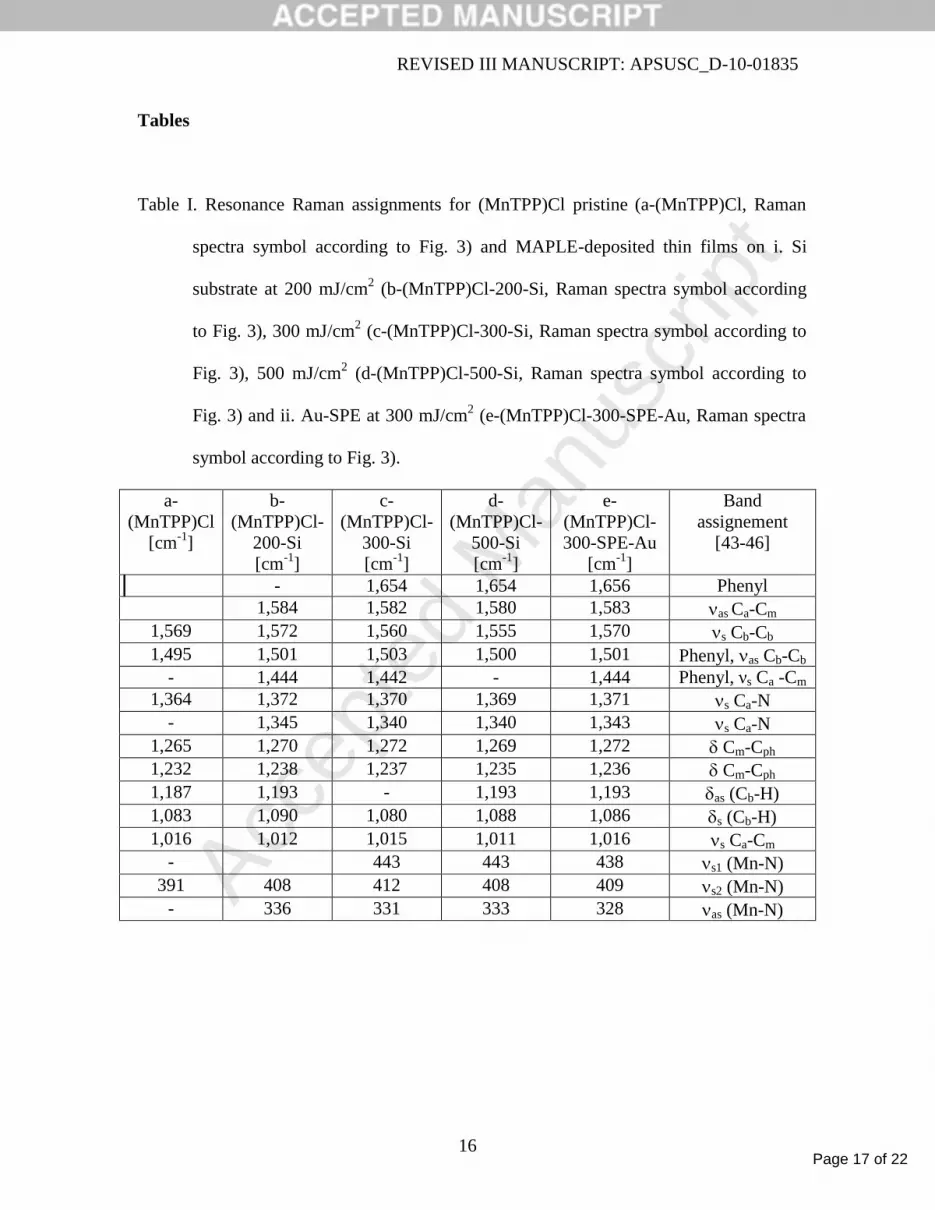
Tables
Table I. Resonance Raman assignments for (MnTPP)Cl pristine (a-(MnTPP)Cl, Raman
spectra symbol according to Fig. 3) and MAPLE-deposited thin films on i. Si
substrate at 200 mJ/cm2 (b-(MnTPP)Cl-200-Si, Raman spectra symbol according
to Fig. 3), 300 mJ/cm2 (c-(MnTPP)Cl-300-Si, Raman spectra symbol according to
Fig. 3), 500 mJ/cm2 (d-(MnTPP)Cl-500-Si, Raman spectra symbol according to
Fig. 3) and ii. Au-SPE at 300 mJ/cm2 (e-(MnTPP)Cl-300-SPE-Au, Raman spectra
symbol according to Fig. 3).
a-
(MnTPP)Cl
[cm-1
]
b-
(MnTPP)Cl-
200-Si
[cm-1
]
c-
(MnTPP)Cl-
300-Si
[cm-1
]
d-
(MnTPP)Cl-
500-Si
[cm-1
]
e-
(MnTPP)Cl-
300-SPE-Au
[cm-1
]
Band
assignement
[43-46]
- 1,654 1,654 1,656 Phenyl
1,584 1,582 1,580 1,583 as Ca-Cm
1,569 1,572 1,560 1,555 1,570 s Cb-Cb
1,495 1,501 1,503 1,500 1,501 Phenyl, as Cb-Cb
- 1,444 1,442 - 1,444 Phenyl, νs Ca -Cm
1,364 1,372 1,370 1,369 1,371 s Ca-N
- 1,345 1,340 1,340 1,343 s Ca-N
1,265 1,270 1,272 1,269 1,272 Cm-Cph
1,232 1,238 1,237 1,235 1,236 Cm-Cph
1,187 1,193 - 1,193 1,193 as (Cb-H)
1,083 1,090 1,080 1,088 1,086 s (Cb-H)
1,016 1,012 1,015 1,011 1,016 s Ca-Cm
- 443 443 438 s1 (Mn-N)
391 408 412 408 409 s2 (Mn-N)
- 336 331 333 328 as (Mn-N)

Page 18 of 22
Accep
ted
Man
uscr
ipt
REVISED III MANUSCRIPT: APSUSC_D-10-01835
17
Figure Caption
Fig. 1. The structure of (5,10,15,20-tetraphenyl)porphinato manganese (III) chloride
((MnTPP)Cl) is shown.
Fig. 2. AFM-tapping phase contrast images of (MnTPP)Cl MAPLE-deposited thin films
on Si wafers at 200 (a), 300 (b) and 500 mJ/cm2 (c).
Fig. 3. Typical Raman spectra of (MnTPP)Cl drop cast (a-(MnTPP)Cl symbol) and
MAPLE-deposited thin films on: i. Si substrate at 200 mJ/cm2 (b-(MnTPP)Cl-
200-Si symbol), 300 mJ/cm2 (c-(MnTPP)Cl-300-Si symbol), 500 mJ/cm
2 (d-
(MnTPP)Cl-500-Si symbol) and ii. SPE-220AT at 300 mJ/cm2 (e-(MnTPP)Cl-
300-SPE-Au symbol).
Fig. 4. Typical cyclic voltammograms of water blank (solid curve), SPE-220AT (long
dashed curve), and MAPLE-deposited thin films on SPE-220AT at 300 mJ/cm2
(short dashed curve).

Page 19 of 22
Accep
ted
Man
uscr
ipt
REVISED III MANUSCRIPT: APSUSC_D-10-01835
18
N N
NN
Mn
Cl
Fig. 1.

Page 20 of 22
Accep
ted
Man
uscr
ipt
REVISED III MANUSCRIPT: APSUSC_D-10-01835
19
Fig. 2.

Page 21 of 22
Accep
ted
Man
uscr
ipt
REVISED III MANUSCRIPT: APSUSC_D-10-01835
20
Fig. 3.

Page 22 of 22
Accep
ted
Man
uscr
ipt
REVISED III MANUSCRIPT: APSUSC_D-10-01835
21
Fig. 4.