Malaria: Basic Biology and Options for Therapy and Prophylaxis
87
RÖ 7 DEFENCE I *m IM DEFENSE Malaria: Basic Biology and Options for Therapy and Prophylaxis Bradley J. Berger Defence Research Establishment Suffield DISTRIBUTION STATEMENT A Approved for Public Release Distribution Unlimited Technical Memorandum DRESTM 2000-055 September 2000 laftcfl National Defense PanaHa " Defence nationale VydllcUacl —_. 20010116 088
-
Upload
khangminh22 -
Category
Documents
-
view
0 -
download
0
Transcript of Malaria: Basic Biology and Options for Therapy and Prophylaxis

RÖ7 DEFENCE I *m IM DEFENSE
Malaria: Basic Biology and Options for Therapy and Prophylaxis
Bradley J. Berger Defence Research Establishment Suffield
DISTRIBUTION STATEMENT A Approved for Public Release
Distribution Unlimited
Technical Memorandum
DRESTM 2000-055
September 2000
laftcfl National Defense PanaHa ■ "■ Defence nationale VydllcUacl
—_. 20010116 088

Malaria: Basic Biology and Options for Therapy and Prophylaxis
Bradley J. Berger Defence Research Establishment Suffield
Defence Research Establishment Suffield
Technical Memorandum
DRES TM 2000-055
September 2000

Author
Bradley J. Berger
Approved by
Dr. C. Boulet
Head/Chemical and Biological Defence Section
Approved for release by
Dr. R. Herring
Chair DRP/DRES
Her Majesty the Queen as represented by the Minister of National Defence, 2000
Sa majeste la reine, representee par le ministre de la Defense nationale, 2000

Abstract
Malaria remains one of the world's greatest health threats with 200 - 300 million infections and 2 - 3 million deaths per year. Increasingly, peace-keeping deployments occur in regions of the world with high incidence rates of malaria, and in areas where resistance to commonly used antimalarials is frequent. This review covers the basic biological properties of malaria parasites and provides information on the biochemistry and pharmacology of the currently available antimalarial therapies. The current status on drug resistance in malaria is also presented.
Resume
Le paludisme reste une des plus grandes menaces de la sante au monde, infectant 200 ä 300 millions de personnes et causant deux ä trois millions de morts par an. De plus en plus, les deploiements pour le maintien de la paix se passent dans des regions du monde oü les taux d'incidence du paludisme sont eleves et dans des zones oü la resistance aux medicaments antipaludiques communs n'est pas rare. La presente etude traite des proprietes biologiques de base des hematozoaires et informe sur la biochimie et la pharmacologie des therapies antipaludiques dont on dispose aujourd'hui. L'etat actuel de la resistance du paludisme aux medicaments y est egalement signale.
DRES TM 2000-055

This page intentionally left blank.
DRES TM 2000-055

Table of contents
Abstract i
Resume i
Table of contents iii
List of figures v
List of tables v
Introduction 1
Biology 2
The Parasites 2
Distribution 5
Pathology 6
Biochemistry 7
In vitro culturing 9
Genetics 9
Detection 10
Antimalarial therapy 12
Quinine 12
Chloroquine 15
Mefloquine 19
Primaquine 21
Proguanil 23
Pyrimethamine/sulfadoxine 25
Tetracycline/doxycycline 28
Halofantrine 30
Artemether 33
Artesunate 35
Quinidine 36
DRES TM 2000-055 III

Pyronaridine 37
Atovaquone 38
Obsolete agents 40
Experimental therapies 41
Recommendations for therapy 41
Recommendations for prophylaxis 42
References 47
IV DRES TM 2000-055

List of figures
Figure 1. Evolutionary relationships of the Apicomplexa. Both upper and lower portions are based on phylogenies constructed by Ayala et al. (1998) using the small subunit ribosomal RNA gene sequence. The upper portion represents the relationships amongst the phyla Dinozoa and Apicomplexa, and the classes, orders, and genuses within the Apicomplexa. The lower portion represents the relationships between selected species within the genus Plasmodium 2
Figure 2. A generalised life cycle of the malaria parasite 4
Figure 3. The digestive process for hemoglobin in the malaria parasite and inhibitors active in the pathway 8
Figure 4. The biosynthetic pathways present in the malaria parasite for the synthesis of pyrimidines and inhibitors active in the pathway 26
List of tables
Table 1. Therapies for malaria (all drug amounts are for the base equivalent of a salt). All doses listed are for adults 45
DRES TM 2000-055

This page intentionally left blank.
VI DRES TM 2000-055

Introduction
Despite decades of attempts to eradicate the disease world-wide, malaria remains one of the planet's greatest sources of mortality and morbidity. Indeed, with the spread of drug-resistant parasites and insecticide-resistant mosquito vectors, the situation regarding malaria has significantly worsened over the last couple of decades. The World Health Organisation estimates that over 2 billion people are at risk of contracting the disease, with over 200 million cases and 2 million deaths per year (WHO, 1999). These values are widely believed to be gross under-estimates, due to the poor level of epidemiological monitoring in sub- Saharan Africa and portions of Southeast Asia.
The increasing amount of international travel undertaken by Canadian businessmen and tourists has led to a corresponding increase in exposure to and infection by malaria. For 1997, Health Canada reported 1036 cases of malaria in returning Canadians (Health Canada, 1997). Per capita, this rate is 10 fold worse than that seen in the United States. Members of the Canadian armed forces are also increasingly at risk for contracting malaria. Many recent peacekeeping operations have been conducted in areas with endemic malaria transmission, and several have taken place in regions known to have high incidence rates of drug-resistant malaria. Given that most of the wars and serious civil disturbances in the world are unfolding in areas with high rates of malaria transmission and occurrence of drug resistant parasites, there remains a high likelihood that future Canadian forces deployments will face the threat of this illness.
This paper reviews the basic biological and medical features of malaria, and provides information on the drugs available for prophylaxis and therapy. The material contained within, while extensive, is not definitive, and additional information on malaria symptomology and pathology, and on current drug dosages and side effects should be obtained before use in a clinical context. In addition, the spread of drug resistance is an active on-going process. This paper was compiled using information available in early 2000, and readers are strongly advised to access updated material on the exact status of malarial drug resistance at future dates.
DRES TM 2000-055

Biology
The Parasites
Malaria parasites are unicellular, eukaryotic organisms belonging to a single genus (Plasmodium) within the phylum Apicomplexa. All apicomplexans are unicellular, parasitic eukaryotes, but not all are obligately intracellular. The unifying feature of all the species within the phylum is the presence of the apical complex at the anterior end of the infectious stage of the organism. This complex consists of three, electron-dense organelles, the roptries, micronemes, and conoid, which are believed to play a role in facilitating invasion of the host cell (Barnwell and Galinski, 1998). There is no clear consensus on the phylogenetic relationship of the different genera within the phylum, but analysis of life-cycles and molecular features (such as ribosomal RNA and mitochondrial DNA sequences) have suggested a broad categorization (Fig. 1).
1 1 Other Protozoa 1
Dinozoa (Dinoflagellates)
i Apicomplexa
1 1
Perkinsus (Oyster Parasi es)
l Coccidea
i
1 Hematozoa
i 1 i I
Piroplasmida
1
i 1 1 Cryptosporidium
ircocystis 1 Toxoplasma
1 s Neospora
I l
Babesia 1
Theileria Plasmodium
berghei malariae I I | 1 (Mouse) (Human) I 1 | 1
vivax cynomolgi knowlesi fragile | | mexicanum reichenowi falciparum (Human) (Monkey) (Monkey) (Monkey) ga|ijnaceum lophurae (Reptile) (Monkey) (Human)
(Bird) (Bird)
Figure 1. Evolutionary relationships of the Apicomplexa. Both upper and lower portions are based on phytogenies constructed byAyala et al. (1998) using the small subunit ribosomal RNA gene sequence. The upper portion represents the relationships amongst the phyla Dinozoa and Apicomplexa, and the classes, orders, and genuses within the Apicomplexa. The lower portion represents the relationships
between selected species within the genus Plasmodium.
DRES TM 2000-055

There are in excess of 100 individual species of malaria, infecting a range of reptiles, birds (such as P. lophurae in ducks, P. gallinaceum in chickens, P. relictum in canaries), and mammals. Within the latter, only mice, primates, and humans have ever been found to maintain plasmodial infections. P. berghei, P. chabaudi, and P. yoelii in mice, and P. cynomolgi, P. simiovale, and P. knowlesi in primates have been used for decades as model infection systems for human malaria. There are four separate species of malaria infectious for man: P.falciparum, P. vivax, P. malariae, and P. ovale. Of these species, P.falciparum is responsible for the vast majority of fatalities, with P. vivax rarely fatal, and P. ovale and P. malariae relatively rare infections (WHO, 1999).
All malaria parasites have essentially the same life-cycle (Fig. 2). When an infected mosquito bites a person, it injects numerous, tiny sporozoites into the bloodstream. It is estimated that as few as 10 sporozoites can establish a human infection. Within an hour, the sporozoites have travelled to the liver and invaded parenchymal cells. Inside the liver cells, the sporozoite matures into a hepatic trophozoite, which feeds on the cytoplasm of the host cell for about one week. The single trophozoite then undergoes several rounds of schizogony to produce as many as 30,000 merozoites. The merozoites burst free from the infected liver cell and are carried throughout the bloodstream, infecting erythrocytes. Within the red blood cell, the merozoite changes into a trophozoite, which begins to feed on the hemoglobin present in the host cell. Young trophozoites resemble rings and mature into ameboid forms which contain visible granules of hemozoin (polymerised heme). The mature trophozoite undergoes a single round of schizogony to produce 6-18 merozoites which burst out of the erythrocyte and infect additional red blood cells. In this manner, the erythrocytic infection is amplified in synchronous waves. In response to unknown stimuli, some merozoites will differentiate into male or female gametocytes after infecting a new erythrocyte, rather than forming the usual trophozoites. The gametocytes mature over 3-10 days and then may persist in the bloodstream for up to several weeks. The mature gametocytes are arrested forms which are preadapted for the mosquito gut. When a competent mosquito bites an infected human, it picks up mature gametocytes, which rapidly transform into gametes. The male gamete's nucleus divides three times and the new nuclei become associated with flagella. These threadlike microgametes tear free from the rest of the male gamete in a rapid process known as exflagellation. The microgametes then swim in search of the female gamete. When fertilisation occurs, the resulting zygote undergoes differentiation over 18 hours to form a motile ookinete. The ookinete crosses the peritrophic membrane of the mosquito midgut, passes through the gut epithelium, and settles beneath the basal lamina, forming the oocyst. The oocyst matures over the next 11 - 60 days in a process involving repeated nuclear division. At the end of this process, the oocyst contains up to 10,000 sporozoites, which then emerge from the oocyst into the mosquito hemolymph. The immature sporozoites then migrate to the salivary glands of the mosquito, where they mature into infective sporozoites. The mosquito then passes the sporozoites on to the human host with the next blood meal. In P. vivax and P. ovale malaria, there is an additional stage, called the hypnozoite. Immediately after sporozoites of this species infect liver cells, some become hypnozoites rather than hepatic trophozoites. The new cell type is essentially a dormant trophozoite, which can reactivate weeks or months later to initiate a new cycle of hepatic schizogony. In this manner, the disease presents as a relapsing form which may resurface even after successful treatment of the primary infection. It is thought that hypnozoite development in these species, which were distributed more widely in temperate regions, was an adaptation to cold weather periods when mosquitos are not present.
DRES TM 2000-055

/. Fertilisation \^ Sporozoites
\——"1 Exflagellation Mosquito Migration to
Salivary Glands
Human * ^\ ^ Sporozoites
Female Male Gametocytes
Mature TrophozoiteV
Hypnozoite
/ 4. Erythrocytic ($$&)Merozoites
""^N. Schizogony \J$ ®
Early.. - — Trophozoite (Ring)
Hepatic Trophozoite
3. Hepatic Schizogony
Hepatic Schizonts
Figure 2. A generalised life cycle of the malaria parasite.
The existence of malaria as a disease of set cycles of fever and chills has a recorded history spanning the last 4000 years, with accurate descriptions found on cuneiform tablets from the library of Ashurbanipal (appr. 2000 BC), Vedic writings (appr. 1500 BC), and the Chinese Nei Ching (appr. 2700 BC) (Bruce-Chwatt, 1988). Numerous descriptions also occur throughout recorded Greek and Roman history. The word "malaria" is Italian, and stems from the common belief that the fevers stemmed from inhaling "bad air" and the term was introduced into English in 1740 by the early novelist Horace Walpole (Russell, 1955). While the word "malaria" is used in a number of francophone publications, the official French equivalent (as used in WHO documents) is "paludisme".
In terms of more recent history, P. vivax malaria was endemic in the United States from the southeastern border north to about Baltimore and in southern Europe. Epidemics of P. vivax malaria regularly occurred in North America as far north as Montreal, and in Europe as far north as the midlands of England and southern Sweden (Bruce-Chwatt, 1988). In all these cases, the presence of the disease was completely eliminated through the draining of swamps and other surface bodies of stagnant water, as recently as the early 20th century in the cases of the southern United States and central Italy. Many areas cleared of malaria still maintain populations of competent mosquito vectors, and aggressive spraying campaigns (such as in south Florida and northern Australia) are common.
DRES TM 2000-055

The erythrocytic stages of the malaria parasite were first discovered by Laveran in 1880 in a blood smear taken from a French soldier in Algeria (Laveran, 1880). Six years later, Golgi discovered the schizogenic cycle in the erythrocytes and demonstrated that the beginning of the fever cycle coincided with the rupture of erythrocytes and the release of the new generation of parasites (Golgi, 1886). In 1897, Ross conclusively demonstrated that female Anopheline mosquitoes were the vector for avian malaria; this discovery was confirmed for human malaria by Grassi in 1898 (Ross, 1897; Grassi, 1900). It was not until 1948 that Shortt and Garnham discovered the initial, hepatic stages of the disease, thus completing the life- cycle (Shortt and Garnham, 1948).
Distribution
At present, over 90 countries are considered malarious (see the section below on Recommendations for Therapy) as reported by WHO and CDC (WHO, 1999; CDC, 2000). About half of these countries, and approximately 90% of the deaths from malaria, are to be found in sub-Saharan Africa. The distribution of P. falciparum and P. vivax are now almost completely identical. Historically, P. ovale was limited to tropical Africa, and P. malariae was found in patches world-wide, but predominantly in Southeast-Asia. At present, both of these species can be found world-wide, but their incidence rate is low relative to P. falciparum or P. vivax, probably due to the shorter time the latter two species require to mature in the mosquito (Beales, 1999).
The phenomenon of resistance to common antimalarials is one of the central reasons behind the recent resurgence of the disease. Chloroquine-resistant P. falciparum is presently found world-wide, with only Mexico and parts of the middle-east and Asia reporting a lack of chloroquine resistance (as of the early 1990s; CDC, 2000). The frequency of chloroquine- resistant infections is particularly high in east Africa (Kenya, Tanzania, Malawi) and southeast Asia (border regions of Thailand, Myanmar, and Cambodia). Chloroquine-resistant P. vivax is a more recent development, and has been reported in Irian Jaya (Indonesia), Papua New Guinea, Myanmar, and Vanuatu (Whitby et al., 1989; Myat-Phone-Kyaw et al., 1993; Schwartz et al., 1991). Throughout New Guinea, over one-third of all P. vivax infections are resistant to chloroquine.
When chloroquine became increasingly ineffective in treating malaria in Southeast Asia, the drug was replaced with the anti-folate combination pyrimethamine/sulfadoxine (Fansidar) as the frontline treatment. Within approximately three years (1977-1980), this new therapy was no longer providing adequate cure rates (Reacher et al., 1982; Pinichpongse et al., 1982). The recent introduction of Fansidar as a frontline therapy for chloroquine in east Africa yielded similar, disturbing results, with resistance reported within one year (Lege-Oguntoye et al., 1990). Resistance to this drug combination is also commonly reported in South America, particularly Amazonia and bordering areas (Kremsner et al., 1988).
With the failure of chloroquine and Fansidar, there has been a heavy reliance on quinine, quinine/tetracycline, and mefloquine. By the early 1990s, almost half of all P. falciparum infections in the border areas of Thailand/Myanmar and Thailand/Cambodia were mefloquine resistant (Ketrangsee et al., 1992; Fontanet et al., 1994). These areas also report incidences of quinine-resistance, with more than 85% of P. falciparum infections in these regions now classified as multidrug resistant (Giboda et al., 1988). Mefloquine-resistance has not yet been reported in South America and Africa, and the incidence rate of quinine-resistance is low, primarily due to the fact that the compounds are not used frequently on these continents.
DRES TM 2000-055
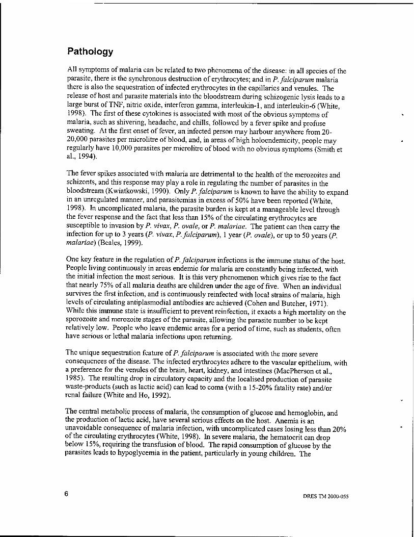
Pathology
All symptoms of malaria can be related to two phenomena of the disease: in all species of the parasite, there is the synchronous destruction of erythrocytes; and in P.falciparum malaria there is also the sequestration of infected erythrocytes in the capillaries and venules. The release of host and parasite materials into the bloodstream during schizogenic lysis leads to a large burst of TNF, nitric oxide, interferon gamma, interleukin-1, and interleukin-6 (White, 1998). The first of these cytokines is associated with most of the obvious symptoms of malaria, such as shivering, headache, and chills, followed by a fever spike and profuse sweating. At the first onset of fever, an infected person may harbour anywhere from 20- 20,000 parasites per microlitre of blood, and, in areas of high holoendemicity, people may regularly have 10,000 parasites per microlitre of blood with no obvious symptoms (Smith et al., 1994).
The fever spikes associated with malaria are detrimental to the health of the merozoites and schizonts, and this response may play a role in regulating the number of parasites in the bloodstream (Kwiatkowski, 1990). Only P.falciparum is known to have the ability to expand in an unregulated manner, and parasitemias in excess of 50% have been reported (White, 1998). In uncomplicated malaria, the parasite burden is kept at a manageable level through the fever response and the fact that less than 15% of the circulating erythrocytes are susceptible to invasion by P. vivax, P. ovale, or P. malariae. The patient can then carry the infection for up to 3 years (P. vivax, P.falciparum), 1 year (P. ovale), or up to 50 years (P. malariae) (Beales, 1999).
One key feature in the regulation ofP.falciparum infections is the immune status of the host. People living continuously in areas endemic for malaria are constantly being infected, with the initial infection the most serious. It is this very phenomenon which gives rise to the fact that nearly 75% of all malaria deaths are children under the age of five. When an individual survives the first infection, and is continuously reinfected with local strains of malaria, high levels of circulating antiplasmodial antibodies are achieved (Cohen and Butcher, 1971). While this immune state is insufficient to prevent reinfection, it exacts a high mortality on the sporozoite and merozoite stages of the parasite, allowing the parasite number to be kept relatively low. People who leave endemic areas for a period of time, such as students, often have serious or lethal malaria infections upon returning.
The unique sequestration feature of P.falciparum is associated with the more severe consequences of the disease. The infected erythrocytes adhere to the vascular epithelium, with a preference for the venules of the brain, heart, kidney, and intestines (MacPherson et al., 1985). The resulting drop in circulatory capacity and the localised production of parasite waste-products (such as lactic acid) can lead to coma (with a 15-20% fatality rate) and/or renal failure (White and Ho, 1992).
The central metabolic process of malaria, the consumption of glucose and hemoglobin, and the production of lactic acid, have several serious effects on the host. Anemia is an unavoidable consequence of malaria infection, with uncomplicated cases losing less than 20% of the circulating erythrocytes (White, 1998). In severe malaria, the hematocrit can drop below 15%, requiring the transfusion of blood. The rapid consumption of glucose by the parasites leads to hypoglycemia in the patient, particularly in young children. The
DRES TM 2000-055

administration of quinine, with its associated induction of insulin secretion, may exacerbate the condition (Davis et al, 1990). Finally, the lactic acid produced by the parasites leads to metabolic acidosis. In severe malaria, the host metabolism may shift to anaerobiosis, leading to addition lactate production from the skeletal muscle. The metabolic acidosis is a major contributor to the death of patients with severe malaria.
Biochemistry
The malaria parasite is a highly specialised, parasitic organism, and has many important differences in its central biochemical processes when compared to host cells. This biochemical specialisation is highly relevant to the action of existing antimalarial drugs, and the development of novel compounds. Almost all modern research into malaria is performed on P. falciparum, as this is the organism responsible for >90% of fatalities, and is also the only species capable of indeterminate growth in vitro. Therefore, unless another species is specified here (or in the general literature), all biochemical and genetic findings are in P. falciparum.
As mentioned before, the central, energy-producing process in Plasmodium spp. is glycolysis. The parasite consumes glucose at a rate approximately 70-100 times faster than uninfected erythrocytes (Roth, 1990), and obtains 2 ATP from each glucose molecule consumed. Malaria lacks a functional tricarboxylic acid cycle (Kreb's cycle) and is unable to utilise the pyruvate produced from glycolysis for further energy production. Instead, the organism converts pyruvate to lactate via lactate dehydrogenase, a process which reduces the nicotinamide cofactors oxidized during glycolysis (Sherman, 1961). Although this incomplete usage of glucose is considered inefficient, it provides sufficient energy for the parasite, so long as an exogenous supply of glucose is maintained. Malaria is known to alter the infected erythrocyte so as to increase the influx of glucose and the efflux of lactate (Ginsburg and Kirk, 1998). The parasite also can utilise the pentose-phosphate pathway for glucose consumption, though to a much lesser degree than glycolysis (Scheibel, 1988).
By utilising the erythrocyte as a host cell, the malaria parasite encounters potential metabolic difficulties. The erythrocyte is, metabolically, a very degenerate cell, and functions solely as a carrier of hemoglobin. The red cell synthesizes no amino acids, nucleic acids, or other essential biochemical intermediates, but does contain nearly 10 mM hemoglobin. The parasite has incorporated this abundance of a single protein into its central metabolism by digesting the host hemoglobin as a source of amino acids (Fig. 3). Malaria parasites contain a specialised, enlarged lysosome called the food vacuole, which contains aspartate and cysteine proteases (plasmepsin I and II, and falcipain) that break down hemoglobin to peptides (Rosenthal et al., 1988; Goldberg et al., 1990; Goldberg et al., 1991). The peptides are then exported to the cell cytoplasm for digestion by endopeptidases, yielding free amino acids for incorporation into malarial proteins (Sherman and Tanagoshi, 1970; Curley et al., 1994). As hemoglobin contains no isoleucine, and little methionine, cysteine, and glutamine, the parasite has an obligate requirement for these amino acids in the plasma (Francis et al., 1994).
DRES TM 2000-055

Plasmepsin I (35 KDal Aspartic Protease) cuts at 33Phe-34Leu
Denatured Hemozoin Hemoglobin
Food \ Cytosol Vacoule ^
Plasmepsin II (35 KDal Aspartic Protease) multiple cleavages
Denatured Globin
Peptides
Amino Acids
Figure 3. The digestive process for hemoglobin in the malaria parasite and inhibitors active in the pathway.
The rapid and massive digestion of hemoglobin by the parasite gives rise to the release of free heme within the food vacuole. As free heme is toxic to cells through inhibition of enzymes and the generation of oxygen radicals (Orjih et al., 1981), it must be deactivated or removed from the parasite. Malaria is known to lack the enzyme heme oxygenase, which mammals use to break down heme, but has evolved a unique solution to the problem of heme toxicity. The free heme is polymerised to an insoluble black-brown paracrystalline material (known as hemozoin or malaria pigment) in the food vacuole (Slater et al., 1991). The hemozoin is left behind when the new generation of merozoites lyse the erythrocyte, and is ultimately deposited in tissues (such as the liver and brain) of the infected patient, giving rise to a grey- black colour in these organs. The mechanism of hemozoin polymerisation has been much debated (Slater and Cerami, 1992; Dorn et al., 1995; Bendrat et al., 1995; Sullivan et al., 1996), but is a central facet of malarial survival. Inhibition of heme polymerisation rapidly leads to the death of the parasite (Fitch et al., 1982).
For its nucleic acid synthesis, the parasite must scavenge purines from the host, primarily through the uptake of hypoxanthine (Asahi et al., 1996). Malaria does, however, synthesize its own pyrimidines (Reyes et al., 1982) and also synthesizes phospholipids, ceramides, and sphingolipids (Ferone, 1977; Vial and Ancelin, 1998).
DRES TM 2000-055
In vitro culturing
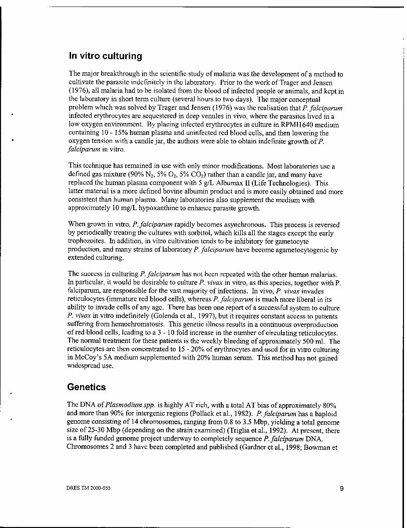
The major breakthrough in the scientific study of malaria was the development of a method to cultivate the parasite indefinitely in the laboratory. Prior to the work of Träger and Jensen (1976), all malaria had to be isolated from the blood of infected people or animals, and kept in the laboratory in short term culture (several hours to two days). The major conceptual problem which was solved by Träger and Jensen (1976) was the realisation that P.falciparum infected erythrocytes are sequestered in deep venules in vivo, where the parasites lived in a low oxygen environment. By placing infected erythrocytes in culture in RPMI1640 medium containing 10 - 15% human plasma and uninfected red blood cells, and then lowering the oxygen tension with a candle jar, the authors were able to obtain indefinite growth of P. falciparum in vitro.
This technique has remained in use with only minor modifications. Most laboratories use a defined gas mixture (90% N2, 5% 02, 5% C02) rather than a candle jar, and many have replaced the human plasma component with 5 g/L Albumax II (Life Technologies). This latter material is a more defined bovine albumin product and is more easily obtained and more consistent than human plasma. Many laboratories also supplement the medium with approximately 10 mg/L hypoxanthine to enhance parasite growth.
When grown in vitro, P.falciparum rapidly becomes asynchronous. This process is reversed by periodically treating the cultures with sorbitol, which kills all the stages except the early trophozoites. In addition, in vitro cultivation tends to be inhibitory for gametocyte production, and many strains of laboratory P. falciparum have become agametocytogenic by extended culturing.
The success in culturing P.falciparum has not been repeated with the other human malarias. In particular, it would be desirable to culture P. vivax in vitro, as this species, togethor with P. falciparum, are responsible for the vast majority of infections. In vivo, P. vivax invades reticulocytes (immature red blood cells), whereas P. falciparum is much more liberal in its ability to invade cells of any age. There has been one report of a successful system to culture P. vivax in vitro indefinitely (Golenda et al., 1997), but it requires constant access to patients suffering from hemochromatosis. This genetic illness results in a continuous overproduction of red blood cells, leading to a 3 - 10 fold increase in the number of circulating reticulocytes. The normal treatment for these patients is the weekly bleeding of approximately 500 ml. The reticulocytes are then concentrated to 15 - 20% of erythrocytes and used for in vitro culturing in McCoy's 5A medium supplemented with 20% human serum. This method has not gained widespread use.
Genetics
The DNA of Plasmodium spp. is highly AT rich, with a total AT bias of approximately 80% and more than 90% for intergenic regions (Pollack et al., 1982). P.falciparum has a haploid genome consisting of 14 chromosomes, ranging from 0.8 to 3.5 Mbp, yielding a total genome size of 25-30 Mbp (depending on the strain examined) (Triglia et al., 1992). At present, there is a fully funded genome project underway to completely sequence P.falciparum DNA. Chromosomes 2 and 3 have been completed and published (Gardner et al., 1998; Bowman et
DRES TM 2000-055

al., 1999), and several other chromosomes are nearing completion. Recent developments in vector construction and transfection techniques have allowed for the transient and stable incorporation of transgenes into malaria and the construction of parasites with selected genes knocked-out (van Dijk et al., 1995; Wu et al., 1995). These processes are still in their infancy and are currently only routinely performed in a few laboratories. By coinfecting a mosquito with two different P.falciparum strains, it is possible to perform a Mendelian analysis on the offspring arising from the sexual stage of the parasite. This type of experiment is difficult and has only been performed twice to date (Walliker et al., 1987; Wellems et al., 1990).
Outside of the nucleus, there are two other genetic elements in P.falciparum, the mitochondrial genome and the apicoplast genome. The former is a 6 kbp molecule, existing as a circle (approximately 1%) or linear multimers (6-30 kbp) and encodes genes for cytochrome b, cytochrome oxidase, and ribosomal RNA (Williamson, 1998). It is one of the smallest genomes discovered in a mitochondrion, and is reflective of the relative inactivity of the parasite mitochondrion. The apicoplast is a tiny, triple-membraned vesicle which is an apparent remnant of an ancestral chloroplast (Wilson and Williamson, 1997). The organelle has no known function, but is known to be essential to malarial survival and has been found in all apicomplexans examined to date. The apicoplast contains a 35 kbp circular genome, which is approximately 20% of the normal size for a chloroplast genome (Wilson et al., 1996). It encodes genes for 2 ribosomal RNAs, 25 transfer RNAs, 19 ribosomal proteins, 3 RNA polymerases, no photosynthetic proteins, and 5 unknown proteins. Recent studies have demonstrated that genes of apparent plastid origin (such as those involved in the shikimate and isoprenoid pathways) are now resident in the parasite nuclear genome (Roberts et al., 1998; Jomaaetal., 1999).
Detection
The traditional procedure for the diagnosis of malaria is the examination of thick and thin blood smears by light microscopy. Detailed instructions for preparation of blood films can be obtained from several sources, such as Garcia and Bruckner (1997). The slides are stained with Giemsa for 30-45 minutes before rinsing and visualisation under an oil immersion lens (600 - 1000 X final magnification, depending on the ocular lenses used). Alternatively, Leishman, Wright, or Diff-Quik stains can be utilised. For routine detection of parasites in blood cultures in vitro, the author prefers the latter due to its rapidity. The standard method for quantification of parasites is via 100 fields of the thick smear with enumeration of both malaria parasites and leukocytes. Based on an average leukocyte count of 8000/ul of blood:
parasites/ul =■ (parasites counted * 8000)/leukocytes counted
For examination of thin smears, both parasitised and uninfected erythrocytes are enumerated, with the results are expressed as percent parasitemia.
The major task in microscopic identification is the speciation of the malarial infection. Misidentification of a P.falciparum infection, or missing P.falciparum in a mixed infection can have lethal consequences. Visual aids for speciating malaria can be found in a number of publications, including Garcia and Bruckner (1997). As a general rule, due to sequestration of infected erythrocytes, P. falciparum infections tend to be visible in circulating blood as rings and early trophozoites. The rings are quite small (about 1/5-1/6 of the red blood cell), may
10 DRES TM 2000-055
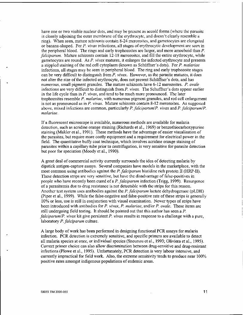
have one or two visible nuclear dots, and may be present as accole forms (where the parasite is closely adjoining the outer membrane of the erythrocyte, and doesn't clearly resemble a ring). When seen, mature schizonts contain 8-24 merozoites, and gametocytes are elongated or banana-shaped. For P. vivax infections, all stages of erythrocytic development are seen in the peripheral blood. The rings and early trophozoites are larger, and more amoeboid than P. falciparum. Mature schizonts contain 12-18 merozoites, and fill the entire erythrocyte, while gametocytes are round. As P. vivax matures, it enlarges the infected erythrocyte and presents a stippled staining of the red cell cytoplasm (known as Schüffner's dots). For P. malariae infections, all stages may be seen in peripheral blood. The ring and early trophozoite stages can be very difficult to distinguish from P. vivax. However, as the parasite matures, it does not alter the size of the infected erythrocyte, does not present Schüffner's dots, and has numerous, small pigment granules. The mature schizonts have 6-12 merozoites. P. ovale infections are very difficult to distinguish from P. vivax. The Schüffner's dots appear earlier in the life cycle than in P. vivax, and tend to be much more pronounced. The later trophozoites resemble P. malariae, with numerous pigment granules, and red cell enlargement is not as pronounced as in P. vivax. Mature schizonts contain 8-12 merozoites. As suggested above, mixed infections are common, particularly P. falciparumlP. vivax and P. falciparumlP. malariae.
If a fluorescent microscope is available, numerous methods are available for malaria detection, such as acridine orange staining (Richards et al., 1969) or benzothiocarboxypurine staining (Makler et al., 1991). These methods have the advantage of easier visualisation of the parasites, but require more costly equipment and a requirement for electrical power in the field. The quantitative buffy coat technique, which involves acridine orange staining of parasites within a capillarytube prior to centrifugation, is very sensitive for parasite detection but poor for speciation (Moody et al., 1990).
A great deal of commercial activity currently surrounds the idea of detecting malaria by dipstick antigen-capture assays. Several companies have models in the marketplace, with the most common using antibodies against the P. falciparum histidine rich protein II (HRP-II). These detection strips are very sensitive, but have the disadvantage of false-positives in people who have recently been cured of & P. falciparum infection (Trigg, 1999). Resurgence of a parasitemia due to drug resistance is not detectable with the strips for this reason. Another test system uses antibodies against the P. falciparum lactate dehydrogenase (pLDH) (Piper et al., 1999). While the false-negative and false-positive rate of these strips is generally 10% or less, use is still in conjunction with visual examination. Newer types of strips have been introduced with antibodies for P. vivax, P. malariae, and/or P. ovale. These items are still undergoing field testing. It should be pointed out that this author has seen a P. falciparumlP. vivax kit give persistent P. vivax results in response to a challenge with a pure, laboratory P. falciparum culture.
A large body of work has been performed in designing functional PCR assays for malaria infection. PCR detection is extremely sensitive, and specific primers are available to detect all malaria species at once, or individual species (Snounou et al., 1993; Oliviera et al., 1995). Correct primer choice can also allow discrimination between drug-sensitive and drug-resistant infections (Plowe et al., 1995). Unfortunately, PCR detection is very labour intensive, and currently impractical for field work. Also, the extreme sensitivity tends to produce near 100% positive rates amongst indigenous populations of endemic areas.
DRES TM 2000-055 11

Antimalarial therapy
Quinine
History. Quinine represents one of the oldest and most effective pharmacological agents known to man. It is the most abundant antimalarial component of the bark of cinchona trees {Cinchona spp., particularly Cinchona ledgeriana), which was used as a folk medicine among Peruvian Indians prior to encounters with the Spanish. As it is currently believed that malaria was introduced to the Americas by Europeans (McNeill, 1976), it is unlikely that the Incas originally used cinchona bark to treat this particular set of symptoms. Nevertheless, the bark proved most effective in curing the recurrent fevers of malaria in colonial Spaniards. By the early 1600's, the bark had been introduced to Spain via the Jesuits, and it rapidly became worth its weight in silver (Gramiccia, 1987). In 1820, Pelletier and Caventou became the first chemists to isolate the principle active ingredient, quinine, from cinchona bark, and the purified material was soon in use in France for the treatment of periodic fevers (Smith, 1976). In the late 1800s, the Dutch established large plantations of high-quinine cinchona in Indonesia, and soon were producing more than 80% of the world's supply of the pure compound. This situation lasted until the outbreak of the second world war, when Japan occupied Indonesia, and quinine became superseded by newer compounds.
Structure. Quinine is a quinolinemethanol and is more completely known as (a-R)-a-(6- methoxy-4-quinolyl)-a-[(2S,4S,5R)-(5-vinylquinuclidin-2-yl)] methanol. The structure is:
H3CO
The free base is only sparingly soluble in water or glycerol, but is soluble in alcohol, chloroform, or ether. The salts of the drug are soluble in water. When in solution, the drug is light and air sensitive. In pharmaceutical formulations, the drug can be found as bisulfate (Biquin, Biquinate, Dentojel, Myoquin, Quinbisan), hydrobromide (Coquelusedal Quinine), hydrochloride (Kinin), or sulfate (Adquin, Kinine, Novoquinine, Quinate, Quine, Quinoctal, Quinsan) salts. A combination of quinine (33.3%), quinidine (33.3%), and cinchonine (33.3%) is also available (Quinimax). Commercial preparations of quinine routinely contain up to 10% of the impurity dihydroquinine (Pukrittayakamee et al., 1997). Quinine is extremely bitter tasting.
12 DRES TM 2000-055

Mechanism of Action. The exact basis of antimalarial activity remains unknown, but the compound only appears to effect late ring, trophozoite, and early schizont stages of the parasite. As with other quinoline antimalarial compounds, quinine is theorised to block the formation of hemozoin in the parasite food vacuole. Quinine is known to form a stable complex with free heme (Gushimana et al, 1993), and this complex is much more soluble in benzene (Warhurst, 1981). It is suspected that heme-quinine complexes cannot polymerise to form hemozoin (Chou and Fitch, 1993), and the complexes then freely enter the parasite membrane where they cause lipid peroxidation (Sugioka and Suzuki, 1991). Quinine also appears to be able to inhibit TNF production by the host (Maruyama et al., 1994; Kwiatkowski and Bate, 1995), thus reducing the more obvious symptoms of malaria.
Pharmacology. Quinine is normally administered orally to patients with uncomplicated malaria, and by intravenous infusion in those suffering from severe or complicated malaria. Rectal administration has also been examined for cases of severe malaria (Barennes et al., 1995; Barennes et al., 1996). As the therapeutic index of quinine is quite low (the therapeutic and toxic levels are close together), the use of a loading dose prior to intravenous infusion should be closely monitored and should be avoided altogether in patients with a history of recent quinine use.
Oral doses of quinine are well absorbed from the digestive tract, with a bioavailability of 64% (Sowunmi et al, 1994), and peak plasma concentrations are reached in 1 - 3 hours (Sowunmi, 1996; White et al., 1983). About 70 - 90% of the drug is bound to plasma proteins and then slowly released as free drug (Karbwang et al., 1993). In healthy adult Africans, a single oral dose of 500 mg quinine base gave a peak plasma concentration of 2.9 ± 0.5 mg/L at 2.0 - 4.0 hr after administration (Sowunmi, 1996). The terminal plasma half-life was 11.7 ± 2.9 hr and the volume of distribution 2.5 ± 0.7 L/kg (Sowunmi, 1996; White et al., 1983; Krishna and White, 1996). These pharmacokinetic properties were found to be linear over 250 -1000 mg oral administration (Sowunmi and Salako, 1996). The values found for intravenous injection (10 mg/kg over 1 hr) in healthy Thai males were found to be very similar, with a terminal plasma half-life of 9.9 hr and a volume of distribution of 3.1 L/kg (Karbwang et al., 1993). Patients with acute, but uncomplicated, malaria given oral quinine had plasma levels two-fold higher than normal, a prolonged elimination half-life, and a lower volume of distribution (Babalola et al., 1998; Krishna and White, 1996). The kinetics and disposition of quinine in patients with complicated malaria have not been as well studied, but cases of cerebral malaria appear to have even higher peak plasma levels of quinine, longer elimination half-life, and much smaller volume of distribution (Krishna and White, 1996). In children, the elimination half-life is shorter than adults, and the volume of distribution is smaller (Sabcharoen et al 1982;Pussardetal., 1999).
Quinine is readily metabolised to 3-hydroxyquinine and a number of additional, minor, oxidative metabolites (Zhao and Ishizaki, 1997). 3-Hydroxyquinine binds less avidly to plasma proteins, with approximately 54% of the metabolite present in an unbound form (Pukrittayakamee et al., 1977). The metabolite has intrinsic antimalarial activity, but is less potent than quinine (Newton et al, 1999). Patients with acute, uncomplicated malaria have a quinine/3-hydroxyquinine ratio of 12, whereas healthy individuals have one of 7 (Pukrittayakamee et al., 1977). This phenomenon appears to be due to the non-specific down- regulation of cytochromes P450 that occurs during a malarial infection and other pathological states (Saxena et al., 19871 Kokwaro et al., 1993). Indeed, the variation in the pharmacokinetic properties of quinine described in the previous paragraph appear entirely due to the resulting lower rate of quinine metabolism.
DRES TM 2000-055 -|3

The principle enzyme involved in quinine metabolism is cytochrome P450 3A4 (Zhao and Ishizaki, 1997), and quinine is also known to be a potent inhibitor of cytochrome P450 2D6 (Krishna and White, 1996). This latter P450 is known to be involved in the detoxification of a number of pharmaceutical agents, such as tricyclic antidepressants (Lennard, 1983). Therefore, administration of quinine can lead to a serious, and potentially fatal, increase in the toxicity of unrelated drugs.
Analytical Methodology. Quinine is routinely quantitated by high-performance liquid chromatography (HPLC), using either reverse-phase or normal-phase methods (Ericsson et al., 1993; Dua et al., 1993). The compound can be detected by fluorescence or ultraviolet spectrophotometry, with the former being more sensitive (concentrations as low as 2 nM are detectable in plasma). Quinine has also been quantified in urine via gas chromatography with mass spectrometric detection (Liddle et al., 1981), and in beverages by electrokinetic capillary chromatography with ultraviolet spectrophotometric detection (Trenerry and Ward, 1996). Older methods for detecting and quantitating quinine include rapid enzyme-linked immunosorbant (ELISA) assay (Rowell and Rowell, 1987), and colorimetric detection following reaction with thalleioquin (Karawya and Diab, 1977). Depending on the source of quinine, analysis of plasma levels or tablet extracts is advised, as fake antimalarials have been reported (Sowunmi et al, 1994).
Adverse Effects. Quinine is known to have a large number of potential side effects. Repeat administration of normal doses often gives rise to cinchonism: tinnitus, headache, nausea, abdominal pain, skin rash, and blurred vision (Bateman and Dyson, 1986). Of these reactions, the ear ringing is particularly common. Transient hypoglycemia is also a very common side effect of quinine administration. More serious quinine intoxification leads to fever, vomiting, confusion, blindness, deafness, and loss of consciousness. Hypotension may occur, and the patient may enter a coma and die from respiratory failure (Orme, 1987). Death from quinine poisoning may occur anywhere from 1 hour to 2 days after ingestion, and the median lethal dose is approximately 8 g. As self-administration of quinine is the norm in many parts of the world, quinine intoxication is rather common.
Patients suffering from chronic malaria or partially treated infections may develop blackwater fever after treatment with quinine (Chau et al., 1996). In this complication, there is a massive, non-specific lysis of erythrocytes and the passage of large amounts of hemoglobin in the urine. This phenomenon presents a very dark red or black urine. Patients with glucose-6- phosphate dehydrogenase deficiency are also at risk for developing blackwater fever following quinine treatment.
Hypersensitivity to quinine does exist, with these patients developing cinchonism, asthma, and other allergic symptoms from small doses of the drug. Thrombocytopenia and hemolytic anemia may also occur in these individuals. While quinine therapy has been suggested to increase spontaneous abortion, recent studies have shown that the drug has no such effect (Phillips-Howard and Wood, 1996). Quinine is one of the few antimalarials indicated for use during pregnancy.
Resistance. Quinine resistance has been reported as early as 1908, but was first properly documented in Brazil in the 1960s (Peters, 1987). Quinine resistance was also demonstrated in Southeast Asia shortly thereafter (Peters, 1987), and clinical failures to the drug have become much more common in the region during the 1980s (Giboda and Denis, 1988). At present, quinine resistance has not spread much beyond Brazil and Southeast Asia, probably
14 DRES TM 2000-055

due to its much lower frequency of use when compared to chloroquine, mefioquine, and pyrimethamine/sulfadoxine (Kouznetsov, 1987). The more rapid plasma half-life of quinine, when compared to chloroquine or pyrimethamine, also mitigates against the development of resistance. As quinine usage is on the increase due to malarial resistance to less toxic agents, the incidence rate of quinine resistance world-wide is likely to increase (Jelinek et al., 1995).
The mechanism of quinine resistance is not completely known. However, the recent work by Reed et al (2000) has clearly shown that mutation of pfmdrl, a transmembrane transport protein of the P-glycoprotein family, leads to an increase in resistance to both mefioquine and quinine. Upregulation of pfmdrl also leads to mefioquine and quinine resistance in vitro (Peel et al., 1994). The product of pfmdrl is thought to act by increasing the efflux of mefloquine/quinine from the parasite, preventing the accumulation of sufficient drug in the food vacuole, but this hypothesis has not been conclusively proven.
Chloroquine
History. One of the world's most consumed drugs, chloroquine was the mainstay of antimalarial therapy for decades, and remains the drug of choice for non-resistant infections. The discovery of chloroquine represents one of the more tortuous paths for any anti-infective. The first concerted efforts to discover novel antimalarials took place in the commercial laboratories of F. Bayer (Leverkeusan, Germany) immediately after the end of the first world war (Greenwood, 1995). Stimulated by the difficulty in obtaining quinine from the Dutch during the war, and the productive development of other antimicrobials from the dye industry, Bayer (after 1925 a subsidiary of IG Farben) synthesized several novel antimalarials. Among these, in 1934, was resochin (chloroquine). Early human trials in Germany uncovered sufficient toxicity that the compound was abandoned for a close structural derivative called sontochin. As part of an agreement with IG Farben, resochin and sontochin were patented in the United States by Winthrop Co. in 1941. When the American supply of quinine was cut off following Pearl Harbor, pharmaceutical companies and university laboratories in the USA pursued novel antimalarials and exchanged information with the existing programs of a similar nature in Britain and Australia. Nevertheless, the Winthrop patent was ignored until American forces captured a supply of Sontochin during the fall of Tunis in 1943. Resochin was then rapidly found to be a superior antimalarial to sontochin, and introduced for use under the name of chloroquine.
Structure. Chloroquine is a 4-aminoquinoline and is more fully known as 7-chloro-4-(4- diethylamino-l-methylbutylamino)quinoline. The chemical structure is:
DRES TM 2000-055 15

The free base is only slightly soluble in water, but is soluble in chloroform, ether, and dilute acids. The salts of the drug are soluble in 3 - 4 parts water. When in solution, the drug is very sensitive to degradation by light. In pharmaceutical preparations, chloroquine is found as the phosphate (Aralis, Avlochlor, Klorokin, Malaviron, Aralen, Chlorochin, Chlorquin, Delagil, Dichinalex, Resochin, Resochine) or sulfate (Nivoquine) salts.
Mechanism of Action. Despite decades of intense research, the mechanism of action of chloroquine has not been conclusively demonstrated. As with other quinolines, chloroquine is thought to interfere with the polymerisation of free hematin into hemozoin. Chloroquine treated parasites have enlarged food vacuoles and reduced amounts of hemozoin, and swelling of the mitochondrion and endoplasmic reticulum (Jacobs et al., 1988). The drug only appears to act on those stages of the parasite actively digesting hemoglobin and using the resulting amino acids for protein synthesis (Deans et al., 1983). A large number of studies have demonstrated that chloroquine accumulates to high concentrations within the food vacuole of the parasite (Ferrari and Cutler, 1991; Bray et al., 1999). In fact, 1 nM external chloroquine can accumulate up to 800-fold. As chloroquine is a diprotic weak base, this accumulation may raise the food vacuole pH from the normal value of 4.5 - 5.0 (Krogstad et al., 1985), and a number of people have suggested that the weak base property of chloroquine is the cause of the accumulation into the food vacuole (Krogstad and Schlesinger, 1986). However, chloroquine does not accumulate in mammalian lysosomes, which have an equivalent pH to malarial food vacuoles (Ohkuma and Poole, 1978), and the pH effect of chloroquine is 1000- fold too high to predict strictly by acid-base chemistry (Krogstad and Schlesinger, 1987). A high affinity binding site for chloroquine appears to be present in the food vacuole. Recently, it has been suggested that the free heme released during hemoglobin digestion is the high affinity binding site (Bray and Ward, 1999).
Previous studies have shown that chloroquine can bind stably to free heme (Chou et al., 1980), and that these complexes either do not incorporate into hemozoin, or do incorporate, but "cap" the heme polymer and prevent further deposition of heme (Sullivan et al., 1996). In either case, the build-up of free heme has toxic effects on the parasite. While chloroquine does have a fairly non-specific ability to intercalate into DNA, the drug exerts no antimicrobial effect on any other organism aside from malaria.
Pharmacology. Chloroquine is normally administered as oral doses for prophylaxis or for patients suffering from uncomplicated malaria. Patients with cerebral malaria are given intravenous infusion or intramuscular injections, with the former strongly preferred, particularly in children.
Oral doses of chloroquine are easily absorbed from the digestive tract (with a bioavailability of 80%) (Gustafsson et al., 1983), and approximately 55% of the circulating drug is bound to plasma proteins (Ofori-Adjei et al., 1986). The drug is very slowly released into the plasma, and accumulates to high concentrations in kidneys, liver, lungs, spleen, and melanin- containing cells. After a single oral or intravenous dose, chloroquine is detectable in plasma for up to 52 days and in urine for 119 days. With such a slow release into plasma, the terminal half-life is hard to accurately define, but is estimated to be anywhere from 10 days to 2 months ( McChesney et al., 1967; Gustafsson et al., 1987; Krishna and White, 1996). The volume of distribution has been mesasured at 200 - 1000 L/kg (Krishna and White, 1996). Peak plasma concentrations of chloroquine are reached within 2 hours after administration (Krishna and White, 1996). Malaria patients have higher peak plasma levels of chloroquine after administration, and there is an increase in elimination half-life (Edwards et al., 1988; Na
16 DRES TM 2000-055

Bangehang et al., 1994). The actual severity of the malarial infection appears to play no further role in altering the pharmacokinetics of the compound (Krishna and White, 1996). As is the case for quinine, the alteration in chloroquine kinetics in the infected state is due to down-regulation of drug metabolising enzymes.
Chloroquine is metabolised primarily to N-monodesethylchloroquine, with up to 40% of circulating drug present as this metabolite (Ducharme and Farinotti, 1996). The metabolite can be further metabolised to N-didesethylchloroquine or N-acetylmonodesethylchloroquine, although these compounds are present in limited amounts. The monodesethyl metabolite has intrinsic antimalarial activity, but it is 2 - 10 times less active than the parent compound (depending on the strain of parasite examined) (Verdier et al., 1984). Limited clinical studies have suggested that interindividual variation in chloroquine metabolism exists (Hellgren et al., 1993), but the biochemical or physiological basis for this variation has not been determined.
The N-desethylation reaction is catalysed by the cytochromes P450 (Augustijns et al., 1999), although the exact isozyme involved is unclear. One report has suggested that P450 3A4 is responsible for the N-desethylation reaction, and chloroquine and desethylchloroquine are known to inhibit P450 2D6 (Ducharme and Farinotti, 1996; Adedoyin et al., 1998). Malaria parasites have been shown to contain at least one gene for cytochrome P450 (Surolia et al, 1993), and several studies have suggested that parasite metabolism of chloroquine by cytochromes P450 may be the basis for chloroquine resistance (Ndifor et al., 1990; 1993). However, detailed examination of parasites with radiolabelled chloroquine has shown that no metabolism of the drug is catalysed by any parasite enzyme (Berger et al., 1995).
Analytical Methodology. Chloroquine and its metabolites are routinely analysed by HPLC. A wide variety of methods are available for extraction of the drugs from blood, plasma, urine, and other tissues using liquid-liquid or solid-phase extraction. The extracted samples can then be analysed using normal- or reverse-phase techniques, with fluorescence or ultraviolet detection (Alvan et al., 1982; Pussard et al., 1986; Berger et al, 1995). Fluorescence detection is more sensitive, with a limit of approximately 1 ng/ml in plasma. Radioimmunoassay and enzyme-linked immunoassay methods for chloroquine and its metabolites have been developed (Escande et al., 1990), and colorimetric and fiuorimetric assays also exist (Adelusi and Salako, 1980; Mount et al., 1987).
Adverse Effects. As a general rule, chloroquine is well tolerated when taken in antimalarial doses, and serious side effects are relatively rare. However, the drug is extremely toxic when overdoses occur, and chloroquine is a common vehicle for suicide in tropical regions of the world (Obafunwa et al., 1994). The drug has a lethal dose of 1 - 4 g orally. Common side effects at normal doses are headache, nausea, diarrhoea, abdominal cramps, and pruritis. These effects are reversible on withdrawal of chloroquine treatment. More rarely, patients may demonstrate convulsions, psychosis, hypotension, or double vision.
Overdoses of chloroquine are associated with severe headache, arrhythmia, shock and visual disturbances. These symptoms are followed by convulsions, respiratory and cardiac arrest, and death. Chloroquine intoxication may occur after intravenous bolus injection or chloroquine administration to children (White et al., 1988). Overdosage of chloroquine must be treated promptly with emptying of the stomach (with or without charcoal), and symptomatic treatment of hypotension, respiratory distress, and cardiac depression.
DRES TM 2000-055 17

Prolonged use of normal doses of chloroquine may lead to corneal opacity and retinal lesions, due to the retention of the drug in the eye for long periods after the cessation of therapy. The risk for retinopathy is present when cumulative oral doses exceed 100 g of chloroquine. The rarer side effects of chloroquine are hair loss, hair bleaching, skin darkening, photosensitivity, tinnitus, aplastic anemia, thrombocytopenia, or neutropenia. Chloroquine is considered safe for routine use in pregnant women, although rare reports of ocular or auditory damage in the children have occurred.
Resistance. Chloroquine resistance in malaria first arose almost simultaneously along the Thailand-Cambodia border and the Colombia-Venezuela border in the late 1960s (Harinasuta et al., 1961; Young and Moore, 1961). From these two foci, resistance has spread outward until almost every country with endemic P.falciparum malaria is now reporting some level of resistance to chloroquine. Given the rapid spread of chloroquine resistance, it is remarkable to note the lengthy period of massive use before resistance first occurred. Chloroquine was replaced by Fansidar as the front-line therapeutic in areas with substantial chloroquine failure rates, with the Thailand-Cambodia border making the switch in 1977, and East Africa in 1993 - 1997.
Chloroquine resistance is defined by WHO as falling into three categories: Rl resistance is where the parasitemia drops below measurable levels after drug treatment, but then recrudesces; R2 resistance is where parasitemia drops, but never becomes undetectable; and R3 resistance is where the parasitemia is unaffected by drug treatment. Many parts of the world, in particular Southeast Asia, have high incidences of R3 resistance.
The mechanism of chloroquine resistance is one of the most intensely studied areas of plasmodial biology. Originally, chloroquine resistance was thought to be mediated by an upregulation of a multidrug resistance P-glycoprotein (Foote et al., 1990). This membrane protein was supposed to confer resistance by increasing the efflux of chloroquine from the parasite, thus preventing an accumulation in the food vacuole. A genetic cross of a chloroquine sensitive P.falciparum clone with a chloroquine resistant clone yielded 16 progeny clones, which were either sensitive or resistant (Wellems et al., 1990). Genetic mapping of ther progeny narrowed the region involved with chloroquine resistance down to 36 Kbp, and definitely excluded pfmdrl or pfmdr2 as being the source of resistance (Wellems et al., 1991). Nevertheless, studies persisted in trying to define a set mutation in pfmdrl which was associated with chloroquine resistance. A very recent publication, which transformed normal or mutant copies of pfmdrl into either chloroquine sensitive or resistant P.falciparum clones has definitively shown that pfmdrl does not play a role in chloroquine resistance, but does act in quinine and mefloquine resistance (Reed et al., 2000).
The 36 Kbp region ofP.falciparum has been sequenced, yielding several putative gene products of unknown function (Su et al., 1997). One of these, cgc2, a 2819 bp gene encoding a putative 330 kDal protein, contains 12 set mutations and a particular repeat polymorphism in chloroquine resistant P.falciparum from Southeast Asia and Africa. Resistant malaria from South America appeared to have a separate set of mutations, suggesting that the simultaneous discovery of resistance in southeast Asia and south America was due to two separate selective processes. The complex number of mutations required in cgc2 to be associated with chloroquine resistance is probably the source for the great length of time it took to generate field resistance despite decades of intensive use, and the stability of the resistance phenotype.
18 DRES TM 2000-055

Mefloquine
History. During their involvement in Vietnam, the American forces had a growing need for novel antimalarial agents. From 1963 to 1976, the Walter Reed Army Institute of Research conducted a massive screening effort for new antimalarials (Greenwood, 1995). Using rodent malaria to screen for activity, the program examined more than 250,000 compounds, with mefloquine (WR142490) being the most notable success. The compound was introduced to general clinical use for areas with chloroquine resistant malaria in the early 1980s.
Structure. Mefloquine is a 4-quinolinemethanol, and is properly known as a-[2,8- bis(trifluoro- methyl)-4-quinolyl-a-(2-piperidyl)methanol. The chemical structure is:
HO. NH
N CF3
CF3
In pharmaceutical formulations, the drug is given as a hydrochloride salt (Lariam), which is slightly soluble in water and soluble in ethanol.
Mechanism of Action. Unlike chloroquine or quinine, mefloquine exerts antimicrobial effects against all the erythrocytic stages of malaria, from young ring stage to schizont. The drug is a particularly effective schizonticide, and has activity against gametocytes. The mechanism of action of mefloquine is not known, but it may interfere with hemozoin formation in the food vacuole (Sullivan et al., 1998; Mungthin et al., 1998). The basis for the differential activity against the schizont and gametocyte stages is not understood.
Pharmacology. Mefloquine is only administered orally, which limits its usefulness for complicated malaria. In addition, gastrointestinal absorption is reduced in cases of cerebral malaria (Karbwang and White, 1990), and the compound is not recommended in these cases. Mefloquine is well absorbed in healthy individuals, with peak plasma concentrations reached in 7 - 24 hours. The drug is extensively bound to plasma proteins, with over 98% retained. The volume of distribution is 13 - 41 L/kg, and the terminal plasma half-life is 14 - 41 days (Karbwang and White, 1990; Na Bangehang et al., 1995; Simpson et al., 1999). Patients with acute, uncomplicated malaria have peak plasma concentrations 2-3 times higher than healthy individuals, there is a reduction in the volume of distribution, and elimination half-life increased (Karbwang and White, 1990). As for the other quinolines, this alteration appears to be due to disease-induced down-regulation of drug metabolising enzymes.
Mefloquine is metabolised to carboxymefloquine by the cytochromes P450, and is then excreted primarily via the fecal route (Mu et al., 1975). While the exact isozyme of P450 involved in mefloquine metabolism has not been conclusively demonstrated, the fact that
DRES TM 2000-055 19

ketoconazole and quinine can inhibit mefloquine metabolism by human liver microsomes implicates cytochrome P450 2D6 (Bangehang et al., 1992). Carboxymefloquine has little intrinsic antimalarial properties.
Analytical Methodology. Mefloquine is routinely analysed by reverse-phase HPLC following liquid-liquid or solid-phase extraction (Bergqvist et al., 1988; Ter Kuile et al., 1994; Green et al., 1999). The limits of detection for these methods are 30 - 50 ng/ml mefloquine in whole blood. The compound has also been quantified by gas chromatography with mass spectrometric detection (Schwartz and Ranaldar, 1981; Neal et al., 1994), and by supercritical fluid chromatography with electron capture detection (Mount et al., 1990).
Adverse Effects. Common effects after a single oral dose include nausea, dizziness, diarrhea, abdominal pain, and cardiac arrythmia. Nausea is by far the most common effect, and care must be taken that the patient does not vomit within the first hour after administration. Larger doses of mefloquine are given as half-doses six hours apart to prevent vomiting. More severe, but not infrequent, side effects include psychosis, hallucination, and seizures. These neurological symptoms are transient and resolve themselves after cessation of therapy. A great deal of attention has been placed recently on the psychological side effects of mefloquine, both in western media and in litigation. It is estimated that the chance of a severe psychiatric reaction from mefloquine is 5 X 10'4 for a 15 mg/kg dose (Ter Kuile et al., 1995). Patients with a history of depression or other psychiatric disturbances are not recommended to take mefloquine.
Mefloquine prophylaxis is not recommended for patients who are likely to require coordination and concentration in the workplace (such as airline pilots), and the drug is not given to patients taking cardioactive compounds unless there are no alternatives. In the latter patients, sinus bradycardia and arrythmia may result. If mefloquine is given to patients who have suffered from an attack of severe malaria within the last 10 days, there is a 5% chance of developing post-malaria neurological syndrome (PMNS), which consists of confusion, psychosis, convulsions, and fine tremors. All these symptoms are transient (Nguyen et al 1996).
Resistance. When mefloquine was first introduced to Southeast Asia in the early 1980's, it was associated with a cure rate of 95%. Within two years, the first reports of resistance were occurring in Thailand, and Rl, R2, and R3 resistance have all been reported on the Thailand- Cambodia border (Fontanet et al., 1994). It is interesting to note that strains of malaria resistant to mefloquine alone were already present in Thailand prior to the clinical introduction of the drug, necessitating its use in combination with pyrimethamine/sulfadoxine (10 mefloquine:20 sulfadoxine: 1 pyrimethamine; Fansimef). This combination is no longer used due to high resistance to pyrimethamine. In areas of mefloquine resistance, increased doses of the compound (1250 mg) are used in conjunction with tetracycline or artemisinin analogues (Looareesuwan et al., 1996). The pre-existence of mefloquine resistant strains clearly highlights the issue of multidrug resistance phenotypes in malaria. Studies on the P- glycoprotein pfmdrl have shown that expression of a mutant version of the protein is associated with increased resistance to mefloquine and quinine (Peel et al., 1994). Transfection of a mutant pfmdrl into P.falciparum led to an increased resistance to mefloquine and quinine (Foote et al., 2000). The exact mechanism of pfmdrl mediated mefloquine resistance is not fully known, but it is presumed that mefloquine resistant parasites have a decreased influx or an increased efflux of the drug, preventing its accumulation in the food vacuole of the parasite.
20 ORES TM 2000-055

Primaquine
History. One of the first successes to arise form the early antimalarial program undertaken by Bayer in Germany was a compound named Plasmochin, which became available in 1926 (Greenwood, 1995). This drug (also known as plasmoquine or pamaquine) was discovered through screening against P. relictum in canaries as a model of human malaria. However, Plasmochin turned out to be less effective in curing human malaria than the avian disease, and displayed serious toxicity (Sinton and Bird, 1928). The major benefit of the drug was its ability to prevent relapse of P. vivax malaria, which was a feature lacking in quinine. The drug was thus recommended by the League of Nations for use, despite its toxicity and lack of effect on erythrocytic stages of the disease, as it killed the stages required to maintain the chain of infection (H.O.L.N., 1933). During the second world war, large amounts of plasmoquine were synthesized by ICI in Britain.
Primaquine, a close derivative of plasmoquine, was one of the compounds which arose from the American participation in cooperative antimalarial research during the war. A large series of plasmoquine analogues had been synthesized and tested, with primaquine being the most active and least toxic. The compound was demonstrated to be effective on curing relapsing P. vivax malaria in prison volunteers and in veterans from the Korean war (Alving et al., 1952; Garrison et al., 1952). Primaquine (and other close analogues) is unique among antimalarials in that it exerts activity against hepatic schizonts (thus preventing progression of malaria to the erythrocytes), gametocytes (preventing passage of the infection to mosquitoes), and hepatic hypnozoites (preventing relapse), but it has little effect on the erythrocytic forms which cause the disease symptoms.
Structure. Primaquine is an 8-aminoquinoline, and is fully known as 8-(4-amino-l- methylbutylamino)-6-methoxyquinoline. The chemical structure is:
The free base is a viscous liquid that is soluble in ether, and the salts have moderate solubility in water. Pharmaceutical preparations of primaquine are phosphate salts.
Mechanism of Action. The mechanism of primaquine's selective action on malaria parasites is not well understood. As these stages of the life cycle do not catabolise hemoglobin, the putative mechanism of action of the 4-aminoquinolines, which involves blockage of heme polymerisation, is not conserved. Primaquine and its metabolites are known to generate
DRES TM 2000-055 21
oxygen radicals and methemoglobin in erythrocytes, but the relevance of this phenomenon to antimicrobial activity is not clear (Fletcher et al., 1988; Bates et al., 1990). Primaquine treated gametocytes have swollen mitochondria and a loss of internal mitochondrial structure (Lanners, 1991), and primaquine treated cells have disruptions in the generation of transport vesicles from the outer membrane (Hiebsch et al., 1991). Both of these phenomena may be involved in the mechanism of action.
Pharmacology. Primaquine is only administered orally to patients that have already responded to blood schizontocidal treatment (such as quinine, chloroquine, or Fansidar), in order to eliminate P. falciparum gametocytes or P. vivaxIP. ovale relapsing forms. Absorption of the drug from the digestive tract is very rapid and the bioavailability is 75 - 100%, with maximal plasma levels reached in 2 - 3 hr (Breckenridge et al., 1987). The terminal plasma half-life is 7 hours and the volume of distribution is 200 L for healthy adult males. The effect of acute or complicated malaria infections on the pharmacokinetics of the drug have not been examined, but as the compound is normally administered following recovery from treatment for the erythrocytic forms, the information on disease alterations is probably not relevant.
Primaquine is very rapidly and extensively metabolised to carboxyprimaquine, 5- hydroxyprimaquine, demethylprimaquine, 5-hydroxydemethylprimaquine, 6-hydroxy-8- aminoquinoline, and 6-methoxy-8-aminoquinoline. Of these metabolites, carboxyprimaquine is produced in the largest amounts, being present almost immediately in plasma at concentrations up to 10 times that of primaquine itself (Breckenridge et al., 1987). Carboxyprimaquine has a longer plasma half-life than the parent compound (30 vs 7 hr), and can accumulate upon repeat dosing. Carboxyprimaquine has very little antimalarial activity (Peters, 1987), and the 5-hydroxy metabolites have been shown to possess all the oxidative activity seen upon primaquine administration. These metabolites convert hemoglobin to methemoglobin, and also oxidise glutathione (Fletcher et al., 1988). As these effects were more pronounced in glucose-starved erythrocytes, these metabolites may be responsible for the enhanced toxicity seen in patients with glucose-6-phosphate dehydrogenase (G6PD) deficiency.
The identity of the enzymes which catalyse the metabolism of primaquine are not known. Basic in vitro studies have shown that primaquine metabolism is not inhibited by carbon monoxide and is not stimulated by NADPH (Strother et al., 1987). These results suggest that the cytochromes P450 are not involved in the formation of the major metabolites (carboxyprimaquine in particular).
Analytical Methodology. Primaquine and its metabolites can be assayed using a number of reverse-phase HPLC techniques using liquid-liquid extraction and ultraviolet spectrophotometric or electrochemical detection (Baker et al., 1982; Nora et al., 1984; Ward et al., 1984). Assays utilising gas chromatography with mass spectrophotometric detection have also been utilised (Baty et al., 1975).
Adverse Effects. When taken on an empty stomach, the most common side effect of primaquine ingestion is nausea and abdominal cramping. Less frequent minor effects are vomiting and jaundice. The most common serious adverse effect is methemoglobin formation and hemolytic anemia, which is particularly pronounced in patients with G6PD deficiency.
22 ORES TM 2000-055

Patients with moderate G6PD deficiency can be given a modified regimen of primaquine (45 mg weekly for 8 weeks) and observed closely, but patients with severe G6PD deficiency should not be given primaquine. The drug is also contraindicated in pregnancy, and should not be administered for P. vivax or P. ovale malaria until after delivery.
Resistance. Numerous reports of primaquine resistance in P. vivax infections have appeared (Rombo et al., 1987; Bunnag et al., 1994; Collins and Price, 1996), and resistance appears to be spreading. It may be possible to combat this phenomenon at present through the utilisation of higher doses of primaquine (Bunnag et al., 1994). This tactic brings with it the increased risk of hemolytic complication. Primaquine resistance is not particularly well documented, primarily due to the inability to grow P. vivax in vitro, and there have been no studies into its underlying mechanism.
Proguanil
History. Proguanil represents the greatest success of the world war two program at ICI in the UK to uncover novel antimalarials (Greenwood, 1995). The compound was discovered in 1944, and was the first of the compounds active against the parasite's dihydrofolate reductase (DHFR). In 1951, it was discovered that proguanil was actually a prodrug, with the antimalarial activity residing in the principle metabolite called cycloguanil (Carrington et al., 1951). More recently, a closely related analogue, chlorproguanil, has also been in clinical use. Although both proguanil and chlorproguanil have been superseded to some degree by pyrimethamine and trimethoprim as DHFR inhibitors, the compounds are still available.
Structure. Proguanil is a biguanide precursor to an arylpyrimidine. The full name of proguanil is l-(p-chlorophenyl)-5-isopropylbiguanide, while cycloguanil is 4,6-diamino-l-(p- chloro phenyl)-l,2-dihydro-2,2-dimethyl-s-triazine, and chlorproguanil is l-(3,4- dichlorophenyl)-5- isopropylbiguanide. The chemical structures are:
NH NH CH3
Proguanil (R=H) Chlorproguanil (R = CI) H2N
Cycloguanil
NH?
The salt of proguanil or chlorproguanil is soluble in alcohol, slightly soluble in water, and insoluble in chloroform. The salt of cycloguanil is very slightly soluble in water. Commercial preparations of proguanil are hydrochloride salts (Diguanyl, Drinupal, Guanatol, Palusil, Paludrine, Paludrinol, Tirian), as are preparations of chlorproguanil (Lapudrine). Cycloguanil was once available as the pamoate salt (Camolar). Proguanil is also available as a combination with atovaquone (Malarone).
DRES TM 2000-055 23
Mechanism of Action. The active compounds derived from proguanil and chlorproguanil, cycloguanil and chlorcycloguanil, are potent inhibitors of DHFR. Blockage of DHFR prevents the formation of dihydrofolic acid from folic acid, and ultimately stops the synthesis of pyrimidines (Schellenberg and Coatney, 1961). The parasite is then unable to complete DNA synthesis and dies. This mechanism of parasite death is significantly slower than that seen with chloroquine or quinine. The compound has activity against the erythrocytic stages of all malarias, and also exerts an effect against the tissue forms of some strains of P. falciparum. In addition, the compounds have a cytostatic effect on P. flaciparum gametocytes, preventing their maturation so long as the drug is present. While proguanil was once given on its own, it is currently recommended to be given in combination with a sulfa compound (usually dapsone) to maximise the antifolic inhibition. Sulfa compounds prevent the synthesis of folic acid, while DHFR inhbitors prevent its recycling to a functional form.
Pharmacology. Proguanil and chlorproguanil are administered orally, while cycloguanil was injected intramuscularly while it was available. Proguanil appears to be well absorbed from the digestive tract, and reaches peak plasma concentrations in 3 - 5 hours (Bybjerg et al., 1987; Wattanagoon et al., 1987; Edstein et al., 1988). The terminal plasma half-life is 14 - 16 hr and the volume of distribution 30 L. The active metabolite, cycloguanil, represents about 30% of the total plasma drug concentration, peaks about 5 - 7 hours after proguanil administration, and has a terminal half-life of 15 hr. Chlorproguanil pharmacokinetics are almost identical to proguanil, but chlorcycloquanil has a terminal elimination half-life of 51 hours (Veenendaal et al., 1988).
Metabolism of proguanil and chlorproguanil is absolutely essential to the antimalarial effect. This metabolism is rapid, but not extensive (only 30% of the drug is converted), and is catalysed primarily by cytochrome P450 2C19 (Bybjerg et al., 1987; Helsby et al., 1993). As this particular isoform of P450 is known to be polymorphic in human populations, normal (extensive) metabolisers will respond better to proguanil administration than poor metabolisers (Helsby et al., 1993). Poor metabolisers tend to have a peak plasma concentration 3 times lower than extensive metabolisers (Helsby et al., 1990). In addition, poor metabolisers have a higher percentage of proguanil converted to the other, inactive, metabolite: 4-chlorophenylbiguanide. The administration of sulfa drugs appears to have no effect on the activation of proguanil, but compounds which are known inhibitors of cytochrome P450 2C19 will antagonise the effectiveness of proguanil.
Analytical Methodology. Proguanil and cycloguanil can be assayed by reverse-phase HPLC following extraction of the compound by liquid-liquid or solid-phase methods (Moody et al., 1980; Taylor et al., 1990; Bergqvist et al., 1998). The limit of detection is about 20 nmol/L for proguanil.
Adverse Effects. When given at normal doses, proguanil and chlroproguanil have no side effects. Large doses can lead to vomiting, gastric discomfort, hematuria, and renal irritation, and prolonged use of DHFR inhibitors can lead to a suppression of hematopoeisis due to inhibition of the human DHFR. When given in combination with sulfa drugs, the patient should be monitored for signs of allergy (see below for sulfadoxine).
Resistance. High levels of resistance to proguanil, chlorproquanil, cycloguanil, and other DHFR inhibitors have developed in numerous parts of the world (Parzy et al., 1997). For this reason, proguanil is not recommended for use on its own, and is only given together with sulfa drugs in areas where resistance to antifolates has not been documented. Recently, a combination of proguanil and atovaquone has been tested (see below under Atovaquone).
24 DRES TM 2000-055

The mechanism of resistance to proguanil is quite well understood. The parasite DHFR is made resistant to cycloguanil by a point mutation that converts Serl08 to Asnl08 (Plowe et al., 1995; Parzy et al., 1997). Other point mutations at residue 51 and 59 appear to potentiate the level of resistance (Parzy et al, 1997). The fact that a single mutation can lead to antifolate resistance explains the relative rapidity of resistance to these drugs upon initiation of large-scale use.
Pyrimethamine/sulfadoxine
History. The success of proguanil led the Burroughs Wellcome company of the UK and USA to examine the antifolates they had been developing as anticancer agents. In 1950, they discovered a series of arylpyrimidines which were potent folic acid antagonists and, coincidentally, turned out to be structurally related to the, as yet, undiscovered cycloguanil. The most successful of the compounds synthesized by Burroughs Wellcome was pyrimethamine. When pyrimethamine was used on its own as an antimalarial, it tended to have a suppressive rather than a curative effect. Therefore, the drug is currently administered as a combination with sulfa drugs, with sulfadoxine and dapsone being the most common.
The sulfa drugs arose out of one of the earliest successful, synthetic antimicrobials: prontosil rubrum. This compound was first synthesized by Domagk in 1935 and shown to have antibacterial properties (Domagk, 1935). It was rapidly demonstrated that this compound was a prodrug which was metabolically activated to sulfanilamide (Trefouel, 1935), and that this latter substance acted as an analogue of p-aminobenzoic acid and inhibited dihydropterate synthase (Woods, 1962).
As the first generation of broad spectrum antibiotics, the sulfa drugs were widely investigated by numerous pharmaceutical companies, leading to a large number of analogues in clinical use. One of these, first synthesized in the early 1960s by Hoffman-La Roche, was sulfadoxine. Sulfadoxine was chosen for use in antimalarial treatment (as well as use in leprosy) due to its relatively long plasma half-life.
Structure. Pyrimethamine is an aryl pyrimidine and is properly known as 2,4-diamino-5-(p- chlorophenyl)-6-ethylpyrimidine, while sulfadoxine is a sulfonamide derivative and has a proper nomenclature of N'-(5,6-dimethoxy-4-pyrimidyl)sulfanilamide. The chemical structures of the two drugs are:
N NH2
Pvrimetharnine Sulfedoxine
DRES TM 2000-055 25

The free base of pyrimethamine is insoluble in water, and slightly soluble in alcohol or chloroform, while the free base of sulfadoxine is slightly soluble in water or alcohol. Both compounds are available as the free base either individually (Fanasil, Fanaril, Fanasulf, Fanzil, or Fontasul for sulfadoxine; Daraprim for pyrimethamine) or as a combination of 25 mg pyrimethamine + 500 mg sulfadoxine (Fansidar). Pyrimethamine is also available as a combination of 12.5 mg + 100 mg dapsone (another sulfonamide regularly used against leprosy) under the trade name Maloprim.
Mechanism of Action. Pyrimethamine is a specific inhibitor of DHFR, with a much higher affinity for the plasmodial enzyme than the mammalian one (Ferone et al., 1969). As mentioned for proguanil, inhibition of DHFR leads to an inability to synthesize thymidine, and ultimately prevents DNA synthesis. Sulfadoxine is a non-metabolisable analogue of p- aminobenzoic acid, and acts as an inhibitor of dihydropteroate synthase. This inhibition leads to the same biochemical end effects as DHFR blockage, and the two compounds are thus synergistic in their prevention of folic acid synthesis (Sirawaraporn and Yuthavong, 1986) (Fig. 4). On their own, pyrimethamine or sulfadoxine are fairly poor antimalarials, but the combination is very effective in killing sensitive parasites. Both pyrimethamine and sulfadoxine exert their effects on the blood stages of all plasmodial species, and pyrimethamine also has some effect on the exoerythrocytic stages of P. falciparum and sporozoites of all species.
HCO, + Glutamine + ATP
1 Carbamoyl phosphate
\f Aspartate
Carbamoyl aspartate
I dihydroorotate s < Ubiquinone Hv
orotate lt\
1 Orotate monophosphate
i
Inhibitors 1. Atovaquone 2. Pyrimethamine/Cycloguanil 3. Sulfadoxine/Dapsone
Ubiquinone
f Cytochrorne be, R,v Z \ Cytochrorne oxidase
.. Cytochrorne be, * \H,0
3*1/2 O,
UTP •
t •■ Cytidine triphosphate -
(CTP)
■ DeoxvllDP - Undine monophosphate —► Uridine diphosphate (UMP) (UDP)
M5-methylTHF -4 — Nä,N'"-methylene-THF
\ ^ 2
+-CDP—► dCDP-
• DeoxyUMP
dCTP
Guanosine triphosphate (GTP)
I Dihydroneopterin —► Dihydroneopterin - triphosphate monophosphate
Tetrahydrofolate -4+1- Dihydrofolate (THF) '' j,
Glutamate -y Deoxythymidine monophosphate
Dihydropteroate
-4- p-Aminobenzoic acid—-**\ 3 Pteridine
pyrophosphate
t • Dihydroneopterin —► Pterine
(dTMP)
1 dTDP
1 I dTTP I
Figure 4. The biosynthetic pathways present in the malaria parasite for the synthesis of pyrimidines and inhibitors active in the pathway.
26 DRES TM 2000-055

Pharmacology. Both compounds are normally administered orally, and both have near complete absorption from the digestive tract. The pharmacokinetics of pyrimethamine and sulfadoxine are known, but have not been as intensively studied as quinine or chloroquine. Pyrimethamine reaches maximal plasma concentrations in 2- 6 hours after administration, while sulfadoxine achieves maximal levels in 3 - 6 hours (Schwartz et al., 1987; Le Liboux et al., 1991; Wang et al., 1990). The terminal plasma half-life of pyrimethamine is approximately 4 days, whereas the half-life of sulfadoxine is 8 - 9 days. The volume of distribution is 2 - 3 L/kg for pyrimethamine. Both drugs can cross the blood-brain and placental barriers and are detectable in breast milk. Little data is available which addresses the effect of malarial infection on the kinetics of either compound.
Animal studies have shown that the primary hepatic metabolite of pyrimethamine is the 3-N- oxide derivative, although other metabolites are thought to be formed (Coleman et al., 1985). Similarly, the major metabolite of sulfadoxine is the N-4-acetyl derivative (Midskov, 1984), and a minor amount is converted to the glucuronide derivative. It is unclear whether the metabolites have intrinsic antimalarial activity or are inactive, and the effect of malarial infection on the rate of drug metabolism has not been addressed.
Analytical Methodology. Pyrimethamine and sulfadoxine can each be quantified in blood, or other tissue samples, by HPLC (Bergqvist et al., 1987; Le Liboux et al., 1991). However, the preferred HPLC methods allow for the simultaneous determination of both compounds, following liquid-liquid or solid-phase extraction and ultraviolet or fluorescent detection (Bergqvist and Ericsson, 1985; Bergqvist et al., 1991; Astier et al., 1997). The limits of detection are 1 -10 ng/ml pyrimethamine and 8-22 ug/ml for sulfadoxine. A thin layer Chromatographie method for pyrimethamine also exists (De Angelis et al., 1975).
Adverse Effects. When given in the normal antimalarial doses, pyrimethamine is associated with few side-effects. With prolonged dosing, however, there may be a suppression of hematopoesis due to inhibition of the human DHFR, leading to anemia, leukopenia, thrombocytopenia, and pancytopenia. Overdoses can lead to vomiting, convulsions, and respiratory failure. Large doses of pyrimethamine have been associated with birth defects in animals, but the compound is not contraindicated for use in pregnancy. It is recommended, however, that pregnant mothers given pyrimethamine are also given folic acid supplements.
Side effects to sulfadoxine, as with all sulfa drugs, occur fairly frequently and can be severe. Common minor effects include vomiting, diarrhea, headache, and skin rashes. Psychological disturbances, such as insomnia, nightmares, and psychosis have also been reported. The most serious adverse effect of sulfa compounds is the potential for an allergic reaction. This complication is most likely to occur 7-10 days after the initiation of therapy for non- sensitized patients, and immediately on treatment of pre-sensitized individuals. Direct exposure to sunshine after sulfadoxine treatment may lead to sensitisation dermititis in patients with sulfa allergy, and may develop into potentially fatal Stevens-Johnson syndrome. As with pyrimethamine, prolonged use of sulfadoxine may lead to suppression of hematopoeisis. The drug may be used in pregnancy, but as it is not recommended for infants, should not be given to mothers due to deliver or who are breast-feeding. Dietary supplementation with p-aminobenzoic acid, or use of procaine local anaesthetics, will antagonise sulfadoxine activity.
DRES TM 2000-055 27

Resistance. The use of Fansidar as the front-line treatment for malaria began in the early 1970s with the development of chloroquine resistance. High level resistance to both compounds rapidly developed in Southeast Asia, and less than 5% of P. falciparum infections in Thailand are currently susceptible to Fansidar (Karbwang et al., 1999). Fansidar use in Africa is much more recent, and high level resistance appears to be developing at this time.
As discussed above for proguanil, pyrimethamine resistance is primarily due to mutation of the parasite DHFR to yield an enzyme with a much lower binding affinity for the drug. Alteration of Serl08 to Asnl08 is the key change leading to resistance, and is also associated with cross-resistance to cycloguanil (Peterson et al., 1988). Additional mutation of Asn51 to Ile51, or Cys 59 to Arg59 leads to an increase in the level of resistance, and further change of Ilel64 to Leul64 to an even higher level (Wu et al., 1996; Sirawarapoon et al., 1997). With all four mutations in the enzyme, the parasite is 700 - 800 times resistant to pyrimethamine and cycloguanil.
The major mechanism of resistance to sulfadoxine is mutation of the parasite dihydropteroate synthase which also gives rise to a decrease in binding affinity for the drug. Alteration of Ala437 to Gly437 is central to the resistance phenotype, yielding a 10 fold level of resistance (Wang et al., 1997a; Wang et al., 1997b). Additional change of Ala581 to Gly581 increases resistance to 30 fold, or a double additional change of Ser436 to Phe436 and Ala613 to Ser613 yielded a 560 fold level of resistance.
Tetracycline/doxycycline
History. After the discovery of penicillin, there was a concerted effort on the part of pharmaceutical companies and academic laboratories to screen soil samples for novel antibiotic producing microorganisms. In 1948, the natural product chlortetracycline was discovered, and, in 1952, its semisynthetic derivative tetracycline was introduced. This latter compound was found to be active against a very broad range of gram-positive and gram- negative bacteria, and became very widely used in clinical therapy of bacterial infections. In 1966, doxycycline, a more lipophilic derivative of tetracycline, with more potency against most infections, was introduced. Due to their relative toxicity, the tetracyclines tended to be used on infections which did not respond well to other antibiotics. Nevertheless, fairly indiscriminate clinical use and widespread addition to animal feed has led to the spread of tetracycline resistant bacteria (Levy, 1989). At present tetracycline is recommended as a front-line therapy for a very limited number of infections, such as Bruceila spp., Yersinia pestis, Vibrio cholerae, Burkholderia pseudomallei, Calymmatobacterium granulomatis, Borrelia recurrentis or B. burgdorferi, and ricketsial diseases.
The use of tetracyclines as antimalarials is a recent development, and represents the dire lack of functional compounds against drug resistant plasmodial strains. The compounds were found to be effective against chloroquine sensitive and resistant P. falciparum in vitro, and were tested in clinical trials in the early 1970s (Rieckman et al., 1971; Colwell et al., 1972a; Colwell et al., 1972b). On its own, tetracycline was found to cure malaria infections slowly, and was soon being tested in combination with chloroquine, mefloquine, or quinine. The latter combination represents the most common form of using tetracycline in malaria therapy. The greatest utility for tetracycline or doxycycline has been in its use for malaria prophylaxis
28 DRES TM 2000-055

in areas of chloroquine and Fansidar resistance, and a number of recent trials have examined the successful use of doxycycline for prophylaxis (Watanasook et al., 1989; Shanks et al., 1995; Andersen et al., 1998).
Structure. Both tetracycline and doxycycline are natural product derivatives with a naphthacene core. Tetracycline is [4S-(4a,4aa,5aa,6ß,12aa)]-4-(dimethylamino)- l,4,4a,5,5a,6-l l,12a-octa hydro-3,6,10,12,12a-pentahydroxy-6-methyl-l,l l-dioxo-2- naphthacenecarboamide, while doxycycline is the 6-dehydroxy analogue of tetracycline. The chemical structures are:
H3C CH3
HO CH3 "N' * /H H =
OH O
Tetracycline
OH OH O
OH
NHo
H3Cs ,CH3
CH3 OH N
OH O
Doxycycline
OH OH O
Both compounds available as the hydrochloride salt, which is soluble in water, unstable in aqueous solution, and available under a vast profusion of trade names (with the most common being Vibramycin).
Mechanism of Action. Tetracycline and doxycycline are taken orally, with the latter preferred for antimalarial use. The compounds are known to exert their antibacterial activity via binding to the 30S ribosomal subunit in a 1:1 stoichiometry (Tritton, 1977). This binding is sufficient to prevent the binding of aminoacyl-tRNA to the A-site of the ribosome, and no proteins can be synthesized. In malaria, the precise mechanism of action has not been conclusively demonstrated. However, it has been shown that tetracycline disturbs fluorescent labelling of the parasite mitochondrion (Kiatfuengfoo et al., 1989), and also depressed dihydroorotate dehydrogenase activity, which is normally found in the parasite mitochondrion (Prapunwattana et al., 1988). It is theorised that tetracycline inhibits mitochondrial protein synthesis (but not cytoplasmic protein synthesis) in the parasite, leading to a decrease in dihydroorotate dehydrogenase activity, which prevents the formation of pyrimidine and, ultimately, blocks parasite nucleic acid synthesis.
Pharmacology. Both tetracycline and doxycycline are absorbed from the digestive tract, with 60 - 80% and 95% bioavailability values respectively, although these values can vary depending on food intake (Barza and Scheife, 1977). Most of the absorption takes place in the stomach and duodenum. After the initial dose, maximal plasma concentrations are reach in approximately 2 - 3 hr, and the compounds have a terminal plasma half-lives of 6 - 12 hr and 16 -18 hr, respectively (Michel et al., 1979; Sande and Maxwell, 1985). Anywhere from 25 % to 90% (tetracycline or doxycycline respectively) of the dose may be bound to plasma proteins, and the compounds are retained in areas of new bone growth and in teeth, leading to
DRES TM 2000-055 29

yellowing (Demers et al., 1968). Doxycycline appears to have a lower affinity for calcium than tetracycline, and promotes less tooth staining (Forti and Benincori, 1983). Both compounds cross the blood-brain and placental barriers, and are also found in breast milk, tears, and saliva. The superior bioavailability, longer half-life, and lower toxicity (see below) are the main reasons for choosing doxycycline over tetracycline.
Doxycycline appears to be less metabolically active than tetracycline (Nelis and DeLeenheer, 1981). One metabolite of doxycycline has been identified: the N-monodemethyl derivative (Bocker, 1983). Both tetracycline and doxycycline are excreted, primarily intact, by both urinary and fecal routes, with urine accounting for at least 60% of excreted material in tetracycline and significantly less in doxycycline (Ylitalo et al., 1977; Mahon et al., 1976). The drugs are reabsorbed from the intestine, so biliary excretion is associated with enterohepatic cycling (Adir, 1975). Doxycycline appears to be excreted in an inactive form in the feces, which may explain its lower rate of diarrheal side-effects.
Analytical Methodology. Tetracycline and doxycycline can be quantified by HPLC, with a multitude of possible methods involving liquid-liquid or solid-phase extraction and ultraviolet or fluorescent detection (Bocker, 1983; Prevosto et al., 1995; Pena et al., 1998). Depending on the method, the limit of detection can be as low as 0.25 ng/ml tetracycline.
Adverse Effects. Tetracycline is associated with a high frequency of toxic effects, which has greatly limited its usefulness. Nausea, vomiting, and diarrhea are common, as are infections by fungi or bacteria resistant to tetracycline (such as Pseudomonas and Proteus). Patients with renal insufficiency will have their symptoms exacerbated by tetracycline. Due to the calcium binding properties of the drug, patients may exhibit yellowing and potential weakening of the teeth. In a similar manner, infants may show weakening of new bone tissue, and the compound is therefore not prescribed to pregnant women and children younger than 8 years old. Also, patients taking tetracycline often display an increased sensitivity to sunburn. Old tetracycline should be avoided, as it may contain degradation products which are more toxic than the parent compound. Ingestion of ethanol can lead to an exacerbation of toxic side effects through an increase in plasma levels of tetracycline (Seitz et al., 1995). Conversely, oral antacids can reduce the plasma levels of the drug (Nguyen et al., 1989). leading to possible treatment failure. The toxic effects of doxycycline are as for tetracycline, except that diarrheal complications are less frequent, and tooth and bone deposition are reduced.
Resistance. To date, there has been no documented evidence of tetracycline/doxycycline resistance in malaria. Most failures for therapy or prophylaxis can be tied to lack of drug compliance. Chloroquine plus tetracycline has been shown to be ineffective against chloroquine resistant P.falciparum (Phillips et al., 1984).
Halofantrine
History. Of the antimalarial agents that resulted from the work in the Bayer laboratories in Germany prior to the second world war, the most immediately successful compound was atebrin (Greenwood, 1995). This acridine based compound, first used in 1932, was an effective blood schizonticide, albeit with substantial toxic effects. Upon the outbreak of world war two, the British (at ICI) successfully managed to duplicate the synthetic process for
30 ORES TM 2000-055

atebrine (called mepacrine in the UK), and were producing over 100,000 pounds of the material by 1943. It has been suggested that the ready availability of mepacrine was central to Allied survival in the Pacific theatre during the war. Nevertheless, the introduction of the much less toxic chloroquine after the war led to the abandonment of mepacrine.
During the large scale search for novel antimalarials at Walter Reed undertaken in the late 1960s, a series of derivatives of mepacrine were examined. Of these, the most active and least toxic compound to arise was halofantrine. This drug was thoroughly tested and entered clinical use in the 1980s. The compound exerts an antimalarial effect on the erythrocytic stages involved in hemoglobin metabolism, but has no effect on gametocytes.
Structure. Halofantrine is a phenanthrene methanol (also known as an acridine), and is more properly called dibutylamino-l,3-dichloro-6-(trifluoromethyl)-9-phenanthrenepropanol. The chemical structure is:
In pharmaceutical formulations, the drug is found as the hydrochloride salt (Halfan).
Mechanism of Action. The exact mechanism of action of halofantrine is unknown, and has not been subjected to much experimentation. The drug is known to bind to free heme (Blauer, 1988), and inhibition of hemoglobin catabolism by protease inhibitors is antagonistic to halofantrine activity (Baird et al., 1995). From these two results, it has been suggested that halofantrine acts by interfering with heme polymerisation.
Pharmacology. Halofantrine is administered orally to patients with uncomplicated malaria, and is often effective against chloroquine resistant strains of malaria. Oral absorption of the drug from the digestive tract is extremely variable, and appears to rely heavily on the presence of fatty food intake (Humberstone et al., 1996; Wasan et al., 1999; Mclntosh et al., 1999). The maximum plasma levels are reached within 4-6 hours, but the actual concentration achieved can vary 3-5 fold (Karbwang and NaBangchang, 1994). The bioavailability is poor, ranging from 5% in fasted beagles to 60% in animals dosed with food (Humberstone et al., 1996). The terminal elimination half-life in healthy humans is 16 days, and this value is significantly shorter (5- 9 days) in patients with malaria infections (Karbwang and NaBangchang, 1994; Ohrt et al., 1995; Watkins et al., 1995). Infected patients also achieve lower maximal plasma concentrations of halofantrine.
DRES TM 2000-055 31

Halofantrine is metabolised to desbutylhalofantrine in the liver, with the metabolite detectable in the plasma almost immediately upon detection of halofantrine (Karbwang and NaBangchang, 1994). However, the levels of the metabolite are always lower than the parent compound. Two other minor metabolites, putatively identified as deamination or oxidation alterations to the dibutylaminopropyl side chain, have been identified, but these are present in very low amounts (Cheng et al, 1992). Studies have shown that halofantrine desbutylation is primarily catalysed by cytochrome P450 3A4, with minor contributions by P450 3A5 and 2C8 (Baune et al., 1999a). In addition, mefloquine, quinine, quinidine, and ketoconazole inhibited halofantrine metabolism (7%, 49%, 26%, and 99% respectively), and coadministration of these compounds with halofantrine might exacerbate toxicity (Baune et al., 1999b).
Analytical Methodology. Halofantrine and its metabolite are detected and quantified by reverse-phase HPLC and ultraviolet spectrophotometry following liquid-liquid extraction from blood samples (Brocks et al., 1995; Onyeji and Aideloje, 1997; Gorichon et al., 1998). The detection limit is as low as 2.5 ng/ml halofantrine. The drug has also been analysed by gas chromatography with mass spectrometric detection (Cheng et al., 1992).
Adverse Effects. The most common side effects of halofantrine ingestion are abdominal pain, diarrhea, and pruritis. Transient elevation in the serum transaminase levels have also been reported. The most important side-effect is prolongation of the QTc interval in the electrocardiogram. Despite the frequency of this reaction (about 80% of patients), there are rarely any cardiac complications (Sowunmi et al., 1998; Sowunmi et al., 1999). However, halofantrine is not advised for patients with a history of heart disease, and an ECG screening is recommended before using the drug. There have been reports of cardiac deaths due to halofantrine,and caution is recommended in its use (Wesche et al., 2000).
Resistance. Failure of halofantrine therapy for both chloroquine sensitive and chloroquine resistant malaria was reported very quickly after its introduction (Basco et al., 1991; Brasseur et al., 1993). It became rapidly apparent that many treatment failures might be due to incomplete absorption of the compound rather than resistance in the parasite. More careful clinical monitoring has shown that halofantrine resistant strains of P. falciparum do exist, and that there is a high correlation between mefloquine resistance and halofantrine resistance (Basco et al., 1991). Conversely, chloroquine resistance is not normally associated with halofantrine resistance. A study in treatment of chloroquine resistant P. vivax found that 6% of halofantrine treatments were unsuccessful (Baird et al., 1995), although the mechanism for this failure was not known.
There is contradictory information on the mechanism of halofantrine resistance. Laboratory strains of P. falciparum selected for mefloquine resistance have increased expression of the P- glycoprotein pfmdrl, and also display halofantrine resistance (Cowman et al., 1994; Peel et al., 1994). However, a P. falciparum clone selected for halofantrine resistance displayed mefloquine cross-resistance without any increased expression or mutation of pfmdrl (Ritchie etal., 1996).
32 DRES TM 2000-055

Artemether
History. Although there is not a great deal of recorded information, the sweet wormwood plant (Artemisia annua) has been used in Chinese herbal medicine for curing fevers for at least 2000 years. Known locally as qinghao, the plant was "rediscovered" in the late 1960s when the People's Republic of China initiated a program to investigate traditional remedies. An ether extract of the plant was found to have activity against murine malaria, and the active component, qinghaosu, was purified in 1972 (Lusha, 1979; Meshnick, 1998). By 1979, the Chinese government had successfully used qinhaosu to treat several thousand patients with malaria, but these results were not published until 1982 (C.C.R.G., 1982a). Initial studies were also performed on two semisynthetic derivatives: artemether and artesunate (C.C.R.G., 1982b). The compounds were not freely available to western scientists until the plant was discovered (by antimalarial researchers from Walter Reed) growing in a park on the banks of the Potomac outside of Washington DC (Meshnick, 1998).
It was very quickly found that qinghousu, renamed artemisinin, was water insoluble and had unfavourable oral pharmacokinetics. The compound was, however, quite effective, but more water soluble derivatives were desired for oral administration. Both artemether and artesunate have the desired properties and have been proved successful in clinical trial. While the compounds are routinely used, particularly in Southeast Asia, they have not been approved for use by regulatory agencies in North America or Europe due to questions over toxicity.
Structure. Artemether is a sesquiterpene lactone, and is more fully called [3R- (3a,5aß,6ß,8aß,9aJ0a,12ß,12aR*)]^cahydro-10-me1hoxyO,6,9-trimethyl-3,12-epoxy- 12H-pyrano[4,3-j]-l,2 -benzodioxepin. The chemical structure is:
H,C
Mechanism of Action. Artemether acts on all blood stages of malaria, but does not effect the hepatic stages of the disease (and thus do not prevent relapse in P. vivax infections). Treatment with artemether alone is associated with an unacceptable rate of recrudescence (as high as 65% in cases of severe malaria), and the drug is usually administered in combination with mefloquine (Bunnag et al., 1992). All analogues of artemisinin have a unique mechanism of action that is dependent on the catabolism of hemoglobin by the parasite. The active form of the drug, dihydroartemisinin, binds to free heme in the food vacuole, leading to iron catalysed formation of a free radical from the endoperoxide and also to alkylation of the heme (Hong et al., 1994; Meshnick et al., 1996). The exact mechanism of free radical toxicity is not known, but the addition of free radical scavengers inhibits drug activity (Krungkai and Yuthavong, 1987). The compound has also been shown to alkylate specific malarial proteins, but the role of this phenomenon in cell death has not been fully determined (Asawamahasakda et al., 1994).
DRES TM 2000-055 33
Pharmacology. Artemether is administered orally, or intramuscularly as an oil suspension. Injections are rapidly absorbed, with maximal plasma concentrations achieved in 6 hours. Oral doses have a bioavailability of 43% and reach maximal plasma concentrations in 1.5 - 3 hours (Karbwang et al., 1997). An 200 mg oral dose gives rise to a maximal plasma concentration of 310 ng/ml artemether (Mordi et al., 1997). The compound demonstrates extremely rapid removal from the plasma, with a terminal half life of 2 -4 hours after oral administration and 7 hours after intramuscular injection (Karbwang et al., 1997; Mordi et al., 1997; van Agtmael et al., 1999). Approximately 77% of the drug is bound to plasma proteins, and the compound can cross the blood-brain barrier. The rapid plasma kinetics have been implicated in the difficulty in obtaining a cure after artemether monotherapy, and increasing the frequency of dosing to 700 mg over 5 days has been shown to be beneficial (Karbwang et al., 1992; Karbwang et al., 1994).
Artemether is very rapidly metabolised in the liver to the active form of the drug: dihydroartemisinin. This active metabolite reaches maximal plasma concentrations 6 hours after oral administration and has a terminal half-life of 2 - 4 hours (Karbwang et al, 1997). A 200 mg oral dose of artemether gives rise to a maximal plasma concentration of 270 ng/ml dihydroartemisinin (Mordi et al., 1997). The half-life of dihydroartemisinin after injection is 7 hours. Artemether can also be hydroxylated at the 9 position, and other positions can be monohydroxylated to form minor metabolites (Maggs et al., 2000). The monohydroxy metabolites and dihydroartemisinin are actively conjugated to glucuronic acid and excreted in the bile. The exact enzymes catalysing dihydroartemisinin formation have not been defined, but cytochromes P450 2D6 and 2C19, two of the important polymorphic enzymes in human populations, are not involved (van Agtmael et al., 1998).
Analytical Methodology. Artemether is normally quantified by HPLC using electrochemical detection and solid-phase or liquid-liquid extraction (Karbwang et al., 1997). Methods have also been published using ultraviolet detection (Thomas et al., 1992), and gas chromatography with mass spectrometric detection (Mohamed et al., 1999).
Adverse Effects. Artemether is associated with few clinical side effects. Transient decreases in neutrophil and reticulocyte levels have been reported, as have increases in plasma transaminase levels. The major concern with all artemisinin analogues relates to neurotoxicity, as animal studies have demonstrated serious neurological complications after extensive dosing (Brewer et al., 1994). To date there is little data suggesting that such complications occur in humans, but close follow up examination is warranted.
Resistance. At present, there is no evidence of any strains of malaria resistant to any artemisinin analogue. It should be pointed out, however, that artemisinin resistant P. falciparum have been easily generated in vitro in the laboratory (Inselburg, 1985). The cure rate for artemether alone is unacceptably low, and seems dependent on the dosage used and the severity of the malaria infection. For this reason, artemether is used in combination with a longer acting agent to completely cure an infection. Most often, mefloquine is used in conjunction with artemether (Bunnag et al., 1995), but benflumetol/artemether combinations are currently undergoing clinical trial (Ezzet et al., 1998; White et al., 1999).
34 DRES TM 2000-055
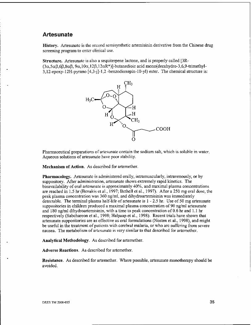
Artesunate
History. Artesunate is the second semisynthetic artemisinin derivative from the Chinese drug screening program to enter clinical use.
Structure. Artesunate is also a sequiterpene lactone, and is properly called [3R- (3a,5ccß,6ß,8aß, 9a,10a,12ß,12aR*)]-butanedioic acid mono(decahydro-3,6,9-trimethyl- 3,12-epoxy-12H-pyrano [4,3-j]-l,2 -benzodioxepin-10-yl) ester. The chemical structure is:
0
H X^CH,
COOH
Pharmaceutical preparations of artesunate contain the sodium salt, which is soluble in water. Aqueous solutions of artesunate have poor stability.
Mechanism of Action. As described for artemether.
Pharmacology. Artesunate is administered orally, intramuscularly, intravenously, or by suppository. After administration, artesunate shows extremely rapid kinetics. The bioavailability of oral artesunate is approximately 40%, and maximal plasma concentrations are reached in 1.5 hr (Benakis et al., 1997; Bethell et al., 1997). After a 250 mg oral dose, the peak plasma concentration was 360 ng/ml, and dihydroartemisinin was immediately detectable. The terminal plasma half-life of artesunate is 1 - 2.5 hr. Use of 50 mg artesunate suppositories in children produced a maximal plasma concentration of 90 ng/ml artesunate and 180 ng/ml dihydroartemisinin, with a time to peak concentration of 0.6 hr and 1.1 hr respectively (Sabchareon et al., 1998; Halpaap et al., 1998). Recent trials have shown that artesunate suppositories are as effective as oral formulations (Nosten et al., 1998), and might be useful in the treatment of patients with cerebral malaria, or who are suffering from severe nausea. The metabolism of artesunate is very similar to that described for artemether.
Analytical Methodology. As described for artemether.
Adverse Reactions. As described for artemether.
Resistance. As described for artemether. Where possible, artesunate monotherapy should be avoided.
DRES TM 2000-055 35

Quinidine
History. Quinidine is the pure D-isomer isolated from cinchona preparations (while pure quinine is the L-isomer), and is more active, more toxic, and more costly to make. Tests in vitro have shown that quinidine is 2 -3 times more potent against P. falciparum than quinine (Druilhe et al., 1988). Due to its expense and toxicity, quinidine is not frequently used for antimalarial therapy. Quinidine is also used as an antiarrhythmic agent.
Structure. Quinidine is (a-S)-a-(6-methoxy-4-quinolyl)-a-[(2S,4S,5R)-(5-vinylquinuclidin 2-yl)] methanol, and the structure is:
H3CO
The free base of quinidine is only sparingly soluble in water, but is very soluble in methanol. Salts of quinidine are soluble in 9 - 10 parts water. Commercial preparations of quinidine are hydrogen sulfate salts (Kiditard, Kinichron, Kinidin, Durules, Quiniduran), sulfate salts (Cin- Quin, Quinidex, Extentabs, Quinicardine, Quinora), gluconate salts (Duraquin, Quinaglute), or polygalacturonate salts (Cardioquin, Galactoquin, Naticardina).
Mechanism of Action. As described for quinine.
Pharmacology. Quinidine is normally given orally, although it can also be administered by intravenous infusion. The drug is well absorbed from the digestive tract, with a bioavailability of 70% (Guentert et al., 1979; Ochs et al., 1980). Maximal plasma concentrations are reached within 20-120 min, and 70 - 95% of the dose is bound to plasma proteins. The terminal plasma half-life is 5 - 12 hours and the volume of distribution is 2 - 5 L/kg (Wooding-Scott et al., 1988; Verme et al., 1992).
As is seen with quinine, 60 - 85% of the drug is converted to metabolites by the cytochrome P450 system (Rakhit et al., 1984a). The primary metabolites are 3-hydroxyquinidine, quinidine-N-oxide, and quinidine-10,11-dihydrodiol, with the bulk of the metabolites and parent compound excreted in an unconjugated form (Rakhit et al., 1984a; Rakhit et al., 1984b). The principle isoform of cytochrome P450 involved in quinidine metabolism is P450 3A4, with minor contributions from P450 2C9 (Damkier et al., 1999). Quinidine is a potent inhibitor of P450 2D6, and may its use may lead to toxic drug interactions when administered to patients taking compounds known to be metabolised by P450 2D6.
36 DRES TM 2000-055

Analytical Methodology. It can be difficult to differentiate quinidine from quinine in analytical assays, but several HPLC and gas Chromatographie methods exist which can separate the two drugs (Furner et al., 1981; Edstein et al., 1990). Methods have also been published for the HPLC separation of quinidine and its metabolites (Pershing et al., 1982).
Adverse Effects. Quinidine displays all of the common side effects seen with quinine. Howvere, quinidine has a more pronounced effect on the extension of QTc time in the electrocardiogram (Karbwang et al., 1993). This cardiac effect has been shown to be directly correlated with the plasma level of quinidine, which is the basis for using lengthy infusion times for intravenous infusion of the drug. Due to the increased cardiotoxicity, quinidine is not often used outside of the hospital for malaria therapy or for prophylaxis.
Resistance. As described for quinine.
Pyronaridine
History. Two of the earlier successes in antimalarial therapy were mepacrine (atebrine, quinacrine), as discussed above in the halofantrine section, and amodiaquine. This latter compound was one of the sister compounds to chloroquine which arose from the American antimalarial program during the second world war. Amodiaquine, like chloroquine, is a 4- aminoquinoline, and actually clears parasitemia faster than the latter drug. Unfortunately, amodiaquine caused a serious, and potentially lethal, agranulocytosis and hepatic toxicity in a small proportion of patients (Naisbitt et al., 1998). Therefore, amodiaquine was discontinued as a frontline therapy, although some countries still continue to use it.
Pyronaridine was created in 1970 in the People's Republic of China, when a compound containing part of mepacrine and part of amodiaquine was synthesized. As was the case with artemisinin, pyronaridine has been studied and used clinically in China in isolation from western drug approval processes. To this day, published data on pyronaridine pharmacokinetics, metabolism, and early clinical trials are in Chinese. The compound is effective against both chloroquine sensitive and resistant parasites (although there is some debate about this fact), and appears to be very well tolerated.
Structure. Pyronaridine is an anilinoderivative of phenanthrene with a chemical structure of:
NH
NOCH3
DRES TM 2000-055 37

Pyronaridine is used as the tetraphosphate salt.
Mechanism of Action. Pyronaridine, and compounds with a similar structure, have been shown to bind to the minor groove of DNA and interfere with the action of topoisomerase II (Chavalitshewinkoon et al., 1993). However, whether this phenomenon is relevant to the antimalarial activity has not been demonstrated. Pyronaridine treated P. falciparum have been shown to have altered morphology of the food vacuole, preventing hemoglobin degradation (Kawai et al., 1996). It is therefore suggested that pyronaridine may interfere with heme polymerisation in a manner similar to halofantrine or the 4-aminoquinolines. As yet, the exact mechanism of action of this compound is not known.
Pharmacology. Detailed kinetic and metabolism studies are only available in Chinese language journals. Similar studies have not appeared in the western scientific literature.
Analytical Methodology. Pyronaridine can be quantified by HPLC using liquid-liquid or solid-phase extraction and fluorescence or electrochemical detection (Wages et al, 1990; Jayaraman et al., 1997). The drug can be detected in plasma samples down to 10 ng/ml.
Adverse Effects. In numerous controlled trials, pyronaridine has not been associated with any serious side effects (Looareesuwan et al., 1996; Ringwald et al., 1999). In particular, the drug does not appear to cause agranulocytemia or hepatotoxicity.
Resistance. The issue of resistance to pyronaridine is unclear. The original Chinese clinical trials stated that the compound was effective against drug sensitive, chloroquine resistant, mefloquine resistant, and multidrug resistant malaria, and the drug has been used there for many years. However, introduction into Thailand was associated with an unacceptably high recrudescence rate (12 - 47%) (Looareesuwan et al., 1996). Tests of the drug in Africa have not shown similar problems with treatment failure (Ringwald et al., 1999). In vitro studies have suggested that a direct correlation exists between sensitivity to pyronaridine and sensitivity to mepacrine, amodiaquine, and chloroquine (Elueze et al., 1996; Ringwald et al., 1999). Clinical isolates of P. malariae and P. ovale have been found to be sensitive to pyronaridine (Basco and le Bras, 1994; Ringwald et al., 1997). Given the potential for treatment failure and the uncertainty over the resistance status against pyronaridine, use of the compound in monotherapy is not recommended. The Chinese have had good success with pyronaridine/pyrimethamine/sulfadoxine combinations (Shao et al., 1991), although this mixture is unlikely to be useful in areas with high resistance to Fansidar.
Atovaquone
History. In the 1940s, the laboratory of Fieser initiated the screening of a series of hydroxynaphthoquinones as antiprotozoals (Fieser et al., 1948). One of these compounds, known as lapinone, was found to be effective against P. vivax (Fawaz and Hadad, 1951), but needed to be administered via injection in large doses. Later, an improved compound, called menoctone, was tested in WHO trials, but suffered from a low oral bioavailability and rapid metabolic inactivation (Vaidya, 1998). Research on this class of compounds continued at the Wellcome Research Laboratories, ultimately yielding 566C80, which was orally active and more stable metabolically (Hudson et al., 1991). This drug, renamed atovaquone, was shown
38 ORES TM 2000-055

to be very effective against chloroquine sensitive and resistant P. falciparum in vitro, and also has demonstrated activity against Toxoplasma gondii and Pneumocystis carinii infections in AIDS patients. Clinical trials against uncomplicated malaria quickly showed that atovaquone was inadequate on its own, as it was associated with a 30% recrudescence rate (Looareesuwan et al., 1996). However, the drug is very effective when given in combination with proguanil, and is currently marketed in this format.
Structure. Atovaquone is a hydroxynaphthoquinone, and is more properly called trans-2- [4- (4-chlorophenyl)cyclohexyl]-3-hydroxy-l,4-naphthoquinone. The chemical structure is:
Atovaquone is commercially available as a 2.5:1 mixture with proguanil (Malarone).
Mechanism of Action. It was shown very early in the development of the hydroxynaphthoquinones that these compounds had an inhibitory effect on mitochondrial respiration in mammals (Wendel, 1946). It was presumed that the antimalarial action was also due to inhibition of mitochondrial respiration. However, while malaria parasites have a single mitochondrion, they do not display any respiratory activity (Fry and Beesley, 1991). Nevertheless, atovaquone was shown to inhibit parasite cytochrome c reductase with an IC50 two orders of magnitude better than for mammalian cytochrome c reductase (Fry and Pudney, 1992).
The discovery of the structure of ubiquitin (coenzyme Q), and the realisation that the hydroxynaphthoquinones were structural analogues, led to the theory that atovaquone was acting as a coenzyme Q antagonist in the parasites (Porter and Folkers, 1974). Atovaquone has been shown to inhibit ubiquinone oxidation by the mitochondrial be 1 complex, which in turn prevents the transfer of electrons to cytochrome c (Vaidya, 1998). The ultimate effect of atovaquone inhibition is the collapse of the A^Fm (mitochondrial membrane potential), which prevents pyrimidine biosynthesis and may trigger programmed cell death (Reed, 1977; Srivastava et al., 1997).
The combination of proguanil and atovaquone is actually synergistic, and it appears that this synergism is independent of proguanil's activity (via cycloguanil) against the parasite DHFR (Srivastava and Vaidya, 1999). Indeed, the biguanide nature of proguanil seems essential to its synergistic activity with atovaquone. Proguanil itself has no action on the parasite A^Fm, but significantly enhances atovaquone's ability to collapse the A^Pm. The exact mechanism by which proguanil assists atovaquone is not yet understood.
DRES TM 2000-055 39
Pharmacology. Atovaquone is normally administered orally, but has a rather low bioavailability (Haile and Flaherty, 1993). Peak plasma concentrations are reached in 6 -24 hours, with a 17 mg/kg dose giving rise to a maximal plasma concentration of 5 ug/ml (Hussein et al., 1997; Sabchareon et al., 1998). The terminal plasma half-life is 32 hr, but has also been reported to be as long as 50 - 72 hr. The volume of distribution has been measured as 25 - 30 L/kg. The drug is excreted almost entirely in the feces, and atovaquone is reported to undergo no significant metabolic alteration in humans (Haile and Flaherty, 1993).
Analytical Methodology. Atovaquone can be quantified from plasma samples using HPLC together with liquid-liquid extraction and ultraviolet spectrophotometric detection (DeAngelis et al., 1994; Hannan et al., 1996; Hansson et al., 1996). Concentrations as low as 100 ng/ml are detectable.
Adverse Effects. To date, there have been no reports of serious side effects due to atovaquone administration. Common minor effects include skin rash, gastrointestinal disturbances, and fever.
Resistance. Drug resistance is stated to arise very easily when atovaquone is used as monotherapy (Looareesuwan et al., 1996). Certainly, P.falciparum pressured in vitro with atovaquone quickly become resistant to the compound (Rathod et al., 1997). Recent studies have suggested that resistance arises from mutation in the cytochrome b gene in the parasite mitochondrion, which leads to an alteration in the binding constant for atovaquone (Srivastava and Vaidya, 1999). Most importantly, parasites made resistant to atovaquone alone are also resistant to atovaquone plus proguanil. Therefore, atovaquone use is recommended to be kept strictly limited, with no widespread, self-administered field use or prophylaxis.
Obsolete agents
There is always a remote potential of encountering local production and use of an antimalarial which is no longer recommended for use in western pharmacopias. Such compounds were superseded due to lower activity and/or higher toxicity than currently used drugs, although the occasional drug was dropped simply because of widespread shift to a new agent. Almost all obsolete drugs are structural analogues of existing chemotherapeutics, and tend to show cross- resistance with their better known counterparts. Analogues of chloroquine include amodiaquine (Camoquin, Basoquin, Flavoquin), amopyroquine (Propoquin), nivaquin (Sontochin) and cycloquine (Cyclochin, Haloquin). It should be pointed out that hydroxychloroquine (Plaquenil, Ercoquin, Quensyl) is, however, an acceptable substitute for chloroquine, although it is normally used for treating lupus. Primaquine was preceded by pamaquine (Plasmoquine, Plasmochin, Aminoquin, Praequine, Beprochine, Gamefar, Quipenyl), and plasmocid (Rhodoquine, Antimalarine). Halofantine is the modern version of mepacrine (Atebrin, Atabrine, Quinacrine).
40 ORES TM 2000-055

Experimental therapies
Almost all classes of compound made for testing against any disease or condition has also been screened against P. falciparum malaria in vitro, and many are also screened in vivo against murine malaria. Therefore, any attempt to cover the types of compound which demonstrate activity against malaria in the laboratory is futile. However, the number of potential targets in the parasite that are persistently examined is much smaller, and several of these have yielded compounds undergoing pre-clinical and clinical trial. The following are pathways which have been highlighted for concerted study: agents which block food vacuole proteases (Bailly et al., 1992), new compounds which interfere with heme polymerisation (De et al., 1997), iron chelation therapy (Gordeuk et al., 1994), compounds which interfere with lipid biosynthesis (Lauer et al., 1995), drugs which act on enzymes involved in amino acid biosynthesis (Roberts et al., 1998), and agents which bind to apicoplast targets (McConkey et al., 1997). Of agents which appear close to clinical use, we have clindamycin or azithromycin, artemether plus benflumetol, and etaquine (as an improved replacement for primaquine).
In terms of active clinical study, it is perhaps predictable that most protocols are involved with obtaining extra life out of the existing, failing chemotherapeutics, rather than investigating novel agents. A large number of papers are published every year on new dosing regimens, novel dosing types and routes, and novel drug combinations. The latter set of studies is perhaps the most interesting, and involves the search for mixtures of new antimicrobial agents with older ones (such as the anticipated benflumetal/halofantrine mixture), and also the mixture of old agents with compounds that reverse resistance. Resistance reversal is a topic that once received extremely intense attention, and then was almost completely abandoned. Chloroquine resistance is associated with a phenomenon where unrelated, non-antimicrobial, compounds can reverse the resistance phenotype (Martin et al., 1987). This feature is also often seen in resistance to anticancer agents, and is thought to be a phenomenon related to blocking of a multidrug efflux pump (Krogstad et al, 1987). While this mechanism is still controversial, compounds such as verapamil, cimetidine, cyproheptadine, desipramine, and chlorpheniramine can reverse the chloroquine resistance phenotype in vitro. However, verapamil was considered too toxic for human use, and initial trials with desipramine or cyproheptadine were disappointing (Basco and Le Bras, 1991; Warsame et al., 1992). Recently, there have been some studies that have made successful use of chloroquine plus chlorpheniramine in Nigeria (Sowunmi et al., 1997; Sowunmi et al., 1998). These trials deserve wider attention.
Recommendations for therapy
The choice for effective antimalarial therapy is determined by the resistance status of the region involved. As resistance patterns are rapidly evolving (eg: the current situation regarding Fansidar use in Africa), it is always important to find current information on recommended treatments. At the time of this article's compilation, the following resistance designations were made by the Centers for Disease Control in Atlanta, USA:
chloroquine sensitive - Algeria, Argentina, Armenia, Azerbaijan, Belize, China (except southern border with Vietnam, Laos, Myanmar and Hainan island), Costa Rica, Dominican
DRES TM 2000-055 41

Republic, Egypt, El Salvador, Guatamala, Haiti, Honduras, Iraq, Libya, Mauritius, Mexico, Morocco, Nicaragua, Panama (except east of canal zone), Paraguay, Phillipines (except Luzon, Basilian, Mindoro, Palawan, Mindanao, and the Sulu Archipelago), Saudi Arabia, South Korea, Syria, Tajikistan, Turkey, United Arab Emirates.
chloroquine resistant - Afghanistan, Angola, Bangladesh, Benin, Bhutan, Bolivia, Botswana, Brazil, Burkino Faso, Burundi, Cambodia (except western border), Cameroon, Cape Verde, Central African Republic, Chad, China (southern border areas and Hainan island), Colombia, Comoros, Congo, Congo (Zaire), Djibouti, Ecuador, Equatorial Guinea, Eritrea, Ethiopia, French Guiana, Gabon, Gambia, Ghana, Guinea, Guinea-Bissau, Guyana, India, Indonesia, Iran, Ivory Coast, Kenya, Laos, Liberia, Madagascar, Malawi, Malaysia, Mali, Mauritania, Mayotte, Mozambique, Myanmar (except eastern border), Namibia, Nepal, Niger, Nigeria, Oman, Pakistan, Panama (east of canal zone), Papua New Guinea, Peru, Philippines (Luzon, Basilian, Mindoro, Palawan, Mindanao, and the Sulu Archipelago), Rwanda, Sao Tome and Principe, Senegal, Sierra Leone, Solomon Islands, Somolia, South Africa, Sri Lanka, Sudan, Surinam, Swaziland, Tanzania, Thailand (except border regions), Togo, Uganda, Vanuatu, Venezuela, Vietnam, Yemen, Zambia, Zimbabwe.
chloroquine and mefloquine resistant - Cambodia-Thai border, Myanmar-Thai border.
Keeping in mind that resistance to compounds other than chloroquine and mefloquine occur in many parts of the world (Fansidar is now useless in Southeast Asia, Brazil, and parts of Africa), a listing of effective antimalarial regimens is shown in Table 1. Depending on what part of the world one is in, it is always wise to have a back-up (or several back-ups) therapy ready in case of drug resistance or drug intolerance. One striking example is of a patient who contracted malaria despite mefloquine prophylaxis, and required numerous rounds of treatment: halofantrine, quinine plus halofantrine, quinine plus doxycycline, halofantrine again, mefloquine plus doxycycline, and finally artesunate plus mefloquine (van Theil et al., 1993).
Recommendations for prophylaxis
The American Centers for Disease Control and Health Canada have identical guidelines on the use of antimalarials for the prevention of disease. In chloroquine sensitive areas (see above), 300 mg chloroquine base (or 310 mg hydroxychloroquine base) should be taken once per week with dosing starting one week before entering the malarious area and finishing four weeks after leaving the region. In chloroquine resistant areas, mefloquine is the drug of choice, and 250 mg base should be taken once a week, starting one week before travel and ending four weeks after travel. In areas of dual chloroquine-mefloquine resistance, or where patients are mefloquine intolerant, doxycycline should be used, and 100 mg taken daily, starting 2 days before travel and ending 4 weeks after travel. In cases where a patient is intolerant of both mefloquine and doxycycline, chloroquine should be taken as described for chloroquine sensitive regions plus 200 mg proguanil per day over the same time frame. This latter prophylactic regimen is not as effective as the recommended courses for chloroquine resistant regions.
42 ORES TM 2000-055

The issue of drug compliance and the spread of drug resistance are very important with malaria prophylaxis. Among Dutch soldiers serving in Cambodia, 16% were stricken with malaria despite good compliance with mefloquine prophylaxis (Hopperus Buma et al., 1996). Milhous (1998) also reports that 5 peace keepers died from malaria during tours through Cambodia. Brazilian peace keepers serving in Angola suffered an incidence rate of 21%, including 3 deaths, despite mefloquine prophylaxis. While there is no proof, this high incidence appears to be related to lack of compliance rather than resistance. There were 48 cases of malaria in American army troops serving in Somolia, all of which were related to a lack of compliance with mefloquine prophylaxis (Wallace et al., 1993). A further 79 cases of malaria developed after army personnel returned home, several of which were partially resistant to primaquine (Smoak et al., 1997). Amongst the marines in Somolia, 112 cases occurred (Newton et al., 1994). The incidence rate of malaria in Canadian soldiers does not appear to be publicly available. It should be pointed out, that despite the research priorities of the military, none of these soldiers were under threat of chemical or biological attack, but were under intense threat of contracting malaria.
DRES TM 2000-055 43
This page intentionally left blank.
44 DRES TM 2000-055

Table 1. Therapies for malaria (all drug amounts are for the base equivalent of a salt). All doses listed are for adults.
Drug (Common Trade Name)
Infection Type
Dose
Chloroquine
(Aralen)
Uncomplicated
Complicated**
Oral: 10 mg/kg on days 1 & 2, then 5 mg/kg on day 3.
i.V.: 10 mg/kg over 8 hr, then 15 mg/kg over 24 hr.
i.m.: 3.5 mg/kg every 6 hr for 7 doses.
Pyrimethamine
Sulfadoxine
(Fansidar)
Uncomplicated
Complicated
Oral: 75 mg pyrimethamine plus 1500 mg sulfadoxine as a single dose.
Not recommended.
Quinine
(Novaquinine)
Uncomplicated
Complicated
Oral: 500 mg 3 times a day plus 250 mg tetracycline 4 times a day for 7 days. Recommended in areas of Fansidar resistance.
Oral: 500 mg 3 times a day for 3 days plus a single dose of 75 mg pyrimethamine & 1500 mg sulfadoxine. Recommended in areas of Fansidar sensitivity.
i.v.*: 16.7 mg/kg over 4 hours (as loading dose) then 8.3 mg/kg over 4 hr 3 times daily for 7 days (switching to oral as soon as possible).
Quinidine Uncomplicated
Complicated
Not recommended
i.v.*: 15 mg/kg in a volume of 250 mL of normal saline infused over 4 hours (loading dose), followed by 7.5 mg/kg over 4 hours every 8 hours for 7 days or until oral quinine can be taken.
Mefloquine
(Lariam)
Uncomplicated
Complicated
Oral: 15 mg/kg as a single dose
Oral: 12.5 mg/kg twice at 6 hr intervals
Not recommended
Halofantrine
(Halfan)
Uncomplicated
Complicated
Oral: 500 mg every 6 hours for 3 doses. Repeat one week later.
Not recommended
Artemether Uncomplicated
Complicated
Oral: 300 mg (loading dose), followed by 100 mg daily for 4 days.
Oral: 300 mg single dose, followed by two doses of 12.5 mg/kg mefloquine (6 hours apart).
i.m.: 3.2 mg/kg (loading dose), followed by 1.6 mg/kg daily for 6 days.
i.m.: 3.2 mg/kg (loading dose), followed by 2 mg/kg daily for 4 days, and 12.5 mg/kg mefloquine twice on day 4 (6 hours apart).
DRES TM 2000-055 45

Table 1. Therapies for malaria (all drug amounts are for the base equivalent of a salt). listed are for adults.
All doses
Drug (Common Trade Name)
Infection Type
Dose
Artesunate Uncomplicated
Complicated
Oral: 300 mg (loading dose), followed by 100 mg daily for 4 days.
Oral: 300 mg single dose, followed by two doses of 12.5 mg/kg mefloquine (6 hours apart).
Oral: 5 mg/kg on the first day, 2.5 mg/kg plus 15 mg/kg mefloquine on the second day, 2.5 mg/kg plus 10 mg/kg mefloquine on the third day.
i.V.: 2.4 mg/kg (loading dose), followed by 1.2 mg/kg at 12 and 24 hours, and then 2 mg/kg daily for 6 days.
Suppository: 200 mg intrarectally at 0, 12, 24, 36, 48, and 60 hours, followed by two doses of 12.5 mg/kg oral mefloquine (6 hours apart).
Primaquine vivax & ovale Oral: 15 mg daily for 14 days
Oral: 25 - 30 mg daily for 14 days in Southeast Asia and Western Pacific
Oral: 45 mg weekly for 8 weeks in patients with known mild G6PD deficiency
Atovaquone
Proguanil
(Malarone)
Uncomplicated
Complicated
Oral: 1000 mg atovaquone plus 400 mg proguanil once daily for 3 days.
Not recommended
*Note: loading doses of quinine or quinidine should not be given to patients who have had quinine, quinidine, or mefloquine in the last 12-24 hours. It is recommended that patients with complicated malaria being given infused quinine or quinidine should also receive doxycycline (100 mg orally 2 times a day for 7 days), Fansidar (one oral dose of 75 mg/kg pyrimethamine plus 1500 mg sulfadoxine), or clindamycin (10 mg/kg i.v. followed by 5 mg/kg every 8 hr until parasitemia clears).
"Note: a case can be considered as complicated if malaria is present plus any one (or more) of the following: impaired consciousness or coma, severe normocytic anemia, renal failure, pulmonary edema, hypoglycemia, circulatory collapse or shock, spontaneous bleeding/disseminated intravascular coagulation, repeated generalized convulsions, acidemia/acidosis, hemoglobinuria, parasitemia of > 5% in non-immune individuals.
46 DRES TM 2000-055

References
1. Adedoyin, A., R.F. Frye, K. Mauro, and R.A. Branch. 1998. Chloroquine modulation of specific metabolizing emzymes activities: investigation with five drug cocktails. Br. J. Clin. Pharmacol. 46:215-219.
2. Adelusi, S.A., and L.A. Salako. 1980. Improved fiuorometric assay of chloroquine in biological samples. J. Pharm. Pharmacol. 32:711-712.
3. Adir, J. 1975. Enterohepatic circulation of tetracycline in rats. J. Pharm. Sei. 64:1847- 1850.
4. Alvan, G., L. Ekman, B. Lindstrom. 1982. Determination of chloroquine and its desethyl metabolite in plasma, red blood cells and urine by liquid chromatography. J. Chromatogr. 229:241-247.
5. Alving, A.S., J. Arnold, and D.H. Robinson. 1952. Mass therapy of subclinical malaria with primaquine. J. Amer. Med. Assoc. 149:1558-1562.
6. Andersen, S.L., A.J. Oloo, D.M. Gordon, O.B. Ragama, G.M. Aleman, J.D. Berman, D.B. Tang, M.W. Dunne, and G.D. Shanks. 1998. Successful double-blinded, randomized, placebo-controlled field trial of azithromycin and doxycycline as prohpylaxis for malaria in western Kenya. Clin. Infect. Dis. 26:146-150.
7. Asahi, H., T. Kanazawa, Y. Kajihara, K. Takahashi, and T. Takahashi. 1996. Hypoxanthine: a low molecular weight factor essential for growth of erythrocytic Plasmodium falciparum in a serum-free medium. Parasitology 113:19-23.
8. Asawamahasakda, W., I. Ittarat, C.C. Chang, P. McElroy, and S.R. Meshnick. 1994. Reaction of malarial emdoperoxides with specific parasite proteins. Mol. Biochem. Parasitol. 67:183-191.
9. Astier, H., C. Renard, V. Cheminel, O. Soares, C. Mounier, F. Peyron, and J.F. Chaulet. 1997. Simultaneous determination of pyrimethamine and sulphadoxine in human plasma by high-performance liquid chromatography after automated liquid-solid extraction. J. Chromatogr. 5 698:217-223.
10. Augustijns, P., L. Peeters, K. Croes, and R.K. Verbeeck. 1999. Stereoselective de- ethylation of chloroquine in rat liver microsomes. Eur. J. Drug Metab. Pharmacokinetics 24:105-108.
11. Ayala, F.J., A.E. Escalante, A.A. Lai, and S.M. Rich. 1998. Evolutionary relationships of human malaria parasites, p. 285-300. In I.W. Sherman (ed.), Malaria: Parasite Biology, Pathogenesis, and Protection. ASM Press, Washington, DC.
DRES TM 2000-055 47

12. Babalola, C.P., O.O. Bolaji, F.A. Ogunbona, A. Sowunmi, and O. Walker. 1998. Pharmacokinetics of quinine in African patients with acute falciparum malaria. Pharm. World Sei. 20:118-122.
13. Bailly, E., R. Jambou, J. Savel, and G. Jaureguiberry. 1992. Plasmodium falciparum: differential sensitivity in vitro to E-64 (cysteine protease inhibitor) and pepstatin A (aspartyl protease inhibitor). J. Protozool. 39:547-571.
14. Baird, J.K., H. Basri, B. Subianto, DJ. Fryauff, P.D. McElroy, B. Leksana, T.L. Richie, S. Masbar, F.S. Wignall, and S.L. Hoffman. 1995. Treatment of chloroquine-resistant Plasmodium vivax with chloroquine and primaquine or halofantrine. J. Infect. Dis. 171:1678-1682.
15. Baker, J.K., J.D. McChesney, CD. Hufford, and A.M. Clark. 1982. High-performance liquid Chromatographie analysis of the metabolism of primaquine and the identification of a new mammalian metabolite. J. Chromatogr. 230:69-77.
16. Bangehang, K.N., J. Karbwang, and D.J. Back. 1992. Mefloquine metabolism by human liver microsomes. Effect of other antimalarial drugs. Biochem. Pharmacol. 43:1957-1961.
17. Barennes, H., F. Kahiatani, E. Pussard, F. Clavier, D. Meynard, S. Njifountawouo, and F. Verdier. 1995. Intrarectal Quinimax (an association of Cinchona alkaloids) for the treatment of Plasmodium falciparum malaria in children in Niger: efficacy and pharmacokinetics. Trans. R. Soc. Trop. Med. Hyg. 89:418-421.
18. Barennes, H., E. Pussard, A. Mahaman Sani, F. Clavier, F. Kahiatani, G. Granic, D. Henzel, L.Ravient, and F. Verdier. 1996. Efficacy and pharmacokinetics of a new intrarectal quinine formulation in children with Plasmodium falciparum malaria. Br. J. Clin. Pharmacol. 41:389-395.
19. Barnwell, J.W., and M.R. Galinski. 1998. Invasion of vertebrate cells: erythrocytes, p. 93-120. In I.W. Sherman (ed.), Malaria: Parasite Biology, Pathogenesis, and Protection. ASM Press, Washington, DC.
20. Barza, M., and R.T. Scheife. 1977. Antimicrobial spectrum, pharmacology, and therapeutic uses of antibiotics. J. Maine Med. Assoc. 68:194-210.
21. Basco, L.K., J. Le Bras, C. Gillotm, P. Ringwald, E. Rabenjarson, F. Gimenez, O. Bouchard, R. Farinotti, and J.P. Coulaud. 1991. Type RI resistance to halofantrine in West Africa. Trop. Med. Parasitol. 42:413-414.
22. Basco, L.K., and J. le Bras. 1994. In vitro susceptibility of Cambodian isolates of Plasmodium falciparum to halofantrine, pyronaridine and artemisinin derivatives. Ann. Trop. Med. Parasitol. 88:137-144.
48 DRES TM 2000-055
23. Basco, L.K., and J. LeBras. 1991. Reversal of chloroquine resistance with cyproheptadine in 'wild' strains of Plasmodium falciparum. Trans. R. Soc. Trop. Med. Hyg. 85:204-205.
24. Bateman, D.N., and E.H. Dyson. 1986. Quinine toxicity. Adverse Drug React. Acute Poisoning Rev. 5:216-233.
25. Bates, M.D., S.R. Meshnick, C.I. Sigler, P. Leland, and M.R. Hollingdale. 1990. In vitro effects of primaquine and primaquine metabolites on exoerythrocytic stages of Plasmodium berghei. Am. J. Trop. Med. Hyg. 42:532-537.
26. Baty, J.D., D.A. Price Evans, and P.A. Robinson. 1975. The identification of 6- methoxy-8-aminoquinoline as a metabolite of primaquine in man. Biomed. Mass Spectrom 2:304-306.
27. Baune, B., J.P. Flinois, V. Furlan, F. Gimenez, A.M. Taburet, L. Becquemont, and R. Farinotti. 1999. Halofantrine metabolism in microsomes in man: major role of CYP 3A4 and CYP 3A5. /. Pharm. Pharmacol. 51:419-426.
28. Baune, B., V. Furlan, A.M. Taburet, and R. Farinotti. 1999. Effect of selected antimalarial drugs and inhibitors of cytochrome P-450 3A4 on halofantrine metabolism by human liver microsomes. Drug Metab. Dispos. 27:565-568.
29. Beales, P.E. 1999. The epidemiology of malaria, p. 121-167. In H.M. Gilles (ed.), Protozoal Diseases. Arnold, London.
30. Benakis, A., M. paris, L. Loutan, C.T. Plessas, and S.T. Plessas. 1997. Pharmacokinetics of artemisinin and artesunate after oral administration in healthy volunteers. Am. J. Trop. Med. Hyg. 56:12-23.
31. Bendrat, K., B J. Berger, and A. Cerami. 1995. Haem polymerization in malaria. Ataure378:138.
32. Berger, B.J., J. Martiney, A.F.G. Slater, A.H. Fairlamb, and A. Cerami. 1995. Chloroquine resistance is not associated with drug metabolism in Plasmodium falciparum. J. Parasitol. 81:1004-1008.
33. Bergqvist, Y., U. Hellgren, and F.C. Churchill. 1988. High-performance liquid Chromatographie assay for the simultaneous monitoring of mefloquine and its acid metabolite in biological samples using protein precipitation and ion-pair extraction. J. Chromatogr. 432:253-263.
34. Bergqvist, Y., L. Funding, B. Krysen, T. Leek, and K. Yvell. 1998. Improved validated assay for the determination of proguanil and its metabolites in plasma, whole blood, and urine using solid-phase extraction and high-performance liquid chromatography. Ther. DrugMonit. 20:325-330.
DRES TM 2000-055 49

35. Bergqvist, Y., and M. Ericsson. 1985. Simultaneous determination of pyrimethamine and sulphadoxine in human plasma by high-performance liquid chromatography. Trans. R. Soc. Trop. Med. Hyg. 79:297-301.
36. Bergqvist, Y., E. Hjelm, and L. Rombo. 1987. Sulfadoxine assay using capillary blood samples dried on filter paper - suitable for monitoring of blood concentrations in the field. Ther. Drug. Monit. 9:203-207.
37. Bergqvist, Y., S. Eckerbom, H. Larsson, and M. Malekzadeh. 1991. Reversed-phase liquid Chromatographie method for the simultaneous determination of the antimalarial drugs sulfadoxine, pyrimethamine, mefloquine, and its major carboxylic metabolite in plasma. J. Chromatogr. 571:169-177.
38. Bethell, D.B., P. Teja-Isavadharm, X.T. Cao, T.T. Pham, T.T. Ta, T.N. Tran, T.T. Nguyen, T.P. Pham, D. Kyle, N.P. Day, and N.J. White. Pharmacokinetics of oral artesunate in children with moderately severe Plasmodium falciparum malaria. Trans. R. Soc. Trop. Med. Hyg. 91:195-198.
39. Blauer, G. 1988. Interaction of ferriprotoporphyrin IX with the antimalarials amodiaquine and halofantrine. Biochem. Int. 17:729-734.
40. Bocker, R. 1983. Analysis and quantitation of a metabolite of doxycycline in mice, rats, and humans by high-performance liquid chromatography. J. Chromatogr. 274:255-262.
41. Bowman. S., D. Lawson, D. Basham, D. Brown, T. Chillingworth et al. 1999. The complete nucleotide sequence of chromosome 3 of Plasmodium falciparum. Nature 400:532-538.
42. Brasseur, P., P. Bitsindou, R.S. Moyou, T.A. Eggelte, G. Samba, L. Penchenier, and P. Druilhe. 1993. Fast emergence of Plasmodium falciparum resistance to halofantrine. Lancet 341:901-902.
43. Bray, P.G., O. Janneh, K.J. Raynes, M. Mungthin, H. Ginsburg, and S.A. Ward. 1999. Cellular uptake of chloroquine is dependent on binding to ferriprotoporphyrin IX and is independent ofNHE activity in Plasmodium falciparum. J. Cell Biol. 145:363-376.
44. Breckenridge, A., D.J. Back, I.G. Edwards, G. Mihaly, M. Orme, H. Purba, and S. Ward. 1987. The clinical and biochemical pharmacology of primaquine, p. 65-76. In W.H. Wernsdorfer and P.I. Trigg (eds.) Primaquine: pharmacokinetics, metabolism, toxicity and activity. John Wiley, New York.
45. Brewer, T.G., S.J. Grate, J.O. Peggins, P.J. Weina, J.M. Petras, B.S. Levine, M.H. Heiffer, and B.G. Schuster. 1994. Fatal neurotoxicity of arteether and artemether. Am. J. Trop. Med. Hyg. 51:251-259.
46. Brocks, D.R., M.J. Dennis, and W.H. Schaefer. 1995. A liquid Chromatographie assay for the stereospecific quantitative analysis of halofantrine in human plasma. J. Pharm. Biomed.Anal. 13:911-918.
50 DRES TM 2000-055

47. Bruce-Chwatt, L.J. 1988. History of malaria from prehistory to eradication, p. 1-59. In W. Wernsdorfer and I. McGregor (eds.), Malaria. Principles and Practice of Malariology, vol 1. Churchill Livingstone, Edinburgh.
48. Bunnag, D., J. Karbwang, A. Thanavibul, S. Chittamas, Y. Ratanapongse, K. Chalermrut, K.N. Bangchang, and T. Harinasuta. 1994. High dose of primaquine in primaquine resistant vivax malaria. Trans. R. Soc. Trop. Med. Hyg. 88:218-219.
49. Bunnag, D., J. Karbwang, and T. Harinasuta. 1992. Artemether in the treatment of multiple drug resistant falciaprum malaria. Southeast Asian J. Trop. Med. Public HEalth 23:762-767.
50. Bunnag, D., T. Kanda, J. Karbwang, K. Thimasam, S. Pungpak, and T. Harinasuta. 1995. Artemether-mefloquine combination in multidrug resistant falciparum malaria. Trans. R. Soc. Trop. Med. Hyg. 89:213-215.
51. Bygbjerg, I., P. Ravn, A. Ronn, H. Flachs, and F.F. Hvidberg. 1987. Human pharmacokinetics of proguanil and its metabolites. Trop. Med. Parasitol. 38:77-80.
52. Carrington, H.C., A.F. Crowther, D.G. Davey, A.A. Levi, and F.L. Rose. 1951. Metabolite of'paludrine' with high antimicrobial activity. Nature 168:1080-1082.
53. Centers for Disease Control. 2000. Regional malaria information. www.cdc.gov/travel/regionalmalaria/index.htm.
54. Chau, T.T.H., N.P. Day, L.V. Chuong, H.N.T. Mai, P.P. Loc, N.H. Phu, D.B. Bethell,, D.X. Sinh, T.T. Hien, and N.J. White. 1996. Blackwater fever in southern Vietnam: a prospective descriptive study of 50 cases. Clin. Infect. Dis. 23:1274-1281.
55. Chavalitshewinkoon, P., P. Wilairat, S.A. Gamage, W.A. Denny, D.P. Figgit, and R.K. Ralph. 1993. Structure-activity relationships and modes of action of 9-anilinoacridines against chloroquine-resistantPlasmodium falciparum in vitro. Antimicrob. Ag. Chemother. 37:403-406.
56. Cheng, K.N., L.F. Elsom, and D.R. Hawkins. 1992. Identification of metabolites of halofantrine, a new candidate anti-malarial drug, by gas-chromatography-mass spectrometry. J. Chromatogr. 581:203-211.
57. China Cooperative Research Group on Qinhaousu and its Derivatives as Antimalarials. 1982a. Chemical studies on qinghaosu (artemisinine). J. Trad. Chin. Med. 2:3-8.
58. China Cooperative Research Group on Qinhaousu and its Derivatives as Antimalarials. 1982b. The chemistry and synthesis of qinhaousu derivatives. J. Trad. Chin. Med. 2:9- 16.
59. Chou, A.C., and CD. Fitch. 1993. Control of heme polymerase by chloroquine and other quinoline derivatives. Biochem. Biophys. Res. Comm. 195:422-427.
DRES TM 2000-055 51

60. Chou, A.C., R. Chevli, and CD. Fitch. 1980. Fernprotoporphyrin IX fulfills the criteria for identification as the chloroquine receptor of malaria parasites. Biochemistry 19:1543-1549.
61. Cohen, S., and G.A. Buthcer. 1971. Serum antibody in acquired malarial immunity. Trans. Roy. Soc. Trop. Med. Hyg. 65:125-128.
62. Coleman, M.D., G.W. Mihaly, S.A. ward, G. Edwards, R.E. Howells, and A.M. Breckenridge. 1985. The disposition of pyrimethamine in the isolated prefused rat liver. Biochem. Pharmacol. 34:2193-2197.
63. Collins, W.E., and G.M. Price. 1996. Primaquine resistance in Plasmodium vivax. Am. J. Trop. Med. Hyg. 55:243-249.
64. Colwell, E.J., R.L. Hickman, R. Intraprasert, and C. Tirabutana. 1972. Minocycline and tetracycline treatment of acute falciparum malaria in Thailand. Am. J. Trop. Med. Hyg. 21:144-149.
65. Colwell, E.J., R.L. Hickman, and S. Kosakal. 1972. Tetracycline treatment of chloroquine- resistant falciparum malaria in Thailand. J. Amer. Med. Assoc. 220:684- 686.
66. Cowman, A.F., D. Galatis, and J.K. Thompson. 1994. Selection for mefloquine resistance in Plasmodium falciparum is linked to amplification of the pfmdrl gene and cross-resistance to halofantrine and quinine. Proc. Natl. Acad. Sei. USA 91:1143-1147.
67. Curley, G.P., S.M. O'Donovan, J. McNally, M. Mullally, H. O'Hara, A. Troy, S.A. O'Callaghan, and J.P. Dalton. 1994. Aminopeptidases from Plasmodium falciparum, Plasmodium chabaudi, and Plasmodium berghei. J. Euk. Microbiol. 41:119-123.
68. Damkier, P., L.L. Hansen, and K. Brosen. 1999. Effect of diclofenac, disulfiram, itraconazloe, grapefruit juice and erythromycin on the pharmacokinetics of quinidine. Br. J. CLin. Pharmacol. 48:829-838.
69. Davis, T.M.E., S. Pukrittayakamee, W. Supunaranond, S. Looareesuwon, S. Krishna, B. Nagachinta, R.C. Turner, and N.J. White. 1990. Glucose metabolism in quinine-treated patients with uncomplicated falciparum malaria. Clin. Endocrinol. 33:739-749.
70. De, D., L.D. Byers, and D.J. Krogstad. 1997. Antimalarials: synthesis of 4- aminoquinolines that circumvent drug resistance in malaria. J. Heterocycl. Chem. 34:315-320.
71. DeAngelis, R.L., W.S. Simmons, and C.A. Nichol. 1975. Quantitative thin-layer chromatography of pyrimethamine and related diaminopyrimidines in body fluids. J. Chromatogr. 106:41-49.
52 DRES TM 2000-055
72. DeAngelis, D.V., J.D. Long, L.L. Kanics. and J.L. Woolley. 1994. High-performance liquid Chromatographie assay for the measurement of atovaquone in plasma. J. Chromatogr. 652:211-219.
73. Deans, J.A., A.W. Thomas, P.M. Inge, and S. Cohen. 1983. Stage-specific protein synthesis by asexual blood parasites of Plasmodium falciparum. Mol Biochem. Parasitol. 8:45-51.
74. Demers, P., D. Fräser, R.B. Goldbloom, J.C. Haworth, J. LaRochelle, R. MacLean, and T.K. Murray. 1968. Effects of tetracyclines on skeletal growth and dentition. A report by the Nutrition Committee of the Canadian Paediarric Society. Can. Med. Assoc. J. 99:849-854.
75. Domagk, G. 1935. Eine neue Klasse von Desininfektionsmitten. Deut. Med. Wochenschr. 61:829-832.
76. Dorn, A., R. Stoffel, H. Matile, A. Bubendorf, and R.G. Ridley. 1995. Malarial haemozoin/ß-hematin supports haem polymerization in the absence of protein. Nature 374:269-271.
77. Druilhe, P., O. Brandicourt, T. Chongsuphajaisiddhi, and J. Berthe. 1988. Activity of a combination of three cinchona bark alkaloiods against Plasmoidum falciparum in vitro. Antimicrob. Ag. Chemother. 32:250-254.
78. Dua, V.K., R. Sarin, and A. Prakash. 1993. Determination of quinine in serum, plasma, red blood cells and whole blood in healthy and Plasmodium falciparum malaria cases by high-performance liquid chromatography. J. Chromatogr. 614:87-93.
79. Ducharme, J., and R. Farinotti. 1996. Clinical pharmacokinetics and metabolism of chloroquine. Focus on recent developments. Clin. Pharmacokinetics 31:257-21'4.
80. Edstein, M.D., J.R. Veenendaal, H.V. Scott, and R.K. Rieckmann. 1988. Steady-state kinetics of proguanil and its active metabolite, cycloguanil, in man. Chemother. 34:385-392.
81. Edstein, M.D., A. Prasitthipayong, A. Abchareon, T. Chongsuphajaisiddhi, and H.K. Webster. 1990. Simultaneous measurement of quinine and quinidine in human plasma, whole blood, and erythrocytes by high-performance liquid chromatography with fluorescence detection. Ther. DrugMonit. 12:493-500.
82. Edwards, G., S. Looareesuwan, AJ. Davies, Y. Wattanagoon, R.E. Phillips, D.A. Warrell. 1988. Pharmacokinetics of chloroquine in Thais: plasma and red-cell concentrations following an intravenous infusion to healthy subjects with Plasmodium vivax malaria. Br. J. Clin. Pharmacol. 22:477-485.
83. Elueze, E.I., S.L. Croft, and D.C. Warhurst. 1996. Activity of pyronaridine and mepacrine against twelve strains of Plasmodium falciparum in vitro. J. Antimicrob. Chemother. 37:511-518.
DRES TM 2000-055 53

84. Ericsson, O., M. Friden, U. Hellgren, and L.L. Gustafsson. 1993. Reversed-phase high- performance liquid chromatography determination of quinine in plasma, whole blood, urine, and samples dried on filter paper. Ther. Drug Monit. 15:334-337.
85. Escande, C, P. Chevalier, F. Verdier, and R. Bourdon. 1990. Sensitive radioimmunoassay and enzyme-linked immunosorbent assay for the simultaneous determination of chloroquine and its metabolites in biological fluids. J. Pharm. Sei. 79:23-27.
86. Ezzet, F., R. Mull, and J. Karbwang. 1998. Population pharmacokinetics and therapeutic response of CGP56697 (artemether + benflumetol) in malaria patients. Br. J. Clin. Pharmacol. 46:553-561.
87. Fawaz, G., and F.S. Haddad. 1951. The effect of lapinone (M-2350) on P. vivax infection in man. Am. J. Trop. Med. Hyg. 31:569-571.
88. Ferone, R. 1977. Folate metabolism in malaria. Bull. WHO 55:291-298.
89. Ferone, R., J.J. Burchill, and G.H. Hitchings. 1969. Plasmodium berghei dihydrofolate reductase. Isolation, properties, and inhibition by antifolates. Mol. Pharmacol. 5:49-59.
90. Ferrari, V., and D.J. Cutler. 1991. Simulation of kinetic data on the influx and efflux of chloroquine by erythrocytes infected with Plasmodium falciparum. Biochem. Pharmacol. 42:5167-5179.
91. Fieser, L.F., E. Berliner, F.J. Bondhus, F.C. Chang, W.G. Dauben, et al. 1948. Naphthoquinone antimalarials. I-XVII. J. Am. Chem. Soc. 70:3151-3244.
92. Fitch, CD., R. Chevli, H.S. Banyal, G. Phillips, M.A. Pfeiler, and D.J. Krogstad. 1982. Lysis of Plasmodium falciparum by ferriprotoporphyrin IX and a chloroquine- ferriprotoporphyrin IX complex. Antimicrob. Ag. Chemother. 21:819-822.
93. Fletcher, K.A., P.F. Barton, and J.A. Kelly. 1988. Studies on the mechanisms of oxidation in the erythrocyte by metabolites of primaquine. Biochem. Pharmacol. 37:2683-2690.
94. Fontanet, A.L., B.D. Johnston, A.M. Walker, Y. Bergqvist, U. Hellgren, and W. Rooney. 1994. Falciparum malaria in eastern Thailand: a randomized trial of the efficacy of a single dose of mefloquine. Bull. WHO 72:73-81.
95. Foote, S.J., D.E. Kyle, R.K. Martin, A.M. Oduola, K. Forsyth, D.J. Kemp, and A.F. Cowman. 1990. Several alleles of the multidrug-resistance gene are closely linked to chloroquine resistance in Plasmodium falciparum. Nature 345:255-258.
96. Forti, G, and C. Benincori. 1969. Doxycycline and the teeth. Lancet 1:782.
97. Francis, S.E., I.Y. Gluzman, A. Oksman, A. Knickerbocker, R. Mueller, M.L. Bryant, D.R. Sherman, D.G. Russell, and D.E. Goldberg. 1994. Molecular characterization and inhibition of & Plasmodium falciparum aspartic hemoglobinase. EMBOJ. 13:306-317.
54 DRES TM 2000-055
98. Fry, M., and J.E. Beesley. 1991. Mitochondria of mammalian Plasmodium species. Parasitology 102:17-26.
99. Fry, M., and M. Pudney. 1992. Site of action of the antimalarial hydroxynaphthoquinone, 2-[trans-4-(4'-chlorophenyl)cyclohexyl]-3-hydroxy-l,4- naphthoquinone (566C80). Biochem. Pharmacol. 43:1545-1553.
100. Furner, R.L., G.B. Brown, and J.W. Scott. 1981. A method for differentiation and analysis of quinine and quinidine by gas chromatography/mass spectrometry. J. Anal. Toxicol. 5:275-278.
101. Garcia, L.S., and D.A. Bruckner. 1997. Diagnostic medical parasitology, 3rd ed.. ASM Press, Washington, DC, USA.
102. Gardner, MJ, H. Tettelin, DJ. Carucci, L.M. Cummings, L. Aravind et al. 1998. Chromosome 2 sequence of the human malaria parasite Plasmodium falciparum. Science 282:1126-1132.
103. Garrison, P.L., D.D. Hankey, W.G. Coker, W.N. Donovan, B. Jastremski, et al. 1952. Status of primaquine: cure of Korean vivax malaria with pamaquine and primaquine. J. Amer. Med. Assoc. 149:1562-1563.
104. Giboda, M., and M.B. Denis. 1988. Response of Kampuchean strains of Plasmodium falciparum to antimalarials: in-vivo assessment of quinine and quinine plus tetracycline; multiple drug resistance in vitro. J. Trop. Med. Hyg. 91:205-211.
105. Ginsburg, H., and K. Kirk. 1998. Membrane transport in the malaria-infected erythrocyte, p. 219-232. In, I.W. Sherman (ed.) Malaria. Parasite biology, pathogenesis, and protection. ASM Press, Washington, DC.
106. Goldberg, D.E., A.F.G. Slater, R. Beavis, B. Chait, A. Cerami, and G.B. Henderson. 1991. Hemoglobin degradation in the human malaria pathogen Plasmodium falciparum: a catabolic pathway initiated by a specific aspartic protease. J. Exp. Med. 173:961-969.
107. Goldberg, D.E., A.F.G. Slater, A. Cerami, and G.B. Henderson. 1990. Hemoglobin degradation in the malaria parasite Plasmodium falciparum: an ordered process in a unique organelle. Proc. Natl. Acad. Sei. USA 87:2931-2935.
108. Golenda, C, and R. Rosenberg. 1997. Continuous in vitro propagation of the malaria parasite Plasmodium vivax. Proc. Natl. Acad. Sei. USA 94:6786-6791.
109. Golgi, C. 1886. Sull'infezione malarica. Arch. Sei. Med. 10:109-135.
110. Gordeuk, V.P., P.E. Thuma, and G.M. Brittenham. 1994. Iron chelation therapy for malaria. Adv. Exp. Med. Biol. 356:371-383.
111. Gorichon, E., C. Martin, K.N. Bangchang, J. Karbwang, A. Thullier, R. Farinotti, and F. Gimenez. 1998. Chiral Chromatographie method to determine the enantiomers of
DRES TM 2000-055 55

halofantrine and its main chiral desbutyl meatbolite in erythrocytes. J. Chromatogr. B 712:259-262.
112. Gramiccia, G. 1987. Quinine: should the past be taken as a guidance for the future? Acta Leiden. 55:15-20.
113. Grassi, B. 1990. Studi di un zoologo sulla malaria. Rome, Italy.
114. Green, M.D., Y. Bergqvist, D.L. Mount, S. Corbett, M.J. D'Souza. 1999. Improved validated assay for the determination of mefloquine and its carboxy metabolite in plasma, serum and whole blood using solid-phase extraction and high-performance liquid chromatography. J. Chromatogr. B 727:159-165.
115. Greenwood, D. 1995. Conflicts of interest: the genesis of synthetic antimalarial agents in peace and war. J. Antimicrob. Chemother. 36:857-872.
116. Guentert, T.W., N.H. Holford, P.E. Coates, R.A. Upton, and S. Riegelman. 1979. Quinidine pharmacokinetics in man: choice of a disposition model and absolute bioavailability studies. J. Pharmacokinet. Biopharm. 7:315-330.
117. Gushimana, Y., B. Doepner, E. Martinez-Hackert, and G. IIgenfritz. 1993. Kinetics of quinine-deuterohemin binding. Biophys. Chem. 47:153-162.
118. Gustafsson, L.L., L. Rombo, G. Alvan, A. Bjorkman, M. Lind, and O. Walker. 1983. On the question of dose-dependent chloroquine elimination of a single dose. Clin. Pharmacol. Ther. 34:383-385.
119. Gustafsson, L.L., B. Lindstrom, A. Grahnen, and G. Alven. 1987. Chloroquine excretion following malaria prophylaxis. Br. J. Clin. Pharmacol. 24:221-224.
120. Haue, L.G., and J.F.Flaherty. 1993. Atovaquone: a review. Ann. Pharmacother. 27:1488-1494.
121. Halpaap, B., M. Ndjave, M. Paris, A. Benakis, and P.G. Kremsner. 1998. Plasma levels of artesunate and dihydroartemisinin in children with Plasmodium falciparum malaria in Gabon after administration of 50-milligram artesunate suppositories. Am. J. Trop. Med. Hyg. 58:365-368.
122. Hannan, S.L., G.A. Ridout, and A.E. Jones. 1996. Determination of the potent antiprotozoal compound atovaquone in plasma using liquid-liquid extraction followed by reversed-phase high-performance liquid chromatography with ultraviolet detection. J. CHromatogr. B 678:297-302.
123. Hansson, A.G., S. Mitchell, P. Jatlow, and P.M. Rainey. 1996. Rapid high-performance liquid Chromatographie assay for atovaquone. J. Chromatogr. B 675:180-182.
124. Harinasuta, T., S. Migasena, and D. Bunnag. 1961. Chloroquine resistance in Plasmodium falciparum in Thailand. Proc. 1st UNESCO Reg. Symp.
56 DRES TM 2000-055

125. Health Canada. 1999. Malaria. Canada Communicable Disease Report 25:Mzrch 15.
126. Heath Organisation of the League of Nations. 1933. The therapeutics of malaria. Third general report of the malaria comission. Quart. Bull. Health Org. League of Nations 2:181-228.
127. Hellgren, U., G. Alvan, and M. Jerling. 1993. On the question of interindividual variations in chloroquine concentrations. Eur. J. Clin. Pharmacol. 45:383-385.
128. Helsby, N.A., G. Edwards, A.M. Breckenridge, and S.A. Ward. 1993. The multiple dose pharmacokinetics ofproguanil. Br. J. Clin. Pharmacol. 35:653-656.
129. Helsby, N.A., S.A. Ward, G. Edwards, R.E. Howells, and A.M. Bereckenridge. 1990. The pharmacokinetics and activation ofproguanil in man: consequences of variability in drug metabolism. Br. J. Clin. Pharmacol. 30:593-598.
130. Hiebsch, R.R., TJ. Raub, and B.W. Wattenberg. 1991. Primaquine blocks transport by inhibiting the formation of functional transport vesicles. Studies in a cell-free assay of protein transport through the Golgi apparatus. J. Biol. Chem. 266:20323-20328.
131. Hong, Y.L., Y.Z. Yang, and S.R. Meshnick. 1994. The interaction of artemisinin with malarial hemozoin. Mol. Biochem. Parasitol. 63:121-128.
132. Hopperus Buma, A.P., P.P. van Theil, H.O. Lobel, C. Ohrt, E.J. van Ameijden, R.L. Veltink,D.C. Tendeloo, T. van Gool, M.D. Green, G.D. Todd, D.E. Kyle, and P.A. Kager. 1996. Long-term malaria chemoprophylaxis with mefloquine in DUtch marines in Cambodia. J. Lab. Clin. Med. 127:574-582.
133. Hudson, A.T., M. Dickens, CD. Ginger, W.E. Gutteridge, T. Holdich, D.B.A. Hutchinson, M. Pudney, A.W. Randall, and V.S. Latter. 1991. 566C80: a broad spectrum anti-infective agent with activity against malaria and opportunistic infections in AIDS patients. Drugs Exp. Clin. Res. 17:427-435.
134. Humberstone, A.J., C.J. Porter, and W.N. Charman. 1996. A physiochemical basis for the effect of food on the absolute oral bioavailability of halofantrine. J. Pharm. Sei. 85:525-529.
135. Hussein, Z., J. Eaves, D.B. Hutchinson, and C.J. Canfield. 1997. Population pharmacokinetics of atovaquone in patients with acute malaria caused by Plasmodium falciparum. Clin. Pharmacol. Ther. 61:518-530.
136. Inselburg, J. 1985. Induction and isolation of artemisinin-resistant mutants of Plasmodium falciparum. Am. J. Trop. Med. Hyg. 34:417-418.
137. Jacobs, G.H., A.M. Oduola, D.E. Kyle, W.K. Milhous, S.K. Martin, and M. Aikawa. 1988. Ultrastructural study of the effects of chloroquine and verapamil on Plasmodium
falciparum. Am. J. Trop. Med. Hyg. 39:15-20.
DRES TM 2000-055 57

138. Jayaraman, S.D., S. Ismail, N.K. Nair, and V. Navaratnam. 1997. Determination of pyronaridine in blood plasma by high-performance liquid chromatography for application in clinical pharmacological studies. J. Chromatogr. B 690:253-257.
139. Jelinek, T., P. Schelbert, T. Loscher, and D. Eichenlaub. 1995. Quinine resistant falciparum malaria acquired in east Africa. Trop. Med. Parasitol. 46:38-40.
140. Jomaa, H., J. Wiesner, S. Sanderbrand, B. Altincicek, C. Weidemeyer, M. Hinton, I Turbachova, M. Ebert, J. Zeidler, H.K. Lichtenthaler, D. Soldati, and E. Beck. 1999. Inhibitors of the nonmevalonate isoprenoid biosynthesis as antimalarial drugs. Science 285:1573-1576.
141. Karawya, M.S., and A.M. Diab. 1977. Colorimetric assay of quinine and quinidine in raw materials, formulations, and biological fluids. J. Pharm. Sei. 66:1317-1319.
142. Karbwang, J., T.M. Davis, S. Looareesuwan, P. Molunto, D. Bunnag, and N.J. White. 1993. A comparison of quinine and quinidine in healthy Thai males. Br. J. Clin. Pharmacol. 35:265-271.
143. Karbwang, J., and N.J. White. 1990. Clinical pharmacokinetics of mefloquine. Clin. Pharmacokinetics 19:264-279.
144. Karbwang, J., and K. Na Bangchang. 1994. Clinical pharmacokinetics of halofantrine. Clin. Pharmackinetics 27:104-119.
145. Karbwang, J., K. Na Bangchang, A. Thanavibul, D. Bunnag, T. Chongsuphajaisiddhi, and T. Harinasuta. 1992. Comparison of oral artemether and mefloquine in acute uncomplicated falciparum malaria. Lancet 340:1245-1248.
146. Karbwang, J., K. Na Bangchang, Y. Wattanagoon, A. Thanavibul, and T. Harinasuta. 1994. Artemether 5 versus 7-day regimen for severe falciparum malaria. Southeast Asian J. Trop. Med. Public Health 25:702-706.
147. Karbwang, J., K. Na Bangchang, K. Congpuong, P. Molunto, and A. Thanavibul. 1997. Pharmacokinetics and bioavailability of oral and intramuscular artemether. Eur. J. Clin. Pharmacol. 52:307-310.
148. Karbwang, J., K. Na-Bangchang, and H.M. Gilles. 1999. Treatment and management, p. 106-122. In H.M. Gilles (ed.) Protozoa! Diseases. Arnold, London.
149. Karbwang, J., K. Na-Bangchang, P. Molunto, V. Banmairuroi, and K. Congpuong. 1997. Determination of artemether and its major metabolite, dihydroartemisinin, in plasma using high performance liquid chromatography with electrochemical detection. J. Chromatogr. B 690:259-265.
150. Kawai, S., S. Kano, C. Chang, and M. Suzuki. 1996. The effects of pyronaridine on the morphology of Plasmodium falciparum in Aotus trivirgatus. Am. J. Trop. Med. Hyg. 55:223-229.
58 ORES TM 2000-055

151. Ketrangsee, S., S. Vijayakadya, P. Yamokgul, S. Jatapadma, K. Thimasarn, and W. Rooney. 1992. Comparative trial on the response of Plasmodium falciparum to halofantrine and mefloquine in Trat Province, Eastern Thailand. Southeast. Asian J. Trop. Med. Pub. Health 23:51-54.
152. Kiatfuengfoo, R., T. Suthiphongchai, P. Prapunwatta, and Y. Yuthavong. 1989. Mitochondria as the site of action of tetracycline on Plasmodium falciparum. Mol. Biochem. Parasitol. 34:109-115.
153. Kokwaro, CO., A.P. Glazier, S.A. Ward, A.M. Breckenridge, and G. Edwards. 1993. Effect of malaria infection and endotoxin-induced fever on phenacetin O-deethylation by rat liver microsomes. Biochem. Pharmacol. 45:1235-1241.
154. Kouznetsov, R.L. 1987. Antimalarial drug requirements for national malaria control programmes in developing countries, 1983-1989. World Health Organisation, Geneva.
155. Kremsner, P.G., G.M. Zotter, H. Felmeier, W. Graninger, R.M. Rocha, and G. Wiedermann. 1988. A comparative trial of three regimens for treating uncomplicated falciparum malaria in Acra, Brazil. J. Infect. Dis. 158:1368-1371.
156. Krishna, S., and N.J. White. 1996. Pharmacokinetics of quinine, chloroquine, and amodiaquine. Clin. Pharmacokinetics 30:263-299.
157. Krogstad, DJ., P.H. Schlesinger, and I.Y. Gluzman. 1985. Antimalarials increase vesicle pH in Plasmodium falciparum. J. Cell. Biol. 101:2302-2309.
158. Krogstad, D.J., and P.H. Schlesinger. 1986. A perspective on antimalarial action: effects of weak bases on Plasmodium falciparum. Biochem. Pharmacol. 35:547-552.
159. Krogstad, DJ., and P.H. Schlesinger. 1987. The basis of antimalarial action: non-weak base effects of chloroquine on vesicle pH. Amer. J. Trop. Med. Hyg. 36:213-220.
160. Krogstad, DJ., I.Y. Gluzman, D.E. Kyle, S.K. Martin, W.K. Milhous, and P.H. Schlesinger. 1987. Efflux of chloroquine from Plasmodium falciparum: mechanism of chloroquine resistance. Science 238:1283-1285.
161. Krungkai, S.R., and Y. Yuthavong. 1987. The antimalarial action on Plasmodium falciparum of qinghaousu and artesunate inn combination with agents which modulate oxidant stress. Trans. R. Soc. Trop. Med. Hyg. 81:710-714.
162. Kwiatkowski, D., and C. Bate. 1995. Inhibition of tumour necrosis factor (TNF) prodcution by antimalarial drugs used in cerebral malaria. Trans. R. Soc. Trop. Med. Hyg. 89:215-216.
163. Kwiatkowski, D. 1990. Tumour necrosis factor, fever, and fatality in falciparum malaria. Immunol. Lett. 25:213-216.
DRES TM 2000-055 59

164. Lanners, H.N. 1991. Effect of the 8-aminoquinoline primaquine on culture-derived gametocytesofthe malaria parasite Plasmodium falciparum. Parasitol. Res. 77:478- 481.
165. Lauer, S.A., N. Ghori, and K. Haldar. 1995. Sphingolipid synthesis as a target for chemotherapy against malaria parasites. Proc. Natl. Acad. Sei. USA 92:9181-9185.
166. Laveran, A. 1880. Note sur un nouveau parasite trouve dans le sang de plusiers malades attemts de fievre palustre. Bull. Acad. Med. 9:1235-1236.
167. Le Liboux, A., H. Duquesne, G. Montay, D. Chassard, E. Pichard, J.J. Thebault, A. Frydman, and J. Gaillot. 1991. Pharmacokinetics of pyrimethamine in healthy young volunteers using a new solid-phase extraction/HPLC method. Eur. J. Drug Metab. Pharmacol. 3:284-290.
168. Lege-Oguntoye, L., L. Adagu, B. Werblinska, W.N. Ogala, and A.B. Slotboom. 1990. Resistance of Plasmodium falciparum to sulfadoxine-pyrimethamine combination in semi-immune children in Zara, northern Nigeria. Trans. Roy. Soc. Trop. Med. Hyg. 84:505-506.
169. Lennard, M.S., L.E. Ramsay, J.H. Silas, G.T. Tucker, and H.E. Woods. 1983. Protecting the poor metabolizer: clinical consequences of a genetic polymorphism of drug oxidation. Pharm. Int. 1983:53-57.
170. Levy, S.B. 1989. Evolution and spread of tetracycline resistance determinants. J. Antimicrob. Chemother. 24:1-3.
171. Liddle, C., G.G. Graham, R.K. Cristopher, S. Bhuwapathanapun, and A.M. Duffield. 1981. Identification of new urinary metabolites in man of quinine using methane chemical ionization gas chromatography-mass spectrometry. Xenobiotica 11:81-87.
172. Looareesuwan, S., D.E. Kyle, C. Viravan, S. Vanijanonta, P. Wilairatana, and W.H. Wernsdorfer. 1996. Clinical study of pyronaridine for the treatment of acute uncomplicated falciparum malaria in Thailand. Am. J. Trop. Med. Hyg. 54:205-209.
173. Looareesuwan, S., C. Viravan, H.K. Webster, D.E. Kyle, D.B. Hutchinson, and CJ. Canfield. 1996. Clinical studies of atovaquone, alone or in combination with other antimalarial drugs, for treatment of acute uncomplicated malaria in Thailand. Am. J. Trop. Med. Hyg. 54:62-66.
174. Looareesuwan, S., S. Vanijanonta, C. Viravan, S. Vanijanonta, P. Pitisuttithum, and D.E. Kyle. 1996. Randomised trial of mefloquine-tetracycline and quinine-tetracycline for acute uncomplicated falciparum malaria. Ada Trop. 57:47-53.
175. Lusha, X. 1979. A new drug for malaria. China Reconstructs 28:48-49.
176. MacPherson, G.G., M.J. Warrell, N.J. White, P. Chantavanich, S. Looareesuwan, M. Agrapart, and S. Tharavanij. 1985. Human cerebral malaria: a quantitative
60 DRES TM 2000-055

ultrastructural analysis of parasitized erythrocyte sequestration. Am. J. Pathol. 119:385- 401.
177. Maggs, J.L., L.P. Bishop, G. Edwards, P.M. O'Neill, S.A. Ward, P.A. Winstanley, and B.K. Park. 2000. Biliary metabolites of beta-artemether in rats: biotransformations of an antimalarial endoperoxide. DrugMetab. Dispos. 28:209-217.
178. Mahon, W.A., G.E, Jonhnson, L. Endrenyl, M.F. Kelly, and S.S. Fenton. 1976. The elimination of tritiated doxycycline in normal subjects and in patients with severely impaired renal function. Scand. J. Infect. Dis. 9:24-31.
179. Makler, M.T., L.K. Ries, J. Ries, R.J. Horton, and D.J. Hinrichs. 1991. Detection of Plasmodium falciparum infection with the fluorescent dye, benzothiocarboxypurine. Amer. J. Trop. Med. Hyg. 44:11-16.
180. Martin, S.K., A.M.J. Oduola, and W.K. Milhous. 1987. Reversal of chloroquine resistance with verapamil. Science 235:799-801.
181. Maruyama, N., Y. Kakuta, K. Yamauchi, Y. Ohkawara, T. Aizawa, T. Ohrui, M. Nara, T. Oshiro, G. Tamura et al. 1994. Quinine inhibits production of tumor necrosis factor- alpha from human alveolar macrophages. Am.J. Respir. Cell Mol. Biol. 10:514-520.
182. McChesney, E.W., M.J. Fasco, and W.F. Banks. 1967. The metabolism of chloroquine in man during and after repeated doses. J. Pharmacol. Exp. Ther. 158:323-331.
183. McConkey, G.A., M.J. Rogers, and T.F. McCutchen. 1997. Inhibition of Plasmodium falciparum protein synthesis. Targeting the plastid-like organelle with thiostrepton. J. Biol.Chem. 211:2046-2049.
184. Mclntosh, M.P., C.J. Porter, K.M. Wasan, M. Ramaswamy, and W.N. Charman. 1999. Differences in the lipoprotein binding profile of halofantrine in fed and fasted human or beagle plasma are dictated by the respective masses of core apolar lipoprotein lipid. J. Pharm. Sei. 88:378-384.
185. McNeill, W.H. 1976. Plagues and Peoples. Penguin Books, London.
186. Meshnick, S.R. 1998. From quinine to qinghaousu: historical perspectives, p. 341-353. In I.W. Sherman (ed.), Malaria: Parasite Biology, Pathogenesis, and Protection. ASM Press, Washington, DC.
187. Meshnick, S.R., T.E. Taylor, and S. Kamchonwongpaison. 1996. Artemisinin and the antimalarial endoperoxides: from herbal remedy to to targeted chemotherapy. Microb. Rev. 60:301-315.
188. Michel, G., J. Mosser., and F. Fauran. 1979. Serum kinetics of doxycycline polyphosphate in dogs. Eur. J. Metab. Pharmacokinetics 4:43-48.
DRES TM 2000-055 61

189. Midskov, C. 1984. High-performance liquid Chromatographie assay of pyrimethamine, sulfadoxine and its N4-acetylated metabolite in serum and urine after ingestion of Suldox. J. Chromatogr. 308:217-227.
190. Milhous, W.K., and D.E. Kyle. 1998. Introduction to the modes of action of and mechanisms of resistance to antimalarials, p. 303-316. In I.W. Sherman (ed.), Malaria: Parasite Biology, Pathogenesis, and Protection. ASM Press, Washington, DC.
191. Mohamed, S.S., S.A. Khalid, S.A. Ward, T.S. Wan, H.P. Tang, M. Zheng, R.K. Haynes, and G. Edwards. 1999. Simultaneous determination of artemether and its major metabolite dihydroartemisinin in plasma by gas chromatography-mass spectrometry- selected ion monitoring. J. Chromatogr. B 731:251-260.
192. Moody, R.R., A.B. Selkirk, and R.B. Taylor. 1980. High-performance liquid chromatography of proguanil, cycloguanil and 4-chlorophenylbiguanide using hydrophobic pairing ion and its application to serum assay. J. Chromatogr. 182:359- 367.
193. Moody, A.H., A. Hunt-Clarke, and P.L. Chiodini. 1990. Experience with the Becton- Dickinson QBC 110 centrifigal haematology analyser for haemoparasites. Trans. R. Soc. Trop. Med. Hyg. 84:782.
194. Mordi, M.N., S.M. Mansour, V. Navaratnam, and W.H. Wernsdorfer. 1997. Single dose pharmacokinetics of oral artemether in healthy Malaysian volunteers. Br. J. Clin. Pharmacol. 43:363-365.
195. Mount, D.L., L.C. Patchen, S.B. Williams, and F.C. Churchill. 1987. Colorimetric and thin-layer Chromatographie methods for field assay of chloroquine and its metabolites in urine. Bull. World Health Org. 65:615-623.
196. Mount, D.L., L.C. Patchen, and F.C. Churchill. 1990. Determination of mefloquine in blood by supercritical fluid chromatography with electron-capture detection. J. Chromatogr. 527:51-58.
197. Mu, J.Y., Z.H. Israili, and P.G. Dayton. 1975. Studies of the disposition and metabolism of mefloquine HC1 (WR142,490), a quinolinemethanol antimalarial, in the rat. Limited studies with an analog, WR30,090. Drug Metab. Dispos. 3:198-210.
198. Mungthin, M., P.G. Bray, R.G. Ridley, and S.A. Ward. 1998. Central role of hemoglobin degradation in mechanisms of action of 4-aminoquinolines, quinoline methanols, and phenanthrene methanols. Antimicrob. Ag. Chemother. 42:2973-2977.
199. Myat-Phone-Kyaw, Myint-Oo, Myint-Lwin, Thaw-Zin, Kyin-Hla-Aye, and Nwe-Nwe- Yin. 1993. Emergence of chloroquine-resistant Plasmodium vivax in Myanmar (Burma). Trans. R. Soc. Trop. Med. Hyg. 87:687.
62 DRES TM 2000-055

200. Na Bangehang, K., L. Limpaibul, A. Thanavibul, P. Tan-Ariya, and J. Karbwang. 1994. The pharmaeokinetics of chloroquine in healthy Thai subjects and patients with Plasmodium vivax malaria. Br. J. Clin. Pharmacol. 38:278-281.
201. Na-Bangchang, K., P. Molunto, V. Banmairuroi, A. Thanavibul, and J. Karbwang. 1995. Pharmaeokinetics of mefloquine when given as a single and two divided-dose regimens. Int. J. Clin. Pharmacol. Res. 15:215-220.
202. Naisbitt, DJ., D.P. Williams, P.M. O'Neill, J.L. Maggs, D.J. Willcock, M. Pirmohamed, and B.K. Park. 1998. Metabolism-dependent neutrophil cytotoxicity of amodiaquine: a comparison with pyronaridine and related antimalarial drugs. Chem. Res. Toxicol. 11:1586-1595.
203. Ndifor, A.M., R.E. Howells, P.G. Bray, J.L. Ngu, and S.A. Ward. 1993. Enhancement of drug susceptibility in Plasmodium falciparum in vitro and Plasmodium berghei in vivo by mixed-function oxidase inhibitors. Antimicrob. Ag. Chemother. 37:1318-1323.
204. Ndifor, A.M., S.A. Ward, and R.E. Howells. 1990. Cytochrome P-450 activity in malarial parasites and its possible relationship to chloroquine resistance. Mol. Biochem. Parasitol. 41:251-258.
205. Neal, J.M., W.N. Howard, K.L. Kunze, R.F. Lawrence, and W.F. Träger. 1994. Application of negative-ion chemical ionization isotope dilution gas chromatography- mass spectrometry to single-dose bioavailability studies of mefloquine. J. Chromatogr. 5 661:263-269.
206. Nelis, H.J., and A.P. De Leenheer. 1981. Evidence for metabolic inertness of doxycycline. J. Pharm. Sei. 70:226-228.
207. Newton, P., D. Keeratithakul, P. Teja-Isavadharm, S. Pukrittayakamee, D. Kyle, and N. White. 1999. Pharmaeokinetics of quinine and 3-hydroxyquinine in severe falciparum malaria with acute renal failure. Trans. R. Soc. Trop. Med. Hyg. 93:69-72.
208. Newton, JA., G.A. Schnepf, M.R. Wallace, H.O. Lobel, C.A. Kennedy, and E.C. Oldfield. 1994. Malaria in US marines returning from Somolia. J. Amer. Med. Assoc. 272:397-399.
209. Nguyen, T.H., N.P. Day, V.C. Ly, D. Waller, H.P. Nguyen, D.B. Bethell, T.H. Tran, and N.J. White. 1996. Post-malaria neurological syndrome. Lancet 348:917-921.
210. Nguyen, V.X., D.E. Nix, S. Gillikin, and J.J. Schentag. 1989. Effect of oral antacid administration on the pharmaeokinetics of intravenous doxycycline. Antimicrob. Ag. Chemother. 33:434-436.
211. Nora, M.V., G.W. Parkhurst, R.T. Thomas, and P.E. Carson. 1984. High performance liquid chromatographic-electrochemical assay method for primaquine in plasma and urine. J. Chromatogr. 307:451-456.
DRES TM 2000-055 63

212. Nosten, F., M. van Vugt, and N.J. White. 1998. Intrarectal artemisinin derivatives. Med. Trop 58:63-64.
213. Obafunwa, J.O., A.M. al-Oqleh, and A. Busuttil. 1994. Suicidal chloroquine poisoning: report of two cases. Med. Sei. Law 34:334-337.
214. Ochs, H.R., D J. Greenblatt, and E. Woo. Clinical pharmacokinetics of quinidine. Clin. Pharmacokinetics 5:150-168.
215. Ofori-Adjei, D., O. Ericsson, B. Lindstrom, and F. Sjoqvist. 1986. Protein binding of chloroquine enantiomers and desethylchloroquine. Br. J. Clin. Pharmacol. 22:356-358.
216. Ohkuma, S., andB.Poole. 1978. Fluorescence probe measurement of the intralysosomal pH in living cells and the perturbation of pH by various agents. Proc. Natl. Acad. Sei. USA 75:3327-3331.
217. Ohrt, C, G. Watt, P. Teja-Isavadharm, D. Keeratithakul, L. Loesuttiviboon, H.K. Webster, B. Schuster, and L. Fleckenstein. 1995. Pharmacokinetics of an extended-dose halofantrine regimen in patients with malaria and in healthy volunteers. Clin. Pharmacol. Ther. 57:525-532.
218. Oliviera, D.A., B.P. Holloway, E.L. Durigan, et al. 1995. Polymerase chain reaction and a liquid-phase non-isotopic hybridization for species-specific and sensitive detection of malaria infection. Amer. J. Trop. Med. Hyg. 52:139-144.
219. Onyeji, CO., and S.O. Aideloje. 1997. Ion-pair reversed-phase high-performance liquid Chromatographie analysis of halofantrine and desbutylhalofantrine in human plasma. Ther. Drug Monit. 19:682-687.
220. Orjih, A.U., H.S. Banyal, R. Chevli, and CD. Fitch. 1981. Hemin lyses malaria parasites. Science 214:667-669.
221. Orme, M.L. 1987. Side effects of quinine and derivatives. Ada Leiden. 55:77'-86.
222. Parzy, D., C. Doering, B. Pradines, A. Rico, T. Fusai, and J.C Doury. 1997. Proguanil resistance in Plasmodium falciparum African isolates: assessment by mutation-specific polymerase chain reaction and in vitro susceptibility. Am. J. Trop. Med. Hyg. 57:646- 650.
223. Peel, S.A., P. Bright, B. Yount, J. Handy, and R.S. Baric. 1994. A strong association between mefloquine and halofantrine resistance and amplification, overexpression, and mutation in the P-glycoprotein gene homolog (pfmdr) of Plasmodium falciparum in vitro. Am. J. Trop. Med. Hyg. 51:648-658.
224. Pena, A., A. Carmona, A. Barbosa, C lino, I. Silveira, and B. Castillo. 1998. Determination of tetracycline and its major degradation products by liquid chromatography with fluorescence detection. J. Pharm. Biomed. Anal. 18:839-845.
64 DRES TM 2000-055

225. Pershing, L.K., M.A. Peat, and B.S. Finkle. 1982. An HPLC method for the quantitation of quinidine and its metabolites in plasma: an application to a quinidine- phenytoin drug interaction study. J. Anal. Toxicol. 6:153-156.
226. Peters, W. 1987. Chemotherapy and Drug Resistance in Malaria, 2nd ed. Academic Press, London.
227. Peters, W., and B.L. Robinson. 1987. The activity of primaquine and its possible metabolites against rodent malaria, 93-101. In W.H. Wernsdorfer and P.I. Trigg (eds.) Primaquine: pharmacokinetics, metabolism, toxicity and activity. John Wiley, New York.
228. Peterson, D.S., D. Walliker, and T.E. Wellems. 1988. Evidence that a point mutation in dihydrofolate reductase-thymidylate synthase confers resistance to pyrimethamine in falciparum malaria. Proc. Natl. Acad. Sei. USA 85:9114-9118.
229. Phillips-Howard, P.A., and D. Wood. 1996. The safety of antimalarial drugs in pregnancy. Drug Safety 14:131-145.
230. Phillips, R.E., S. Looareesuwan, J. Karbwang, D.A. Warrell, N.J. White, P. Kasemsarn, and D.C. Warhurst. 1984. Failure of chloroquine-erythromycin and chloroquine- tetracycline combinations in treatment of chloroquine-resistant falciparum malaria in eastern Thailand. Lancet 1:300-302.
231. Pinichpongse, S., E.B. Doberstyn, J.R. Cullen, L. Yisunri, Y. Thongsombun, and K. Thimasarn. 1982. An evaluation of five regimens for the out-patient therapy of falciparum malaria in Thailand. Bull. WHO 60:907-912.
232. Piper, R., J. Lebras, L. Wentworth, A. Hunt-Cooke, S. Houze, P. Chiodini, and M. Makler. 1999. Immunocapture diagnostic assays for malaria using Plasmodium lactic dehydrogenase. Amer. J. Trop. Med. Hyg. 60:109-118.
233. Plowe, C.V., A. Djimbe, M. Bouare, O. Doumbo, and T.E. Wellems. 1995. Pyrimethamine and proguanil resistance-conferring mutations in Plasmodium falciparum dihydrofolate reductase: polymerase chain reaction methods for surveillance in Africa. Am. J. Trop. Med. Hyg. 52:565-568.
234. Plowe, C.V., A. Djimbe, M. Bouare, O. Doumbo, and T.E. Wellems. 1995. Pyrimethamine and proguanil resistance-conferring mutations in Plasmodium falciparum dihydrofolate reductase: polymerase chain reaction methods for surveillance in Africa. Amer. J. Trop. Med. Hyg. 52:565-568.
235. Pollack, Y., A.L. Katzen, D.T. Spira, and J. Golenser. 1982. The genome of Plasmodium falciparum I: DNA base composition. Nucl. Acids Res. 10:539-546.
236. Porter, T.H., and K. Folkers. 1974. Antimetabolites of coenzyme Q: their potential application as antimalarials. Angew. Chem. 13:559-569.
DRES TM 2000-055 65

237. Prapunwatta, P., W.J. O'Sullivan, and Y. Yuthavong. 1988. Depression of Plasmodium falciparum dihydroorotate dehydrogenase activity in in vitro culture by tetracycline. Mol. Biochem. Parasitol. 27:119-124.
238. Prevosto, J.M., B. Beraud, V. ChemineL Y. Gaillard, C. Mounier, and J.F. Chaulet. 1995. Determination of doxycycline in human plasma and urine samples by high performance liquid chromatography. Application for drug monitoring in malaria chemoprophylaxis. Ann. Biol. Clin. 53:29-32.
239. Pukrittayakamaa, S., S. Looareesuwan, D. Keeratithakul, T.M. Davis, P. Teja- Isavadharm, B. Nagachinta, A. Weber, A.L. Smith, D. Kyle, and N.J. White. 1977. A study of the factors affecting the metabolic clearance of quinine in malaria. Eur. J. Pharmacol. 52:487-493.
240. Pussard, E., H. Barennes, H. Daouda, F. Clavier, A.M. Sani, M. Osse, G. Granic, and F. Verdier. 1999. Quinine disposition in globally malnourished children with cerebral malaria. Clin. Pharmacol. Ther. 65:500-510.
241. Pussard, E., F. Verdier, and M.-C. Blayo. 1986. Simultaneous determination of chloroquine, amodiaquine, and their metabolites in human plasma, red blood cells, whole blood and urine by column liquid chromatography. J. Chromatogr. 374:111-118.
242. Rakhit, A., N.H. Holford, T.W. Guentert, K. Maloney, and S. Riegelman. 1984. Pharmacokinetics of quinidine and three of its metabolites in man. J. Pharmacokinet. Biopharm. 12:1-21.
243. Rakhit, A., T.W. Guentert, N.H. Holford, J. Verhoeven, and S. Riegelman. 1984. Pharmacokinetics and pharmadynamics of quinidine and its metabolite, quinidine-N- oxide, in beagle dogs. Eur. J. DrugMetab. Pharmacokinet. 9:315-324.
244. Rathod, P.K., T. McErlean, and P.C. Lee. 1997. Variations in the frequencies of drug resistance in Plasmodium falciparum. Proc. Natl. Acad. Sei. USA 94:9389-9393.
245. Reacher, M., C.C. Campbell, J. Freeman, E.B. Doberstyn, and A.D. Brandling-Bennett. 1980. Drug therapy for falciparum malaria resistant to pyrimethamine/sulfadoxine. A study of alternate regimens in Eastern Thailand. Lancet 2:1066-1069.
246. Reed, J.C. 1997. Cytochrome c: can't live with it - can't live without it. Cell 91:559- 562.
247. Reed, M.B., K.J. Saliba, S.R. Caruana, K. Kirk, and A.F. Cowman. 2000. Pghl modulates sensitivity and resistance to multiple antimalrials in Plasmodium falciparum. Nature 403:906-909.
248. Reyes, P., P.K. Rathod, DJ. Sanchez, J.E.K. Mrema, K.H. Rieckmann, and H-G Heidrich. 1982. Enzymes of purine and pyrimidine metabolism from the human malaria parasite Plasmodium falciparum. Mol. Biochem. Parasitol. 5:275-290.
66 ORES TM 2000-055

249. Richards, D.F., D.T. Hunter, and B. Janis. 1969. Detection of plasmodia by acridine orange stain. Am. Tech. Bull. Regist. Med. Tech. 39:4-7.
250. Rieckmann, K.H., R.D. Powell, J.V. McNamara, D. Willerson, L. Lass, F. Frischer, and P.E. Carson. 1971. Effects of tetracycline against chloroquine-resistant and chloroquine-sensitive Plasmodium falciparum. Am. J. Trop. Med. Hyg. 20:811-815.
251. Ringwald, P., E.C. Eboumbou, J. Bickii, and L.K. Basco. 1999. In vitro activities of pyronaridine, alone and in combination with other antimalarial drugs, against Plasmodium falciparum. Antimicrob. Ag. Chemother. 43:1525-1527.
252. Ringwald, P., J. Bickii, A. Same-Ekobo, and L.K. Basco. 1997. Pyronaridine for treatment of Plasmodium ovale and Plasmodium malariae infections. Antimicrob. Ag. Chemother. 41:2317-2319.
253. Ritchie, G.Y., M. Mungthin, J.E. Green, P.G. Bray, S.R. Hawley, and S.A. Ward. 1996. In vitro selection of halofantrine resistance in Plasmodium falciparum is not associated with increased expression of Pghl. Mol. Biochem. Parasitol. 83:35-46.
254. Roberts, F., C.W. Roberts, J.J. Johnson, D.E. Kyle, J.R. Coggins, G.H. Coombs, W.K. Milhous, S. Tzipori, DJ. Ferguson, D. Chakrabarti, and R. MacLeod. 1998. Evidence for the shikimate pathway in apicomplexan parasites. Nature 393:801-805.
255. Rombo, L., G. Edwards, S.A. Ward, G. Eriksson, L. Lindqvist, A. Lindberg, A. Runehagen, A. Bjorkman, and N.O. Hylander. 1987. Seven patients with relapses of Plasmodium vivax or P. ovale despite primaquine treatment. Trop. Med. Parasitol. 38:49-50.
256. Rosenthal, P.J., J.H. McKerrow, M. Aikawa, H. Nagasawa, and J.H. Leetch. 1988. A malarial cysteine protease is necessary for hemoglobin degradation by Plasmodium falciparum. J. Clin. Invest. 82:1560-1566.
257. Ross, R. 1897. On some peculiar pigmented cells found in two mosquitoes fed on malarial blood. Br. Med. J. 2:1786-1788.
258. Roth, E. 1990. Plasmodium falciparum carbohydrate metabolism: a connection between host cell and parasite. Blood Cells 16:453-460.
259. Rowell, V., and FJ. Rowell. 1987. Rapid enzyme-linked immunosorbent assay (ELISA) with a visual end-point for detecting quinine in urine, serum and dried blood spots. Analyst 112:1437-1439.
260. Russell, P.F. 1955. Man's Mastery of Malaria. Oxford University Press, London.
261. Sabchareon, A., P. Attanah, P. Chanthavanich, P. Phanuaksook, V. Prarinyanupharb, Y. Poonpanich, D. Mookmanee, P. Teja-Isavadharm, D.G. Heppner, T.G. Brewer, and T. Chongsuphajaisiddhi. 1998. Comparative clinical trial of artesunate suppositories and
DRES TM 2000-055 67

oral artesunate in combination with mefloqume in the treatment of children with acute falciparum malaria. Am. J. Trop. Med. Hyg. 58:11-16.
262. Sabchareon, A., P. Attanath, P. Phanuaksook, P. Chanthavanich, Y. Poonpanich, D. Mookmanee, T. Chongsuphajaisiddhi, B.M. Sadler, Z. Hussein, C.J. Canfield, and D.B. Hutchinson. 1998. Efficacy and pharmacokinetics of atovaquone and proguanil in children with multidrug resistant Plasmodium falciparum. Trans. R. Soc. Trop. Med. Hyg. 92:201-206.
263. Sabcharoen, A., T. Chongsuphajaisiddhi, and P. Attanath. 1982. Serum quinine concentrations following the initial dose in children with falciparum malaria. S. E. Asian J. trop. Med. Pub. Hyg. 13:556-562.
264. Sande, M.A., and G.L. Madell. 1985. Antimicrobial agents. Tetracyclines, chloramphenicol, erythromycin, and miscellaneous antimicrobial agents, p.l 170-1198. In A.G. Gilman, L.S. Goodman, T.W. Rail, and F. Murad (eds.) Goodman and Gilman 's The Pharmacoligical Basis of Therapeutics, 7th ed. Macmillan Publishing Co., New York.
265. Saxena, N., A. Saxena, G.P. Dutta, S. Ghatak, and V.C. Pandey. 1987. Effect of Plasmodium yoelii nigeriensis infection and chloroquine on the hepatic mixed function oxidase system of mice. Mol. Biochem. Parasitol. 24:283-287.
266. Scheibel, L. 1988. Plasmodial metabolism and related organellar function during various of the life cycle: carbohydrates, p. 219-252. In, W. Wernsdorfer and I. McGregor (eds.), Malaria, Principles and Practice ofMalariology. Churchill Livingstone, Edinburgh.
267. Schellenberg, K.A., and G.R. Coatney. 1961. The influence of antimalarial drugs on nucleic acid synthesis in Plasmodium gallinaceum and Plasmodium berghei. Biochem. Pharmacol. 6:143-152.
268. Schwartz, I.K., E.M. Lackritz, and L.C. Patchen. 1991. Chloroquine-resistant Plasmodium vivax from Indonesia. N. Engl. J. Med. 324:927.
269. Schwartz, D.E., and U.B. Ranalder. 1981. Highly sensitive and specific determination of mefloquine in biological fluids using gas chromatography mass spectrometry with selected ion monitoring. Biomed. Mass Spectrom. 8:589-592.
270. Schwartz, D.E., E. Weidekamm, I. Mimica, P. Heizmann, and R. Portmann. 1987. Multiple-dose pharmacokinetics of the antimalarial drug Fasnimef (pyrimethamine + sulfadoxine + mefloquine) in healthy subjects. Chemother. 33:1-8.
271. Seitz, C, P. Garcia, and A. Arancibia. 1995. Influence of ethanol ingestion on tetracycline kinetics. Int. J. Clin. Pharmacol. Ther. 33:462-464.
68 DRES TM 2000-055
272. Shanks, G.D., P. Roessler, M.D. Edstein, and K.H. Rieckmann. 1995. Doxycycline for malaria prophylaxis in Australian soldiers deployed to United Nations missions in Somolia and Cambodia. Mil. Med. 160:443-445.
273. Shao, B., Z.S. Huang, X.H. Shi, and F. Meng. 1991. A 5-year surveillance of sensitivity in vivo of Plasmodium falciparum to pyronaridine/sulfadoxine/pyrimethamine in Diaoluo area, Hainan Province. Southeast Asian J. Trop. Med. Pub. Health 22:65-67.
274. Sherman, I.W. 1961. Molecular heterogeneity of lactic dehydrogenase in avian malaria {Plasmodium lophurae). J. Exp. Med. 114:1049-1062.
275. Sherman, I.W., and L. Tanigoshi. 1970. Incorporation of 14C-amino acids by malaria {Plasmodium lophurae). IV. In vivo utilization of host cell hemoglobin. Int. J. Biochem. 1:635-637.
276. Shortt, H.E., and P.C.C. Garnham. 1948. The pre-erythrocytic development of Plasmodium cynomolgi and Plasmodium vivax. Trans. R. Soc. Trop. Med. Hyg. 41:785- 795.
277. Simpson, J.A., R. Price, F. ter Kuile, P. Tej-Isavatharm, F. Nosten, T. Chongsuphajaisiddhi, S. Looareesuwan, L. Aarons, and N.J. White. 1999. Population pharmacokinetics of mefloquine in patients with acute falciparum malaria. Clin. Pharmacol. Ther. 66:472-484.
278. Sinton, J.A., and W. Bird. 1928. Studies in malaria with special reference to treatment. Plasmoquine in the treatment of malaria. Ind. J. Med. Res. 16:159-177.
279. Sirawarapom, W., T. Sathitkul, R. Sirawaraporn, Y, Yuthavong, andD.V. Santi. 1997. Antifolate-resistant mutants of Plasmodium falciparum dihydrofolate reductase. Proc. Natl. Acad. Sei. USA 94:1124-1129.
280. Sirawaraporn, W., and Y. Yuthavong. 1986. Potentiating effect ofpyrimethamine and sulfadoxine against dihydrofolate reductase from pyrimethamine-sesnitive and pyrimethamine-resistantP/aiOTo^Mw chabaudi. Antimicrob. Ag. Chemother. 29:899- 905.
281. Slater, A.F.G., W.J. Swiggard, B.R. Orton, W.D. Flitter, D.E. Goldberg, A. Cerami, and G.B. Henderson. 1991. An iron-carboxylate bond links the heme units of malaria pigment. Proc. Natl. Acad. Sei. USA 88:325-329.
282. Slater, A.F.G., and A. Cerami. 1992. Inhibition by chloroquine of a novel haem polymerase enzyme activity in malaria trophozoites. Nature 355:167-169.
283. Smith, D.C. 1976. Quinine and fever: the development of the effective dose. J. Hist. Med. 31:343-367.
DRES TM 2000-055 69
284. Smith, T.B., B. Genton, K. Bala, N. Gibson, J. Taime, A. Narara, F. Al Yaman, H.P. Beck, J. Hii, and M. Alpers. 1994. Relationship between Plasmodium falciparum infection and morbidity in a highly endemic area. Parasitology 109:539-549.
285. Smoak, B.L., R.F. DeFraites, A.J. Magill, K.C. Bain, and B.T. Welde. 1997. Plasmodium vivax infections in U.S. Army troops: failure of primaquine to prevent relapse in studies from Somolia. Am. J. Trop. Med. Hyg. 56:231-234.
286. Snounou, G., S. Viriyakosol, X.P. Zhu, W. Jarra, L. Pinheiro, V.E. do Rosario, S. Thaithong,and K.N. Brown. 1993. High sensitivity of detection of human malaria parasites by the use of nested polymerase chain reactions. Mol. Biochem. Parasitol. 61:315-320.
287. Sowunmi, A., L.A. Salako, and F.A. Ogunbuna. 1994. Bioavailability of sulphate and dihydrochloride salts of quinine. Afr. J. Med. Med. Sei. 23:275-278.
288. Sowunmi, A. 1996. Disposition of quinine in plasma after a single oral and intramuscular dose in healthy adult Africans. East Afr. Med. J. 73:111-114.
289. Sowunmi, A., and L.A. Salako. 1996. Effect of dose size on the pharmacokinetics of orally administered quinine. Eur. J. Clin. Pharmacol. 49:383-386.
290. Sowunmi, A., CO. Falade, A.M. Oduola, O.A. Ogundahunsi, F.A. Fehintola, G.O. Gbotosho, P. Larcier, and L.A. Salako. 1998. Cardiac effects of halofantrine in children suffering from acute uncomplicated falciparum malaria. Trans. R. Soc. Trop. Med. Hyg. 92:446-448.
291. Sowunmi, A., F.A. Fehintola, O.A. Ogundahusi, A.B. Ofi, T.C. Happi, and A.M. Oduola. 1999. Comparative cardiac effects of halofantrine and chloroquine plus chlorpheniramine in children with acute uncomplicated falciparum malaria. Trans. R. Soc. Trop. Med. Hyg. 93:78-83.
292. Sowunmi, A., A.M.J. Odoula, O.A.T. Ogundahunsi, CO. Falade, G.O. Gbotosho, and L.A. Salako. 1997. Enhanced efficacy of chloroquine-chlorpheniramine combination in acute uncomplicated falciparum malaria in children. Trans. R. Soc. Trop. Med. Hyg. 91:63-67.
293. Sowunmi, A., A.M.J. Oduola, O.A.T. Ogundahunsi, and L.A. Salako. 1998. Comparative efficacy of chloroquine plus chlorpheniramine and pyrimethamine/sulfadoxme in acute uncomplicated falciparum malaria in Nigerian children. Trans. R. Soc. Trop. Med. Hyg. 92:77-81.
294. Srivastava, I.K., H. Rottenberg, and A.B. Vaidya. 1997. Atovaquone. a broad spectrum antiparasitic drug, collpases mitochondrial membrane potential in a malarial parasite. J. Biol. Chem. 272:3961-3966.
295. Srivastava, I.K., and A.B. Vaidya. 1999. A mechanism for the synergistic antimalarial action of atovaquone and proguanil. Antimicrob. Ag. Chemother. 43:1334-1339.
70 DRES TM 2000-055

296. Srivastava, I.K., J.M. Morrisey, E. Darrouzet, F. Daldal, and A.B. Vaidya. 1999. Resistance mutations reveal the atovaquone-binding domain of cytochrome b in malaria parasites. Mol. Microbiol. 33:704-711.
297. Strother, A., J. Buchholz, S. Abu-El-Haj, R. Allahyari, and I.M. Fräser. 1987. Metabolism of primaquine by various animal species, p. 27-48. In W.H. Wernsdorfer and P.I. Trigg (eds.) Primaquine: pharmacokinetics, metabolism, toxicity and activity. John Wiley, New York.
298. Su, X.Z., L.A. Kirkman, H. Fujioka, and T.E. Wellems. 1997. Complex polymorphisms in a 330 kDa protein are linked to chloroquine-resistant Plasmodium falciparum in Southeast Asia and Africa. Cell 91:591-603.
299. Sugioka, Y., and M. Suzuki. 1991. The chemical basis for the ferriprotoporphyrin DC- chloroquine complex induced lipid peroxidation. Biochim. Biophys. Acta 1074:19-24.
300. Sullivan, DJ., H. Matile, R.G. Ridley, and D.E. Goldberg. 1998. A common mechanism for blockade of heme polymerization by antimalarial quinolines. J. Biol. Chem. 273:31103-31107.
301. Sullivan, DJ., I.Y. Gluzman, and D.E. Goldberg. 1996. Plasmodium hemozoin formation mediation by histidine-rich proteins. Science 271:219-222.
302. Sullivan, DJ., I.Y. Gluzman, D.G. Russell, and D.E. Goldberg. 1996. On the molecular mechanism of chloroquine's antimalarial action. Proc. Natl. Acad. Sei. USA 93:11865- 11870.
303. Surolia, N., G. Karthikeyan, and G. Padmanaban. 1993. Involvement of cytochrome P- 450 in conferring chloroquine resistance to the malarial parasite Plasmodium falciparum. Biochem. Biophys. Res. Comm. 197:562-569.
304. Taylor, R.B., R. Behrens, R.R. Moody, and J. Wangboonskul. 1990. Assay method for the simultaneous determination of proguanil, chloroquine and their major metabolites in biological fluids. J. Chromatogr. 527:490-497.
305. Ter Kuile, F.O., P. Teja-Isavatharm, M.D. Edstein, D. Keeratithakul, G. Dolan, F. Nosten, L. Phaipun, H.K. Webster, and N.J. White. 1994. Comparison of capillary whole blood, venous whole blood, and plasma concentrations of mefloquine, halofantrine, and desbutyl-halofantrine measured by high-performance liquid chromatography. Am. J. Trop. Med. Hyg. 51:778-784.
306. Ter Kuile, F.O., F. Nosten., C. Luxemburger, D. Kyle, P. Teja-Isavatharm, L. Phaipan, R. Price, T. Chonsuphajaisiddhi, and N.J. White. 1995. Mefloquine treatment of acute falciparum malaria: a prospective study of non-serious adverse effects in 3673 patients. Bull WHO 73:631-642.
DRES TM 2000-055 71

307. Thomas, CG., S.A. Ward, and G. Edwards. 1992. Selective determination, in plasma, of artemether and its major metabolite, dihydroartemisinin, by high-performance liquid chromatography with ultraviolet detection. J. Chromatogr. 583:131-136.
308. Träger, W., and J.B. Jensen. 1976. Human malaria parasites in continuous culture. Science 193:673-675.
309. Trefouel, J. 1935. Compt. Rend. Soc. Biol. 120:756.
310. Trenerry, V.C., and CM. Ward. 1996. The separation and determination of quinine in bitter drinks by micellar electrokinetic chromatography. J. Capillary Electrophor. 3:271-274.
311. Trigg, P.I. 1999. Diagnosis, p. 88-104.7« H.M. Gilles (ed.), Protozoal Diseases. Arnold, London.
312. Triglia, T., T.E. Wellems, and D.J. Kemp. 1992. Towards a high-resolution map of the Plasmodium falciparum genome. Parasitol. Today 8:225-229.
313. Tritton, T.R. 1977. Ribosome-tetracycline interactions. Biochemistry 16:4133-4188.
314. Vaidya, A.B. 1998. Mitochondrial physiology as a target for atovaquone and other antimalarials, p. 355-368. In I.W. Sherman (ed.), Malaria: Parasite Biology, Pathogenesis, and Protection. ASM Press, Washington, DC
315. van Agtmael, M.A., S. ChengQi, J.X. Qing, R. Mull, and C.J. van Boxtel. 1999. Multiple dose pharmacokinetics of artemether in Chinese patients with uncomplicated falciparum malaria. Int. J. Antimicrob. Ag. 12:151-158.
316. van Agtmael, M.A., CA. van der Graaf, T.K. Dien, R.P. Koopmans, and CJ. van Boxtel. 1998. The contribution of the enzymes CYP2D6 and CYP2C19 in the demethylation of artemether in healthy subjects. Eur. J. DrugMetab. Pharmacokinet. 23:429-436.
317. van Theil, P.P., T. van Gool, A.P. Hopperus Buma, CH. Tendeloo, A. Leentvaar- Kuijpers, and P.A. Kager. 1993. Artemisinin compounds in treatment of malaria. Lancet 341:1034-1035.
318. van Dijk, M.R., A.P. Waters, and CJ. Janse. 1995. Stable transfection of malaria parasite blood stages. Science 268:1358-1362.
319. Veenendaal, J.R., M.D. Edstein, and K.H. Rieckmann. 1988. Pharmacokinetics of chlorproguanil in man after a single oral dose of Lapudrine. Chemother. 34:275-283.
320. Verdier, F., J. Le Bras, F. Clavier, and I. Hatin. 1984. Blood levels and in vitro activity of desethylchloroquine against Plasmodium falciparum. Lancet 1984:1186-1187.
321. Verme, C.N., T.M. Ludden, W.A. Clementi, and S.C Harris. 1992. Pharmacokinetics of quinidine in male patients. A poulation analysis. Clin. Pharmacokinetics 22:468-480.
72 ORES TM 2000-055

322. Vial, HJ., and M.L. Ancelin. 1998. Malarial lipids, p. 159-175. In, I.W. Sherman (ed.) Malaria. Parasite biology, pathogenesis, and protection. ASM Press, Washington, DC.
323. Wages, S.A., L.C. Patchen, and F.C. Churchill. 1990. Analysis of blood and urine samples from Macaca mulata for pyronaridine by high-performance liquid chromatography with electrochemical detection. J. Chromatogr. 527:115-126.
324. Wallace, M.R., T.W. Sharp, B. Smoak, C. Iriye, P. Rozmajzl, S.A. Thornton, R. Batchelor, AJ. Magill, H.O. Lobel, C.F. Longer, and J.P. Burans. 1995. Malaria among United States troops in Somolia. An. R. Acad. Nac. Med. 112:617-645.
325. Walliker, D., I.A. Quakyi, T.E. Wellems, T.F. McCutchan, A. Szarfman, W.T. London, L.M. Corcoran, T.R. Burkot, and R. Carter. 1987. Genetic analysis of the human malaria parasite Plasmodium falciparum. Science 236:1661-1666.
326. Wang, N.S., X.B. Guo, Q.D. Liu, L.C. Fu, G.Q. Li, and K. Arnold. 1990. Pharmacokinetics of the combination pyrimethamine with sulfadoxine and mefloquine (Fansimef) in Chinese volunteers and the relative bioavailability of a lacquered tablet. Chemother. 36:177-184.
327. Wang, P., C.S. Lee, R. Bayoumi, A. Djimbe, O. Doumbo, G. Swedberg, L. Dao, H. Mshinda, M. Tanner, W.M. Watkins, P.F.G. Sims, and J.E. Hyde. 1997. Resistance to antifolates in Plasmodium falciparum monitored by sequence analysis of dihydropteroate synthetase and dihydrofaloate reductase alleles in a large number of field samples of divers origins. Mol. Biochem. Parasitol. 89:161-177.
328. Wang, P., M. Read, P.F.G. Sims, and J.E. Hyde. 1997. Sulfadoxine resistance in the human malaria parasite Plasmodium falciparum is determined by mutations in dihydropteroate synthetase and an additional factor associated with folate utilisation. Mol. Microbiol. 23:979-986.
329. Ward, S.A., G. Edwards, M. Orme, and A.M. Breckenridge. 1984. Determination of primaquine in biological fluids by reversed-phase high-performance liquid chromatography. J. Chromatogr. 305:239-243.
330. Warhurst, D.C. 1981. The quinine-haemin interaction and its relationship to antimalarial activity. Biochem. Pharmacol. 30:3323-3327.
331. Warsame, M., W.H. Wernsdorfer, and A. Bjorkman. 1992. Lack of effect of desipramine on the response to chloroquine of patients with chloroquine-resistant falciparum malaria. Trans. R. Soc. Trop. Med. Hyg. 86:235-236.
332. Wasan, K.M., M. Ramaswamy, M.P. Mclntosh, C.J.H. Porter, and W.N. Charman. 1999. Differences in the lipoprotein distribution of halofantrine are regulated by lipoprotein apolar lipid and protein cancentration and lipid transfer protein I activity: in vitro studies in normolipidemic and dyslipidemic human plasmas. J. Pharm. Sei. 88:185-190.
DRES TM 2000-055 73
333. Watanasook, C, P. Singharaj, V. Suriyamongkol, JJ. Karwacki, D. Shanks, P. Phintuyothin, S. Pilungkasa, and P. Wasuwat. 1989. Malaria prophylaxis with doxycycline in soldiers deployed to the Thai-Kampuchean border. Southeast Asian J. Trop. Med. Pub. Health 20:61-64.
334. Watkins, W.M., P.A. Winstanley, E.K. Mberu, G. Kokwaro, S.A. Ward, C.J. Newton, I. Mwangi, D. Forster, and K. Marsh. 1995. Halofantrine pharmacokinetics in Kenyan children with non-severe and sever malaria. Br. J. Clin. Pharmacol. 39:283-287.
335. Wattanagoon, Y, R.B. Taylor, R.R. Moody, N.A. Ochekpe, S. Looareesuwan, and N.J. White. 1987. Single dose pharmacokinetics of proguanil and its metabolites in healthy subjects. Br. J. Clin. Pharmacol. 24:775-780.
336. Wellems, T.E., L.J. Panton, I.Y. Gluzman, V.E. do Rosario, R. Gwadz, A. Walker- Jonah, and D.J. Krogstad. 1990. Chloroquine resistance not linked to mdr-like genes in a Plasmodium falciparum cross. Nature 345:253-255.
337. Wellems, T.E., A. Walker-Jonah, and L.J.H. Panton. 1991. Genetic mapping of the chloroquine-resistance locus on Plasmodium falciparum chromosome 7. Proc. Nat I. Acad. Sei. USA 88:3382-3386.
338. Wellems, T.E., L.J. Panton, I.Y. Gluzman, V.E. Do Rosario, R.W. Gwadz, A. Walker- Jonah, and D.J. Krogstad. 1990. Chloroquine resistance not linked to mdr-like genes in a Plasmodium falciparum cross. Nature 345:253-255.
339. Wendel, W.B. 1946. The influence of naphthoquinones upon the respiratory and carbohydrate metabolism of malaria parasites. Fed. Proc. 5:406-407.
340. Wesche, D.L., B.G. Schuster, W.X. Wang, and R.L. Woosley. 2000. Mechanism of cardiotoxicity of halofantrine. Clin. Pharmacol. Ther. 67:521-529.
341. Whitby, M., G. Wood, J.R. Veenendaal, and K.R. Rieckman. 1989. Chloroquine- resistant Plasmodium vivax. Lancet 2:1395.
342. White, N.J., P. Chantavanich, S. Krishna, C. Bunch, and K. Silamut. 1983. Quinine disposition kinetics. Br. J. Clin. Pharmacol. 16:399-404.
343. White, N.J., K.D. Miller, F.C. Churchill, C. Berry, J. Brown, S.B. Williams, and B.M. Greenwood. 1988. Chloroquine treatment of severe malaria in children. Pharmacokinetics, toxicity, and new dosage recommendations. N. Engl. J. Med. 319:1493-1500.
344. White, N.J., M. van Vugt, and F. Ezzet. 1999. Clinical pharmacokinetics and pharmacodynamics of artemether-lumefantrine. Clin. Pharmacokinetics 37:105-125.
345. White, N.J. 1998. Malaria pathophysiology, p. 371-385. In, I.W. Sherman (ed.) Malaria. Parasite biology, pathogenesis, and protection. ASM Press, Washington, DC.
74 DRES TM 2000-055
346. White, N.J., and M. Ho. 1992. The pathophysiology of malaria, p. 84-175. In, R.M.J.R. Baker (ed.), Advances in Parasitology. Academic Press, London.
347. Williamson, D.H. 1998. Extrachromosomal DNA: the mitochondrion, p. 267-275. In, I.W. Sherman (ed.) Malaria. Parasite biology, pathogenesis, and protection. ASM Press, Washington, DC.
348. Wilson, R.J.M., and D.H. Williamson. 1997. Extrachromosomal DNA in the Apicomplexa. Microbiol. Mol. Biol. Rev. 61:1-16.
349. Wilson, R.J.M., P.W. Denny, P.R. Preiser, K. Rangachari, K. Roberts, A. Roy, A. Whyte, M.Strath, D. Moore, P.W. Moore, and D.H. Williamson. 1996. Complete gene map of the plastid-like DNA of the malaria parasite Plasmodium falciparum. J. Mol. Biol. 261:155-172.
350. Wooding-Scott, R.A., J. Smalley, J. Visco, and R.L. Slaughter. 1988. The pharmacokinetics and pharmacodynamics of quinidine and 3-hydroxyquinidine. Br. J. Clin. Pharmacol. 26:415-421.
351. Woods, D.D. 1962. The biochemical mode of action of the sulphonamide drugs. J. Gen. Microbiol. 29:687-702.
352. World Health Organisation. 1999. Tropical Disease Research: Progress 1997-1998. World Health Organisation, Geneva.
353. Wu, Y., L.A. Kirkman, and T.E. Wellems. 1996. Transformation of Plasmodium falciparum malaria parasites by homologous integration of plasmids that confer resistance to pyrimethamine. Proc. Natl. Acad. Sei. USA. 93:1130-1134.
354. Wu, Y., CD. Sifri, H.H. Lei, X-Z Su, and T.E. Wellems. 1995. Transfection of Plasmodium falciparum within human red blood cells. Proc. Natl. Acad. Sei. USA 92:973-977.
355. Ylito, P., H. Hinkka, and P.J. Newonen. 1977. Effect of exercise on the serum level and urinary excretion of tetracycline, doxycycline, and sulphamethazole. Eur. J. Clin. Pharmacol. 12:367-373.
356. Young, M.D., and D.V. Moore. 1961. Chloroquine resistance in Plasmodium falciparum. Am. J. Trop. Med. Hyg. 10:317-320.
357. Zhao, X.J., and T. Ishizaki. 1997. The in vitro hepatic metabolism of quinine in mice, rats, and dogs: comparison with human liver microsomes. J. Pharmacol. Ther. 283:1168-1176.
DRES TM 2000-055 75

UNCLASSIFIED SECURITY CLASSIFICATION OF FORM
(highest classification of Title, Abstract, Keywords)
DOCUMENT CONTROL DATA (Security classification of title, body of abstract and indexing annotation must be entered when the overall document is classified)
1. ORIGINATOR (the name and address of the organization preparing the document. Organizations for who the document was prepared, e.g. Establishment sponsoring a contractor's report, or tasking agency, are entered in Section 8.)
DRES
2. SECURITY CLASSIFICATION (overall security classification of the document, including special warning terms if applicable)
UNCLASSIFIED
3. TITLE (the complete document title as indicated on the title page. Its classification should be indicated by the appropriate abbreviation (S, C or U) in parentheses after the title).
Malaria: Basic Biology and Options for Therapy and Prophylaxis
4. AUTHORS (Last name, first name, middle initial. If military, show rank, e.g. Doe, Maj. John E.)
Berger, Bradley J.
5. DATE OF PUBLICATION (month and year of publication of document)
September 2000
6a. NO. OF PAGES (total containing information, include Annexes, Appendices, etc)
83
6b. NO. OF REFS (total cited in document)
357
7. DESCRIPTIVE NOTES (the category of the document, e.g. technical report, technical note or memorandum If appropriate, enter the type of report, e.g. interim, progress, summary, annual or final. Give the inclusive dates when a specific reporting period is covered.)
Technical Memorandum
8. SPONSORING ACTIVITY (the name of the department project office or laboratory sponsoring the research and development. Include the address.)
9a. PROJECT OR GRANT NO. (If appropriate, the applicable research and development project or grant number under which the document was written. Please specify whether project or grant.)
10a. ORIGINATOR'S DOCUMENT NUMBER (the official document number by which the document is identified by the originating activity. This number must be unique to this document.)
TM 2000-055
9b. CONTRACT NO. (If appropriate, the applicable number under which the document was written.)
10b. OTHER DOCUMENT NOs. (Any other numbers which may be assigned this document either by the originator or by the sponsor.)
11. DOCUMENT AVAILABILITY (any limitations on further dissemination of the document, other than those imposed by security classification)
Unlimited distribution Distribution limited to defence departments and defence contractors; further distribution only as approved Distribution limited to defence departments and Canadian defence contractors; further distribution only as approved Distribution limited to government departments and agencies; further distribution only as approved Distribution limited to defence departments; further distribution only as approved Other (please specify):
12. DOCUMENT ANNOUNCEMENT (any limitation to the bibliographic announcement of this document. This will normally corresponded to the Document Availability (11). However, where further distribution (beyond the audience specified in 11) is possible, a wider announcement audience may be selected).
UNCLASSIFIED SECURITY CLASSIFICATION OF FORM

UNCLASSIFIED SECURITY CLASSIFICATION OF FORM
13. ABSTRACT (a brief and factual summary of the document. It may also appear elsewhere in the body of the document itself. It is highly desirable that the abstract of classified documents be unclassified. Each paragraph of the abstract shall begin with an indication of the security classification of the information in the paragraph (unless the document itself is unclassified) represented as (S), (C) or (U). It is not necessary to include here abstracts in both official languages unless the text is bilingual).
Malaria remains one of the world's greatest health threats with 200-300 million infections and 2-3 million deaths per year. Increasingly, peace kieeping deployments occur in regions of the world with high incidence rates of malaria, and in areas where resistance to commonly used antimalarials is frequent. This review covers the basic biological properties of malaria parasites and provides information on the biochemistry and pharmacology of the currently available antimalarial therapies. The current status on drug resistance in malaria is also presented.
14. KEYWORDS, DESCRIPTORS or IDENTIFIERS (technically meaningful terms or short phrases that characterize a document and could be helpful in cataloguing the document. They should be selected so that no security classification is required. Identifies, such as equipment model designation, trade name, military project code name, geographic location may also be included. If possible keywords should be selected from a published thesaurus, e.g. Thesaurus of Engineering and Scientific Terms (TEST) and that thesaurus-identified. If it is not possible to select indexing terms which are Unclassified, the classification of each should be indicated as with the title.)
malaria, biology, drug resistance, therapy, prophylaxis, quinine, chloroquine, mefloquine, primaquine, proguanil, pyrimethamine, sulfadoxine, tetracycline, doxycycline, halofantrine, artemether, artesunate, quinidine, pyronaridine, atovaquone
UNCLASSIFIED SECURITY CLASSIFICATION OF FORM

The Defence Research
and Development Branch
provides Science and
Technology leadership
in the advancement and
maintenance of Canada's
defence capabilities.
Leader en sciences et
technologie de la defense,
la Direction de la recherche
et du developpement pour
la defense contribue
ä maintenir et ä
accroitre les competences
du Canada dans
ce domaine.
DEFENCE I < ■ m DEFENSE
www.crad.dnd.ca