Unusual antimalarial meroditerpenes from tropical red macroalgae
Upload
khangminh22Category
view
1download
0
antibiotics
Review
Macroalgae as a Source of Valuable AntimicrobialCompounds: Extraction and Applications
Aurora Silva 1,2 , Sofia A. Silva 3, M. Carpena 1 , P. Garcia-Oliveira 1,4 , P. Gullón 1,M. Fátima Barroso 2 , M.A. Prieto 1,* and J. Simal-Gandara 1,*
1 Nutrition and Bromatology Group, Department of Analytical and Food Chemistry, Faculty of Food Scienceand Technology, University of Vigo, Ourense Campus, E32004 Ourense, Spain; [email protected] (A.S.);[email protected] (M.C.); [email protected] (P.G.-O.);[email protected] (P.G.)
2 REQUIMTE/LAQV, Instituto Superior de Engenharia do Porto, Instituto Politécnico do Porto, Rua DrAntónio Bernardino de Almeida 431, 4200-072 Porto, Portugal; [email protected]
3 Departamento de Química, Universidade de Aveiro, 3810-168 Aveiro, Portugal; [email protected] Centro de Investigação de Montanha (CIMO), Instituto Politécnico de Bragança, Campus de Santa Apolonia,
5300-253 Bragança, Portugal* Correspondence: [email protected] (M.A.P.); [email protected] (J.S.-G.)
Received: 2 August 2020; Accepted: 22 September 2020; Published: 25 September 2020�����������������
Abstract: In the last few decades, attention on new natural antimicrobial compounds has arisen dueto a change in consumer preferences and the increase in the number of resistant microorganisms.Macroalgae play a special role in the pursuit of new active molecules as they have been traditionallyconsumed and are known for their chemical and nutritional composition and their biologicalproperties, including antimicrobial activity. Among the bioactive molecules of algae, proteins andpeptides, polysaccharides, polyphenols, polyunsaturated fatty acids and pigments can be highlighted.However, for the complete obtaining and incorporation of these molecules, it is essential to achieve easy,profitable and sustainable recovery of these compounds. For this purpose, novel liquid–liquid andsolid–liquid extraction techniques have been studied, such as supercritical, ultrasound, microwave,enzymatic, high pressure, accelerated solvent and intensity pulsed electric fields extraction techniques.Moreover, different applications have been proposed for these compounds, such as preservativesin the food or cosmetic industries, as antibiotics in the pharmaceutical industry, as antibiofilm,antifouling, coating in active packaging, prebiotics or in nanoparticles. This review presents the mainantimicrobial potential of macroalgae, their specific bioactive compounds and novel green extractiontechnologies to efficiently extract them, with emphasis on the antibacterial and antifungal data andtheir applications.
Keywords: antimicrobial applications; antimicrobial compounds; bioactive compounds; macroalgae;novel technologies
1. Introduction
Approximately 70% of the Earth’s surface is covered by marine waterand thus, the marine worldis home to a huge diversity of species. Several organisms have been proposed as sources of knownbeneficial compounds and other new molecules with biological potential [1,2]. Nowadays, there aremore than 200,000 eukaryotic marine species validated, among which, algae contribute nearly 44,000described species [3]. Among algae, macroalgae (also called seaweed) constitute a new source ofcompounds, as they have been used traditionally for nutritional or medicinal purposes [4]. They aredefined as marine macroscopic eukaryote photosynthetic organisms. Among them, plenty of divisions
Antibiotics 2020, 9, 642; doi:10.3390/antibiotics9100642 www.mdpi.com/journal/antibiotics

Antibiotics 2020, 9, 642 2 of 41
can be established depending on the chosen criteria, however the most common classification dividesmacroalgae in three groups depending on their pigments: green (Chlorophyceae), red (Rhodophyceae)and brown algae (Ochrophyta) [5,6].
More recently, functional products and especially, natural functional ingredients have enjoyed aboost in consumer demand. These products are usually preferred by the client over synthetic ingredients;a trend that is growing not only in the food industry but also in other sectors. In this context, macroalgaeentail a source of valuable compounds for their nutritional and chemical composition [7]. Algae’snutritional profile usually consists of minerals (7–36%), lipids (1–5%), polysaccharides (15–76%) andproteins (5–47%) [3,8,9]. Concretely, algae polysaccharides (namely, agar, alginate or carrageenans,among others) have been widely studied for their food applications as thickener, stabilizer or emulsifieragents [10,11]. On the other hand, even though macroalgae have a low lipid content, they have a highproportion of poly-unsaturated fatty acids (PUFAs) and other lipid compounds with beneficial healthproperties [9,12]. Moreover, they also show an elevated content of micro-nutrients such as vitamins andother secondary metabolites, usually antioxidants, such as polyphenols or pigments [10]. In sight of thevariety of active molecules reported in algae, their extracts have been submitted to different bioactivitytests showing plenty of biological properties, such as: anti-inflammatory, antioxidant, antimicrobial,antidiabetic, anticancer, neuroprotective and photoprotective, among others [13–15]. Regarding allthese aspects, macroalgae may be considered as a source of active molecules with biological propertiesand with a huge potential for application in food, cosmetic and pharmacological industries, not onlybecause of their composition but also for their diversity and the availability of resources [16–18].
During the last decades, two main trends have stimulated interest in new natural antimicrobialcompounds. First, natural ingredients with preservative properties have experienced an increasingdemand, in replacement of the use of synthetic ingredients, to prevent microbial contamination as theyare safer, ecofriendly, they possess a wide spectra of actions and they avoid some of the side-effectsassociated with synthetic antimicrobials [19,20]. Second, in recent decades, an increase in the numberof pathogens (bacteria and fungi) resistant to antimicrobial drugs has occurred. This issue is considerednow as a public health problem since traditional antibiotics and antifungals have lost efficacy [21].
In Figure 1, a schematic summary of the main resistance acquisition pathways (fundamentally bymutation or by acquiring mobile genetic elements with resistance genes) [22] and the main mechanismsof resistance to antibiotics is shown. In this context, marine macroalgae have shown antimicrobialpotential and in some cases, a synergistic effect with conventional antimicrobial agents againstdrug-resistant pathogens [1]. Thus, this association can be applied to the pharmaceutical sector andalso to the food industry, where consumer resistance is also a reality and food-spoilage-microorganismscontrol is a must in the food chain supply [23]. Nevertheless, the search for antimicrobial compoundsin algae is not a recent idea. A study carried out in 1974, screened 151 species of British marine algaeagainst different microorganisms in order to find new alternatives for the production of antibiotics [24].However, considering the reviewed bibliography, the vast majority of the studies have focused onthe general screening of the antimicrobial properties of algae extracts, whereas information about thepurified molecule’s specific mechanism of action is quite scarce, with polyphenols being the moleculesmost studied [20,23]. Moreover, research has mostly focused on clinical bacteria, not on food relatedpathogens [25].

Antibiotics 2020, 9, 642 3 of 41
Antibiotics 2020, 9, x FOR PEER REVIEW 3 of 40
Figure 1. Summary of the resistance acquisition pathways: mutation and mobile genetic elements: by transduction, transformation or conjugation. Schema of the six main mechanisms of antibiotics resistance. Modified from [1,26,27].
Given the actual interest in identifying antimicrobial compounds from algae, it is essential to achieve profitable and sustainable recovery of these compounds, in an easy and fast process [28]. Sometimes these compounds could be synthesized chemically, however, regarding the availability of algae, their recovery using green extraction technologies is an economic and environmentally friendly alternative that also avoids the use of dangerous chemical compounds. The extraction of bioactive compounds has always been a fundamental step in recovering these molecules from vegetal matrixes. New techniques have been developed in order to reduce extraction time, energy consumption, quantity of solvent, environmental implications, economical cost and waste productions while increasing the extraction efficiency and the quality of the obtained extract [29].
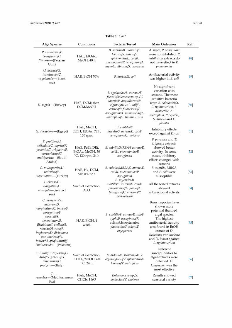
The most common classic technique applied to extract bioactive compounds from macroalgae is the maceration at different temperatures, followed by Soxhlet. It is also notable that the most frequently chosen solvents are methanol and ethanol even though other solvents with different characteristics are also successfully used. In this regard, Table 1 presents a compilation of the last 10 years of literature, refereeing studied algae species, the focus on the extraction conditions and highlighting the major outcomes achieved. Nowadays, novel liquid–liquid and solid–liquid extraction techniques are currently more applied, including: supercritical, ultrasound, microwave, enzymatic, high pressure, accelerated solvent and intensity pulsed electric fields extraction techniques [30]. In this regard, the combination of green technologies and safe environmental solvents is desirable to obtain efficient extraction of biocompounds, preserving their biological properties and opening the door to their implementation in the food, cosmetic and pharmaceutical industries. Once the target compounds are efficiently well extracted, the resultant extracts can be incorporated into different products. In general, the most common application would be the incorporation into food matrixes as preservatives [25], to cosmetic products [31] or as antibiotics with a synergistic effect on the pharmaceutical industry [1]. Moreover, other applications have been investigated such as antibiofilm [32], antifouling [33], coating in active packaging [34] or as prebiotics [11], among others. Therefore, the aim of this article is to review the main compounds responsible for the antimicrobial activity of algae, the novel extraction techniques for obtaining them and their applications.
Figure 1. Summary of the resistance acquisition pathways: Mutation and mobile genetic elements:By transduction, transformation or conjugation. Schema of the six main mechanisms of antibioticsresistance. Modified from [1,26,27].
Given the actual interest in identifying antimicrobial compounds from algae, it is essential toachieve profitable and sustainable recovery of these compounds, in an easy and fast process [28].Sometimes these compounds could be synthesized chemically, however, regarding the availability ofalgae, their recovery using green extraction technologies is an economic and environmentally friendlyalternative that also avoids the use of dangerous chemical compounds. The extraction of bioactivecompounds has always been a fundamental step in recovering these molecules from vegetal matrixes.New techniques have been developed in order to reduce extraction time, energy consumption, quantityof solvent, environmental implications, economical cost and waste productions while increasing theextraction efficiency and the quality of the obtained extract [29].
The most common classic technique applied to extract bioactive compounds from macroalgae isthe maceration at different temperatures, followed by Soxhlet. It is also notable that the most frequentlychosen solvents are methanol and ethanol even though other solvents with different characteristicsare also successfully used. In this regard, Table 1 presents a compilation of the last 10 years ofliterature, refereeing studied algae species, the focus on the extraction conditions and highlighting themajor outcomes achieved. Nowadays, novel liquid–liquid and solid–liquid extraction techniques arecurrently more applied, including: supercritical, ultrasound, microwave, enzymatic, high pressure,accelerated solvent and intensity pulsed electric fields extraction techniques [30]. In this regard, thecombination of green technologies and safe environmental solvents is desirable to obtain efficientextraction of biocompounds, preserving their biological properties and opening the door to theirimplementation in the food, cosmetic and pharmaceutical industries. Once the target compounds areefficiently well extracted, the resultant extracts can be incorporated into different products. In general,the most common application would be the incorporation into food matrixes as preservatives [25],to cosmetic products [31] or as antibiotics with a synergistic effect on the pharmaceutical industry [1].Moreover, other applications have been investigated such as antibiofilm [32], antifouling [33], coatingin active packaging [34] or as prebiotics [11], among others. Therefore, the aim of this article is toreview the main compounds responsible for the antimicrobial activity of algae, the novel extractiontechniques for obtaining them and their applications.

Antibiotics 2020, 9, 642 4 of 41
Table 1. Antimicrobial activity of algae crude extracts obtained using classic extraction technologiesfrom 2010 to 2020.
Alga Species Conditions Bacteria Tested Main Outcomes Ref.
L. brandenii—(India)HAE, MeOH:
CHCl3 (6:4), 35 ◦C,120 rpm, 7 days
S. aureus/B.subtilis/M.luteus/R.
rhodochrous/E.coli/P.aeruginosa/Vibrio
cholerae/Salmonellatyphi/Streptococcus
pneumoniae
All organisms wereinhibited. High
activity against B.subtilis whereas it
was moderateagainst E. coli
[35]
G. ornata—(Brazil) HAE, H2O, 25 ◦C,24 h
B. subtilis/S. aureus/E.aerogens/E. coli/P.
aeruginosa/S. choleraesuis/S.typhi
Only exhibitsinhibition to E. coli [36]
C. rubrum—(Chile) HAE (3x), 96%EtOH, 24 h. S. parasitica/Y. ruckeri
Antibacterial andantifungal activities
against fishpathogens
[37]
G. changii—(Malaysia)Solid liquid
extraction, MeOH,4 days.
P. aeruginosaMinimal inhibitoryconcentration (MIC)
6. 25 mg/mL[38]
P. capillacea/O.obtusiloba—(Brazil)
Solid liquidextraction, cold Hx
and 70% EtOH.
E. coli/S. aureus/Salmonellasp./V. harveyi
No antimicrobialactivity. [39]
P.gymnospora—(Brazil)
Percolation withMeOH S. aureus MIC 500 µg/mL [40]
S. latifolium/S.platycarpum/C.
socialis—(ArabianGulf)
HAE extraction,MeOH and AcO 25◦C, 150 rpm, 7 days
S. aureus/S.xylosus/MRSA/E. faecalis/B.
subtilis/E. coli/P.aeruginosa/Salmonella sp./K.
pneumoniae/C. albicans
Higher activityagainst Gram
positive bacteria thanGram negative
[41]
L. japonica—(Korea,Japan, China)
HAE (x3), EtOH, 25◦C, 1 day.
S. mutans/S. sobrinus/A.naeslundii/A.
odontolyticus/A.actinomycetemcomitas/F.nucleatum/P.gingivalis
Inhibitory activityagainst all
microorganisms[42]
D. membranacea—(Mediterranean Sea)
Column extraction,EtOH, AcO and
MeOH/DCM
S. aureus/S. agalactiae/B.subtilis/E. faecium/E.
faecalis/E. coli/C. albicans
EtOH and AcOshowed higher
antimicrobial activity[43]
S.oligocystum—(Persian
Gulf)
HAE, hot and coldH2O and glycerin
S. aureus/S. epidermidis/P.aeruginosa/E. coli
Hot water extractexhibited activity
against S. aureus, S.epidermidis, and P.
aeruginosa
[44]
C. myrica/C. trinodis/P.gymnospora/S.
dentifolium/S. hystrix/A.fragilis/C. racemosa/C.
fragile—(Red sea)
HAE, MeOH, 25◦C, 50 rpm, 7 days.
E. coli/S. aureus/E.faecalis/Salmonella sp./B.
cereus/P. aeruginosa.
MeOH extracts P.gymnospora and C.fragile showed thehighest activities
[45]
S. polycystum/P.australis—(Malasya)
HAE, Hx, DCM,MeOH, 72 h
S. aureus/B. cereus/E. coli/E.coli/P. aeruginosa
S. polycystum extractsexhibited higher
bacteriostatic activity[46]
Gracilariasp.—(Malaysiafarmed algae)
HAE, MeOH, 48 hB. subtilis/S. aureus/S.epidermidis/E. coli/V.
cholera/E. cloacae.
Moderateantibacterial activity
but S. aureus, Sepidermidis, E. cloacaewere not inhibited
[47]
Antibiotics 2020, 9, 642 5 of 41
Table 1. Cont.
Alga Species Conditions Bacteria Tested Main Outcomes Ref.
P. antillarum/P.boergesenii/U.
flexuosa—(PersianGulf)
HAE, EtOAc,MeOH; 48 h
B. subtilis/B. pumulis/E.faecalis/S. aureus/S.
epidermidis/E. coli/K.pneumoniae/P. aeruginosa/A.niger/C. albicans/S. cerevisiae
A. niger, P. aeruginosawere not inhibited. P.antillarum extracts donot have effect in K.
pneumoniae
[48]
U. lactuca/U.intestinales/C.
vagabunda—(Blacksea)
HAE, EtOH 70% S. aureus/E. coli Antibacterial activitywas higher in E. coli [49]
U. rigida—(Turkey) HAE, DCM; thenDCM/MeOH
S. agalactiae./S. aureus./E.faecalis/Micrococcus sp./V.tapetis/V. anguillarum/V.
alginolyticus E. coli/P.cepacia/P. fluorescens/P.
aeruginosa/A. salmonicida/A.hydrophila/S. typhimurium
No significantvariation with
seasons. The mostsensitive bacteria
were A. salmonicida,S. typhimurium, S.
agalactiae, A.hydrophila, P. cepacia,
S. aureus and E.faecalis
[50]
G. doryphora—(Egypt)HAE, MeOH,
EtOH, EtOAc, 72 h,150 rpm.
B. subtilis/E.faecalis/S. aureus/E. coli/P.
aeruginosa/C. albicans
Inhibitory effectsexcept against E. coli [51]
E. prolifera/U.reticulata/C. myrica/P.pavonica/T. triquetra/S.
portieriatum/G.multipartita—(Saudi
Arabia)
HAE, PeEt, DEt,EtOAc, MeOH, 30◦C, 120 rpm, 24 h
B. subtilis/MRSA/S aureus/E.coli/K. pneumoniae/P.
aeruginosa
P. pavonica and T.triquetra extractsshowed better
activity. In somecases, inhibitory
effects changed withseasons
[52]
G. multipartita/U.reticulata/S.
marginatum—(Turkey)
HAE, Hx, DCM,MeOH, 72 h
B. subtilis/MRSA/S. aureus/E.coli/K. pneumoniae/P.
aeruginosa
B. subtilis, MRSA,and E. coli were
susceptible[53]
L. obtusa/C.elongatum/C.
multifida—(Adriactsea)
Soxhlet extraction,AcO
B. mycoides/B.subtilis/S. aureus/E. coli/K.pneumoniae/A. flavus/A.fumigatus/C. albicans/P.
verrucosum
All the tested extractsshowed
antimicrobial activity[54]
C. iyengarii/S.asperum/S.
marginatum/C. indica/S.variegatum/S.
swartzii/S.tenerrimum/S.
ilicifolium/I. stellata/S.robusta/H. tuna/R.
implexum/D. dichotomavar. intricata/D.
indica/M. afaqhusainii/J.laminarioides—(Pakistan)
HAE, EtOH, 1week
B. subtilis/S. aureus/E. coli/S.typhi/P. aeruginosa/R.solani/Macrophominaphaseolina/F. solani/F.
oxysporum
Brown species haveshown more
potential than redalgal species.The highest
antibacterial activitywas found in EtOH
extract of D.dichotoma var intricataand D. indica against
S. typhimurium
[55]
C. linum/C. rupestris/G.dura/G. gracilis/G.
longissima/U.prolifera—(Italy)
Soxhlet extraction,CHCl3/MeOH, 60
◦C, 24 h
V. ordalii/V. salmonicida V.alginolyticus/V. splendidus/V.
harveyi/V. vulnificus
Differentsusceptibilities to
algal extracts weredetected. G.
longissima was themost effective
[56]
C.rupestris—(Mediterranean
Sea)
HAE, MeOH,CHCl3, H2O
Enterococcus sp./S.agalactiae/V. cholerae
Results showedseasonal variety [57]

Antibiotics 2020, 9, 642 6 of 41
Table 1. Cont.
Alga Species Conditions Bacteria Tested Main Outcomes Ref.
G. longissima—(Mediterranean Sea)
Soxhlet extraction,CHCl3/MeOH (2:1),
60 ◦C, 24 h
P. aeruginosa/Enterococcussp./S. agalactiae/V.
salmonicida/V. fluvialis/V.vulnificus/V. cholerae/V.
alginolyticus/C. albicans/C.famata/C. glabrata
Moderateantimicrobial effect
except on V.salmonicida andfungal species
[58]
C. antemmina/C.peltata/C.
scalpelliformis/D.dichotoma/S.
marginatum/A.specifera/G.
lithophilia/G.corticata—(India)
HAE, MeOHE. coli/P.
aeruginosa/S. aureus/K.pneumoniae
G. lithophila presentsthe most promising
results[59]
J. rubens/C. elongata/P.capillacea/U. fasciata/U.lactuca/E. compressa/E.
linza/S. vulgare/C.sinuosa—(Egypt)
HAE, EtOH 70%,MeOH 70% AcO
70%, 150 rpm, 72 h
B. subtilis/S. aureus/E. coli/S.typhi/K.
pneumoniae/C. albicans.
In all the tests, AcOshowed the biggest
inhibition halos[60]
D. flabellata/P.concrescens/L.johnstonii/G.
martinensis/U.lactuca/C.
fragile—(Mexico)
HAE, AcO:MeOH E. coli/S. aureus/B. cereus/B.subtilis/S. epidermidis
L. johnstonii, D.flabellata and U.
lactuca presentedactivity against
pathogenic bacteriatested
[61]
E. bicyclis—(SouthKorea)
HAE, MeOH, 70◦C, 3 h
C. acnes/S. aureus/S.epidermidis/P. aeruginosa
Inhibitory effectsexcept against P.
aeruginosa[62]
C. trinodis—(PersianGulf)—(Persian Gulf)
HAE,DEt:EtOH:Hx
S. aureus/S. epidermidis/E.coli/P. aeruginosa
The best Inhibitoryeffect was against S.epidermidis was the
worst against P.aeruginosa
[63]
C. glomerata,/E. linza/U.rigida/C. barbata/P.
pavonica/C. ciliatum/C.officinalis—(Black sea
Turkey)
HAE, 95% EtOH
S. aureus/B. cereus/A. niger/Styphimurium/L.
monocytogenes/E.coli/C. albicans/P. aeruginosa
All alga extractspresent antimicrobial
activity[64]
S. vulgare/C. hirsutus/R.verruculosa—(Coast of
Algeria)
Soxhlet extraction,MeOH, MeOH:
CHCl3, 6h
B. cereus/S. aureus/M.luteus/P. aeruginosa/E. coli/K.
pneumoniae/C. albicans
Positiveantimicrobial resultsagainst S. aureus and
B. cereus
[65]
Laurencia ssp.(aldingensis/catarinensis/
dendroidea/intricata/translucida)–(Brazil)
HAE, Hx, CHCl3,MeOH, H2O
C. albicans/C. parapsilosis/C.neoformans
L. aldingensis showedthe best antifungal
effects[66]
D.membranacea—(Tunisia)
HAE, H2O, CHCl3,EtOAc
S. aureus/S. epidermidis/L.monocytogenes/M. luteus/E.
faecium/E. coli/P.aeruginosa/S.
typhimurium/C. albicans/C.kefyr/C. krusei/C.
dubliniensis/C. glabrata
Inhibitory effectsagainst M. luteus,
S. aureus, S.epidermidis, L.
monocytogenes, C.krusei, C. dubliniensis
and C. kefyr
[67]
S. wightii/C. linum/P.gymnospora—(India)
HAE, Hx, EtOAc,AcO, MeOH
P. aeruginosa/S. typhi/E.amylovora/E. aerogens/P.vulgaris/K. pneumonia/E.coli/MRSA/B. subtilis/E.
faecalis
EtOAc and AcOextracts were more
efficient, but noinhibitory effectswere observed
against S. paratyphiand K. pneumonia.
[68]

Antibiotics 2020, 9, 642 7 of 41
Table 1. Cont.
Alga Species Conditions Bacteria Tested Main Outcomes Ref.
Fucus spp./P.elongata/Rhodomela
confervoides/S.latissima./C. rupestris/D.
contorta/F.vesiculosus/C.
rubrum/M. stellatus/L.digitata—(Germany)
HAE, DCME. amylovora/E. coli/P.
aeruginosa/B. subtilis/S.lentus
The macroalgaepresented
antibacterial activityagainst at least one of
the test strains
[69]
H. tuna/C. barbata/C.bursa—(Montenegro)
HAE, DCM:MeOH,48 h
E. coli/S. aureus/B. subtilis/E.faecalis/C. albicans
C. barbatademonstrated ashaving the best
antimicrobial activityfor S. aureus and B.
subtilis
[70]
G. corticata/G.edulis—(India)
HAE, DMSO, 70%MeOH, 130 rpm, 16
h
E. coli/Photobacterium sp./P.fluorescens/S. aureus/B.
subtilis
MeOH and DMSOextracts inhibited B.
subtilis[71]
L. digitata/S.latissima/H. elongata/P.
palmata/C.crispus—(Ireland)
HAE, MeOH,EtOH, AcO, 2 h
L. monocytogenes/S. abony/E.faecalis/P. aeruginosa
The extraction ofantimicrobials from
macroalgae weresolvent dependent
[72]
S.marginatum—(India)
HAE, DCM, EtOAc,AcO, MeOH Candida spp. Low antifungal
properties. [73]
S. lomentaria/P.pavonica/C.mediterranea/
H. musciformis/S.filamentosa—(Turkey)
HAE, MeOH, 8 h,200 rpm
S. aureus/S. typhimurium/E.coli/E. faecalis/C. albicans
S. lomentariainhibited S.
typhimurium. C.mediterrranea
inhibited C. albicans
[74]
U. lactuca/E.intestinalis—(Adriaticcoast of Montenegro)
HAE, Hx, DCM,MeOH, 72 h
B. mycoides/B. subtilis/E.coli/K.
pneumoniae/S. aureus/A.flavus/A.
fumigatus/C. albicans/P.purpurascens/P. verrucosum
Inhibitory effectswere observed
against B. mycoidesand B. subtilis
[75]
A. fragilis/C.a myrica/H.cuneiformes/L.
papillosa/S. cinereum/Tturbinata—(Egypt)
HAE, 80% MeOH,25 ◦C
B. subtilis/S. aureus/E.coli/C. albicans
H. cuneiformis extractshowed stronger
activity[76]
E.cava—(Korea)HAE, EtOH, n-Hx,
DCM, EtOAc,n-BuOH, H2O
S. aureus/MRSA/S. typhi/S.enteritidis/S. gallinarum
EtOH hadantibacterial activityS. aureus, MRSA and
Salmonella spp.
[77]
C. barbata—(Red Sea,Egypt)
Soxhlet extraction,EtOH
B. subtilis/S. aureus/M.luteus/E. coli/P.
aeruginosa/Serratia.marcescens/S. typhi/Vibrio
sp./A. hydrophila/C. albicans
Inhibitory activityexcept against M.
luteus[78]
K. alvarezii—(Malaysia) HAE, EtOH, H2O E. coli/B. cereusB. cereus was
inhibited but no E.coli.
[79]
U. lactuca/D.dichotoma/P.
gymnospora/S.vulgare/H. musciformis/D. simplex—(Brazil)
HAE, DCM,MeOH, EtOH, H2O
T. rubrum/T. tonsurans/T.mentagrophytes/M. canis/M.
gypseum/E.flocossum/C. albicans/C.
krusei/C. guilliermondi/C.parapsilosis/
EtOH and MeOHextracts were the
most effective[80]
B.bifurcata—(Portugal)
HAE, MeOH,DCM, 12 h
E. coli/P. aeruginosa/B.subtilis/S. aureus/S. cerevisiae
MeOH extracts hadinhibitory effects in
all themicroorganisms
[81]

Antibiotics 2020, 9, 642 8 of 41
Table 1. Cont.
Alga Species Conditions Bacteria Tested Main Outcomes Ref.
H. flagelliformis/C.myrica/S.
boveanum—(PersianGulf)
HAE, DCM, 48 h
E. coli:/K. pneumonia/S.typhi/S. aureus/S.
epidemidis/B. subtilis/A.niger/C. albicans
The antimicrobialactivity was
solvent-dependent[82]
T. conoides—(India)HAE, n-Hx, MeOHand EtOH: H2O, 72
h.
S. aureus/S. epidermidis/E.coli/P. aeruginosa/A.
niger/C. albicans
MeOH and EtOH:H2O extracts werethe most effective
against themicroorganisms
studied
[83]
D. dichotoma/P.pavonica/S.
vulgare—(AdriaticSea)
HAE, AcO, 50 ◦C;4h
B. mycoides/B.subtilis/S. aureus/E. coli/K.pneumoniae/A. flavus/A.fumigatus/C. albicans/P.
purpurescens/P. verrucosum
All crude extractshave a statistically
significant inhibitoryeffect on microbial
growth
[84]
C. racemosa/C. sertularioides/
K. alvarezii—(Malaysian coast)
HAE, Hx, CHCl3,EtOAc, EtOH,
MeOH, H2O, 1 day
B. cereus/S. aureus/A.baumannii/E. coli/K.
pneumoniae/P.aeruginosa/C. albicans/C.parapsilosis/C. krusei/C.
neoformans/A. fumigatus/T.interdigitale
Inhibitory effectsexcept against A.
fumigatus[85]
U. lactuca—(Gulf ofMaine) HAE, MeOH, 70 ◦C S. aureus/S. epidermidis Inhibitory effects
against both species [86]
T. ornata/T. decurrens/T. conoides/
S. polycystum/S.incisifolium/S.ilicifolium/H.a
cuneiformis—(Madagascar)
HAE, MeOH,EtOAc
B. cereus/S. aureus/S.pneumoniae/E. cloacae/K.oxytoca/S. boydii/E. coli/S.
enteridis/P.aeruginosa/C. albicans/C.
membranaefaciens/C.neoformans/T. mucoides
Antimicrobial tests ofthe crude extractsrevealed a strongactivity againstS. aureus and S.
pneumoniae
[87]
A.specifera/Cladophoropsis
sp./L.paniculata/Tydemania
sp./U. prolifera
Soxhlet extraction,EtOH and PeEt,
24 h
C. albicans/A. niger/Mucorsp./Paeciliomyces sp.
EtOH extract of L.paniculata showed
the best antimicrobialactivity
[88]
H. esperi/C.prolifera—(Egypt)
Soxhlet extraction,MeOH, 40 ◦C, 24 h
E. coli/P. aeruginosa/S.typhimurium/A.
hydrophila/B.subtilis/S. aureus
Inhibitory effectsagainst B. subtilis andS. aureus growth but
no against P.aeruginosa and S.
typhimurium,
[89]
Grateloupia sp./G.corticata/Halymeniasp./Metamastophora
sp./Spyridia sp.
HAE, MeOH, 24 h
E. cloacae/K. oxytoca/E. coli/S.enteridis/B.
cereus/S. aureus/S.pneumoniae/C. albicans.
All the crude extractsobtained can inhibitmicrobe’s growth.
[90]
H. elongata—(Ireland) HAE, H2O, MeOH,40 ◦C, 100 rpm, 2 h
L. monocytogenes/S. abony/E.faecalis/P. aeruginosa
60% MeOH extractshowed the best
results.[91]
F. serratus/F.vesiculosus—(Ireland)
HAE, H2O, MeOH,EtOAc, AcO MRSA 28 strains
Both species presentantibacterial activity
against severalMRSA strains.
[92]
U.reticulata—(Vietnam)
HAE,MeOH:CHCl3:
H2O
B. cereus/S. faecalis/E.cloace/S. aureus/E. coli/P.
aeruginosa/V. haveyi
U. reticulata showedhigh antimicrobialactivity, against E.
cloace and against E.coli.
[93]

Antibiotics 2020, 9, 642 9 of 41
Table 1. Cont.
Alga Species Conditions Bacteria Tested Main Outcomes Ref.
U. rigida—(Tunisia) HAE, EtOH:H2O,48
B. subtilis/B.cerus/S. aureus/S.
epidermis/E. faecalis/L.monocytogenes/E. coli/P.
aeruginosa/K. pneumoniae/A.niger/F. graminearum/F.
culmorum/F.oxysporum/C. albicans
Antimicrobialactivity varied
depending on theseason
[94]
U. fasciata/G.salicornia—(Honolulu,
USA)HAE, EtOH
E. faecalis/V. alginolyticus/V.cholerae/S. aureus/S.typhimurium/E. coli
U. fasciata hadsignificantly higher
antimicrobial activitycompared to G.
salicornia
[95]
U. lactuca/D.dichotoma/C.
elongata—(Algeria)
HAE, MeOH, DEt,CHCl3
E. coli/S. aureus/Salmonella/C. albicans/Penicillium sp.
CHCl3 extracts of U.lactuca and C.
elongata had thehighest activity
against E. coli andSalmonella sp. MeOHof all species showed
antifungal activityfor C. albicans.
[96]
Conditions: Heat-assisted extraction (HAE) Acetone (AcO), Ethanol (EtOH), Methanol (MeOH); Dichloromethane(DCM), Water (H2O), Hexane (Hex), Dimethilsulfoxide (DMSO), Chloroform (CHCl3), Petroleum ether (PeEt), Ethylacetatete (EtOAc), Diethyl ether (DEt), n- Butanol (n-BuOH), n-Hexane (n-Hx). Main outcomes: Minimal inhibitoryconcentration (MIC).
2. Macroalgae as a Promising Source of Valuable Antimicrobial Compounds
The nutritional composition of seaweed is strongly influenced by factors such as the species,environmental conditions, geographical place, seasonality and characteristics of the growth mediumas well as by the developmental stage [97]. Seaweeds are an excellent source of biologically activecompounds that includes proteins and peptides, polysaccharides, polyphenols, PUFAs and pigments [98,99].The resistance of pathogenic microorganisms to synthetic antibiotics has become a concern of publichealth systems [33] and therefore, it is imperative to explore new alternatives to solve this problem.In particular, among the different compounds that exhibit bioactivity that are present in macroalgae,interest in their antimicrobial potential has increased in the last few years in order to develop newantimicrobial therapies with less secondary effects, that are more cost effective and with minor toxicity,when compared to the synthetic antibiotics [33]. Although research on the antimicrobial properties ofseaweed compounds is a topic of great interest, until now the attribution of a particular compoundto such activity was a challenge as they are usually evaluated as extracts and not as a compoundconstituted by different biomolecules; in most cases, the antimicrobial effect is probably a consequenceof a synergic effect between these compounds. The main components of seaweeds are described in thefollowing sections.
2.1. Protein and Peptides
The protein content is highly variable, ranging between 10 to 30% of the dry weight (DW) in redseaweeds, from 5 to 15% DW in brown seaweeds and from 3 to 47% DW in green seaweeds [9,97].Moreover, these contents vary depending on the season, founding the highest concentration during thewinter–early spring and the lowest during summer–early autumn [97,100,101]. The proteins that areactively functional in seaweeds belong to two groups, namely, lectins and phycobiliproteins [14,102].Particularly, whereas lectins have been identified in several species of seaweeds, phycobiliproteins arefound in red macroalgae [102].

Antibiotics 2020, 9, 642 10 of 41
Protein from seaweeds contains all amino acids, chiefly glycine, alanine, arginine, proline, glutamicand aspartic acids [103]. However, seaweeds present reduced content of lysine, threonine, tryptophan,cysteine and methionine in comparison with other protein foods [102]. Peptides obtained from seaweedshave experienced an increasing interest in recent years due to their multiple bioactivity compounds(angiotensin converting enzyme inhibition, antihypertensive, antioxidative and antidiabetic) [104].
Regarding the antimicrobial activity, the only seaweed protein described in the literature asantibacterial is lectin [14,105]. The information about bioactive peptides with antibacterial properties isscarce. For example, Beaulieu et al. (2015) obtained antibacterial peptides after hydrolysis of proteinsfrom macroalgae S. longicruris. These authors evaluated the antibacterial activity of the >10 kDa proteinhydrolysate fraction against S. aureus and observed a significant decrease in the maximum specificgrowth rate when using a quantity between 0.31 mg/mL to 2.5 mg/mL [105]. Another example is thestudy by Cordeiro et al. (2006), which extracted protein fractions rich in lectin from the red seaweedH. musciformis and assessed its antifungal properties against human pathogen yeasts C. albicans andC. guilliermondii. Specifically, the F40/70 fraction demonstrated the capacity to inhibit the growthof C. guilliermondii at 45, 100 and 450 µg protein/mL, but a poor inhibition was observed againstC. albicans independent of the evaluated concentrations [106]. Lectin was also isolated from redmacroalgae S. filiformis [107]. Antibacterial activity was assessed on the growth of eight Gram-negative(E. coli, S. marcescens, S. typhi, S. typhimurium, K. pneumoniae, E. aerogenes, Proteus spp, and Pseudomonasaeruginosa) and three Gram-positive (B. cereus, B. subtilis and S. aureus). The authors found differentresults regarding the effects of lectin against Gram-negative and Gram-positive bacteria growth.The lectin at 500 µg/mL stimulated the growth of B. cereus and inhibited the growth of S. marcescens, S.typhi, K. pneumoniae, E. aerogenes, Proteus sp, and P. aeruginosa at 1000 µg/mL. The compound did notexhibit an effect, at any of the tested concentrations, on the growth of S. aureus and B. subtilis, or on E.coli and S. typhimurium. The authors concluded that more studies are necessary to evaluate the actionof lectin on bacterial growth in order to evaluate further clinical applications.
2.2. Polysaccharides
Polysaccharides are the main components of seaweeds, having a structural role in algal cellwalls [108]. Polysaccharides can be neutral or acidic, linear or branched [108]. Green, red and brownalgae are defined by the presence of sulfated polysaccharides with a high degree of complexity in therange from 4% to 76% DW [109]. In green seaweeds, polysaccharides represent between 38 and 54% oftheir dry matter [110]. These macroalgae are rich in ulvan (another sulfate polysaccharide), sulfatedrhamnans and sulfated galactans. The main component of these polysaccharides are sulfate groupsand moreover they contain repeating units of rhamnose, xylose and uronic acids (glucuronic andiduronic acids) and minor contents of glucose, galactose, rhamnose and arabinose [110]. Red seaweedspresent different sulfated galactans, sulfated rhamnans or mannans, carrageenans and agars. The mainpolysaccharides found in red macroalgae are galactans with a backbone composed of repeating unitsof three-linked β-D-galactopyranosyl and four-linked α-galactopyranosyl units: if the configurationof the four-linked α-galactopyranosyl units is L the polysaccharide is agar and if the configurationof the four-linked α-galactopyranosyl units is D then the polysaccharide is carrageenan. Agars aretypically low in sulfate ester substitution, whereas carrageenans are comparatively rich in sulfateester substitution [111]. Brown macroalgae are a rich source of alginate with a content that variesbetween 14 and 40% of their dry mass [7]. After the alginates, β-glucans (laminarans), cellulose andheteroglycans are present in important amounts in brown algae [7]. Laminarans are β-D-glucanswith low molecular weight that are linear polysaccharides of glucose linked by 1→3 β-glycosidicbonds. There are different types of laminarans classed in accordance of their length and branchingdegree. Laminarans contain some 6-O-branches in their backbone and some β-(1→6)-intrachain links.Fucoidans are a family of sulfated homo and heteropolysaccharides that are mainly composed of(1→3)-linked-L-fucopyranose units. Moreover, fucoidans can present a main chain of alternating (1→3)and (1→4)-linked-L-fucopyranose units, and sulfate groups located mainly at C2, C4, or disubstituted

Antibiotics 2020, 9, 642 11 of 41
at both C2 and C4. In addition, acetyl groups and D-galactose, D-xylose, D-mannose, L-rhamnose andD-glucuronic acid residues were identified as components of fucoidans [112].
The antimicrobial activity of polysaccharides from seaweeds depends on factors such as molecularweight, charge density, sulfated content in the case of sulfated polysaccharides and structural andconformation characteristics [33]. Several studies have been reported. For example, sulphatedpolysaccharides (alginates, fucoidans and laminaran) extracted from different seaweeds, such asL. japonica, A. nodosum, or U. pinnafitida have demonstrated an inhibitory effect on the growth ofpathogenic bacteria [113]. Similarly, extracts rich in either laminarin or fucoidan isolated from Laminariaspp, decreased the fecal E. coli populations in piglets (0.3 and 0.24 g/kg, respectively), observing areduction in the initial bacterial load in derived raw meat products [114].
2.3. Fatty Acids
Seaweeds produce PUFAs and are also important sources of essential fatty acids [115–117].The most predominant PUFAs are ω3 such as 16:4 ω3 and 18:4 ω3 and some species alsopresent important amounts of eicosapentaenoic acid (20:5 ω3, EPA) [118], α-linolenic (18:3 ω-3),octadecatetraenoic (18:4ω-3) and arachidonic (20:4ω-6) [116]. Several studies have reported theantimicrobial properties of macroalgal fatty acids. For example, different sulfolipid classes isolatedfrom the total lipids of two species of U. fasciata (Chlorophyta), L. papillosa, G. cylindriea (Rhodophyta),D. fasciola and T. atomaria (Ochrophyta). Authors assessed the antimicrobial activity of these compoundsand they observed a high inhibition of the growth of B. subtilis and E. coli, using 100 µg/well. However,these compounds did not exhibit any inhibition against the fungi or yeast tested [119]. Another studyobtained G. vermiculophylla, P. dioica and C. crispus using solvents with different polarity (DEt < EtOAc< MeOH:H2O (1:1)). The authors identified the fatty acid profile of ethyl acetate extracts and observedthat saturated fatty acids (SFA), especially palmitic acid (16:0) was the more abundant, followedby polyunsaturated fatty acids (PUFA) and monounsaturated fatty acids (MUFA). They tested theantimicrobial activity against Gram-positive bacteria (L. innocua, B. cereus, E. faecalis, L. brevis, S. aureusand Gram negative (E. coli, S. enteritidis, P. aeruginosa) and the yeast Candida sp. The EtOAc extractshowed the high inhibition capacity of the tested strains [17].
2.4. Polyphenolic Compounds
Polyphenolic compounds are secondary metabolites of seaweeds whose composition vary fromsimple phenolic acids to complex molecules such as phlorotannins [120]. Macroalgae are excellentsources of catechins, flavonols and phlorotannins [121,122]. Green and red algae contain highproportions of bromophenols, phenolic acids and flavonoids. Brown algae present predominantpolyphenolic compounds such us phlorotannins, which are complex polymers of phloroglucinol(1,3,5-trihydroxybenzene), and are classified into different groups in function of the bonds of thephloroglucinol units. They are divided in eckols and carmalols (dibenzodioxin linkage), fuhalols (etherbonds and hydroxyl groups), fucophlorethols (ether and phenyl bonds), phlorethols (ether bonds),fucols (phanyl linkages), and ishofuhalols [121,123]. In addition, some brown algae can contain bromo-,chloro- and iodo-phlorotannins [123]. These compounds represent 20% DW of algae [120] and haveonly been described in the composition of brown algae [123]. Other polyphenols such as catechins,flavonoids and flavonol glycosides have been identified in brown seaweeds [124].
Referring to the antimicrobial activity of polyphenols, several authors have studied the propertiesof phlorotanins. In this context, Nagayama et al. (2002) [125] evaluated the antibacterial effectof phlorotannins from E. kurome against several food-borne pathogenic bacteria, different MRSAstrains and S. pyogenes. They observed that the phlorotannins were effective against MRSA and alsoCampylobacter spp., which presented the highest susceptibility to these compounds. The minimalbactericidal concentration of the crude phlorotannins, dieckol and 8,8′-bieckol against C. jejuni were50 mg/L, 0.03 µmol/mL and 0.03 µmol/mL, respectively [125].

Antibiotics 2020, 9, 642 12 of 41
2.5. Pigments
Based on their content in pigments, macroalgae are classified into three groups: green algae(Chlorophyta, ca. 1200 species), red algae (Rhodophyta, ca. 6000 species) and brown algae (Ochrophyta,ca. 1750 species). Macroalgae are described to present three types of natural pigments: chlorophylls,carotenoids and phycobilins. Chlorophylls are greenish lipid-soluble natural pigments which containa porphyrin ring and are found in all algae [102]. Four forms of chlorophylls have been identifiedin macroalgae with the most important being chlorophyll a; chlorophyll b and c are also describedand the chlorophyll d is present in red algae [102]. Phycobiliproteins are other natural pigmentsand are water soluble fluorescent proteins present in seaweeds [126,127]. There are three types ofphycobiliproteins: phycocyanins (blue pigment), phycoerytrins (red pigment) and allophycocyanins(light-blue pigment) [102,126], with phycoerytrins being the most abundant in many red macroalgaespecies. Carotenoids are linear polyenes and can be classified in carotenes (α yβ-carotene, lycopene) andxanthophylls (fucoxanthin, violaxanthin, antheraxanthin, zeaxanthin, lutein, neoxanthin). The mostabundant carotenoid is fucoxanthin, a brown pigment that confers the coloration to brown algae [102].
Regarding the antimicrobial activity of pigments, there are few studies that evaluate the activity ofisolated compounds. A recent study has evaluated the antimicrobial properties of fucoxanthin againstdifferent Gram-positive and Gram-negative bacteria. The results showed that the compound was moreeffective against Gram-positive bacteria, with S. agalactiae being the most affected bacteria (with a MICof 62.5 µg/mL), followed by S. epidermidis and S. aureus [128].
3. Mechanisms of Action of Antimicrobial Compounds
In the previous section, numerous compounds extracted from macroalgae with provenantimicrobial activity are mentioned. In the following paragraphs, the mechanisms of action ofthese compounds will be explained. A summary of these mechanisms is presented in (Figure 2).In Table 2, several studies demonstrating the antimicrobial properties of macroalgae compounds havebeen compiled.
Antibiotics 2020, 9, x FOR PEER REVIEW 12 of 40
minimal bactericidal concentration of the crude phlorotannins, dieckol and 8,8′-bieckol against C. jejuni were 50 mg/L, 0.03 µmol/mL and 0.03 µmol/mL, respectively [125].
2.5. Pigments
Based on their content in pigments, macroalgae are classified into three groups: green algae (Chlorophyta, ca. 1200 species), red algae (Rhodophyta, ca. 6000 species) and brown algae (Ochrophyta, ca. 1750 species). Macroalgae are described to present three types of natural pigments: chlorophylls, carotenoids and phycobilins. Chlorophylls are greenish lipid-soluble natural pigments which contain a porphyrin ring and are found in all algae [102]. Four forms of chlorophylls have been identified in macroalgae with the most important being chlorophyll a; chlorophyll b and c are also described and the chlorophyll d is present in red algae [102]. Phycobiliproteins are other natural pigments and are water soluble fluorescent proteins present in seaweeds [126,127]. There are three types of phycobiliproteins: phycocyanins (blue pigment), phycoerytrins (red pigment) and allophycocyanins (light-blue pigment) [102,126], with phycoerytrins being the most abundant in many red macroalgae species. Carotenoids are linear polyenes and can be classified in carotenes (α y β-carotene, lycopene) and xanthophylls (fucoxanthin, violaxanthin, antheraxanthin, zeaxanthin, lutein, neoxanthin). The most abundant carotenoid is fucoxanthin, a brown pigment that confers the coloration to brown algae [102].
Regarding the antimicrobial activity of pigments, there are few studies that evaluate the activity of isolated compounds. A recent study has evaluated the antimicrobial properties of fucoxanthin against different Gram-positive and Gram-negative bacteria. The results showed that the compound was more effective against Gram-positive bacteria, with S. agalactiae being the most affected bacteria (with a MIC of 62.5 µg/mL), followed by S. epidermidis and S. aureus [128].
3. Mechanisms of Action of Antimicrobial Compounds
In the previous section, numerous compounds extracted from macroalgae with proven antimicrobial activity are mentioned. In the following paragraphs, the mechanisms of action of these compounds will be explained. A summary of these mechanisms is presented in (Figure 2). In Table 2, several studies demonstrating the antimicrobial properties of macroalgae compounds have been compiled.
Figure 2. Schematic illustration of the main action mechanisms of antimicrobial compounds extracted from macroalgae species. Figure 2. Schematic illustration of the main action mechanisms of antimicrobial compounds extractedfrom macroalgae species.

Antibiotics 2020, 9, 642 13 of 41
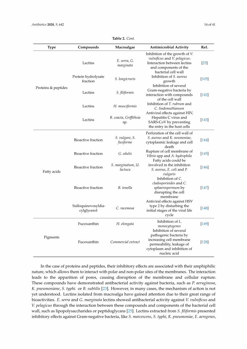
Table 2. Antimicrobial properties of compounds extracted from macroalgae.
Type Compounds Macroalgae Antimicrobial Activity Ref.
Polyphenols
Phlorotannins F. vesiculosus
Alteration of the cellmembrane and cell
destruction of S. aureus,S. pneumonia and P.
aeruginosa
[129]
Phlorotannins S. thunbergii
Alteration of the cellmembrane, cytoplasm’s
leakage and cell destructionof V. parahaemolyticus
[130]
Phlorofucofuroeckol E. bicyclis
Cell membrane damage andsuppression of genes relatedto methicillin resistance in
S. aureus
[131]
Bromophenols K. alvareziiDownregulation ofpathogenic genes of
P. gingivalis[132]
Dieckol E. clava Alteration of cell integrityand metabolism of T. rubrum [133]
PhlorotanninsC. nodicaulis,
C. usneoides, F.spiralis
Alterations of the cell wallcomposition, increased
mitochondrial respiration.Inhibition of the formation
of the germ tube ofC. albicans
[134]
Phlorotannins E. clavaInhibition of the enzyme
neuraminidase of theInfluenza A virus
[135]
Polyphenolic richextracts
E. arborea,S. filiformis
Inhibition of the viralparticle [136]
Polysaccharides
Depolymerizedfucoidans L. japonica
Interaction with protein ofthe cell membrane and
cellular rupture of E. coli andS. aureus
[137]
Fucoidan F. vesiculosusInhibition of dental plaque
bacteria and foodbornepathogens.
[138]
Laminarin richextracts
A. nodosum, L.hyperborea
Inhibition of S. aureus, L.monocytogenes, E. coli and S.
typhimurium.[139]
Water solublepolysaccharide
extracts
P. capillacae, D.membranacea
Inhibition of F. oxysporiumInhibition of C. albicans and
M. phaseli[140]
Sulfatedpolysaccharides
G. skottbergii
Obstruction of herpessimplex virus type 1 and 2
attachment to the cells [141]Interference with fusion
between HIV infected cells.Inhibition of the viral
enzyme reverse transcriptase
C. okamuranus
Inhibition of dengue virusby interaction with the
glycoprotein of the viralenvelop
Antibiotics 2020, 9, 642 14 of 41
Table 2. Cont.
Type Compounds Macroalgae Antimicrobial Activity Ref.
Proteins & peptides
Lectins E. serra, G.marginata
Inhibition of the growth of V.vulnificus and V. pelagicus.
Interaction between lectinsand components of the
bacterial cell wall
[25]
Protein hydrolysatefraction S. longicruris Inhibition of S. aureus
growth [105]
Lectins S. filiformis
Inhibition of severalGram-negative bacteria by
interaction with compoundsof the cell wall
[142]
Lectins H. musciformis Inhibition of T. rubrum andC. lindemuthianum
Lectins B. coacta, Griffithsiasp.
Antiviral effects against HIV,Hepatitis C virus and
SARS-CoV by preventingthe entry in the host cells
[143]
Fatty acids
Bioactive fraction S. vulgare, S.fusiforme
Perforation of the cell wall ofS. aureus and K. neumoniae,
cytoplasmic leakage and celldeath
[144]
Bioactive fraction G. edulis Rupture of cell membrane ofVibrio spp and A. hydrophila [145]
Bioactive fraction S. marginatum, U.lactuca
Fatty acids could beinvolved in the inhibition
S. aureus, E. coli and P.vulgaris
[146]
Bioactive fraction B. tenella
Inhibition of C.cladosporioides and C.sphaerospermum bydisrupting the cell
membrane
[147]
Sulfoquinovosyldia-cylglycerol C. racemosa
Antiviral effects against HSVtype 2 by disturbing the
initial stages of the viral lifecycle
[148]
Pigments
Fucoxanthin H. elongata Inhibition of L.monocytogenes [149]
Fucoxanthin Commercial extract
Inhibition of severalpathogenic bacteria by
increasing cell membranepermeability, leakage of
cytoplasm and inhibition ofnucleic acid
[128]
In the case of proteins and peptides, their inhibitory effects are associated with their amphiphilicnature, which allows them to interact with polar and non-polar sites of the membranes. The interactionleads to the apparition of pores, causing disruption of the membrane and cellular rupture.These compounds have demonstrated antibacterial activity against bacteria, such as P. aeruginosa,K. pneumoniane, S. typhi. or B. subtilis [23]. However, in many cases, the mechanism of action is notyet understood. Lectins isolated from macroalga have gained attention due to their great range ofbioactivities. E. serra and G. marginata lectins showed antibacterial activity against V. vulnificus andV. pelagicus through the interaction between these compounds and components of the bacterial cellwall, such as lipopolysaccharides or peptidoglycans [25]. Lectins extracted from S. filiformis presentedinhibitory effects against Gram-negative bacteria, like S. marcescens, S. typhi, K. pneumoniae, E. aerogenes,

Antibiotics 2020, 9, 642 15 of 41
Proteus sp. and P. aeruginosa. This effect was associated with the interaction between glycol-compoundspresent on the cell wall. In a similar way, lectins extracted from macroalga H. musciformis exhibitedantifungal activity against T. rubrum and C. lindemuthianum [142]. They have also displayed antiviraleffects against human immunodeficiency, hepatitis C, severe acute respiratory syndrome coronavirus(SARS-CoV) viruses, mainly by preventing the entry of the virus in the host cells and thereby theirpropagation [143].
The antimicrobial properties of macroalgae polysaccharides are attributed to the interactionbetween glyco-receptors of the bacterial cell wall, compounds of the membrane and nucleic acids andthe polysaccharides. Those interactions lead to the disruption of the membrane stability and cellularfunctions [34]. Several factors have been shown to influence this activity, such as the molecular weight,charge density, structure and conformation [20]. Sulfated polysaccharides have demonstrated theirantibacterial activity in several studies. For example, depolymerized fucoidans of L. japonica showedantibacterial activity against E. coli and S. aureus, which is caused by the interaction of fucoidanswith membrane proteins, leading to membrane rupture and further cell death [137]. In other study,sulfated polysaccharides were extracted from different marine macroalgae and their antibacterialand antibiofilm properties were assessed against dental plaque bacteria. Fucoidan extracted fromF. vesiculosus inhibited the mentioned bacteria and foodborne pathogens. In this case, the resultssuggest that fucoidan may not present a direct killing effect and may act by trapping nutrients, reducingthe bioavailability [138]. To our knowledge, few studies have evaluated the antifungal properties andmechanisms of algal polysaccharides. Water soluble polysaccharides extracted from P. capillacea andD. membranacea displayed antifungal activity against different yeast and fungi. P. capillacea inhibitedthe growth of F. oxysporium, while D. membranacea inhibited C. albicans and M. phaseli. In future studies,it is expected that the compounds involved in this activity, as well as their mechanisms of action willbe identified [140]. Regarding antiviral effects, these have been studied more extensively. Macroalgalpolysaccharides can inhibit the multiplication of viruses such as the herpes simplex virus (HSV),human immunodeficiency virus (HIV) or the dengue virus. They can also obstruct the interactionbetween viruses and cells and inhibit enzymes [141]
The antibacterial activity of algal lipids and fatty acids has been attributed to their ability toinhibit the electron transport chain and oxidative phosphorylation in cell membranes, leading to theformation of peroxidation and auto-oxidation degradation products and the cellular lysis [34,145].To our knowledge, no studies have isolated and then tested the antibacterial activity of macroalgafatty acids, but they have been successfully identified in bioactive extracts. In the study by El Shafayet al. [144], fatty acids were identified in the bioactive fraction of the extracts of S. vulgare and S.fusiforme, but no isolation was performed. The analysis demonstrated that the cell wall of S. aureus andK. pneumoniae was perforated, which resulted in the rupture of the cell wall, leakage of the cytoplasmand further cell death. Similarly, fatty acids were detected in the active fractions extracted from thered algae G. edulis. Although fatty acids were not isolated and their antimicrobial activity was notseparately verified, authors attributed the antibacterial effects against Vibrio spp. and A. hydrophilato these compounds [145]. In the case of fungi, it has been proposed that fatty acids may act indisrupting the cell membrane, inhibiting the reproduction. Antifungal activity has been observedagainst C. cladosporioides and C. sphaerospermum, in addition to antiprotozoal effects against T. cruzi andL. amazonensis [147]. A sulfoquinovosyldiacylglycerol isolated from the n-BuOH fraction of C. racemosashowed antiviral effect against HSV type two by disturbing the early stage of the viral life cycle [148].
Among the literature, the most studied antimicrobial compounds are the polyphenols. Theirantimicrobial action has been associated with their ability to alter membrane permeability (causingcell lysis), inhibit enzymes and different metabolic pathways, bind to surface molecules and othermechanisms. This activity seems to be related to the number of hydroxyl groups and also the degreeof polymerization [20,23]. Several studies have assessed the antimicrobial properties and the diverseaction mechanisms of polyphenols. For example, a recent study evaluated the antibactericidal actionof phlorotannins (a type of polyphenol found in macroalgae of the class Phaeophyta) extracted from

Antibiotics 2020, 9, 642 16 of 41
F. vesiculosus. The results showed that these compounds presented a significant bactericidal effectagainst S. aureus, S. pneumonia and P. aeruginosa. Phlorotannins presented a higher effectivity againstGram-positive bacteria than against Gram-negative bacteria, probably due the differences betweentheir cell membranes, since Gram-negative bacteria are surrounded by an outer membrane which isrich in polysaccharides. The authors attributed the observed effects to the ability of phlorotannins toinhibit bacterial growth by the alteration of the cell membrane [129]. Similarly, phlorotannins fromS. thunbergii have ben demonstrated to inhibit V. parahaemolyticus, causing damage to the cell walland the membrane, which increased the membrane permeability and caused further leakage anddestruction of the bacterial cells [130]. A phlorofucofuroeckol of the brown macroalga E. bycliclis hasshown antibacterial effects against methicillin-resistant S. aureus. This compound produced damage inthe cell membrane, leading to the leakage of cytoplasm and cell death. Furthermore, this compoundsuppressed the expression of genes related to resistance to methicillin in a dose-dependent manner [131].Bromophenols, extracted from the red macroalga K. alvarezii, showed activity against P. gingivalis, theprincipal agent of chronic periodontitis. These compounds were able to downregulate the expressionof the proteins involved in the infectious pathways of the bacteria [132]. Regarding yeast and fungi, thephlorotannin dieckol, extracted from E. clava, was tested against the fungi T. rubrum, associated withdermatophytic nail infections. The results exhibited alterations in membrane integrity and also in cellmetabolism [133]. Another study evaluated the antifungal properties of the phlorotannins extractedfrom the brown macroalgae C. nodicaulis, C. usneoides and F. spiralis against different pathogenic yeastand fungi. Antifungal activity against all the studied species was observed, with the yeast C. albicansATCC 10231 being the most susceptible, while the most susceptible fungi were E. floccosum andT. rubrum. The action mechanisms of phlorotannins were also evaluated. In the case of phlorotanninsof C. nodicalus and C. usneoides, the results indicated a lower ergosterol content in the cell membraneof the yeast and fungi, respectively, which disrupted cellular integrity and functions. On the otherhand, F. spiralis phlorotannins reduced the chitin content of the fungi cell wall, an essential wallcomponent. Furthermore, they inhibited the formation of the germ tube of C. albicans, reducing itsvirulence and its capacity to adhere to epithelial cells. Finally, all phlorotannins increased the activity ofthe mitochondrial respiratory rate, which may increase the production of reactive oxygen species [134].Phlorotannins also present antiviral activity. Five phlorotannins extracted from the brown algae E. clavadisplayed inhibitory effects against the Influenza A virus, through the inhibition of the neuraminidase,a critical enzyme for the life cycle of the virus [135]. Polyphenolic rich extracts from E. arborea andS. filiformis have demonstrated antiviral effects against Measles virus. Authors have proposed thatpolyphenols act by direct inactivation of the viral particle, which prevents the infection of cells [136].
Finally, in the case of pigments, the antimicrobial mechanism has not been fully understood.The most studied pigments are the carotenoids, which are supposed to act trough the accumulation oflysozyme, an enzyme able to digest bacterial cell walls [20]. Among carotenoids, fucoxanthin standsout and its antimicrobial properties have been tested against different pathogenic bacteria [34]. Recently,this compound has been tested against E. coli, B. cereus, B. subtilis, K. pneumoniae, S. aureus, P. aeruginosaand L monocytogenes. The proposed mechanisms consist of an increase in permeability, leakage of thecytoplasm and inhibition of nucleic acid formation [128]. Regarding antifungal activity of pigments,chlorophyll extracts from S. pallidum was tested against fungi S. glycines and A. niger and shown topossess low antifungal activity. However, the action mechanism has not been elucidated [150].
4. Novel Liquid–Liquid and Solid–Liquid Extraction Technologies to Efficiently Extract AlgalBioactive Compounds
Over the years, significant research efforts have been made to efficiently extract algae bioactivecompounds by applying different methodologies. Conventional extraction methods (solid–liquidextraction) have numerous limitations, e.g., lower efficacy, high energy cost and low yield, thus newstate-of-the-art extraction methodologies are required. Table 3 presents several articles in respect of theantimicrobial activity of macroalgae crude extracts, using the technologies mentioned below.

Antibiotics 2020, 9, 642 17 of 41
Table 3. Antimicrobial activity of macroalgae crude extracts obtained using emergingextraction technologies.
Method Conditions Macroalgae BioactiveCompound Main Outcomes Ref.
SFE314 bar, 10 ◦C D. salina Fucoidans
Inhibition of E. coli,S. aureus, and C. albicans,
growth[151]
300 bar, 50 ◦C F. vesiculosus Fucosterol Inhibition of Fusariumsp. [152]
UAE 200 W, 20 kHz; 55 ◦C;20 min N. zanardinii Fucoidans
No activity against E.coli, L. monocytogenes P.aeruginosa and S. aureus
[153]
MAE1500 W; 150 ◦C; 10
min (× 2) N. zanardinii Fucoidans Positive activity againstE. coli [153]
50 ◦C, 500 W, 10 min,MeOH, EtOH
Oedogoniumsp./Stigeoclonium
sp./Ulothrixsp./Nitzschia sp.
n.d.All extracts inhibited atleast one microorganism
tested[154]
UMAE 65 ◦C, 3 h (× 2) N. zanardinii Fucoidans
Inhibition of P.aeruginosa, but no effect
on E. coli L.monocytogenes and
S. aureus
[153]
EAE
Alcalase: 2.5 mL (2.4U/g), pH 8, 50 ◦C, 24 hFlavourzyme: 2.5 mL(500 U/g), pH 7, 50 ◦C,
24 hCellulase: 2.5 g (3
U/mg), pH 4.5, 50 ◦C,24 h
Viscozyme: 2.5 mL100 fungal
β-glucanase U/mL,pH 4.5, 50 ◦C, 24 h
N. zanardinii FucoidansNo activity against E. coli
L. monocytogenes, P.aeruginosa and S. aureus
[153]
Viscozyme L: pH 4.5(0.1M, AB), 50 ◦C;
AMG 300 L: pH 4.5(0.1M, AB), 60 ◦C;Celluclast: pH 4.5(0.1M, AB), 50 ◦C;
Termamyl: pH 6 (0.1M, SPB), 60 ◦C;
Ultraflo: pH 6 (0.1 M,SPB), 40 ◦C;
Flavourzyme: pH 7(0.1 M, SPB), 50 ◦C;
Alcalase: pH 8 (0.1 M,SPB), 50 ◦C;
Neutrase: pH 8 (0.1M, SPB), 50 ◦C;
S. boveanum, S.angustifolium, P.gymnospora, C.cervicornis, C.
sinuosa, I.stellate, F.irregularis
Polyphenols/polysaccharides
F. irregularis extractsobtained with
Viscozyme, Celluclasand Flavourzyme
inhibited S. aureus. P.gymnospora and C.
sinuosa extracts obtainedwith Celluclast inhibited
E. feacalis.
[155]
EUAE 65 ◦C, 3 h (× 2) N. zanardinii Fucoidans
Inhibition of P.aeruginosa, but no effect
on E. coli L.monocytogenes and
S. aureus
[153]
SWE 1500 W, 150 ◦C, 10min (× 2) N. zanardinii Fucoidans
Inhibition of E. coli and P.aeruginosa, but no effecton L. monocytogenes and
S. aureus
[153]
SWH200–280 ◦C, 1.3–6.0
MPa, Catalyst -Aceticacid
L. japonica n.d.Strong antibacterialactivity against S.
typhimurium and E. coli,[156]

Antibiotics 2020, 9, 642 18 of 41
Table 3. Cont.
Method Conditions Macroalgae BioactiveCompound Main Outcomes Ref.
PLE
Hx, EtOAc, AcO,EtOH, EtOH: H2O
(50:50)
U. intestinalis,U. lactuca, F.
vesiculosus, D.dichotoma, C.
baccata, H.elongata
Fatty acidsF. vesiculosus extractexhibited the best
antimicrobial properties[157]
Hx, EtOH, W; 200 ◦C,20 min H. elongata Fatty
acids/pigments
All extracts presentedantimicrobial activity
against S. aureus, E. coli,C. albicans and A. niger
[158]
H2O, MeOH, DCMTemperature (20, 40,
60 ◦C)F. vesiculosus
Phlorotannins/phosphatidylcholine/
betaine/lipids/chlorophylls/carotenoids
F. vesiculos extract wasonly effective as an
antimicrobial agent toMRSA
[159]
Conditions: Acetone (AcO), Ethanol (EtOH), Methanol (MeOH); Dichloromethane (DCM), Water (H2O), Hexane(Hex), Ethyl acetatete (EtOAc). n.d: not described. Methods: Supercritical fluid extraction (SFE), UltrasoundAssisted Extraction (UAE), Microwave-assisted extraction (MAE), Ultrasonic-Microwave Assisted Extraction(UMAE), Enzymatic-Assisted Extraction (EAE), Enzymatic-Ultrasound Assisted Extraction (EUAE), SubcriticalWater Extraction (SWE), Subcritical Water Hydrolysate (SWH), Pressurized Liquid Extraction (PLE).
4.1. Supercritical Fluid Extraction (SFE)
Supercritical fluid extraction (SFE) is a green analytical methodology used for the extraction ofhigh-value bioactive compounds from complex matrixes [160]. SFE uses supercritical fluids, whichabove their critical point exhibit liquid-like characteristics such as solvent power, and negligible surfacetension, as well as gas-like features such as enhanced transport properties.
Comparing SFE with other conventional extraction techniques, SFE presents several advantages,namely the use of minimal solvents, great extraction selectivity, short processing time, and a lowdegradability of the extract, showing a broad application for different bioactive compounds [161].
The thermodynamics properties of carbon dioxide (CO2) make it the preferred solvent forSFE-based extraction processes [151]. Moreover, due to its low toxicity, low cost, low explosivity, facileavailability and environmentally friendly nature, it also presents major factors favoring the choice ofCO2 as the SFE solvent [162]. Considering the physical characteristics, CO2 can only be used as theextraction solvent for the extraction of nonpolar or low polarity compounds (as supercritical CO2 is anonpolar solvent). Nevertheless, the polarity of CO2 can be modulated using co-solvents such as smallamounts of ethanol or methanol, increasing the extraction yields of polar compounds [162]. Recently,several reports described the application of SFE to extract high value bioactive molecules in arctic brownalgae of the species F. vesiculosus. The arctic brown fraction extracts present a predominant contentof fatty acids [151,163], polyphenols [163,164], carotenoids and chlorophylls [164]. Moreover, theseartic brown algae SFE extracts also possess pronounced bacterial, fungicidal and immunostimulantactivities [163].
Despite the potential of the SFE technique and its suitability to extract high-value bioactivecompounds from algae, clearly the extraction depends on the nature of the target compounds. Usingthe SFE technique, a study concluded that D. salina extracts in the presence of SFE at 314 bar and9.8 ◦C showed a substantial antimicrobial activity against E. coli, S. aureus, C. albicans and A. niger.As indicated in the work, this notable antimicrobial activity could be attributed to the presence ofindolic compounds, PUFAs, and carotene metabolism, such as β-ionone and neophytadiene in the SFED. salina extracts [151].
The antifungal potential of the brown algal F. vesiculosus was studied. To perform this study, algalextracts were obtained using SFE at a temperature of 50 ◦C and a pressure of 300 bar. Using aqueousalgal extracts at the concentrations of 0.5% and 1.0%, a 100% growth inhibition of macroconidia within

Antibiotics 2020, 9, 642 19 of 41
144 h was obtained. Moreover, F. vesiculosus SFE extracts also promoted a 48% and 72% mycelialgrowth of phytopathogenic F. oxysporum and F. culmorum, respectively, after 168 h of incubation [152].
4.2. Ultrasound Assisted Extraction (UAE)
Ultrasound assisted extraction (UAE) technique uses acoustic waves in the kilohertz range (20 kHzto 100 kHz) that travel through the solvent producing cavitation bubbles. When the cavitation bubblesburst at the surface of the complex sample matrix, a shockwave induces damage to the sample cellwall enhancing the mass transfer of high-value bioactive compounds across cellular membranes intothe solution [165]. Two different types of equipment can be used to carry out UAE: the ultrasonic bath(indirect sonication) that operates at a frequency between 40–50 kHz using a power of 50–500 W andthe ultrasonic probe which operates only at 20 kHz. The principal difference between this equipmentis the way that the ultrasound wave affects the sample [166].
The UAE technique is considered a cold extraction technique, as temperature during the extractionprocess is comparatively low and does not affect the stability of extracted compounds. UAE presentsseveral advantages, such as the potential to reduce or eliminate the use of toxic chemical solventsand it is a more economic process (no need of supplementary energy to separate phases and toeliminate solvent). Moreover, using UAE, full extractions can be completed in a few minutes with highreproducibility, simplifying the manipulation and work-up, giving a higher purity to the final productand eliminating post-treatment of water waste [167].
UAE was the method of choice to extract bioactive compounds (total phenolics, fucose anduronic acid) from A. nodosum. To investigate the effect of process variables (extraction time, acidconcentration, ultrasonic amplitude) response surface methodology (RSM) was employed. Higherextraction yields were obtained for total phenolics, fucose and uronic acid, respectively, at optimizedextraction conditions of 25 min 0.03 M HCl and 114 µm of ultrasonic amplitude. Furthermore,it was demonstrated that UAE can be used to enhance the extraction of bioactive compounds fromseaweed [168].
The extraction of phenolic compounds including gallic acid, catechins and their galloylated esters(gallates) in red and brown edible seaweeds, Palmaria sp., Porphyra sp., H.elongata, L. ochroleuca andU. pinnatifida, was carried out using ultrasonic bath, magnetic stirring and water bath with constantshaking [169].
UAE was also used to extract polysaccharides (fucose and glucans) from L. digitata [170] andL. obtuse [171]. In this case, the RSM was used to investigate the effect of the UAE variables (temperature,time and ultrasonic amplitude) on the macroalgal extracts to enhance the yields of polysaccharidesand its antioxidant activities. A study observed that the UAE studied parameters showed significantinfluence on the levels of fucose. The highest fucose levels were obtained at optimized conditions of76 ◦C during 10 min and ultrasonic amplitude of 100% using 0.1 M HCl as solvent [170]. While, theoptimum UAE extraction parameters for the maximum phenolic content in L. obtusa extracts were asolvent seaweed ratio of 30:1; extraction temperature of 50 ◦C and extraction time of 42.8 min [171].
The same experimental design approach was used to compare UAE and microwave-assistedextraction (MAE), where the combination of both methodologies generated higher yields of compoundextraction when compared to the use of UAE and MAE methods separately [172].
4.3. Microwave Assisted Extraction (MAE)
Microwave-assisted extraction (MAE) is one of the most advanced techniques used for theextraction of bioactive compounds from numerous seaweeds [173,174]. Microwaves are a nonionizingradiation with wavelengths ranging from as short as 1 mm to as long as 1 m and frequencies between300 MHz and 300 GHz. Microwaves induce molecular motion in materials and solvents with dipoles,leading to subsequent heating of the sample. This heating leads plant cells to lose their moisturecontent through evaporation; the steam produced swells and ultimately ruptures the cells, releasingtheir bioactive components more easily. MAE of bioactive compounds might be affected by numerous

Antibiotics 2020, 9, 642 20 of 41
factors, such as the frequency, power, time of extraction, moisture content and particle size of thesample, type and concentration of the solvent, ratio of solid to liquid, extraction temperature, extractionpressure, and number of extraction cycles [174,175].
Carrageenans from S. chordalis (Rhodophyceae,) harvested from the Brittany coast (France) weresuccessfully extracted by MAE methodology. Native carrageenan extracted by MAE had the highestyield (29.3%) after 10 min at 90 ◦C. Evaluation of the antiviral activity of S. chordalis carrageenan againstHSV-1 (Herpes simplex virus type one) showed a EC50 of the iota-carrageenans fractions in the rangeof 3.2 to 54.4 µg/mL (MOI 0.01 ID50/mL) with no cytotoxicity in that range of concentrations [176].
Microwave-assisted aqueous two-phase extraction was utilized for simultaneous extraction andseparation of polysaccharides from S. pallidum. Using the optimal extraction conditions of 21% ethanol(w/w) and 22% ammonium sulfate (w/w), ratio of material to liquid 1:60 (g/mL), extraction time of15 min, microwave power of 830 W, and extraction temperature 95 ◦C, an aqueous extracts rich in fucose,galactose, mannose, and glucuronic acid was obtained [177]. This approach demonstrated to be ahigh-efficient and practical method for the bioactive compounds extraction from seaweeds [178]. Othersseaweeds such as S. thunbergii, and red algae P. haitanensis [179]; G. lemaneiformis [180]; U. pertusa [181];S. ceylonensis, U. lactuca, G. lemaneiformis and Durvillaea antarctica, [180] were also subject to MAEto obtain polysaccharides. Shuntaro Tsubaki et al. [182] proved the efficacy of microwave-assistedhydrothermal extraction for the production of sulfated polysaccharides from U. meridionalis, U. ohnoiand M. latissimum [182].
Four seaweed species: A. nodosum, L. japonica, L. trabeculata and L. nigrecens were investigatedfor phenolic compounds extraction and their antioxidant capacity was also evaluated by MAE.These extracts presented a higher crude yield and higher total phenolic content when compared toconventional extraction techniques. MAE was also employed for the antioxidant extraction fromgreen algae Chaetomorpha sp. [183]. Alternative microwave-assisted configurations such as microwavehydrodiffusion and gravity (MHG), were also used for the extraction of phenolic compounds inL. ochroleuca a brown seaweed [184].
Phlorotannin was obtained from C. flexuosum, C. plumosum and E. radiata by MAE. Using wateras extraction solvent a most efficient extraction process with shorter processing times and a higherpurity product was obtained [185]. The same compounds were also attained from the brown seaweedC. sedoides [186].
Pigments like fucoxanthin, were also recovered by MAE from L. japonica, U. pinnatifida andS. fusiforme [187]. MAE under optimum extraction conditions was an effective method to recoverfucoidan from F. vesiculosus. [188] and from E. radiata [189].
Fucoidans from brown alga N. zanardinii [153] were isolated using conventional andnon-conventional extraction procedures (subcritical water extraction, UAE and MAE), in orderto evaluate the effects of the recent introduced technologies on the biochemical characteristics andsaccharide composition of the extracts, along with their antibacterial, antiviral and cytotoxic properties.The highest and lowest fucoidan yields were obtained by sub critical water extraction and UAE,respectively. It has been reported that the use of different extraction methods resulted in theachievement of fucoidans with various chemical compositions and molecular weights. The algalextracts were tested against E. coli, P. aeruginosa, L. monocytogenes and S. aureus. Fucoidans extracted byMAE and sub critical water extraction were able to inhibit the growth of E. coli. However, fucoidansisolated by EUAE, UAE and MAE showed inhibitory effects against P. aeruginosa [153].
A comparison between hot reflux extraction and MAE was conducted, leading to the conclusionthat the amount of polysaccharides achieved by both techniques were similar [190]. Studies usinghydrodistillation SFE and focused microwave-assisted hydrodistillation indicated that the highestextraction yield was obtained when SFE was used, even though the bioactive terpenes and fatty acidswere obtained in greater quantity by the MAE method.

Antibiotics 2020, 9, 642 21 of 41
In other studies, MAE and UAE methodologies were shown to be the more efficient techniquesfor the chlorophyll and carotenoid isolation from freshwater green algae: C. glomerata, C. rivularis andU. flexuosa compared to the Soxhlet extraction and solid phase extraction [191].
The algal contents of Oedogonium sp., Stigeoclonium sp., Ulothrix sp. and Nitzschia sp. wereextracted by MAE, using methanol an ethanol as extracting agents. The obtained crude extracts weretested against E. coli, S. aureus and S. typhi. All the four algal species inhibited Gram-positive andGram-negative bacteria, except S. aureus which was resistant to the algal extracts. Only the ethanolicextracts of Nitzschia sp. and Ulothrix sp. showed antibacterial activity against this strain [154].
4.4. Enzymatic-Assisted Extractions (EAE)
Enzymatic-assisted extraction (EAE) is based on the innate ability that enzymes have to catalyzereactions with a high specificity, selectivity and an ability to function under mild processing conditionsin aqueous solutions [192], the EAE of bioactive compounds from numerous sources, including marineones, has received much attention in recent years. As compared to other reported conventionalextraction methods, EAE offers some noteworthy advantages namely, high selectivity, overall efficacy,rapid extraction, eco-friendly procedures, low-energy consumption, minimal usage of harsh chemicalsand process recyclability [160]. A range of enzymes including ligninolytic, cellulolytic and proteolyticenzymes has been extensively used as EAE catalysts. Enzyme-based pre-treatment or catalysis easilycauses the breakdown and/or hydrolysis of complex materials on the cell walls and membranes, thusalso supporting the recovery of intracellular bioactive constituents which are not easily extractablethrough conventional extraction methods [193].
A study used an EAE coupled with UAE to achieve the eco-friendly extraction of soluble bioactivefractions from the macroalga S. muticum. Using a mixture of commercial complex enzyme formulations,these authors obtained an S. muticum extract rich in bioactive components such as carbohydrates,monosaccharides, oligosacaccharides and polyphenols [194]. A similar strategy was used by otherauthors [195] to extract bioactive compounds from green and brown seaweed C. tomentosum, S. muticumand O. pinnatifida. Extracts obtained from those seaweeds were evaluated for proximate characterizationand biological properties. According to the authors, high extraction yields of cellulase, viscozyme,nitrogen, total phenolics and sugars and sulfated polysaccharide were measured.
EAE was used (using five carbohydrases and three proteases) to extract multiple bioactivecompounds such as polyphenols, proteins and polysaccharides from several seaweeds, namelyS. boveanum, S. angustifolium, P. gymnospora, C. cervicornis, C. sinuosa, I. stellata and F. irregularis [155].Several fractions of the seaweed extracts were used against S. aureus, E. faecalis, S. cerevisiae, B. cereus andA. hydrophila. Although antimicrobial activities of the enzymatic extracts were low, flavourzyme resultedin a higher number of seaweed extracts with antimicrobial activity against foodborne pathogens [155].
4.5. Accelerated Solvent Extraction (ASE)
Accelerated solvent extraction (ASE) also referred as pressurized liquid extraction (PLE) is deemedan excellent technique for the extraction of polar compounds [196,197]. ASE is a solid–liquid extractionprocess performed at high temperatures (50–200 ◦C) and high pressures (10–15 MPa). Its mainadvantages over traditional extraction methods are the dramatic decrease in the amount of solventused and extraction time [198].
A recent study used ASE to extract phenolic contents (using water, ethanol:water and acetone:wateras extraction solvents) from algae A. nodosum, P. canaliculata, F. spiralis and U. intestinalis [199]. Thispaper compared ASE with the conventional solid–liquid extraction technique. It was observed thatASE was more effective for the extraction of polyphenols when acetone:water (80:20) was used assolvent. However, the traditional solid–liquid extraction (using ethanol:water (80:20) or 100% of water)resulted in higher phenolic content in brown macroalgal extracts.

Antibiotics 2020, 9, 642 22 of 41
Another experiment used ethyl acetate and water by ASE to extract bioactive compounds fromP. pavonica seaweed. High-performance thin-layer chromatography was used to analyze the ethyl acetateand water ASE P. pavonica extracts. Chromatograms indicated that several families of compounds(terpenes, flavonoids and amino acids) were extracted from the studied seaweed but only the ethylacetate extracts contained polyphenols and lipids [200].
In another case, the antimicrobial activity of H. elongata ASE extracts was assessed. In this work,the use of several solvents (ethanol, hexane, and water) demonstrated that ASE technology wasable to extract a variety of bioactive molecules namely, fatty acids, tocopherols, alkanes and phenolsfrom H. elongata. The highest yield was reached when only water was used as the extraction solvent.Additionally, the antibiotic and antifungal activity were also tested in S. aureus, E. coli and fungusC. albicans and A. niger. In this case, the best results were obtained when ethanol was used as the ASEsolvent [158].
ASE was also used to extract several bioactive compounds such as phlorotannins,phosphatidylcholine, betaine lipids, chlorophylls and carotenoids from the algae S. muticum [201,202].A study used a combination of ASE and water extraction to withdrawal molecules of interest fromseaweeds. Furthermore, those authors [159] also tested the antimicrobial activity of F. vesiculosus extractsagainst E. faecium, MRSA, K. pneumoniae, A. baumannii, P. aeruginosa, E. coli, C. albicans, C. neoformans,V. anguillarum, P. bacteriolytica and P. elyakovii. Although F. vesiculosus extracts produced evident MRSAgrowth inhibition, radical scavenging and pro-apoptotic activities, this study highlighted the significanteffect of seasonal sampling on these activities [159].
4.6. Intensity Pulsed Electric Fields (IPEF)
In intensity pulsed electric fields (IPEF), the complex biological samples are placed between twoelectrodes hosted in a treatment chamber and exposed to high intensity electric fields (10–50 kV/cm),applied in the form of repetitive pulses of very short duration (from several nanoseconds to a fewmilliseconds), inducing the permeabilization of the cell membranes by electroporation, easing thesubsequent release of intracellular matter as demonstrated in a recent study with microalgae [203,204].The advantages of IPEF are short treatment time, low treatment temperature, increase in shelf life,increased extractive yield, use of both batch and continuous processes and improved metaboliteextraction [203].
Polikovsky and co-workers [205] investigated a new technology to process macroalgae intobiorefinery, employing efficient energy and zero waste conversion of macroalgae biomass into food,chemicals and fuels. For this purpose, a selective extraction of proteins from Ulva genus greenmacroalgae was carried out using an IPEF process. Using 75 pulses with an average electric fieldstrength of 3 kV/cm and pulse duration of 6 µs, several proteins were extracted from the Ulva genus,namely, calreticulin, ferredoxin-NADP+ reductase, fructose-1,6-bisphosphatase, fructose-bisphosphatealdolase 1, phosphoglycerate kinase and ribosomal protein L12 (chloroplast). Polikovsky demonstratedthat IPEF process, despite some proteins were partially or completely degraded by the pulse electricfield treatment, this method is selective and efficient [205].
5. Applications
5.1. Food Industry and Animal Feed
Nowadays, it has been demonstrated that synthetic antimicrobials, like sodium benzoate, sodiumnitrite or sorbic acid, used in the food industry, can produce negative effects on the consumer’s health.Thus, the search for new antimicrobials, obtained from natural sources is gaining importance [206].Different studies have reported the inhibitory effects of macroalgae compounds against food-bornepathogens such as E. coli, L. monocytogenes, S. aureus or Salmonella sp. Antimicrobials could be includedin both the food products as novel functional ingredients, or in the packaging material [207]. In thefirst case, antimicrobials could be added to the food directly to prevent the spoilage due to the growth

Antibiotics 2020, 9, 642 23 of 41
of microorganisms. Recently, the polysaccharide fucoidan extracted from F. vesiculosus showed in vitroinhibitory effects against L. monocytogenes and S. enterica serovar Typhimurium. Furthermore, thiscompound was included in the formulation of a functional apple beverage. The results showedthat fucoidan was significantly effective in preventing the spoilage produced by the mentionedpathogens [208]. On the other hand, antibacterial compounds isolated from macroalgae could be usedin the active packaging sector, which faces the same spoilage produced by microorganisms. Studiesin this area have been focused on the incorporation of antimicrobial compounds into the packagingmaterial to eliminate or reduce the growth of pathogens, extending the shelf life of the products.Currently, several edible films contain alginates and carrageenans to prevent microbial spoilage. Forexample, carrageenan and chitosan containing films demonstrated inhibitory effects against C. jejuniand lactic acid bacteria in raw chicken breast. Biodegradable films containing F. spiralis extract andsorbic acid have been developed to enhance the shelf life of food products [34].
Regarding animal feed, antimicrobial compounds like laminarin and fucoidan could be used inthe animal feeding to substitute commercial antibiotics (which may cause negative effects both onanimals treated and environment), enhance the animal survival and also the safety and nutritionalcharacteristics of animal products consumed by humans [209]. For example, the administration offucoidan to monogastric animals inhibited the attachment of bacterial species in the gut and alsoobstructed the binding of Enterococci and Streptococci sp. to the extracellular matrix, preventing thecolonization of the mucosa [209]. Likewise, fucoidan and other antimicrobial compounds are usefultools in aquaculture, which can increase the resistance of the aquatic species against bacterial and viraldiseases. For instance, dietary supplementation with fucoidan, alginate and ulvan have demonstratedthe ability to improve the survival of several fish and crustaceans species against bacteria and virusesthat threaten aquaculture production [210].
5.2. Cosmetics and Pharmaceuticals
Considering the large amount of studies that have demonstrated that some compounds inmacroalgae might have antimicrobial capabilities, there is a possibility that these compounds maybe used in the development of treatments against pathogens and diseases. In Table 4, someexamples of the antimicrobial properties against bacteria, antibiotic-resistant bacteria, fungi andviruses have been shown. For example, polyphenols could be used in dental products for thetreatment of chronic periodontitis [132], nail treatments to eliminate fungi [134] or in the developmentof new antivirals [135,136], among other interesting applications. Polysaccharides have shownantimicrobial effects against bacteria and fungi, but their antiviral properties are gaining attentionin the pharmaceutical sector [132]. In the case of other compounds, like proteins and pigments,their antimicrobial properties have not yet been widely studied and understood, so their currentpharmaceutical applications are limited. Regarding cosmetic applications, several macroalgalcompounds could be interesting to develop safe treatments against acne. A β-D-galactosyl O-linkedglycolipid has been identified as the main compound responsible of the inhibitory effects of the ethylacetate extract of F. evanescens against C. acnes. This bacterium can colonize the skin and is related withacne. Finally, the phenolic compound fucofuroeckol-A, isolated from methanolic extract of E. bicyclisalso showed antimicrobial properties against the mentioned microorganism [33]. Nevertheless, mostof antimicrobial studies are in vitro and thus, to ensure the efficacy and safety of these compounds,further in vivo studies are required.

Antibiotics 2020, 9, 642 24 of 41
Table 4. Macroalgae-based nanoparticles (NPs) with antimicrobial properties.
Macroalgae NPs Size (nm) Antimicrobial Activity Ref.
S. muticum Ag 43–79Growth inhibition of B.
subtilis, K. pneumoniae and S.typhi
[211]
G. amansii Ag 27–54
Antifouling activity againstP. aeruginosa,
V. parahaemolyticus, E. coli, A.hydrophila, B. pumilus and
S. aureus
[212]
G. corneum Ag 20–50Antimicrobial and
antibiofilm activity againstC. albicans and E. coli
[22]
G. corticata Ag 18–46 Antifungal activity againstCandida spp. [213]
G. birdiae Ag 20–95 Antimicrobial activityagainst E. coli [214]
S. wightii Ag 55–70Maximum growth inhibition
against M. luteus > S.marcescens
[215]
V. pachynema Ag 30–40Moderate growth inhibition
against M. luteus > S.marcescens
[215]
P. hornemannii Ag 70–75
Antimicrobial activityagainst the fish pathogens:
V. parahaemolyticus, V.vulnificus, V. harveyii and V.
anguillarum
[216]
U. faciata Ag 7Bacterial reduction in textilefabrics against S. aureus and
E. coli[217]
P. capillacea Ag 7Bacterial reduction in textilefabrics against S. aureus and
E. coli[217]
J. rubens Ag 12Bacterial reduction in textilefabrics against S. aureus and
E. coli[217]
C. sinusa Ag 20Bacterial reduction in textilefabrics against S. aureus and
E. coli[217]
S. plagiophyllum AgCl 18–42 Growth inhibition of E.a coli [218]
S. marginatum Au 18–94
Growth inhibition of P.aeruginosa, K. oxytoca, E.faecalis, K. pneumoniae,V. parahaemolyticus, V.
cholerae, S. typhi, S. paratyphi,and P. vulgaris
[219]
S. plagiophyllum Au 65–66Antibacterial activity against
S. typhi and E. coli bybacteria membrane lysis
[220]
C. sinuosa Fe3O4 11–34
Excellent antifungal activityagainst A. flavus and F.
oxysporum. Antibacterialactivity against E. coli, P.
aeruginosa, S. typhi, V. cholera,B. subtilis and S. aureus
[221]
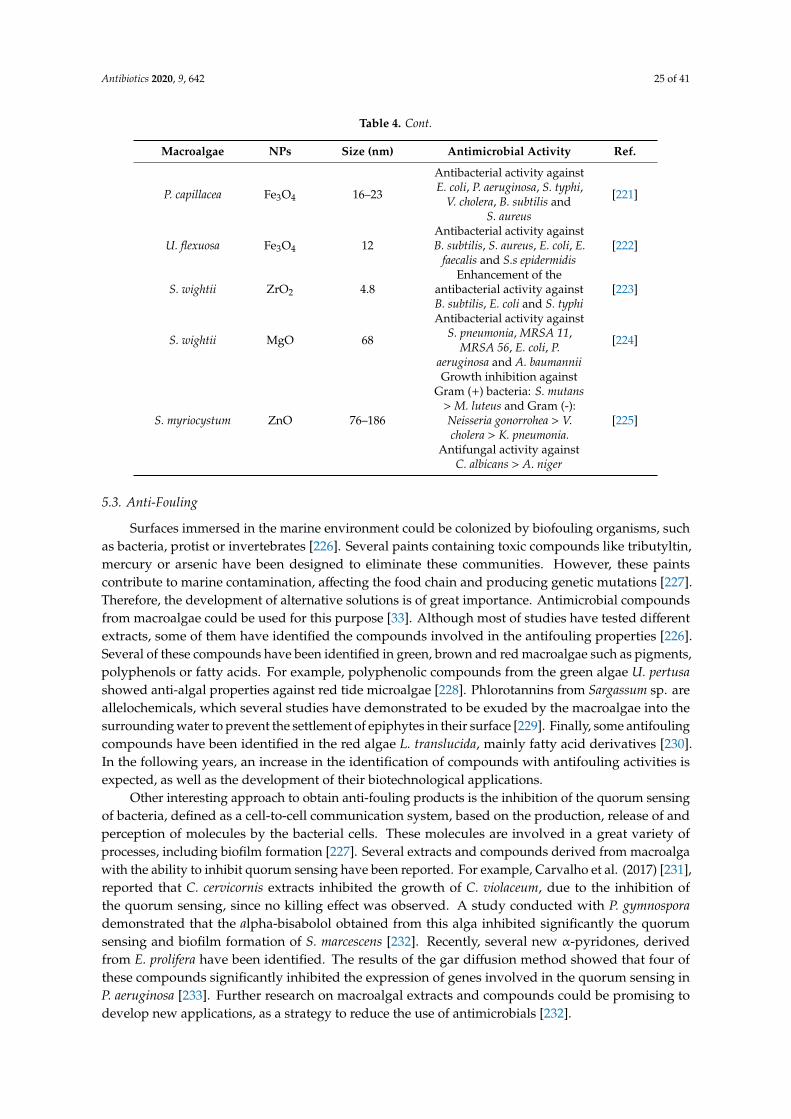
Antibiotics 2020, 9, 642 25 of 41
Table 4. Cont.
Macroalgae NPs Size (nm) Antimicrobial Activity Ref.
P. capillacea Fe3O4 16–23
Antibacterial activity againstE. coli, P. aeruginosa, S. typhi,
V. cholera, B. subtilis andS. aureus
[221]
U. flexuosa Fe3O4 12Antibacterial activity againstB. subtilis, S. aureus, E. coli, E.
faecalis and S.s epidermidis[222]
S. wightii ZrO2 4.8Enhancement of the
antibacterial activity againstB. subtilis, E. coli and S. typhi
[223]
S. wightii MgO 68
Antibacterial activity againstS. pneumonia, MRSA 11,
MRSA 56, E. coli, P.aeruginosa and A. baumannii
[224]
S. myriocystum ZnO 76–186
Growth inhibition againstGram (+) bacteria: S. mutans
> M. luteus and Gram (-):Neisseria gonorrohea > V.cholera > K. pneumonia.
Antifungal activity againstC. albicans > A. niger
[225]
5.3. Anti-Fouling
Surfaces immersed in the marine environment could be colonized by biofouling organisms, suchas bacteria, protist or invertebrates [226]. Several paints containing toxic compounds like tributyltin,mercury or arsenic have been designed to eliminate these communities. However, these paintscontribute to marine contamination, affecting the food chain and producing genetic mutations [227].Therefore, the development of alternative solutions is of great importance. Antimicrobial compoundsfrom macroalgae could be used for this purpose [33]. Although most of studies have tested differentextracts, some of them have identified the compounds involved in the antifouling properties [226].Several of these compounds have been identified in green, brown and red macroalgae such as pigments,polyphenols or fatty acids. For example, polyphenolic compounds from the green algae U. pertusashowed anti-algal properties against red tide microalgae [228]. Phlorotannins from Sargassum sp. areallelochemicals, which several studies have demonstrated to be exuded by the macroalgae into thesurrounding water to prevent the settlement of epiphytes in their surface [229]. Finally, some antifoulingcompounds have been identified in the red algae L. translucida, mainly fatty acid derivatives [230].In the following years, an increase in the identification of compounds with antifouling activities isexpected, as well as the development of their biotechnological applications.
Other interesting approach to obtain anti-fouling products is the inhibition of the quorum sensingof bacteria, defined as a cell-to-cell communication system, based on the production, release of andperception of molecules by the bacterial cells. These molecules are involved in a great variety ofprocesses, including biofilm formation [227]. Several extracts and compounds derived from macroalgawith the ability to inhibit quorum sensing have been reported. For example, Carvalho et al. (2017) [231],reported that C. cervicornis extracts inhibited the growth of C. violaceum, due to the inhibition ofthe quorum sensing, since no killing effect was observed. A study conducted with P. gymnosporademonstrated that the alpha-bisabolol obtained from this alga inhibited significantly the quorumsensing and biofilm formation of S. marcescens [232]. Recently, several new α-pyridones, derivedfrom E. prolifera have been identified. The results of the gar diffusion method showed that four ofthese compounds significantly inhibited the expression of genes involved in the quorum sensing inP. aeruginosa [233]. Further research on macroalgal extracts and compounds could be promising todevelop new applications, as a strategy to reduce the use of antimicrobials [232].

Antibiotics 2020, 9, 642 26 of 41
6. New Approaches and Future Perspectives
In the previous sections and throughout the manuscript a series of applications of the antimicrobialcompounds obtained from algae is given. Among all these applications, the most common is the use ofthese molecules or extracts as preservatives of natural origin in the food, cosmetic or pharmaceuticalindustry. Namely, in the food industry new trends are rapidly being incorporated to the market suchas active packaging or the use of antibiofilm compounds [14,34]. Additionally, research on naturalantimicrobials has also increased due to the current problem related to resistant strains. In this regard,polyphenols represent molecules widely employed by in vitro studies for their synergistic effect whenacting with other conventional antimicrobials and thus, resulting in a higher efficacy, lower dosesand side-effects reduction [234]. However, to our knowledge, few studies have assessed the in vivoproperties of polyphenols or other enriched extracts obtained from algae.
On the other hand, nanoparticles (NPs) and nanomaterials have recently gained popularity bothin engineering and medical sciences. Algae have been proposed as alternative nanobiofactories forthe synthesis of NPs since physicochemical synthesis is usually difficult and expensive. Algae canfacilitate metal reduction as they can accumulate metal ions and stabilize and remodel them. Likewise,algae extracts are known for their biological properties and particularly, they have been studied fortheir antibacterial and antifungal effects [235]. Their mechanism of action consists basically of the NPsadhesion to the microorganism surface and their penetration into cells. Once there, different interactionscan occur with the bacterial components: disruption of the enzymatic activity or the production ofreactive oxygen species (ROS) that can result in mitochondrial, protein or DNA damage [236]. The useof algae in NPs synthesis is quite a new trend and therefore, different authors have performed studieson characterizing, synthetizing and researching their antimicrobial potential. Table 4 shows a series ofexamples of algae-based NPs and their antimicrobial activity. For instance, a study found that silverNPs based on four different algae (U. faciata, P. capillacae, J. rubens and C. sinusa) could reduce thebacterial growth of S. aureus and E. coli on textile cotton fabrics. This capacity could be useful for thedesign of antiseptic dressings with biomedical applications [217]. In another case, also Ag-based NPsobtained from the red algae G. amansii, showed potential as antifouling for their antimicrobial activityagainst different microorganisms [212]. Among all the examples given on Table 4, the majority of thestudies have been carried out on silver-NPs, however other approaches are being investigated tooand their antimicrobial activity has been proved against plenty of microorganisms. Moreover, otherapproaches apart from antimicrobial or antifouling applications have been proposed, such as theiruse in bioremediation [237]. On the other hand, the use of algae extracts as prebiotics has also beenstudied. Marine algae have been pointed out as a source of oligosaccharides and polysaccharides andeven though these compounds possess several bioactivities, their use as prebiotics is rising because oftheir high content of dietary fiber, enhancing the growth of those beneficial bacteria in the intestinaltract [11,238].
Taken together, all the information given in the different manuscript sections and consideringthe favorable consumer perception about these products, there is a huge potential antimicrobials,extracted from algae, to be incorporated into different applications. Future perspectives should focuson the development of further in vivo and toxicological studies and in the transformation of the wholeprocess into a cost-effective and reproducible alternative which goes through the improvement, furtherstudy and optimization of the extraction techniques of the bioactive compounds responsible for theantimicrobial activity [239,240].
7. Conclusions
The use of bioactive compounds as a constituent in several food–cosmetic- and medical-basedproducts will soon be the norm. Among different sources, macroalgae biomass as a marine by-producthas been considered as a promising and viable source of a broader range of bioactive compounds.As evident from this review, algae bio-products include: proteins, peptides, polysaccharides,polyphenols, fatty acids, pigments, and the highest treasure antimicrobial compounds. To obtain these

Antibiotics 2020, 9, 642 27 of 41
valuable bioactive compounds, several innovative green extraction methodologies have been used toefficiently recover them from algae biomass. This review also comprises the recent advancements in theextraction techniques such as supercritical-fluid extraction, accelerated solvent extraction, ultrasound-,microwave-, and enzymatic-assisted extraction. Most of the literature concerning the antimicrobialactivity of algae is based on classic extraction techniques. Furthermore, new future approachesshould focus on the utilization of natural antimicrobial compounds obtained from algae into thepharmaceutical industry, due to the current problem related to the resistance of the drugs to pathogenicmicroorganisms. Moreover, algae have been also implicated as a possible alternative for nanoparticlesand nanomaterials synthesis, which will raise the possibility to use this by-product in engineering andmedical science.
Author Contributions: Formal analysis, M.F.B., M.A.P. and J.S.-G.; Investigation, A.S., S.A.S., M.C., P.G.-O., P.G.,M.F.B., M.A.P. and J.S.-G.; Methodology, A.S., S.A.S., M.C., P.G.-O., P.G.; Supervision M.F.B., M.A.P.and J.S.-G.;Validation, M.F.B., M.A.P. and J.S.-G.; Writing—original draft A.S., S.A.S., M.C., P.G.-O., P.G., M.F.B., M.A.P. andJ.S.-G.; Writing—review and editing, M.F.B., M.A.P. and J.S.-G. All authors have read and agreed to the publishedversion of the manuscript.
Funding: This research received no external funding.
Acknowledgments: The research leading to these results received financial support from Programa deCooperación Interreg V-A España—Portugal (POCTEP) 2014–2020 (projects Ref. 0181_NANOEATERS_01_Eand Ref. 0377_IBERPHENOL_6_E); from MICINN supporting the Ramón and Cajal grant for M.A. Prieto(RYC-2017-22891); from Xunta de Galicia and University of Vigo supporting the pre-doctoral grant of P.García-Oliveira (ED481A-2019/295); by EcoChestnut Project (Erasmus + KA202) supporting the work of M.Carpena and from postdoctoral support for P. Gullón working under the project Bio Based Industries JointUndertaking (JU) under grant agreement No 888003 UP4HEALTH Project (H2020-BBI-JTI-2019). This work alsoreceived financial support from REQUIMTE/LAQV, National Funds (FCT, Fundação para a Ciência e Tecnologia)through project UID/QUI/50006/2019 and by FEDER—Fundo Europeu de Desenvolvimento Regional fundsthrough the COMPETE 2020—Operational Programme for Competitiveness and Internationalisation (POCI),and by Portuguese funds through FCT—Fundação para a Ciência e a Tecnologia in the framework of the projectPOCI-01-0145-FEDER-030240—PTDC/OCE-ETA/30240/2017.
Conflicts of Interest: The authors declare no conflict of interest.
Abbreviations
GenericATCC American type culture collectionDW Dry weightFW Fresh weightHVS Herpes simplex virusMIC Minimal inhibition concentrationMRSA Methicillin-resistant Staphylococcus aureusMOI Multiplicity of infectionNPs NanoparticlesRSM Response surface methodologyRT Room temperatureSARS Severe acute respiratory syndromeUV UltravioletExtraction techniquesASE Accelerated solvent extractionEAE Enzyme-assisted extractionEUAE Enzymatic ultrasound assisted extractionHAE Heat-assisted extractionIPEF Intensity pulsed electric fieldsMAE Microwave-assisted extractionPLE Pressurized liquid extractionSFE Supercritical fluid extractionUAE Ultrasound assisted extraction
Antibiotics 2020, 9, 642 28 of 41
CompoundsDEt Diethyl etherDCM DichloromethaneDPPH 1,1-diphenyl-2-picryl hydrazylEtOAc Ethyl acetateEtOH EthanolH2O WaterHx HexaneMeOH Methanoln-BuOH n- Butanoln-Hx n-HexanePUFAs Poly-unsaturated fatty acidsBacteria speciesAcinetobacter (A.): A. baumannii. Actinobacillus (A.): A. actinomycetemcomitas. Actinomyces (A.): A.naeslundii, A. odontolyticus. Aeromonas (A.): A. hydrophyla, A. salmonicida. Aspergillus (A.): A. flavus, A.fumigatus, A. niger. Bacillus (B.): B. cereus, B. mycoides, B. pumilus, B. subtilis. Candida (C.): C. albicans, C.dublinensis, C. famata, C. glabrata, C. guilliermondi, C. kefyr, C. krusei, C. membranaefaciens, C. parapsilosis.Campylobacter (C.): C. jejuni. Chromobacterium (C.): C. violaceum. Colletotrichum (C.): C. lindemuthianum.Cryptococcus (C.): C. neoformans. Cultibacterium (C.): C. acnes. Enterobacter (E.): E. aerogens, E. cloacae.Enterococcus (E.): E. faecalis, E. faecium. Epidermophyton (E.): E. flocossum. Erwinia (E.): E. amylovora.Escherichia (E.): E. coli. Fusarium (F.): F. graminearum, F. culmorum, F. oxysporum, F. solani. Fusobacterium (F.):F. nucleatum. Klebsiella (K.): K. oxytoca, K. pneumonia. Lactobacillus (L.): L. brevis. Listeria (L.): L. innocua, L.monocytogenes. Leishmania (L.): L. amazonensis. Macrophomina (M.): M. phaseolina. Methicillin-resistantStaphylococcus aureus (MRSA). Micrococcus (M.): M. luteus. Microsporum (M.): M. canis, M. gypseum.Penicillium (P.): P. verrucosum. Porphyromonas (P.): P. gingivalis. Pseudoalteromonas (P.): P. bacteriolytica.Proteus (P.): P. vulgaris. Pseudomonas (P.): P. aeruginosa, P. cepacia, P. fluorescens. Rhizoctonia (R.): R. solani.Rhodococcus (R.): R. rhodochrous. Saccharomyces (S.): S. cerevisiae. Salmonella (S.): S. abony, S. choleraesuis, S.gallinarum, S. typhi, S. typhimurium. Septoria (S.): S. glycines. Serratia (S.): S. marcescens. Shigella (S.): S. boydii.Staphylococcus (S.): S. aureus, S. enterica, S. epidermidis, S. lentus, S. xylosus. Streptococcus (S.): S. agalactiae, S.epidermis, S. mutans, S. pneumonia, S. pyogenes, S. sobrinus. Streptomyces (S.): S. purpurascens. Syspastospora(S.): S. parasitica. Trichophyton (T.): T. interdigitale, T. mentagrophytes, T. rubrum, T. tonsurans, Trichosporon (T.):T. mucoides. Trypanosoma (T.): T. cruzi. Vibrio (V.): V. alginolyticus, V. anguillarum, V. cholerae, V. fluvialis, V.haryevi, V. ordalii, V. parahaemolyticus, V. salmonicida, V. splendidus, V. tapetis, V. vulnificus. Yersinia (Y.): Y. ruckeri.Macroalgae speciesChlorophyta: Boodlea (B.): B. coacta. Caulerpa (C.): C. peltata, C. prolifera, C. racemosa, C. scalpelliformis, C.
sertularioides. Cladophora (C.): C. glomerata, C. rivularis, C. rupestris, C. socialis, C. vagabunda. Codium (C.): C.bursa, C. elongatum, C. fragile, C. iyengarii, C. tomentosum. Enteromorpha (E.): E. linza, E. prolifera. Halimeda(H.): H. tuna. Monostroma (M.): M. latissimum. Rhizoclonium (R.): R. implexum. Ulva (U.): U. fasciata, U.flexuosa, U. intestinalis, U. lactuca, U. meridionalis, U. ohnoi, U. pertusa, U. prolifera, U. rigida, U. reticulata.Valonopsis (V.): V. pachynema.Rhodophyta: Acanthophora (A.): A. specifera. Actinotrichia (A.): A. fragilis. Ascophyllum (A.): A. nodosum.
Ceramium (C.): C. ciliatum, C. rubrum. Chaetomorpha (C.): C. antemmina, C. linum. Chondrus (C.): C. crispus.Corallina (C.): C. elongata, C. officinalis. Digenea (D.): D. simplex. Dumontia (D.): D. contorta. Eucheuma (E.): E.serra. Galaxaura (G.): G. cylindriea, G. marginata. Gelidium (G.): G. amansii, G. corneum. Gigartina (G.): G.skottsbergii. Gracilaria (G.): G. birdiae, G. changii, G. corticata, G. dura, G. edulis, G. gracilis, G. lemaneiformis, G.multipartita, G. ornata, G. vermiculophylla. Gracilariopsis (G.): G. longissima. Grateloupia (G.): G. doryphora, G.lithophilia. Hypnea (H.): H. esperi, H. flagelliformis, H. musciformis. Jania (J.): J. rubens. Kappaphycus (K.):K. alvarezii. Laurencia (L.): L. aldingensis, L. brandenii, L. catarinensis, L. dendroidea, L. intricata, L. johnstonii, L.obtusa, L. paniculata, L. papillosa, L. translucida. Mastocarpus (M.): M. stellatus. Melanothamnus (M.): M.afaqhusainii. Osmundaria (O.): O. obtusiloba. Osmundea (O.): O. pinnatifida. Palmaria (P.): P. palmata.Polysiphonia (P.): P. elongata. Porphyra (P.): P. dioica, P. haitanensis. Portieria (P.): P. hornemannii. Pterocladia(P.): P. capillacea. Pterocladiella (P.): P. capillacea. Rissoella (R.): R. verruculosa. Rhodomela (R.): R. confervoides.Sarcodia (S.): S. ceylonensis.

Antibiotics 2020, 9, 642 29 of 41
Ochrophyta: Bifurcaria (B.): B. bifurcata. Canistrocarpus (C.): C. cervicornis. Cladosiphon (C.): C. okamuranus.
Cladostephus (C.): C. hirsutus. Carpophyllum (C.): C. flexuosum, C. plumosum. Colpomenia (C.): C. sinuosa.Cutleria (C.): C. multifida. Cystoseira (C.): C. barbata, C. indica, C. mediterranea, C. myrica, C. nodicaulis, C.sedoides, C. trinodis, C. usneoides. Dictyopteris (D.): D. membranacea. Dictyota (D.): D. dichotoma, D. flabellata, D.indica. Dilophus (D.): D. fasciola. Ecklonia (E.): E. arborea, E. cava, E. kurome, E. radiata. Eisenia (E.): E. bicyclis.Feldmannia (F.): F. irregularis. Fucus (F.): F. evanescens, F. serratus, F. spiralis, F. vesiculosus. Gymnogongrus(G.):G. martinensis. Himanthalia (H.): H. elongata. Hormophysa (H.): H. cuneiformes. Iyengaria (I.): I. stellata.Jolyna (J.): J. laminarioides. Laminaria (L.): L. digitata, L. hyperborea, L. japonica. Lessonia (L.): L. nigrecens, L.trabeculata. Nizamuddinia (N.): N. zanardinii. Padina (P.): P. australis, P. concrescens, P. gymnospora. Pelvetia(P.): P. canaliculata. Porolithon (P.): P. antillarum, P. boergesenii. Saccharina (S.): S. latissima, S. longicruris.Sargassum (S.): S. angustifolium, S. boveanum, S. cinereum, S. dentifolium, S. hystrix, S. ilicifolium, S. incisifolium, S.latifolium, S. marginatum, S. muticum, S. myriocystum, S. oligocystum, S. pallidum, S. platycarpum, S. polycystum, S.portieriatum, S. swartzii, S. tenerrimum, S. thunbergii, S. variegatum, S. vulgare, S. wightii. Scytosiphon (S.): S.lomentaria. Solieria (S.): S. chordalis, S. filiformis, S. robusta. Spatoglossum (S.): S. asperum. Spyridia (S.): S.filamentosa. Stoechospermum (S.): S. marginatum. Taonia (T.): T. atomaria. Turbinaria (T.): T. conoides, T.decurrens, T. ornata, T. triquetra. Undaria (U.): U. pinnafitida.
References
1. Barbosa, F.; Pinto, E.; Kijjoa, A.; Pinto, M.; Sousa, E. Targeting antimicrobial drug resistance with marinenatural products. Int. J. Antimicrob. Agents 2020, 5, 106005. [CrossRef] [PubMed]
2. Gnanavel, V.; Roopan, S.M.; Rajeshkumar, S. Aquaculture: An overview of chemical ecology of seaweeds(food species) in natural products. Aquaculture 2019, 507, 1–6. [CrossRef]
3. Lourenço-Lopes, C.; Fraga-Corral, M.; Jimenez-Lopez, C.; Pereira, A.G.; Garcia-Oliveira, P.; Carpena, M.;Prieto, M.A.; Simal-Gandara, J. Metabolites from Macroalgae and Its Applications in the Cosmetic Industry:A Circular Economy Approach. Resources 2020, 9, 101. [CrossRef]
4. Leandro, A.; Pereira, L.; Gonçalves, A.M.M. Diverse applications of marine macroalgae. Mar. Drugs 2020, 18,17. [CrossRef]
5. Spalding, H.L.; Amado-Filho, G.M.; Bahia, R.G.; Ballantine, D.L.; Fredericq, S.; Leichter, J.J.; Nelson, W.A.;Slattery, M.; Tsuda, R.T. Macroalgae. In Mesophotic Coral Ecosystems; Springer: Cham, Switzerland, 2019;ISBN 978-3-319-92734-3.
6. Pepper, I.L.; Gentry, T.J. Microorganisms Found in the Environment. In Environmental Microbiology: ThirdEdition; Academic Press: Cambridge, MA, USA, 2015; pp. 9–36, ISBN 9780123946263.
7. Wells, M.L.; Potin, P.; Craigie, J.S.; Raven, J.A.; Merchant, S.S.; Helliwell, K.E.; Smith, A.G.; Camire, M.E.;Brawley, S.H. Algae as nutritional and functional food sources: Revisiting our understanding. J. Appl. Phycol.2017, 29, 949–982. [CrossRef]
8. Anyanwu, R.C.; Rodriguez, C.; Durrant, A.; Olabi, A.G. Micro-Macroalgae Properties and Applications.Ref. Modul. Mater. Sci. Mater. Eng. 2018, 1–28. [CrossRef]
9. Praveen, M.A.; Parvathy, K.R.K.; Balasubramanian, P.; Jayabalan, R. An overview of extraction and purificationtechniques of seaweed dietary fibers for immunomodulation on gut microbiota. Trends Food Sci. Technol.2019, 92, 46–64. [CrossRef]
10. Suleria, H.A.R.; Gobe, G.; Masci, P.; Osborne, S.A. Marine bioactive compounds and health promotingperspectives; innovation pathways for drug discovery. Trends Food Sci. Technol. 2016, 50, 44–55. [CrossRef]
11. Gurpilhares, D.d.B.; Cinelli, L.P.; Simas, N.K.; Pessoa, A.; Sette, L.D. Marine prebiotics: Polysaccharides andoligosaccharides obtained by using microbial enzymes. Food Chem. 2019, 280, 175–186. [CrossRef]
12. Belghit, I.; Rasinger, J.D.; Heesch, S.; Biancarosa, I.; Liland, N.; Torstensen, B.; Waagbø, R.; Lock, E.J.;Bruckner, C.G. In-depth metabolic profiling of marine macroalgae confirms strong biochemical differencesbetween brown, red and green algae. Algal Res. 2017, 26, 240–249. [CrossRef]
13. Vuong, D.; Kaplan, M.; Lacey, H.J.; Crombie, A.; Lacey, E.; Piggott, A.M. A study of the chemical diversity ofmacroalgae from South Eastern Australia. Fitoterapia 2018, 126, 53–64. [CrossRef] [PubMed]
14. Bedoux, G.; Hardouin, K.; Burlot, A.S.; Bourgougnon, N. Bioactive components from seaweeds: CosmeticApplications and Future Development; Elsevier: Amsterdam, The Netherlands, 2014; Volume 71,ISBN 9780124080621.

Antibiotics 2020, 9, 642 30 of 41
15. Hamed, I.; Özogul, F.; Özogul, Y.; Regenstein, J.M. Marine Bioactive Compounds and Their Health Benefits:A Review. Compr. Rev. Food Sci. Food Saf. 2015, 14, 446–465. [CrossRef]
16. Silva, M.; Vieira, L.; Almeida, A.P.; Kijjoa, A. The Marine Macroalgae of the Genus Ulva: Chemistry, BiologicalActivities and Potential Applications. J. Oceanogr. Mar. Res. 2013, 1, 1–6. [CrossRef]
17. Mendes, M.; Pereira, R.; Sousa Pinto, I.; Carvalho, A.P.; Gomes, A.M. Antimicrobial activity and lipid profileof seaweed extracts from the North Portuguese Coast. Int. Food Res. J. 2013, 20, 3337–3345.
18. Buschmann, A.H.; Camus, C.; Infante, J.; Neori, A.; Israel, Á.; Hernández-González, M.C.; Pereda, S.V.;Gomez-Pinchetti, J.L.; Golberg, A.; Tadmor-Shalev, N.; et al. Seaweed production: Overview of the globalstate of exploitation, farming and emerging research activity. Eur. J. Phycol. 2017, 52, 391–406. [CrossRef]
19. Sharma, K.; Guleria, S.; Razdan, V.K.; Babu, V. Synergistic antioxidant and antimicrobial activities of essentialoils of some selected medicinal plants in combination and with synthetic compounds. Ind. Crop. Prod. 2020,154, 112569. [CrossRef]
20. Abu-Ghannam, N.; Rajauria, G. Antimicrobial activity of compounds isolated from algae. In FunctionalIngredients from Algae for Foods and Nutraceuticals; Woodhead Publishing Limited: Shaston, UK, 2013;pp. 287–306, ISBN 9780857095121.
21. Roca, I.; Akova, M.; Baquero, F.; Carlet, J.; Cavaleri, M.; Coenen, S.; Cohen, J.; Findlay, D.; Gyssens, I.;Heure, O.E.; et al. The global threat of antimicrobial resistance: Science for intervention. New Microbes NewInfect. 2015, 6, 22–29. [CrossRef]
22. Yılmaz Öztürk, B.; Yenice Gürsu, B.; Dag, I. Antibiofilm and antimicrobial activities of green synthesizedsilver nanoparticles using marine red algae Gelidium corneum. Process Biochem. 2020, 89, 208–219. [CrossRef]
23. Bhowmick, S.; Mazumdar, A.; Moulick, A.; Adam, V. Algal metabolites: An inevitable substitute forantibiotics. Biotechnol. Adv. 2020, 43, 107571. [CrossRef]
24. Hornsey, I.S.; Hide, D. The production of antimicrobial compounds by British marine algae I.Antibiotic-producing marine algae. Br. Phycol. J. 1974, 9, 353–361. [CrossRef]
25. Pina-Pérez, M.C.; Rivas, A.; Martínez, A.; Rodrigo, D. Antimicrobial potential of macro and microalgaeagainst pathogenic and spoilage microorganisms in food. Food Chem. 2017, 235, 34–44. [CrossRef] [PubMed]
26. Chellat, M.F.; Raguž, L.; Riedl, R. Targeting Antibiotic Resistance. Angew. Chemie Int. Ed. 2016, 55, 6600–6626.[CrossRef] [PubMed]
27. Lewis, K. Platforms for antibiotic discovery. Nat. Rev. Drug Discov. 2013, 12, 371–387. [CrossRef] [PubMed]28. Raguraman, V.; Stanley, A.L.; MubarakAli, D.; Narendrakumar, G.; Thirugnanasambandam, R.;
Kirubagaran, R.; Thajuddin, N. Unraveling rapid extraction of fucoxanthin from Padina tetrastromatica:Purification, characterization and biomedical application. Process Biochem. 2018, 73, 211–219. [CrossRef]
29. Chemat, F.; Abert-Vian, M.; Fabiano-Tixier, A.S.; Strube, J.; Uhlenbrock, L.; Gunjevic, V.; Cravotto, G. Greenextraction of natural products. Origins, current status, and future challenges. TrAC Trends Anal. Chem. 2019,118, 248–263. [CrossRef]
30. Belwal, T.; Chemat, F.; Venskutonis, P.R.; Cravotto, G.; Jaiswal, D.K.; Bhatt, I.D.; Devkota, H.P.; Luo, Z. Recentadvances in scaling-up of non-conventional extraction techniques: Learning from successes and failures.TrAC Trends Anal. Chem. 2020, 127, 115895. [CrossRef]
31. Baweja, P.; Kumar, S.; Sahoo, D.; Levine, I. Biology of Seaweeds. In Seaweed in Health and Disease Prevention;Elsevier Inc.: Amsterdam, The Netherlands, 2016; pp. 41–106, ISBN 9780128027936.
32. Cloutier, M.; Mantovani, D.; Rosei, F. Antibacterial Coatings: Challenges, Perspectives, and Opportunities.Trends Biotechnol. 2015, 33, 637–652. [CrossRef]
33. Pérez, M.J.; Falqué, E.; Domínguez, H. Antimicrobial action of compounds from marine seaweed. Mar. Drugs2016, 14, 52. [CrossRef]
34. Shannon, E.; Abu-Ghannam, N. Antibacterial derivatives of marine algae: An overview of pharmacologicalmechanisms and applications. Mar. Drugs 2016, 14. [CrossRef]
35. Manilal, A.; Sujith, S.; Sabarathnam, B.; Kiran, G.S.; Selvin, J.; Shakir, C.; Lipton, A.P. Biological activity of thered alga Laurencia brandenii. Acta Bot. Croat. 2011, 70, 81–90. [CrossRef]
36. dos Santos Amorim, R.d.N.; Rodrigues, J.A.G.; Holanda, M.L.; Quinderé, A.L.G.; de Paula, R.C.M.;Melo, V.M.M.; Benevides, N.M.B. Antimicrobial effect of a crude sulfated polysaccharide from the redseaweed Gracilaria ornata. Brazilian Arch. Biol. Technol. 2012, 55, 171–181. [CrossRef]
Antibiotics 2020, 9, 642 31 of 41
37. Cortés, Y.; Hormazábal, E.; Leal, H.; Urzúa, A.; Mutis, A.; Parra, L.; Quiroz, A. Novel antimicrobialactivity of a dichloromethane extract obtained from red seaweed Ceramium rubrum (Hudson) (Rhodophyta:Florideophyceae) against Yersinia ruckeri and Saprolegnia parasitica, agents that cause diseases in salmonids.Electron. J. Biotechnol. 2014, 17, 126–131. [CrossRef]
38. Sasidharan, S.; Darah, I.; Noordin, M.K.M.J. In vitro antimicrobial activity against Pseudomonas aeruginosa andacute oral toxicity of marine algae Gracilaria changii. N. Biotechnol. 2010, 27, 390–396. [CrossRef] [PubMed]
39. de Alencar, D.B.; de Carvalho, F.C.T.; Rebouças, R.H.; dos Santos, D.R.; dos Santos Pires-Cavalcante, K.M.;de Lima, R.L.; Baracho, B.M.; Bezerra, R.M.; Viana, F.A.; dos Fernandes Vieira, R.H.S.; et al. Bioactiveextracts of red seaweeds Pterocladiella capillacea and Osmundaria obtusiloba (Floridophyceae: Rhodophyta)with antioxidant and bacterial agglutination potential. Asian Pac. J. Trop. Med. 2016, 9, 372–379. [CrossRef]
40. Baliano, A.P.; Pimentel, E.F.; Buzin, A.R.; Vieira, T.Z.; Romão, W.; Tose, L.V.; Lenz, D.; de Andrade, T.U.;Fronza, M.; Kondratyuk, T.P.; et al. Brown seaweed Padina gymnospora is a prominent natural wound-careproduct. Rev. Bras. Farmacogn. 2016, 26, 714–719. [CrossRef]
41. Moubayed, N.M.S.; Al Houri, H.J.; Al Khulaifi, M.M.; Al Farraj, D.A. Antimicrobial, antioxidant propertiesand chemical composition of seaweeds collected from Saudi Arabia (Red Sea and Arabian Gulf). Saudi J. Biol.Sci. 2017, 24, 162–169. [CrossRef]
42. Kim, Y.H.; Kim, J.H.; Jin, H.J.; Lee, S.Y. Antimicrobial activity of ethanol extracts of Laminaria japonica againstoral microorganisms. Anaerobe 2013, 21, 34–38. [CrossRef]
43. Akremi, N.; Cappoen, D.; Anthonissen, R.; Verschaeve, L.; Bouraoui, A. Phytochemical and in vitroantimicrobial and genotoxic activity in the brown algae Dictyopteris membranacea. S. Afr. J. Bot. 2017, 108,308–314. [CrossRef]
44. Tajbakhsh, S.; Pouyan, M.; Zandi, K.; Bahramian, P.; Sartavi, K.; Fouladvand, M.; Asayesh, G.; Barazesh, A.In vitro study of antibacterial activity of the alga Sargassum oligocystum from the Persian Gulf. Eur. Rev. Med.Pharmacol. Sci. 2011, 15, 293–298.
45. Salem, W.M.; Galal, H.; Nasr El-deen, F. Screening for antibacterial activities in some marine algae from thered sea (Hurghada, Egypt). African J. Microbiol. Res. 2011, 5, 2160–2167. [CrossRef]
46. Chiao-Wei, C.; Siew-Ling, H.; Ching-Lee, W. Antibacterial activity of Sargassum polycystum C. Agardh andPadina australis Hauck (Phaeophyceae). Afr. J. Biotechnol. 2011, 10, 14125–14131. [CrossRef]
47. Assaw, S.; Rosli, N.L.; Azmi, N.A.M.; Mazlan, N.W.; Ismail, N. Antioxidant and antibacterial activities ofpolysaccharides and methanolic crude extracts of local edible red seaweed Gracilaria sp. Malaysian Appl. Biol.2018, 47, 135–144.
48. Mashjoor, S.; Yousefzadi, M.; Esmaeili, M.A.; Rafiee, R. Cytotoxicity and antimicrobial activity of marine macroalgae (Dictyotaceae and Ulvaceae) from the Persian Gulf. Cytotechnology 2016, 68, 1717–1726. [CrossRef][PubMed]
49. Sirbu, R.; Stanciu, G.; Tomescu, A.; Ionescu, A.M.; Cadar, E. Evaluation of antioxidant and antimicrobialactivity in relation to total phenolic content of green algae from Black Sea. Rev. Chim. 2019, 70, 1197–1203.[CrossRef]
50. Ismail, A.; Ktari, L.; Ben Redjem Romdhane, Y.; Aoun, B.; Sadok, S.; Boudabous, A.; El Bour, M. AntimicrobialFatty Acids from Green Alga Ulva rigida (Chlorophyta). Biomed Res. Int. 2018, 2018. [CrossRef]
51. Abdel-Latif, H.H.; Shams El-Din, N.G.; Ibrahim, H.A.H. Antimicrobial activity of the newly recorded redalga Grateloupia doryphora collected from the Eastern Harbor, Alexandria, Egypt. J. Appl. Microbiol. 2018, 125,1321–1332. [CrossRef]
52. Omar, H.H.; Shiekh, H.M.; Gumgumjee, N.M.; El-Kazan, M.M.; El-Gendy, A.M. Antibacterial activity ofextracts of marine algae from the Red Sea of Jeddah, Saudi Arabia. Afr. J. Biotechnol. 2012, 11, 13576–13585.[CrossRef]
53. Al Khazan, M.M.; Omar, H.H.; Gumgumjee, N.M.; Shiekh, H.M.; El-Gendy, A.M. Marine macroalgae as apotential source of bioactive natural products with antibacterial activity. Main Group Chem. 2016, 15, 139–151.[CrossRef]
54. KOSANIC, M.; RANKOVIC, B.; Stanojkovic, T. Evaluation of antioxidant, antimicrobial and anticancereffects of three selected marine macroalgae. Rom. Biotechnol. Lett. 2018, 1–10.
55. Ambreen; Hira, K.; Tariq, A.; Ruqqia, R.; Sultana, V.; Jehan, A. Evaluation of biochemical component andantimicrobial activity of some seaweeeds occurring at Karachi coast. Pakistan J. Bot. 2012, 44, 1799–1803.

Antibiotics 2020, 9, 642 32 of 41
56. Cavallo, R.A.; Acquaviva, M.I.; Stabili, L.; Cecere, E.; Petrocelli, A.; Narracci, M. Antibacterial activity ofmarine macroalgae against fish pathogenic Vibrio species. Cent. Eur. J. Biol. 2013, 8, 646–653. [CrossRef]
57. Stabili, L.; Acquaviva, M.I.; Biandolino, F.; Cavallo, R.A.; De Pascali, S.A.; Fanizzi, F.P.; Narracci, M.; Cecere, E.;Petrocelli, A. Biotechnological potential of the seaweed Cladophora rupestris (Chlorophyta, Cladophorales)lipidic extract. New Biotechnol. 2014, 31, 436–444. [CrossRef] [PubMed]
58. Stabili, L.; Acquaviva, M.I.; Biandolino, F.; Cavallo, R.A.; de Pascali, S.A.; Fanizzi, F.P.; Narracci, M.;Petrocelli, A.; Cecere, E. The lipidic extract of the seaweed Gracilariopsis longissima (Rhodophyta, Gracilariales):A potential resource for biotechnological purposes? New Biotechnol. 2012, 29, 443–450. [CrossRef] [PubMed]
59. Manikandan, S.; Ganesapandian, S.; Singh, M.; Sangeetha, N.; Kumaraguru, A.K. Antimicrobial activity ofseaweeds against multi drug resistant strains. Int. J. Pharmacol. 2011, 7, 522–526. [CrossRef]
60. Osman, M.E.H.; Abushady, A.M.; Elshobary, M.E. In vitro screening of antimicrobial activity of extractsof some macroalgae collected from Abu-Qir bay Alexandria, Egypt. Afr. J. Biotechnol. 2010, 9, 7203–7208.[CrossRef]
61. Águila-Ramírez, R.N.; Arenas-González, A.; Hernández-Guerrero, C.J.; González-Acosta, B.;Borges-Souza, J.M.; Véron, B.; Pope, J.; Hellio, C. Antimicrobial and antifouling activities achieved byextracts of seaweeds from Gulf of California, Mexico. Hidrobiologica 2012, 22, 8–15.
62. Lee, J.-H.; Eom, S.-H.; Lee, E.-H.; Jung, Y.-J.; Kim, H.-J.; Jo, M.-R.; Son, K.-T.; Lee, H.-J.; Kim, J.H.; Lee, M.-S.;et al. In vitro antibacterial and synergistic effect of phlorotannins isolated from edible brown seaweed Eiseniabicyclis against acne-related bacteria. ALGAE 2014. [CrossRef]
63. Tajbakhsh, S.; Ilkhani, M.; Rustaiyan, A.; Larijani, K.; Sartavi, K.; Tahmasebi, R.; Asayesh, G. Antibacterialeffect of the brown alga Cystoseira trinodis. J. Med. Plant Res. 2011, 5, 4654–4657.
64. Ertürk, Ö.; Tas, B. Antibacterial and antifungal effects of some marine algae. Kafkas Univ. Vet. Fak. Derg.2011, 17, 121–124.
65. Ghania, A.; Nabila, B.B.; Larbi, B.; Elisabeth, M.; Philippe, G.; Mariem, B.; Khadidja, K.K.; Wacila, B.R.;Fawzia, A.B. Antimicrobial and antiparasitic activities of three algae from the northwest coast of Algeria.Nat. Prod. Res. 2019, 33, 742–745. [CrossRef]
66. Stein, E.M.; Colepicolo, P.; Afonso, F.A.K.; Fujii, M.T. Screening for antifungal activities of extracts of theBrazilian seaweed genus Laurencia (Ceramiales, Rhodophyta). Braz. J. Pharmacogn. 2011, 21, 290–295.[CrossRef]
67. Aoun, Z.B.; Said, R.B.; Farhat, F. Anti-inflammatory, antioxidant and antimicrobial activities of aqueous andorganic extracts from Dictyopteris membranacea. Bot. Mar. 2010, 53, 259–264. [CrossRef]
68. Rosaline, X.D.; Sakthivelkumar, S.; Rajendran, K.; Janarthanan, S. Screening of selected marine algae fromthe coastal Tamil Nadu, South India for antibacterial activity. Asian Pac. J. Trop. Biomed. 2012, 2. [CrossRef]
69. Goecke, F.; Labes, A.; Wiese, J.; Imhoff, J.F. Dual effect of macroalgal extracts on growth of bacteria in WesternBaltic Sea. Rev. Biol. Mar. Oceanogr. 2012, 47, 75–86. [CrossRef]
70. Milovic, S.; Kundakovic, T.; Macic, V.; Stankovic, J.A.; Grozdanic, N.; Ðuricic, I.; Stankovic, I.; Stanojkovic, T.Anti α-glucosidase, antitumour, antioxidative, antimicrobial activity, nutritive and health protective potentialof some seaweeds from the Adriatic coast of Montenegro. Farmacia 2017, 65, 731–740.
71. Arulkumar, A.; Rosemary, T.; Paramasivam, S.; Rajendran, R.B. Phytochemical composition, in vitroantioxidant, antibacterial potential and GC-MS analysis of red seaweeds (Gracilaria corticata and Gracilariaedulis) from Palk Bay, India. Biocatal. Agric. Biotechnol. 2018, 15, 63–71. [CrossRef]
72. Cox, S.; Abu-Ghannam, N.; Gupta, S. An assessment of the antioxidant and antimicrobial activity of sixspecies of edible Irish seaweeds. Int. Food Res. J. 2010, 17, 205–220. [CrossRef]
73. Arumugam, G.; Rajendran, R. Anti-candidal activity and synergetic interaction of antifungal drugs withdifferential extract of brown algae Stocheospermum marginatum. Biocatal. Agric. Biotechnol. 2019, 19, 101145.[CrossRef]
74. Taskin, E.; Caki, Z.; Ozturk, M.; Taskin, E. Assessment of in vitro antitumoral and antimicrobial activities ofmarine algae harvested from the eastern Mediterranean sea. Afr. J. Biotechnol. 2010, 9, 4272–4277. [CrossRef]
75. Kosanic, M.; Rankovic, B.; Stanojkovic, T. Biological activities of two macroalgae from Adriatic coast ofMontenegro. Saudi J. Biol. Sci. 2015, 22, 390–397. [CrossRef]
76. Osman, N.A.H.K.; Siam, A.A.; El-manawy, I.M.; Jeon, Y. Anti-microbial and Anti-diabetic Activity of SixSeaweeds Collected from the Red Sea, Egypt. Catrina Int. J. Environ. Sci. 2019, 19, 55–60.
Antibiotics 2020, 9, 642 33 of 41
77. Kang, O.; Brice, O.; Lee, Y.; Chae, H. Antibacterial activity of Ecklonia cava against methicillin-resistantStaphylococcus aureus and Salmonella spp. Foodborne Pathog. Dis. 2010, 7, 435–441. [CrossRef]
78. Abdel-Raouf, N.; Mohamed, H.; Mostafa, S.; Ibraheem, I. Controlling of Microbial Growth by UsingCystoseira barbata Extract. Egypt. J. Bot. 2017, 57, 469–477. [CrossRef]
79. Bhuyar, P.; Rahim, M.H.; Sundararaju, S.; Maniam, G.P.; Govindan, N. Antioxidant and antibacterial activityof red seaweed; Kappaphycus alvarezii against pathogenic bacteria. Glob. J. Environ. Sci. Manag. 2020, 5,47–58. [CrossRef]
80. Guedes, E.A.C.; dos Santos Araújo, M.A.; Souza, A.K.P.; de Souza, L.I.O.; de Barros, L.D.; de AlbuquerqueMaranhão, F.C.; Sant’Ana, A.E.G. Antifungal Activities of Different Extracts of Marine Macroalgae AgainstDermatophytes and Candida Species. Mycopathologia 2012, 174, 223–232. [CrossRef]
81. Alves, C.; Pinteus, S.; Simões, T.; Horta, A.; Silva, J.; Tecelão, C.; Pedrosa, R. Bifurcaria bifurcata: A keymacro-alga as a source of bioactive compounds and functional ingredients. Int. J. Food Sci. Technol. 2016, 51,1638–1646. [CrossRef]
82. Jassbi, A.R.; Mohabati, M.; Eslami, S.; Sohrabipour, J.; Miri, R. Biological activity and chemical constituentsof red and brown algae from the Persian Gulf. Iran. J. Pharm. Res. 2013, 12, 339–348. [CrossRef]
83. Shanmugam, S.K.; Kumar, Y.; Yar, K.M.S.; Gupta, V.; de Clercq, E. Antimicrobial and cytotoxic activities ofTurbinaria conoides (J.Agardh) Kuetz. Iran. J. Pharm. Res. 2010, 9, 411–416. [CrossRef]
84. Kosanic, M.; Rankovic, B.; Stanojkovic, T. Brown macroalgae from the Adriatic Sea as a promising source ofbioactive nutrients. J. Food Meas. Charact. 2019, 13, 330–338. [CrossRef]
85. Chan, Y.S.; Ong, C.W.; Chuah, B.L.; Khoo, K.S.; Chye, F.Y.; Sit, N.W. Antimicrobial, antiviral and cytotoxicactivities of selected marine organisms collected from the coastal areas of Malaysia. J. Mar. Sci. Technol. 2018,26, 128–136. [CrossRef]
86. Deveau, A.M.; Miller-Hope, Z.; Lloyd, E.; Williams, B.S.; Bolduc, C.; Meader, J.M.; Weiss, F.; Burkholder, K.M.Antimicrobial activity of extracts from macroalgae Ulva lactuca against clinically important Staphylococciis impacted by lunar phase of macroalgae harvest. Lett. Appl. Microbiol. 2016, 62, 363–371. [CrossRef][PubMed]
87. Rahelivao, M.P.; Gruner, M.; Andriamanantoanina, H.; Bauer, I.; Knölker, H.J. Brown Algae (Phaeophyceae)from the Coast of Madagascar: Preliminary Bioactivity Studies and Isolation of Natural Products. Nat. ProductsBioprospect. 2015, 5, 223–235. [CrossRef]
88. Mickymaray, S.; Alturaiki, W. Antifungal efficacy of marine macroalgae against fungal isolates from bronchialasthmatic cases. Molecules 2018, 23, 3032. [CrossRef]
89. Selim, S.; Amin, A.; Hassan, S.; Hagazey, M. Antibacterial, cytotoxicity and anticoagulant activities fromHypnea esperi and Caulerpa prolifera marine algae. Pak. J. Pharm. Sci. 2015, 28, 525–530. [PubMed]
90. Rahelivao, M.P.; Gruner, M.; Andriamanantoanina, H.; Andriamihaja, B.; Bauer, I.; Knölker, H.J. Red algae(Rhodophyta) from the coast of Madagascar: Preliminary bioactivity studies and isolation of natural products.Mar. Drugs 2015, 13, 4197–4216. [CrossRef]
91. Rajauria, G.; Jaiswal, A.K.; Abu-Gannam, N.; Gupta, S. Antimicrobial, antioxidant and free radical-scavengingcapacity of brown seaweed Himanthalia elongata from western coast of Ireland. J. Food Biochem. 2013, 37,322–335. [CrossRef]
92. Higgins Hoare, A.; Tan, S.P.; McLoughlin, P.; Mulhare, P.; Hughes, H. The Screening and Evaluation of Fucusserratus and Fucus vesiculosus Extracts against Current Strains of MRSA Isolated from a Clinical HospitalSetting. Sci. Rep. 2019, 9, 1–9. [CrossRef]
93. Van Tran, T.T.; Truong, H.B.; Tran, N.H.V.; Quach, T.M.T.; Nguyen, T.N.; Bui, M.L.; Yuguchi, Y.; Thanh, T.T.T.Structure, conformation in aqueous solution and antimicrobial activity of ulvan extracted from green seaweedUlva reticulata. Nat. Prod. Res. 2018, 32, 2291–2296. [CrossRef]
94. Trigui, M.; Gasmi, L.; Zouari, I.; Tounsi, S. Seasonal variation in phenolic composition, antibacterial andantioxidant activities of Ulva rigida (Chlorophyta) and assessment of antiacetylcholinesterase potential.J. Appl. Phycol. 2013, 25, 319–328. [CrossRef]
95. Vijayavel, K.; Martinez, J.A. In vitro antioxidant and antimicrobial activities of two Hawaiian marine Limu:Ulva fasciata (Chlorophyta) and Gracilaria salicornia (Rhodophyta). J. Med. Food 2010, 13, 1494–1499. [CrossRef]
96. Zouaoui, B.; Ghalem, B.R. The Phenolic Contents and Antimicrobial Activities of Some Marine Algae fromthe Mediterranean Sea (Algeria). Russ. J. Mar. Biol. 2017, 43, 491–495. [CrossRef]
Antibiotics 2020, 9, 642 34 of 41
97. Vieira, E.F.; Soares, C.; Machado, S.; Correia, M.; Ramalhosa, M.J.; Oliva-teles, M.T.; Paula Carvalho, A.;Domingues, V.F.; Antunes, F.; Oliveira, T.A.C.; et al. Seaweeds from the Portuguese coast as a source ofproteinaceous material: Total and free amino acid composition profile. Food Chem. 2018, 269, 264–275.[CrossRef] [PubMed]
98. Cherry, P.; Yadav, S.; Strain, C.R.; Allsopp, P.J.; Mcsorley, E.M.; Ross, R.P.; Stanton, C. Prebiotics fromseaweeds: An ocean of opportunity? Mar. Drugs 2019, 17, 327. [CrossRef] [PubMed]
99. Ciko, A.M.; Jokic, S.; Šubaric, D.; Jerkovic, I. Overview on the application of modern methods for theextraction of bioactive compounds from marine macroalgae. Mar. Drugs 2018, 16, 348. [CrossRef] [PubMed]
100. Bak, U.G.; Nielsen, C.W.; Marinho, G.S.; Gregersen, Ó.; Jónsdóttir, R.; Holdt, S.L. The seasonal variation innitrogen, amino acid, protein and nitrogen-to-protein conversion factors of commercially cultivated FaroeseSaccharina latissima. Algal Res. 2019, 42, 101576. [CrossRef]
101. Stack, J.; Tobin, P.R.; Gietl, A.; Harnedy, P.A.; Stengel, D.B.; FitzGerald, R.J. Seasonal variation in nitrogenouscomponents and bioactivity of protein hydrolysates from Porphyra dioica. J. Appl. Phycol. 2017, 29, 2439–2450.[CrossRef]
102. Pangestuti, R.; Kim, S. Seaweed proteins, peptides, and amino acids. In Seaweed Sustainablity; Elsevier Inc.:Amsterdam, The Netherlands, 2015; pp. 125–140, ISBN 9780124186972.
103. Cerná, M. Seaweed Proteins and Amino Acids as Nutraceuticals. In Advances in Food and Nutrition Research;Elsevier Inc.: Amsterdam, The Netherlands, 2011; Volume 64, pp. 297–312, ISBN 9780123876690.
104. Admassu, H.; Abdalbasit, M.; Yang, R.; Zhao, W.; War, W. Bioactive Peptides Derived from Seaweed Proteinand Their Health Benefits: Antihypertensive, Antioxidant, and Antidiabetic Properties. J. Food Sci. 2018, 83,6–16. [CrossRef]
105. Beaulieu, L.; Bondu, S.; Doiron, K.; Rioux, L.; Turgeon, S.L. Characterization of antibacterial activityfrom protein hydrolysates of the macroalga Saccharina longicruris and identification of peptides implied inbioactivity. J. Funct. Foods 2015, 17, 685–697. [CrossRef]
106. Cordeiro, R.A.; Gomes, V.M.; Carvalho, A.F.U.; Melo, V.M.M. Effect of proteins from the red seaweed Hypneamusciformis (Wulfen) lamouroux on the growth of human pathogen yeasts. Braz. Arch. Biol. Technol. 2006, 49,915–921. [CrossRef]
107. Holanda, M.L.; Melo, V.M.M.; Silva, L.M.C.M.; Amorim, R.C.N.; Pereira, M.G.; Benevides, N.M.B. Differentialactivity of a lectin from Solieria filiformis against human pathogenic bacteria. Brazilian J. Med. Biol. Res.2005, 38, 1769–1773. [CrossRef]
108. Xu, S.; Huang, X.; Cheong, K. Recent Advances in Marine Algae Polysaccharides: Isolation, Structure, andActivities. Mar. Drugs 2017, 15, 388. [CrossRef] [PubMed]
109. Berri, M.; Olivier, M.; Holbert, S.; Dupont, J.; Demais, H.; Le Goff, M.; Collen, P.N. Ulvan from Ulva armoricana(Chlorophyta) activates the PI3K/Akt signalling pathway via TLR4 to induce intestinal cytokine production.Algal Res. 2017, 28, 39–47. [CrossRef]
110. Cho, M.; You, S. Sulfated Polysaccharides from Green Seaweeds. In Springer Handbook of Marine Biotechnology;Springer: Berlin/Heidelberg, Germany, 2015; pp. 941–953.
111. Bouhlal, R.; Haslin, C.; Chermann, J.C.; Colliec-Jouault, S.; Sinquin, C.; Simon, G.; Cerantola, S.; Riadi, H.;Bourgougnon, N. Antiviral activities of sulfated polysaccharides isolated from Sphaerococcus coronopifolius(Rhodophytha, Gigartinales) and Boergeseniella thuyoides (Rhodophyta, Ceramiales). Mar. Drugs 2011, 9,1187–1209. [CrossRef] [PubMed]
112. Ermakova, S.; Men’shova, R.; Vishchuk, O.; Kim, S.M.; Um, B.H.; Isakov, V.; Zvyagintseva, T. Water-solublepolysaccharides from the brown alga Eisenia bicyclis: Structural characteristics and antitumor activity. AlgalRes. 2013, 2, 51–58. [CrossRef]
113. De Jesus Raposo, M.F.; De Morais, A.M.B.; De Morais, R.M.S.C. Marine polysaccharides from algae withpotential biomedical applications. Mar. Drugs 2015, 13, 2967–3028. [CrossRef] [PubMed]
114. O’Doherty, J.V.; McDonnell, P.; Figat, S. The effect of dietary laminarin and fucoidan in the diet of theweanling piglet on performance and selected faecal microbial populations. Livest. Sci. 2010, 134, 208–210.[CrossRef]
115. Belattmania, Z.; Engelen, A.H.; Pereira, H.; Serrão, E.A.; Custódio, L.; Varela, J.C.; Zrid, R.; Reani, A.;Sabour, B. Fatty acid composition and nutraceutical perspectives of brown seaweeds from the Atlantic coastof Morocco. Int. Food Res. J. 2018, 25, 1520–1527.
Antibiotics 2020, 9, 642 35 of 41
116. Kendel, M.; Wielgosz-Collin, G.; Bertrand, S.; Roussakis, C.; Bourgougnon, N.B.; Bedoux, G. Lipid composition,fatty acids and sterols in the seaweeds ulva armoricana, and solieria chordalis from brittany (France): Ananalysis from nutritional, chemotaxonomic, and antiproliferative activity perspectives. Mar. Drugs 2015, 13,5606–5628. [CrossRef]
117. Schmid, M.; Guihéneuf, F.; Stengel, D.B. Fatty acid contents and profiles of 16 macroalgae collected from theIrish Coast at two seasons. J. Appl. Phycol. 2014, 26, 451–463. [CrossRef]
118. Cardoso, C.; Ripol, A.; Afonso, C.; Freire, M.; Varela, J.; Quental-Ferreira, H.; Pousão-Ferreira, P.; Bandarra, N.Fatty acid profiles of the main lipid classes of green seaweeds from fish pond aquaculture. Food Sci. Nutr.2017, 5, 1186–1194. [CrossRef]
119. El Baz, F.K.; El Baroty, G.S.; Abd El Baky, H.H.; Abd El-Salam, O.I.; Ibrahim, E.A. Structural characterizationand biological activity of sulfolipids from selected marine algae. Grasas y Aceites 2013, 64, 561–571. [CrossRef]
120. Mekinic, I.G.; Skroza, D.; Šimat, V.; Hamed, I.; Cagalj, M.; Perkovic, Z.P. Phenolic content of brown algae(Pheophyceae) species: Extraction, identification, and quantification. Biomolecules 2019, 9, 244. [CrossRef]
121. Gómez-Guzmán, M.; Rodríguez-Nogales, A.; Algieri, F.; Gálvez, J. Potential role of seaweed polyphenols incardiovascular-associated disorders. Mar. Drugs 2018, 16, 250. [CrossRef] [PubMed]
122. Cassani, L.; Gomez-zavaglia, A.; Jimenez-lopez, C.; Prieto, M.A.; Simal-gandara, J. Seaweed-based naturalingredients: Stability of phlorotannins during extraction, storage, passage through the gastrointestinal tractand potential incorporation into functional foods. Food Res. Int. 2020, 109676. [CrossRef]
123. Montero, L.; del Pilar Sánchez-Camargo, A.; Ibáñez, E.; Gilbert-López, B. Phenolic Compounds from EdibleAlgae: Bioactivity and Health Benefits. Curr. Med. Chem. 2017, 25, 4808–4826. [CrossRef]
124. Yuan, Y.; Zhang, J.; Fan, J.; Clark, J.; Shen, P.; Li, Y.; Zhang, C. Microwave assisted extraction of phenoliccompounds from four economic brown macroalgae species and evaluation of their antioxidant activities andinhibitory effects on α-amylase, α-glucosidase, pancreatic lipase and tyrosinase. Food Res. Int. 2018, 113,288–297. [CrossRef]
125. Nagayama, K.; Iwamura, Y.; Shibata, T.; Hirayama, I.; Nakamura, T. Bactericidal activity of phlorotanninsfrom the brown alga Ecklonia kurome. J. Antimicrob. Chemother. 2002, 50, 889–893. [CrossRef]
126. Aryee, A.N.; Agyei, D.; Akanbi, T.O. Recovery and utilization of seaweed pigments in food processing. Curr.Opin. Food Sci. 2018, 19, 113–119. [CrossRef]
127. Michalak, I.; Chojnacka, K. Algae as production systems of bioactive compounds. Eng. Life Sci. 2015, 15,160–176. [CrossRef]
128. Karpinski, T.M.; Adamczak, A. Fucoxanthin—An Antibacterial Carotenoid. Antioxidants 2019, 8, 239.[CrossRef]
129. Bogolitsyn, K.; Dobrodeeva, L.; Druzhinina, A.; Ovchinnikov, D.; Parshina, A.; Shulgina, E. Biological activityof a polyphenolic complex of Arctic brown algae. J. Appl. Phycol. 2019, 31, 3341–3348. [CrossRef]
130. Wei, Y.; Liu, Q.; Xu, C.; Yu, J.; Zhao, L.; Guo, Q. Damage to the Membrane Permeability and Cell Death ofVibrio parahaemolyticus Caused by Phlorotannins with Low Molecular Weight from Sargassum thunbergii.J. Aquat. Food Prod. Technol. 2016, 25, 323–333. [CrossRef]
131. Eom, S.H.; Lee, D.S.; Jung, Y.J.; Park, J.H.; Choi, J.I.; Yim, M.J.; Jeon, J.M.; Kim, H.W.; Son, K.T.; Je, J.Y.; et al.The mechanism of antibacterial activity of phlorofucofuroeckol-A against methicillin-resistant Staphylococcusaureus. Appl. Microbiol. Biotechnol. 2014, 98, 9795–9804. [CrossRef] [PubMed]
132. Cherian, C.; Jannet Vennila, J.; Sharan, L. Marine bromophenols as an effective inhibitor of virulent proteins(peptidyl arginine deiminase, gingipain R and hemagglutinin A) in Porphyromas gingivalis. Arch. Oral Biol.2019, 100, 119–128. [CrossRef] [PubMed]
133. Lee, M.H.; Lee, K.B.; Oh, S.M.; Lee, B.H.; Chee, H.Y. Antifungal activities of dieckol isolated from the marinebrown alga Ecklonia cava against Trichophyton rubrum. J. Appl. Biol. Chem. 2010, 53, 504–507. [CrossRef]
134. Lopes, G.; Pinto, E.; Andrade, P.B.; Valentão, P. Antifungal Activity of Phlorotannins against Dermatophytesand Yeasts: Approaches to the Mechanism of Action and Influence on Candida albicans Virulence Factor. PLoSONE 2013, 8, e72203. [CrossRef]
135. Ryu, Y.B.; Jeong, H.J.; Yoon, S.Y.; Park, J.Y.; Kim, Y.M.; Park, S.J.; Rho, M.C.; Kim, S.J.; Lee, W.S. Influenzavirus neuraminidase inhibitory activity of phlorotannins from the edible brown alga Ecklonia cava. J. Agric.Food Chem. 2011, 59, 6467–6473. [CrossRef]
Antibiotics 2020, 9, 642 36 of 41
136. Morán-Santibañez, K.; Peña-Hernáncez, M.A.; Cruz-Suárez, L.E.; Ricque-Marie, D.; Skouta, R.; Vasquez, A.H.;Rodríguez-Padilla, C.; Trejo-Avila, L.M. Virucidal and Synergistic Activity of Polyphenol-Rich Extracts ofSeaweeds against Measles Virus. Viruses 2018, 10, 465. [CrossRef]
137. Liu, M.; Liu, Y.; Cao, M.; Liu, G.; Chen, Q.; Sun, L.; Chen, H. Antibacterial activity and mechanisms ofdepolymerized fucoidans isolated from Laminaria japonica. Carbohydr. Polym. 2017, 172, 294–305. [CrossRef]
138. Id, J.J.; Jung, M.; Jeong, I.; Yamazaki, K.; Kawai, Y. Antimicrobial and Antibiofilm Activities of SulfatedPolysaccharides from Marine Algae against Dental. Mar. Drugs 2018, 16, 301. [CrossRef]
139. Kadam, S.U.; Donnell, C.P.O.; Rai, D.K.; Hossain, M.B.; Burgess, C.M.; Walsh, D.; Tiwari, B.K. Laminarinfrom Irish brown seaweeds Ascophyllum nodosum and Laminaria hyperborea: Ultrasound assisted extraction,characterization and bioactivity. Mar. Drugs 2015, 13, 4270–4280. [CrossRef] [PubMed]
140. Zeid, A.H.A.; Aboutabl, E.A.; Sleem, A.A.; El-rafie, H.M. Water soluble polysaccharides extracted fromPterocladia capillacea and Dictyopteris membranacea and their biological activities. Carbohydr. Polym. 2014, 113,62–66. [CrossRef] [PubMed]
141. Siahaan, E.A.; Pangestuti, R.; Kim, S.-K. Seaweeds: Valuable Ingredients for the Pharmaceutical Industries;Springer: Cham, Siwtzerland, 2018; ISBN 9783319690759.
142. Singh, R.S.; Walia, A.K. Lectins from red algae and their biomedical potential. J. Appl. Phycol. 2018, 30,1833–1858. [CrossRef] [PubMed]
143. Cheung, R.C.F.; Wong, J.H.; Pan, W.; Chan, Y.S.; Yin, C.; Dan, X.; Ng, T.B. Marine lectins and their medicinalapplications. Appl. Microbiol. Biotechnol. 2015, 99, 3755–3773. [CrossRef]
144. El Shafay, S.M.; Ali, S.S.; El-Sheekh, M.M. Antimicrobial activity of some seaweeds species from Red sea,against multidrug resistant bacteria. Egypt. J. Aquat. Res. 2016, 42, 65–74. [CrossRef]
145. Kasanah, N.; Amelia, W.; Mukminin, A.; Triyanto; Isnansetyo, A. Antibacterial activity of Indonesian redalgae Gracilaria edulis against bacterial fish pathogens and characterization of active fractions. Nat. Prod. Res.2019, 33, 3303–3307. [CrossRef]
146. Anjali, K.P.; Sangeetha, B.M.; Devi, G.; Raghunathan, R.; Dutta, S. Bioprospecting of seaweeds (Ulva lactucaand Stoechospermum marginatum): The compound characterization and functional applications in medicine-acomparative study. J. Photochem. Photobiol. B Biol. 2019, 200, 111622. [CrossRef]
147. de Felício, R.; de Albuquerque, S.; Young, M.C.M.; Yokoya, N.S.; Debonsi, H.M. Trypanocidal, leishmanicidaland antifungal potential from marine red alga Bostrychia tenella J. Agardh (Rhodomelaceae, Ceramiales).J. Pharm. Biomed. Anal. 2010, 52, 763–769. [CrossRef]
148. Wang, H.; Li, Y.; Shen, W.; Rui, W.; Ma, X.; Cen, Y. Antiviral activity of a sulfoquinovosyldiacylglycerol (SQDG ) compound isolated from the green alga Caulerpa racemosa. Bot. Mar. 2007, 50, 185–190. [CrossRef]
149. Rajauria, G.; Abu-ghannam, N. Isolation and Partial Characterization of Bioactive Fucoxanthin fromHimanthalia elongata Brown Seaweed: A TLC-Based Approach. Int. J. Anal. Chem. 2013, 2013. [CrossRef]
150. Gerasimenko, N.I.; Martyyas, E.A.; Logvinov, S.V.; Busarova, N.G. Biological activity of lipids andphotosynthetic pigments of Sargassum pallidum C. Agardh. Appl. Biochem. Microbiol. 2014, 50, 73–81.[CrossRef]
151. Mendiola, J.A.; Santoyo, S.; Cifuentes, A.; Reglero, G.; Ibáñez, E.; Javier Señoráns, F. Antimicrobial activityof sub- and supercritical CO2 extracts of the green alga Dunaliella salina. J. Food Prot. 2008, 71, 2138–2143.[CrossRef] [PubMed]
152. Tyskiewicz, K.; Tyskiewicz, R.; Konkol, M.; Rój, E.; Jaroszuk-Sciseł, J.; Skalicka-Wozniak, K. AntifungalProperties of Fucus vesiculosus L. Supercritical Fluid Extract against Fusarium culmorum and Fusariumoxysporum. Molecules 2019, 24. [CrossRef]
153. Alboofetileh, M.; Rezaei, M.; Tabarsa, M.; Rittà, M.; Donalisio, M.; Mariatti, F.; You, S.G.; Lembo, D.;Cravotto, G. Effect of different non-conventional extraction methods on the antibacterial and antiviral activityof fucoidans extracted from Nizamuddinia zanardinii. Int. J. Biol. Macromol. 2019, 124, 131–137. [CrossRef][PubMed]
154. Munir, N.; Rafique, M.; Altaf, I.; Sharif, N.; Naz, S. Antioxidant and antimicrobial activities of extracts fromselected algal species. Bangladesh J. Bot. 2018, 47, 53–61.
155. Sabeena, S.F.; Alagarsamy, S.; Sattari, Z.; Al-Haddad, S.; Fakhraldeen, S.; Al-Ghunaim, A.; Al-Yamani, F.Enzyme-assisted extraction of bioactive compounds from brown seaweeds and characterization. J. Appl.Phycol. 2020, 32, 615–629. [CrossRef]

Antibiotics 2020, 9, 642 37 of 41
156. Meillisa, A.; Siahaan, E.A.; Park, J.N.; Woo, H.C.; Chun, B.S. Effect of subcritical water hydrolysate in thebrown seaweed Saccharina japonica as a potential antibacterial agent on food-borne pathogens. J. Appl. Phycol.2013, 25, 763–769. [CrossRef]
157. Otero, P.; Quintana, S.E.; Reglero, G.; Fornari, T.; García-Risco, M.R. Pressurized Liquid Extraction (PLE) asan innovative green technology for the effective enrichment of galician algae extracts with high quality fattyacids and antimicrobial and antioxidant properties. Mar. Drugs 2018, 16. [CrossRef]
158. Plaza, M.; Santoyo, S.; Jaime, L.; García-Blairsy Reina, G.; Herrero, M.; Señoráns, F.J.; Ibáñez, E. Screening forbioactive compounds from algae. J. Pharm. Biomed. Anal. 2010, 51, 450–455. [CrossRef]
159. Heavisides, E.; Rouger, C.; Reichel, A.F.; Ulrich, C.; Wenzel-Storjohann, A.; Sebens, S.; Tasdemir, D. Seasonalvariations in the metabolome and bioactivity profile of fucus vesiculosus extracted by an optimised,pressurised liquid extraction protocol. Mar. Drugs 2018, 16, 503. [CrossRef]
160. Sosa-Hernández, J.E.; Escobedo-Avellaneda, Z.; Iqbal, H.M.N.; Welti-Chanes, J. State-of-the-art extractionmethodologies for bioactive compounds from algal biome to meet bio-economy challenges and opportunities.Molecules 2018, 23. [CrossRef] [PubMed]
161. Gallego, R.; Bueno, M.; Herrero, M. Sub- and supercritical fl uid extraction of bioactive compounds fromplants, food-by-products, seaweeds and microalgae—An update. Trends Anal. Chem. 2019, 116, 198–213.[CrossRef]
162. Saravana, P.S.; Getachew, A.T.; Cho, Y.J.; Choi, J.H.; Park, Y.B.; Woo, H.C.; Chun, B.S. Influence of co-solventson fucoxanthin and phlorotannin recovery from brown seaweed using supercritical CO2. J. Supercrit. Fluids2017, 120, 295–303. [CrossRef]
163. Bogolitsyn, K.G.; Kaplitsin, P.A.; Dobrodeeva, L.K.; Druzhinina, A.S.; Ovchinnikov, D.V.; Parshina, A.E.;Shulgina, E.V. Fatty Acid Composition and Biological Activity of Supercritical Extracts from Arctic BrownAlgae Fucus vesiculosus. Russ. J. Phys. Chem. B 2017, 11, 1144–1152. [CrossRef]
164. Fabrowska, J.; Messyasz, B.; Pankiewicz, R.; Wilinska, P.; Łeska, B. Seasonal differences in the content ofphenols and pigments in thalli of freshwater Cladophora glomerata and its habitat. Water Res. 2018, 135, 66–74.[CrossRef]
165. Tiwari, B.K. Ultrasound: A clean, green extraction technology. TrAC Trends Anal. Chem. 2015, 71, 100–109.[CrossRef]
166. Adam, F.; Abert-Vian, M.; Peltier, G.; Chemat, F. “Solvent-free” ultrasound-assisted extraction of lipids fromfresh microalgae cells: A green, clean and scalable process. Bioresour. Technol. 2012, 114, 457–465. [CrossRef]
167. Chemat, F.; Rombaut, N.; Sicaire, A.G.; Meullemiestre, A.; Fabiano-Tixier, A.S.; Abert-Vian, M. Ultrasoundassisted extraction of food and natural products. Mechanisms, techniques, combinations, protocols andapplications. A review. Ultrason. Sonochem. 2017, 34, 540–560. [CrossRef]
168. Kadam, S.U.; Tiwari, B.K.; Smyth, T.J.; O’Donnell, C.P. Optimization of ultrasound assisted extraction ofbioactive components from brown seaweed Ascophyllum nodosum using response surface methodology.Ultrason. Sonochem. 2015, 23, 308–316. [CrossRef]
169. Rodríguez-Bernaldo de Quirós, A.; Lage-Yusty, M.A.; López-Hernández, J. Determination of phenoliccompounds in macroalgae for human consumption. Food Chem. 2010, 121, 634–638. [CrossRef]
170. Garcia-Vaquero, M.; Rajauria, G.; Tiwari, B.; Sweeney, T.; O’Doherty, J. Extraction and yield optimisation offucose, glucans and associated antioxidant activities from laminaria digitata by applying response surfacemethodology to high intensity ultrasound-assisted extraction. Mar. Drugs 2018, 16. [CrossRef] [PubMed]
171. Topuz, O.K.; Gokoglu, N.; Yerlikaya, P.; Ucak, I.; Gumus, B. Optimization of Antioxidant Activity andPhenolic Compound Extraction Conditions from Red Seaweed (Laurencia obtuse). J. Aquat. Food Prod.Technol. 2015, 25, 414–422. [CrossRef]
172. Garcia-Vaquero, M.; Ummat, V.; Tiwari, B.; Rajauria, G. Exploring ultrasound, microwave andultrasound-microwave assisted extraction technologies to increase the extraction of bioactive compoundsand antioxidants from brown macroalgae. Mar. Drugs 2020, 18, 172. [CrossRef]
173. Simsek, M.; Sumnu, G.; Sahin, S. Microwave Assisted Extraction of Phenolic Compounds from Sour CherryPomace. Sep. Sci. Technol. 2012, 47, 1248–1254. [CrossRef]
174. Zhang, H.F.; Yang, X.H.; Wang, Y. Microwave assisted extraction of secondary metabolites from plants:Current status and future directions. Trends Food Sci. Technol. 2011, 22, 672–688. [CrossRef]

Antibiotics 2020, 9, 642 38 of 41
175. Mirzadeh, M.; Arianejad, M.R.; Khedmat, L. Antioxidant, antiradical, and antimicrobial activities ofpolysaccharides obtained by microwave-assisted extraction method: A review. Carbohydr. Polym. 2020, 229,115421. [CrossRef]
176. Boulho, R.; Marty, C.; Freile-Pelegrín, Y.; Robledo, D.; Bourgougnon, N.; Bedoux, G. Antiherpetic (HSV-1)activity of carrageenans from the red seaweed Solieria chordalis (Rhodophyta, Gigartinales) extracted bymicrowave-assisted extraction (MAE). J. Appl. Phycol. 2017, 29, 2219–2228. [CrossRef]
177. Ren, B.; Chen, C.; Li, C.; Fu, X.; You, L.; Liu, R.H. Optimization of microwave-assisted extraction of Sargassumthunbergii polysaccharides and its antioxidant and hypoglycemic activities. Carbohydr. Polym. 2017, 173,192–201. [CrossRef]
178. Cao, C.; Huang, Q.; Zhang, B.; Li, C.; Fu, X. Physicochemical characterization and in vitro hypoglycemicactivities of polysaccharides from Sargassum pallidum by microwave-assisted aqueous two-phase extraction.Int. J. Biol. Macromol. 2018, 109, 357–368. [CrossRef]
179. Chen, Y.Y.; Xue, Y.T. Optimization of microwave assisted extraction, chemical characterization and antitumoractivities of polysaccharides from porphyra haitanensis. Carbohydr. Polym. 2019, 206, 179–186. [CrossRef]
180. He, J.; Xu, Y.; Chen, H.; Sun, P. Extraction, structural characterization, and potential antioxidant activity ofthe polysaccharides from four seaweeds. Int. J. Mol. Sci. 2016, 17. [CrossRef]
181. Le, B.; Golokhvast, K.S.; Yang, S.H.; Sun, S. Optimization of microwave-assisted extraction of polysaccharidesfrom ulva pertusa and evaluation of their antioxidant activity. Antioxidants 2019, 8. [CrossRef] [PubMed]
182. Tsubaki, S.; Oono, K.; Hiraoka, M.; Onda, A.; Mitani, T. Microwave-assisted hydrothermal extractionof sulfated polysaccharides from Ulva spp. and Monostroma latissimum. Food Chem. 2016, 210, 311–316.[CrossRef] [PubMed]
183. Safari, P.; Rezaei, M.; Shaviklo, A.R. The optimum conditions for the extraction of antioxidant compoundsfrom the Persian gulf green algae (Chaetomorpha sp.) using response surface methodology. J. Food Sci. Technol.2015, 52, 2974–2981. [CrossRef] [PubMed]
184. López-Hortas, L.; Gannon, L.; Moreira, R.; Chenlo, F.; Domínguez, H.; Torres, M.D. Microwave hydrodiffusionand gravity (MHG) processing of Laminaria ochroleuca brown seaweed. J. Clean. Prod. 2018, 197, 1108–1116.[CrossRef]
185. Zhang, R.; Yuen, A.K.L.; Magnusson, M.; Wright, J.T.; de Nys, R.; Masters, A.F.; Maschmeyer, T. A comparativeassessment of the activity and structure of phlorotannins from the brown seaweed Carpophyllum flexuosum.Algal Res. 2018, 29, 130–141. [CrossRef]
186. Abdelhamid, A.; Lajili, S.; Elkaibi, M.A.; Ben Salem, Y.; Abdelhamid, A.; Muller, C.D.; Majdoub, H.; Kraiem, J.;Bouraoui, A. Optimized Extraction, Preliminary Characterization and Evaluation of the in Vitro AnticancerActivity of Phlorotannin-Rich Fraction from the Brown Seaweed, Cystoseira sedoides. J. Aquat. Food Prod.Technol. 2019, 28, 892–909. [CrossRef]
187. Xiao, X.; Si, X.; Yuan, Z.; Xu, X.; Li, G. Isolation of fucoxanthin from edible brown algae by microwave-assistedextraction coupled with high-speed countercurrent chromatography. J. Sep. Sci. 2012, 35, 2313–2317.[CrossRef]
188. Rodriguez-Jasso, R.M.; Mussatto, S.I.; Pastrana, L.; Aguilar, C.N.; Teixeira, J.A. Microwave-assisted extractionof sulfated polysaccharides (fucoidan) from brown seaweed. Carbohydr. Polym. 2011, 86, 1137–1144.[CrossRef]
189. Lorbeer, A.J.; Lahnstein, J.; Fincher, G.B.; Su, P.; Zhang, W. Kinetics of conventional and microwave-assistedfucoidan extractions from the brown alga, Ecklonia radiata. J. Appl. Phycol. 2015, 27, 2079–2087. [CrossRef]
190. Zhong-rui Li Influence of preparation methods on the yield, components and antioxidant activities ofpolysaccharides from Palmaria palmata. African J. Pharm. Pharmacol. 2012, 6, 1157–1165. [CrossRef]
191. Fabrowska, J.; Messyasz, B.; Szyling, J.; Walkowiak, J.; Łeska, B. Isolation of chlorophylls and carotenoidsfrom freshwater algae using different extraction methods. Phycol. Res. 2018, 66, 52–57. [CrossRef]
192. Puri, M.; Sharma, D.; Barrow, C.J. Enzyme-assisted extraction of bioactives from plants. Trends Biotechnol.2012, 30, 37–44. [CrossRef] [PubMed]
193. Alam, M.A.; Sarker, M.Z.I.; Ghafoor, K.; Happy, R.A.; Ferdosh, S. Bioactive Compounds and ExtractionTechniques. In Recovering Bioactive Compounds from Agricultural Wastes; Nguyen, V.T., Ed.; John Wiley & SonsLtd.: Hoboken, NJ, USA, 2017; pp. 33–53.
194. Casas, M.P.; Conde, E.; Domínguez, H.; Moure, A. Ecofriendly extraction of bioactive fractions from Sargassummuticum. Process Biochem. 2019, 79, 166–173. [CrossRef]
Antibiotics 2020, 9, 642 39 of 41
195. Rodrigues, D.; Sousa, S.; Silva, A.; Amorim, M.; Pereira, L.; Rocha-Santos, T.A.P.; Gomes, A.M.P.; Duarte, A.C.;Freitas, A.C. Impact of enzyme- and ultrasound-assisted extraction methods on biological properties ofred, brown, and green seaweeds from the Central West Coast of Portugal. J. Agric. Food Chem. 2015, 63,3177–3188. [CrossRef] [PubMed]
196. Herrero, M.; Sánchez-Camargo, A.d.P.; Cifuentes, A.; Ibáñez, E. Plants, seaweeds, microalgae and foodby-products as natural sources of functional ingredients obtained using pressurized liquid extraction andsupercritical fluid extraction. TrAC Trends Anal. Chem. 2015, 71, 26–38. [CrossRef]
197. Harrysson, H.; Hayes, M.; Eimer, F.; Carlsson, N.G.; Toth, G.B.; Undeland, I. Production of protein extractsfrom Swedish red, green, and brown seaweeds, Porphyra umbilicalis Kützing, Ulva lactuca Linnaeus, andSaccharina latissima (Linnaeus) J. V. Lamouroux using three different methods. J. Appl. Phycol. 2018, 30,3565–3580. [CrossRef]
198. Sun, H.; Ge, X.; Lv, Y.; Wang, A. Application of accelerated solvent extraction in the analysis of organiccontaminants, bioactive and nutritional compounds in food and feed. J. Chromatogr. A 2012, 1237, 1–23.[CrossRef]
199. Tierney, M.S.; Smyth, T.J.; Hayes, M.; Soler-Vila, A.; Croft, A.K.; Brunton, N. Influence of pressurised liquidextraction and solid-liquid extraction methods on the phenolic content and antioxidant activities of Irishmacroalgae. Int. J. Food Sci. Technol. 2013, 48, 860–869. [CrossRef]
200. Fayad, S.; Nehmé, R.; Tannoury, M.; Lesellier, E.; Pichon, C.; Morin, P. Macroalga Padina pavonica waterextracts obtained by pressurized liquid extraction and microwave-assisted extraction inhibit hyaluronidaseactivity as shown by capillary electrophoresis. J. Chromatogr. A 2017, 1497, 19–27. [CrossRef]
201. Montero, L.; Sánchez-Camargo, A.P.; García-Cañas, V.; Tanniou, A.; Stiger-Pouvreau, V.; Russo, M.;Rastrelli, L.; Cifuentes, A.; Herrero, M.; Ibáñez, E. Anti-proliferative activity and chemical characterizationby comprehensive two-dimensional liquid chromatography coupled to mass spectrometry of phlorotanninsfrom the brown macroalga Sargassum muticum collected on North-Atlantic coasts. J. Chromatogr. A 2016,1428, 115–125. [CrossRef] [PubMed]
202. Anaëlle, T.; Serrano Leon, E.; Laurent, V.; Elena, I.; Mendiola, J.A.; Stéphane, C.; Nelly, K.; Stéphane, L.B.;Luc, M.; Valérie, S.P. Green improved processes to extract bioactive phenolic compounds from brownmacroalgae using Sargassum muticum as model. Talanta 2013, 104, 44–52. [CrossRef] [PubMed]
203. Carullo, D.; Abera, B.D.; Casazza, A.A.; Donsì, F.; Perego, P.; Ferrari, G.; Pataro, G. Effect of pulsed electricfields and high pressure homogenization on the aqueous extraction of intracellular compounds from themicroalgae Chlorella vulgaris. Algal Res. 2018, 31, 60–69. [CrossRef]
204. La, H.J.; Choi, G.G.; Cho, C.; Seo, S.H.; Srivastava, A.; Jo, B.H.; Lee, J.Y.; Jin, Y.S.; Oh, H.M. Increased lipidproductivity of Acutodesmus dimorphus using optimized pulsed electric field. J. Appl. Phycol. 2016, 28, 931–938.[CrossRef]
205. Polikovsky, M.; Fernand, F.; Sack, M.; Frey, W.; Müller, G.; Golberg, A. Towards marine biorefineries: Selectiveproteins extractions from marine macroalgae Ulva with pulsed electric fields. Innov. Food Sci. Emerg. Technol.2016, 37, 194–200. [CrossRef]
206. Gupta, S.; Abu-Ghannam, N. Recent developments in the application of seaweeds or seaweed extracts as ameans for enhancing the safety and quality attributes of foods. Innov. Food Sci. Emerg. Technol. 2011, 12,600–609. [CrossRef]
207. Sofi, S.A.; Singh, J.; Rafiq, S.; Ashraf, U.; Dar, B.N.; Nayik, G.A. A Comprehensive Review on AntimicrobialPackaging and its Use in Food Packaging. Curr. Nutr. Food Sci. 2018, 14, 305–312. [CrossRef]
208. Poveda-castillo, G.C.; Rodrigo, D.; Mart, A.; Consuelo, M. Bioactivity of Fucoidan as an Antimicrobial Agentin a New Functional Beverage. Beverages 2018, 4, 64. [CrossRef]
209. Øverland, M.; Mydland, L.T.; Skrede, A. Marine macroalgae as a source of protein and bioactive compoundsin feed for monogastric animals. J. Sci. Food Agric. 2019, 99, 13–24. [CrossRef]
210. Mohan, K.; Ravichandran, S.; Muralisankar, T.; Uthayakumar, V.; Chandirasekar, R.; Seedevi, P.; Abirami, R.G.;Rajan, D.K. Application of marine-derived polysaccharides as immunostimulants in aquaculture: A reviewof current knowledge and further perspectives. Fish Shellfish Immunol. 2019, 86, 1177–1193. [CrossRef]
211. Madhiyazhagan, P.; Murugan, K.; Kumar, A.N.; Nataraj, T.; Dinesh, D.; Panneerselvam, C.; Subramaniam, J.;Kumar, P.M.; Suresh, U.; Roni, M.; et al. Sargassum muticum synthesized silver nanoparticles: An effectivecontrol tool against mosquito vectors and bacterial pathogens. Parasitol. Res. 2015, 114, 4305–4317. [CrossRef]
Antibiotics 2020, 9, 642 40 of 41
212. Pugazhendhi, A.; Prabakar, D.; Jacob, J.M.; Karuppusamy, I.; Saratale, R.G. Synthesis and characterizationof silver nanoparticles using Gelidium amansii and its antimicrobial property against various pathogenicbacteria. Microb. Pathog. 2018, 114, 41–45. [CrossRef] [PubMed]
213. Kumar, P.; Senthamil Selvi, S.; Govindaraju, M. Seaweed-mediated biosynthesis of silver nanoparticles usingGracilaria corticata for its antifungal activity against Candida spp. Appl. Nanosci. 2013, 3, 495–500. [CrossRef]
214. de Aragão, A.P.; de Oliveira, T.M.; Quelemes, P.V.; Perfeito, M.L.G.; Araújo, M.C.; Santiago, J.d.A.S.;Cardoso, V.S.; Quaresma, P.; de Souza de Almeida Leite, J.R.; da Silva, D.A. Green synthesis of silvernanoparticles using the seaweed Gracilaria birdiae and their antibacterial activity. Arab. J. Chem. 2019, 12,4182–4188. [CrossRef]
215. Selvaraj, P.; Neethu, E.; Rathika, P.; Jayaseeli, J.P.R.; Jermy, B.R.; AbdulAzeez, S.; Borgio, J.F.; Dhas, T.S.Antibacterial potentials of methanolic extract and silver nanoparticles from marine algae. Biocatal. Agric.Biotechnol. 2020, 28, 101719. [CrossRef]
216. Fatima, R.; Priya, M.; Indurthi, L.; Radhakrishnan, V.; Sudhakaran, R. Biosynthesis of silver nanoparticlesusing red algae Portieria hornemannii and its antibacterial activity against fish pathogens. Microb. Pathog.2020, 138, 103780. [CrossRef] [PubMed]
217. El-Rafie, H.M.; El-Rafie, M.H.; Zahran, M.K. Green synthesis of silver nanoparticles using polysaccharidesextracted from marine macro algae. Carbohydr. Polym. 2013, 96, 403–410. [CrossRef]
218. Stalin Dhas, T.; Ganesh Kumar, V.; Karthick, V.; Jini Angel, K.; Govindaraju, K. Facile synthesis of silverchloride nanoparticles using marine alga and its antibacterial efficacy. Spectrochim. Acta Part A Mol. Biomol.Spectrosc. 2014, 120, 416–420. [CrossRef]
219. Arockiya Aarthi Rajathi, F.; Parthiban, C.; Ganesh Kumar, V.; Anantharaman, P. Biosynthesis of antibacterialgold nanoparticles using brown alga, Stoechospermum marginatum (kützing). Spectrochim. Acta Part A Mol.Biomol. Spectrosc. 2012, 99, 166–173. [CrossRef]
220. Dhas, T.S.; Sowmiya, P.; Kumar, V.G.; Ravi, M.; Suthindhiran, K.; Borgio, J.F.; Narendrakumar, G.; Kumar, V.R.;Karthick, V.; Kumar, C.M.V. Antimicrobial effect of Sargassum plagiophyllum mediated gold nanoparticles onEscherichia coli and Salmonella typhi. Biocatal. Agric. Biotechnol. 2020, 26, 101627. [CrossRef]
221. Salem, D.M.S.A.; Ismail, M.M.; Aly-Eldeen, M.A. Biogenic synthesis and antimicrobial potency of iron oxide(Fe3O4) nanoparticles using algae harvested from the Mediterranean Sea, Egypt. Egypt. J. Aquat. Res. 2019,45, 197–204. [CrossRef]
222. Mashjoor, S.; Yousefzadi, M.; Zolgharnain, H.; Kamrani, E.; Alishahi, M. Organic and inorganic nano-Fe3O4:Alga Ulva flexuosa-based synthesis, antimicrobial effects and acute toxicity to briny water rotifer Brachionusrotundiformis. Environ. Pollut. 2018, 237, 50–64. [CrossRef] [PubMed]
223. Kumaresan, M.; Vijai Anand, K.; Govindaraju, K.; Tamilselvan, S.; Ganesh Kumar, V. Seaweed Sargassumwightii mediated preparation of zirconia (ZrO 2 ) nanoparticles and their antibacterial activity against grampositive and gram negative bacteria. Microb. Pathog. 2018, 124, 311–315. [CrossRef] [PubMed]
224. Pugazhendhi, A.; Prabhu, R.; Muruganantham, K.; Shanmuganathan, R.; Natarajan, S. Anticancer,antimicrobial and photocatalytic activities of green synthesized magnesium oxide nanoparticles (MgONPs)using aqueous extract of Sargassum wightii. J. Photochem. Photobiol. B Biol. 2019, 190, 86–97. [CrossRef]
225. Nagarajan, S.; Arumugam Kuppusamy, K. Extracellular synthesis of zinc oxide nanoparticle using seaweedsof gulf of Mannar, India. J. Nanobiotechnol. 2013, 11, 1–11. [CrossRef]
226. Dang, H.; Lovell, C.R. Microbial Surface Colonization and Biofilm Development in Marine Environments.Microbiol. Mol. Biol. Rev. 2016, 80, 91–138. [CrossRef]
227. Batista, D.; Carvalho, A.P.; Costa, R.; Coutinho, R.; Dobretsov, S. Extracts of macroalgae from the Braziliancoast inhibit bacterial quorum sensing. Bot. Mar. 2014, 57, 441–447. [CrossRef]
228. Ying-ying, S.; Hui, W.; Gan-lin, G.; Yin-fang, P. Green alga Ulva pertusa — a new source of bioactivecompounds with antialgal activity. Environ. Sci. Pollut. Res. 2015, 22, 10351–10359. [CrossRef]
229. Nakajima, N.; Sugimoto, N.; Ohki, K.; Kamiya, M. Diversity of phlorotannin profiles among sargassasaceanspecies affecting variation and abundance of epiphytes. Eur. J. Phycol. 2016, 51, 307–316. [CrossRef]
230. Dahms, H.U.; Dobretsov, S. Antifouling Compounds from Marine Macroalgae. Mar. Drugs 2017, 15, 265.[CrossRef]
231. Carvalho, A.P.; Batista, D.; Dobretsov, S.; Coutinho, R. Extracts of seaweeds as potential inhibitors of quorumsensing and bacterial growth. J. Appl. Phycol. 2017, 29, 789–797. [CrossRef]

Antibiotics 2020, 9, 642 41 of 41
232. Sethupathy, S.; Shanmuganathan, B.; Kasi, P.D.; Karutha Pandian, S. Alpha-bisabolol from brown macroalgaPadina gymnospora mitigates biofilm formation and quorum sensing controlled virulence factor production inSerratia marcescens. J. Appl. Phycol. 2016, 28, 1987–1996. [CrossRef]
233. Du, Y.; Sun, J.; Gong, Q.; Wang, Y.; Fu, P.; Zhu, W. New α-Pyridones with Quorum-Sensing Inhibitory Activityfrom Diversity-Enhanced Extracts of a Streptomyces sp. Derived from Marine Algae. J. Agric. Food Chem.2018, 66, 1807–1812. [CrossRef] [PubMed]
234. Daglia, M. Polyphenols as antimicrobial agents. Curr. Opin. Biotechnol. 2012, 23, 174–181. [CrossRef][PubMed]
235. Khanna, P.; Kaur, A.; Goyal, D. Algae-based metallic nanoparticles: Synthesis, characterization andapplications. J. Microbiol. Methods 2019, 163, 105656. [CrossRef] [PubMed]
236. Sri, V.; Prakash, S.; Ramasubburayan, R. Enzyme and Microbial Technology Seaweeds: A resource for marinebionanotechnology. Enzyme Microb. Technol. 2016, 95, 45–57. [CrossRef]
237. Sudhakar, M.S.; Aggarwal, A.; Sah, M.K. Engineering biomaterials for the bioremediation: Advancesin nanotechnological approaches for heavy metals removal from natural resources. In EmergingTechnologies in Environmental Bioremediation; Elsevier Inc.: Amsterdam, The Netherlands, 2020; pp. 323–340,ISBN 9780128198605.
238. Gómez-Ordóñez, E.; Jiménez-Escrig, A.; Rupérez, P. Dietary fibre and physicochemical properties of severaledible seaweeds from the northwestern Spanish coast. Food Res. Int. 2010, 43, 2289–2294. [CrossRef]
239. Olszewska, M.A.; Gedas, A.; Simões, M. Antimicrobial polyphenol-rich extracts: Applications and limitationsin the food industry. Food Res. Int. 2020, 134, 109214. [CrossRef]
240. Lourenço-Lopes, C.; Garcia-Oliveira, P.; Carpena, M.; Fraga-Corral, M.; Jimenez-Lopez, C.; Pereira, A.G.;Prieto, M.A.; Simal-Gándara, J. Scientific Approaches on Extraction, Purification and Stability for theCommercialization of Fucoxanthin Recovered from Brown Algae. Foods 2020, 9, 1113. [CrossRef]
© 2020 by the authors. Licensee MDPI, Basel, Switzerland. This article is an open accessarticle distributed under the terms and conditions of the Creative Commons Attribution(CC BY) license (http://creativecommons.org/licenses/by/4.0/).
Copyright © 2022 FDOKUMEN