Maccarone sn Pos DMS JLR 2014
10
1668 Journal of Lipid Research Volume 55, 2014 Copyright © 2014 by the American Society for Biochemistry and Molecular Biology, Inc. This article is available online at http://www.jlr.org glycerophospholipids (GPs). Numerous accounts have ex- amined the role of GPs in cellular biochemistries including membrane permeability, protein aggregation, and recep- tor activation (1–6). MS is a powerful tool for GP structure elucidation and is commonly used in contemporary lipido- mics studies in complex biological extracts. MS/MS that uses collision-induced dissociation (CID) is central to most protocols in modern lipidomics and can identify headgroup class, acyl chain length, and degree of acyl chain unsaturation (7–11). However, there are numerous important structural features of GPs that are not easily dis- cerned by CID, including identification of carbon-carbon double bond position(s), the stereochemistry of carbon- carbon double bonds, and the position of substitution of each acyl chain on the glycerol backbone (i.e., sn-position) (9, 12, 13). The inability to discriminate between sn- positional isomers, or even to unequivocally exclude the presence of both isomers, is an impediment to our under- standing of the roles of these distinct molecular structures in biological systems. Recent reports point to specific arrangements of acyl chains in GPs being responsible for structural interactions that induce specific activity. This has been noted particu- larly in the interactions of GPs toward nuclear receptor proteins. For example, Liu et al. (14) examined the diur- nal variation in fat metabolism in mice and suggested that the phosphatidylcholine (PC) (18:0/18:1), and not its iso- mer PC (18:1/18:0) (where the nomenclature indicates sn-1/ sn-2 positions), acts as a trigger for the mediation of Abstract Glycerophospholipids (GPs) that differ in the relative position of the two fatty acyl chains on the glycerol backbone (i.e., sn-positional isomers) can have distinct physi- cochemical properties. The unambiguous assignment of acyl chain position to an individual GP represents a signifi- cant analytical challenge. Here we describe a workflow where phosphatidylcholines (PCs) are subjected to ESI for charac- terization by a combination of differential mobility spectrom- etry and MS (DMS-MS). When infused as a mixture, ions formed from silver adduction of each phospholipid isomer {e.g., [PC (16:0/18:1) + Ag] + and [PC (18:1/16:0) + Ag] + } are transmitted through the DMS device at discrete compensa- tion voltages. Varying their relative amounts allows facile and unambiguous assignment of the sn-positions of the fatty acyl chains for each isomer. Integration of the well-resolved ion populations provides a rapid method (< 3 min) for relative quantification of these lipid isomers. The DMS-MS results show excellent agreement with established, but time-consum- ing, enzymatic approaches and also provide superior accu- racy to methods that rely on MS alone. The advantages of this DMS-MS method in identification and quantification of GP isomer populations is demonstrated by direct analysis of complex biological extracts without any prior fraction- ation.—Maccarone, A. T., J. Duldig, T. W. Mitchell, S. J. Blanksby, E. Duchoslav, and J. L. Campbell. Characterization of acyl chain position in unsaturated phosphatidylcholines using differential mobility-mass spectrometry. J. Lipid Res. 2014. 55: 1668–1677. Supplementary key words lipid isomers • sn-positional isomers • mass spectrometry • differential mobility spectrometry Differences in molecular structure are well under- stood to profoundly influence the biological function of This work was supported by the Australian Research Council (ARC) and AB SCIEX through the Linkage Scheme LP110200648 (S.J.B. and T.W.M.). T. W. Mitchell is an ARC Future Fellow (FT110100249), and S. J. Blanksby is sup- ported by the ARC Centre of Excellence for Free Radical Chemistry and Biotech- nology (CE0561607). Manuscript received 8 January 2014 and in revised form 29 May 2014. Published, JLR Papers in Press, June 17, 2014 DOI 10.1194/jlr.M046995 Characterization of acyl chain position in unsaturated phosphatidylcholines using differential mobility-mass spectrometry Alan T. Maccarone,* Jackson Duldig,* Todd W. Mitchell, † Stephen J. Blanksby, 1,§ Eva Duchoslav,** and J. Larry Campbell 1, ** School of Chemistry* and School of Medicine, † University of Wollongong, New South Wales 2522, Australia; Central Analytical Research Facility, § Queensland University of Technology , Queensland 4000, Australia; and AB SCIEX,** Concord, Ontario L4K 4V8, Canada Abbreviations: CE, collision energy; CID, collision-induced disso- ciation; CV, compensation voltage; DG, diacylglycerol; DMS, differen- tial mobility spectrometry; GP, glycerophospholipid; IMS, ion mobility spectrometry; LPC, lysophosphatidylcholine; OzID, ozone-induced dis- sociation; PC, phosphatidylcholine; PG, phosphatidylglycerol; PLA 2 , phospholipase A 2 ; SV, separation voltage. 1 To whom correspondence should be addressed. e-mail: [email protected] (S.J.B.); larry.campbell@ absciex.com (J.L.C.) The online version of this article (available at http://www.jlr.org) contains supplementary data in the form of five figures. at Univ of Wollongong (CAUL), on July 31, 2014 www.jlr.org Downloaded from .html http://www.jlr.org/content/suppl/2014/06/17/jlr.M046995.DC1 Supplemental Material can be found at:
Transcript of Maccarone sn Pos DMS JLR 2014

1668 Journal of Lipid Research Volume 55, 2014
Copyright © 2014 by the American Society for Biochemistry and Molecular Biology, Inc.
This article is available online at http://www.jlr.org
glycerophospholipids (GPs). Numerous accounts have ex-amined the role of GPs in cellular biochemistries including membrane permeability, protein aggregation, and recep-tor activation ( 1–6 ). MS is a powerful tool for GP structure elucidation and is commonly used in contemporary lipido-mics studies in complex biological extracts. MS/MS that uses collision-induced dissociation (CID) is central to most protocols in modern lipidomics and can identify headgroup class, acyl chain length, and degree of acyl chain unsaturation ( 7–11 ). However, there are numerous important structural features of GPs that are not easily dis-cerned by CID, including identifi cation of carbon-carbon double bond position(s), the stereochemistry of carbon-carbon double bonds, and the position of substitution of each acyl chain on the glycerol backbone (i.e., sn -position) ( 9, 12, 13 ). The inability to discriminate between sn -positional isomers, or even to unequivocally exclude the presence of both isomers, is an impediment to our under-standing of the roles of these distinct molecular structures in biological systems.
Recent reports point to specifi c arrangements of acyl chains in GPs being responsible for structural interactions that induce specifi c activity. This has been noted particu-larly in the interactions of GPs toward nuclear receptor proteins. For example, Liu et al. ( 14 ) examined the diur-nal variation in fat metabolism in mice and suggested that the phosphatidylcholine (PC) (18:0/18:1), and not its iso-mer PC (18:1/18:0) (where the nomenclature indicates sn -1/ sn -2 positions), acts as a trigger for the mediation of
Abstract Glycerophospholipids (GPs) that differ in the relative position of the two fatty acyl chains on the glycerol backbone (i.e., sn -positional isomers) can have distinct physi-cochemical properties. The unambiguous assignment of acyl chain position to an individual GP represents a signifi-cant analytical challenge. Here we describe a workfl ow where phosphatidylcholines (PCs) are subjected to ESI for charac-terization by a combination of differential mobility spectrom-etry and MS (DMS-MS). When infused as a mixture, ions formed from silver adduction of each phospholipid isomer {e.g., [PC (16:0/18:1) + Ag] + and [PC (18:1/16:0) + Ag] + } are transmitted through the DMS device at discrete compensa-tion voltages. Varying their relative amounts allows facile and unambiguous assignment of the sn -positions of the fatty acyl chains for each isomer. Integration of the well-resolved ion populations provides a rapid method (< 3 min) for relative quantifi cation of these lipid isomers. The DMS-MS results show excellent agreement with established, but time-consum-ing, enzymatic approaches and also provide superior accu-racy to methods that rely on MS alone. The advantages of this DMS-MS method in identifi cation and quantifi cation of GP isomer populations is demonstrated by direct analysis of complex biological extracts without any prior fraction-ation. —Maccarone, A. T., J. Duldig, T. W. Mitchell, S. J. Blanksby, E. Duchoslav, and J. L. Campbell. Characterization of acyl chain position in unsaturated phosphatidylcholines using differential mobility-mass spectrometry. J. Lipid Res . 2014. 55: 1668–1677.
Supplementary key words lipid isomers • sn -positional isomers • mass spectrometry • differential mobility spectrometry
Differences in molecular structure are well under-stood to profoundly infl uence the biological function of
This work was supported by the Australian Research Council (ARC) and AB SCIEX through the Linkage Scheme LP110200648 (S.J.B. and T.W.M.). T. W. Mitchell is an ARC Future Fellow (FT110100249), and S. J. Blanksby is sup-ported by the ARC Centre of Excellence for Free Radical Chemistry and Biotech-nology (CE0561607).
Manuscript received 8 January 2014 and in revised form 29 May 2014.
Published, JLR Papers in Press, June 17, 2014 DOI 10.1194/jlr.M046995
Characterization of acyl chain position in unsaturated phosphatidylcholines using differential mobility-mass spectrometry
Alan T. Maccarone , * Jackson Duldig , * Todd W. Mitchell , † Stephen J. Blanksby , 1,§ Eva Duchoslav , ** and J. Larry Campbell 1, **
School of Chemistry* and School of Medicine, † University of Wollongong , New South Wales 2522, Australia ; Central Analytical Research Facility, § Queensland University of Technology , Queensland 4000, Australia ; and AB SCIEX ,** Concord, Ontario L4K 4V8, Canada
Abbreviations: CE, collision energy ; CID, collision-induced disso-ciation; CV, compensation voltage; DG, diacylglycerol; DMS, differen-tial mobility spectrometry; GP, glycerophospholipid; IMS, ion mobility spectrometry; LPC, lysophosphatidylcholine; OzID, ozone-induced dis-sociation; PC, phosphatidylcholine; PG, phosphatidylglycerol; PLA 2 , phospholipase A 2 ; SV, separation voltage.
1 To whom correspondence should be addressed. e-mail: [email protected] (S.J.B.); [email protected] (J.L.C.)
The online version of this article (available at http://www.jlr.org) contains supplementary data in the form of fi ve fi gures.
at Univ of W
ollongong (CA
UL), on July 31, 2014
ww
w.jlr.org
Dow
nloaded from
.html http://www.jlr.org/content/suppl/2014/06/17/jlr.M046995.DC1Supplemental Material can be found at:
Characterization of PC regioisomers by DMS-MS 1669
achieved if the target lipid could be purifi ed, or at least the pool of lipids signifi cantly simplifi ed, prior to the enzyme assay. Yoshikawa and coworkers ( 29 ) succeeded in this by examining only the lipids selectively bound to bovine heart cytochrome C oxidase upon crystallization. From this simplifi ed pool, the GP component was further puri-fi ed and subjected to both MS and phospholipase A 2 (PLA 2 )-catalyzed hydrolysis. This analysis allowed defi ni-tive assignment of the acyl chain positions within the PG (16:0/18:1) associated with the protein. This approach, while defi nitive in assigning molecular structure to GPs, cannot be universally applied and requires signifi cant sam-ple amounts. Such results serve to highlight the need for a technique that can rapidly and unambiguously assign sn -position in GPs on diminishingly small amounts of crude lipid extract.
Gas phase separation of isomeric compounds has been demonstrated using ion mobility spectrometry (IMS) with mixtures of ionized molecules resolved based on several physicochemical properties of the ions, including m/z , size and shape, and dipole moment ( 30, 31 ). In a recent criti-cal evaluation of the current tools for lipidomics, some of us have suggested that differentiation of isomeric lipids could be achieved by combining IMS and MS workfl ows ( 12 ), considering that different IMS technologies have previously been deployed for lipid analysis ( 32 ). For ex-ample, Jackson et al. ( 33 ) successfully separated GP classes using drift tube IMS followed by MS identifi cation, while Kim and colleagues ( 34 ) showed some resolution of PCs based on the degree of unsaturation using traveling-wave IMS. To this point, however, there have been no reports of the application of IMS to resolving sn -positional isomers in GPs. Recently, there has been renewed interest in a type of IMS known as differential mobility spectrometry (DMS), which in other compound classes has been shown to achieve separation of structural isomers ( 35, 36 ), stereo-isomers ( 37 ), isotopomers ( 38 ), and even tautomers ( 39 ). But most germane to the current discussion, Shvartsburg et al. ( 40 ) used a DMS device coupled to an ion-trap mass spectrometer to resolve the ionized lipid diacylglycerols (DGs) (16:0/12:0/OH) and (16:0/OH/12:0), which dif-fer only in the position of the acyl chains on the glycerol backbone. Motivated by this demonstration of rapid gas phase sn -positional isomer separation on a planar DMS-MS platform, the workfl ow described herein was devel-oped for the separation and relative quantitation of sn -positional isomeric PCs from complex biological ex-tracts on a commercially available system that couples pla-nar DMS with triple quadrupole ion-trap MS.
MATERIALS AND METHODS
Nomenclature The shorthand notation for lipids suggested recently by Liebisch
and coworkers ( 22 ) that builds on prior recommendations ( 41, 42 ) is used extensively throughout this manuscript when denot-ing lipid structure. For example, 1-palmitoyl-2-oleoyl- sn -glycero-3-phosphatidylcholine is represented as PC (16:0/18:1), where
FA breakdown in muscles via PPAR � signaling. Elsewhere, Ingraham and colleagues ( 15 ) have reported the crystal structure of the phosphatidylglycerol (PG) (18:1/16:1) bound to the receptor steroidogenic factor 1 indicating that GP ligands with this arrangement of acyl chains on the glycerol backbone may be required for the protein to function in steroid synthesis. These, and related studies, have used CID to examine the acyl chain composition and glycerol backbone position of the target GPs. Such assign-ments rely on general trends in the product ion abun-dances in CID mass spectra and are based on literature precedent ( 16, 17 ). In some instances, peak intensity ra-tios have been shown to reveal the relative amounts of each sn -positional isomer by benchmarking against enzy-matic hydrolysis methods ( 18 ). This is necessary because relative ion abundances in CID spectra are infl uenced by numerous factors including instrument type and experi-mental confi guration ( 19 ). However, instrument calibra-tion, which could be used to standardize the instrument response, can be confounded by the diffi culty in obtaining isomerically pure GPs. Even synthetic preparations that target GPs with a specifi c acyl chain confi guration can give rise to a signifi cant amount of the alternate sn -positional isomer ( 20 ). With CID mass spectra often ambiguous for assignment of acyl chain position, separation of isomers prior to MS analysis is desirable. Separation of GP sn -posi-tional isomers by conventional reversed-phase LC, how-ever, is only possible where one of the acyl chains has a high degree of unsaturation ( 21 ). In the absence of rapid and defi nitive methods for the determination of acyl chain position, assigning sn -position in GPs is often based on the convention of the more unsaturated acyl chain occupying the sn -2 position (see below). This raises concerns that some reported GP structures may be entirely incorrect or ignore the likelihood of both isomers being present in the sample. Indeed, it has recently been suggested that GP no-tation be modifi ed to refl ect whether the sn -position of the acyl chains has been explicitly determined ( 22 ).
Current knowledge of the most common acyl chain dis-tribution patterns within GPs has been developed over the past 40 years and is based primarily on digestion within lipid extracts (or subfractions thereof) by enzymes that hy-drolyze the ester moieties at select positions on the glyc-erol backbone ( 23 ). These techniques work well for determining the distribution of different FAs at the sn -1 and sn -2 of all GPs in an extract and/or a targeted sub-class. Numerous accounts detailing the study of eukaryotic lipids have led to the convention of assigning saturated and unsaturated acyl chains at the sn- 1 and sn -2 positions, respectively ( 24–27 ). However, it should be noted that ex-ceptions to this generality have been documented, such as the “unusual” sn -distribution in PGs from the bacterial strain Mycoplasma gallisepticum reported by Rottem and Markowitz ( 28 ) where unsaturated fatty acyl chains were found to be prevalent at the sn -1 position.
Enzymatic hydrolysis of complex lipid mixtures gener-ally falls short of allowing sn -position assignment for a spe-cifi c combination of fatty acyl chains within a GP subclass. It follows that structural assignment at this level could be
at Univ of W
ollongong (CA
UL), on July 31, 2014
ww
w.jlr.org
Dow
nloaded from
.html http://www.jlr.org/content/suppl/2014/06/17/jlr.M046995.DC1Supplemental Material can be found at:
1670 Journal of Lipid Research Volume 55, 2014
The fundamental mechanisms and general operation of this particular form of DMS have been described elsewhere ( 31, 37, 39, 44 ). Typically, the DMS was operated at a fi xed optimal sepa-ration voltage (SV = 4,100 V), while the compensation voltage (CV) was ramped from +9 to +14 V. During each 0.10 V step in CV, data were acquired in either an MS 2 (enhanced product ion) or MS 3 mode. In either case, fi ve scans were summed at each CV step for a total acquisition time of 4 to 5 min. The resulting iono-grams were smoothed once using a Gaussian algorithm with a 1 point width prior to extracting peak areas. Manual integration was necessary in some cases where the signal intensity of a feature was relatively low. In complex extracts where the ions represent-ing [PC (34:2) + 109 Ag] + and [PC (34:1) + 107 Ag] + were isobaric, isotope corrections were carried out as described in supplemen-tary Fig. V. In certain experiments, both SV and CV were set for the collection of CID spectra from Q1-isolated precursor ions { m/z 866.5 for [PC (16:0_18:1) + Ag] + }, and in these cases, data were acquired for 3 min. In between different samples, the syringe and line were fl ushed with solvent to eliminate cross-contamination of results, which was verifi ed by observing no analytical signal while sampling a blank solution.
The QTRAP 5500 has been modifi ed for ozone-induced dis-sociation (OzID) in a similar fashion as described previously ( 45 ). Here, a combination CID/OzID workfl ow was used as this has been shown capable of revealing acyl chain sn -position in phospholipids ( 13 ). Sodiated PC (16:0_18:1) cations were gener-ated by ESI and were selected in Q1 at m/z 782.2. These ions were accelerated into q2 [collision energy (CE) = 38 eV Lab , i.e., the value set for CE in Analyst TM that corresponds to the voltage off-set between the q0 and q2 rods] where they could fragment upon collisions with target gas consisting of a mixture of N 2 , O 3 , and O 2 . The fragment ions, as well as residual intact PC ions, were then trapped in q2 for 1 s. Product ions from the CID/OzID fragmentation processes were cooled and transferred to Q3, where they were analyzed by mass-selective axial ejection at 10,000 Th s � 1 . All spectra reported here represent the average over 50 scans, totaling 1 min of acquisition time.
Experiments were also conducted with the QTRAP 5500 using the MS 3 workfl ow developed by Ekroos et al. ( 18 ) to determine relative regioisomeric content of PCs. Ions of m/z 818.5 corre-sponding to [PC (16:0_18:1) + CH 3 COO] � were formed during negative ion ESI. The ESI probe was maintained at � 4,500 V, the source at a temperature of 150°C, the nebulizing gas pressure at 20 psi, and auxiliary gas pressure at 5 psi. Nitrogen was used as the curtain gas (55 psi) and CID target gas (3, arbitrary units). The CE was set to 30 eV Lab for optimal production of [M � 15] � ions at m/z 744.4, while an excitation amplitude of 0.077 V was used to fragment these ions in the Q3 linear ion trap ( 46 ). Prod-uct ions were scanned out for mass analysis at 2,000 Th s � 1 . The spectra reported here represent the average over 200 scans total-ing 3 min of acquisition time.
PLA 2 -catalyzed hydrolysis The PLA 2 digestion procedure was based on methods de-
scribed by Bergmeyer et al. ( 47 ) and a protocol from Sigma-Al-drich ( 48 ). Salt-free, lyophilized powder of PLA 2 was dissolved in 1:1 water-glycerol (v/v) containing 75 mM NaCl and 10 mM Tris buffer (pH 8.0) to produce a PLA 2 concentration of 0.89 U � l � 1 . Five mixtures varying in theoretical molar amounts of PC (16:0/18:1) and (18:1/16:0) were prepared in LC/MS-grade methanol such that each contained 1.2 ml of 10 � M total PC. These mixtures were prepared from the same stock solutions and at the same fi ve theoretical mole percentages [0, 25, 50, 75, and 100% (PC 18:1/16:0)] as those used for direct MS analyses. Each mixture was divided into three separate aliquots of 400 � l and placed in a 1.5 ml Eppendorf tube (Hamburg, Germany) for drying
“PC” represents the PC subclass of the GP class, the “18:1” indi-cates the number of carbon atoms:number of double bonds, and its placement after the forward slash assigns the position of esteri-fi cation specifi cally at sn -2 on the glycerol backbone. Analo-gously, the 16:0 positioning before the forward slash indicates a palmitoyl chain esterifi ed at the sn -1 position. Where the assign-ment of the sn -position of the acyl chains is uncertain or a mix-ture of both possible isomers is present, we have adopted the PC (16:0_18:1) notation ( 22 ). Liebisch and coworkers do not recom-mend the use of parentheses unless specifying double bond posi-tion and stereochemistry within a specifi c acyl chain. However, in this manuscript parentheses are used to encapsulate a given sn -1 and sn -2 acyl chain pair to aid distinction between frequently mentioned sn -positional isomers. The term “regioisomer” is used exclusively in this manuscript to refer to one in a given pair of GP sn -positional isomers. Silver-adducted lipids denoted as [M + Ag] + in this work refer to those formed from the 107 Ag isotope unless otherwise specifi ed.
Reagents and materials Synthetic PCs (16:0/18:1), (18:1/16:0), (16:0/18:0), (18:0/16:0),
(18:0/18:1), (18:1/18:0), and (16:0/18:2) were obtained from Avanti Polar Lipids Inc. (Alabaster, AL). Silver acetate, lithium acetate, glycerol, sodium chloride, Tris base, PLA 2 from honey-bee venom ( Apis mellifera ), and the PC fraction from chicken egg yolk were obtained from Sigma-Aldrich (St. Louis, MO). Analyti-cal grade ammonium acetate, calcium chloride, dichlorometh-ane, and LC/MS-grade methanol were purchased from Thermo Fisher Scientifi c (Scoresby, Victoria, Australia), while distilled deionized water (18 M � ) was produced in-house using a Synergy UV purifi cation system (Millipore, North Ryde, New South Wales, Australia). All chemicals listed previously were used with-out further purifi cation. Bovine ( Bos taurus L.) brain and kidneys were collected from the Wollondilly Abattoir (Picton, New South Wales, Australia) immediately following the death of the animals, and the lipids were extracted as previously de-scribed ( 43 ).
MS All solutions were prepared for ESI and varied slightly in con-
centration depending on the experiment. In the positive-mode experiments, cow brain and kidney extracts were infused at a concentration of 0.1 � M total lipids in methanol containing 50 � M silver acetate, while the egg yolk PC fraction and synthetic lipid solutions contained 0.05 � M (also in 50 � M silver acetate in methanol). For the assay following enzymatic hydrolysis, the lyso-phosphatidylcholine (LPC) mixtures contained 0.4 � M total LPC in methanol doped with 5 mM ammonium acetate. Syn-thetic lipid mixtures and all extracts were made up to 0.05 � M total PC in 45:45:10 dichloromethane-methanol-water contain-ing 12 mM ammonium acetate for use in negative-mode experi-ments. The fl ow rate in all cases was 15–20 � l min � 1 .
A differential mobility spectrometer system (SelexION TM , AB SCIEX, Concord, Ontario, Canada) was mounted in the atmo-spheric pressure region between the sampling orifi ce of a QTRAP ® 5500 system (AB SCIEX) and a TurboV TM ESI source ( 37, 39 ). All mass spectral data were acquired and analyzed using Analyst TM software version 1.5.2. The following parameters were set unless noted otherwise. The ESI probe was maintained at 5,500 V, with a source temperature of 150°C, nebulizing gas pres-sure of 20 psi, and auxiliary gas pressure of 5 psi. Nitrogen was used as the curtain gas (20 psi), resolving gas (0 to 35 psi), and CID target gas with inlet set to 3 (arbitrary units, pressure � 3 mTorr) for the MS 2 and MS 3 experiments. A constant gas fl ow in the DMS cell is achieved by the vacuum pumping of the MS sys-tem, and the DMS temperature was maintained at 225°C.
at Univ of W
ollongong (CA
UL), on July 31, 2014
ww
w.jlr.org
Dow
nloaded from
.html http://www.jlr.org/content/suppl/2014/06/17/jlr.M046995.DC1Supplemental Material can be found at:

Characterization of PC regioisomers by DMS-MS 1671
( Fig. 1A ). To verify this fi nding, a solution containing syn-thetic PC (18:1/16:0) in the presence of silver acetate was infused into the instrument and subjected to the same DMS-MS analysis. The resulting ionogram is shown in Fig. 1B where the location of two peaks is similar to the trace in Fig. 1A , but the feature at CV = 10.7 V now dominates. The result from repeating the same experiment on a 1:1 mix-ture of the two synthetic PC regioisomers is shown in Fig. 1C , where near baseline resolution is apparent. The collective data in Fig. 1 allow assignment of the peak at CV = 10.7 V to the silver-adducted PC (18:1/16:0) and that at 12.3 V to the PC (16:0/18:1) regioisomer. Interestingly, an analysis of the peak areas corresponding to these two ionogram features reveals that the contribution of the isomeric “im-purity” in PC (16:0/18:1) (13%, Fig. 1A ) is greater than that present in PC (18:1/16:0) (1%, Fig. 1B ). This fi nding is consistent with PLA 2 assays (see later) and may refl ect a difference in the rates of acyl chain migration during synthesis.
Separation via DMS afforded the opportunity to exam-ine the CID mass spectrum of each sn -positional isomer in isolation. Fig. 2A , B shows the MS 2 spectra obtained from the 1:1 mixture of synthetic PC regioisomers with the CV across the DMS cell set to values of 12.3 and 10.7 V, re-spectively. At these voltages, the overall ion current is di-minished by 20-fold compared with having the DMS deactivated. Diffusive losses in the DMS cell due to in-creased resolving gas necessary for separation of these ion-ized regioisomers account for 4-fold of this reduction. Despite this, the excellent signal-to-noise in the tandem mass spectra facilitates analysis. The silver-adduct ions of a pure regioisomer fragment via loss of the neutral phos-
under nitrogen at 37°C. Each sample was reconstituted with 100 µL of the PLA 2 solution and 5 � l of aqueous 100 mM CaCl 2 solu-tion just prior to complete methanol evaporation from the sam-ples. The resulting aqueous solution of lipids, salts, and enzyme was vortexed at 1,400 rpm for 7 min at room temperature fol-lowed by dilution of 50 � l of each sample in a glass vial (Thermo Fisher Scientifi c) with 5 mM ammonium acetate in LC/MS-grade methanol to a fi nal volume of 1 ml for storage at � 80°C.
Following positive ion ESI, both MS and precursor ion scans for m/z 184.1 were used to assay the relative amount of LPCs formed during PLA 2 -assisted hydrolysis. Both scan types were used to verify that the hydrolysis had gone to completion. The MS scans were run once per sample to serve as a check against the precursor ion scans (run in triplicate) in the calculation of relative regioisomeric content. The ESI probe was maintained at 5,500 V, with a source temperature of 150°C, nebulizing gas pres-sure of 25 psi, auxiliary gas pressure of 20 psi, and curtain gas set to 10 psi. The CE was set to 35 eV Lab and the CID target gas inlet to 9 ( � 6 mTorr) for the precursor ion experiments. Ions were scanned out of Q3 at 200 Th s � 1 , and 175 spectra were averaged over 5 min of acquisition time for all samples in each assay.
RESULTS
Differential mobility allows rapid separation of PC sn -positional isomers
To investigate the ability of DMS to separate PC regioiso-mers, a simple mixture of synthetic lipids comprising 50% PC (16:0/18:1) and 50% PC (18:1/16:0) was prepared and analyzed. Initially, this solution was subjected to ESI in posi-tive ion mode to yield ions of m/z 760.4, corresponding to formation of [PC (34:1) + H] + ions in the gas phase. This ionic form of the PCs was not amenable to DMS separation even when the SV was maximized at 4,100 V and resolving gas was used to increase the ion residence time in the DMS cell. While the use of chemical modifi ers (e.g., polar gases or volatile liquids) has been shown to enhance ion separa-tion in some cases ( 49 , 50 ), no separation of the protonated PC regioisomers was achieved when the DMS was operated with several of the more common modifi ers, including iso-propanol, acetone, and acetonitrile (data not shown).
Aside from changing the chemical environment of the DMS cell, alteration of the specifi c form of an ionized mol-ecule can change its mobility. Silver-adducted PC (34:1) cations were formed in the electrospray ion source when the solutions containing the synthetic PCs were doped with silver acetate. When a solution containing synthetic PC (16:0/18:1) was analyzed under these conditions, two distinct features became visible in the resulting ionogram ( Fig. 1 ). These features were observed at CVs of 10.7 and 12.3 V when scanning this voltage in the DMS and moni-toring product ions from CID of m/z 866.5 corresponding to the [PC (16:0/18:1) + Ag] + cation ( Fig. 1A ). Some groups have highlighted the diffi culties with synthesizing isomerically pure PCs ( 20 ) and furthermore analyzing their regioisomeric content ( 18 ). Therefore, it was ex-pected that some amount of PC (18:1/16:0) would be present in the PC (16:0/18:1) sample, and hence, the smaller peak at CV = 10.7 V was attributed to this component
Fig. 1. Total ionograms resulting from DMS-based separation of [PC (16:0_18:1) + Ag] + sn -positional isomers formed during posi-tive-mode ESI of silver acetate-doped solutions of synthetic lipids PC (16:0/18:1) (A), PC (18:1/16:0) (B), and a 1:1 mixture of the two (C). *Relative intensity represents the total ion abundance re-sulting from CID of m/z 866.5 [M + Ag] + ions. The dashed lines (** both magnifi ed 50 times) are extracted ionograms and represent the ion abundance for m/z 525.1 and 551.2 product ions.
at Univ of W
ollongong (CA
UL), on July 31, 2014
ww
w.jlr.org
Dow
nloaded from
.html http://www.jlr.org/content/suppl/2014/06/17/jlr.M046995.DC1Supplemental Material can be found at:

1672 Journal of Lipid Research Volume 55, 2014
DMS-based separation enables the relative quantitation of PC sn -positional isomers
With these initial demonstrations of the DMS-based sepa-ration of PC (16:0/18:1) from PC (18:1/16:0), the quantita-tive accuracy of this approach was compared against a range of alternative methods. This was done by fi rst preparing mixtures of synthetic PC (16:0/18:1) and PC (18:1/16:0) in different ratios and dividing these for analysis by one of four methods, namely i ) enzymatic digestion with the enzyme PLA 2 , ii ) DMS-MS, iii ) negative ion CID, and iv ) a recently described gas-phase ozonolysis approach.
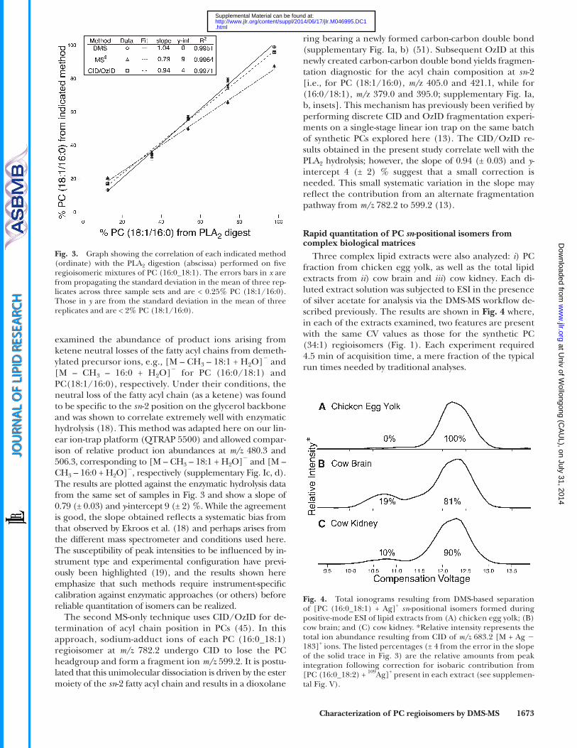
The relative amounts of synthetic PC (16:0/18:1) and PC (18:1/16:0) were determined in the fi ve mixtures by com-paring the relative amounts of the LPCs (16:0_OH) and (18:1_OH) generated from PLA 2 hydrolysis. Precursor ion scans ( m/z 184.1) were used to assay the reaction ( 18 ) by monitoring the abundance of ion signals at m/z 496.4 and 522.4, corresponding to [LPC (16:0_OH) + H] + and [LPC (18:1_OH) + H] + , respectively. Peak heights were used to calculate the relative percentage of PC (18:1_16:0) regio-siomers. For the DMS workfl ow, MS 3 spectra recorded dur-ing CV scanning were used to generate an ionogram from which the area under each peak was used to calculate the relative percentage of each isomer. The correlation be-tween the two methods is described by the straight-line data fi t ( Fig. 3 ) where the slope of 1.04 (± 0.04) and y -intercept of 0 (± 3) % indicate excellent agreement between the two workfl ows. More importantly, these results reveal that there is no need for calibration of the DMS method, as the rela-tive peak areas in the ionogram provide accurate relative abundances of the two regioisomers.
Two previously described MS-based techniques were benchmarked against the enzymatic hydrolysis. The method of Ekroos et al. ( 18 ) used MS 3 on [M + CH 3 COO] � precur-sor ions of PCs in a linear ion-trap mass spectrometer and
phocholine headgroup ( � 183 Da) to produce the base peak at m/z 683.5 in each case under identical MS condi-tions. Peaks appearing at m/z 525.1 and 551.2 correspond to neutral losses of 282 (FA 18:1) and 256 Da (FA 16:0) from the [M + Ag � N(CH 3 ) 3 ] + ( m/z 807.4) product ions, respectively. Despite being of low abundance (note the 50 times magnifi cation), the relative intensity ratio between these two peaks is seen to switch between regioisomers; that is, m/z 551.2 dominates for isolated [PC (16:0/18:1) + Ag] + while m/z 525.1 is more abundant for [PC (18:1/16:0) + Ag] + . Importantly, both of these product ions are formed from both sn -positional isomers. This fact is emphasized by the data shown in Fig. 1C where the extracted ionogram for each of the m/z 525.1 and 551.2 product ions reveals features in the total ionogram for both isomers. Although the neutral loss of FAs from the sn -1 position dominates, this product channel is not exclusive to an individual re-gioisomer. This is most easily seen at CV = 12.3 V where losses of fatty acyl chains from each of the sn -1 and sn -2 positions in the isolated [PC (16:0/18:1) + Ag] + are of sim-ilar abundance ( Fig. 1C ). MS 3 protocols that examine rela-tive abundances from CID of primary product ions have also been used to assign acyl chain backbone position in GPs ( 51 ). The DMS separation of the isomers afforded the opportunity to examine the selectivity of such fragmenta-tion from isomerically pure ion populations. Subsequent dissociation (i.e., MS 3 ) of the primary [M + Ag � 183] + product ion at m/z 683.2 was undertaken for each of the isolated [M + Ag] + regioisomers ( Fig. 2C, D ). Ions at m/z 371.3 and 389.3 correspond to [FA 18:1 � H 2 O + Ag] + and [FA 18:1 + Ag] + ( 52 ), respectively. Although their rela-tive intensity switches between pure isomers, both prod-uct ions are present in each spectrum indicating that neither is entirely diagnostic of the isomeric form of the phospholipid.
Fig. 2. CID spectra recorded following DMS-based isolation of [PC (16:0/18:1) + Ag] + with CV set to 12.3 V (A and C) or [PC (18:1/16:0) + Ag] + with CV set to 10.7 V (B and D); ions were formed from ESI of a 1:1 mixture of the two sn -positional isomers in the presence of silver acetate. The MS 2 spectra (A and B) show product ions from CID of m/z 866.5 [M + Ag] + ions, while the MS 3 spectra (C and D) show subsequent products from CID of m/z 683.2 [M + Ag � 183] + ions.
at Univ of W
ollongong (CA
UL), on July 31, 2014
ww
w.jlr.org
Dow
nloaded from
.html http://www.jlr.org/content/suppl/2014/06/17/jlr.M046995.DC1Supplemental Material can be found at:
Characterization of PC regioisomers by DMS-MS 1673
ring bearing a newly formed carbon-carbon double bond (supplementary Fig. Ia, b) ( 51 ). Subsequent OzID at this newly created carbon-carbon double bond yields fragmen-tation diagnostic for the acyl chain composition at sn -2 [i.e., for PC (18:1/16:0), m/ z 405.0 and 421.1, while for (16:0/18:1), m/ z 379.0 and 395.0; supplementary Fig. Ia, b, insets]. This mechanism has previously been verifi ed by performing discrete CID and OzID fragmentation experi-ments on a single-stage linear ion trap on the same batch of synthetic PCs explored here ( 13 ). The CID/OzID re-sults obtained in the present study correlate well with the PLA 2 hydrolysis; however, the slope of 0.94 (± 0.03) and y -intercept 4 (± 2) % suggest that a small correction is needed. This small systematic variation in the slope may refl ect the contribution from an alternate fragmentation pathway from m/z 782.2 to 599.2 ( 13 ).
Rapid quantitation of PC sn -positional isomers from complex biological matrices
Three complex lipid extracts were also analyzed: i ) PC fraction from chicken egg yolk, as well as the total lipid extracts from ii ) cow brain and iii ) cow kidney. Each di-luted extract solution was subjected to ESI in the presence of silver acetate for analysis via the DMS-MS workfl ow de-scribed previously. The results are shown in Fig. 4 where, in each of the extracts examined, two features are present with the same CV values as those for the synthetic PC (34:1) regioisomers ( Fig. 1 ). Each experiment required 4.5 min of acquisition time, a mere fraction of the typical run times needed by traditional analyses.
examined the abundance of product ions arising from ketene neutral losses of the fatty acyl chains from demeth-ylated precursor ions, e.g. , [M – CH 3 – 18:1 + H 2 O] � and [M – CH 3 – 16:0 + H 2 O] � for PC (16:0/18:1) and PC(18:1/16:0), respectively. Under their conditions, the neutral loss of the fatty acyl chain (as a ketene) was found to be specifi c to the sn -2 position on the glycerol backbone and was shown to correlate extremely well with enzymatic hydrolysis ( 18 ). This method was adapted here on our lin-ear ion-trap platform (QTRAP 5500) and allowed compar-ison of relative product ion abundances at m/z 480.3 and 506.3, corresponding to [M – CH 3 – 18:1 + H 2 O] � and [M – CH 3 – 16:0 + H 2 O] � , respectively (supplementary Fig. Ic, d). The results are plotted against the enzymatic hydrolysis data from the same set of samples in Fig. 3 and show a slope of 0.79 (± 0.03) and y -intercept 9 (± 2) %. While the agreement is good, the slope obtained refl ects a systematic bias from that observed by Ekroos et al. ( 18 ) and perhaps arises from the different mass spectrometer and conditions used here. The susceptibility of peak intensities to be infl uenced by in-strument type and experimental confi guration have previ-ously been highlighted ( 19 ), and the results shown here emphasize that such methods require instrument-specifi c calibration against enzymatic approaches (or others) before reliable quantitation of isomers can be realized.
The second MS-only technique uses CID/OzID for de-termination of acyl chain position in PCs ( 45 ). In this approach, sodium-adduct ions of each PC (16:0_18:1) regioisomer at m/z 782.2 undergo CID to lose the PC headgroup and form a fragment ion m/z 599.2. It is postu-lated that this unimolecular dissociation is driven by the ester moiety of the sn -2 fatty acyl chain and results in a dioxolane
Fig. 3. Graph showing the correlation of each indicated method (ordinate) with the PLA 2 digestion (abscissa) performed on fi ve regioisomeric mixtures of PC (16:0_18:1). The errors bars in x are from propagating the standard deviation in the mean of three rep-licates across three sample sets and are < 0.25% PC (18:1/16:0). Those in y are from the standard deviation in the mean of three replicates and are < 2% PC (18:1/16:0).
Fig. 4. Total ionograms resulting from DMS-based separation of [PC (16:0_18:1) + Ag] + sn -positional isomers formed during positive-mode ESI of lipid extracts from (A) chicken egg yolk; (B) cow brain; and (C) cow kidney. *Relative intensity represents the total ion abundance resulting from CID of m/z 683.2 [M + Ag � 183] + ions. The listed percentages (± 4 from the error in the slope of the solid trace in Fig. 3 ) are the relative amounts from peak integration following correction for isobaric contribution from [PC (16:0_18:2) + 109 Ag] + present in each extract (see supplemen-tal Fig. V ).
at Univ of W
ollongong (CA
UL), on July 31, 2014
ww
w.jlr.org
Dow
nloaded from
.html http://www.jlr.org/content/suppl/2014/06/17/jlr.M046995.DC1Supplemental Material can be found at:

1674 Journal of Lipid Research Volume 55, 2014
depletion. The need for silver in the successful separation of these regioisomers might arise from the known abilities of this metal ion to bind not only to lone pairs of electrons present on heteroatoms (like O and N), but also to � -electrons present in carbon-carbon multiple bonds, such as the alkene group present in one acyl chain of the PCs studied here ( 53 ).
It was clear that the presence of the silver cation was critical to the ability to separate the two PC regioisomers by DMS. Given the ability of silver to coordinate with car-bon-carbon double bonds, the potential role of the car-bon-carbon double bond on the oleoyl chain was investigated. Similar DMS-based analysis was conducted on a related pair of regioisomeric lipids, each of which has fully saturated acyl chains: PC (16:0/18:0) and PC (18:0/16:0). As shown in supplementary Fig. II, no DMS-based separation of [M + Ag] + regioisomers from a 1:1 mixture of PC (16:0/18:0) and PC (18:0/16:0) containing silver acetate was achieved using conditions that separated PC (16:0/18:1) from PC (18:1/16:0). This result suggests that the carbon-carbon double bond present in the latter pair plays an integral role in the DMS-based separation of these PC regioisomers. This is supported by other research results inferring silver affi nity for carbon-carbon double bonds in both the liquid and gas phases ( 52–54 ). A 1:1 mixture of PC (18:0/18:1) with PC (18:1/18:0) was also subjected to the same workfl ow in order to examine the effect of chain length on separation (supplementary Fig. III). The results indicate that this mixture of regioisomers, where both acyl chains have identical length, can also be well separated by DMS provided that at least one of the chains is unsaturated.
The standard residence time for the PC ions within the DMS cell ( � 7 ms) was not suffi cient to separate the silver-adducted PC (16:0_18:1) regioisomers during CV voltage scanning. However, we could extend the residence time for these ions by introducing an impeding gas fl ow from the terminus of the DMS cell (i.e., in between the DMS cell and the orifi ce where ions enter the mass spectrome-ter). As indicated in Fig. 5B for the [M + Ag] + ions, no separation was observed for resolving gas pressures up to 20 psi. However, when the resolving gas pressure was in-creased to 25 psi, two features at 10.7 and 12.3 V in CV space emerged. Further increasing the resolving gas pres-sure to 35 psi provided clear separation of the two regioi-someric lipid ions. The need for additional residence time (ca. 12 ms when resolving gas pressure was 35 psi) to sepa-rate these ion populations suggests subtle structural differ-ences between these two regioisomeric ions in the gas phase.
DISCUSSION
The DMS-MS workfl ow developed here provides for unambiguous differentiation of sn -positional isomers of unsaturated PCs and is compatible with both LC- and di-rect infusion-MS protocols ( 8, 9, 11 ). The amount of sam-ple and the preparation time required for this method are
The area under the curves in Fig. 4 allows relative regioiso-meric quantifi cation of the PC (34:1) component within these complex biological samples. Chicken egg yolk was found to be more enriched in the PC (16:0/18:1) regioiso-mer than even the commercially available synthetic lipid. For the two bovine organs studied, the relative proportions of the two regioisomers varies by a factor of two, emphasizing a vary-ing isomeric distribution between organs of this particular mammal. Precedent for this observation has been reported by Pham et al. ( 13 ) who showed similar changes in isomer ratios for the same tissues using the MS-based CID/OzID technique, and by Taguchi and coworkers ( 21 ) who showed that the relative amounts of PC (16:0_22:6) regioisomers var-ied signifi cantly between brain and liver tissues in mice.
Factors infl uencing DMS separation of PC sn -positional isomers
Ultimately, the successful DMS-based separation of PC (16:0/18:1) from PC (18:1/16:0) was afforded by three key factors: i ) cationization of the PCs with silver, ii ) the presence of a carbon-carbon double bond on an acyl chain of the PC, and iii ) use of resolving gas to enhance resolu-tion. To examine the effect of the cation type for the sepa-ration of the PC regioisomers, we generated several different forms of the ionized lipids by ESI, including the protonated form and several metal cation adducts with the formula [M + X] + , where X = Li + , Na + , K + , or Ag + . Upon examination, only the silver-adduct ions of PCs were sepa-rable by DMS ( Fig. 5A ). While the Li + adducts appeared to demonstrate some degree of separation, it required more rigorous DMS conditions that resulted in greater signal
Fig. 5. The effect on the [M + X] + regioisomeric separation capa-bility in the differential mobility spectrometer of X in a 1:1 mixture of PC (16:0/18:1) and (18:1/16:0) (A) and resolving gas pressure in a 1:1 mixture of PC (16:0/18:1) and (18:1/16:0) with X = Ag + (B). *Relative intensity in each trace in A represents the extracted ion abundance for those products corresponding to neutral loss of 183 Da from CID of [M + X] + ions. Each trace in B represents the total ion abundance resulting from CID of m/z 866.5 [M + Ag] + ions. In both panels, the data have been approximately normalized to the respective solid black trace.
at Univ of W
ollongong (CA
UL), on July 31, 2014
ww
w.jlr.org
Dow
nloaded from
.html http://www.jlr.org/content/suppl/2014/06/17/jlr.M046995.DC1Supplemental Material can be found at:

Characterization of PC regioisomers by DMS-MS 1675
[PC + X] + where X = Na, Li, Ag) infl uence the resolving power of the planar DMS cell used here. Using this proto-col, at least one degree of unsaturation is required to sepa-rate sn -positional isomers, and, of the metal ions investigated, only silver-adduct ions were found to provide suffi cient separation for quantitative workfl ows. In considering the scope of this approach, DMS conditions were also opti-mized to induce separation of regioisomeric forms of the representative polyunsaturated lipid PC (16:0_18:2) (see supplemental Fig. IV). Future efforts to optimize and benchmark the DMS-MS approach for other PC isomers or other classes of glycerolipids would be greatly assisted by increased availability of pairs of synthetic isomers. The use of silver adducts was found to be effective for increasing the resolving power of the DMS method by analogy with tradi-tional silver ion-chromatographic approaches ( 53, 54 ). The presence of two abundant silver isotopes separated in mass by 2 Da does introduce some complexity to the analysis, but methods to account for these isobars in quantitative work-fl ows are demonstrated here (see supplemental Fig. V) and are similar to approaches already used for isotope correc-tions in low-resolution shotgun lipidomics protocols. As the theory of DMS further evolves, it is expected that the scope of this approach for the analysis of isomeric lipids will ex-pand to encompass a broader range of lipid classes and may enable its use with a wider variety of ion types.
Even within the scope of lipids presented here, the abil-ity of DMS to fi lter for a targeted lipid regioisomer repre-sents a signifi cant advance toward the goal of total lipid structure elucidation within the complex molecular makeup of a crude biological extract. The precise molecu-lar information afforded by this approach could play a critical role in understanding which individual lipid mol-ecules are responsible for particular cellular functions. Further structural motif identifi cation (i.e., double bond location and stereochemistry) may be possible in the near future when DMS is coupled with other MS methods. The work here builds on previous effort examining the ion mo-bility of lipids ( 32 ) and supports the idea that coupling mobility with MS may produce powerful tools for applica-tion toward total structure elucidation within the fi eld of lipidomics ( 12 ).
The authors thank the following people for their assistance in the development of the enzymatic hydrolysis procedure and subsequent assay implemented here: Prof. Nick Dixon [University of Wollongong (UOW )] and his group members Dr. Zhi-Qiang Xu and Mr. Nicholas Horan; and Drs. Simon Brown (UOW) and Kim Ekroos (Zora Biosciences, Finland). We also acknowledge Dr. Jessica Hughes (UOW) for providing the bovine organ lipid extracts. J. L. Campbell would like to thank Drs. Yves Le Blanc and Jim Hager for insightful discussions regarding the mobility experiments.
REFERENCES
1 . Gurr , M. I. , J. L. Harwood , and K. N. Frayn . 2002 . Lipid Biochemistry. Blackwell Science, Oxford. 215–263.
2 . Lee , A. 2001 . Membrane structure. Curr. Biol. 11 : R811 – R814 .
signifi cantly reduced when compared with those involving enzymatic hydrolysis of GPs ( 24, 55 ). Furthermore, the relative quantitation of PC regioisomers obtained by DMS-MS shows excellent agreement with values derived from these well-established wet chemical assays. The demonstra-tion that isomeric forms of ionized lipids can be well sepa-rated in the gas phase indicates that calibration against classical methods is not required and thus presents a sig-nifi cant advantage over methods relying on MS alone.
The separation of PC (16:0/18:1) from PC (18:1/16:0) as silver-adducted ions has allowed interrogation of the frag-mentation processes associated with each isomer in isola-tion. Given the diffi culty in obtaining a single GP isomer from either synthetic or biological sources, the tandem mass spectra presented here may be among the fi rst ob-tained from an isomerically pure lipid. These data reveal that while the abundance of product ions arising from ion-ized forms of PC are affected by the substitution pattern on the glycerol backbone, no single product ion is found to be an exclusive indicator of sn -position. Rather, these results refl ect competition among dissociation pathways (e.g., neu-tral losses from sn -1 and sn -2 positions) even within a single GP isomer. Indeed, it has recently been shown that for re-lated ionized glycerolipids (i.e., triacylglycerols) the ener-getic and entropic dependence of the dissociation pathways are a function of the sn- positional distribution ( 56 ). The existence of competing pathways for dissociation is consis-tent with the observations that i ) product ions arising from competing dissociation pathways are affected by instrument confi guration and experimental parameters ( 19 ), and ii ) under typical instrument conditions, product ions are not exclusive to a particular isomer. Taken together, this sug-gests that tandem mass spectrometric techniques that ex-amine peak abundances alone, and in the absence of calibration, should not be used to assign a single acyl chain substitution pattern in GPs. Instead, product ion abun-dances should be used as a guide to indicate only the most abundant isomer present in the sample.
Where mass spectral data provide the GP class and the stoichiometry of the two acyl chains the assignment of fatty acyl chain position on the glycerol backbone is sometimes undertaken using rules of thumb (e.g., the more unsatu-rated chain is at the sn -2 position). It is worth remembering that the precedents for these conventions derive from enzy-matic assays conducted on lipid extracts or class fractions and are thus insensitive to relative populations of each pair of regioisomers. The data presented here, along with previ-ous studies ( 18, 21 ), demonstrate that in biological extracts GPs are most often present as a mixture of both regioiso-mers. As such, assignment of the structure exclusively to one isomer based on convention alone may be entirely in-correct or serve to mask the true molecular diversity of the lipidome. We thus support the recent suggestion of Liebisch et al. ( 22 ) that the notation for lipid structural assignment should precisely refl ect the information provided by the specifi c analyses undertaken (e.g., PC (16:0_18:1) where the sn -positions are not explicitly determined).
Results described herein demonstrate that the degree of unsaturation and the ionized form of a particular PC (i.e.,
at Univ of W
ollongong (CA
UL), on July 31, 2014
ww
w.jlr.org
Dow
nloaded from
.html http://www.jlr.org/content/suppl/2014/06/17/jlr.M046995.DC1Supplemental Material can be found at:
1676 Journal of Lipid Research Volume 55, 2014
25 . Connor , W. E. , D. S. Lin , G. Thomas , F. Ey , T. DeLoughery , and N. Zhu . 1997 . Abnormal phospholipid molecular species of erythro-cytes in sickle cell anemia. J. Lipid Res. 38 : 2516 – 2528 .
26 . Van Deenen , L. L. 1971 . Chemistry of phospholipids in relation to biological membranes. Pure Appl. Chem. 25 : 25 – 56 .
27 . Lands , W. E. M. 2000 . Stories about acyl chains. Biochim. Biophys. Acta . 1483 : 1 – 14 .
28 . Rottem , S. , and O. Markowitz . 1979 . Membrane lipids of Mycoplasma gallisepticum : a disaturated phosphatidylcholine and a phosphati-dylglycerol with an unusual positional distribution of fatty acids. Biochemistry. 18 : 2930 – 2935 .
29 . Shinzawa-Itoh , K. , H. Aoyama , K. Muramoto , H. Terada , T. Kurauchi , Y. Tadehara , A. Yamasaki , T. Sugimura , S. Kurono , K. Tsujimoto , et al . 2007 . Structures and physiological roles of 13 in-tegral lipids of bovine heart cytochrome c oxidase. EMBO J. 26 : 1713 – 1725 .
30 . Kanu , A. B. , P. Dwivedi , M. Tam , L. Matz , and H. H. Hill , Jr . 2008 . Ion mobility-mass spectrometry. J. Mass Spectrom. 43 : 1 – 22 .
31 . Eiceman , G. A. , and Z. Karpas . 2005 . Ion Mobility Spectrometry. CRC Press, Boca Raton, FL.
32 . Kliman , M. , J. C. May , and J. A. McLean . 2011 . Lipid analysis and lipidomics by structurally selective ion mobility-mass spectrometry. Biochim. Biophys. Acta . 1811 : 935 – 945 .
33 . Jackson , S. N. , M. Ugarov , J. D. Post , T. Egan , D. Langlais , J. A. Schultz , and A. S. Woods . 2008 . A study of phospholipids by ion mobility TOF/MS. J. Am. Soc. Mass Spectrom. 19 : 1655 – 1662 .
34 . Kim , H. I. , H. Kim , E. S. Pang , E. K. Ryu , L. W. Beegle , J. A. Loo , W. A. Goddard , and I. Kanik . 2009 . Structural characterization of unsaturated phosphatidylcholines using traveling wave ion mobil-ity spectrometry. Anal. Chem. 81 : 8289 – 8297 .
35 . Barnett , D. A. , B. Ells , R. Guevremont , and R. W. Purves . 1999 . Separation of leucine and isoleucine by electrospray ionization–high fi eld asymmetric waveform ion mobility spectrometry–mass spectrometry. J. Am. Soc. Mass Spectrom. 10 : 1279 – 1284 .
36 . Blagojevic , V. , A. Chramow , B. B. Schneider , T. R. Covey , and D. K. Bohme . 2011 . Differential mobility spectrometry of isomeric pro-tonated dipeptides: modifi er and fi eld effects on ion mobility and stability. Anal. Chem. 83 : 3470 – 3476 .
37 . Schneider , B. B. , T. R. Covey , S. L. Coy , E. V. Krylov , and E. G. Nazarov . 2010 . Planar differential mobility spectrometer as a pre-fi lter for atmospheric pressure ionization mass spectrometry. Int. J. Mass Spectrom. 298 : 45 – 54 .
38 . Shvartsburg , A. A. , D. E. Clemmer , and R. D. Smith . 2010 . Isotopic effect on ion mobility and separation of isotopomers by high-fi eld ion mobility spectrometry. Anal. Chem. 82 : 8047 – 8051 .
39 . Campbell , J. L. , J. C. Y. Le Blanc , and B. B. Schneider . 2012 . Probing electrospray ionization dynamics using differential mobil-ity spectrometry: the curious case of 4-aminobenzoic acid. Anal. Chem. 84 : 7857 – 7864 .
40 . Shvartsburg , A. A. , G. Isaac , N. Leveque , R. D. Smith , and T. O. Metz . 2011 . Separation and classifi cation of lipids using differ-ential ion mobility spectrometry. J. Am. Soc. Mass Spectrom. 22 : 1146 – 1155 .
41 . Fahy , E. , S. Subramaniam , H. A. Brown , C. K. Glass , A. H. Merrill , R. C. Murphy , C. R. H. Raetz , D. W. Russell , Y. Seyama , W. Shaw , et al . 2005 . A comprehensive classifi cation system for lipids. J. Lipid Res. 46 : 839 – 861 .
42 . Fahy , E. , S. Subramaniam , R. C. Murphy , M. Nishijima , C. R. H. Raetz , T. Shimizu , F. Spener , G. van Meer , M. J. O. Wakelam , and E. A. Dennis . 2009 . Update of the LIPID MAPS comprehensive clas-sifi cation system for lipids. J. Lipid Res. 50 : S9 – S14 .
43 . Nealon , J. R. , S. J. Blanksby , T. W. Mitchell , and P. L. Else . 2008 . Systematic differences in membrane acyl composition associ-ated with varying body mass in mammals occur in all phospho-lipid classes: an analysis of kidney and brain. J. Exp. Biol. 211 : 3195 – 3204 .
44 . Shvartsburg , A. A. 2009 . Differential Ion Mobility Spectrometry: Nonlinear Ion Transport and Fundamentals of FAIMS. CRC Press, Boca Raton, FL. 1–293.
45 . Poad , B. L. J. , H. T. Pham , M. C. Thomas , J. R. Nealon , J. L. Campbell , T. W. Mitchell , and S. J. Blanksby . 2010 . Ozone-induced dissociation on a modifi ed tandem linear ion-trap: observations of different reactivity for isomeric lipids. J. Am. Soc. Mass Spectrom. 21 : 1989 – 1999 .
46 . Collings , B. A. , and M. A. Romaschin . 2009 . MS/MS of ions in a low pressure linear ion trap using a pulsed gas. J. Am. Soc. Mass Spectrom. 20 : 1714 – 1717 .
3 . Janmey , P. A. , and P. K. J. Kinnunen . 2006 . Biophysical properties of lipids and dynamic membranes. Trends Cell Biol. 16 : 538 – 546 .
4 . Gross , R. W. , C. M. Jenkins , J. Y. Yang , D. J. Mancuso , and X. L. Han . 2005 . Functional lipidomics: the roles of specialized lipids and lipid-protein interactions in modulating neuronal function. Prostaglandins Other Lipid Mediat. 77 : 52 – 64 .
5 . Menon , A. K. 2008 . Lipid modifi cations of proteins. In Biochemistry of Lipids, Lipoproteins and Membranes. D. E. Vance and J. E. Vance, editors. Elsevier, Sydney. 39–58.
6 . Dowhan , W. , M. Bogdanov , and E. Mileykovskaya . 2008 . Functional roles of lipids in membranes. In Biochemistry of Lipids, Lipoproteins, and Membranes. D. E. Vance and J. E. Vance, edi-tors. Elsevier, Sydney. 1–37.
7 . Brügger , B. , G. Erben , R. Sandhoff , F. T. Wieland , and W. D. Lehmann . 1997 . Quantitative analysis of biological membrane lip-ids at the low picomole level by nano-electrospray ionization tan-dem mass spectrometry. Proc. Natl. Acad. Sci. USA . 94 : 2339 – 2344 .
8 . Han , X. , K. Yang , and R. W. Gross . 2012 . Multi-dimensional mass spectrometry-based shotgun lipidomics and novel strategies for lipidomic analyses. Mass Spectrom. Rev. 31 : 134 – 178 .
9 . Blanksby , S. J. , and T. W. Mitchell . 2010 . Advances in mass spec-trometry for lipidomics. Annu. Rev. Anal. Chem. 3 : 433 – 465 .
10 . Shevchenko , A. , and K. Simons . 2010 . Lipidomics: coming to grips with lipid diversity. Nat. Rev. Mol. Cell Biol. 11 : 593 – 598 .
11 . Pulfer , M. , and R. C. Murphy . 2003 . Electrospray mass spectrom-etry of phospholipids. Mass Spectrom. Rev. 22 : 332 – 364 .
12 . Brown , S. H. J. , T. W. Mitchell , A. J. Oakley , H. T. Pham , and S. J. Blanksby . 2012 . Time to face the fats: what can mass spectrometry reveal about the structure of lipids and their interactions with pro-teins? J. Am. Soc. Mass Spectrom. 23 : 1441 – 1449 .
13 . Pham , H. T. , A. T. Maccarone , M. C. Thomas , J. L. Campbell , T. W. Mitchell , and S. J. Blanksby . 2014 . Structural characterization of glycerophospholipids by combinations of ozone- and collision-induced dissociation mass spectrometry: the next step towards “top-down” lipidomics. Analyst . 139 : 204 – 214 .
14 . Liu , S. , J. D. Brown , K. J. Stanya , E. Homan , M. Leidl , K. Inouye , P. Bhargava , M. R. Gangl , L. Dai , B. Hatano , et al . 2013 . A diurnal serum lipid integrates hepatic lipogenesis and peripheral fatty acid use. Nature . 502 : 550 – 554 .
15 . Krylova , I. N. , E. P. Sablin , J. Moore , R. X. Xu , G. M. Waitt , J. A. MacKay , D. Juzumiene , J. M. Bynum , K. Madauss , V. Montana , et al . 2005 . Structural analyses reveal phosphatidyl inositols as ligands for the NR5 orphan receptors SF-1 and LRH-1. Cell . 120 : 343 – 355 .
16 . Hsu , F-F. , and J. Turk . 2009 . Electrospray ionization with low-en-ergy collisionally activated dissociation tandem mass spectrometry of glycerophospholipids: mechanisms of fragmentation and struc-tural characterization. J. Chromatogr. B Analyt. Technol. Biomed. Life Sci. 877 : 2673 – 2695 .
17 . Huang , Z. H. , D. A. Gage , and C. C. Sweeley . 1992 . Characterization of diacylglycerylphosphocholine molecular species by FAB-CAD-MS/MS: a general method not sensitive to the nature of the fatty acyl groups. J. Am. Soc. Mass Spectrom. 3 : 71 – 78 .
18 . Ekroos , K. , C. S. Ejsing , U. Bahr , M. Karas , K. Simons , and A. Shevchenko . 2003 . Charting molecular composition of phosphati-dylcholines by fatty acid scanning and ion trap MS3 fragmentation. J. Lipid Res. 44 : 2181 – 2192 .
19 . Hou , W. , H. Zhou , M. B. Khalil , D. Seebun , S. A. L. Bennett , and D. Figeys . 2011 . Lyso-form fragment ions facilitate the determination of stereospecifi city of diacyl glycerophospholipids. Rapid Commun. Mass Spectrom. 25 : 205 – 217 .
20 . Plückthun , A. , and E. A. Dennis . 1982 . Acyl and phosphoryl migra-tion in lysophospholipids: importance in phospholipid synthesis and phospholipase specifi city. Biochemistry. 21 : 1743 – 1750 .
21 . Nakanishi , H. , Y. Iida , T. Shimizu , and R. Taguchi . 2010 . Separation and quantifi cation of sn-1 and sn-2 fatty acid positional isomers in phosphatidylcholine by RPLC-ESIMS/MS. J. Biochem. 147 : 245 – 256 .
22 . Liebisch , G. , J. A. Vizcaíno , H. Köfeler , M. Trötzmüller , W. J. Griffi ths , G. Schmitz , F. Spener , and M. J. O. Wakelam . 2013 . Shorthand notation for lipid structures derived from mass spec-trometry. J. Lipid Res. 54 : 1523 – 1530 .
23 . Stern , W. , and M. E. Pullman . 1978 . Acyl-CoA-sn-glycerol-3-phosphate acyltransferase and positional distribution of fatty acids in phospholipids of cultured cells. J. Biol. Chem. 253 : 8047 – 8055 .
24 . Kiełbowicz , G. , W. Gladkowski , A. Chojnacka , and C. Wawrzeńczyk . 2012 . A simple method for positional analysis of phosphatidylcho-line. Food Chem. 135 : 2542 – 2548 .
at Univ of W
ollongong (CA
UL), on July 31, 2014
ww
w.jlr.org
Dow
nloaded from
.html http://www.jlr.org/content/suppl/2014/06/17/jlr.M046995.DC1Supplemental Material can be found at:
Characterization of PC regioisomers by DMS-MS 1677
47 . Bergmeyer , H. U. , M. Graßl , and W. Hans-Elmar . 1983 . Enzymes. In Methods of Enzymatic Analysis. H. U. Bergmeyer, editor. Verlag Chemie, Basel. 283–284.
48 . Sigma-Aldrich . 1993 . Enzymatic Assay of Phospholipase A2 (EC 3.1.1.4).
49 . Schneider , B. B. , T. R. Covey , S. L. Coy , E. V. Krylov , and E. G. Nazarov . 2010 . Chemical effects in the separation process of a differential mobility/mass spectrometer system. Anal. Chem. 82 : 1867 – 1880 .
50 . Campbell , J. L. , M. Zhu , and W. S. Hopkins . 2014 . Ion-molecule clustering in differential mobility spectrometry: lessons learned from tetraalkylammonium cations and their isomers . J. Am. Soc. Mass Spectrom. 25. doi: 10.1007/s13361-014-0939-3.
51 . Hsu , F. F. , and J. Turk . 2003 . Electrospray ionization/tandem quadrupole mass spectrometric studies on phosphatidylcholines: the fragmentation processes. J. Am. Soc. Mass Spectrom. 14 : 352 – 363 .
52 . Yoo , H. J. , and K. Hakansson . 2011 . Determination of phospholipid regiochemistry by Ag(I) adduction and tandem mass spectrometry. Anal. Chem. 83 : 1275 – 1283 .
53 . Damyanova , B. , S. Momtchilova , S. Bakalova , H. Zuilhof , W. W. Christie , and J. Kaneti . 2002 . Computational probes into the conceptual basis of silver ion chromatography: I. Silver(I) ion complexes of unsaturated fatty acids and esters. J. Mol. Struct. THEOCHEM . 589–590 : 239 – 249 .
54 . Morris , L. J. 1966 . Separations of lipids by silver ion chromatogra-phy. J. Lipid Res. 7 : 717 – 732 .
55 . Chen , S. , and P. V. Subbaiah . 2013 . Regioisomers of phosphatidyl-choline containing DHA and their potential to deliver DHA to the brain: role of phospholipase specifi cities. Lipids . 48 : 675 – 686 .
56 . Renaud , J. B. , S. Overton , and P. M. Mayer . 2013 . Energy and entropy at play in competitive dissociations: the case of uneven positional dis-sociation of ionized triglycerides. Int. J. Mass Spectrom. 352 : 77 – 86 .
at Univ of W
ollongong (CA
UL), on July 31, 2014
ww
w.jlr.org
Dow
nloaded from
.html http://www.jlr.org/content/suppl/2014/06/17/jlr.M046995.DC1Supplemental Material can be found at: