Morphine Preconditioning Protects Against LPS-Induced Neuroinflammation and Memory Deficit
Immune deficiencies, infection, and systemic immune disorders
LPS-responsive beige-like anchor (LRBA) gene mutation in afamily with inflammatory bowel disease and combinedimmunodeficiency
Abdullah Alangari, MD,a* Abdulrahman Alsultan, MD,a* Nouran Adly, BSc,b* Michel J. Massaad, PhD,d
Iram Shakir Kiani, MD,c Abdulrahman Aljebreen, MD,c Emad Raddaoui, MD,d Abdul-Kareem Almomen, MD,c
Saleh Al-Muhsen, MD,a Raif S. Geha, MD,e and Fowzan S. Alkuraya, MDa,b,f Riyadh, Saudi Arabia, and Boston, Mass
Background: Clinical immunology has traditionally relied onaccurate phenotyping of the patient’s immune dysfunction forthe identification of a candidate gene or genes for sequencingand molecular confirmation. Although this is also true for otherbranches of medicine, the marked variability in immune-relatedphenotypes and the highly complex network of molecules thatconfer normal host immunity are challenges that clinicalimmunologists often face in their quest to establish a specificgenetic diagnosis.Objective: We sought to identify the underlying genetic cause ina consanguineous family with chronic inflammatory boweldisease–like disorder and combined immunodeficiency.Methods: We performed exome sequencing followed byautozygome filtration.Results: A truncating mutation in LPS-responsive beige-likeanchor (LRBA), which abolished protein expression, wasidentified as the most likely candidate variant in this family.Conclusion: The combined exome sequencing and autozygositymapping approach is a powerful tool in the study of atypicalimmune dysfunctions. We identify LRBA as a novelimmunodeficiency candidate gene the precise role of which inthe immune system requires future studies. (J Allergy ClinImmunol 2012;130:481-8.)
Key words: LPS-responsive beige-like anchor (LRBA), chronicdiarrhea, common variable immunodeficiency, autoimmunity
Immune-related disorders are important contributors to diseaseburden in human subjects not only in their rare Mendelian forms(eg, immunodeficiency disorders) but also in their frequentlyencountered ‘‘common’’ forms (eg, autoimmune disorders)through the complex interaction of genes in oligogenic or poly-genic models with the environment.1 The relative ease with whichMendelian immunologic disorders can be studied has been an im-portant factor in propelling gene discovery in this category of dis-eases, in which medically actionable information can be providedto patients and their families once disease-causing mutations areidentified. The identification of the likely candidate gene is usuallycontingent on careful immunologic phenotyping, which involvesclinically validated functional assays of the various componentsof the immune system.2 However, marked variability in the clinicalphenotype of many of these disorders often complicates the clini-cian’s ability to order relevant immunologic assays that can facili-tate the identification of the correct candidate gene.3 In addition,although an impressive number ofMendelian immune dysfunctiongenes has been identified in the past few years,4 many more are yetto be discovered. Thus, evenwhen a specific immunologic defect isidentified clinically, the underlying genetic heterogeneity will stillpose a significant diagnostic challenge.Until recently, linkage analysis and autozygosity mapping of
families with immune disorders has been one of themost powerfultools in identifying novel disease genes that underlie known oratypical immune disorders, and much has been learned aboutfactors that contribute to the normal development and mainte-nance of the immune system through these discoveries. The recentavailability of massively parallel (next-generation) sequencinghas revolutionized the gene discovery process in Mendeliandisorders, including those that involve immune dysfunction.5
The ability to identify the underlying causative mutation even insimplex cases by using these sequencing techniques has obviatedthe historical requirement of large and/or multiple consanguine-ous pedigrees.Common variable immunodeficiency (CVID) is a heteroge-
neous group of disorders with variable age of onset and ischaracterized by hypogammaglobulinemia and poor antibodyresponses. CVID affects approximately 1:25,000 white subjects.Patients with CVID have low serum IgG and IgA levels, and abouthalf of the patients have reduced serum IgM levels.2,6 Autoimmu-nity affects up to 25% of these patients and might be the present-ing feature in some. The most common autoimmune
From the Departments of aPediatrics, cInternal Medicine, and dPathology, King KhalidUniversity Hospital and College of Medicine, King Saud University, Riyadh; bthe De-partment of Genetics, King Faisal Specialist Hospital and Research Center, Riyadh;ethe Division of Immunology, Children’s Hospital Boston and Harvard MedicalSchool, Boston; and fthe Department of Anatomy and Cell Biology, College of Med-icine, Alfaisal University, Riyadh.
*These authors contributed equally to this work.This work was supported in part by an intramural fund from KFSHRC (to F.S.A.), Na-tional Institutes of Health grants AI-076210 and AI094017 (to R.S.G.), the Perkin-El-mer Foundation (to R.S.G.), and the Dubai-Harvard Foundation for Medical Research(to R.S.G. and F.S.A.).
Disclosure of potential conflict of interest: N. Adly has received grants from and isemployed by the Research Center at King Faisal Specialist Hospital & ResearchCentre. R. S. Geha has received grants from the National Institutes of Health, isemployed by Children’s Hospital Boston, has patents for Quickchange and Stratogene,and receives royalties for the book Pediatric Allergy Cases in Immunology. F. S. Alkur-aya has received grants from the Dubai Harvard Foundation forMedical Research. Therest of the authors declare that they have no relevant conflicts of interest.
Received for publication April 28, 2012; revised May 29, 2012; accepted for publicationMay 29, 2012.
Available online June 19, 2012.Corresponding author: Fowzan S. Alkuraya, MD, Developmental Genetics Unit, KingFaisal Specialist Hospital and Research Center, MBC-03 PO BOX 3354, Riyadh11211, Saudi Arabia. E-mail: [email protected].
0091-6749/$36.00! 2012 American Academy of Allergy, Asthma & Immunologydoi:10.1016/j.jaci.2012.05.043
481
Abbreviations usedBEACH: Beige and Chediak-Higashi syndrome
CHS: Chediak-Higashi syndromeCID: Combined immunodeficiency
ConA: Concanavalin ACVID: Common variable immunodeficiencyIBD: Inflammatory bowel diseaseIVIG: Intravenous immunoglobulin
LRBA: LPS-responsive beige-like anchorLYST: Lysosomal trafficking regulatorNK: Natural killer
complications are hematologic, namely thrombocytopenia andhemolytic anemia. Up to half of the patients with CVID will havechronic diarrhea with malabsorption. This might be secondary toinfection, granuloma formation, celiac disease, or intestinalinflammation with features of Crohn disease.7
In this study we describe an apparently novel immune defi-ciency and immune dysregulation phenotype in 5 affectedmembers who belonged to 2 branches of a large consanguineouspedigree. The phenotype is characterized by an immunodefi-ciency with features of CVID, immune dysregulation, or bothmanifesting as inflammatory bowel disease (IBD) with chronicdiarrhea, autoimmune cytopenia, and EBV-induced lymphopro-liferative disease. Combined exome sequencing and autozygomefiltration in this consanguineous family revealed a null mutationin LPS-responsive beige-like anchor (LRBA), which segregateswith the disease. This report links LRBA to a human diseaseand suggests an important role of LRBA in maintaining normalB-cell, and possibly other immune cell, function.
METHODSHuman subjects
All patients were fully evaluated by a clinical immunologist who is certifiedby the American Board of Allergy and Immunology (A.A. or S.A.). Theirimmunologic assessment included immunoglobulin levels, lymphocyte subsetenumeration, measurement of specific antibody titers to antigens, and T-cellproliferation determination by using a tritiated thymidine incorporation assay.Patients and their relatives were recruited by using written informed consentapproved by the Internal ReviewBoard of King SaudUniversity andKFSHRC(RAC #2121053).
Autozygosity analysisDNA samples from affected and unaffected members of the family were
genotyped on an Axiom Chip platform per the manufacturer’s protocol(Affymetrix, Santa Clara, Calif). Runs of homozygosity of greater than 2 Mbthat span 107 single nucleotide polymorphisms were used as surrogates ofautozygosity (ie identical by descent) given the consanguineous nature of thefamily determined by using autoSNPa.8 The entire set of autozygosity blocks(autozygome) was determined for each affected member, and potential over-lap between the autozygomes of affected members was pursued.
Exome sequencingExome capture was performed with the TruSeq Exome Enrichment kit
(Illumina, San Diego, Calif), according to the manufacturer’s protocol.Samples were prepared as an Illumina sequencing library, and in the secondstep the sequencing libraries were enriched for the desired target by using theIllumina Exome Enrichment protocol. The captured libraries were sequencedwith the Illumina HiSeq2000 Sequencer. The reads were mapped against
UCSC hg19 (http://genome.ucsc.edu/) by using BWA (http://bio-bwa.sourceforge.net/). The single nucleotide polymorphisms and indels were de-tected by using SAMTOOLS (http://samtools.sourceforge.net/).
Immunophenotyping and proliferation response tomitogens
PBMCs were isolated on a Ficoll-Paque PLUS gradient (GE Healthcare,Fairfield, Conn). Lymphocytes were surface stained with anti-human CD3,CD4, CD8, CD19, CD16, and CD56 (BioLegend, San Diego, Calif) andanalyzedwith anLSRFortessaflowcytometer (BDBiosciences, San Jose,Calif)and FlowJo software (Tree Star, Ashland, Ore). PBMCs were placed in aliquotsin 96-well tissue-culture plates at a density of 23 105 cells per well and stim-ulated with 1 mg/mL PHA (Sigma-Aldrich, St Louis, Mo), 1 mg/mL concanav-alin A (ConA), or 10 ng/mL anti-CD3 mAb (OKT3; eBioscience, San Diego,Calif). Cultures were pulsed at 96 hours with tritiated thymidine (1 mCi perwell), and tritiated thymidine uptake into DNAwas determined 18 hours later.
PCR and RT-PCRThe coding sequence of LRBA exon 44 (NM_006726) was amplified by us-
ing specially designed primers on genomic DNA. In addition, cDNA primerswere also used to check for the stability of the aberrant transcript. The sourceof RNAwas lymphoblasts extracted with the QIAamp RNAMini Kit (Qiagen,Germantown, Md) and DNase treated with the RNase-Free DNase Set (Qia-gen), according to the manufacturer’s recommendations, and used forcDNA synthesis with the iScript cDNA synthesis kit and Poly T oligonucleo-tide primers (Applied Biosystems, Carlsbad, Calif). Amplicons were se-quenced on DNA Analyzer 3700xl (Applied Biosystems).
Immunoblot analysisCell lysates were prepared with RIPA buffer (Sigma) containing a protease
inhibitor cocktail mix (Roche, Mannheim, Germany). Samples were normal-ized by measuring on a SmartSpec Plus spectrophotometer (Bio-Rad Labo-ratories, Hercules, Calif) using the Protein Assay Dye Reagent (Bio-Rad).Laemmli buffer (Bio-Rad Laboratories) with 5% b-mercaptoethanol wasadded to the samples. Thesewere then boiled, electrophoresed on a 12% SDS-PAGE (National Diagnostics, Charlotte, NC), and transferred onto apolyvinylidene difluoride membrane (Hybond; GE Healthcare Biosciences,Pittsburgh, Pa).Membraneswere blocked for 1 hour in PBS-Tween containing5% nonfat milk at room temperature and incubated with a primary rabbitantibody raised against amino acid residues 907-1038 of human LRBA (ie,N-terminal to the truncation [no. ab121765; Abcam, Cambridge, Mass]),according to the manufacturer’s instructions, and then incubated with horse-radish peroxidase–conjugated secondary antibodies (Pierce, Cheshire, UnitedKingdom). Immunosignals were detected by using SuperSignal West PicoChemiluminescent (Pierce). b-Actin antibody was used as a loading control(Cell Signaling, Danvers, Mass).
RESULTSIdentification of a novel immunodeficiencyphenotype
The family pedigree of the 5 affected patients is shown in Fig1. The index patient (VI:5) is a 7-year-old male offspring offirst-cousin parents and has 1 affected sister (VI:2). The 3 otherpatients (V:3, V:4, and V:5) are sisters, also offspring of second-cousin parents. The index patient’s mother is a paternal cousinof the 3 affected sisters. Thus a common set of grandparents isshared between the 2 nuclear families. Chronic diarrhea was thecommon clinical feature among all 5 affected family members.Below is a clinical summary of each of the 5 affected members.Patient VI:5 is a 7-year-old boy with chronic diarrhea since
infancy. He had onset of nonbloody diarrhea at 2 months of age
J ALLERGY CLIN IMMUNOL
AUGUST 2012
482 ALANGARI ET AL

that got worse after introducing solid foods. Duodenal biopsyperformed at 3 years of age showed partial villous blunting withintraepithelial lymphocytic infiltration. Results of serologic testsfor celiac disease were negative, and diarrhea persisted despitetrial of a gluten-free diet. The interpretation of a repeat duodenalbiopsy at 6 years of age was similar to the previous one, and acolonic biopsy specimen showed patchy thickening of the sub-epithelial collagenous plate, mucin depletion, and expansion ofthe lamina propria by lymphocytic infiltration, which is sugges-tive of collagenous colitis. His history was also remarkable forautoimmune pancytopenia at 3 years of age, which responded tointravenous immunoglobulin (IVIG) replacement therapy and ashort course of prednisone. He also had 2 episodes of EBV-associated lymphoproliferative disease with intense infiltrationwith B cells evident on histopathologic analysis of lymph nodebiopsy specimens (see Fig E1 in this article’s Online Repository atwww.jacionline.org) at ages 5 and 6 years. The EBV PCR titerwas high on both occasions, a lymph node biopsy specimenshowed marked follicular lymphoid hyperplasia with prominentgerminal centers, and lymph nodes were mostly infiltrated by Bcells, as shown by using CD20 immunohistochemical staining(see Fig E1). EBV-associated lymphoproliferative episodeswere transient and improved spontaneously with no treatment.He had no history of recurrent infections. Immunologic investiga-tions revealed normal serum IgG, IgA, IgM, and IgE levels andnormal numbers of CD31 T lymphocytes, CD41 and CD81
T-cell subsets, B cells, and natural killer (NK) cells. He had anormal T-cell proliferation to the mitogens PHA and ConA(Table I) and a normal increase in antibody titers after vaccinationwith tetanus toxoid, diphtheria, toxoid and Haemophilus influen-zae capsular antigens (data not shown).Patient VI:2 is a 19-year-old woman with a history of nephrotic
syndrome diagnosed at the age of 2 years and treated with steroidsuntil she was 8 years old. Shortly after stopping steroids, shestarted to have mucous nonbloody stools, which persisted on agluten-free diet. Duodenal biopsy performed at 16 years of age
revealed partial villous atrophy and marked inflammation, andcolonic biopsy performed at the same time showed mild chroniccolitis with lymphocytic infiltration of the lamina propria (Fig 2,A and B). She was started on prednisone and azathioprine withpoor compliance, and her diarrhea persisted. Her history was re-markable for megaloblastic anemia caused by vitamin B12 defi-ciency, which improved after starting monthly intramuscularvitamin B12 injections. She also had evidence of growth hormonedeficiency and is currently receiving human growth hormone re-placement. She had no history of recurrent infections. Clubbingwas noted on her clinical examination. Immunologic investiga-tion at 17 years of age revealed normal serum IgG, IgA, IgM,and IgE levels and normal numbers of CD31 T lymphocytes,CD41 and CD81 T-cell subsets, B lymphocytes, and NK cells.She had a normal T-cell proliferation to the mitogens PHA andConA (Table I) and a normal increase in antibody titers after vac-cination with pneumococcal vaccine (data not shown).Patient V:3 is a 22-year-old woman with a history of recurrent
otitis media and pneumonia since the age of 4 years. Sheeventually experienced bilateral bronchiectasis and finger club-bing. She also had nonmucous and nonbloody chronic diarrheaand poor appetite since the age of 18 years, which resulted in poorweight gain. A large-bowel biopsy specimen showed mucosalinflammation with lymphocytic infiltration but no granulomas orulcerations. Her chronic diarrhea improved on oral prednisone(1 mg/kg/d). She also had a history of recurrent arthritis in thelarge joints, mainly the knees. Synovial fluid analysis wasconsistent with inflammation, with no evidence of infection.Her investigations revealed low serum IgG and IgA levels anddecreased B-cell numbers but normal numbers of T cells, T-cellsubsets, and NK cells (Table I). T-cell proliferation in response toPHA and anti-CD3 mAb was markedly reduced (Table I).Patient V:4 is a 19-year-old woman who has had chronic
nonbloody and nonmucous diarrhea since the age of 3 years,which did not improve on a gluten-free diet. Her large-bowelbiopsy was similar to that of her older sister (Fig 2, C and D).
FIG 1. Pedigree of a multiplex consanguineous family with a novel phenotype of CVID, immunedysregulation, or both characterized by IBD. Solid symbols denote affected status.
J ALLERGY CLIN IMMUNOL
VOLUME 130, NUMBER 2
ALANGARI ET AL 483
Duodenal biopsy showed villous atrophy. She had autoimmunethrombocytopenia and autoimmune hemolytic anemia at theage of 13 years, both of which responded to treatment with ste-roids and rituximab. She has no history of recurrent infections.Her investigations at the age of 14 years, when she was referredto us, revealed low serum IgG and IgA levels and decreasedB-cell numbers but normal numbers of T cells, T-cell subsets,and NK cells (Table I). T-cell proliferation to PHA and anti-CD3 mAb was markedly reduced (Table I).
Patient V:5 is a 15-year-old girl who has had chronic diarrheasince shewas 2 years old, with no blood ormucus in stool. She hasno history of recurrent infections or autoimmune hematologicmanifestations. Her investigations revealed low serum IgG andIgA levels and normal numbers of T cells, T-cell subsets, B cells,and NK cells (Table I). T-cell proliferation in response to PHAand anti-CD3 mAb was markedly reduced (Table I).All 3 sisters (patients V:3, V:4, and V:5) were started on IVIG
replacement immediately after the diagnosis of hypogammaglob-ulinemia was made. Serum antibody titers were not available forthem before institution of IVIG replacement therapy.
Identification of a null LRBA mutation in a familywith a novel immunodeficiency/immunedysregulation syndrome
Because the affected members from the 2 branches of thefamily share a common set of grandparents and because thedisease affects both sexes, we hypothesized that their phenotype isautosomal recessive and caused by inheriting 2 copies of anancestral haplotype that harbors an autosomal recessive mutation(ie, autozygosity).9 Thereforewe implemented autozygositymap-ping as a tool to identify the entire set of autozygous blocks withinthe genome (autozygome) and searched for autozygosity blocks,which are exclusively shared by the affected members, becausesuch blocks are highly likely to harbor the disease-causing muta-tion. Because patients VI:3 and VI:5 were recruited first, autozy-gome analysis was initially carried out on these 2 patients, andseveral autozygous intervals were found to be exclusively sharedbetween them when compared with their unaffected siblings.
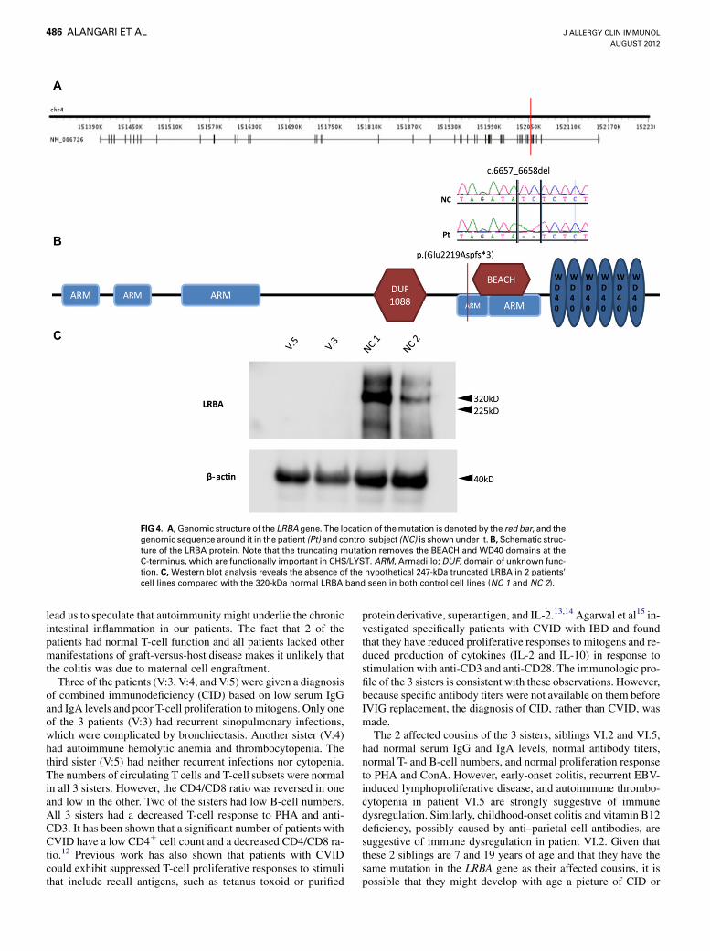
To identify the causative mutation within these blocks ofautozygosity, exome sequencing was performed on patient VI:5,and the results were filtered as outlined in the scheme shown inFig 3. We identified 1245 homozygous variants (of >80,000 to-tal exomic variants) that reside within the shared autozygome.By only considering coding/splicing variants (synonymouschanges were excluded), we were left with just 13 variants, ofwhich only 1 was found to be novel (ie, absent in publicdbSNP and our in-house collection of 183 Saudi exomes).Thus our strategy effectively narrowed the search to a singlevariant in exon 44 of LRBA. This 2-bp deletion,NM_006726:c.6657_6658del, predicts a frameshift and prema-ture truncation of the protein p.(Glu2219Aspfs*3). Segregationof this variant with the disease was confirmed within this familyby means of Sanger sequencing.The structure of the LRBA gene and LRBA protein and the
location of the mutation in the gene and its product are shownin Fig 4. The protein has 5 Armadillo (ARM) domains, 3 of whichare located at the N terminus, a beige and Chediak-Higashi syn-drome (BEACH) domain, and 5 WD40 repeat (WD40) domainslocated at the C-terminus. The BEACH and WD domains inLRBA are homologous to those in the protein encoded by theCHS1 gene, which is mutated in patients with Chediak-HigashiSyndrome (CHS). The truncating mutation in our patients re-moves the BEACH and WD domains, which are known to befunctionally important in the CHS protein, which is also knownas lysosomal trafficking regulator (LYST).10,11 To examine the ef-fect of the mutation on the RNA and protein level, we performedRT-PCR on patients’ lymphoblasts and showed that there is noevidence of significant nonsense-mediated decay (see Fig E2 inthis article’s Online Repository at www.jacionline.org). Westernblot analysis revealed a 320-kDa band in lymphoblast lysatesfrom 2 healthy donors, which corresponds in size to LRBA. Incontrast, this band was absent from lymphoblast lysates of thepatients. More importantly, patients’ lymphoblast lysates lackeda 247-kDa band that would correspond to the putative truncated2220-amino-acid mutant protein, suggesting that the truncatedmutant protein is rapidly degraded and unstable. Thus the muta-tion we describe appears to be nullimorphic in nature.
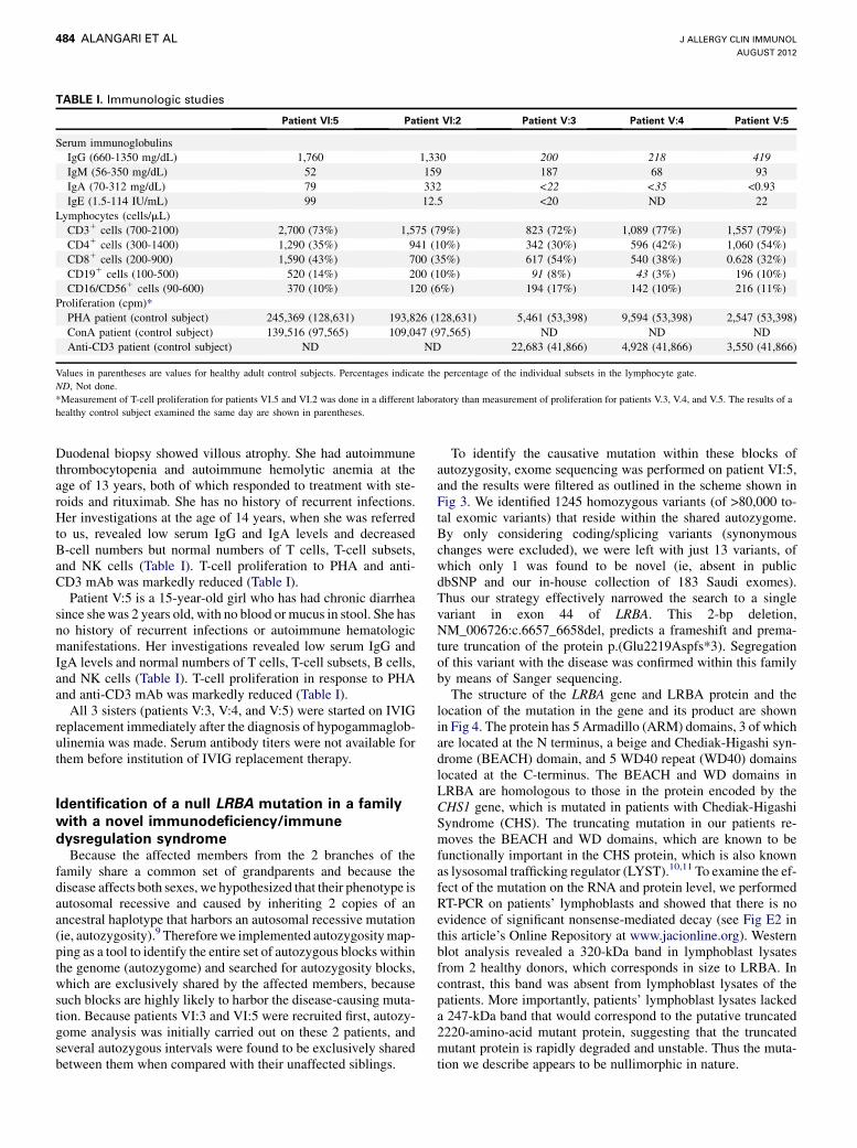
TABLE I. Immunologic studies
Patient VI:5 Patient VI:2 Patient V:3 Patient V:4 Patient V:5
Serum immunoglobulinsIgG (660-1350 mg/dL) 1,760 1,330 200 218 419IgM (56-350 mg/dL) 52 159 187 68 93IgA (70-312 mg/dL) 79 332 <22 <35 <0.93IgE (1.5-114 IU/mL) 99 12.5 <20 ND 22
Lymphocytes (cells/mL)CD31 cells (700-2100) 2,700 (73%) 1,575 (79%) 823 (72%) 1,089 (77%) 1,557 (79%)CD41 cells (300-1400) 1,290 (35%) 941 (10%) 342 (30%) 596 (42%) 1,060 (54%)CD81 cells (200-900) 1,590 (43%) 700 (35%) 617 (54%) 540 (38%) 0.628 (32%)CD191 cells (100-500) 520 (14%) 200 (10%) 91 (8%) 43 (3%) 196 (10%)CD16/CD561 cells (90-600) 370 (10%) 120 (6%) 194 (17%) 142 (10%) 216 (11%)
Proliferation (cpm)*PHA patient (control subject) 245,369 (128,631) 193,826 (128,631) 5,461 (53,398) 9,594 (53,398) 2,547 (53,398)ConA patient (control subject) 139,516 (97,565) 109,047 (97,565) ND ND NDAnti-CD3 patient (control subject) ND ND 22,683 (41,866) 4,928 (41,866) 3,550 (41,866)
Values in parentheses are values for healthy adult control subjects. Percentages indicate the percentage of the individual subsets in the lymphocyte gate.ND, Not done.*Measurement of T-cell proliferation for patients VI.5 and VI.2 was done in a different laboratory than measurement of proliferation for patients V.3, V.4, and V.5. The results of ahealthy control subject examined the same day are shown in parentheses.
J ALLERGY CLIN IMMUNOL
AUGUST 2012
484 ALANGARI ET AL

DISCUSSIONWe report a novel immune deficiency phenotype, immune
dysregulation phenotype, or both, which segregates with atruncating mutation in LRBA.
All 5 patients in the family studied had early onset of colitis andIBD-like presentation. Intestinal inflammation was documented
by histology of colonic biopsy specimens in the 4 patients fromwhom a colonic biopsy specimen was obtained. Early-onsetcolitis is often a presentation of immune deficiency, immunedysregulation, or both. The dense lymphocytic infiltrate observedon histopathologic examination of the intestinal mucosa, as wellas the dramatic response to steroids in the 3 compliant patients,
FIG 2. Histology of patients’ intestinal biopsy specimens. A, Duodenal biopsy specimen from patient VI:2showing marked villous atrophy and increased chronic inflammatory lymphoplasma cells in the laminapropria (hematoxylin and stain, original magnification320). B, High-power view of a duodenal biopsy spec-imen from patient VI:2 showing increased intraepithelial lymphocytes (arrows; hematoxylin and eosin stain,original magnification 360). C, Colonic biopsy specimen from patient V:4 (transverse colon) showing a re-markable increase in mixed acute and chronic inflammatory cell numbers in the lamina propria and a de-creased number of crypts and fibrosis, mainly subepithelial (arrow; hematoxylin and eosin stain, originalmagnification 320). D, High-power view of a colonic biopsy specimen from patient V:4 (transverse colon)showing increased intraepithelial lymphocyte numbers (arrows; hematoxylin and eosin stain, original mag-nification 360).
FIG 3. Filtration scheme of the exome variants identified in patient VI:5. Note the dramatic reduction in thenumber of candidate variants when filtered by the coordinates of the autozygosity intervals that areexclusively shared between the affected members. Only 1 of the 13 coding/splicing variants was novel (ie,not found in publically available single nucleotide polymorphism databases or our in-house 183 Saudiexomes).
J ALLERGY CLIN IMMUNOL
VOLUME 130, NUMBER 2
ALANGARI ET AL 485
lead us to speculate that autoimmunity might underlie the chronicintestinal inflammation in our patients. The fact that 2 of thepatients had normal T-cell function and all patients lacked othermanifestations of graft-versus-host disease makes it unlikely thatthe colitis was due to maternal cell engraftment.Three of the patients (V:3, V:4, and V:5) were given a diagnosis
of combined immunodeficiency (CID) based on low serum IgGand IgA levels and poor T-cell proliferation tomitogens. Only oneof the 3 patients (V:3) had recurrent sinopulmonary infections,which were complicated by bronchiectasis. Another sister (V:4)had autoimmune hemolytic anemia and thrombocytopenia. Thethird sister (V:5) had neither recurrent infections nor cytopenia.The numbers of circulating T cells and T-cell subsets were normalin all 3 sisters. However, the CD4/CD8 ratio was reversed in oneand low in the other. Two of the sisters had low B-cell numbers.All 3 sisters had a decreased T-cell response to PHA and anti-CD3. It has been shown that a significant number of patients withCVID have a low CD41 cell count and a decreased CD4/CD8 ra-tio.12 Previous work has also shown that patients with CVIDcould exhibit suppressed T-cell proliferative responses to stimulithat include recall antigens, such as tetanus toxoid or purified
protein derivative, superantigen, and IL-2.13,14 Agarwal et al15 in-vestigated specifically patients with CVID with IBD and foundthat they have reduced proliferative responses to mitogens and re-duced production of cytokines (IL-2 and IL-10) in response tostimulation with anti-CD3 and anti-CD28. The immunologic pro-file of the 3 sisters is consistent with these observations. However,because specific antibody titers were not available on them beforeIVIG replacement, the diagnosis of CID, rather than CVID, wasmade.The 2 affected cousins of the 3 sisters, siblings VI.2 and VI.5,
had normal serum IgG and IgA levels, normal antibody titers,normal T- and B-cell numbers, and normal proliferation responseto PHA and ConA. However, early-onset colitis, recurrent EBV-induced lymphoproliferative disease, and autoimmune thrombo-cytopenia in patient VI.5 are strongly suggestive of immunedysregulation. Similarly, childhood-onset colitis and vitamin B12deficiency, possibly caused by anti–parietal cell antibodies, aresuggestive of immune dysregulation in patient VI.2. Given thatthese 2 siblings are 7 and 19 years of age and that they have thesame mutation in the LRBA gene as their affected cousins, it ispossible that they might develop with age a picture of CID or
FIG 4. A,Genomic structure of the LRBA gene. The location of themutation is denoted by the red bar, and thegenomic sequence around it in the patient (Pt) and control subject (NC) is shown under it.B, Schematic struc-ture of the LRBA protein. Note that the truncating mutation removes the BEACH and WD40 domains at theC-terminus, which are functionally important in CHS/LYST. ARM, Armadillo; DUF, domain of unknown func-tion. C, Western blot analysis reveals the absence of the hypothetical 247-kDa truncated LRBA in 2 patients’cell lines compared with the 320-kDa normal LRBA band seen in both control cell lines (NC 1 and NC 2).
J ALLERGY CLIN IMMUNOL
AUGUST 2012
486 ALANGARI ET AL
CVID and T-cell dysfunction. Alternatively, modifier genes mightaccount for the development of CID in the 3 sisters who belong tothe other branch of the family. It is not clear at present whether thenephrotic syndrome and growth hormone deficiency in patientVI.2 are part of the clinical spectrum of LRBA deficiency.The variability of the clinical and immunologic phenotype in
the 5 patients is likely to be secondary to genetic, epigenetic, andenvironmental factors, as is often the case in patients with primaryimmune deficiency diseases. Orange et al16 showed, using agenome-wide association approach in 363 patients with CVID,that disease heterogeneity could be related to several variationsin the genetic background, including variations in the MHC re-gion, a hyperpolymorphic region of DNA.Thus far, mutations in 7 genes, inducible costimulator
(ICOS),17 transmembrane activator and calcium modulator andcyclophilin ligand interactor (TACI),18,19 CD19,20 B-cell activat-ing factor receptor (BAFFR),21 CD81,22 CD20,23 and CD21,24
have been found to cause CVID. However, more than 80% of pa-tients with CVID have no identifiable mutation in these genes, atestimony to the marked genetic heterogeneity that characterizesthis disorder. This study shows that LRBA can bemutated in a sub-set of patients with features of CVID, adding a potential novel ge-netic cause for CVID.The product of the LRBA gene is a cytosolic protein expressed
in many tissues. LRBAwas shown to translocate on activation ofB cells to the membrane of vesicles that include the trans-Golginetwork, lysosomes, and endoplasmic reticulum, and to plasmamembrane.25 LRBAwas also shown to be involved in endocytosisof ligand-activated receptors rather than in vesicular exocyto-sis.25,26 Endocytosis is important in both lymphocyte receptor–mediated activation and inhibition.27 It is possible that impairedreceptor internalization in LRBA-deficient lymphocytes mightlead to a defect in B-cell activation and immunoglobulin synthesisand impaired T-cell activation. In addition, failure of signaling byinhibitory receptors that depend on receptor internalization mightlead to failure of immune tolerance and autoimmunity. Interest-ingly, it was shown that defects in LRBA decrease the sensitivityof tumor cells to apoptosis.28 Thus it is tantalizing to speculatethat autoimmunity observed in our patients might be caused bya defect in the elimination of autoreactive lymphocytes, resultingin inefficient peripheral, central, or both tolerance andautoimmunity.LRBA is partly homologous to CHS/LYST, the protein defec-
tive in patients with CHS.25 The 2 proteins share the BEACH andWD40 domains located at their carboxy terminus. The truncatingmutation we report in LRBA removes both of these domains.29 Pa-tients with CHS present early in childhood with partial hair andskin albinism, neurologic dysfunction, bleeding diathesis, and re-current infections and eventually have an accelerated phase of he-mophagocytic lymphohistiocytosis. They also have giantgranules in their peripheral blood leukocytes and bone marrow,indicating failure of lysosomal degranulation.30 Thus, the CHSphenotype is clearly different from that of our patients. AlthoughLRBA appears to be involved in endocytosis, CHS/LYST plays animportant role in vesicle trafficking and exocytosis. This differ-ence might explain the difference in phenotypes between patientswith CHS and our patients. Further work is needed to determinethe precise role of LRBA in the immune system.Our observations suggest that LRBA deficiency should be
considered in patients with CID or CVID, as well as in childrenwith early-onset colitis and autoimmunity. Long-term follow-up
and additional reports of patients with LRBAmutations will delin-eate the full phenotypic spectrum of disease caused by LRBAdeficiency.
We thank the family for their enthusiastic participation. We also thank allhealth care providers who helped in the management of these patients. We areindebted to the Genotyping and Sequencing Core Facilities at KFSHRC fortheir technical help. We also thank Ms Mais Hashem and Ms Niema Ibrahimfor their help as research coordinators and Ms Tarfa Alshiddi for her help inestablishing lymphoblast cell lines.
Clinical implications: LRBA deficiency should be considered inpatients with CID and/or early-onset colitis and autoimmunity.
REFERENCES1. Fischer A. Human primary immunodeficiency diseases. Immunity 2007;27:835-45.2. Cunningham-Rundles C. Human B cell defects in perspective. Immunol Res [Epub
ahead of print].3. Salzer U, Unger S, Warnatz K. Common variable immunodeficiency (CVID): ex-
ploring the multiple dimensions of a heterogeneous disease. Ann N Y Acad Sci2012;1250:41-9.
4. Al-Herz W, Casanova J-L, Chapel H, Conley ME, Cunningham-Rundles C, EtzioniA, et al. Primary immunodeficiency diseases: an update on the classification fromthe International Union of Immunological Societies Expert Committee for PrimaryImmunodeficiency. Front Primary Immunodef 2011;2:1-26.
5. Bamshad MJ, Ng SB, Bigham AW, Tabor HK, Emond MJ, Nickerson DA, et al.Exome sequencing as a tool for Mendelian disease gene discovery. Nat Rev Genet2011;12:745-55.
6. Park JH, Resnick ES, Cunningham-Rundles C. Perspectives on common variableimmune deficiency. Ann N Y Acad Sci 2011;1246:41-9.
7. Park MA, Li JT, Hagan JB, Maddox DE, Abraham RS. Common variable immu-nodeficiency: a new look at an old disease. Lancet 2008;372:489-502.
8. Carr IM, Flintoff KJ, Taylor GR, Markham AF, Bonthron DT. Interactive visualanalysis of SNP data for rapid autozygosity mapping in consanguineous families.Hum Mutat 2006;27:1041-6.
9. Alkuraya FS. Autozygome decoded. Genet Med 2010;12:765-71.10. Ward DM, Shiflett SL, Kaplan J. Chediak-Higashi syndrome: a clinical and mo-
lecular view of a rare lysosomal storage disorder. Curr Mol Med 2002;2:469-77.11. Zarzour W, Kleta R, Frangoul H, Suwannarat P, Jeong A, Kim SY, et al. Two novel
CHS1 (LYST) mutations: clinical correlations in an infant with Chediak-Higashisyndrome. Mol Genet Metab 2005;85:125-32.
12. Moratto D, Gulino AV, Fontana S, Mori L, Pirovano S, Soresina A, et al. Combineddecrease of defined B and T cell subsets in a group of common variable immuno-deficiency patients. Clin Immunol 2006;121:203-14.
13. Fischer MB, Hauber I, Eggenbauer H, Thon V, Vogel E, Schaffer E, et al. A defectin the early phase of T-cell receptor-mediated T-cell activation in patients withcommon variable immunodeficiency. Blood 1994;84:4234-41.
14. Stagg AJ, Funauchi M, Knight SC, Webster AD, Farrant J. Failure in antigen re-sponses by T cells from patients with common variable immunodeficiency(CVID). Clin Exp Immunol 1994;96:48-53.
15. Agarwal S, Smereka P, Harpaz N, Cunningham-Rundles C, Mayer L. Character-ization of immunologic defects in patients with common variable immunodefi-ciency (CVID) with intestinal disease. Inflamm Bowel Dis 2011;17:251-9.
16. Orange JS, Glessner JT, Resnick E, Sullivan KE, Lucas M, Ferry B, et al. Genome-wide association identifies diverse causes of common variable immunodeficiency.J Allergy Clin Immunol 2011;127:1360-7.e6.
17. Grimbacher B, Hutloff A, Schlesier M, Glocker E, Warnatz K, Drager R, et al. Ho-mozygous loss of ICOS is associated with adult-onset common variable immuno-deficiency. Nat Immunol 2003;4:261-8.
18. Salzer U, Chapel HM, Webster AD, Pan-Hammarstrom Q, Schmitt-Graeff A,Schlesier M, et al. Mutations in TNFRSF13B encoding TACI are associatedwith common variable immunodeficiency in humans. Nat Genet 2005;37:820-8.
19. Castigli E, Wilson SA, Garibyan L, Rachid R, Bonilla F, Schneider L, et al. TACI ismutant in common variable immunodeficiency and IgA deficiency. Nat Genet2005;37:829-34.
20. van Zelm MC, Reisli I, van der Burg M, Castano D, van Noesel CJ, van Tol MJ,et al. An antibody-deficiency syndrome due to mutations in the CD19 gene. N EnglJ Med 2006;354:1901-12.
21. Warnatz K, Salzer U, Rizzi M, Fischer B, Gutenberger S, Bohm J, et al. B-cellactivating factor receptor deficiency is associated with an adult-onset antibody
J ALLERGY CLIN IMMUNOL
VOLUME 130, NUMBER 2
ALANGARI ET AL 487

deficiency syndrome in humans. Proc Natl Acad Sci U S A 2009;106:13945-50.
22. van Zelm MC, Smet J, Adams B, Mascart F, Schandene L, Janssen F, et al. CD81gene defect in humans disrupts CD19 complex formation and leads to antibody de-ficiency. J Clin Invest 2010;120:1265-74.
23. Kuijpers TW, Bende RJ, Baars PA, Grummels A, Derks IA, Dolman KM, et al.CD20 deficiency in humans results in impaired T cell-independent antibody re-sponses. J Clin Invest 2010;120:214-22.
24. Thiel J, Kimmig L, Salzer U, Grudzien M, Lebrecht D, Hagena T, et al. GeneticCD21 deficiency is associated with hypogammaglobulinemia. J Allergy Clin Im-munol 2012;129:801-10.e6.
25. Wang JW, Howson J, Haller E, Kerr WG. Identification of a novellipopolysaccharide-inducible gene with key features of both A kinase anchor pro-teins and chs1/beige proteins. J Immunol 2001;166:4586-95.
26. de Souza N, Vallier LG, Fares H, Greenwald I. SEL-2, the C. elegans neuro-beachin/LRBA homolog, is a negative regulator of lin-12/Notch activity and af-fects endosomal traffic in polarized epithelial cells. Development 2007;134:691-702.
27. Yuseff MI, Lankar D, Lennon-Dumenil AM. Dynamics of membrane traffickingdownstream of B and T cell receptor engagement: impact on immune synapses.Traffic 2009;10:629-36.
28. Wang JW, Gamsby JJ, Highfill SL, Mora LB, Bloom GC, Yeatman TJ, et al. Deregu-lated expression of LRBA facilitates cancer cell growth. Oncogene 2004;23:4089-97.
29. Kaplan J, De Domenico I, Ward DM. Chediak-Higashi syndrome. Curr Opin Hem-atol 2008;15:22-9.
30. Huizing M, Helip-Wooley A, Westbroek W, Gunay-Aygun M, Gahl WA. Disordersof lysosome-related organelle biogenesis: clinical and molecular genetics. AnnuRev Genomics Hum Genet 2008;9:359-86.
J ALLERGY CLIN IMMUNOL
AUGUST 2012
488 ALANGARI ET AL

FIG E1. A, Lymph node (left axilla) shows marked follicular lymphoid hy-perplasia with prominent germinal centers (hematoxylin and eosin stain,original magnification 320). B, The same lymph node. A CD20 immunohis-tochemistry stain specific for B cells (Novocastra, Newcastle Upon Tyne,United Kingdom) shows dense staining (CD20 marker, immunohistochem-ical stain, original magnification 320).
J ALLERGY CLIN IMMUNOL
VOLUME 130, NUMBER 2
ALANGARI ET AL 488.e1
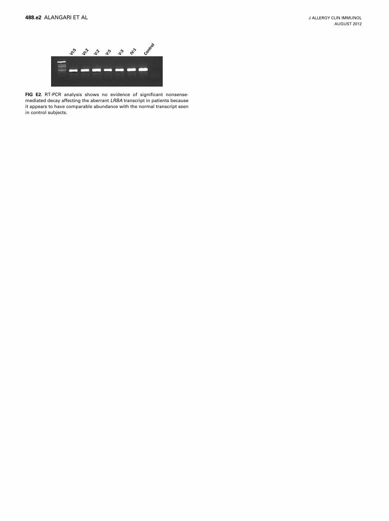
FIG E2. RT-PCR analysis shows no evidence of significant nonsense-mediated decay affecting the aberrant LRBA transcript in patients becauseit appears to have comparable abundance with the normal transcript seenin control subjects.
J ALLERGY CLIN IMMUNOL
AUGUST 2012
488.e2 ALANGARI ET AL
Copyright © 2022 FDOKUMEN