Lossless PDE-based Compression of 3D Medical Images ⋆
12
Lossless PDE-based Compression of 3D Medical Images ? Ikram Jumakulyyev [0000-0002-7096-8717] and Thomas Schultz [0000-0002-1200-7248] University of Bonn, Germany {ijumakulyyev,schultz}@cs.uni-bonn.de Abstract. Inpainting with Partial Differential Equations (PDEs) has previously been used as a basis for lossy image compression. For med- ical images, lossless compression is often considered to be safer, given that even subtle details could be diagnostically relevant. In this work, we introduce a PDE-based codec that achieves competitive compression rates for lossless image compression. It is based on coding the differences between the original image and its PDE-based reconstruction. These differences often have lower entropy than the original image, and can therefore be coded more efficiently. We optimize this idea via an itera- tive reconstruction scheme, and a separate coding of empty space, which takes up a considerable fraction of the field of view in many 3D medical images. We demonstrate that our PDE-based codec compares favorably to previously established lossless codecs. We also investigate the individ- ual benefit from each ingredient of our codec on multiple examples, ex- plore the effect of using homogeneous, edge enhancing, and fourth-order anisotropic diffusion, and discuss the choice of contrast parameters. Published in Scale Space and Variational Methods in Computer Vision The final authenticated version is published by Springer and available online at https://doi.org/10.1007/978-3-030-75549-2_36 1 Introduction The overall size of neuroimaging data that is acquired each year has been re- ported to grow exponentially [5], due to the proliferation of medical imaging devices, their increased resolution, and the increasing use of multiple contrasts or channels. This makes the development of compression schemes for the storage of 3D medical images an important and timely research goal. The use of diffusion-based inpainting has been explored for the lossy com- pression of images [6,24,23], videos [11,22,1], and audio [21]. This paradigm is based on storing information only for a sparse subset of the original samples, and interpolating it to approximate the remaining parts of the original signal. Interpolation is often done via Partial Differential Equations (PDEs) that are inspired by the well-known heat transfer equation, in analogy to how radiators that are sparsely distributed in a room would heat up the space in between them. ? Supported by the German Academic Exchange Service (DAAD). The brain MR images were kindly provided by Tobias Schmidt-Wilcke, University of D¨ usseldorf. The foot CT dataset is courtesy of Philips Research.
-
Upload
khangminh22 -
Category
Documents
-
view
0 -
download
0
Transcript of Lossless PDE-based Compression of 3D Medical Images ⋆

Lossless PDE-based Compressionof 3D Medical Images ?
Ikram Jumakulyyev[0000−0002−7096−8717] andThomas Schultz[0000−0002−1200−7248]
University of Bonn, Germany{ijumakulyyev,schultz}@cs.uni-bonn.de
Abstract. Inpainting with Partial Differential Equations (PDEs) haspreviously been used as a basis for lossy image compression. For med-ical images, lossless compression is often considered to be safer, giventhat even subtle details could be diagnostically relevant. In this work,we introduce a PDE-based codec that achieves competitive compressionrates for lossless image compression. It is based on coding the differencesbetween the original image and its PDE-based reconstruction. Thesedifferences often have lower entropy than the original image, and cantherefore be coded more efficiently. We optimize this idea via an itera-tive reconstruction scheme, and a separate coding of empty space, whichtakes up a considerable fraction of the field of view in many 3D medicalimages. We demonstrate that our PDE-based codec compares favorablyto previously established lossless codecs. We also investigate the individ-ual benefit from each ingredient of our codec on multiple examples, ex-plore the effect of using homogeneous, edge enhancing, and fourth-orderanisotropic diffusion, and discuss the choice of contrast parameters.
Published in Scale Space and Variational Methods in Computer VisionThe final authenticated version is published by Springer and available online athttps://doi.org/10.1007/978-3-030-75549-2_36
1 Introduction
The overall size of neuroimaging data that is acquired each year has been re-ported to grow exponentially [5], due to the proliferation of medical imagingdevices, their increased resolution, and the increasing use of multiple contrastsor channels. This makes the development of compression schemes for the storageof 3D medical images an important and timely research goal.
The use of diffusion-based inpainting has been explored for the lossy com-pression of images [6,24,23], videos [11,22,1], and audio [21]. This paradigm isbased on storing information only for a sparse subset of the original samples,and interpolating it to approximate the remaining parts of the original signal.Interpolation is often done via Partial Differential Equations (PDEs) that areinspired by the well-known heat transfer equation, in analogy to how radiatorsthat are sparsely distributed in a room would heat up the space in between them.
? Supported by the German Academic Exchange Service (DAAD). The brain MRimages were kindly provided by Tobias Schmidt-Wilcke, University of Dusseldorf.The foot CT dataset is courtesy of Philips Research.

2 I. Jumakulyyev et al.
Almost all previous works on PDE-based compression have focused on 2Dnatural images or videos. Only a single example has considered a 3D extension[20]. Even more importantly, all above-mentioned codecs are for lossy compres-sion, and their benefit relative to established transform-based codecs like JPEG[18] and JPEG2000 [25] tends to be most pronounced at high compression rates[24]. However, compression schemes that lead to visually noticeable changes areless suitable for medical images, since potentially subtle, but diagnostically rel-evant details might be perturbed. Therefore, lossless compression is often pre-ferred and is sometimes even legally required [9,10,16], since it guarantees notto interfere with interpretation or quantification of the image contents.
Our work is the first to explore the potential of PDE-based methods for thelossless compression of 3D medical images. In Section 3, we present a losslessPDE-based codec that stores the residuals between the PDE-based reconstruc-tion and the original values. Its success rests on three key ideas: First, we usea simple regular grid as the initial inpainting mask, so that the locations of themask voxels do not have to be stored explicitly. Second, we encode and decodethe image iteratively, alternating between PDE-based reconstructions and a di-lation of the inpainting mask. Compared to a single reconstruction, this furtherreduces the entropy of the residuals that have to be stored. Third, we optionallycode regions of empty space separately, since they take up a substantial fractionof the field of view in many medical images.
In Section 4, we demonstrate that our codec achieves a higher compressionrate than several established codecs on three Magnetic Resonance Images withdifferent characteristics, as well as a Computed Tomography image. Moreover,we study the effect of several variations of our codec, using different PDEs,iteration modes, and contrast parameters.
2 Related Work
Several lossless compression standards are widely used in medical imaging. TheDigital Imaging and Communications in Medicine (DICOM) standard definesa unified image file format for different devices, manufacturers, and modalities[12]. DICOM accounts for lossless compression with JPEG-LS, as well as lossyand lossless JPEG and JPEG2000. Consequently, these are most often used as areference to which new codecs are compared: Lossy JPEG and JPEG2000 in caseof lossy and hybrid or near-lossless medical image compression schemes [28,20],the lossless JPEG family for lossless compression [10,9].
The Neuroimaging Informatics Technology Initiative (NIfTI) defines an al-ternative file format that has been widely adopted for brain imaging. It consistsof a header, followed by a binary representation of voxel intensities. NIfTI filesare commonly compressed by simply applying GZIP [4] to them. GZIP is basedon the Deflate algorithm [3], which is in turn based on the LZ77 and Huffmancompression schemes, which have occasionally been used as an additional refer-ence for lossless image compression [9]. In Section 4.1, we will compare our owncodec to JPEG-LS, lossless JPEG and JPEG2000, as well as GZIP.

Lossless PDE-based Compression of 3D Medical Images 3
Fig. 1. An overview of the individual steps taken to encode (blue) or decode (red) a3D image. At the core of our codec is an iteration that alternates between PDE-basedreconstruction from an inpainting mask, and a dilation of that mask.
Prior to our work, the only PDE-based image compression codec for 3Dmedical images was C-EED [20]. It is based on edge-enhancing diffusion (EED)[26] and a cuboid subdivision scheme that extends the rectangular subdivision inthe previously proposed R-EED codec [24]. Since it aims for lossy compression,C-EED is based on very different design decisions than our codec. In particular,it applies brightness optimizations and quantization to the mask voxel values,which makes it more efficient to store them but, in our context, would requirestoring residuals even for the voxels in the inpainting mask.
Recent work has demonstrated the potential of deep learning for losslesscompression of natural images [15]. Adapting such an approach to 3D medicalimages will have to account for data privacy, which makes it difficult to obtainlarge-scale training data and raises concerns about inference attacks [17]. Weconsider this to be a separate line of research which is outside of our scope.
3 Our Proposed Lossless Codec
Figure 1 shows a high-level overview of our PDE-based lossless codec. Section 3.1will provide details on the first two steps, in which the encoder (blue) constructsan initial inpainting mask. The next two steps are the core of our approach.They alternate between a PDE-based reconstruction and a dilation of the mask,and will be discussed in Section 3.2. Finally, the initial mask and residuals arestored in compressed form (Section 3.4). The decoder (red) mirrors the encoderin that it again alternates between reconstruction and mask dilation.
3.1 Constructing the Initial Mask
In most lossy PDE-based image compression schemes, a substantial effort goesinto selecting a suitable small subset of pixels as an inpainting mask from whichthe original image can be approximately reconstructed. To increase image qual-ity, semantically important image features such as edges and corners are typically

4 I. Jumakulyyev et al.
included in the mask [14,13], and optimal inpainting masks have been approxi-mated by sophisticated mask selection methods [6,7,24,23].
Our lossless PDE-based codec restores the original image exactly by alsocoding the residual with respect to the PDE-based reconstruction. This strategyyields an advantage in terms of compression rate since the residuals are morecompressible than the original intensities. However, our strategy only achievesa net benefit as long as the cost of coding the initial mask does not exceed thegain from increased compressibility of the residuals.
Therefore, we simply use a regular grid as our initial mask, which has theadvantage of not having to store any voxel locations. In particular, for a 3D inputimage of size nx × ny × nz, our initial mask is the hexahedral grid consistingof voxels (4i, 4j, 4k), where i ∈ {0, 1, . . . , b(nx − 1)/4c}, and j, k are definedaccordingly. This amounts to storing the intensities of approximately 1.6% ofall voxels. We attempted to use more sophisticated masks that exploit edgeinformation, but found that, even though it yielded even more compressibleresiduals, the cost of coding the masks grew disproportionally.
Our codec exploits the fact that many medical images contain a substantialamount of empty space, which typically yields the lowest possible intensity, andcan be coded efficiently as a run length encoded binary mask. In the following,we assume that the minimum intensity will be zero. In practice, our encoderdeals with negative intensities, as they arise in computed tomography (CT), bysubtracting the minimum from the original input and storing it, so that thedecoder can add it again to its output. In some cases, the gain from includingvoxels with zero intensity in the preliminary inpainting mask M0 is substantial.In others, its cost outweighs its benefit, because intensities within empty spaceare perturbed by strong measurement noise, or the image contains little or noempty space. In this case, our encoder simply sets M0 := ∅. The initial inpaintingmask M1 arises as the union of M0 and the voxels on the above-described grid.We only store the intensities of grid voxels outside of M0.
3.2 Iterative Reconstruction and Residual Coding
A straightforward lossless PDE-based codec would reconstruct the image fromthe inpainting mask M1, and it would code the residuals in all non-mask voxels.However, we found the initial reconstruction to be so coarse that this does notyet yield a competitive compression rate. This reflects the fact that our initialmask does not adequately sample all semantically relevant image structures. Wecompensate for this by an iterative reconstruction and coding of residuals.
In each iteration, we first reconstruct the image from the current inpaintingmask Mi. We then store the residuals in the immediate vicinity of the currentmask. Those residuals are typically the most compressible, since the uncertaintyin the inpainting result tends to increase with distance away from the knownpart of the image. Voxels whose residuals are stored are added to the inpaintingmask Mi+1 that will be used in the next iteration. The decoder mirrors thisiterative reconstruction, again starting with the initial mask M1, then addingthe stored residuals from the immediate neighbors to the reconstruction results.

Lossless PDE-based Compression of 3D Medical Images 5
This yields the original intensities in a subdomain of the image that grows witheach iteration, until all voxels have become part of the mask.
We grow the mask by applying a morphological dilation to it. We exper-imented with two different structuring elements. The first is a cube, whichamounts to a 3 × 3 × 3 neighborhood. We call this Mode 0. The second is across, which amounts to the six face-connected neighbors. We call this Mode 1.Compression in Mode 0 requires two or three iterations, while Mode 1 takessix or seven iterations, depending on boundary effects. Section 4.3 will investi-gate the effect of the two different modes on the final compression rates. Thecomputational cost of later iterations decreases with the number of remainingunknown voxels, and because we initialize them with the inpainting result fromthe previous iteration.
Residuals could be positive or negative. We avoid having to store them assigned integers by performing subtractions (in the encoder) and additions (inthe decoder) in modular arithmetic, with the maximum value as the modulus.As mentioned above, the minimum intensity at this point will always be zero.
3.3 Choice of PDE and its Parameters
Our compression strategy bears a certain conceptual resemblance to some es-tablished lossless codecs, such as JPEG-LS, which predict the values that havenot yet been coded from the ones that are already known, and only code theresiduals. Whether we can beat their compression rate should partly dependon whether PDE-based predictions are more successful at decreasing residualentropy compared to the simpler predictor used in JPEG-LS.
We experimentally determined the suitability of three different PDEs forlossless compression: Linear homogeneous diffusion as a simple baseline, edge-enhancing diffusion (EED), which is a popular choice for PDE-based lossy com-pression [24], and a recently introduced fourth-order generalization of EED [8].
Second-order diffusion can be stated as
∂tu = div(D · ∇u), (1)
where u denotes the image intensity as a function of location within the imagedomain, and of diffusion time t. Diffusion-based inpainting uses the intensitiesin the mask voxels as Dirichlet boundary conditions, and obtains the inpaintedresult as the steady state that is attained as t→∞ [7].
In linear homogeneous diffusion, the diffusion tensor D is the identity. ForEED, it is a symmetric matrix field that encodes directional dependence, so thatdiffusion across images edges is decreased depending on the gradient magni-tude, while diffusion along the edge is free. In fourth-order EED, the first-orderdivergence and gradient operators in Eq. (1) are replaced with second-ordercounterparts, and the second-order diffusion tensor D is replaced with a fourth-order tensor that acts on the Hessian. For the sake of brevity, we refer the readerto [26,8] for the full mathematical details and the numerical implementation ofthese PDEs.

6 I. Jumakulyyev et al.
The definitions of diffusion tensors for second- and fourth-order EED involvea diffusivity function, which determines the diffusivity across the edge as a func-tion of gradient magnitude. As it is customary in PDE-based inpainting, weselect the Charbonnier diffusivity function,
g(s2) =1√
1 + s2
λ2
, (2)
where s = ‖∇σu‖ is the gradient magnitude, computed with a certain amountof pre-smoothing. In our experiments, we fixed it at σ = 1. We also tried othervalues of σ, but found that this had only a very minor effect on the compressionrate. This agrees with experimental findings in lossy image compression [23].
A second parameter in Eq. (2) is the contrast parameter λ, which correspondsto the scale of ‖∇σu‖ at which g switches from high to low diffusivity. Thisparameter affects the quality of the PDE-based reconstructions, and therefore,the compression rate. Which contrast parameter value is optimal depends on theimage contents, inpainting mask, and PDE. Some lossy PDE-based codecs haveoptimized λ by trying out different candidate values [24,23].
Empirically optimizing λ causes a noticeable computational expense and, aswill be reported in more detail in Section 4.4, we found its benefit in the contextof lossless compression to be relatively minor. Therefore, we rely on a heuristicchoice of λ. It is based on one suggested by Perona and Malik [19], who proposedto set λ to the 90th percentile of the gradient magnitudes in the input image.We adapt this in two ways: First, we only consider gradient magnitudes outsideof the initial mask M1, in order to exclude the potentially large flat regionsof empty space. Second, we need to account for the fact that inpainting from asparse mask results in an image that is much smoother than the original one. Forthis reason, we divide the value at the 90th percentile by the empirical divisor25. All reported results are based on this simple heuristic.
3.4 Compressed File Format
Our compressed files consist of a header, the locations of zero intensity voxels (ifseparating them yielded a benefit), the values at the initial mask voxels, as wellas the residuals at non-mask voxels. We assume that intensity values are 16 bitintegers, as it is common in medical imaging. If zero intensity voxels are codedseparately, this is done as a binary mask, which is compressed using run lengthencoding, followed by the Deflate algorithm. The mask intensities and residualsare compressed using the Deflate algorithm or pure Huffman coding, dependingon which choice resulted in the smaller size.
To ensure that comparisons to compressed NIfTI files are fair, we add thefull NIfTI header (348 bytes), which includes the image dimensions among otherinformation. In addition, we have to store the original minimum and maximumvalues (4 bytes), sizes of the compressed data streams for zero voxel binary maskand mask intensities (8 bytes), the contrast parameter (4 bytes), as well as singlebyte that encodes the type of PDE (2 bits), the dilation mode (1 bit), and thetypes of encoding for mask intensities and residuals (2 bits).

Lossless PDE-based Compression of 3D Medical Images 7
Table 1. A comparison of different variants of our PDE-based codec to establishedlossless standards. Positive percentages indicate a relative benefit from our codec.
Image PDE Codec GZIP JPEG JPEG2000 JPEG-LS
B0 R-ILH-1 +26.489% +29.747% +17.238% +2.980%B0 R-IEED-1 +28.036% +31.225% +18.979% +5.022%B0 R-IFOEED-1 +28.784% +31.940% +19.821% +6.009%
B700 R-ILH-1 +23.778% +6.922% −4.461% +7.123%B700 R-IEED-1 +27.167% +11.061% +0.184% +11.253%B700 R-IFOEED-1 +27.552% +11.530% +0.711% +11.721%
T1 R-ILH-1 +32.294% +31.912% −5.142% −1.650%T1 R-IEED-1 +35.615% +35.252% +0.015% +3.336%T1 R-IFOEED-1 +37.925% +37.575% +3.602% +6.804%
CT R-ILH-1 +16.954% +37.111% +11.198% +3.527%CT R-IEED-1 +19.886% +39.332% +14.334% +6.934%CT R-IFOEED-1 +20.658% +39.916% +15.158% +7.830%
4 Results
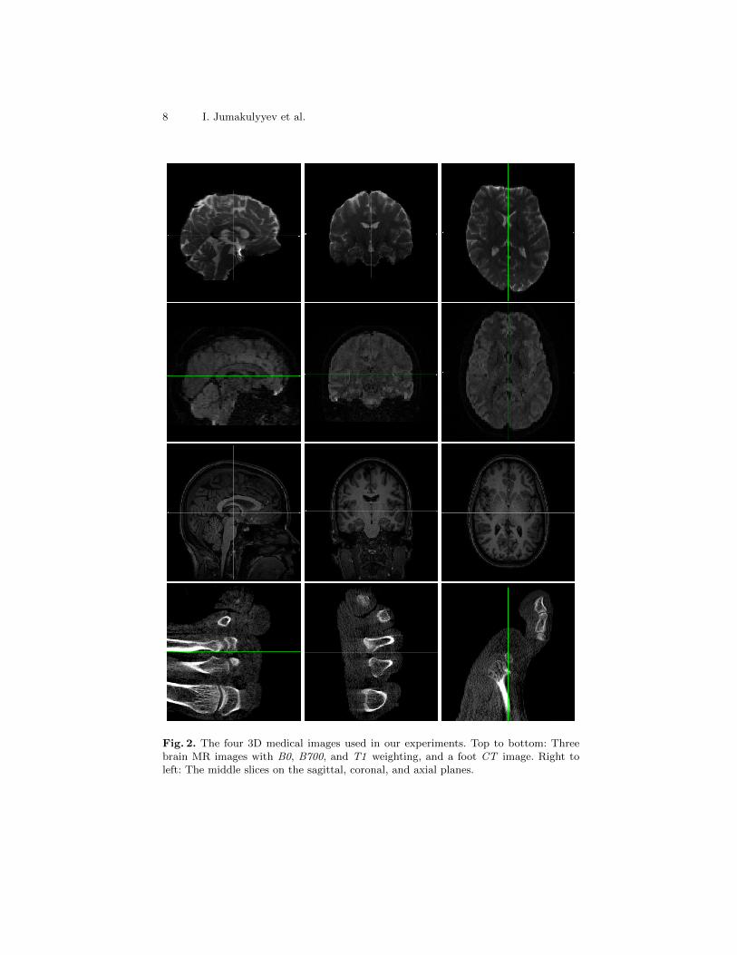
For our experiments, we chose four 3D medical images which are illustrated inFigure 2. Even though three of them are from brain imaging, they have beenchosen to represent diverse contrasts and properties.
1. A scan from diffusion MRI [2] with diffusion weight b = 0 and 136×136×84voxels. A brain extraction algorithm has zeroed out 73% of the voxels. Thisresults in a test case that is analogous to hybrid compression, in which onlya clinically relevant region of interest (ROI) is losslessly compressed [28,27].
2. A diffusion MRI scan with b = 700 and 104× 104× 72 voxels. This time, nobrain extraction has been performed, and there is substantial measurementnoise in the background, yielding less than 10% voxels with exactly zerointensity. This should provide a challenging test case for our codec, since thenoisy background region should be difficult to compress losslessly.
3. A T1 weighted MR image with 256× 256× 220 voxels. No brain extractionhas been performed, but there is much less noise, leading to more than 65%zero voxels. Due to the higher spatial resolution, we expected a larger degreeof spatial dependencies which could be exploited by a PDE-based inpainting.
4. A foot CT image with 256 × 256 × 256 voxels, which we expected to bechallenging due to the noisy appearance within the foreground region.
4.1 Comparison to Other Codecs
Table 1 compares the compression rate of our proposed codec to four establishedalternatives, by specifying the relative differences in final file sizes. Positive val-ues indicate a benefit of our codec. Details on the four lossless codecs includedin our comparison are given in Section 2. Results consider different variants ofour codec, using linear homogeneous (LH), second-order edge enhancing (EED),
8 I. Jumakulyyev et al.
Fig. 2. The four 3D medical images used in our experiments. Top to bottom: Threebrain MR images with B0, B700, and T1 weighting, and a foot CT image. Right toleft: The middle slices on the sagittal, coronal, and axial planes.

Lossless PDE-based Compression of 3D Medical Images 9
Table 2. Delta coding of intensities already reduced entropy in all test images. Weseparately report this for the whole images, and within their non empty space regions.
Image ImageEntropy
Delta Coded ImageEntropy
Nonzero RegionEntropy
Delta Coded NonzeroRegion Entropy
B0 3.63997 3.50099 10.33056 9.63958B700 5.34271 5.01050 5.39311 5.11973T1 3.14553 2.73150 6.46612 5.54785CT 2.81012 2.66688 6.71209 6.15826
or fourth-order edge enhancing diffusion (FOEED). All cases use iterative re-construction from a regular grid (R-I) in Mode 1 (see Section 3.2). The effect ofusing fewer or no iterations will be studied separately in Section 4.3.
In all four examples, we observe a clear improvement when moving from basiclinear homogeneous diffusion to anisotropic diffusion. Highest compression rateswere achieved with the recently introduced fourth-order EED. It allowed us toachieve higher lossless compression rates than any established codec. In manycases, the margin was considerable.
However, FOEED also had the highest computational cost. For the B700image, iterative 3D reconstruction on a single 3.3 GHz CPU core took 27 s withLH diffusion, 478 s with EED, and 6185 s with FOEED. We expect that thesetimes could be shortened significantly by a parallel implementation. This wasnot the focus of our current work.
4.2 Non-PDE Baseline for Further Comparisons
We require a suitable baseline to pinpoint the effect that the iterative reconstruc-tion and tuning of the contrast parameter have on the final compression rate.Since we use Huffman coding or the Deflate algorithm for the final encoding,file sizes achieved with GZIP are a natural reference. In addition, we designed abaseline codec that is “in between” GZIP and our PDE-based codec.
First, we found that, when 3D images contain a substantial amount of emptyspace, coding it separately can increase compression rates. Therefore, our base-line codec performs the same run length encoding as our PDE-based codec if itdecreases the overall file size. This was the case in all examples except B700.
Second, we were wondering how much we can benefit already from a very sim-ple, non PDE-based prediction of voxel intensities. To this end, we performed adelta coding, i.e., we fed differences between subsequent voxel intensities insteadof the intensities themselves into the final compression. Table 2 shows that, inall cases, this decreased the entropy. It also slightly increased compression rates.
Finally, as in our PDE-based codec, our baseline codec uses either Deflate orpure Huffman coding, depending on what results in a smaller file. We used thesame implementations from zlib and dippykit, respectively. Table 3 shows thatthis baseline already improves considerably over GZIP.

10 I. Jumakulyyev et al.
Table 3. Our non-PDE baseline that makes use of delta coding and optional emptyspace coding already results in a clear improvement over GZIP.
Image GZIP (bytes) Non-PDE Baseline Zero Density Zero Mask (bytes)
B0 692.372 +21.431% 72.92% 9.928B700 610.968 +18.015% 9.07% 43.681T1 5.207.535 +26.955% 65.70% 290.969CT 4.515.257 +8.347% 71.10% 315.173
Table 4. Compared to the non-PDE baseline, direct coding of residuals after a singlereconstruction with second-order EED does not yet result in a clear benefit. However,an iterative reconstruction as described in Section 3.2 does.
Image Direct Residual Iteration in Mode 0 Iteration in Mode 1
B0 +0.095% +4.367% +8.406%B700 +1.784% +6.847% +11.164%T1 −0.698% +6.715% +11.845%CT −6.476% +7.286% +12.590%
4.3 Effect of Iterative Construction of Residuals
Table 4 shows how the iterative alternation between reconstruction and residualcoding that is described in Section 3.2 affects the overall file sizes achieved withour codec. Differences are relative to the non-PDE baseline from the previoussection. In this experiment, second-order EED has been used for reconstruction.Positive values indicate a benefit of PDE-based predictions over delta coding.
Results indicate that a single reconstruction from a sparse regular grid isnot sufficient to obtain a benefit from second-order anisotropic diffusion. Onthe other hand, the proposed iterative reconstruction achieves a clear additionalreduction in compressed file size. It is most pronounced in Mode 1, which dilatesthe inpainting mask with a cross-shaped structuring element and consequentlyrequires more iterations than Mode 0, which dilates with a box.
4.4 Effect of Contrast Parameter
Even though it can be seen from Table 1 that moving from isotropic to anisotropicdiffusion noticeably improved compression rates, we found that fine-tuning thecontrast parameter in the diffusivity function is less important. Table 5 exploresthe effect of varying the ad-hoc threshold value of 90% that was used in Sec-tion 3.3 to two other values, 60% and 30%. For each image and threshold, thetable reports the corresponding values of contrast parameter λ, as well as theresulting improvement over the non-PDE baseline.
All differences due to the contrast parameter are below 0.5%. This supportsour decision to rely on a heuristic setting of the contrast parameter for losslesscompression, rather than spending computational resources on trying to optimizeit. Results in Table 5 used second-order EED with iteration mode 0, becausefine-tuning the contrast parameter would be even more costly in mode 1.

Lossless PDE-based Compression of 3D Medical Images 11
Table 5. Results from varying the threshold that our heuristic uses to set the contrastparameter λ. Despite a noticeable effect on λ itself, the corresponding differences incompression rates are rather small. Improvements are relative to the non-PDE baseline.
30% Threshold 60% Threshold 90% ThresholdImage λ Improvement λ Improvement λ Improvement
B0 2.04741 +4.272% 4.42198 +4.362% 11.83980 +4.367%B700 0.01379 +6.468% 0.10905 +6.794% 0.56127 +6.847%T1 0.12347 +7.108% 0.30572 +7.017% 0.58445 +6.715%CT 0.12464 +7.060% 0.22596 +7.225% 0.65364 +7.286%
5 Conclusion
PDE-based inpainting has previously been shown to have a strong potential forlossy image compression, especially at high compression rates. We demonstratedthat this approach also holds promise for lossless compression of 3D medicalimages. In particular, we propose a codec that beats state-of-the-art alternativesby combining a simple yet efficient to code initial inpainting mask with iterativereconstruction and coding of residuals, as well as a separate coding of emptyspace. In the future, we are planning to extend our work to exploit redundanciesalong the fourth axis that arises in diffusion MRI, i.e., orientation of the diffusiongradient [2]. This will require operating on the space of positions and orientation.
References
1. Andris, S., Peter, P., Weickert, J.: A proof-of-concept framework for PDE-basedvideo compression. In: Picture Coding Symposium (PCS). pp. 1–5. IEEE (2016)
2. Basser, P.J., Mattiello, J., Le Bihan, D.: Estimation of the effective self-diffusiontensor from the NMR spin echo. J. of Magnetic Resonance B(103), 247–254 (1994)
3. Deutsch, P.: RFC1951: DEFLATE compressed data format specification version1.3 (1996)
4. Deutsch, P.: RFC1952: GZIP file format specification version 4.3 (1996)
5. Dinov, I.D.: Volume and value of big healthcare data. Journal of medical statisticsand informatics 4, 3 (2016)
6. Galic, I., Weickert, J., Welk, M., Bruhn, A., Belyaev, A., Seidel, H.P.: TowardsPDE-based image compression. In: Int’l Workshop on Variational, Geometric, andLevel Set Methods in Computer Vision. pp. 37–48. Springer (2005)
7. Galic, I., Weickert, J., Welk, M., Bruhn, A., Belyaev, A., Seidel, H.P.: Image com-pression with anisotropic diffusion. Journal of Mathematical Imaging and Vision31(2-3), 255–269 (2008)
8. Jumakulyyev, I., Schultz, T.: Fourth-order anisotropic diffusion for inpainting andimage compression. In: Anisotropy Across Fields and Scales, pp. 99–123. Mathe-matics and Visualization, Springer (2021)
9. Kil, S.K., Lee, J.S., Shen, D., Ryu, J., Lee, E., Min, H., Hong, S.: Lossless medicalimage compression using redundancy analysis. International Journal of ComputerScience and Network Security 6(1), 50–56 (2006)

12 I. Jumakulyyev et al.
10. Kim, Y.S., Pearlman, W.A.: Lossless volumetric medical image compression. In:Applications of Digital Image Processing XXII. vol. 3808, pp. 305–312. Interna-tional Society for Optics and Photonics (1999)
11. Kostler, H., Sturmer, M., Freundl, C., Rude, U.: PDE based video compression inreal time. In: Tech. Rep. 07-11, Lehrstuhl fur Informatik 10. University Erlangen–Nurnberg (2007)
12. Larobina, M., Murino, L.: Medical image file formats. Journal of digital imaging27(2), 200–206 (2014)
13. Liu, D., Sun, X., Wu, F., Li, S., Zhang, Y.Q.: Image compression with edge-based inpainting. IEEE Transactions on Circuits and Systems for Video Technology17(10), 1273–1287 (2007)
14. Mainberger, M., Weickert, J.: Edge-based image compression with homogeneousdiffusion. In: Int’l Conf. on Computer Analysis of Images and Patterns. pp. 476–483(2009)
15. Mentzer, F., Agustsson, E., Tschannen, M., Timofte, R., Gool, L.V.: Practical fullresolution learned lossless image compression. In: Proc. IEEE Conf. on ComputerVision and Pattern Recognition (CVPR). pp. 10629–10638 (2019)
16. Miaou, S.G., Ke, F.S., Chen, S.C.: A lossless compression method for medicalimage sequences using JPEG-LS and interframe coding. IEEE Transactions onInformation Technology in Biomedicine 13(5), 818–821 (2009)
17. Nasr, M., Shokri, R., Houmansadr, A.: Comprehensive privacy analysis of deeplearning: Passive and active white-box inference attacks against centralized andfederated learning. In: Proc. IEEE Symp. on Security and Privacy. pp. 739–753(2019)
18. Pennebaker, W.B., Mitchell, J.L.: JPEG: Still Image Data Compression Standard.Springer (1993)
19. Perona, P., Malik, J.: Scale-space and edge detection using anisotropic diffusion.IEEE Trans. on Pattern Analysis and Machine Intelligence 12(7), 629–639 (1990)
20. Peter, P.: Three-dimensional data compression with anisotropic diffusion. In: Ger-man Conf. on Pattern Recognition. pp. 231–236. Springer (2013)
21. Peter, P., Contelly, J., Weickert, J.: Compressing audio signals with inpainting-based sparsification. In: Scale Space and Variational Methods. LNCS, vol. 11603,pp. 92–103. Springer (2019)
22. Peter, P., Schmaltz, C., Mach, N., Mainberger, M., Weickert, J.: Beyond purequality: Progressive modes, region of interest coding, and real time video decodingfor PDE-based image compression. Journal of Visual Communication and ImageRepresentation 31, 253–265 (2015)
23. Schmaltz, C., Peter, P., Mainberger, M., Ebel, F., Weickert, J., Bruhn, A.: Under-standing, optimising, and extending data compression with anisotropic diffusion.Int’l Journal of Computer Vision 108(3), 222–240 (2014)
24. Schmaltz, C., Weickert, J., Bruhn, A.: Beating the quality of JPEG 2000 withanisotropic diffusion. In: Joint Pattern Recognition Symposium. pp. 452–461.Springer (2009)
25. Taubman, D., Marcellin, M.: JPEG2000: Image Compression Fundamentals, Stan-dards and Practice. Springer (2002)
26. Weickert, J.: Anisotropic diffusion in image processing. Teubner Stuttgart (1998)27. Yee, D., Soltaninejad, S., Hazarika, D., Mbuyi, G., Barnwal, R., Basu, A.: Medi-
cal image compression based on region of interest using better portable graphics(BPG). In: IEEE Int’l Conf. on Systems, Man, and Cybernetics. pp. 216–221 (2017)
28. Zukoski, M.J., Boult, T., Iyriboz, T.: A novel approach to medical image compres-sion. Int’l J. of Bioinformatics Research and Applications 2(1), 89–103 (2006)