Longitudinal Assessment of Human Immunodeficiency Virus Type 1 (HIV-1)-Specific Gamma Interferon...
10
JOURNAL OF VIROLOGY, July 2005, p. 8121–8130 Vol. 79, No. 13 0022-538X/05/$08.000 doi:10.1128/JVI.79.13.8121–8130.2005 Copyright © 2005, American Society for Microbiology. All Rights Reserved. Longitudinal Assessment of Human Immunodeficiency Virus Type 1 (HIV-1)-Specific Gamma Interferon Responses during the First Year of Life in HIV-1-Infected Infants Barbara L. Lohman, 1,2 * Jennifer A. Slyker, 7 Barbra A. Richardson, 3,5 Carey Farquhar, 2,4 Jenniffer M. Mabuka, 1 Christopher Crudder, 1 Tao Dong, 7 Elizabeth Obimbo, 1 Dorothy Mbori-Ngacha, 1 Julie Overbaugh, 5,6 Sarah Rowland-Jones, 7 and Grace John-Stewart 1,2,4 Department of Paediatrics, University of Nairobi, Nairobi, Kenya 1 ; Departments of Epidemiology, 2 Biostatisics, 3 and Medicine, 4 University of Washington, Seattle, Washington; Divisions of Public Health Sciences 5 and Human Biology, 6 Fred Hutchinson Cancer Research Center, Seattle, Washington; and Human Immunology Unit, Weatherall Institute of Molecular Medicine, Oxford University, Oxford, England 7 Received 3 December 2004/Accepted 13 March 2005 Human immunodeficiency virus type 1 (HIV-1) infection results in different patterns of viral replication in pediatric compared to adult populations. The role of early HIV-1-specific responses in viral control has not been well defined, because most studies of HIV-1-infected infants have been retrospective or cross-sectional. We evaluated the association between HIV-1-specific gamma interferon (IFN-) release from the cells of infants of 1 to 3 months of age and peak viral loads and mortality in the first year of life among 61 Kenyan HIV-1-infected infants. At 1 month, responses were detected in 7/12 (58%) and 6/21 (29%) of infants infected in utero and peripartum, respectively (P 0.09), and in 50% of infants thereafter. Peaks of HIV-specific spot-forming units (SFU) increased significantly with age in all infants, from 251/10 6 peripheral blood mononuclear cells (PBMC) at 1 month of age to 501/10 6 PBMC at 12 months of age (P 0.03), although when limited to infants who survived to 1 year, the increase in peak HIV-specific SFU was no longer significant (P 0.18). Over the first year of life, infants with IFN- responses at 1 month had peak plasma viral loads, rates of decline of viral load, and mortality risk similar to those of infants who lacked responses at 1 month. The strength and breadth of IFN- responses at 1 month were not significantly associated with viral containment or mortality. These results suggest that, in contrast to HIV-1-infected adults, in whom strong cytotoxic T lymphocyte responses in primary infection are associated with reductions in viremia, HIV-1-infected neonates generate HIV-1-specific CD8 -T-cell responses early in life that are not clearly associated with improved clinical outcomes. CD8 cytotoxic T lymphocytes (CTL) are responsible for clearing acute viral infections such as cytomegalovirus (CMV) and measles virus and play variable roles in chronic viral in- fections, depending on the site and degree of ongoing viral replication (reviewed in references 38 and 64). The CD8 CTL response to human immunodeficiency virus (HIV) has been extensively studied in humans and in the rhesus macaque- simian immunodeficiency virus (SIV) model for association with virus levels and disease progression. HIV-1- and SIV- specific CD8 -T lymphocyte numbers rise during acute infec- tion, and the peak number of CD8 -T cells coincides with the decline in plasma viremia (30, 31, 48). In the SIV model, the depletion of CD8 -T lymphocytes in either acute or chronic infection leads to an increase in viral replication which is cur- tailed by the regeneration of CD8 -T cells (26, 56). The con- clusion that HIV-1-specific CTL are an important component of the host immune response to infection is supported by several notable findings. First, antiviral CTL, frequently gag- specific, are associated with control of HIV-1 viral replication in adults and children of 10 years old (10, 13, 19, 39, 40, 45). Second, levels of circulating HIV-1-specific CTL are main- tained in long-term nonprogressors (24, 51). Finally, in both acute and chronic HIV-1 infections, HIV isolates have evolved mutations allowing escape from CTL recognition, indicating immune pressure on viral replication (6, 22, 47). The study of HIV-1-specific CD8 -T-cell responses in ver- tically infected infants is complicated by several factors absent in horizontal HIV-1 transmission. The patterns of HIV-1 peak and set-point plasma viral loads are very different in adults and infants (49). In infants, the levels of HIV-1 plasma viremia are persistently high, with declines not seen until the second year of life (17, 18, 37). In the absence of antiretroviral therapy, vertically infected infants have a bimodal distribution of dis- ease progression, with approximately 25% progressing to AIDS within 1 year of life (reviewed in reference 35). Factors that may influence the levels of viral replication and disease progression in infants include the phenotype of the transmitted virus, the high number of target cells available for HIV-1 infection, and an immature immune system. Infants are likely infected with a viral variant modified by maternal immune pressure due to the half-match in major histocompatibility complex alleles (21, 55). In addition, infants have high levels of thymic output, and their immune systems are predominantly naı ¨ve (8, 15, 57), although the role of the thymus in the disease * Corresponding author. Mailing address: IARTP, 325 Ninth Ave., Box 359909, Seattle, WA 98104. Phone: (206) 543-4278. Fax: (206) 543-4818. E-mail: [email protected]. 8121 on December 5, 2015 by guest http://jvi.asm.org/ Downloaded from
-
Upload
independent -
Category
Documents
-
view
4 -
download
0
Transcript of Longitudinal Assessment of Human Immunodeficiency Virus Type 1 (HIV-1)-Specific Gamma Interferon...

JOURNAL OF VIROLOGY, July 2005, p. 8121–8130 Vol. 79, No. 130022-538X/05/$08.00�0 doi:10.1128/JVI.79.13.8121–8130.2005Copyright © 2005, American Society for Microbiology. All Rights Reserved.
Longitudinal Assessment of Human Immunodeficiency Virus Type 1(HIV-1)-Specific Gamma Interferon Responses during the
First Year of Life in HIV-1-Infected InfantsBarbara L. Lohman,1,2* Jennifer A. Slyker,7 Barbra A. Richardson,3,5 Carey Farquhar,2,4
Jenniffer M. Mabuka,1 Christopher Crudder,1 Tao Dong,7 Elizabeth Obimbo,1Dorothy Mbori-Ngacha,1 Julie Overbaugh,5,6 Sarah Rowland-Jones,7
and Grace John-Stewart1,2,4
Department of Paediatrics, University of Nairobi, Nairobi, Kenya1; Departments of Epidemiology,2 Biostatisics,3 andMedicine,4 University of Washington, Seattle, Washington; Divisions of Public Health Sciences5 and
Human Biology,6 Fred Hutchinson Cancer Research Center, Seattle, Washington; and HumanImmunology Unit, Weatherall Institute of Molecular Medicine,
Oxford University, Oxford, England7
Received 3 December 2004/Accepted 13 March 2005
Human immunodeficiency virus type 1 (HIV-1) infection results in different patterns of viral replication inpediatric compared to adult populations. The role of early HIV-1-specific responses in viral control has notbeen well defined, because most studies of HIV-1-infected infants have been retrospective or cross-sectional. Weevaluated the association between HIV-1-specific gamma interferon (IFN-�) release from the cells of infants of1 to 3 months of age and peak viral loads and mortality in the first year of life among 61 Kenyan HIV-1-infectedinfants. At 1 month, responses were detected in 7/12 (58%) and 6/21 (29%) of infants infected in utero andperipartum, respectively (P � 0.09), and in �50% of infants thereafter. Peaks of HIV-specific spot-formingunits (SFU) increased significantly with age in all infants, from 251/106 peripheral blood mononuclear cells(PBMC) at 1 month of age to 501/106 PBMC at 12 months of age (P � 0.03), although when limited to infantswho survived to 1 year, the increase in peak HIV-specific SFU was no longer significant (P � 0.18). Over thefirst year of life, infants with IFN-� responses at 1 month had peak plasma viral loads, rates of decline of viralload, and mortality risk similar to those of infants who lacked responses at 1 month. The strength and breadthof IFN-� responses at 1 month were not significantly associated with viral containment or mortality. Theseresults suggest that, in contrast to HIV-1-infected adults, in whom strong cytotoxic T lymphocyte responses inprimary infection are associated with reductions in viremia, HIV-1-infected neonates generate HIV-1-specificCD8�-T-cell responses early in life that are not clearly associated with improved clinical outcomes.
CD8� cytotoxic T lymphocytes (CTL) are responsible forclearing acute viral infections such as cytomegalovirus (CMV)and measles virus and play variable roles in chronic viral in-fections, depending on the site and degree of ongoing viralreplication (reviewed in references 38 and 64). The CD8� CTLresponse to human immunodeficiency virus (HIV) has beenextensively studied in humans and in the rhesus macaque-simian immunodeficiency virus (SIV) model for associationwith virus levels and disease progression. HIV-1- and SIV-specific CD8�-T lymphocyte numbers rise during acute infec-tion, and the peak number of CD8�-T cells coincides with thedecline in plasma viremia (30, 31, 48). In the SIV model, thedepletion of CD8�-T lymphocytes in either acute or chronicinfection leads to an increase in viral replication which is cur-tailed by the regeneration of CD8�-T cells (26, 56). The con-clusion that HIV-1-specific CTL are an important componentof the host immune response to infection is supported byseveral notable findings. First, antiviral CTL, frequently gag-specific, are associated with control of HIV-1 viral replicationin adults and children of �10 years old (10, 13, 19, 39, 40, 45).
Second, levels of circulating HIV-1-specific CTL are main-tained in long-term nonprogressors (24, 51). Finally, in bothacute and chronic HIV-1 infections, HIV isolates have evolvedmutations allowing escape from CTL recognition, indicatingimmune pressure on viral replication (6, 22, 47).
The study of HIV-1-specific CD8�-T-cell responses in ver-tically infected infants is complicated by several factors absentin horizontal HIV-1 transmission. The patterns of HIV-1 peakand set-point plasma viral loads are very different in adults andinfants (49). In infants, the levels of HIV-1 plasma viremia arepersistently high, with declines not seen until the second yearof life (17, 18, 37). In the absence of antiretroviral therapy,vertically infected infants have a bimodal distribution of dis-ease progression, with approximately 25% progressing toAIDS within 1 year of life (reviewed in reference 35). Factorsthat may influence the levels of viral replication and diseaseprogression in infants include the phenotype of the transmittedvirus, the high number of target cells available for HIV-1infection, and an immature immune system. Infants are likelyinfected with a viral variant modified by maternal immunepressure due to the half-match in major histocompatibilitycomplex alleles (21, 55). In addition, infants have high levels ofthymic output, and their immune systems are predominantlynaı̈ve (8, 15, 57), although the role of the thymus in the disease
* Corresponding author. Mailing address: IARTP, 325 Ninth Ave.,Box 359909, Seattle, WA 98104. Phone: (206) 543-4278. Fax: (206)543-4818. E-mail: [email protected].
8121
on Decem
ber 5, 2015 by guesthttp://jvi.asm
.org/D
ownloaded from
progression of HIV-1-infected infants is not well understood(7). The ability of the neonate to respond effectively to infec-tion is thought to be limited by the number of circulatingmature T and antigen-processing cells (50, 54). Cellular im-mune responses in HIV-1-infected infants have been inconsis-tently detected in infants younger than 6 months (33, 34, 36, 46,61). The paucity of CTL responses in infants less than 1 yearold has been suggested (i) to be due to diminished Th1 re-sponses, in particular a deficiency in gamma interferon(IFN-�) secretion (58, 62, 63) or (ii) to be influenced by age,CD4 counts, and antigen processing (53). The interpretation ofthe earlier reports is limited by the lack of longitudinal dataand the imprecise detection of the timing of infection in theinfants.
We had the opportunity, with a prospective observationalcohort of infants born to HIV-1-infected women, to identifyinfants infected before 1 month of life and to measure HIV-1-specific CD8�-T-cell responses together with viral loads overthe first year of life. We hypothesized that sustained highHIV-1 viral loads observed in perinatal transmission were con-sistent with a deficiency in virus-specific CD8�-T-cell re-sponses. We examined the HIV-1-specific IFN-� release fromCD8�-T cells at one to five time points during the first year oflife of 61 Kenyan infants diagnosed with HIV-1 infection at orbefore the first month of life and investigated the relationshipbetween the timing and presence of early anti-HIV-1 CD8�-T-cell responses and peak viral loads and mortality in theinfants.
MATERIALS AND METHODS
Patient cohort. As part of a larger cohort of infants born to HIV-1-infectedwomen between 1999 and 2003, 61 infants with HIV-1 RNA or DNA detected inblood obtained during the first month of life were included in this cohort. Detailsof the larger cohort of infants born to HIV-1-infected women have been pre-sented elsewhere (20, 41). Written informed consent was obtained from allmothers on behalf of themselves and their infants. Mothers were recruitedduring pregnancy and were provided with zidovudine beginning between the 34thand 36th weeks of gestation for the prevention of infant HIV-1 infection (59).Infants were breast or formula fed as per maternal preference. Infants wereexamined by clinicians, and 1 to 3 ml of peripheral blood was obtained at birthand at months 1, 3, 6, 9, and 12. Seven infants born to HIV-1-seronegativewomen were included as controls and were bled at 3 months of life. This researchcomplied with all U.S. and Kenyan guidelines and policies.
Blood collection. Freshly collected EDTA-anticoagulated blood was centri-fuged for 10 min at 1,800 rpm to separate plasma, which was aliquoted and storedat �80°C. The remaining blood was then diluted with an equal volume of RPMI1640 (Gibco-BRL), layered on a Ficoll gradient (Lymphocyte Separation Me-dium; Organon Teknika, West Chester, PA), and centrifuged for 30 min at 2,000rpm. Peripheral blood mononuclear cells (PBMC) at the interface of the gradi-ent were isolated, washed in RPMI 1640, and used directly in the enzyme-linkedimmunospot (ELISPOT) assay. The mean numbers � standard errors of themeans of PBMC recovered at months 1, 3, 6, 9, and 12 were (13.6 � 1.0) � 106,(17.7 � 1.1) � 106, (18.6 � 1.7) � 106, (16.8 � 1.6) � 106, and (15.5 � 1.5) �106, respectively.
HIV-1 diagnosis and determination of timing of infection. Infant HIV-1 in-fection status was determined by PCR amplification of HIV-1 gag DNA se-quences from dried blood spotted on filter paper (44) or by quantitative analysisof infant plasma HIV-1 RNA using a transcription-mediated amplificationmethod sensitive for the detection of multiple HIV-1 subtypes (Gen-ProbeHIV-1 viral load assay) (43). Results relative to HIV RNA copies/ml plasmawere considered positive if there were �100 copies per ml or, in cases where lessthan 500 �l was available for testing, if there were �50 RNA copies per reaction.The timing of an infection was categorized as in utero if the specimen collectedwithin the first 48 h of life was positive for either HIV-1 DNA or HIV-1 RNA.The timing of infection was defined as peripartum if plasma HIV-1 RNA was
undetectable within the first 48 h of life and positive at 1 month of age. Quan-titative plasma viral loads were determined using the Gen-Probe assay.
HLA typing. Molecular HLA typing was performed on DNA extracted from 5� 106 infant PBMC using amplification refractory mutation system PCR withsequence-specific primers designed for East African populations (9).
Peptides. Sixty-eight peptides spanning five regions of HIV-1 were synthesizedby Fmoc (9-fluorenylmethoxy carbonyl) chemistry at the Peptide Core Facility atthe Weatherall Institute of Molecular Medicine (Tao Dong, Oxford University).The peptides were chosen based on predefined CTL epitopes of the prevalentHIV-1 clades in Kenya (A and D). Twenty-seven peptides were from gag, 18 werefrom pol, 19 were from env, 13 were from nef, and 1 was from rev (Table 1).Epitopes were chosen based on responses previously reported in HIV-1 infectionand included those present in acute infection, long-term nonprogressors, andthose associated with viral control (29). These peptides bind 29 common HLAclass 1 alleles (12 HLA-A, 15 HLA-B, and 2 HLA-C) representative of EastAfrican populations. The identities of peptides tested for each individual werebased on the infant’s HLA type. In the event of a limited number of cells,peptides were prioritized to test a complete panel for each HLA allele.
IFN-� ELISPOT assay. An ELISPOT assay was used to detect HIV-1-specificIFN-� release from PBMC following overnight incubation with peptides. Briefly,96-well Millipore plates (MAIP45; Millipore SA, Molsheim, France) were coatedwith 7.5 �g monoclonal antibody to IFN-� (1-DIK; Mabtech Ab, Nacka, Swe-den) for 2 h at 37°C. Excess antibody was removed by washing six times withRPMI 1640 and blocked with RPMI 1640 containing L-glutamine and supple-mented with 10% fetal calf serum (all from Gibco-BRL), designated R10, for 30min at room temperature before cells were added. Duplicate wells containing 2� 105 PBMC/well were stimulated with 20 �g/ml peptide, 10 �g/ml phytohe-magglutinin (PHA) (positive control) (Murex Biotech Limited, Dartford, UnitedKingdom), or R10 media alone (negative control). The mean numbers � stan-dard errors of the means of PBMC used per assay at months 1, 3, 6, 9, and 12were (6.5 � 0.5) � 106, (6.4 � 0.4) � 106, (6.9 � 0.4) � 106, (6.7 � 0.5) � 106,and (6.8 � 0.4) � 106, respectively. After overnight stimulation in a humidifiedincubator at 37°C and with 5% CO2, cells were removed from the plates bywashing with phosphate-buffered saline containing 0.05% Tween 20 (Sigma, St.Louis, MO), followed in sequence by the application of biotinylated anti-IFN-�antibody (1:1,000; 7-B6-1 biotin; Mabtech) for 3 h at room temperature, washing,and the application of streptavidin alkaline phosphatase (1:1,000; Mabtech) for1.5 h at room temperature. After the final washing, spot-forming units (SFU)were visualized by the addition of alkaline phosphatase (Bio-Rad Laboratories,Hercules, CA) for approximately 10 min or until an intense blue reaction wasvisible in the wells stimulated with PHA. Additional color development wasprevented by washing the plates under running water. Plates were allowed to dryovernight before reading. Plates were read visually until January 2001, afterwhich an automated ELISPOT reader was used (Autoimmun Diagnostika,Strassberg, Germany). The number of HIV-specific SFU per 106 cells was de-fined as the average number of SFU per 106 cells from peptide-stimulated wellsminus the average number of SFU per 106 cells in wells containing R10 (back-ground control). The following criteria were used to determine a positive assay:(i) a response to PHA of �100 SFU after the subtraction of the background, (ii)the number of HIV-specific SFU per 106 cells being greater than or equal to 50,and (iii) the number of SFU in peptide-stimulated wells being greater than orequal to two times the number of background control SFU (4). The qualities ofthe ELISPOT responses were compared in two ways: relative to the mean ofpositive responses and relative to the peak of positive responses.
Detection of antigen-specific CD8�-T cells. PBMC from an infant expressingHLA-A2 and -B8 were stained with phycoerythrin-conjugated-HLA class I tet-rameric complexes refolded with either HIV-1 nef-FLKEKGGL (B8-nef) orCMV pp65-NLVPMVATV (A2-CMV) (Tao Dong, Oxford University). Briefly,for each stain, 150 �l of whole blood was incubated with phycoerythrin-conju-gated tetramer for 15 min at 37°C, followed by the addition of peridin chlorophyllprotein-labeled anti-CD8 and fluorescein isothiocyanate-labeled anti-CD45R0(both from Becton Dickenson, San Diego, CA) for an additional 15 min. Threemilliliters of FACSLysis solution (Becton Dickenson) was added per tube, andthe tubes were left for 5 min at room temperature, after which time the tubeswere centrifuged for 5 min at 1,500 rpm. The cell pellet was washed once inphosphate-buffered saline containing 0.5% fetal calf serum and 0.5 mM EDTA,suspended in 170 �l of FACSfix (Becton Dickenson), and stored overnight in thedark at 4°C prior to analysis with CellQuest software (Becton Dickenson).
Statistical analysis. To compare differences in the IFN-� responses of infantsinfected with HIV in utero to the responses of those infected peripartum, dif-ferences in the distributions of background SFU, numbers of mean and peakpositive HIV-specific SFU, numbers of peptides tested, and numbers of peptides
8122 LOHMAN ET AL. J. VIROL.
on Decem
ber 5, 2015 by guesthttp://jvi.asm
.org/D
ownloaded from
recognized were determined using Mann-Whitney U tests. Pearson’s chi-squarestatistic was used for the comparison of the proportion of infants with positiveHIV-1-specific ELISPOT results who were infected in utero to that of those whowere infected peripartum. Data was log transformed for analysis of viral loadsand changes in the numbers of HIV-specific SFU over time. Differences betweenthe mean and peak log10s of the HIV-1 RNA plasma viral loads of (i) infantsinfected with HIV-1 in utero and those of the infants infected peripartum or (ii)between infants who had and those who lacked HIV-1-specific ELISPOT re-sponses were assessed using t tests for independent samples. To model thechanges in peak numbers of HIV-specific SFU over the first year of life ofinfected infants, a linear mixed-effect model was used. For determination of thechange in viral load over time, a linear mixed-effect model with a compoundsymmetric covariance structure was used with the log10 of the HIV RNA viralload between month 1 and month 12 as the outcome variable. Linear regressionanalysis was used to compare peak plasma HIV-1 RNA loads in infants who hador lacked HIV-1-specific IFN-� responses at 1 month of age, controlling for viralload at 1 month of age after testing for and excluding nonsignificant effectmodification. Cox regression analysis was used to model the association betweenmortality and (i) the presence of either HIV-1-specific IFN-� responses at 1month of age or (ii) weak HIV-specific IFN-� responses (�500 HIV-specificSFU), controlling for viral load at 1 month of age and in utero infection.
RESULTS
Concordance between peptide-stimulated IFN-� secretionand detection of antigen-specific CD8�-T cells. The presenceof antigen-specific CD8�-T cells and the relationship betweenthe detection of these cells by major histocompatibility com-plex-tetrameric complexes and detection by IFN-� ELISPOTassay were confirmed in three individuals. Figure 1 illustratesrepresentative results obtained from one HIV-1-infected in-fant at 9 and 12 months of age, demonstrating the presence ofboth A2-CMV and B8-nef-specific CD8�-T cells in this infanttogether with peptide-specific ELISPOT assay results. At 9months of age, we detected tetramer-positive A2-CMV-spe-cific and B8-nef-specific CD8�-T cells, comprising 0.68% and0.27%, respectively, of total lymphocytes. In the concordantELISPOT assay, the frequency of IFN-�-producing cells re-sponding to A2-CMV peptide was 1,497 per 106 PBMC, andthe frequency of cells responding to B8-nef-peptide was 303
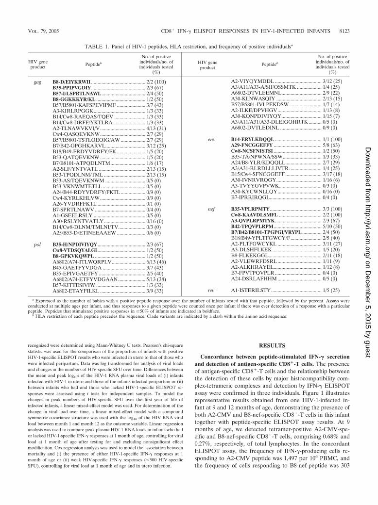
TABLE 1. Panel of HIV-1 peptides, HLA restriction, and frequency of positive individualsa
HIV geneproduct Peptideb
No. of positiveindividuals/no. ofindividuals tested
(%)
HIV geneproduct
Peptideb
No. of positiveindividuals/no. ofindividuals tested
(%)
gag B8-D/EIYKRWII .......................................... 2/2 (100)B35-PPIPVGDIY.......................................... 2/3 (67)B57-I/LSPRTLNAWL.................................. 2/4 (50)B8-GGKKKYR/KL ...................................... 1/2 (50)B57/B5801-KAFSPEVIPMF ...................... 3/7 (43)A3-KIRLRPGGK........................................ 1/3 (33)B14/Cw8-RAEQAS/TQEV ........................ 1/3 (33)B14/Cw8-DRFF/YKTLRA......................... 1/3 (33)A2-TLNAWVKVI/V................................... 4/13 (31)Cw4-QASQEVKNW................................... 2/7 (29)B57/B5801-TSTLQEQIG/AW ................... 2/7 (29)B7/B42-GPGHKARVL............................... 3/12 (25)B18/B49-FRDYVDRFY/FK ...................... 1/5 (20)B53-QATQEVKNW ................................... 1/5 (20)B7/B8101-ATPQDLNTM........................... 1/6 (17)A2-SLF/YNTVATL .................................... 2/13 (15)B53-TPQDLNM/TML ................................ 2/13 (15)B53-AS/TQEVKNWM ............................... 0/5 (0)B53 VKNWMTETLL ................................. 0/5 (0)A24/B44-RDYVDRFY/FKTL ................... 0/9 (0)Cw4-KYRLKHLVW................................... 0/9 (0)A26-YVDRFFKTL ..................................... 0/1 (0)B7-SPRTLNAWV ....................................... 0/4 (0)A1-GSEELRSLY ........................................ 0/5 (0)A30-RSLYNTVATLY................................ 0/16 (0)B14/Cw8-DLNM/TMLNI/TV..................... 0/3 (0)A25/B53-D/ETINEEAAEW ...................... 0/6 (0)
pol B35-H/NPDIVIYQY ..................................... 2/3 (67)Cw8-VTDSQYALGI ..................................... 1/2 (50)B8-GPKVKQWPL ....................................... 1/2 (50)A6802/A74-ITLWQRPLV.......................... 6/13 (46)B45-GAETFYVDGA ................................. 3/7 (43)B35-EPIVGAETFY .................................... 2/5 (40)A6802/A74-ETFYVDGAAN ..................... 5/13 (38)B57-KITTESIVIW ...................................... 1/3 (33)A6802-ETAYFILKL ................................... 3/9 (33)
a Expressed as the number of babies with a positive peptide response over the number of infants tested with that peptide, followed by the percent. Assays wereconducted at multiple ages per infant, and thus responses to a given peptide were counted once per infant if there was ever detection of a response with a particularpeptide. Peptides that stimulated positive responses in �50% of infants are indicated in boldface.
b HLA restriction of each peptide precedes the sequence. Clade variants are indicated by a slash within the amino acid sequence.
A2-VIYQYMDDL .................................... 3/12 (25)A3/A11/A33-A/SIFQSSMTK ................... 1/4 (25)A6802-DTVLEEMNL............................... 2/9 (22)A30-KLNWASQIY ................................... 2/13 (15)B57/B5801-IVLPEKDSW......................... 1/7 (14)A2-ILKE/DPVHGV.................................. 1/13 (8)A30-KQNPDIVIYQY............................... 1/15 (7)A3/A11/A31/A33-DLEIGQHRTK .......... 0/5 (0)A6802-DVTLEDINL ................................ 0/9 (0)
env B14-ERYLKDQQL .................................... 1/1 (100)A29-FNCGGEFFY ..................................... 5/8 (63)Cw8-NCSFNISTSI ..................................... 1/2 (50)B35-TA/NPWNA/SSW.............................. 1/3 (33)A24/B8-YLR/KDQQLL............................ 2/7 (29)A3/A31-RLRDLLLIVTR......................... 1/4 (25)B15/Cw4-SFNCGGEFF ............................ 3/17 (18)A30-IVNRVRQGY................................... 1/16 (6)A3-TVYYGVPVWK................................. 0/3 (0)A30-KYCWNLLQY.................................. 0/16 (0)B7-IPRRIRQGL........................................ 0/4 (0)
nef B35-VPLRPMTY ........................................ 3/3 (100)Cw8-KAAVDLSMFL ................................. 2/2 (100)A3-QVPLRPMTYK.................................... 2/3 (67)B42-TPQVPLRPM..................................... 5/10 (50)B7/B42/B8101-TPGPGI/VRYPL ............... 2/4 (50)B18/B49-YPLTFGWCY/F........................ 2/5 (40)A2-PLTFGWCYKL .................................. 3/11 (27)A3-DLSHFLKEK ...................................... 1/5 (20)B8-FLKEKGGL......................................... 2/11 (18)A2-VLEWRFDSRL .................................. 1/11 (9)A2-ALKHRAYEL..................................... 1/12 (8)B7-FPVTPQVPLR .................................... 0/4 (0)A24-DSRLAFHHM .................................. 0/5 (0)
rev A1-ISTERILSTY....................................... 1/5 (25)
VOL. 79, 2005 CD8� IFN-� ELISPOT RESPONSES IN HIV-1-INFECTED INFANTS 8123
on Decem
ber 5, 2015 by guesthttp://jvi.asm
.org/D
ownloaded from

per 106 PBMC. When the assays were repeated 3 months later,the frequencies of tetramer-positive CD8�-T cells were 0.35%and 0.38% of total lymphocytes specific for A2-CMV and B8-nef, respectively. The corresponding frequencies of A2-CMVand B8-nef peptide-specific IFN-�-producing cells were 1,025per 106 PBMC and 450 per 106 PBMC, respectively. Differ-ences between the percentage of CD8�-T cells detected bytetramer and the corresponding functional response followingcognate peptide stimulation have been observed before (3,29a), and the detection of IFN-� ELISPOT responses is likelyrepresentative of the presence of CD8� antigen-specific T cellsin circulation.
HIV-1-specific ELISPOT responses in infants infected inutero or peripartum. We measured HIV-1-specific IFN-� se-cretions from PBMC isolated from infants infected with HIV-1in utero or peripartum and followed those responses over thefirst year of life. The peptides were used either singly or pairedby clade variants and are presented by decreasing frequency ofresponse in Table 1.
Using this panel, we estimated the prevalence of the detec-tion of HIV-1-specific IFN-� responses during the first year oflife in HIV-1-infected infants (Table 2). The number of infantstested at each time point varied because of clinic attendanceand mortality. We were able to test all HLA-restricted peptides inour panel on 29/33 (88%) infants tested at month 1 and on 43/45(96%), 40/41 (97%), 26/29 (90%), and 24/26 (92%) infants atmonths 3, 6, 9, and 12, respectively. At 1 month of age, there wasa trend for more-likely detection of HIV-1-specific IFN-� re-sponses in infants infected in utero compared to infants infectedperipartum. Seven of 12 (58%) infants infected in utero haddetectable responses versus 6 (29%) of 21 infants infected peri-partum (P 0.09). Although the differences did not reach signif-icance due to small sample sizes, this trend is likely explained by
the duration of virus infection. By 3 months of age, the prevalenceof HIV-1-specific IFN-� responses was approximately 50% ineither group and, at this and subsequent ages, there was no sig-nificant difference in the prevalences of the detection of responsesin infants with respect to the timing of infection with HIV-1.
To determine if the timing of HIV-1 infection affected thequality of the cellular immune response, we compared thebreadths and strengths of IFN-� responses in infants infectedin utero versus peripartum (Table 3). We chose the month 3time point for comparisons because the greatest number ofinfants (45/61 [75%]) were tested at that time. To determineHIV-1 specificity, we compared the prevalence of positive as-says in HIV-1-infected infants with that in HIV-1 unexposed,uninfected infants. None of seven 3-month-old HIV-1 unex-posed, uninfected infants had positive ELISPOT responses,suggesting a high specificity for the assay. We observed trendsfor infants infected peripartum to have stronger HIV-1-specificIFN-� responses detected at 3 months of age, although thedata did not reach significance. For infants infected in utero,the median magnitudes of mean and peak responses were 151and 155 HIV-specific SFU per 106 PBMC, respectively, versus384 and 458 per 106 PBMC, respectively, in infants infectedperipartum (P 0.06 for both).
HIV-1-specific IFN-� responses over the first year of life. Ina subset of 18 infants, IFN-� responses and plasma viral loadmeasurements were conducted at every time point up to month12 or death. These results are shown in Fig. 2a and b, respec-tively, and demonstrate a diverse pattern of immune recogni-tion and viral replication. Three infants were completely lack-ing in detectable responses (B1-276, B1-160, and B1-005). Twoinfants demonstrated strengthening and broadening responsesto infection over time (B1-454, B1-473). Two infants lost earlyresponses, but the loss was not associated with mortality (B1-093, B1-259). In one infant, the loss of early responses pre-ceded the infant’s death (B1-424). To interpret the changes inHIV-1-specific IFN-� responses over the first year of life in allthe infants, we employed a model that adjusted for age, in-complete and repeated measures, and the timing of infections.Log10-transformed peak HIV-specific SFU numbers were usedto model the responses over time. We found a significantchange in peak HIV-1-specific IFN-� responses in infants dur-
FIG. 1. Assays of CMV- and HIV-specific tetramer staining andIFN-� ELISPOT responses. Responses detected from an HIV-1-in-fected infant at 9 (upper panels) and 12 (lower panels) months of age.PBMC were stained with anti-CD8 monoclonal antibody (y axes) andwith the relevant HLA class I/peptide tetramer (x axes) or stimulatedwith peptides in an overnight ELISPOT assay to detect IFN-� secre-tion. A2-CMVpp65-NLVPMVATV (left panels) and B8-HIV-1 nef-FLKEDGGL (right panels) responses were detected by both assays. Thetetramer-positive CD8� T cells, as percentages of total lymphocytes,are indicated adjacent to each plot. The antigen-specific IFN-� ELI-SPOT assay results are shown above each plot.
TABLE 2. Prevalence of detection of HIV-1-specific IFN-�ELISPOT responses during the first year of life of
61 HIV-1-infected infants
Age(months)
No. tested(% of cohort)
Prevalence of HIV-1-specificresponses [no. of positive
infants/no. of infants infected(%)]
P valueb
In uteroa Peripartum
1 33 (54) 7/12 (58) 6/21 (29) .093 45 (74) 12/18 (67) 14/27 (52) .46 41 (67) 6/15 (40) 13/26 (50) .49 29 (48) 6/11 (54) 8/18 (44) .512 26 (43) 5/10 (50) 10/16 (63) .6
a The timing of infection of infants was categorized as in utero if HIV-1 DNAor RNA was detected in dried blood spots or plasma collected within the first48 h of life. Infants were categorized as infected peripartum if HIV-1 DNA orRNA was undetectable within the first 48 h. of life and detectable at 1 month oflife.
b P values determined by Pearson’s chi-square statistic.
8124 LOHMAN ET AL. J. VIROL.
on Decem
ber 5, 2015 by guesthttp://jvi.asm
.org/D
ownloaded from

ing the first year of life, independent of the timing of infectionwith HIV-1 (P 0.03) (Fig. 3). Peak HIV-specific SFU in-creased twofold with increasing age, from a mean of 251/106
PBMC (2.4 log10) at 1 month of age to mean of 501/106 PBMC(2.7 log10) at 12 months of age. To address the potential bias ofsurvivor effect, we also limited the analysis to 30 infants whosurvived to month 12. We found a similar increase in peakIFN-� responses with age, but the change was no longer sta-tistically significant (P 0.18). The strength of the peak HIV-specific SFU increased 1.5-fold with increasing age in the in-fants who survived to month 12, from a mean of 343/106 PBMC(2.53 log10) at 1 month of age to a mean of 540/106 PBMC(2.73 log10) at 12 months of age.
Early HIV-1-specific IFN-� responses and HIV-1 replicationkinetics. The emergence of HIV- and SIV-specific CD8�-T-cell immune responses in primary infection has been shown tocorrelate with declines in levels of viral replication. Therefore,we sought to determine the relationship between early HIV-1-specific IFN-� responses and outcomes of HIV-1 infection ininfants, as measured by peak viral loads, rates of decline ofviral replication, and risk of mortality. The patterns of HIV-1RNA plasma viral replication over time in infants with orwithout HIV-1-specific IFN-� responses at 1 month of age,depicted by timing of infection, are shown in Fig. 4.
Peak HIV-1 plasma load is an important marker of diseaseprogression in HIV-1-infected infants (52). Peak viral load wasdefined as the highest viral load detected within 6 monthspostinfection. We found no significant difference in mean log10
peak viral loads for infants infected in utero versus those in-fected peripartum. The mean peak viral load for 23 infantswith defined in utero infection was 6.59 log10 � 0.84 versus6.86 log10 � 0.67 among 37 infants infected peripartum (P 0.2). Thirty-two infants had concurrent HIV-1-specific IFN-�ELISPOT assays and HIV-1 RNA plasma viral loads measuredat 1 month of age. As the timing of HIV-1 infection was shownnot to significantly affect peak viral load, all 32 infants wereconsidered as one group for the subsequent analyses. Therewas not a significant difference in mean log10 peak viral loadsfor infants with or without detectable HIV-1-specific IFN-�responses at 1 month of life, after controlling for the baselinemonth 1 viral load. The mean peak viral load for 12 infants
with detectable HIV-1-specific IFN-� responses at 1 month ofage was 6.82 log10 � 0.18 versus 6.62 log10 � 0.18 for 20 infantswho lacked detectable responses at 1 month of age (P 0.5).There were no significant differences in peak viral loads forinfants with broad (2 peptides) or strong (500 HIV-1 SFU)IFN-� responses at 1 month compared to those with narrower,weaker, or negative responses (data not shown).
Having seen no difference in the peak viral loads, we nextinvestigated the relationship between the presence of HIV-1-specific IFN-� responses at 1 month of age and the change inviral load over time. The presence of detectable HIV-1-specificIFN-� responses at 1 month of life was not associated with adifference in the rate of change of viral load over the first yearof life compared to the rate for infants who lacked such re-sponses at 1 month (decline of 0.04 versus 0.02 log10 HIV-1RNA copies/ml plasma/month, respectively; P 0.2).
We lastly investigated the relationship between the presenceand strength of HIV-1-specific IFN-� responses at 1 month ofage and infant mortality during the first year of life. There wasa trend correlating the presence of HIV-1-specific IFN-� re-sponses at 1 month of life and increased mortality found withunivariate analysis (hazard ratio [HR] 2.72; 95% confidenceinterval [CI], 0.89 to 8.36; P 0.08). There was also a trend forhigher mortality in those infants with IFN-� responses of �500HIV-1 SFU (HR 3.77; 95% CI, 0.94 to 15.13; P 0.06).After controlling for the viral load at month 1 and the timingof infection in multivariate analysis, there were no statisti-cally significant relationships either between mortality andthe presence of month 1 HIV-1-specific IFN-� responses(HR, 2.20; 95% CI, 0.62 to 7.87; P 0.2) or betweenmortality and IFN-� responses of �500 HIV-1 SFU (HR,2.08; 95% CI, 0.70 to 6.17; P 0.2). Thus, the presence orstrength of HIV-1-specific IFN-� responses in the firstmonths of life had no effect on peak viral load, on the rateof decline in HIV-1 plasma viral load, or on survival in thiscohort of HIV-1-infected infants.
DISCUSSION
We addressed a mechanism hypothesized to contribute tothe sustained high viral loads measured in perinatal HIV-1
TABLE 3. HIV-1-specific IFN-� responses in HIV-1 unexposed, uninfected infants and HIV-1-infected infants measured at 3 months of age
Parameter
InfantP
valuecUnexposeda
Exposed-infectedb
In utero Peripartum
No. of individuals tested 7 18 27No. of peptides tested (range of median) 5 (1–9) 11 (6–18) 9 (1–19) .2Background SFU/106 PBMC median (25th–75th quartile) 57 (20–90) 34 (19–64) 20 (14–40) .08No. with positive responses (%) 0 12 (67) 14 (52) .4No. of positive peptides (range of median) 0 2 (1–6) 2 (1–6) .3Mean positive HIV SFU/106 PBMC median (25th–75th quartile) n.a.d 151 (83–396) 384 (216–522) .06Peak positive HIV SFU/106 PBMC median (25th–75th quartile) n.a. 155 (103–693) 458 (216–953) .06
a Unexposed infants were born to HIV-1-seronegative women. The infection status of an infant was confirmed by testing for HIV-1 DNA in dried blood spotscollected at the time of the visit, as described in Materials and Methods.
b Infants were categorized as infected in utero if HIV-1 DNA or RNA was detected in dried blood spots or plasma collected within the first 48 h of life. Infants werecategorized as infected peripartum if HIV-1 DNA or RNA was undetectable within the first 48 h of life and detectable at 1 month of life.
c The P values for differences in responses between infants infected in utero and peripartum was determined by nonparametric test for differences in medians fromindependent samples, except for comparisons of numbers of individuals with positive responses, in which the P values were calculated from the chi-square statistic.
d n.a., not applicable.
VOL. 79, 2005 CD8� IFN-� ELISPOT RESPONSES IN HIV-1-INFECTED INFANTS 8125
on Decem
ber 5, 2015 by guesthttp://jvi.asm
.org/D
ownloaded from

infection, the adequacy of the CD8�-T-cell immune response.We were able to establish the timing of infection in the infantsas occurring either in utero or peripartum and to compare thedevelopment of CD8�-T-cell effector function and viral loads.Infants infected peripartum had a tendency to generate stron-ger but not broader responses to HIV-1 peptides than didinfants infected in utero. Both groups had similar increases inthe magnitudes of HIV-1-specific IFN-� responses over theirfirst year of life, an indication that infants infected in utero arenot more immunosuppressed than infants infected peripartumand that both groups possess the capacity for continuing im-mune maturation. Infants infected in utero did not demon-strate higher peak viral loads than infants infected peripartum,as has also been described in the Abidjan ANRS 049 DitrameStudy (52). A primary report describing the bimodal disease
progression in perinatally infected infants suggests that inutero infection may interfere with the maturation of the im-mune system and increase the rate of development of immu-nosuppression (5), something we do not show with our data.Despite a rapid and relatively robust immune response toinfection, induction of these early responses did not appearbeneficial to infants during the first year of life, regardless ofthe timing of infection. Specifically, there was no relationshipbetween the detection of these responses and a reduction inpeak viral load, rate of decline of viral replication, or risk ofmortality (independent of viral load).
Previous studies have relied on cross-sectional studies inolder children, where there may be bias towards those whosurvived. The approach here, using a longitudinal analysis of acohort of HIV-1-infected infants with well-defined timing of
FIG. 2. Spectrum of HIV-1-specific peptide responses in HIV-1-infected infants with measurements at every time point to 1 year (A) or death(B). Individual graphs present HIV-1-specific SFU/106 PBMC (stacked colored bars) and HIV-1 RNA copies/ml plasma (closed circles) asfunctions of age. The numbers above each bar or above the horizontal axes indicate the numbers of peptides tested at each time point, while theheights of each of the colored sections of the bars indicate the strengths of the peptide-specific responses. The number of peptides was the lowestat month 1, due to limitations in cell numbers, and remained constant from month 3 to 1 year or death. The notation of death indicates the infantdied 1 to 3 months after the last measurement.
8126 LOHMAN ET AL. J. VIROL.
on Decem
ber 5, 2015 by guesthttp://jvi.asm
.org/D
ownloaded from
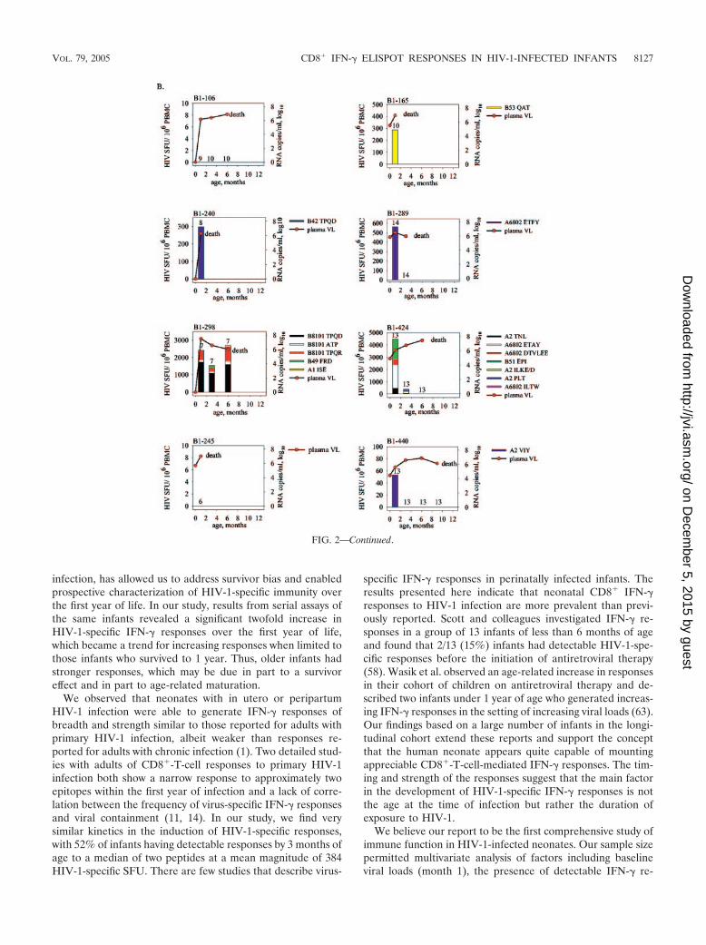
infection, has allowed us to address survivor bias and enabledprospective characterization of HIV-1-specific immunity overthe first year of life. In our study, results from serial assays ofthe same infants revealed a significant twofold increase inHIV-1-specific IFN-� responses over the first year of life,which became a trend for increasing responses when limited tothose infants who survived to 1 year. Thus, older infants hadstronger responses, which may be due in part to a survivoreffect and in part to age-related maturation.
We observed that neonates with in utero or peripartumHIV-1 infection were able to generate IFN-� responses ofbreadth and strength similar to those reported for adults withprimary HIV-1 infection, albeit weaker than responses re-ported for adults with chronic infection (1). Two detailed stud-ies with adults of CD8�-T-cell responses to primary HIV-1infection both show a narrow response to approximately twoepitopes within the first year of infection and a lack of corre-lation between the frequency of virus-specific IFN-� responsesand viral containment (11, 14). In our study, we find verysimilar kinetics in the induction of HIV-1-specific responses,with 52% of infants having detectable responses by 3 months ofage to a median of two peptides at a mean magnitude of 384HIV-1-specific SFU. There are few studies that describe virus-
specific IFN-� responses in perinatally infected infants. Theresults presented here indicate that neonatal CD8� IFN-�responses to HIV-1 infection are more prevalent than previ-ously reported. Scott and colleagues investigated IFN-� re-sponses in a group of 13 infants of less than 6 months of ageand found that 2/13 (15%) infants had detectable HIV-1-spe-cific responses before the initiation of antiretroviral therapy(58). Wasik et al. observed an age-related increase in responsesin their cohort of children on antiretroviral therapy and de-scribed two infants under 1 year of age who generated increas-ing IFN-� responses in the setting of increasing viral loads (63).Our findings based on a large number of infants in the longi-tudinal cohort extend these reports and support the conceptthat the human neonate appears quite capable of mountingappreciable CD8�-T-cell-mediated IFN-� responses. The tim-ing and strength of the responses suggest that the main factorin the development of HIV-1-specific IFN-� responses is notthe age at the time of infection but rather the duration ofexposure to HIV-1.
We believe our report to be the first comprehensive study ofimmune function in HIV-1-infected neonates. Our sample sizepermitted multivariate analysis of factors including baselineviral loads (month 1), the presence of detectable IFN-� re-
FIG. 2—Continued.
VOL. 79, 2005 CD8� IFN-� ELISPOT RESPONSES IN HIV-1-INFECTED INFANTS 8127
on Decem
ber 5, 2015 by guesthttp://jvi.asm
.org/D
ownloaded from

sponses, and the timing of infection in the model. The presenceof HIV-1-specific IFN-� responses was not associated with thecontrol of HIV-1 infection in neonates. One explanation forthis finding is the possibility that although IFN-� secretion iswidely used as a surrogate for CTL activity, it may not predictCTL levels and may be an inadequate marker to use as ameasure of HIV-1-specific immunity. The ability of cells toproduce IFN-� in response to HIV-1 peptide stimulation hasbeen shown not to fully predict levels of functional CTL. Thequality of the CD8�-T-cell response is affected by perforin and
granzyme production (2, 23), T-cell receptor flexibility (32),and the quality of CD4� T-cell help (27), and all these factorsare likely important for the control of HIV-1 replication. Ad-ditionally, the repertoire of cytokine production from humanCD8�-T cells stimulated by either vaccination or natural in-fection is broad and diverse, and limiting analyses to one cy-tokine reduces the ability to detect associations (3, 16). Alter-natively, IFN-� secretion may accurately reflect the CD8�-T-cell responses to pediatric HIV-1 infection, but the immunepressure may promote generation of CTL escape variants.High levels of HIV-1 replication in the setting of antiviral CTLresponses have been linked to the rapid emergence of CTLescape variants (6, 12, 22, 42). Additionally, viral variantsadapted to the maternal CTL responses may be transmitted tothe infant, limiting the capacity of the infant’s CTL response tohave an effect on viral replication. The appearance of viralvariants not recognized by the neonatal immune system maycontribute significantly to the high viral loads we observed inthis cohort. Delineation of viral and host factors in the patho-genesis of pediatric HIV-1 infection will hopefully generatebetter options for treatment and care of this population.
The lack of detectable responses in almost half of the indi-viduals over the course of the study may not reflect a completeabsence of HIV-1-specific IFN-� responses, because of our useof defined CD8�-T-cell epitopes rather than overlapping pep-tides spanning the HIV genome. By its nature, the peptidepanel does not fully represent the HIV-1 viral genome orrecombinant variants, limiting our ability to detect responses toundefined epitopes. Also, by testing individual peptides ratherthan peptide pools, we maximized our chances of detectinglow-level responses (4), and we were able to test peptidesderived from HIV-1 subtypes A and D, previously known toelicit responses in HIV-1-infected individuals from Kenya (28),including many defined as being in a state of acute infection(14).
We find no link between the detection of early HIV-1-spe-
FIG. 3. HIV-1-specific IFN-� responses increase with age in HIV-1-infected infants. Individual peak HIV-1 SFU responses are plottedon the y axis with respect to age of the infant, which is plotted on thex axis. Infants infected with HIV-1 in utero are represented by trian-gles; those infected peripartum are represented by circles. The linearmixed-effect regression lines are shown for changes in peak HIV-1SFU numbers over time for infants infected in utero (dotted) andperipartum (solid) and are not significantly different from each other.
FIG. 4. HIV-1 RNA plasma viral loads during the first year of life in infants with or without HIV-1-specific IFN-� responses detected at 1month of age. (A) Twelve infants infected with HIV-1 in utero: 7 with month 1 HIV-1-specific IFN-� early responses, 5 without. (B) Twenty infantsinfected peripartum: 6 with HIV-1-specific IFN-� responses, 14 without. Open symbols/dashed lines represent individuals who lacked detectableHIV-1-specific IFN-� responses at 1 month of life. Closed symbols/solid lines represent those who had month 1 HIV-1-specific IFN-� responses.The mean log10s of HIV-1 RNA copies per ml plasma for infants with month 1 HIV-1-specific IFN-� responses are indicated by bold solid lines;the mean log10s of HIV-1 RNA copies/ml plasma for infants without month 1-specific responses are represented by bold dashed lines.
8128 LOHMAN ET AL. J. VIROL.
on Decem
ber 5, 2015 by guesthttp://jvi.asm
.org/D
ownloaded from

cific immune responses and viral pathogenesis, but this lack ofapparent protective effect in the setting of infection does notindicate vaccine-induced cellular responses will be nonprotec-tive, as the outcome of neonatal exposure to antigen is likelydetermined by the antigenic dose and the timing and route ofexposure. Our observation that neonates have the capacity tomount antiviral immune responses in the first months of lifethat are comparable to levels observed in adults with primaryinfection supports the concept of newborn immunization strat-egies. Immunization of newborn rhesus macaques results inthe emergence of SIV-specific immune responses, and immu-nized infants demonstrated prolonged survival after challengevirus (60). In the lymphocytic choriomeningitis model, DNAimmunization of newborn mice results in rapidly generatedCD8�-T-cell responses within the first weeks of life as well aslong-lived, fully functional responses detectable 1 year postim-munization (25, 65). However, our data support a cautionaryapproach with vaccines designed to mimic HIV-1-specificIFN-� responses observed in infected individuals reliant onIFN-� secretion as the sole immune correlate. These responsesmay be ineffective in the control of early HIV-1 replication andmay not be representative of the spectrum of immune re-sponses generated by vaccination.
ACKNOWLEDGMENTS
We thank Sandy Emery for viral load measurements, Julius Oyugiand Sammy Wambua for HLA typing, and Rose Bosire, PhelgonaOtieno, Dalton Wamalwa, Grace Wariua, and Christine Gichuhi forclinical examinations. We are grateful to Tim Rostron for assistancewith HLA typing, training, and reagents. We thank Rupert Kaul andMarie Reilly for helpful discussions and critical reading of the manu-script. We acknowledge the women and infant participants in this studyand peer counselors for their contributions.
This work was supported by AIDS International Training and Re-search Program, NIH Fogarty International Center grant D43 TW00007, and NIH grants TW06080 (B.L.L.) and HD23412-14 (G.J.-S.).
REFERENCES
1. Addo, M. M., X. G. Yu, A. Rathod, D. Cohen, R. L. Eldridge, D. Strick, M. N.Johnston, C. Corcoran, A. G. Wurcel, C. A. Fitzpatrick, M. E. Feeney, W. R.Rodriguez, N. Basgoz, R. Draenert, D. R. Stone, C. Brander, P. J. R. Goul-der, E. S. Rosenberg, M. Altfeld, and B. D. Walker. 2003. Comprehensiveepitope analysis of human immunodeficiency virus type 1 (HIV-1)-specificT-cell responses directed against the entire expressed HIV-1 genome dem-onstrate broadly directed responses, but no correlation to viral load. J. Virol.77:2081–2092.
2. Appay, V., P. R. Dunbar, M. Callan, P. Klenerman, G. M. A. Gillespie, L.Papagno, G. S. Ogg, A. King, F. Lechner, C. A. Spina, S. Little, D. V. Havlir,D. D. Richman, N. Gruener, G. Pape, A. Waters, P. Easterbrook, M. Salio,V. Cerundolo, A. J. McMichael, and S. L. Rowland-Jones. 2002. MemoryCD8� T cells vary in differentiation phenotype in different persistent virusinfections. Nat. Med. 8:379–385.
3. Appay, V., D. F. Nixon, S. M. Donahoe, G. M. A. Gillespie, T. Dong, A. King,G. S. Ogg, H. M. L. Spiegel, C. Conlon, C. A. Spina, D. V. Havlir, D. D.Richman, A. Waters, P. Easterbrook, A. J. McMichael, and S. L. Rowland-Jones. 2000. HIV-specific CD8� T cells produce antiviral cytokines but areimpaired in cytolytic function. J. Exp. Med. 192:63–75.
4. Beattie, T., R. Kaul, T. Rostron, T. Dong, P. Easterbrook, W. Jaoko, J.Kimani, F. Plummer, A. McMichael, and S. Rowland-Jones. 2004. Screeningfor HIV-specific T-cell responses using overlapping 15-mer peptide pools oroptimized epitopes. AIDS 18:1–12.
5. Blanche, S., M. Tardieu, A.-M. Duliege, C. Rouzioux, F. Le Deist, K. Fuku-naga, M. Caniglia, C. Jacomet, A. Messiah, and C. Griscelli. 1990. Longi-tudinal study of 94 symptomatic infants with perinatally acquired humanimmunodeficiency virus infection. Am. J. Dis. Child. 144:1210–1215.
6. Borrow, P., H. Lewicki, X. Wei, M. S. Horwitz, N. Peffer, H. Meyers, J. A.Nelson, J. E. Gairin, B. H. Hahn, M. B. A. Oldstone, and G. M. Shaw. 1997.Antiviral pressure exerted by HIV-1-specific cytotoxic T lymphocytes (CTLs)during primary infection demonstrated by rapid selection of CTL escapevirus. Nat. Med. 3:205–211.
7. Brenchley, J. M., B. J. Hill, D. R. Ambrozak, D. A. Price, F. J. Guenaga, J. P.Casazza, J. Kuruppu, J. Yazdani, S. A. Migueles, M. Connors, M. Roederer,D. C. Douek, and R. A. Koup. 2004. T-cell subsets that harbor humanimmunodeficiency virus (HIV) in vivo: implications for HIV pathogenesis.J. Virol. 78:1160–1168.
8. Buck, R. H., C. T. Cordle, D. J. Thomas, T. R. Winship, J. P. Schaller, andJ. E. Dugle. 2002. Longitudinal study of intracellular T cell cytokine produc-tion in infants compared to adults. Clin. Exp. Immunol. 128:490–497.
9. Bunce, M., C. M. O’Neill, M. C. Barnardo, P. Krausa, M. J. Browning, P. J.Morris, and K. I. Welsh. 1995. Phototyping: comprehensive DNA typing forHLA-A, B, C, DRB1, DRB3, DRB4, DRB5, and DQB1 by PCR with 144primer mixes utilizing sequence specific primers (PCR-SSP). Tissue Anti-gens 46:355–367.
10. Buseyne, F., J. Le Chenadec, B. Corre, F. Porrot, M. Burgard, C. Rouzioux,S. Blanche, M.-J. Mayaux, and Y. Riviere. 2002. Inverse correlation betweenmemory gag-specific cytotoxic T lymphocytes and viral replication in humanimmunodeficiency virus-infected children. J. Infect. Dis. 186:1589–1596.
11. Cao, J., J. McNevin, S. Holte, L. Fink, L. Corey, and M. J. McElrath. 2003.Comprehensive analysis of human immunodeficiency virus type 1 (HIV-1)-specific gamma interferon-secreting CD8� T cells in primary HIV-1 infec-tion. J. Virol. 77:6867–6878.
12. Cao, J., J. McNevin, U. Malhotra, and M. J. McElrath. 2003. Evolution ofCD8� T cell immunity and viral escape following acute HIV-1 infection.J. Immunol. 171:3837–3846.
13. Chouquet, C., B. Autran, E. Gomard, J.-M. Bouley, V. Calvez, C. Katlama,D. Costagliola, Y. Riviere, and the IMMUNOCO Study Group. 2002. Cor-relation between breadth of memory HIV-specific cytotoxic T cells, viral loadand disease progression in HIV infection. AIDS 16:2399–2407.
14. Dalod, M., M. Dupuis, J.-C. Deschemin, C. Goujard, C. Deveau, L. Meyer,N. Ngo, C. Rouzioux, J.-G. Guillet, J.-F. Delfraissy, M. Sinet, and A. Venet.1999. Weak anti-HIV CD8� T-cell effector activity in HIV primary infec-tion. J. Clin. Investig. 104:1431–1439.
15. Delespesse, G., L. P. Yang, Y. Ohshima, C. Demeure, U. Shu, D. G. Byun,and M. Sarfati. 1998. Maturation of human neonatal CD4� and CD8� Tlymphocytes into Th1/Th2 effectors. Vaccine 16:1415–1419.
16. De Rosa, S., F. X. Lu, J. Yu, S. P. Perfetto, J. Falloon, S. Moser, T. G. Evans,R. Koup, C. J. Miller, and M. Roederer. 2004. Vaccination in humansgenerates broad T cell cytokine responses. J. Immunol. 173:5372–5380.
17. De Rossi, A., S. Masiero, C. Giaquinto, E. Ruga, M. Comar, M. Giacca, andL. Chieco-Bianchi. 1996. Dynamics of viral replication in infants with verti-cally acquired human immunodeficiency virus type 1 infection. J. Clin. In-vestig. 97:323–330.
18. Dickover, R. E., M. Dillon, K.-M. Leung, P. Krogstad, S. Plaeger, S. Kwok,C. Christopherson, A. Deveikis, M. Keller, E. R. Steihm, and Y. J. Bryson.1998. Early prognostic indicators in primary perinatal human immunodefi-ciency virus type 1 infection: importance of viral RNA and the timing oftransmission on long-term outcome. J. Infect. Dis. 178:375–387.
19. Edwards, B. H., A. Bansal, S. Sabbaj, J. Bakari, M. J. Mulligan, and P. A.Goepfert. 2002. Magnitude of functional CD8� T-cell responses to the gagprotein of human immunodeficiency virus type 1 correlates inversely withviral load in plasma. J. Virol. 76:2298–2305.
20. Farquhar, C., T. C. VanCott, D. A. Mbori-Ngacha, L. Horani, R. K. Bosire,J. K. Kreiss, B. A. Richardson, and G. C. John-Stewart. 2002. Salivarysecretory leukocyte protease inhibitor is associated with reduced transmis-sion of human immunodeficiency virus type 1 through breast milk. J. Infect.Dis. 186:1173–1176.
21. Goulder, P. J. R., C. Pasquier, E. C. Holmes, B. Liang, Y. Tang, J. Izopet, K.Saune, E. S. Rosenberg, S. A. Burchett, K. McIntosh, M. C. Barnardo, M.Bunce, B. D. Walker, C. Brander, and R. E. Phillips. 2001. Mother-to-childtransmission of HIV infection and CTL escape through HLA-A2-SLYNTVATL epitope sequence variation. Immunol. Lett. 79:109–116.
22. Goulder, P. J. R., R. E. Phillips, R. A. Colbert, S. McAdam, G. S. Ogg, M. A.Nowak, P. Giangrande, G. Luzzi, B. Morgan, A. Edwards, A. J. McMichael,and S. Rowland-Jones. 1997. Late escape from an immunodominant cyto-toxic T-lymphocyte response associated with progression to AIDS. Nat. Med.3:212–217.
23. Haridas, V., T. W. McCloskey, R. Pahwa, and S. Pahwa. 2003. Discordantexpression of perforin and granzyme A in total and HIV-specific CD8 Tlymphocytes of HIV infected children and adolescents. AIDS 17:2313–2322.
24. Harrer, T., E. Harrer, S. A. Kalams, P. Barbosa, A. Trocha, R. P. Johnson,T. Elbeik, M. B. Feinberg, S. P. Buchbinder, and B. D. Walker. 1996.Cytotoxic T lymphocytes in asymptomatic long-term nonprogressing HIV-1infection. J. Immunol. 156:2616–2623.
25. Hassett, D. E., J. Zhang, M. Slifka, and J. L. Whitton. 2000. Immuneresponses following neonatal DNA vaccination are long-lived, abundant, andqualitatively similar to those induced by conventional immunization. J. Virol.74:2620–2627.
26. Jin, X., D. E. Bauer, S. E. Tuttleton, S. Lewin, A. Gettie, J. Blanchard, C. E.Irwin, J. T. Safrit, J. Mittler, L. Weinberger, L. G. Kostrikis, L. Zhang, A. S.Perelson, and D. D. Ho. 1999. Dramatic rise in plasma viremia after CD8�T cell depletion in simian immunodeficiency virus-infected rhesus macaques.J. Exp. Med. 189:991–998.
VOL. 79, 2005 CD8� IFN-� ELISPOT RESPONSES IN HIV-1-INFECTED INFANTS 8129
on Decem
ber 5, 2015 by guesthttp://jvi.asm
.org/D
ownloaded from
27. Kalams, S. A., S. P. Buchbinder, E. S. Rosenberg, J. M. Billingsley, D. S.Colbert, N. G. Jones, A. K. Shea, A. K. Trocha, and B. D. Walker. 1999.Association between virus-specific cytotoxic T-lymphocyte and helper responsesin human immunodeficiency virus type 1 infection. J. Virol. 73:6715–6720.
28. Kaul, R., T. Dong, F. A. Plummer, J. Kimani, T. Rostron, P. Kiama, E. Njagi,E. Irungu, B. Farah, J. Oyugi, R. Chakraborty, K. S. MacDonald, J. J.Bwayo, A. McMichael, and S. L. Rowland-Jones. 2001. CD8� lymphocytesrespond to different HIV epitopes in seronegative and infected subjects.J. Clin. Investig. 107:1303–1310.
29. Korber, B. T. M., C. Brander, B. F. Haynes, R. Koup, J. P. Moore, B. D.Walker, and D. I. Watkins. 2003. HIV immunology and HIV/SIV vaccinedatabases 2003. Los Alamos National Laboratory, Theoretical Biology andBiophysics, Los Alamos, New Mexico.
29a.Kostense, S., G. S. Ogg, E. H. Manting, G. Gillespie, J. Joling, K. Vanden-berghe, E. Z. Veenhof, D. van Baarle, S. Jurriaans, M. R. Klein, and F.Miedema. 2001. High viral burden in the presence of major HIV-specificCD8� T cell expansions: evidence for impaired CTL effector function. Eur.J. Immunol. 31:677–686.
30. Koup, R. A., J. T. Safrit, Y. Cao, C. A. Andrews, G. McLeod, W. Borkowsky,C. Farthing, and D. D. Ho. 1994. Temporal association of cellular immuneresponses with the initial control of viremia in primary human immunode-ficiency virus type 1 syndrome. J. Virol. 68:4650–4655.
31. Kuroda, M. J., J. E. Schmitz, W. A. Charini, C. E. Nickerson, M. A. Lifton,C. I. Lord, M. A. Forman, and N. L. Letvin. 1999. Emergence of CTLcoincides with clearance of virus during primary simian immunodeficiencyvirus infection in rhesus monkeys. J. Immunol. 162:5127–5133.
32. Lopes, A. R., A. Jaye, L. Dorrell, S. Sabally, A. Alabi, N. A. Jones, D. R.Flower, A. De Groot, P. Newton, R. M. Lascar, I. Williams, H. Whittle, A.Bertoletti, P. Borrow, and M. K. Maini. 2003. Greater CD8� TCR hetero-geneity and functional flexibility in HIV-2 compared to HIV-1 infection.J. Immunol. 171:307–316.
33. Luzuriaga, K., D. Holmes, A. Hereema, J. Wong, D. L. Panicali, and J. L.Sullivan. 1995. HIV-1-specific cytotoxic T lymphocyte responses in the firstyear of life. J. Immunol. 154:433–443.
34. Luzuriaga, K., R. A. Koup, C. A. Pikora, D. B. Brettler, and J. S. Sullivan.1991. Deficient human immunodeficiency virus type 1-specific cytotoxic Tcell responses in vertically infected children. J. Pediatr. 119:230–236.
35. Luzuriaga, K., M. McManus, M. Catalina, S. Mayack, M. Sharkey, M.Stevenson, and J. L. Sullivan for the PACTG 356 Investigators. 2000. Earlytherapy of vertical human immunodeficiency virus type 1 (HIV-1) infection:control of viral replication and absence of persistent HIV-1-specific immuneresponses. J. Virol. 74:6984–6991.
36. Luzuriaga, K., P. McQuilken, A. Alimenti, M. Somasundaran, R. Hesselton,and J. L. Sullivan. 1993. Early viremia and immune responses in vertical humanimmunodeficiency virus type 1 infection. J. Infect. Dis. 167:1008–1013.
37. Luzuriaga, K., H. Wu, M. McManus, P. Britto, W. Borkowsky, S. A. Bur-chett, B. Smith, L. Mofenson, J. L. Sullivan, and the PACTG 356 Investi-gators. 1999. Dynamics of human immunodeficiency virus type 1 replicationin vertically infected infants. J. Virol. 73:362–367.
38. Mims, C., A. Nash, and J. Stephen. 2001. Mim’s pathogenesis of infectiousdisease, 5th ed. Academic Press, San Diego, Calif.
39. Mollet, L., T.-S. Li, A. Samri, C. Tournay, R. Tubiana, V. Calvez, P. Debre,C. Katlama, B. Autran, et al. 2000. Dynamics of HIV-specific CD8� Tlymphocytes with changes in viral load. J. Immunol. 165:1692–1704.
40. Novitsky, V., P. Gilbert, T. Peter, M. F. McLane, S. Gaolekwe, N. Rybak, I.Thior, T. Ndung’u, R. Marlink, T. H. Lee, and M. Essex. 2003. Associationbetween virus-specific T-cell responses and plasma viral load in humanimmunodeficiency virus type 1 subtype C infection. J. Virol. 77:882–890.
41. Obimbo, E. M., D. A. Mbori-Ngacha, J. O. Ochieng, B. A. Richardson, P. A.Otieno, R. Bosire, C. Farquhar, J. Overbaugh, and G. C. John-Stewart. 2004.Predictors of early mortality in a cohort of HIV-1 infected African children.Pediatr. Infect. Dis. J. 23:536–543.
42. Oxenius, A., D. A. Price, A. Trkola, C. Edwards, E. Gostick, H.-T. Zhang,P. J. Easterbrook, T. Tun, A. Johnson, A. Waters, E. C. Holmes, and R. E.Phillips. 2004. Loss of viral control in early HIV-1 infection is temporallyassociated with sequential escape from CD8� T cell responses and decreasein HIV-1-specific CD4� and CD8� T cell frequencies. J. Infect. Dis. 190:713–721.
43. Panteleeff, D. D., S. Emery, B. A. Richardson, C. Rousseau, S. Benki, S.Bodrug, J. K. Kreiss, and J. Overbaugh. 2002. Validation of performance ofthe Gen-Probe human immunodeficiency virus type 1 viral load assay withgenital swabs and breast milk samples. J. Clin. Microbiol. 40:3929–3937.
44. Panteleeff, D. D., G. C. John, R. Nduati, D. Mbori-Ngacha, B. A. Richardson,J. K. Kreiss, and J. Overbaugh. 1999. Rapid method for screening driedblood samples on filter paper for human immunodeficiency virus type 1DNA. J. Clin. Microbiol. 37:350–353.
45. Patke, D. S., S. J. Langan, L. M. Carruth, S. M. Keating, B. P. Sabundayo,J. B. Margolick, T. C. Quinn, and R. C. Bollinger. 2002. Association ofgag-specific T lymphocyte responses during the early phase of human im-munodeficiency virus type 1 infection and lower virus set point. J. Infect. Dis.186:1177–1180.
46. Pikora, C. A., J. L. Sullivan, D. Panicali, and K. Luzuriaga. 1997. Early
HIV-1 envelope-specific cytotoxic T lymphocyte responses in vertically in-fected infants. J. Exp. Med. 185:1153–1161.
47. Price, D. A., P. J. R. Goulder, P. Klenerman, A. K. Sewell, P. J. Easterbrook,M. Troop, C. R. M. Bangham, and R. E. Phillips. 1997. Positive selection ofHIV-1 cytotoxic T lymphocytes escape variants during primary infection.Proc. Natl. Acad. Sci. USA 94:1890–1895.
48. Reimann, K. A., K. Tenner-Racz, P. Racz, D. C. Montefiori, Y. Yasutomi, W.Lin, B. J. Ransil, and N. L. Letvin. 1994. Immunopathogenic events in acuteinfection of rhesus monkeys with simian immunodeficiency virus of ma-caques. J. Virol. 68:2362–2370.
49. Richardson, B. A., D. Mbori-Ngacha, L. Lavreys, G. C. John-Stewart, R.Nduati, D. D. Panteleeff, S. Emery, J. K. Kreiss, and J. Overbaugh. 2003.Comparison of human immunodeficiency virus type 1 viral load in Kenyanmen, women, and infants during primary and early infection. J. Virol. 77:7120–7123.
50. Ridge, J. P., E. J. Fuchs, and P. Matzinger. 1996. Neonatal tolerance revis-ited: turning on newborn T cells with dendritic cells. Science 271:1723–1726.
51. Rinaldo, C., X.-L. Huang, Z. Fan, M. Ding, L. Beltz, A. Logar, D. Panicali,G. Mazzara, J. Liebmann, M. Cottrill, and P. Gupta. 1995. High levels ofanti-human immunodeficiency virus type 1 (HIV-1) memory cytotoxic T-lymphocyte activity and low viral load are associated with lack of disease inHIV-1-infected long-term nonprogressors. J. Virol. 69:5838–5842.
52. Rouet, F., C. Sakarovitch, P. Msellati, N. Elenga, C. Montcho, I. Viho, S.Blanche, C. Rouzioux, F. Dabis, and V. Leroy for the Abidjan ANRS Ditrame049 Study Group. 4 October 2003, posting date. Pediatric viral human im-munodeficiency virus type 1 RNA levels, timing of infection, and diseaseprogression in African HIV-1-infected children. Pediatrics 112:e289. [On-line.] http://pediatrics.aappublications.org/cgi/content/full/112/4/e289.
53. Sandberg, J. K., N. M. Fast, K. A. Jordan, S. N. Furlan, J. D. Barbour, G.Fennelly, J. Dobroszycki, H. M. L. Spiegel, A. Wiznia, M. G. Rosenberg, andD. F. Nixon. 2003. HIV-specific CD8� T cell function in children withvertically acquired HIV-1 infection is critically influenced by age and thestate of the CD4� T cell compartment. J. Immunol. 170:4403–4410.
54. Sarzotti, M., D. S. Robbins, and P. M. Hoffman. 1996. Induction of protec-tive CTL responses in newborn mice by a murine retrovirus. Science 271:1726–1728.
55. Scarlatti, G., T. Leitner, E. Halapi, J. Wahlberg, P. Marchisio, M. A. Clerici-Schoeller, H. Wigzell, E. M. Fenyo, J. Albert, M. Uhlen, and P. Rossi. 1993.Comparison of variable region 3 sequences of human immunodeficiencyvirus type 1 from infected children with the RNA and DNA sequences of thevirus populations of their mothers. Proc. Natl. Acad. Sci. USA 90:1721–1725.
56. Schmitz, J. E., M. J. Kuroda, S. Santa, V. G. Sasseville, M. A. Simon, M. A.Lifton, P. Racz, K. Tenner-Racz, M. Dalesandro, B. J. Scallon, J. Ghrayeb,M. A. Forman, D. C. Montefiori, E. P. Reiber, N. L. Letvin, and K. A.Reimann. 1999. Control of viremia in simian immunodeficiency virus infec-tion by CD8� lymphocytes. Science 283:857–860.
57. Schonland, S. O., J. K. Zimmer, C. Lopez-Benitez, T. Widmann, K. D.Ramin, J. J. Goronzy, and C. M. Weyand. 2003. Homeostatic control ofT-cell generation in neonates. Blood 102:1428–1434.
58. Scott, Z. A., E. G. Chadwick, L. L. Gilbson, M. D. Catalina, M. M. McMa-nus, R. Yogev, P. Palumbo, J. L. Sullivan, P. Britto, H. Gay, K. Luzuriaga,and PACTG 345 Investigators. 2001. Infrequent detection of HIV-1-specific,but not cytomegalovirus-specific, CD8� T cell responses in young HIV-1-infected infants. J. Immunol. 167:7134–7140.
59. Shaffer, N., R. Chuahchoowong, P. A. Mock, C. Bhadrakom, W. Siriwasin,N. L. Young, T. Chotpitayasunondh, S. Chearskul, A. Roongpisuthipong, P.Chinayon, J. Karon, T. D. Mastro, R. J. Simonds, et al. 1999. Short-coursezidovudine for perinatal HIV-1 transmission in Bangkok, Thailand: a ran-domized clinical trial. Lancet 353:773–780.
60. Van Rompay, K. K. A., J. L. Greenier, K. S. Cole, P. Earl, B. Moss, J. D.Steckbeck, B. Pahar, T. Rourke, R. C. Montelaro, D. R. Canfield, R. P.Tarara, C. J. Miller, M. B. McChesney, and M. L. Marthas. 2003. Immuni-zation of newborn rhesus macaques with simian immunodeficiency virus(SIV) vaccines prolongs survival after oral challenge with virulent SIV-mac251. J. Virol. 77:179–190.
61. Wasik, T. J., J. Bratosiewicz, A. Wierzbicki, V. E. Whiteman, R. R. Rutstein,S. E. Starr, S. D. Douglas, D. Kaufman, A. V. Sison, M. Polansky, H. W.Lischner, and D. Kozbor. 1999. Protective role of �-chemokines associatedwith HIV-specific Th responses against perinatal HIV transmission. J. Im-munol. 162:4355–4364.
62. Wasik, T. J., P. P. Jagodzinski, E. M. Hyjek, J. Wustner, G. Trinchieri, H. W.Lischner, and D. Kozbor. 1997. Diminished HIV-specific CTL activity isassociated with lower type 1 and enhanced type 2 responses to HIV-specificpeptides during perinatal HIV infection. J. Immunol. 158:6029–6036.
63. Wasik, T. J., A. Wierzbicki, V. E. Whiteman, G. Trinchieri, H. W. Lischner,and D. Kozbor. 2000. Association between HIV-specific T helper responsesand CTL activities in pediatric AIDS. Eur. J. Immunol. 30:117–127.
64. Wherry, E. J., and R. Ahmed. 2004. Memory CD8 T-cell differentiationduring viral infection. J. Virol. 78:5535–5545.
65. Zhang, J., N. Silvestri, J. L. Whitton, and D. E. Hassett. 2002. Neonatesmount robust and protective adult-like CD8�-T-cell responses to DNAvaccines. J. Virol. 76:11911–11919.
8130 LOHMAN ET AL. J. VIROL.
on Decem
ber 5, 2015 by guesthttp://jvi.asm
.org/D
ownloaded from