Long-Term Nitric Oxide Exposure Enhances Lung Cancer Cell Migration
9
Hindawi Publishing Corporation BioMed Research International Volume 2013, Article ID 186972, 9 pages http://dx.doi.org/10.1155/2013/186972 Research Article Long-Term Nitric Oxide Exposure Enhances Lung Cancer Cell Migration Arpasinee Sanuphan, Preedakorn Chunhacha, Varisa Pongrakhananon, and Pithi Chanvorachote Department of Pharmacology and Physiology, Faculty of Pharmaceutical Sciences and Cell-based Drug and Health Product Development Research Unit, Chulalongkorn University, Bangkok, ailand Correspondence should be addressed to Pithi Chanvorachote; pithi [email protected] Received 25 April 2013; Revised 27 June 2013; Accepted 28 June 2013 Academic Editor: Silvia Gregori Copyright © 2013 Arpasinee Sanuphan et al. is is an open access article distributed under the Creative Commons Attribution License, which permits unrestricted use, distribution, and reproduction in any medium, provided the original work is properly cited. Nitric oxide (NO) found in the vicinity of lung cancer cells may play a role in the regulation of cancer cell behaviors. To explore the possible effects of NO on cell motility, human lung cancer cells were exposed to nontoxic concentrations of NO for 0–14 days, and the migratory characteristics of the cells were determined. e present study found that long-term treatment with NO significantly enhanced cell migration in a dose- and time-dependent manner. Furthermore, we found that the increased migratory action was associated with the increased expression of caveolin-1 (Cav-1), which in turn activated the focal adhesion kinase (FAK) and ATP- dependent tyrosine kinase (Akt) pathways. Notably, the NO-treated cells exhibited an increased number of filopodia per cell, as well as an increase in the levels of cell division cycle 42 (Cdc42) protein. Together, these results indicate that extended NO exposure has a novel effect on cell migration through a Cav-1-dependent mechanism, a finding that strengthens our understanding of cancer biology. 1. Introduction e cancer microenvironment has been reported to have a significant impact on cancer cells in many ways [1]. Indeed, in such an active environment, cell signaling molecules as well as mediators including proinflammatory cytokines and reactive species are found to be intensified [2]. Among them, the con- centrations of nitric oxide (NO), a reactive nitrogen species synthesized by many cells, such as endothelial, immune, and tumor cells, are found to be dramatically increased in lung cancer environments [3, 4]. Excessive and uncontrolled NO production is associated with the pathogenesis of lung cancer [5]. Additionally, clinical observation has shown that NO levels in the lungs of lung cancer patients were increased in comparison to those of normal subjects [6, 7]. While cytokines have been shown to have significant effects on the behavior of cancer cells within microenvironment, the effects of long-term nitric oxide exposure on lung cancer cell motility remain unknown. e ability of cancer cells to migrate is an important hallmark of successful metastasis [8]. e metastasis cascade is a multistep process that consists of five components: local migration and invasion, intravasation, circulation, extrava- sation, and colony formation at secondary sites [9]. Tumor cells need to be motile to invade tissues; this motility is achieved by changing their cell-cell adhesion properties and by reorganizing their cytoskeletons. ese cellular mecha- nisms are regulated by various signaling molecules, including the Rho family of small GTPases, caveolin-1 (Cav-1), and focal adhesion kinase (FAK) [10, 11]. FAK is activated by an initial autophosphorylation at the Tyr 397 residue, and its activation is essential for the regulation of focal adhesion turnover and cell protrusion [12, 13]. Studies have reported that FAK mediates cells motility through the activation of the downstream Akt signaling pathway [14]. Furthermore, evidence has suggested that Cdc42 overexpression increased cell motility by inducing the formation of filopodia [11, 15, 16]. Recently, caveolin-1 (Cav-1), a 21–24 kDa integral membrane
Transcript of Long-Term Nitric Oxide Exposure Enhances Lung Cancer Cell Migration

Hindawi Publishing CorporationBioMed Research InternationalVolume 2013, Article ID 186972, 9 pageshttp://dx.doi.org/10.1155/2013/186972
Research ArticleLong-Term Nitric Oxide Exposure EnhancesLung Cancer Cell Migration
Arpasinee Sanuphan, Preedakorn Chunhacha,Varisa Pongrakhananon, and Pithi Chanvorachote
Department of Pharmacology and Physiology, Faculty of Pharmaceutical Sciences and Cell-based Drug andHealth Product Development Research Unit, Chulalongkorn University, Bangkok, Thailand
Correspondence should be addressed to Pithi Chanvorachote; pithi [email protected]
Received 25 April 2013; Revised 27 June 2013; Accepted 28 June 2013
Academic Editor: Silvia Gregori
Copyright © 2013 Arpasinee Sanuphan et al. This is an open access article distributed under the Creative Commons AttributionLicense, which permits unrestricted use, distribution, and reproduction in any medium, provided the original work is properlycited.
Nitric oxide (NO) found in the vicinity of lung cancer cells may play a role in the regulation of cancer cell behaviors. To explore thepossible effects of NO on cell motility, human lung cancer cells were exposed to nontoxic concentrations of NO for 0–14 days, andthe migratory characteristics of the cells were determined.The present study found that long-term treatment with NO significantlyenhanced cell migration in a dose- and time-dependent manner. Furthermore, we found that the increased migratory action wasassociated with the increased expression of caveolin-1 (Cav-1), which in turn activated the focal adhesion kinase (FAK) and ATP-dependent tyrosine kinase (Akt) pathways. Notably, the NO-treated cells exhibited an increased number of filopodia per cell, aswell as an increase in the levels of cell division cycle 42 (Cdc42) protein. Together, these results indicate that extended NO exposurehas a novel effect on cell migration through a Cav-1-dependent mechanism, a finding that strengthens our understanding of cancerbiology.
1. Introduction
The cancer microenvironment has been reported to have asignificant impact on cancer cells inmanyways [1]. Indeed, insuch an active environment, cell signalingmolecules aswell asmediators including proinflammatory cytokines and reactivespecies are found to be intensified [2]. Among them, the con-centrations of nitric oxide (NO), a reactive nitrogen speciessynthesized by many cells, such as endothelial, immune, andtumor cells, are found to be dramatically increased in lungcancer environments [3, 4]. Excessive and uncontrolled NOproduction is associated with the pathogenesis of lung cancer[5]. Additionally, clinical observation has shown that NOlevels in the lungs of lung cancer patients were increasedin comparison to those of normal subjects [6, 7]. Whilecytokines have been shown to have significant effects onthe behavior of cancer cells within microenvironment, theeffects of long-term nitric oxide exposure on lung cancer cellmotility remain unknown.
The ability of cancer cells to migrate is an importanthallmark of successful metastasis [8]. The metastasis cascadeis a multistep process that consists of five components: localmigration and invasion, intravasation, circulation, extrava-sation, and colony formation at secondary sites [9]. Tumorcells need to be motile to invade tissues; this motility isachieved by changing their cell-cell adhesion properties andby reorganizing their cytoskeletons. These cellular mecha-nisms are regulated by various signalingmolecules, includingthe Rho family of small GTPases, caveolin-1 (Cav-1), andfocal adhesion kinase (FAK) [10, 11]. FAK is activated by aninitial autophosphorylation at the Tyr 397 residue, and itsactivation is essential for the regulation of focal adhesionturnover and cell protrusion [12, 13]. Studies have reportedthat FAK mediates cells motility through the activation ofthe downstream Akt signaling pathway [14]. Furthermore,evidence has suggested that Cdc42 overexpression increasedcell motility by inducing the formation of filopodia [11, 15, 16].Recently, caveolin-1 (Cav-1), a 21–24 kDa integral membrane
2 BioMed Research International
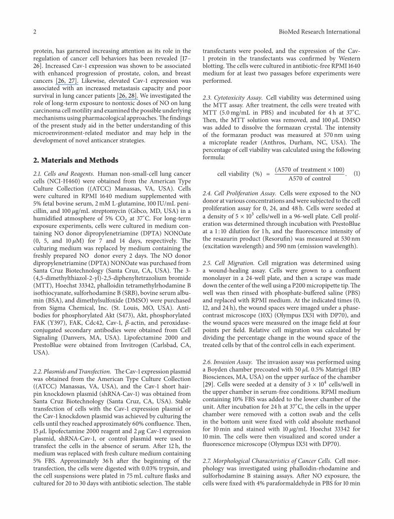
protein, has garnered increasing attention as its role in theregulation of cancer cell behaviors has been revealed [17–26]. Increased Cav-1 expression was shown to be associatedwith enhanced progression of prostate, colon, and breastcancers [26, 27]. Likewise, elevated Cav-1 expression wasassociated with an increased metastasis capacity and poorsurvival in lung cancer patients [26, 28]. We investigated therole of long-term exposure to nontoxic doses of NO on lungcarcinoma cellmotility and examined the possible underlyingmechanisms using pharmacological approaches.Thefindingsof the present study aid in the better understanding of thismicroenvironment-related mediator and may help in thedevelopment of novel anticancer strategies.
2. Materials and Methods
2.1. Cells and Reagents. Human non-small-cell lung cancercells (NCI-H460) were obtained from the American TypeCulture Collection ((ATCC) Manassas, VA, USA). Cellswere cultured in RPMI 1640 medium supplemented with5% fetal bovine serum, 2mM L-glutamine, 100 IU/mL peni-cillin, and 100 𝜇g/mL streptomycin (Gibco, MD, USA) in ahumidified atmosphere of 5% CO
2at 37∘C. For long-term
exposure experiments, cells were cultured in medium con-taining NO donor dipropylenetriamine (DPTA) NONOate(0, 5, and 10 𝜇M) for 7 and 14 days, respectively. Theculturing medium was replaced by medium containing thefreshly prepared NO donor every 2 days. The NO donordipropylenetriamine (DPTA) NONOate was purchased fromSanta Cruz Biotechnology (Santa Cruz, CA, USA). The 3-(4,5-dimethylthiazol-2-yl)-2,5-diphenyltetrazolium bromide(MTT), Hoechst 33342, phalloidin tetramethylrhodamine Bisothiocyanate, sulforhodamine B (SRB), bovine serum albu-min (BSA), and dimethylsulfoxide (DMSO) were purchasedfrom Sigma Chemical, Inc. (St. Louis, MO, USA). Anti-bodies for phosphorylated Akt (S473), Akt, phosphorylatedFAK (Y397), FAK, Cdc42, Cav-1, 𝛽-actin, and peroxidase-conjugated secondary antibodies were obtained from CellSignaling (Danvers, MA, USA). Lipofectamine 2000 andPrestoBlue were obtained from Invitrogen (Carlsbad, CA,USA).
2.2. Plasmids andTransfection. TheCav-1 expression plasmidwas obtained from the American Type Culture Collection((ATCC) Manassas, VA, USA), and the Cav-1 short hair-pin knockdown plasmid (shRNA-Cav-1) was obtained fromSanta Cruz Biotechnology (Santa Cruz, CA, USA). Stabletransfection of cells with the Cav-1 expression plasmid orthe Cav-1 knockdown plasmid was achieved by culturing thecells until they reached approximately 60% confluence.Then,15 𝜇L lipofectamine 2000 reagent and 2𝜇g Cav-1 expressionplasmid, shRNA-Cav-1, or control plasmid were used totransfect the cells in the absence of serum. After 12 h, themedium was replaced with fresh culture medium containing5% FBS. Approximately 36 h after the beginning of thetransfection, the cells were digested with 0.03% trypsin, andthe cell suspensions were plated in 75mL culture flasks andcultured for 20 to 30 days with antibiotic selection.The stable
transfectants were pooled, and the expression of the Cav-1 protein in the transfectants was confirmed by Westernblotting.The cells were cultured in antibiotic-free RPMI 1640medium for at least two passages before experiments wereperformed.
2.3. Cytotoxicity Assay. Cell viability was determined usingthe MTT assay. After treatment, the cells were treated withMTT (5.0mg/mL in PBS) and incubated for 4 h at 37∘C.Then, the MTT solution was removed, and 100 𝜇L DMSOwas added to dissolve the formazan crystal. The intensityof the formazan product was measured at 570 nm usinga microplate reader (Anthros, Durham, NC, USA). Thepercentage of cell viability was calculated using the followingformula:
cell viability (%) = (A570 of treatment × 100)A570 of control
. (1)
2.4. Cell Proliferation Assay. Cells were exposed to the NOdonor at various concentrations andwere subjected to the cellproliferation assay for 0, 24, and 48 h. Cells were seeded ata density of 5 × 10³ cells/well in a 96-well plate. Cell prolif-eration was determined through incubation with PrestoBlueat a 1 : 10 dilution for 1 h, and the fluorescence intensity ofthe resazurin product (Resorufin) was measured at 530 nm(excitation wavelength) and 590 nm (emission wavelength).
2.5. Cell Migration. Cell migration was determined usinga wound-healing assay. Cells were grown to a confluentmonolayer in a 24-well plate, and then a scrape was madedown the center of the well using a P200micropipette tip.Thewell was then rinsed with phosphate-buffered saline (PBS)and replaced with RPMI medium. At the indicated times (0,12, and 24 h), the wound spaces were imaged under a phase-contrast microscope (10X) (Olympus IX51 with DP70), andthe wound spaces were measured on the image field at fourpoints per field. Relative cell migration was calculated bydividing the percentage change in the wound space of thetreated cells by that of the control cells in each experiment.
2.6. Invasion Assay. The invasion assay was performed usinga Boyden chamber precoated with 50𝜇L 0.5% Matrigel (BDBiosciences, MA, USA) on the upper surface of the chamber[29]. Cells were seeded at a density of 3 × 10⁴ cells/well inthe upper chamber in serum-free conditions. RPMI mediumcontaining 10% FBS was added to the lower chamber of theunit. After incubation for 24 h at 37∘C, the cells in the upperchamber were removed with a cotton swab and the cellsin the bottom unit were fixed with cold absolute methanolfor 10min and stained with 10𝜇g/mL Hoechst 33342 for10min. The cells were then visualized and scored under afluorescence microscope (Olympus IX51 with DP70).
2.7. Morphological Characteristics of Cancer Cells. Cell mor-phology was investigated using phalloidin-rhodamine andsulforhodamine B staining assays. After NO exposure, thecells were fixed with 4% paraformaldehyde in PBS for 10min

BioMed Research International 3
at 37∘C, permeabilized with 0.1% Triton-X100 in PBS for4min, rinsed with PBS, and then blocked with 0.2% BSAfor 30min. The cells were then incubated with either a 1 : 100dilution of phalloidin-rhodamine in PBS or 0.4% sulforho-damine B in 1% acetic acid for 15min; the cells were thenrinsed 3 times with PBS and mounted with 50% glycerol. Thecellmorphologywas imaged using a fluorescencemicroscope(Olympus IX51 with DP70).
2.8. Western Blotting. After specific treatment, cells wereincubated with lysis buffer containing 20mM Tris⋅HCl (pH7.5), 1% Triton X-100, 150mM sodium chloride, 10% glyc-erol, 1mM sodium orthovanadate, 50mM sodium fluo-ride, 100mM phenylmethylsulfonyl fluoride, and proteaseinhibitor cocktail (RocheMolecular Biochemicals) for 30minon ice. The cell lysates were collected and determined forprotein content using the BCA protein assay kit (PierceBiotechnology, Rockford, IL, USA). Equal amounts of proteinfrom each sample (40 𝜇g) were denatured by heating at 95∘Cfor 5min with Laemmli loading buffer and loaded onto10% SDS-polyacrylamide gel electrophoresis. After separa-tion, proteins were transferred onto 0.45𝜇m nitrocellulosemembranes (Bio-Rad). The transferred membranes wereblocked in 5% nonfat dry milk in TBST (25mM Tris-HCl(pH 7.5), 125mMNaCl, and 0.05% Tween 20) for 30min andincubated with the appropriate primary antibodies overnightat 4∘C. Membranes were washed three times with TBST for10min and incubated with horseradish peroxidase- (HRP-) labeled secondary antibodies for 1 h at room temperature.The immune complexeswere detected by chemiluminescence(SupersignalWest Pico; Pierce, Rockford, IL, USA) and quan-tified using analyst/PC densitometry software (Bio-Rad).
2.9. Statistical Analysis. The mean data from independentexperiments were normalized to the results of the controlcells. The values are presented as the mean ± standarddeviation (SD) from three or more independent experimentsand were analyzed using one-way ANOVA with a post-hoctest (Tukey’s test) at a significance level of 𝑃 < 0.05 usingSPSS version 16.0.
3. Results
3.1. Effect of NO Donor on the Viability of the HumanLung Cancer H460 Cell Line. We first characterized theeffects of NO donor on the viability of the human lungcancer H460 cell line. The H460 cells were cultured inthe presence and absence of DPTA NONOate (1–20𝜇M),a slow-releasing NO donor compound, for 24 h, and cellviability was determined. Figure 1(a) shows that when cellswere treated with the NO donor, at concentrations ranging1–10𝜇M, neither cytotoxicity nor proliferative effects wereobserved in the cells. A significant decrease in viability wasfirst detected in cells treated with 20𝜇M DPTA NONOate;however, approximately 90% of the cells still remained viable.Accordingly, our results indicated that at the indicated doses,the NO donor did not cause a significant effect on cellviability up to 72 h of NO exposure (data not shown). To
0
20
40
60
80
100
120
Control 1 2 2.5 5 10 20
Cel
l via
bilit
y (%
) ∗
NO (𝜇M)
(a)
0
0.5
1
1.5
2
2.5
0 24 48
Rela
tive p
rolif
erat
ion
Time (h)
ControlNO 5𝜇MNO 10𝜇M
(b)
Figure 1: Effect of NO donor on cytotoxicity in lung carcinomaH460 cells. (a) Effect of DPTA NONOate on H460 cell viability.H460 cells were treated with various concentrations (0–20𝜇M) ofDPTA NONOate for 24 h. The cell viability was analyzed usingthe MTT assay. (b) Proliferative effect of DPTA NONOate onH460 cells. Cell proliferation for 24 and 48 h was determined usingPrestoBlue. The data are the mean ± SD (𝑛 = 3). ∗𝑃 < 0.05 versusthe nontreated control.
investigate the effect of long-term NO treatment on cellproliferation, H460 cells were cultured in their optimalconditions supplemented with 5 or 10 𝜇M NO donor, andtheir proliferative behavior was evaluated using PrestoBlue.As Figure 1(b) indicates, the NO-treated cells exhibited nosignificant changes in cell proliferation during the test period.
3.2. Long-Term NO Exposure Potentiates Migration and Inva-sion of H460 Cells. To investigate the effect of NO on cellmigration, we performed scratchwound-healing assays. Cellswere exposed to NO for 7 or 14 days and were subjectedto the migration assay for 12 and 24 h. Figures 2(a) and2(b) show that long-term treatment with the NO donorsignificantly enhanced the motility of the cells in dose- andtime-dependentmanners as comparedwith theH460 controlcells. Treatment with 10𝜇M DPTA NONOate for 14 days

4 BioMed Research International
Control
Day 7 Day 14
0h
12h
24h
NO 5𝜇M NO 10𝜇M Control NO 5𝜇M NO 10𝜇M
(a)
Control
0
1
2
3
Rela
tive m
igra
tion
#
#
#
#
Day 7 Day 14 Day 7 Day 14
12h 24h
∗ ∗∗
∗∗
∗
∗∗
NO 5𝜇MNO 10𝜇M
(b)
Figure 2: Effect of nitric oxide exposure on H460 cell migration. (a) Cells were exposed to NO donor at various concentrations for 7 or 14days and subjected to amigration assay. Phase-contrast images were captured at 0, 12, and 24 h. (b)The relative cell migration was determinedby comparing the relative change in wound space to the control cells. The data are the mean ± SD (𝑛 = 3). ∗𝑃 < 0.05 versus the control cells,#𝑃 < 0.05 versus NO-treated cells at 7 days.
potentiated the migration of the cells approximately 2.5-fold as compared with the nontreated cells, as shown inFigure 2(b).
In addition, we investigated the effect of NO onH460 cellinvasion using a precoated Matrigel Transwell unit, and wefound that treatment with the NO donor at various concen-trations (0, 5, and 10 𝜇M) for the indicated times significantlystimulatedH460 cell invasion through theMatrigel, as shownin Figures 3(a) and 3(b).
3.3. NO Enhances Filopodia Formation in Lung Cancer Cells.Filopodia are generated through actin polymerization andrearrangement of actin filaments, and the formation of filopo-dia has been linked to increased tumor cell migration. Toevaluate the effect of NO treatment on filopodia formation,
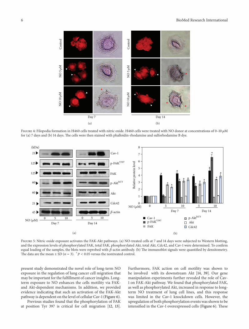
cells were exposed to NO as previously described, and thepresence of filopodia was determined using a phalloidin-rhodamine staining assay. In addition to this staining, thecytoskeletal actin was also stained with sulforhodamine Bdye. Figures 4(a) and 4(b) indicate that, whenH460 cells werecultured in the presence of the NO donor, the cells exhibitedan altered actin alignment and an increased number offilopodia.
3.4. The Long-Term NO Exposure Induces Cav-1-DependentFAK and Akt Activation. Having demonstrated the poten-tiating effect of NO exposure on lung cancer cell motility,we next examined the underlying mechanism, focusing onthe expression levels of the proteins known to play roles incell migration. Cancer cells were treated with NO donor at

BioMed Research International 5
ControlD
ay 1
4 D
ay 7
NO 5𝜇M NO 10𝜇M
(a)
0
100
200
300
400
500
Inva
ding
cells
(% o
f con
trol)
Control
Day 14 Day 7
∗
∗
∗
∗
NO 5𝜇MNO 10𝜇M
(b)
Figure 3: Effect of nitric oxide on H460 cell invasion. The invasionassay was performed using a Boyden chamber. (a) The cells thatinvaded the underside of the membrane were stained with 10𝜇g/mLHoechst 33342 for 10min and visualized using a fluorescencemicro-scope. (b) The relative cell invasion was determined as describedin Materials and Methods. The data are the mean ± SD (𝑛 = 3).∗
𝑃 < 0.05 versus the control cells.
different concentrations for 7 and 14 days and were analyzedby Western blotting. Expression levels of the migration-related proteins, namely, Cav-1, FAK, Akt, and Cdc42, wereevaluated. Figures 5(a) and 5(b) show that NO exposurefor 7 and 14 days significantly increased the levels of Cav-1, phosphorylated FAK (Tyr 397), phosphorylated Akt (Ser473), and Cdc42, whereas NO exposure had no significanteffect on the levels of total FAK and total Akt. Interestingly,the effects of NO on the mentioned proteins appeared tobe dose- and time-dependent; cells treated with 10𝜇M NOdonor for 14 days exhibited the most pronounced changesin protein levels as compared to cells treated with 5 𝜇M NOdonor or cells that were treated for a shorter period of time.
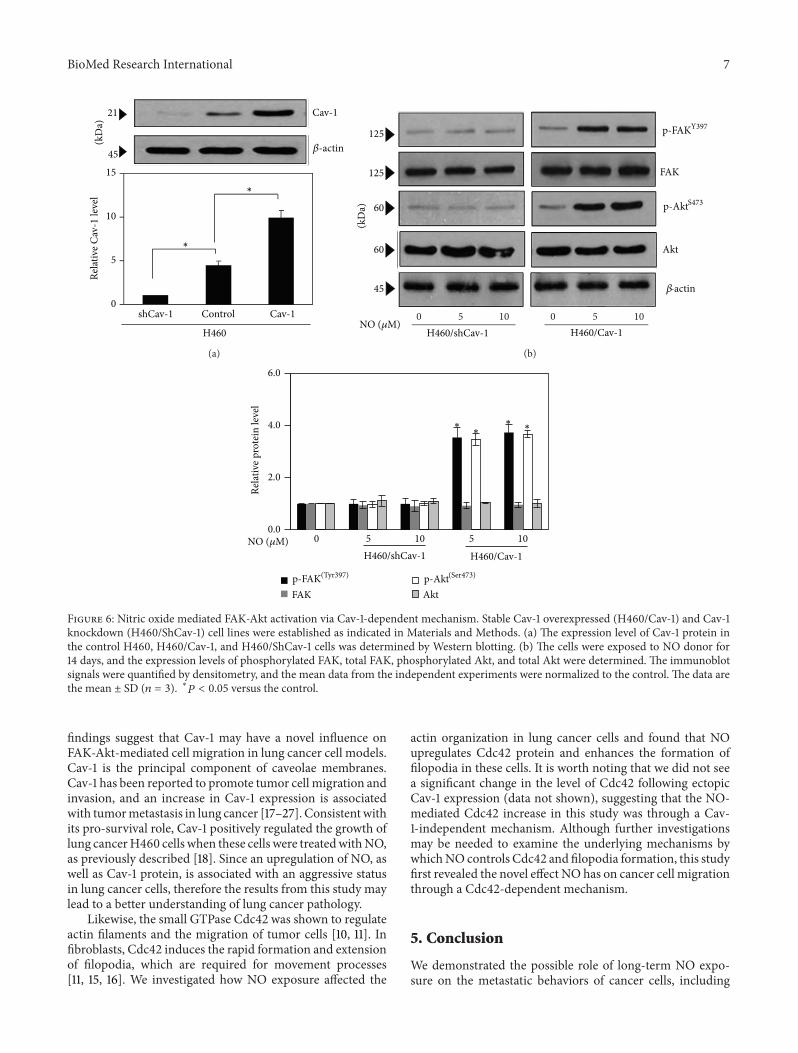
As Cav-1 has been shown to function as an adaptor pro-tein that regulates the activities of other proteins as previouslydescribed [21], we tested whether the upregulation of theproteinsmentioned previously was throughCav-1-dependentmechanism. Using gene manipulation approaches, Cav-1 overexpressed and knockdown cells were generated as
described in Materials and Methods. As expected, West-ern blot analysis of Cav-1 expression showed a substantialincrease in Cav-1 protein level in the Cav-1-transfected cells,whereas a significant decrease in Cav-1 level was observedin the shRNA-Cav-1-transfected cells as compared with thecontrol-transfected cells (Figure 6(a)). The Cav-1 overex-pressing cells (H460/Cav-1), the Cav-1 knockdown cells(H460/ShCav-1), and the control H460 cells were culturedin the presence or absence of NO (5–10𝜇M) for 14 days,and the levels of phosphorylated FAK (Tyr 397), phospho-rylated Akt (Ser 473), and their total protein levels weredetermined. Figure 6(b) shows that the Cav-1 overexpressedcells (H460/Cav-1) exhibited a significantly increased level ofphosphorylated FAK and phosphorylated Akt, whereas thetotal FAK and total Akt levels were not affected. In contrast,the NO-mediated FAK and Akt phosphorylation events weresuppressed in the cells in which Cav-1 was knocked down(H460/ShCav-1 cells). These results indicate that long-termNO exposure in H460 cells induces FAK and Akt activationin a Cav-1-dependent manner.
4. Discussion
Worldwide, lung cancer is a leading cause of cancer-relateddeath in both men and women [30], and approximately90% of non-small-cell lung cancer deaths are attributed tocancer metastasis [28, 31]. Among the multiple steps ofmetastasis, migration of the cancer cells has been recognizedas an important hallmark for the successful spread of cancerthroughout the body [8, 9]. However, information regardingthe key mediators that control the migratory activities ofthe cancer cells remains largely unknown. An increase inNO production has frequently been observed in the tissuesurrounding the tumor and may be critical for some cancercells behavior [3–7]. In addition, elevated NO production hasbeen observed in the lung tissue of lung cancer patients incomparisonwith that of normal subjects [6, 7].These findingshave strengthened the idea thatNOpresent in the lung cancerenvironment may affect the behavior of cancer cells.
NO is a gaseous molecule that is able to diffuse deeplyinto tissues; indeed, such a substance has been shown toregulate cell behaviors inmany ways, including the relaxationof vascular smooth muscle [3, 32]. Controversial roles of NOhave been reported for normal cell motility. NO was shownto inhibit vascular smooth muscle cell migration [32, 33];however, the opposite effect was observed in the microgliacell model [32, 34]. Accordingly, both the inhibitory effectand promoting effect of NO on cancer cells have beenreported [34, 35]. The variable effects of NO in tumors maydepend on the localization of NO synthase and its activity,the concentration and duration of NO exposure, and thecellular sensitivity to NO [3–5, 32, 34, 36]. While the long-term effects of NO on lung cancer cell migration are stillunknown, Hickok et al. showed that short-term treatmentwith an NO donor for 4, 6, and 24 h inhibited breast cancercell migration through N-Myc downstream-regulated gene-1 (NDRG1) expression [35, 37]. However, in prostate cancercells, NO was shown to potentiate cell motility [37, 38]. The
6 BioMed Research International
Con
trol
Day 7
NO5𝜇
MN
O10
𝜇M
(a)C
ontro
l
Day 14
NO5𝜇
MN
O10
𝜇M
(b)
Figure 4: Filopodia formation in H460 cells treated with nitric oxide. H460 cells were treated with NO donor at concentrations of 0–10 𝜇Mfor (a) 7 days and (b) 14 days. The cells were then stained with phalloidin-rhodamine and sulforhodamine B dye.
125
125
60
60
21
45
21
FAK
p-FAKY397
p-AktS473
Akt
Cdc42
𝛽-actin
Cav-1
0 5 10 0 5 10Day 7
(kDa)
Day 14 NO (𝜇M)
(a)
0 5 10 5 10Day 7 Day 14
Relat
ive p
rote
in le
vel
8
7
6
5
4
3
2
1
0
∗
∗∗
∗
∗ ∗ ∗∗
∗∗ ∗
∗
∗
∗
∗
FAKp-FAKY397
p-AktS473
AktCdc42
Cav-1
NO (𝜇M)
(b)
Figure 5: Nitric oxide exposure activates the FAK-Akt pathways. (a) NO-treated cells at 7 and 14 days were subjected to Western blotting,and the expression levels of phosphorylated FAK, total FAK, phosphorylated Akt, total Akt, Cdc42, and Cav-1 were determined. To confirmequal loading of the samples, the blots were reprobed with 𝛽-actin antibody. (b) The immunoblot signals were quantified by densitometry.The data are the mean ± SD (𝑛 = 3). ∗𝑃 < 0.05 versus the nontreated control.
present study demonstrated the novel role of long-term NOexposure in the regulation of lung cancer cell migration thatmay be important for the fulfillment of cancer insights. Long-term exposure to NO enhances the cells motility via FAK-and Akt-dependent mechanisms. In addition, we providedevidence indicating that such an activation of the FAK-Aktpathway is dependent on the level of cellular Cav-1 (Figure 6).
Previous studies found that the phosphorylation of FAKat position Tyr 397 is critical for cell migration [12, 13].
Furthermore, FAK action on cell motility was shown tobe involved with its downstream Akt [14, 39]. Our genemanipulation experiments further revealed the role of Cav-1 on FAK-Akt pathway. We found that phosphorylated FAK,as well as phosphorylated Akt, increased in response to long-term NO treatment of lung cell lines, and this responsewas limited in the Cav-1 knockdown cells. However, theupregulation of both phosphorylation events was shown to beintensified in the Cav-1 overexpressed cells (Figure 6). These
BioMed Research International 7
shCav-1 Control
(kD
a)21
45
Cav-1
H460
Cav-1
∗
∗
𝛽-actin
Relat
ive C
av-1
leve
l
15
10
5
0
(a)(k
Da)
125
125
60
60
45
FAK
p-FAKY397
p-AktS473
Akt
𝛽-actin
0 5 10 0 5 10H460/Cav-1H460/shCav-1
NO (𝜇M)
(b)
0.0
2.0
4.0
6.0
Relat
ive p
rote
in le
vel
FAK Akt
H460/Cav-1H460/shCav-1
p-FAK(Tyr397) p-Akt(Ser473)
∗∗∗
∗
105 1050NO (𝜇M)
Figure 6: Nitric oxide mediated FAK-Akt activation via Cav-1-dependent mechanism. Stable Cav-1 overexpressed (H460/Cav-1) and Cav-1knockdown (H460/ShCav-1) cell lines were established as indicated in Materials and Methods. (a) The expression level of Cav-1 protein inthe control H460, H460/Cav-1, and H460/ShCav-1 cells was determined by Western blotting. (b) The cells were exposed to NO donor for14 days, and the expression levels of phosphorylated FAK, total FAK, phosphorylated Akt, and total Akt were determined. The immunoblotsignals were quantified by densitometry, and the mean data from the independent experiments were normalized to the control. The data arethe mean ± SD (𝑛 = 3). ∗𝑃 < 0.05 versus the control.
findings suggest that Cav-1 may have a novel influence onFAK-Akt-mediated cell migration in lung cancer cell models.Cav-1 is the principal component of caveolae membranes.Cav-1 has been reported to promote tumor cell migration andinvasion, and an increase in Cav-1 expression is associatedwith tumormetastasis in lung cancer [17–27]. Consistentwithits pro-survival role, Cav-1 positively regulated the growth oflung cancerH460 cells when these cells were treatedwithNO,as previously described [18]. Since an upregulation of NO, aswell as Cav-1 protein, is associated with an aggressive statusin lung cancer cells, therefore the results from this study maylead to a better understanding of lung cancer pathology.
Likewise, the small GTPase Cdc42 was shown to regulateactin filaments and the migration of tumor cells [10, 11]. Infibroblasts, Cdc42 induces the rapid formation and extensionof filopodia, which are required for movement processes[11, 15, 16]. We investigated how NO exposure affected the
actin organization in lung cancer cells and found that NOupregulates Cdc42 protein and enhances the formation offilopodia in these cells. It is worth noting that we did not seea significant change in the level of Cdc42 following ectopicCav-1 expression (data not shown), suggesting that the NO-mediated Cdc42 increase in this study was through a Cav-1-independent mechanism. Although further investigationsmay be needed to examine the underlying mechanisms bywhichNO controls Cdc42 and filopodia formation, this studyfirst revealed the novel effect NO has on cancer cell migrationthrough a Cdc42-dependent mechanism.
5. Conclusion
We demonstrated the possible role of long-term NO expo-sure on the metastatic behaviors of cancer cells, including

8 BioMed Research International
migration and invasion. NO exposure activated the FAK-Aktsignaling pathway through a Cav-1-dependent mechanismand increased filopodia formation. Elevated NO levels havebeen observed in cancer environments; thus the knowledgegained from the present studymay benefit our understandingof cancer biology and may be useful in the development ofcancer therapies.
Abbreviations
Akt: ATP-dependent tyrosine kinaseCav-1: Caveolin-1Cdc42: Cell division cycle 42FAK: Focal-adhesion kinaseNO: Nitric oxideMTT: 3-(4,5-Dimethylthiazol-2-yl)-2,5-
diphenyltetrazoliumbromide
p-Akt: Phosphorylated AktPBS: Phosphate-buffered salinep-FAK: Phosphorylated-FAKTBST: Tris-buffered saline with 0.1% Tween.
Conflict of Interests
The authors declare that there is no conflict of interests in thisresearch.
Acknowledgments
This work was supported by grants from the ThailandResearch Fund (P. Chanvorachote) and the Ratchadaphisek-somphot Endowment Fund, Chulalongkorn University (RE-S560530132-HR). The authors would like to thank Mr. KrichRajprasit for his assistance in proof-reading.
References
[1] D.Hanahan andR.A.Weinberg, “Thehallmarks of cancer,”Cell,vol. 100, no. 1, pp. 57–70, 2000.
[2] G. Manda, M. T. Nechifor, and T.-M. Neagu, “Reactive oxygenspecies, cancer and anti-cancer therapies,” Current ChemicalBiology, vol. 3, no. 1, pp. 22–46, 2009.
[3] A.Keibel, V. Singh, andM.C. Sharma, “Inflammation,microen-vironment, and the immune system in cancer progression,”Current Pharmaceutical Design, vol. 15, no. 17, pp. 1949–1955,2009.
[4] P. K. Lala and C. Chakraborty, “Role of nitric oxide in carcino-genesis and tumour progression,” Lancet Oncology, vol. 2, no. 3,pp. 149–156, 2001.
[5] F. Masri, “Role of nitric oxide and its metabolites as potentialmarkers in lung cancer,” Annals of Thoracic Medicine, vol. 5, no.3, pp. 123–127, 2010.
[6] F. A. Masri, S. A. A. Comhair, T. Koeck et al., “Abnormalities innitric oxide and its derivatives in lung cancer,”American Journalof Respiratory andCritical CareMedicine, vol. 172, no. 5, pp. 597–605, 2005.
[7] H. Esme, M. Cemek, M. Sezer et al., “High levels of oxidativestress in patients with advanced lung cancer,” Respirology, vol.13, no. 1, pp. 112–116, 2008.
[8] D. H. Geho, R. W. Bandle, T. Clair, and L. A. Liotta, “Phys-iological mechanisms of tumor-cell invasion and migration,”Physiology, vol. 20, no. 3, pp. 194–200, 2005.
[9] L. A. Mina and G.W. Sledge Jr., “Rethinking the metastatic cas-cade as a therapeutic target,” Nature Reviews Clinical Oncology,vol. 8, no. 6, pp. 325–332, 2011.
[10] M. Parri and P. Chiarugi, “Rac and Rho GTPases in cancer cellmotility control,” Cell Communication and Signaling, vol. 8, no.23, pp. 1–14, 2010.
[11] C. D. Nobes and A. Hall, “Rho, Rac, and Cdc42 GTPases regu-late the assembly of multimolecular focal complexes associatedwith actin stress fibers, lamellipodia, and filopodia,”Cell, vol. 81,no. 1, pp. 53–62, 1995.
[12] B. Serrels, A. Serrels, V. G. Brunton et al., “Focal adhesion kinasecontrols actin assembly via a FERM-mediated interaction withthe Arp2/3 complex,”Nature Cell Biology, vol. 9, no. 9, pp. 1046–1056, 2007.
[13] L. A. Cary, J. F. Chang, and J.-L. Guan, “Stimulation of cellmigration by overexpression of focal adhesion kinase and itsassociation with Src and Fyn,” Journal of Cell Science, vol. 109,no. 7, pp. 1787–1794, 1996.
[14] S. K.Mitra, D. A. Hanson, andD.D. Schlaepfer, “Focal adhesionkinase: in command and control of cellmotility,”Nature ReviewsMolecular Cell Biology, vol. 6, no. 1, pp. 56–68, 2005.
[15] W. E. Allen, G. E. Jones, J. W. Pollard, and A. J. Ridley, “Rho,Rac and Cdc42 regulate actin organization and cell adhesion inmacrophages,” Journal of Cell Science, vol. 110, no. 6, pp. 707–720, 1997.
[16] K. Kaibuchi, S. Kuroda, and M. Amano, “Regulation of thecytoskeleton and cell adhesion by the Rho family GTPases inmammalian cells,” Annual Review of Biochemistry, vol. 68, pp.459–486, 1999.
[17] T. M. Williams and M. P. Lisanti, “Caveolin-1 in oncogenictransformation, cancer, and metastasis,” American Journal ofPhysiology—Cell Physiology, vol. 288, no. 3, pp. C494–C506,2005.
[18] P. Chanvorachote, U. Nimmannit, Y. Lu, S. Talbott, B.-H. Jiang,and Y. Rojanasakul, “Nitric oxide regulates lung carcinoma cellanoikis through inhibition of ubiquitin-proteasomal degrada-tion of caveolin-1,” Journal of Biological Chemistry, vol. 284, no.41, pp. 28476–28484, 2009.
[19] S. Luanpitpong, S. J. Talbott, Y. Rojanasakul et al., “Regulationof lung cancer cell migration and invasion by reactive oxygenspecies and caveolin-1,” Journal of Biological Chemistry, vol. 285,no. 50, pp. 38832–38840, 2010.
[20] P. Rungtabnapa, U. Nimmannit, H. Halim, Y. Rojanasakul, andP. Chanvorachote, “Hydrogen peroxide inhibits non-small celllung cancer cell anoikis through the inhibition of caveolin-1degradation,” American Journal of Physiology—Cell Physiology,vol. 300, no. 2, pp. C235–C245, 2011.
[21] K. Pongjit and P. Chanvorachote, “Caveolin-1 sensitizescisplatin-induced lung cancer cell apoptosis via superoxideanion-dependent mechanism,” Molecular and Cellular Bio-chemistry, vol. 358, no. 1-2, pp. 365–373, 2011.
[22] P. Chunhacha, V. Pongrakhananon, Y. Rojanasakul, andP. Chanvorachote, “Caveolin-1 regulates Mcl-1 stability andanoikis in lung carcinoma cells,” American Journal of Physiolo-gy—Cell Physiology, vol. 302, no. 9, pp. C1284–C1292, 2012.
[23] T. Songserm, V. Pongrakhananon, and P. Chanvorachote, “Sub-toxic cisplatin mediates anoikis resistance through hydrogenperoxide-induced caveolin-1 up-regulation in non-small cell

BioMed Research International 9
lung cancer cells,” Anticancer Research, vol. 32, no. 5, pp. 1659–1669, 2012.
[24] H. Halim, S. Luanpitpong, and P. Chanvorachote, “Acquisitionof anoikis resistance up-regulates caveolin-1 expression inhuman non-small cell lung cancer cells,” Anticancer Research,vol. 32, no. 5, pp. 1649–1658, 2012.
[25] W. Suchaoin and P. Chanvorachote, “Caveolin-1 attenuateshydrogen peroxide-induced oxidative damage to lung carci-noma cells,” Anticancer Research, vol. 32, no. 2, pp. 483–490,2012.
[26] P. Chunhacha and P. Chanvorachote, “Roles of caveolin-1 onanoikis resistance in non small cell lung cancer,” InternationalJournal of Physiology, Pathophysiology and Pharmacology, vol.4, no. 3, pp. 149–155, 2012.
[27] C.-C. Ho, P.-H. Huang, H.-Y. Huang, Y.-H. Chen, P.-C. Yang,and S.-M.Hsu, “Up-regulated caveolin-1 accentuates themetas-tasis capability of lung adenocarcinoma by inducing filopodiaformation,” American Journal of Pathology, vol. 161, no. 5, pp.1647–1656, 2002.
[28] F. Sotgia, U. E.Martinez-Outschoorn, A.Howell, R. G. Pestell, S.Pavlides, and M. P. Lisanti, “Caveolin-1 and cancer metabolismin the tumor microenvironment: markers, models, and mecha-nisms,”Annual Review of Pathology:Mechanisms of Disease, vol.7, pp. 423–467, 2012.
[29] V. P. Terranova, E. S. Hujanen, D. M. Loeb, G. R. Martin, L.Thornburg, and V. Glushko, “Use of a reconstituted basementmembrane to measure cell invasiveness and select for highlyinvasive tumor cells,” Proceedings of the National Academy ofSciences of the United States of America, vol. 83, no. 2, pp. 465–469, 1986.
[30] D. M. Parkin, F. Bray, J. Ferlay, and P. Pisani, “Global cancerstatistics, 2002,” CA: A Cancer Journal for Clinicians, vol. 55, no.2, pp. 74–108, 2005.
[31] M. Paesmans, J. P. Sculier, P. Libert et al., “Prognostic factorsfor survival in advanced non-small-cell lung cancer: univariateand multivariate analyses including recursive partitioning andamalgamation algorithms in 1,052 patients,” Journal of ClinicalOncology, vol. 13, no. 5, pp. 1221–1230, 1995.
[32] S. Moncada and A. Higgs, “The L-arginine-nitric oxide path-way,”The New England Journal of Medicine, vol. 329, no. 27, pp.2002–2012, 1993.
[33] R. Sarkar, E. G. Meinberg, J. C. Stanley, R. D. Gordon, and R. C.Webb, “Nitric oxide reversibly inhibits themigration of culturedvascular smooth muscle cells,” Circulation Research, vol. 78, no.2, pp. 225–230, 1996.
[34] A. Chen, S. M. Kumar, C. L. Sahley, and K. J. Muller, “Nitricoxide influences injury-inducedmicroglialmigration and accu-mulation in the leech CNS,” Journal of Neuroscience, vol. 20, no.3, pp. 1036–1043, 2000.
[35] A. Dhar, J. M. Brindley, C. Stark, M. L. Citro, L. K. Keefer,and N. H. Colburn, “Nitric oxide does not mediate but inhibitstransformation and tumor phenotype,”Molecular Cancer Ther-apeutics, vol. 2, no. 12, pp. 1285–1293, 2003.
[36] D. Fukumura, S. Kashiwagi, and R. K. Jain, “The role of nitricoxide in tumour progression,”Nature Reviews Cancer, vol. 6, no.7, pp. 521–534, 2006.
[37] J. R. Hickok, S. Sahni, Y. Mikhed, M. G. Bonini, and D. D.Thomas, “Nitric oxide suppresses tumor cell migration throughN-Mycdownstream-regulated gene-1 (NDRG1) expression: roleof chelatable iron,” Journal of Biological Chemistry, vol. 286, no.48, pp. 41413–41424, 2011.
[38] C. Polytarchou, M. Hatziapostolou, E. Poimenidi et al., “Nitricoxide stimulates migration of human endothelial and prostatecancer cells through up-regulation of pleiotrophin expressionand its receptor protein tyrosine phosphatase𝛽/𝜁,” InternationalJournal of Cancer, vol. 124, no. 8, pp. 1785–1793, 2009.
[39] J. Tureckova, M. Vojtechova, M. Krausova, E. Sloncova, andV. Korınek, “Focal adhesion kinase functions as an akt down-stream target in migration of colorectal cancer cells,” Transla-tional Oncology, vol. 2, no. 4, pp. 281–290, 2009.