Individual notions of distributive justice and relative economic status
Chapter 8Long-Term Evolution of Histone Families:Old Notions and New Insights into TheirMechanisms of Diversification AcrossEukaryotes
Jose M. Eirın-Lopez, Rodrigo Gonzalez-Romero, Deanna Dryhurst,Josefina Mendez, and Juan Ausio
Abstract In eukaryotes and some archaebacteria, DNA is found associated withhistones in a nucleoprotein complex called chromatin, which allows for a highextent of compaction of genomic DNA within the limited space of the nucleus.Early studies led to the notion that histones exhibit a conserved structural geneorganization and limited protein diversity. However, research has been progres-sively accumulating to demonstrate that the structure, configuration and copynumber of histone genes varies widely across organisms as a result of a long-termevolutionary process that promotes genetic variation. This genetic diversity ismirrored by the structural and functional diversity exhibited by the protein mem-bers of the different histone families that is, in most instances, concomitant with thecomplexity of the organism. The present chapter is aimed at providing a compre-hensive review of the most recent information on the origin of eukaryotic histonemultigene families. Particular attention is paid to the structural and functionalconstraints acting on histones and their relevance for the progressive diversificationof histone variants during evolution, especially as it pertains to histone geneorganization and expression.
R. Gonzalez-Romero and J. MendezDepartamento de Biologıa Celular y Molecular, Universidade da Coruna, E15071, A Coruna,SpainD. Dryhurst and J.AusioDepartment of Biochemistry and Microbiology, University of Victoria, V8W 3P6 Victoria,CanadaJ.M. Eirın-LopezDepartamento de Biologıa Celular y Molecular, Facultade de ciencias, Universidade da Coruna,Campus de A Zapateria s/n, E15071 A Coruna, Spaine-mail: [email protected]
P. Pontarotti (ed.), Evolutionary Biology: Concept, Modeling, and Application,DOI: 10.1007/978-3-642-00952-5_8, # Springer!Verlag Berlin Heidelberg 2009
139

8.1 Introduction
In eukaryotes and some archaebacteria, DNA is found associated with histonesin a nucleoprotein complex called chromatin. Chromatin allows for a high extentof compaction of genomic DNA within the limited space of the nucleus andalso provides the support on which most DNA metabolic functions (i.e., replica-tion, transcription and repair) take place. The repetitive subunit of chromatin,the nucleosome, consists of an octamer of core histones (two of each H2A, H2B,H3 and H4) around which two left handed superhelical turns of DNA arewrapped (van Holde 1988). The nucleosome core particles (NCPs) are joinedtogether in the chromatin fiber by short stretches of linker DNA that, interactwith linker H1 histones, resulting in an additional folding of the chromatin fiber.Although all the domains in eukaryotic chromatin share a common nucleosomalstructure, the dynamic processes responsible for the local heterogeneityobserved across the genome are regulated by three principal mechanisms: thereplacement of canonical histones with specialized variants that have dedicatedfunctions (Malik and Henikoff 2003), the occurrence of histone posttranslationalmodifications (Jenuwein and Allis 2001) and the association with remodelingcomplexes responsible for nucleosome mobilization (Owen-Hughes 2003). Thewide range of possible configurations that facilitate different chromatin meta-bolic processes are the result of the synergistic action of the aforementionedmechanisms.
Early studies on histone genes led to the formulation of several general dogmasabout their structure: (a) histone genes were considered to be intronless, encodinghighly conserved proteins; (b) they existed in multiple copies closely clustered inthe genome and organized into tandemly repeated blocks; (c) except for the case ofrelatively few replacement histone variants, the mRNAs transcribed from the genesof the canonical histones were not polyadenylated and their expression was largelycoupled to S phase of the cell cycle; and (d) the evolution of the histone genefamilies was thought to be subject to a process of concerted evolution through rapidinterlocus recombination or gene conversion (Hentschel and Birnstiel 1981; Kedes1979; Maxson et al. 1983a; Ohta 1983). However, research has been progressivelyaccumulating to demonstrate that the organization and copy number of eukaryotichistone genes varies widely across organisms. Additional evidence that arguesagainst the assumptions made by these dogmas was found in the case of specializedhistone variants. These proteins are encoded by intron-containing genes and areexpressed constantly at basal levels throughout the cell cycle (Ausio 2006). Thesediverse patterns of organization and expression differ even among closely relatedorganisms and most likely reflect the presence of different regulatory mechanismsand a complex mode of gene family evolution (Doenecke et al. 1997; Khochbin2001; Wells and Kedes 1985). The high degree of structural and functional diversi-fication observed across different histone types provides a compelling argumentagainst the notion of concerted evolution (homogenization) as the major forceguiding long-term histone evolution.
140 J.M. Eirın-Lopez et al.
The present chapter is aimed at providing a comprehensive review of the mostrecent information on the origin of eukaryotic histone multigene families and thesubsequent mechanisms guiding their long-term evolution. Special attention is paid tothe structural and functional constraints acting on histone proteins and their relevanceto the progressive diversification of histone variants during evolution, especially asthey pertain to histone gene organization and expression. Finally, a model summariz-ing the process of histone diversification and differentiation from their archael origin tothe wide diversity of specialized variants in eukaryotes is presented.
8.2 Histone Genes Display Highly Heterogeneous OrganizationPatterns Across Eukaryotic Genomes
Histone gene organization has been studied for more than 30 years (Hentschel andBirnstiel 1981; Isenberg 1979; Kedes 1979; Maxson et al. 1983a) and during mostof this early period, the concept of a homogeneous gene organization was believedto be the normal arrangement in the genomes of most model organisms studied atthe time, especially Drosophila and sea urchin (Lifton et al. 1977; Maxson et al.1983b). However, with the progressive characterization of histone genes in abroader range of organisms, a more complex picture started to emerge showing adispersal and diversification of the genes that was apparently concomitant with theposition of the different organisms on the phylogenetic tree (Doenecke et al. 1997).A great diversity of histone gene organization patterns was observed, however, itwas not until histone variants were first identified and their specific functionsprogressively deciphered that such an extensive heterogeneity was definitivelydetermined to be an intrinsic feature of the different histone families. In sharpcontrast to canonical histones, the histone variants have a unique genomic organi-zation (solitary genes), copy number (single-copy), gene structure (presence ofintrons) and regulation (basal continuous expression throughout the cell cycle)(Ausio 2006). Clearly there is no doubt that the diversification process experiencedby histone genes has allowed for the progressive differentiation of histone variantswith dedicated functions and this has shaped the complex, efficient and tightlycontrolled mechanisms of DNA packaging and regulation of chromatin dynamics inthe cell nucleus. In order to illustrate the relevance of such diversity, we will nextsummarize the major modes of histone gene organization across representativegroups of eukaryotes.
8.2.1 Prokaryotic Chromatin and the Origin of Histones
Most studies on prokaryotic chromatin have initially focused on the eubacteriaEscherichia coli, which has a 4.6 Mb circular chromosome organized into
8 Long-Term Evolution of Histone Families 141
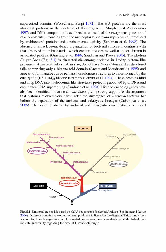
supercoiled domains (Worcel and Burgi 1972). The HU proteins are the mostabundant proteins in the nucleoid of this organism (Murphy and Zimmerman1997) and DNA compaction is achieved as a result of the exogenous pressure ofmacromolecular crowding from the nucleoplasm and from supercoiling introducedby architectural proteins and topoisomerase activity (Sandman et al. 1998). Theabsence of a nucleosome-based organization of bacterial chromatin contrasts withthat observed in archaebateria, which contain histones as well as other chromatinassociated proteins (Grayling et al. 1996; Sandman and Reeve 2005). The phylumEuryarchaea (Fig. 8.1) is characteristic among Archaea in having histone-likeproteins that are relatively small in size, do not have N- or C-terminal unstructuredtails comprising only a histone-fold domain (Arents and Moudrianakis 1995) andappear to form analogous or perhaps homologous structures to those formed by theeukaryotic (H3 + H4)2 histone tetramers (Pereira et al. 1997). These proteins bindand wrap DNA into nucleosomal-like structures protecting about 60 bp of DNA andcan induce DNA supercoiling (Sandman et al. 1998). Histone-encoding genes havealso been identified in marine Crenarchaea, giving strong support for the argumentthat histones evolved very early, after the divergence of Bacteria-Archaea butbefore the separation of the archaeal and eukaryotic lineages (Cubonova et al.2005). The ancestry shared by archaeal and eukaryotic core histones is indeed
Fig. 8.1 Universal tree of life based on rRNA sequences of selected Archaea (Sandman and Reeve2006). Different domains as well as archaeal phyla are indicated in the diagram. Thick fancy linesaccount for those lineages in which histone-fold sequences have been identified while dashed linesindicate uncertainty regarding the time of histone-fold origin
142 J.M. Eirın-Lopez et al.
manifested in their conserved histone-fold region as well as in the amino acidresidues that are important for histone–DNA and histone dimer–dimer interactions.Furthermore, archaeal histone sequences exhibit a degree of variation that providesclues to the molecular requirements of the evolutionary transition from prokaryoticto eukaryotic chromatin (Sandman and Reeve 2006; Zlatanova 1997).
8.2.2 The Transition Toward the Eukaryotic Cell and theAppearance of Pluricellularity in Light of HistoneDiversification
Histones have been extensively characterized in eukaryotic genomes where, withthe only exception of some protozoa such as dinoflagellates (Herzog and Soyer1981), they organize chromatin into a repetitive nucleosome structure. Amonglower eukaryotes, yeast is unique in its small number of histone genes, havingonly two gene copies of each of the four core histones. The H2A and H2B genes areadjacent to one another, they are divergently transcribed and they exist as twogenetically unlinked copies encoding two structural variants. This is in contrast tothe H3 and H4 genes that encode identical products (van Holde 1988). There is alsoa candidate yeast linker histone (Hho1p) encoded by a gene that is co-expressedwith the core histone genes in S phase (Landsman 1996). This protein has anunusual tertiary structure with two regions (GI and GII) homologous to the singleglobular domain of the linker histone of higher eukaryotes (Ali and Thomas 2004).The organization of histone genes is also known in other lower eukaryotes such asthe ciliates Stylonychia lemnae, Tetrahymena thermophila and closely relatedspecies, where the histone genes are unclustered in the genome and the H1 genesare apparently absent in the micronucleus (Allis et al. 1979; Prescott 1994). Also,single-copy H1 genes independently organized from core histone genes weredescribed in Volvox (Lindauer et al. 1993).
Despite the classical notion of histone genes being clustered and tandemly repeated,many arrangements are observed in metazoan genomes as indicated in Fig. 8.2. Forinstance, in the cnidarian Acropora formosa (coral) histone genes consist in clusters ofthe four core histones (quartets) that are reiterated about 150 times (Miller et al. 1993).A similar organization has also been described in the nematode Caenorhabditiselegans that has independently organized single-copy H1 genes (Sanicola et al.1990). Similarly, the histone genes of annelids are organized into clusters containingH1 and the four core histone genes (quintets) but quartets of core histones that arereiterated about 600–650 times in the genome are also present (del Gaudio et al. 1998;Sellos et al. 1990). However, molluscs represent the paradigm of diversity amongmetazoans showing up to three different arrangements of histone genes even within asingle species. This is the case with the musselMytilus galloprovincialis (Eirın-Lopezet al. 2004b), which displays clustered quintets linked to 5S rRNA genes, quartets ofcore histones and independent clusters containing replication independent H1 genes
8 Long-Term Evolution of Histone Families 143
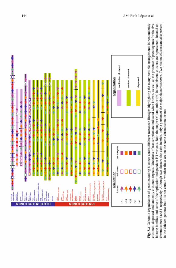
Fig.8
.2Genomicorganizationofgenes
encodinghistones
across
differentmetazoan
lineages
highlightingthemanypossiblearrangem
entsin
nontandem
lyclustered,dispersedandtandem
lyclustered
repeats.D
irectionsoftranscriptionareindicated
when
known,aswellas
thepresence
ofpseudogenes
forthefive
histonefamiliesandsomeofthereplication-independentH1variants.Both
themajor(M
)andminor(m
)human
histoneclustersarerepresented,locatedon
chromosomes
6and1,respectively.A
lthoughtwoclustersalso
existinmice,onlyaportionofthemajorclusterisshown.T
wohistoneclustersarealso
present
inthechicken
genomebutitisnotcertainwhether
theseareonthesamechromosomeornot
144 J.M. Eirın-Lopez et al.

(Drabent et al. 1999; Eirın-Lopez et al. 2002). In insects, the clustered quintetscontaining H1 genes are present in Drosophila (Kremer and Henning 1990; Liftonet al. 1977; Tsunemoto and Matsuo 2001) and in the midge Chironomus (Hankeln andSchmidt 1991). However, clustered quartets of core histones and independent H1histone genes are present in Drosophila virilis (Domier et al. 1986; Schienman et al.1998) and Chironomus thummi (Hankeln and Schmidt 1993).
Echinoderms are probably one of the groups that have been more extensivelyanalyzed among deuterostomes. The sea urchins Strongylocentrotus purpuratusand Psammechinus miliaris display around 700 repetitions of quintets tandemlyarranged and expressed at early embryonic stages (Sures et al. 1978), which coexistin the genome with additional clusters without any regular organization that areexpressed later on during development (Maxson et al. 1983b). However, onlyclustered quartets with variable arrangements have been reported in the starfishspecies Pisaster ochraceus, P. brevispinus and Dermasterias imbricata (Cool et al.1988). Although histone genes still remain organized in clusters in many vertebrategenomes, such regular organization appears to be gradually lost during the courseof evolution. For instance, amphibians display histone genes organized either inclustered quartets (newts) or quintets (Xenopus) with variable copy number(Ruberti et al. 1982; Stephenson et al. 1981; Turner and Woodland 1983; Turneret al. 1983; van Dongen et al. 1981), as do fishes such as Salmo gairdnerii, whichhave quintets repeated about 150 times in their genome (Connor et al. 1984).Although chickens also contain clustered histone quintets repeated about 10times, they also have solitary and replication independent H1-type genes encodingthe specialized H5 histone typical of nucleated erythrocytes (D’Andrea et al. 1985;Scott and Wells 1976). The tandem arrangement of histone genes is definitively lostin mammals. Histones are organized in either a single cluster (located on chromo-some 17 in the rat [Walter et al. 1996]) or in two physically independent major andminor clusters on chromosomes 6 and 3 and chromosomes 13 and 3 in humans andmice, respectively (Albig et al. 1997a, b; Wang et al. 1997). In addition, solitarysingle-copy replication independent H1-type genes are also present in both humanand murine genomes, encoding histone H10 that has dedicated functions in termi-nally differentiated cellular systems (Doenecke and Alonso 1996).
8.3 Histone Variants Impart Specific Functions to Nucleosomes
8.3.1 Linker Histones
The histone H1 family encompasses one of the largest numbers of isoforms amonghistones. This diversity is especially evident in mammals where 11 different linkerhistones have been identified: 7 somatic (H1.1–H1.5, H10 [Ausio and Abbott 2004;Parseghian and Hamkalo 2001], and H1x [Happel et al. 2005]), 3 sperm-specific(H1t [Seyedin andKistler 1979], H1t2 [Martianov et al. 2005] and HILS1 [Iguchi et al.2003; Yan et al. 2003]); and the oocyte-specific variant H1oo (Tanaka et al. 2001).
8 Long-Term Evolution of Histone Families 145
Other cleavage-specific H1 variants homologous to H1oo have also been described inamphibians (embryonic linker histone H1M or B4 from Xenopus [Cho and Wolffe1994]) and echinoderms (histone CS from sea urchin [Mandl et al. 1997]).
The evolution of H1 has favored the differentiation of other highly specializedisoforms such as histone H5 (Ruiz-Carrillo et al. 1983), a replication-independentH1 variant restricted to the nucleated erythrocytes of birds, which also appears to bepresent in amphibians (Khochbin 2001) and reptiles. Therefore, H5 is structurallyrelated to H10, a histone that replaces somatic H1 isoforms in terminally differ-entiated cells of many vertebrates (Panyim and Chalkey 1969). These variants havebeen extensively studied and they share characteristic conserved elements in theirpromoter regions, which are involved in their replication-independent pattern ofexpression. These include a UCE element (Upstream Conserved Element), an H1box followed by a G/C rich segment and an H4 box (Eirın-Lopez et al. 2005;Schulze and Schulze 1995). Although both the H10 and H5 genes encode poly-adenylated mRNAs, the H5 transcript contains two additional stem-loop signals inthe 30 UTR region (Doenecke and Alonso 1996). Until recently, the occurrence ofreplication independent H1 variants had been restricted exclusively to deuteros-tomes. Although different studies postulated the existence of H1 histone variantsin several protostomes (Ausio 1999; Barzotti et al. 2000; del Gaudio et al. 1998;Eirın-Lopez et al. 2002, 2004b; Hankeln and Schmidt 1993) the presence ofreplication-independent H1 forms has only been studied in detail in molluscs(Eirın-Lopez et al. 2005).
8.3.2 Core Histones
Since histones must be synthesized in stoichiometric amounts for the assembly ofchromatin onto newly replicated DNA, transcription of histone genes is tightlyregulated during the cell cycle and the bulk of their translation is coordinated withDNA replication during S phase (Marzluff 1992). A unique feature of thesereplication-dependent histone mRNAs is their lack of polyadenylated tails(replaced by a stem-loop signal) and the regulation of their expression at threedifferent levels including transcriptional, mRNA processing and mRNA stability(Doenecke et al. 1997). Although these regulatory mechanisms are characteristic ofmost histone genes, there is a small fraction of histones encoded by solitary single-copy genes whose expression prevails in nonproliferating cells, the so-calledreplacement histones or histone variants (Henikoff and Ahmad 2005; Smith et al.1984). Histone variant genes exhibit constant basal replication-independent expres-sion throughout the cell cycle, their mRNAs contain long 30 UTR regions as well aspolyadenylated tails that bind the poly(A) binding protein that increases theirstability (Marzluff 1992).
Although core histones are far more conserved than H1 histones (Isenberg1979), this does not preclude the existence of a marked functional differentiationamong their variants. This is particularly noticeable in the case of the H2A family
146 J.M. Eirın-Lopez et al.
(Ausio and Abbott 2002), which includes the heavily studied variants H2A.X andH2A.Z. These two variants are involved in the maintenance of genome integrity andin the regulation of chromatin dynamics. Histone H2A.X is present in almost alleukaryotes and is particularly expressed in germinal cells where it has functionsrelated to DNA repair and chromosome condensation (Li et al. 2005). This variantis encoded by intron-less genes that are expressed as two different types oftranscripts: a short replication-dependent type with a stem-loop signal and a longerreplication-independent type that is polyadenylated (Alvelo-Ceron et al. 2000).Histone H2A.Z has been ascribed multiple functions that may differ among species.A growing body of evidence suggests that it participates in regulation of geneexpression (Barski et al. 2007; Bruce et al. 2005) but it also plays an importantrole in the heterochromatin structure of the centromere (Greaves et al. 2007).In vertebrates, H2A.Z exists as a mixture of two protein forms H2A.Z-1(previously H2A.Z) and H2A.Z-2 (previously H2A.F/Z or H2A.V) that differ bythree amino acids (Coon et al. 2005). These two proteins are encoded by separategenes that contain four introns in humans and are expressed through polyadenylatedmRNAs (Hatch and Bonner 1988, 1990). The H2A family also includes anotherhighly specialized variant, macroH2A. This variant is characterized by a long non-histone C-terminal tail and has been shown to be involved in female X chromosomeinactivation in mammals and birds (Costanzi and Pehrson 1998; Ellegren 2002).In contrast, histone H2A.Bbd is a highly variable quickly evolving mammalianH2A variant (Eirın-Lopez et al. 2008), which is markedly deficient in the inactiveX-chromosome and participates in the destabilization of nucleosomes and in theunfolding of the chromatin fiber (Gonzalez-Romero et al. 2008b).
Other core histone variants include histone H3.3, CENPA and H3.t of the H3family. In mammals, H3.3 differs from canonical replication-dependent H3.1 byonly four amino acids and is enriched in actively transcribed regions of the genomein somatic cells (Mito et al. 2005). Two identical proteins, H3.3A and H3.3B, whichare encoded by two different solitary genes, participate in the transition fromhistones to protamines during spermiogenesis in mammals (Henning 2003; Doe-necke et al. 1997; Wells and Kedes 1985). Centromeric protein A (CENPA) isinvolved in the packaging of chromatin at eukaryotic centromeres (Govin et al.2005; Palmer et al. 1987) and a testis-specific H3 histone (H3t) has been describedin humans (Witt et al. 1996). With regard to the other histone families, the humanH2B.1 gene represents the only replication-independent H2B isoform known andencodes transcripts alternatively processed at 30 UTR regions, yielding replication-dependent and replication-independent mRNAs (Collart et al. 1992). Although noreplacement subtypes have been described for the most highly conserved family ofhistones, the H4 family, an H4 protein with replication-independent properties hasbeen described in Drosophila (Akhmanova et al. 1996).
In striking contrast to the animal kingdom, plant histones provide a verydifferent example of histone gene structure and regulation (Kanazin et al. 1996).This is demonstrated by the existence of an intron containing H3 genes in soybean,barley and wheat, which were found to be expressed in different plant organs in arelatively replication-independent fashion. Plant histones do not have stem-loop
8 Long-Term Evolution of Histone Families 147
signals and are transcribed into polyadenylated mRNAs (reviewed by Chabouteet al. [1993]) suggesting that the mechanisms regulating histone expression are verydifferent between the plant and animal kingdoms. The regulation of histone pro-duction in plants would essentially occur at the transcriptional level.
8.4 Eukaryotic Histones Arose from Archaeal HistonesFollowing a Recurrent Gene Duplication Process
The evolutionary origin of eukaryotic histones can be traced back to prokaryotes.Histones resembling H2A and H4, as well as the DNA topoisomerase V(a prokaryotic counterpart of eukaryotic topoisomerase I) were found in thehyperthermophilic archaebacteria Methanopyrus kandleri (Slerasev et al. 1984)and also archaeal RNA polymerases show common features with eukaryoticRNA polymerases (Reeve et al. 1997). In addition, archaeal histones exhibitsome similarities with the central domain of the CBF-A eukaryotic transcriptionfactor subunit (CCAAT-binding factor subunit A), suggesting that the eukaryoticmodes of transcription and DNA packaging may have originated before the eukar-yotes themselves (Ouzounis and Kyrpides 1996). Consistent with this observation,phylogenetic trees reconstructed from rRNA data place Archaea and Eukarya onthe same branch, indicating the existence of a common ancestor shared by bothgroups after the divergence of Archaea and Eubacteria (Sandman and Reeve 1998,2006).
The presence of histone-like genes in Euryarchaeotawas explained by Sandmanand Reeve (1998) in terms of the ‘hydrogen hypothesis for the first eukaryote’(Martin and Muller 1998). Accordingly, the origin of the eukaryotic cell wasproposed to have been derived from a symbiotic association between a methanogenarchaebacterium and a proteobacterium. Thus, while the eukaryotic nucleus wouldhave been derived from the archaebacterium (including the proteins involved inDNA packaging), most of the cellular metabolism would have been contributed bythe proteobacterium. Consequently, the evolution of histones and DNA condensa-tion into nucleosomes apparently occurred in the euryarchaeotal lineage, before thearchaeal-eukaryal divergence, facilitating the genome expansion and the develop-ment of Eukarya (Sandman and Reeve 2005, 2006).
It appears that DNA duplication has been the basis underlying the evolutionarymechanisms driving histone evolution from the early appearance of these proteinsin Archaea and the evolutionary process shaping this variability can be summarizedin three major stages as indicated in Fig. 8.3. Firstly, the evolution of histones inArchaea would have been based on their generic role in packaging the genomicDNA. In this context, the first important event of gene duplication would haveresulted in at least two histone-like genes in Archaea (such as HMfA and HMfBgenes identified in Methanothermus fervidus, Fig. 8.3a) capable of forming tetra-meric complexes to efficiently compact DNA (Sandman et al. 1990; Starich et al.1996). The next critical duplication event would be represented by the intragenic
148 J.M. Eirın-Lopez et al.
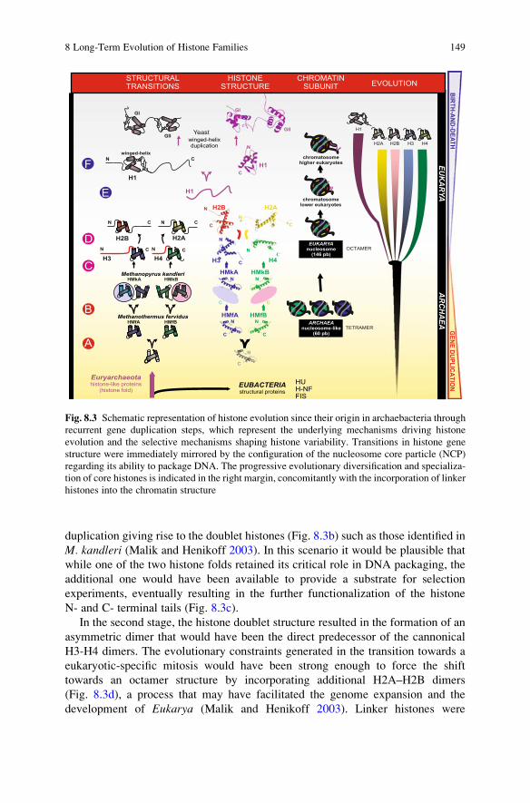
duplication giving rise to the doublet histones (Fig. 8.3b) such as those identified inM. kandleri (Malik and Henikoff 2003). In this scenario it would be plausible thatwhile one of the two histone folds retained its critical role in DNA packaging, theadditional one would have been available to provide a substrate for selectionexperiments, eventually resulting in the further functionalization of the histoneN- and C- terminal tails (Fig. 8.3c).
In the second stage, the histone doublet structure resulted in the formation of anasymmetric dimer that would have been the direct predecessor of the cannonicalH3-H4 dimers. The evolutionary constraints generated in the transition towards aeukaryotic-specific mitosis would have been strong enough to force the shifttowards an octamer structure by incorporating additional H2A–H2B dimers(Fig. 8.3d), a process that may have facilitated the genome expansion and thedevelopment of Eukarya (Malik and Henikoff 2003). Linker histones were
Fig. 8.3 Schematic representation of histone evolution since their origin in archaebacteria throughrecurrent gene duplication steps, which represent the underlying mechanisms driving histoneevolution and the selective mechanisms shaping histone variability. Transitions in histone genestructure were immediately mirrored by the configuration of the nucleosome core particle (NCP)regarding its ability to package DNA. The progressive evolutionary diversification and specializa-tion of core histones is indicated in the right margin, concomitantly with the incorporation of linkerhistones into the chromatin structure
8 Long-Term Evolution of Histone Families 149
likely the last component to join this structure, providing the maximum level ofcompaction of DNA. However, it is still not clear how this process took place. TheC-terminal tail of eukaryotic H1 histones is able to compact chromatin by itself andthis domain constitutes the whole protein in the case of early ancestral eukaryotessuch as trypanosomes (Gruter and Betschart 2001). This supports the hypothesisthat, in contrast to core histones, ancestral H1 histones were composed of only aC-terminal region (Fig. 8.3e) and the core domain containing the winged-helixmotif would have been acquired later in evolution as indicated in Fig. 8.3f(Kasinsky et al. 2001).
In the third stage, the differentiation of the five metazoan histones (H1, H2A,H2B, H3 and H4) would have marked the beginning of the final stage in histoneevolution. Although gene duplication has prevailed as the major mechanism inproviding the eukaryotic cell with the required amounts and diversity of histones(Malik and Henikoff 2003), it has been shown that concerted evolution does notplay a major role in their evolution (Eirın-Lopez et al. 2004a; Eirın-Lopez et al.2005; Piontkivska et al. 2002; Rooney et al. 2002). Crucial to this stage is thediversification as a result of recurrent gene duplications followed by a strongpurifying selection process acting at the protein level. This process is known asbirth-and-death evolution (Nei and Hughes 1992; Nei et al. 1997) and represents themajor mode of long-term evolution in eukaryote histone families as well as in manyother gene families (Nei and Rooney 2006), as discussed in detail in the followingsection. The evolutionary refinement resulting from such diversification wouldhave been determined by the cellular specialization associated with the appearanceof multicellular organisms. Histone variability in these organisms is required inorder to accommodate the different packing needs and regulation of gene expres-sion in different cell types and developmental stages.
8.5 The Long-Term Evolution of Histone Genes Is Guidedby a Birth-and-Death Process That PromotesGenetic Diversity
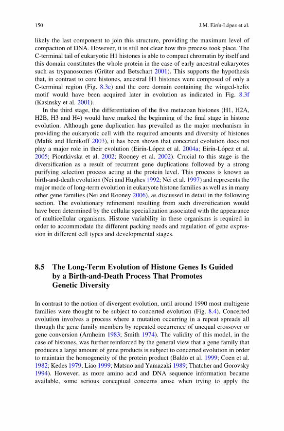
In contrast to the notion of divergent evolution, until around 1990 most multigenefamilies were thought to be subject to concerted evolution (Fig. 8.4). Concertedevolution involves a process where a mutation occurring in a repeat spreads allthrough the gene family members by repeated occurrence of unequal crossover orgene conversion (Arnheim 1983; Smith 1974). The validity of this model, in thecase of histones, was further reinforced by the general view that a gene family thatproduces a large amount of gene products is subject to concerted evolution in orderto maintain the homogeneity of the protein product (Baldo et al. 1999; Coen et al.1982; Kedes 1979; Liao 1999; Matsuo and Yamazaki 1989; Thatcher and Gorovsky1994). However, as more amino acid and DNA sequence information becameavailable, some serious conceptual concerns arose when trying to apply the
150 J.M. Eirın-Lopez et al.
concepts of concerted evolution to some gene families. These include genes involvedin immune systems and disease-resistance (Hughes and Nei 1990; Ota and Nei 1994;Zhang et al. 2000), as well as highly conserved gene families such as ubiquitins andhistones (Eirın-Lopez et al. 2004a, 2005; Gonzalez-Romero et al. 2008a; Nei et al.2000; Piontkivska et al. 2002; Rooney et al. 2002).
As mentioned in the previous section, the differentiation of the five eukaryotichistone families, together with the subsequent specialization of the histone variants,represented an evolutionary breakthrough that allowed for a maximal level ofchromatin compaction and structural and functional diversification. The broadgene and protein diversity of histones is seemingly contradictory to what wouldbe expected from a concerted evolution model since such a model would predictclose intraspecific relationships among histone genes. Several studies have dealtwith this paradigm during recent years by analyzing protein and nucleotide varia-tion levels within the different histone gene families across different groupings ofeukaryotes. However, none of these studies found any support for a process ofhomogenization being involved in histone evolution and there are at least threemajor lines of evidence that argue against such a process. Firstly, phylogeneticinference of the evolutionary history of histone genes revealed that different histoneisoforms cluster by type instead of by species in the phylogenetic trees. Further-more, the analyses of nucleotide sequences failed to detect any signal of homoge-nization among histone genes. For instance, comparisons between human andmouse H1 genes showed that paralogous genes are not more closely relatedthan orthologous H1 genes from both species, indicating that the functional
Fig. 8.4 Schematic representation of the three major models of multigene family evolutionproposed for histone gene families since their discovery (Nei and Rooney 2006). Divergentevolution was the first mechanism proposed in order to explain the long-term evolution ofhemoglobin a, b, g and d chains and myoglobin, whose encoding genes have diverged graduallyas the duplicate genes acquired new functions. However, the close intraspecific relationship amongribosomal RNA genes was very difficult to reconcile with the aforementioned mechanism, leadingto an idea based on a process of unequal crossover or gene conversion responsible for thehomogenization of the family members, known as concerted evolution. Later on, the availabilityof DNA and protein sequences allowed to revisit the mechanisms guiding the evolution of somegene families such as histones, questioning the applicability of concerted evolution and suggestinga model called birth-and-death, based on recurrent gene duplications (open and solid circlesindicate active genes and pseudogenes in this model, respectively)
8 Long-Term Evolution of Histone Families 151
differentiation of these genes is most likely due to a process involving selectionrather than homogenization (Eirın-Lopez et al. 2004a). A second line of evidencearose from the analyses of the nature of nucleotide variation occurring amonghistone genes, which indicated that the synonymous nucleotide divergence wasalways significantly larger than the nonsynonymous variation (Eirın-Lopez et al.2004a; Gonzalez-Romero et al. 2008a; Piontkivska et al. 2002; Rooney et al. 2002).This suggests the absence of a concerted evolution process and is again consistentwith a selective process acting at the protein level. Finally, pseudogene evolutionprovides a powerful tool for examining the presence of homogenization acrosshistone repeats, given that they are expected to have a lower level of divergencecompared to active genes. In this regard, all the studies focused on the long-termevolution of histone families have detected significant levels of divergence inpseudogenes (Eirın-Lopez et al. 2004a; Gonzalez-Romero et al. 2008a; Piontkivska
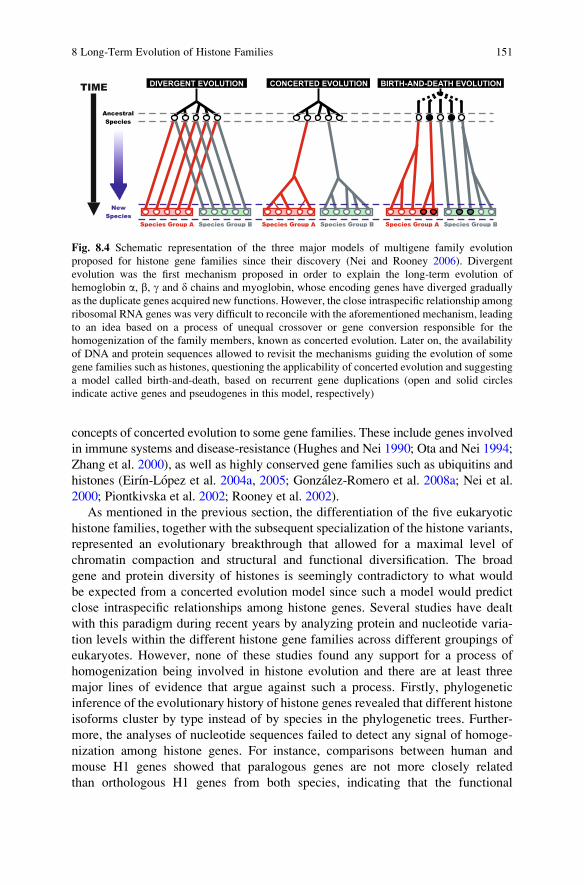
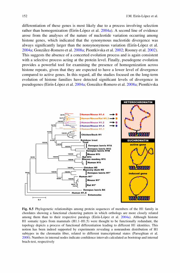
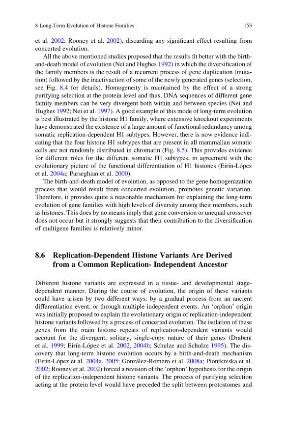
Fig. 8.5 Phylogenetic relationships among protein sequences of members of the H1 family inchordates showing a functional clustering pattern in which orthologs are more closely relatedamong them than to their respective paralogs (Eirın-Lopez et al. 2004a). Although histoneH1 somatic types from mammals (H1.1–H1.5) were thought to be functionally redundant, thetopology depicts a process of functional differentiation leading to different H1 identities. Thisnotion has been indeed supported by experiments revealing a nonrandom distribution of H1subtypes in the chromatin fiber, related to different transcriptional states (Parseghian et al.2000). Numbers in internal nodes indicate confidence intervals calculated as bootstrap and internalbrach-test, respectively
152 J.M. Eirın-Lopez et al.
et al. 2002; Rooney et al. 2002), discarding any significant effect resulting fromconcerted evolution.
All the above mentioned studies proposed that the results fit better with the birth-and-death model of evolution (Nei and Hughes 1992) in which the diversification ofthe family members is the result of a recurrent process of gene duplication (muta-tion) followed by the inactivaction of some of the newly generated genes (selection,see Fig. 8.4 for details). Homogeneity is maintained by the effect of a strongpurifying selection at the protein level and thus, DNA sequences of different genefamily members can be very divergent both within and between species (Nei andHughes 1992; Nei et al. 1997). A good example of this mode of long-term evolutionis best illustrated by the histone H1 family, where extensive knockout experimentshave demonstrated the existence of a large amount of functional redundancy amongsomatic replication-dependent H1 subtypes. However, there is now evidence indi-cating that the four histone H1 subtypes that are present in all mammalian somaticcells are not randomly distributed in chromatin (Fig. 8.5). This provides evidencefor different roles for the different somatic H1 subtypes, in agreement with theevolutionary picture of the functional differentiation of H1 histones (Eirın-Lopezet al. 2004a; Parseghian et al. 2000).
The birth-and-death model of evolution, as opposed to the gene homogenizationprocess that would result from concerted evolution, promotes genetic variation.Therefore, it provides quite a reasonable mechanism for explaining the long-termevolution of gene families with high levels of diversity among their members, suchas histones. This does by no means imply that gene conversion or unequal crossoverdoes not occur but it strongly suggests that their contribution to the diversificationof multigene families is relatively minor.
8.6 Replication-Dependent Histone Variants Are Derivedfrom a Common Replication- Independent Ancestor
Different histone variants are expressed in a tissue- and developmental stage-dependent manner. During the course of evolution, the origin of these variantscould have arisen by two different ways: by a gradual process from an ancientdifferentiation event, or through multiple independent events. An ‘orphon’ originwas initially proposed to explain the evolutionary origin of replication-independenthistone variants followed by a process of concerted evolution. The isolation of thesegenes from the main histone repeats of replication-dependent variants wouldaccount for the divergent, solitary, single-copy nature of their genes (Drabentet al. 1999; Eirın-Lopez et al. 2002, 2004b; Schulze and Schulze 1995). The dis-covery that long-term histone evolution occurs by a birth-and-death mechanism(Eirın-Lopez et al. 2004a, 2005; Gonzalez-Romero et al. 2008a; Piontkivska et al.2002; Rooney et al. 2002) forced a revision of the ‘orphon’ hypothesis for the originof the replication-independent histone variants. The process of purifying selectionacting at the protein level would have preceded the split between protostomes and
8 Long-Term Evolution of Histone Families 153
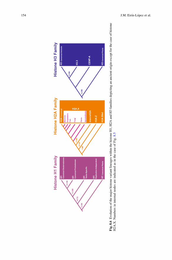
Fig.8.6
Evolutionofthemajorhistonevariantlineages
within
thehistoneH1,H
2AandH3familiesdepictingan
ancientorigin
exceptforthecase
ofhistone
H2A.X.Numbersin
internal
nodes
areindicated
asin
thecase
ofFig.8.5
154 J.M. Eirın-Lopez et al.
deuterostomes allowing for the subsequent transposition of replication-independenthistone variants to solitary locations in the genome, where they would graduallycontinue their evolution (Eirın-Lopez et al. 2004a, 2005).
Different histone families seem to be subject to different rates of ‘birth-and-death’ evolution as indicated by the high levels of diversity exhibited by membersof the H1 and H2A families in contrast to the H2B, H3 and H4 families. Althoughmost replication-independent histone variants have an ancient origin, there arenotable exceptions to this rule, such as the case of histone H2A.X (Fig. 8.6). It isvery likely that H2A.X genes arose separately and recurrently during evolutionhaving totally replaced canonical H2A in organisms such as Saccharomyces cere-visiae while being completely absent in C. elegans (Thatcher and Gorovsky 1994).Less is known about the H3 genes, where a replication independent gene was initiallyproposed to be the progenitor of all H3 genes through a single differentiation eventthat took place early in evolution (Wells et al. 1986). This hypothesis is supported bya study that suggests that a gene similar to that of histone H3 from the protistPhreatamoeba, the closest relative to animal and plant H3 genes, may have beenthe ancestor of the animal, plant and fungal H3 sequences. Nevertheless, the appear-ance of H3 variants independently in animals, plants and Tetrahymenawas also takenas evidence for the multiple origin of H3 variants (Thatcher and Gorovsky 1994).
When considering long-term histone evolution it is important to bear in mind thelarge differences exhibited by the plant and animal kingdoms, which clearly reflectthe different evolutionary strategies followed by different organisms despite havingall gone through the same histone gene duplication and selection mechanisms.Plant histones show very unique features that clearly differentiate them from thereplication-dependent histones from animals but they are very closely related toanimal replication-independent histone variants with which they share commontraits such as the presence of introns and expression through polyadenylatedtranscripts (Chaboute et al. 1993). The most plausible explanation to account forthis relies on the fact that plant cells exhibit a much longer cell cycle than animalcells. Therefore, a rapid change in the levels of histone gene expression duringS-phase is no longer needed and transcription control has a predominant role overposttranscriptional regulation. All this raises the question of whether ancestralhistones were expressed through polyadenylated transcripts. Different authorshave suggested that the major plant histone genes evolved from a common poly-adenylated ancestor prior to the differentiation between plants and animals (Chabouteet al. 1993) and that animal histone genes would have acquired specific posttransla-tional regulatory mechanisms (necessary to ensure the rapid histone biosynthesis inrapidly dividing cells) later on during evolution. This hypothesis is further supportedby the polyadenylated nature of different histone transcripts in ancestral eukaryotespreceding the differentiation of the metazoan variants. A good example of this isprovided by the histone H1 from trypanosomes (Gruter and Betschart 2001).
The mechanisms of transcriptional regulation of histone genes play a critical rolein the specialization of histone variants in different tissues and developmentalstages. The bulk of histone mRNA translation is coordinated with DNA replicationduring S phase of the cell cycle and this process is mediated by the presence of
8 Long-Term Evolution of Histone Families 155
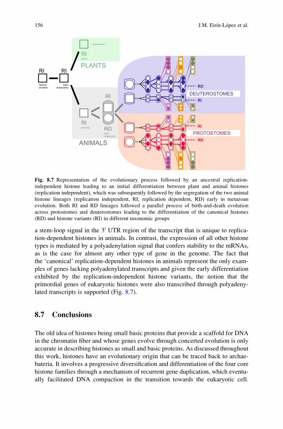
a stem-loop signal in the 30 UTR region of the transcript that is unique to replica-tion-dependent histones in animals. In contrast, the expression of all other histonetypes is mediated by a polyadenylation signal that confers stability to the mRNAs,as is the case for almost any other type of gene in the genome. The fact thatthe ‘canonical’ replication-dependent histones in animals represent the only exam-ples of genes lacking polyadenylated transcripts and given the early differentiationexhibited by the replication-independent histone variants, the notion that theprimordial genes of eukaryotic histones were also transcribed through polyadeny-lated transcripts is supported (Fig. 8.7).
8.7 Conclusions
The old idea of histones being small basic proteins that provide a scaffold for DNAin the chromatin fiber and whose genes evolve through concerted evolution is onlyaccurate in describing histones as small and basic proteins. As discussed throughoutthis work, histones have an evolutionary origin that can be traced back to archae-bateria. It involves a progressive diversification and differentiation of the four corehistone families through a mechanism of recurrent gene duplication, which eventu-ally facilitated DNA compaction in the transition towards the eukaryotic cell.
Fig. 8.7 Representation of the evolutionary process followed by an ancestral replication-independent histone leading to an initial differentiation between plant and animal histones(replication independent), which was subsequently followed by the segregation of the two animalhistone lineages (replication independent, RI; replication dependent, RD) early in metazoanevolution. Both RI and RD lineages followed a parallel process of birth-and-death evolutionacross protostomes and deuterostomes leading to the differentiation of the canonical histones(RD) and histone variants (RI) in different taxonomic groups
156 J.M. Eirın-Lopez et al.
The extraordinary structural and functional diversity observed among members ofthe different histone families is, in most instances, concomitant with the complexityof the organism, owing to the critical role histones play in the evolution ofbiological systems. Such diversity provides compelling evidence in support of amechanism directing the long-term evolution of histone families that is gearedtowards the generation of genetic diversity (birth-and-death), rather than one thatinduces gene homogenization (concerted evolution). Two major histone lineagesmust have already been differentiated very early in eukaryotic evolution, oneleading to the canonical replication-dependent histones and the other to the repli-cation-independent histone variants. Both lineages appear to share a commonreplication-independent ancestor containing introns, which most likely was thepreferred histone choice in plants. It is important to bear in mind that the loss ofintrons and the appearance of a replication-dependent expression must have hadcritical consequences for the evolutionary constraints acting upon the proteins. Itprobably involved a dramatic temporal switch towards strong positive selection ofthe replication-dependent histone genes that would result in an oddly similarsignature to that arising from purifying selection. The current availability ofgenome sequences for a broad range of eukaryotic organisms opens a door tofurther examine the implications of such an intriguing phenomenon, especially asit pertains to the distribution and evolution of introns in early eukaryotes and earlymetazoans in relation to the selective pressures acting on histones.
Acknowledgments This work was funded in part by the Canadian Institutes of Health Research(CIHR) Grant MOP-57718 to Juan Ausio and by the Marie Curie Outgoing International Fellow-ship (MOIF-CT-2005–021900) within the 6th Framework Programme (European Union) and by acontract within the Isidro Parga Pondal Program (Xunta de Galicia) to Jose M. Eirın-Lopez.Rodrigo Gonzalez-Romero is the recipient of a fellowship from the Diputacion de A Coruna.
References
Akhmanova A, Miedema K, Henning W (1996) Identification and characterization of theDrosophila histone H4 replacement gene. FEBS Lett 388:219–222
Albig W, Meergans T, Doenecke D (1997a) Characterization of the H1.5 genes completes the setof human H1 subtype genes. Gene 184:141–148
Albig W, Kioschis P, Poutska A, Meergans T, Doenecke D (1997b) Human histone gene organi-zation: non-regular arrangement within a large cluster. Genomics 40:314–322
Ali T, Thomas JO (2004) Distinct properties of the two putative ‘‘globular domains’’ of the yeastlinker histone, Hho1p J Mol Biol 337:1123–1135
Allis CD, Glover CVC, Gorovsky MA (1979) Micronuclei of Tetrahymena contain two types ofhistone H3. Proc Natl Acad Sci USA 76:4857–4861
Alvelo-Ceron D, Niu L, Collart DG (2000) Growth regulation of human variant histone genes andacetylation of the encoded proteins. Mol Biol Rep 27:61–71
Arents G, Moudrianakis E (1995) The histone fold: a ubiquitous architectural motif utilized inDNA compaction and protein dimerization. Proc Natl Acad Sci USA 92:11170–11174
Arnheim N (1983) Concerted evolution of multigene families. In: Nei M, Koehn RK (eds)Evolution of Genes and Proteins. Sinauer, Sunderland, MA, pp 38–61
8 Long-Term Evolution of Histone Families 157
Ausio J (1999) Histone H1 and evolution of sperm nuclear basic proteins. J Biol Chem274:31115–31118
Ausio J (2006) Histone variants: the structure behind the function. Brief Funct Genomic Proteomic5:228–243
Ausio J, Abbott DW (2002) The many tales of a tail: carboxyl-terminal tail heterogeneityspecializes histona H2A variants for defined chromatin function. Biochemistry 41:5945–5949
Ausio J, Abbott DW (2004) The role of histone variability in chromatin stability and folding. In:Zlatanova J, Leuba SH (eds) Chromatin Structure and Dynamics: State-of-the-Art. Elsevier,New York, pp 241–290
Baldo AM, Les DH, Strausbaugh LD (1999) Potentials and limitations of histone repeat sequencesfor phylogenetic reconstruction of Sophophora. Mol Biol Evol 16:1511–1520
Barski A, Cuddapah S, Cui K, Roth TY, Schones DE, Wang Z, Wei G, Chepelev I, Zhao K (2007)High-resolution profiling of histone methylations in the human genome. Cell 129:823–837
Barzotti R, Pelliccia F, Bucchiarelli E, Rocchini A (2000) Organization, nucleotide sequence, andchromosomal mapping of a tandemly repeated unit containing the four core histone genes and a5S rRNA gene in an isopod crustacean species. Genome 43:341–345
Bruce K, Myers FA, Mantouvalou E, Lefevre P, Greaves I, Bonifer C, Tremethick DJ,Thorne AW, Crane-Robinson C (2005) The replacement histone H2A.Z in hyperacetylatedform is a feature of active genes in chicken. Nucleic Acids Res 33:5633–5639
Chaboute ME, Chaubet N, Gigot C, Phillips G (1993) Histones and histone genes in higher plants:Structure and genomic organization. Biochimie 75:523–531
Cho H, Wolffe AP (1994) Xenopus laevis B4, an intron-containing oocyte-specific linker histone-encoding gene. Gene 143:233–238
Coen E, Strachan T, Dover GA (1982) Dynamics of concerted evolution of ribosomal DNAand histone gene families in the melanogaster species subgroup of Drosophila. J Mol Biol158:17–35
Collart D, Pockwinse S, Lian JB, Stein JL, Stein GS, Romain PL, Pilapil S, Heubner K,Cannizzaro LA, Croce CM (1992) A human histone H2B.1 Variant gene, located on chromo-some 1, utilizes alternative 3 end processing. J Cell Biochem 50:374–385
Connor W, Mezquita J, Winkfein RJ, States JC, Dixon GH (1984) Organization of the histonegenes in the rainbow trout (Salmo gairdnerii). J Mol Evol 20:272–285
Cool D, Banfield D, Honda BM, Smith MJ (1988) Histone genes in three sea star species: Clusterarrangement, transcriptional polarity and analysis of the flanking regions of H3 and H4 genes.J Mol Evol 27:36–44
Coon JJ, Ueberheide B, Syka JE, Dryhurst DD, Ausio J, Shabanowitz J, Hunt DF (2005) Proteinidentification using sequential ion/ion reactions and tandem mass spectrometry. Proc NatlAcad Sci USA 102:9463–9468
Costanzi C, Pehrson JR (1998) Histone macroH2A1 is concentrated in the inactive X chromosomeof female mammals. Nature 393:599–601
Cubonova L, Sandman K, Hallam SJ, DeLong EF, Reeve JN (2005) Histones in Crenarchaea.J Bacteriol 187:5482–5485
D’Andrea R, Coles LS, Lesnikowski C, Tabe L, Wells JRE (1985) Chromosomal organizationof chicken histone genes: preferred associations and inverted duplications. Mol Cell Biol5:3108–3115
del Gaudio N, Potenza N, Stefanoni P, Chiusano ML, Geraci G (1998) Organization and nucleo-tide sequence of the cluster of five histone genes in the polichaete worm Chaetopterusvariopedatus: first record of a H1 histone gene in the phylum annelida. J Mol Evol 46:64–73
Doenecke D, Alonso A (1996) Organization and expression of the developmentally regulated H10
histone gene in vertebrates. Int J Dev Biol 40:423–431Doenecke D, Albig W, Bode C, Drabent B, Franke K, Gavenis K, Witt O (1997) Histones: genetic
diversity and tissue-specific gene expression. Histochem Cell Biol 107:1–10Domier LL, Rivard JJ, Sabatini LM, Blumenfeld M (1986)Drosophila virilis histone gene clusters
lacking H1 coding segments. J Mol Evol 23:149–158
158 J.M. Eirın-Lopez et al.
Drabent B, Kim J-S, Albig W, Prats E, Cornudella L, Doenecke D (1999) Mytilus edulis histonegene clusters containing only H1 genes. J Mol Evol 49:645–655
Eirın-Lopez JM, Gonzalez-Tizon AM, Martınez A, Mendez J (2002) Molecular and evolutionaryanalysis of mussel histone genes (Mytilus spp.): possible evidence of an ‘‘orphon origin’’ for H1histone genes. J Mol Evol 55:272–283
Eirın-Lopez JM, Gonzalez-Tizon AM, Martınez A, Mendez J (2004a) Birth-and-death evolutionwith strong purifying selection in the histone H1 multigene family and the origin of orphon H1genes. Mol Biol Evol 21:1992–2003
Eirın-Lopez JM, Ruiz MF, Gonzalez-Tizon AM, Martınez A, Sanchez L, Mendez J (2004b)Molecular evolutionary characterization of the mussel Mytilus histone multigene family:first record of a tandemly repeated unit of five histone genes containing an H1 subtype with‘‘orphon’’ features. J Mol Evol 58:131–144
Eirın-Lopez JM, Ruiz MF, Gonzalez-Tizon AM, Martınez A, Ausio J, Sanchez L, Mendez J(2005) Common evolutionary origin and birth-and-death process in the replication-independenthistone H1 isoforms from vertebrate and invertebrate genomes. J Mol Evol 61:398–407
Eirın-Lopez JM, Ishibashi T, Ausio J (2008) H2A.Bbd: a quickly evolving hypervariable mam-malian histone that destabilizes nucleosomes in an acetylation-independent way. FASEB J22:316–326
Ellegren H (2002) Dosage compensation: do birds do it as well? Trends Genet 18:25–28Gonzalez-Romero R, Ausio J, Mendez J, Eirın-Lopez JM (2008a) Early evolution of histone
genes: prevalence of an ‘orphon’ H1 lineage in protostomes and birth-and-death process in theH2A family. J Mol Evol 66:505–518
Gonzalez-Romero R, Mendez J, Ausio J, Eirın-Lopez JM (2008b) Quickly evolving histones,nucleosome stability and chromatin folding: All about histone H2A.Bbd. Gene 413:1–7
Govin J, Caron C, Rousseaux S, Khochbin S (2005) Testis-specific histone H3 expression insomatic cells. Trends Biochem Sci 30:357–359
Grayling RA, Sandman K, Reeve JN (1996) Histones and chromatin structure in hyperthermo-philic Archaea. FEMS Microbiol Rev 18:203–213
Greaves IK, Rangasamy D, Ridgway P, Tremethick DJ (2007) H2A.Z contributes to the unique 3Dstructure of the centromere. Proc Natl Acad Sci USA 104:525–530
Gruter E, Betschart B (2001) Isolation, characterisation and organisation of histone H1 genes inAfrican trypanosomes. Parasitol Res 87:977–984
Hankeln T, Schmidt ER (1991) The organization and nucleotide sequence of the histone genes ofthe midge Chironomus thummi. Chromosoma 105:25–31
Hankeln T, Schmidt ER (1993) Divergent evolution of an ‘‘orphon’’ histone gene cluster inChironomus. J Mol Biol 234:1301–1307
Happel N, Schulze E, Doenecke D (2005) Characterization of human histone H1x. Biol Chem386:541–551
Hatch CL, Bonner WM (1988) Sequence of cDNAs from mammalian H2A.Z, an evolutionarilydiverged but highly conserved basal histone H2A isoprotein species. Nucleic Acids Res16:1113–1124
Hatch CL, Bonner W (1990) The human histone H2A.Z gene. Sequence and regulation. J BiolChem 265:15211–15218
Henikoff S, Ahmad K (2005) Assembly of variant histones into chromatin. Annu Rev Cell DevBiol 21:133–153
Henning W (2003) Chromosomal proteins in the spermatogenesis of Drosophila. Chromosoma111:489–494
Hentschel CC, Birnstiel ML (1981) The organization and expression of histone gene families. Cell25:301–313
Herzog M, Soyer MO (1981) Distinctive features of dinoflegellate chromatin. Absence of nucleo-somes in a primitive species Prorocentrum micans. Eur J Cell Biol 23:295–302
Hughes AL, Nei M (1990) Evolutionary relationships of class II major-histocompatibility complexgenes in mammals. Mol Biol Evol 7:491–514
8 Long-Term Evolution of Histone Families 159
Iguchi N, Tanaka H, Yomogida K, Nishimune Y (2003) Isolation and characterization of a novelcDNA encoding a DNA-binding protein (Hils1) specifically expressed in testicular haploidgerm cells. Int J Androl 26:354–365
Isenberg I (1979) Histones. Annu Rev Genet 48:159–191Jenuwein T, Allis CD (2001) Translating the histone code. Science 293:1074–1080Kanazin V, Blake T, Shoemaker RC (1996) Organization of the histone H3 genes in soybean,
barley, and wheat. Mol Gen Genet 250:137–147Kasinsky HE, Lewis JD, Dacks JB, Ausio J (2001) Origin of H1 histones. FASEB J 15:34–42Kedes L (1979) Histone genes and histone messengers. Annu Rev Biochem 225:501–510Khochbin S (2001) Histone H1 diversity: bridging regulatory signals to linker histone function.
Gene 271:1–12Kremer H, Henning W (1990) Isolation and characterization of a Drosophila hydei histone DNA
repeat unit. Nucleic Acids Res 18:1573–1580Landsman D (1996) Histone H1 in Saccharomyces cerevisiae – a double mystery solved. Trends
Biochem Sci 21:287–288Li A, Eirın-Lopez JM, Ausio J (2005) H2AX: tailoring histone H2A for chromatin-dependent
genomic integrity. Biochem Cell Biol 83:505–515Liao D (1999) Concerted evolution: molecular mechanism and biological implications. Am J Hum
Genet 64:24–30Lifton RP, Goldberg ML, Karp RW, Hogness DS (1977) The organization of the histone genes in
Drosophila melanogaster: functions and evolutionary implications. Cold Spring Harbor SympQuant Biol 42:1047–1051
Lindauer A, Muller K, Schmidt R (1993) Two histone H1-encoding genes of the green alga Volvoxcarteri with features intermediate between plant and animal genes. Gene 239:15–27
Malik HS, Henikoff S (2003) Phylogenomics of the nucleosome. Nat Struct Biol 10:882–891Mandl B, Brandt WF, Superti-Furga G, Graninger PG, Birnstiel M, Busslinger M (1997) The five
cleavage-stage (CS) histones of the sea urchin are encoded by a maternally expressed family ofreplacement histone genes: functional equivalence of the CS H1 and frog H1M (B4) proteins.Mol Cell Biol 17:1189–1200
Martianov I, Brancorini S, Catena R, Gansmuller A, Kotaja N, Parvinen M, Sassone-Corsi P,Davidson I (2005) Polar nuclear localization of H1T2, a histone H1 variant, required forspermatid elongation and DNA condensation during spermiogenesis. Proc Natl Acad SciUSA 102:2808–2813
Martin W, Muller M (1998) The hydrogen hypothesis for the first eukaryote. Nature 392:37–41Marzluff WF (1992) Histone 30 ends: essential and regulatory functions. Gene Express 2:93–97Matsuo Y, Yamazaki T (1989) Nucleotide variation and divergence in the histone multigene
family in Drosophila melanogaster. Genetics 122:87–97Maxson R, Cohn R, Kedes L, Mohun T (1983a) Expression and organization of histone genes.
Annu Rev Genet 17:239–277Maxson R, Mohun T, Gormezano G, Childs G, Kedes L (1983b) Distinct organizations and
patterns of expression of early and late histone gene sets in the sea urchin. Nature 301:120–125Miller DJ, Harrison PL, Mahony TJ, McMillan JP, Miles A, Odorico DM, ten Lohuis MR (1993)
Feature of histone gene structure and organization are common to diploblastic and tripoblasticmetazoans. J Mol Evol 37:245–253
Mito Y, Henikoff JG, Henikoff S (2005) Genome-scale profiling of histone H3.3 replacementpatterns. Nat Genet 37:1090–1097
Murphy LD, Zimmerman SB (1997) Stabilization of compact spermidine nucleoids fromEscherichia coli under crowded conditions: implications for in vivo nucleoid structure. J StructBiol 119:336–346
Nei M, Hughes AL (1992) Balanced polymorphism and evolution by the birth-and-death processin the MHC loci. In: Tsuji K, Aizawa M, Sasazuki T (eds) 11th Histocompatibility Workshopand Conference. Oxford University Press, Oxford/England, pp 27–38
Nei M, Rooney AP (2006) Concerted and birth-and-death evolution in multigene families. AnnuRev Genet 39:121–152
160 J.M. Eirın-Lopez et al.

Nei M, Gu X, Sitnikova T (1997) Evolution by the birth-and-death process in multigene families ofthe vertebrate immune system. Proc Natl Acad Sci USA 94:7799–7806
Nei M, Rogozin IB, Piontkivska H (2000) Purifying selection and birth-and-death evolution in theubiquitin gene family. Proc Natl Acad Sci USA 97:10866–10871
Ohta T (1983) On the evolution of multigene families. Theor Popul Biol 23:216–240Ota T, Nei M (1994) Divergent evolution and evolution by the birth-and-death process in the
immunoglobulin VH gene family. Mol Biol Evol 11:469–482Ouzounis CA, Kyrpides NC (1996) Parallel origin of the nucleosome core and eukaryotic
transcription from archaea. J Mol Evol 42:234–239Owen-Hughes T (2003) Colworth memorial lecture. Pathways for remodelling chromatin.
Biochem Soc Trans 31:893–905Palmer DK, O’Day K, Wener MH, Andrews BS, Margolis RL (1987) A 17-kD centromere
protein (CENP-A) copurifies with nucleosome core particles and with histones. J Cell Biol104:805–815
Panyim S, Chalkey R (1969) High resolution acrylamide gel electrophoresis of histones. ArchBiochem Biophys 63:265–297
Parseghian MH, Hamkalo BA (2001) A compendium of the H1 family of somatic subtypes: anelusive cast of characters and their characteristics. Biochem Cell Biol 79:289–304
Parseghian MH, Newcomb RL, Winokur ST, Hamkalo BA (2000) The distribution of somatic H1subtypes is nonrandom on active vs. inactive chromatin: distribution in human fetal fibroblasts.Chromosome Res 8:405–424
Pereira SL, Grayling RA, Lurz R, Reeve JN (1997) Archaeal nucleosomes. Proc Natl Acad SciUSA 94:12633–12637
Piontkivska H, Rooney AP, Nei M (2002) Purifying selection and birth-and-death evolution in thehistone H4 gene family. Mol Biol Evol 19:689–697
Prescott DM (1994) The DNA of ciliated protozoa. Microbiol Rev 58:233–267Reeve JN, Sandman K, Daniels CJ (1997) Archaeal histones, nucleosomes, and transcription
initiation. Cell 89:999–1002Rooney AP, Piontkivska H, Nei M (2002) Molecular evolution of the nontandemly repeated genes
of the histone 3 multigene family. Mol Biol Evol 19:68–75Ruberti I, Fragapane P, Pierandrei-Arnaldi P, Beccari E, Arnaldi F, Bozzoni I (1982) Characteri-
zation of histone genes isolated from Xenopus laevis and Xenopus tropicalis genomic libraries.Nucleic Acids Res 10:1544–1550
Ruiz-Carrillo A, Affolter M, Renaud J (1983) Genomic organization of the genes coding for themain six histones of the chicken: complete sequence of the H5 gene. J Mol Biol 170:843–859
Sandman K, Reeve JN (1998) Origin of the eukaryotic nucleus. Science 281:501–503Sandman K, Reeve JN (2005) Archaeal chromatin proteins: different structures but common
function? Curr Opin Microbiol 8:656–661Sandman K, Reeve JN (2006) Archaeal histones and the origin of the histone fold. Curr Opin
Microbiol 9:520–525Sandman K, Krzycki JA, Dobrinski B, Lurz R, Reeve JN (1990) HMf, a DNA-binding protein
isolated from the hyperthermophilic archaeon Methanothermus fervidus, is most closelyrelated to histones. Proc Natl Acad Sci USA 87:5788–5791
Sandman K, Pereira SL, Reeve JN (1998) Diversity of prokaryotic chromosomal proteins and theorigin of the nucleosome. Cell Mol Life Sci 54:1350–1364
Sanicola M, Ward S, Childs G, Emmons SW (1990) Identification of a Caenorhabditis eleganshistone H1 gene family. Characterization of a family member containing an intron andencoding poly(A) + mRNA. J Mol Biol 212:259–268
Schienman JE, Lozovskaya ER, Strausbaugh LD. (1998) Drosophila virilis has atypical kinds andarrangements of histone repeats. Chromosoma 107:529–539
Schulze E, Schulze B (1995) The vertebrate linker histones H10, H5, and H1M are descendants ofinvertebrate ‘‘orphon’’ histone H1 genes. J Mol Evol 41:833–840
Scott AC, Wells JR (1976) Reiteration frequency of the gene for tissue-specific histone H5 in thechicken genome. Nature 259:635–638
8 Long-Term Evolution of Histone Families 161
Sellos D, Krawetz SA, Dixon GH (1990) Organization and complete nucleotide sequences of thecore-histone-gene cluster of the annelid Platynereis dumerilii. Eur J Biochem 190:21–29
Seyedin SM, Kistler WS (1979) H1 histone subfractions of mammalian testes. 1. Organ specificityin the rat. Biochemistry 18:1371–1375
Slerasev AI, Belova GI, Kozyavkin SA, Lake JA (1984) Evidence for an early prokaryotic originof histones H2A and H4 prior to the emergence of eukaryotes. Nucleic Acids Res 26:427–430
Smith BJ, Harris MR, Sigournay CM, Mayes ELV, Bustin M (1984) A survey of H10- and H5-likeprotein structure and distribution in lower and higher eukaryotes. Eur J Biochem 138:309–317
Smith GP (1974) Unequal crossover and the evolution of multigene families. Cold Spring HarborSymp Quant Biol 38:507–513
Starich MR, Sandman K, Reeve JN, Summers MF (1996) NMR Structure of the HMfB from thehyperthermophile, Methanothermus fervidus, confirms that this archaeal protein is a histone.J Mol Biol 255:187–203
Stephenson E, Erba H, Gall J (1981) Characterization of a cloned histone gene cluster of the newtNotophtalmus viridescens. Nucleic Acids Res 9:2281–2295
Sures I, Lowry J, Kedes L (1978) The DNA sequence of the sea urchin (S. purpuratus) H2A, H2Band H3 histone coding and spacer regions. Cell 15:1033–1044
Tanaka M, Hennebold JD, MacFarlane J, Adashi EY (2001) A mammalian oocyte-specific linkerhistone gene H1oo: homology with the genes for oocyte-specific cleavage stage histones (cs-H1) of sea urchin and the B4/H1M histone of the frog. Development 128:655–664
Thatcher TH, Gorovsky MA (1994) Phylogenetic analysis of the core histones H2A, H2B, H3, andH4. Nucleic Acids Res 22:174–179
Tsunemoto K, Matsuo Y (2001) Molecular evolutionary analysis of a histone gene repeating unitfrom Drosophila simulans. Genes Genet Syst 76:355–361
Turner PC, Woodland HR (1983) Histone gene number and organisation in Xenopus: Xenopusborealis has a homogeneous major cluster. Nucleic Acids Res 11:971–986
Turner PC, Aldridge TC, Woodland HR, Old RW (1983) Nucleotide sequences of H1 histonegenes from Xenopus laevis. A recently diverged pair of H1 genes and an unusual H1 pseudo-gene. Nucleic Acids Res 11:4093–4106
van DongenW, de Laaf L, Zaal R, Moorman AF, Destree O (1981) The organization of the histonegenes in the genome of Xenopus laevis. Nucleic Acids Res 25:2297–2311
van Holde KE (1988) Chromatin. Springer-Verlag, New YorkWalter L, Klinga-Levan K, Helou K, Albig W, Drabent B, Doenecke D, Gunther E, Levan G
(1996) Chromosomal mapping of rat histone genes H1fv (H10), H1d, H1t, Th2a and Th2b.Cytogenet Cell Genet 75:136–139
Wang ZF, Sirotkin AM, Buchold GM, Skoultchi AI, Marzluff WF (1997) The mouse histone H1genes: gene organization and differential regulation. J Mol Biol 271:124–138
Wells JRE, Kedes L (1985) Structure of a human histone cDNA: evidence that basally expressedhistone genes have intervening sequences and encode polyadenylated mRNAs. Proc Natl AcadSci USA 82:2834–2838
Wells JRE, Bains W, Kedes L (1986) Codon usage of histone gene families in higher eukaryotesreflects functional rather than phylogenetic relationships. J Mol Evol 23:224–241
Witt O, Albig W, Doenecke D (1996) Testis-specific expression of a novel human H3 histonegene. Exp Cell Res 229:301–306
Worcel A, Burgi E (1972) On the structure of the folded chromosome of Escherichia coli. J MolBiol 71:127–147
Yan W, Lang M, Burns KH, Matzuk MM (2003) HILS1 is a spermatid-specific linker histoneH1-like protein implicated in chromatin remodeling during mammalian spermiogenesis. ProcNatl Acad Sci USA 100:10546–10551
Zhang J, Dyer KD, Rosenberg HF (2000) Evolution of the rodent eosinophil-associatedRNase gene family by rapid gene sorting and positive selection. Proc Natl Acad Sci USA97:4701–4706
Zlatanova J (1997) Archaeal chromatin: virtual or real? Proc Natl Acad Sci USA 94:12251–12254
162 J.M. Eirın-Lopez et al.
Copyright © 2022 FDOKUMEN