Localized RanGTP Accumulation Promotes Microtubule Nucleation at Kinetochores in Somatic Mammalian...
28
Localized RanGTP accumulation promotes microtubule nucleation at kinetochores in somatic mammalian cells. Liliana Torosantucci, Maria De Luca, Giulia Guarguaglini, Patrizia Lavia and Francesca Degrassi * IBPM Institute of Molecular Biology and Pathology, CNR National Research Council, c/o University ‘La Sapienza’, Via degli Apuli 4, 00185 Rome, Italy * Address correspondence to: Francesca Degrassi ([email protected]) Running title: microtubule nucleation at kinetochores Key words: Kinetochore, microtubule nucleation, RanGTP, Plk1 silencing, spindle assembly. 1 http://www.molbiolcell.org/content/suppl/2008/02/20/E07-10-1050.DC1.html Supplemental Material can be found at:
-
Upload
independent -
Category
Documents
-
view
0 -
download
0
Transcript of Localized RanGTP Accumulation Promotes Microtubule Nucleation at Kinetochores in Somatic Mammalian...
Localized RanGTP accumulation promotes microtubule nucleation at kinetochores
in somatic mammalian cells.
Liliana Torosantucci, Maria De Luca, Giulia Guarguaglini, Patrizia Lavia and Francesca
Degrassi*
IBPM Institute of Molecular Biology and Pathology, CNR National Research Council, c/o
University ‘La Sapienza’, Via degli Apuli 4, 00185 Rome, Italy
* Address correspondence to: Francesca Degrassi ([email protected])
Running title: microtubule nucleation at kinetochores
Key words: Kinetochore, microtubule nucleation, RanGTP, Plk1 silencing, spindle assembly.
1 http://www.molbiolcell.org/content/suppl/2008/02/20/E07-10-1050.DC1.html
Supplemental Material can be found at:

ABSTRACT
Centrosomes are the major sites for microtubule nucleation in mammalian cells, although both
chromatin- and kinetochore-mediated microtubule nucleation have been observed during spindle
assembly. As yet, it is still unclear whether these pathways are co-regulated and the molecular
requirements for microtubule nucleation at kinetochore are not fully understood. This work
demonstrates that kinetochores are initial sites for microtubule nucleation during spindle reassembly
after nocodazole. This process requires local RanGTP accumulation concomitant with
delocalization from kinetochores of the hydrolysis factor RanGAP1. Kinetochore-driven
microtubule nucleation is also activated after cold-induced microtubule disassembly when
centrosome nucleation is impaired, e.g. after Polo-like kinase 1 depletion, indicating that dominant
centrosome activity normally masks the kinetochore-driven pathway. In cells with unperturbed
centrosome nucleation, defective RanGAP1 recruitment at kinetochores after treatment with the
Crm1 inhibitor leptomycin B activates kinetochore microtubule nucleation after cold. Finally,
nascent microtubules associate with the RanGTP regulated microtubule-stabilizing protein HURP in
both cold- and nocodazole-treated cells. These data support a model for spindle assembly in which
RanGTP-dependent abundance of nucleation/stabilization factors at centrosomes and kinetochores
orchestrates the contribution of the two spindle assembly pathways in mammalian cells. The
complex between RanGTP, the export receptor Crm1 and NES-bearing proteins regulates
microtubule nucleation at kinetochores.
2

INTRODUCTION
It is now clear that mitotic spindle assembly can occur via different pathways (for reviews
see Gadde and Heald 2004; Wadsworth and Khodjakov, 2004; Rieder, 2005, O’ Connel and
Khodjakov, 2007). In most somatic cell types, centrosomes nucleate microtubules (MTs) to build a
mitotic spindle. Functional spindles also form in cells lacking centrosomes, e.g. in higher plants and
some animal oocytes. In Xenopus eggs, spindles assemble through MT nucleation from chromatin
in a process requiring the GTPase Ran (Carazo-Salas et al., 1999; Kalab et al., 1999) which, in its
GTP-bound form, is generated near chromatin under the localized action of the nucleotide exchange
factor RCC1 (Moore et al., 2002; Li et al., 2003; Chen et al., 2007). In the Xenopus system,
RanGTP releases spindle assembly factors containing NLS signals, such as NuMa and TPX2 (Gruss
et al., 2001; Nachury et al., 2001; Wiese et al., 2001), from the inhibitory binding to importin α/β,
in the immediate vicinity of chromatin, thus promoting nucleation of MTs therein (reviewed by
Weis, 2003; Ciciarello et al., 2007). This has led to propose that spindle formation in acentrosomal
cells depends on a gradient of RanGTP, concentrating around chromosomes and diluting away from
them (Caudron et al., 2005; Kalab et al., 2006). Local activity of RanGTP and importin α/β also
contribute to spindle assembly in centrosome-driven cells (Nachury et al., 2001; Ciciarello et al.,
2004, Kalab et al., 2006). In these cells, chromatin-associated MT nucleation is normally
suppressed by the dominant centrosome activity, yet remains an intrinsic potential that can be
resumed under certain circumstances, as a functional spindle still form when centrosomes are
removed by laser ablation (Khodjakov et al., 2000), or centrosome activity is abolished by specific
mutations (Bonaccorsi et al., 1998).
Kinetochores were also reported to direct MT nucleation in mammalian cells after spindle
disrupting drugs, but this idea was subsequently questioned, based on concerns for possible
artifactual effects of the drugs (Rieder, 2005 and references therein). More recently, in vivo imaging
of mitosis has shown that unattached kinetochores facing away from centrosomes can promote the
formation of MTs during mitotic spindle assembly in both mammalian (Khodjakov et al., 2003) and
Drosophila cells (Maiato et al., 2004). In both cases, chromosome arms shielded the distal
kinetochore from interaction with microtubules generated at centrosomes, possibly providing time
for kinetochore nucleation to occur. However, the importance of this kinetochore-driven MT
nucleation for spindle assembly in centrosome-containing cells is not clearly defined, as only a few
examples were recorded in the live cells studies (Khodjakov et al., 2003; Maiato et al., 2004).
Furthermore, the molecular requirements for MT formation at kinetochore are not fully understood.
Recently, Tulu et al. (2006) observed microtubule formation at Bub1-positive sites after release
3

from nocodazole (NOC)-induced spindle disassembly in live cell imaging experiments (Tulu et al.,
2006). Formation of MT foci in the chromatin region was severely inhibited when the activity of the
Ran-dependent spindle assembly factor TPX2 was suppressed by either microinjection of excess
importin β, or by direct TPX2 depletion (Tulu et al., 2006). These results suggest that the
dissociation of import complexes entrapping NLS-bearing spindle assembly factors contributes to
the genesis of MT foci. However, the limited resolution of current live cell imaging methods did not
allow discriminating whether the inhibition of extracentrosomal MT foci reflected the suppression
of RanGTP-dependent MT nucleation specifically at kinetochores or, rather, a disruption of the
overall RanGTP gradient, which is generated along the entire chromosome region by the persisting
binding of RCC1 to mitotic chromatin. Furthermore, it remains to be determined what localizes
extracentrosomal MT nucleation to the kinetochore area, given that the exchange factor for Ran is
distributed along the entire length of the chromosome. We have re-addressed these questions by
assessing mitotic spindle re-assembly after microtubule depolymerization by nocodazole or after
cold-induced spindle disassembly in cells with impaired centrosome-nucleating activity. We now
report that the localized accumulation of RanGTP at kinetochores is responsible for MT nucleation
therein. The complex between RanGTP, the export receptor Crm1 and NES-bearing proteins
modulates MT nucleation at kinetochores.
MATERIALS AND METHODS
Cell cultures and treatments
Human lung diploid fibroblasts (MRC-5 cells) and human osteosarcoma U2OS cells were obtained
from the American Type Culture Collection (Rockville, MD) and maintained in Minimum Essential
Medium supplemented with 10 % fetal calf serum, antibiotics, L-glutamine, non-essential
aminoacids and Hepes. Cells were grown at 37°C, in a humidified atmosphere, with 5% CO2.
MRC5 or U2OS cultures were treated with 1.5 μM NOC for 16 hours to completely disassembly
MTs. tsBN2 cells (a gift of M.Dasso) were grown at 32°C and received 4 μM NOC for 3 hours
either at 32°C or at 39°C. At the end of the NOC incubation, cells were washed three times in
prewarmed medium, re-incubated in fresh medium to release the mitotic block and fixed at the
indicated times after NOC release. For MT regrowth after ice-induced depolymerization, U2OS
cells were incubated for 60 minutes on ice, prewarmed media was then added and cells were
incubated at 37°C for the indicated times. When needed, 20 nM leptomycin B was added to culture
medium 2 h before incubation on ice.
4

RNAi experiments
Small interfering RNA (siRNA) oligonucleotides targeting Plk1 or firefly luciferase gene sequences
(GL2) were from Qiagen. SiRNAs were mostly used at the final concentration of 80 nM using
oligofectamine (Invitrogen) as transfection reagent. SiRNA sequences and conditions of
interference were described (De Luca et al., 2006). All analyses were performed 40 h from
transfection.
Immunoblotting
An equal number of cells from tsBN2 cultures grown to 85% confluence at 32° C, or shifted to
39°C during the last 4 h of culture, was pelleted by centrifugation for 10 min at 1500 rpm and
washed in phosphate-buffered saline (PBS). Cells were extracted with RIPA buffer (50 mM Tris-
HCl pH 8.0, 150 mM NaCl, 1%, NP40, 1 mM EGTA, 0.25% sodium deoxycholate) supplemented
with 1 μg/ml Leupeptin, 1 μg/ml Pepstatin, 1 μg/ml Aprotinin, 1 mM PMSF, 1 mM DTT, 1 mM
NaVO4 and 1 mM NaF. Proteins were resolved by electrophoresis on a 10% SDS-polyacrylamide
gel, and transferred to a nitrocellulose membrane (Whatman Schleicher & Schuell PROTRAN)
using a semi-dry blotting system (BIO-RAD) in transfer buffer (48 mM Tris, 39 mM Glycin,
0,037% SDS and 20% Methanol). 60 μg of extract per lane were loaded. Blocking and antibody
incubations were in 5% low-fat dry milk in TBS (10 mM TrisHCl pH 8, 150 mM NaCl) containing
0,1% Tween 20. Membranes were blocked for 30 min, primary antibodies were incubated for 1 h,
secondary antibodies for 30 min, at RT. Primary antibodies were anti-RCC1 (Santa Cruz
Biotechnology, 0,5μg/ml) and anti-RanBP1 (Santa Cruz Biotechnology, 0,5μg/ml). U2OS cells
were lysed, resolved and transferred under similar conditions. 40 μg of extract per lane were loaded.
Primary antibodies were mouse anti-α-tubulin (ascite fluid, clone B-5-1-2, Sigma, 1:2000) and
mouse anti-Plk1 (ab 14210, Abcam,1μg/ml).
Immunostaining
Cells were rinsed in PHEM buffer (60 mM Pipes, 25 mM Hepes, pH 6.9, 10 mM EGTA, 4 mM
MgSO4), then lysed for 45 seconds in 0.1% Triton-X in PHEM buffer (MRC-5, U2OS) or for 5
minutes in 0.5% Triton-X (tsBN2). Cells were fixed for 10 minutes in 3,7% formaldehyde in
PHEM, then rinsed in PBS and post-fixed in ice cold MetOH for 5 minutes. After fixation, cells
were washed, blocked for 30 minutes in 20% goat serum at room temperature and incubated at
37°C for 1 hour with primary antibodies diluted in 5% goat serum. Primary antibodies were: anti-α-
tubulin and anti-γ−tubulin (Sigma) diluted 1:300 and 1:2000 respectively, CREST serum
5
(Antibodies Incorporated, 1:50), anti-NuMA (Oncogene, 1:50), anti-p150Glued (BD
Biosciences,1:100), anti-Plk1(14210 and 14209, ABCAM, 1:100), anti-IAK1/Aurora-A (clone 4,
Transduction Laboratories, 1:1000) anti-α-tubulin FITC conjugate (Sigma, 1:100), anti RanGAP1
(Santa Cruz, 1:200). Anti-Kif2a (1:1000), anti-centrin 2 (1:1125), anti-HURP (1:300) and anti-
RanGTP (AR12,1:50) were kindly provided by D. Compton, J. Salisbury, E.A. Nigg, H.Sillje and I.
Macara, respectively. DNA was counterstained in 0.1 µg/ml DAPI. For RanGTP staining (Tedeschi
et al. 2007), cells were pretreated 1 min in 1% Triton-X in PHEM buffer, incubated with AR12
antibody in PHEM buffer containing NOC during the final 30 min of the NOC treatment and then
fixed immediately as described above or released in fresh medium before fixation to analyze MT
regrowth.
Fluorescence Microscopy and Image Acquisition
All preparations were examined under an Olympus Vanox microscope equipped with a 100X (1.35
NA) oil immersion objective and a SPOT CCD camera (Diagnostic Instruments). Colour encoded
images were acquired using ISO 2000 software (Deltasistemi) and processed with Adobe Photoshop
7 software. Three dimensional (3-D) deconvolution and reconstruction was performed on 0.3-µm Z-
serial optical sections using AutoDeblur 9.3 (AutoQuant Imaging Inc.) image processing program
6

RESULTS
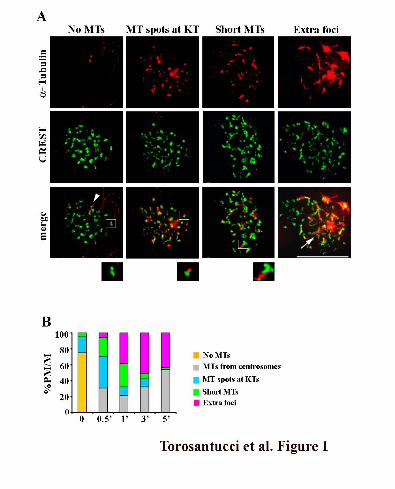
MT nucleation re-starts at kinetochores after NOC.
To dissect the relative contribution of chromatin- and kinetochore-mediated MT nucleation
during spindle assembly in mammalian cells, we took advantage of the reversibility of NOC-
induced MT disassembly and visualized kinetochores at the very early stages of spindle re-
assembly. Cells were first treated with NOC to completely disassemble MTs. The drug was then
carefully removed and the initial stages of spindle reassembly were followed in a time course
analysis. The experiments were performed in human fibroblast MRC-5 cells, but similar results
were obtained in other mammalian cell lines, such as osteosarcoma cells (U2OS, Supplemental
Figure 1). Combined α−tubulin and kinetochore staining enabled us to unambiguously identify the
generation of MTs at kinetochores at very early times after NOC removal, in the form of α−tubulin
positive spots at kinetochores (Figure 1A, MT spots at KTs), short MTs emanating from
kinetochores (Figure 1A, Short MTs) or supernumerary tubulin foci at the centre of a group of
kinetochores (Figure 1A, Extra foci). Thirty seconds after NOC release, about 70 % of cells
exhibited evidence of MT nucleation at kinetochores (Figure 1B), demonstrating that MT
nucleation at kinetochores is the predominant pathway for MT formation immediately after NOC
removal. Over time, the frequency of cells showing kinetochore spots and short kinetochore MTs
decreased, with a proportional increase in the frequency of cells with extra foci (Figure 1B),
suggesting that kinetochore-nucleated MTs are then organized at their minus ends into pole-like
structures. This idea is also supported by the localization to these supernumerary tubulin foci of
several non-centrosomal MT minus end-associated proteins with MT-focusing activity
(Supplemental Figure 2). The frequency of cells showing only centrosome-based MT arrays
increased at later times (Figure 1B), indicating that extra centrosomal foci generated at early times
during spindle reassembly are then captured by centrosomal MTs to form a bipolar spindle. Thus,
our detailed analysis of MT formation in conjunction with the visualization of the CREST
kinetochore marker enabled us to unambiguously identify the generation of MT at kinetochores as
the initial moment in MT regrowth in cells recovering from spindle disassembly. This information
was still lacking in previous live cell studies (Tulu et al., 2006) that left out the possibility that
association of MT to kinetochores was successive to MT nucleation around chromatin. These
results, together with those of Tulu et al. (Tulu et al., 2006), identify kinetochores as the initial sites
for MT nucleation after NOC-induced spindle reassembly.
7

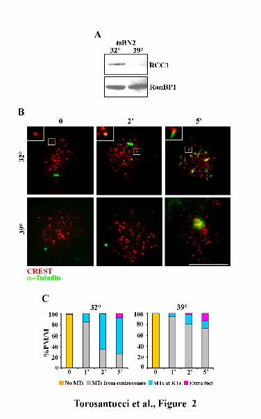
Localized accumulation of RanGTP at kinetochores promotes MT nucleation
To get some insight into the molecular requirements for MT nucleation at kinetochores we
analysed several regulators of microtubule nucleation during spindle reassembly after NOC. We
initially wondered whether NOC displaced regulators of MT nucleation from centrosomes to
chromosomes and whether that did in turn promote kinetochore-dependent MT nucleation. We
found that both Aurora-A and Plk1 kinases maintained their localization to centrosomes (Aurora-A,
Plk1) and kinetochores (Plk1), with no variation during NOC treatment (Supplemental Figure 3),
ruling out the possibility that NOC impaired the association of these crucial regulators with
centrosomes and that this in turn activated nucleation from kinetochores.
We next investigated Ran, which is clearly required for MT nucleation from chromatin in oocytes
and in mammalian cells (reviewed in Ciciarello et al., 2007) and has been implicated in the
kinetochore pathway after NOC (Tulu et al., 2006). To directly investigate the requirement for
RanGTP in the kinetochore nucleation pathway, we made use of tsBN2 cells, a baby hamster
kidney-derived conditional cell line harbouring a mutant version of the chromatin-bound exchange
factor RCC1 that is degraded at the non-permissive temperature (Ohtsubo et al., 1989). Formation
of MTs at kinetochores was investigated in tsBN2 cells at the permissive and restrictive
temperatures, by combining kinetochore and α-tubulin antibody staining at very early times from
NOC release (Figure 2). When NOC release took place at 32°C, when RCC1 is functional (Figure
2A), MT nucleation was activated at kinetochores (Figure 2B, 32°C).The fraction of cells showing
MT regrowth at kinetochores increased from 1 to 5 min after NOC release (Figure 2C, 32°C),
indicating that MT nucleation from kinetochores greatly contributes to the reformation of the
mitotic spindle after NOC release when RanGTP is present. On the contrary, MT nucleation from
kinetochores was suppressed at the restrictive temperature for RCC1 activity in the vast majority of
the cells that exhibited MT growing from asters only (Figure 2, B and C, 39°C). This demonstrated
that RanGTP production is required for MT nucleation at kinetochores, in addition to its role in
nucleation around chromatin.
To further substantiate the requirement for RanGTP at kinetochores in MT nucleation, we
decided to directly visualize the intracellular localization of RanGTP both during NOC exposure
and after drug removal. This was achieved by using of a conformational antibody that specifically
reacts with the GTP-bound form of Ran (Richards et al., 1995). That antibody had been previously
employed to reveal RanGTP at centrosomes and along spindle MTs in mitotic human cells (Keryer
et al., 2003; Tedeschi et al., 2007). The specificity of the antibody for the GTP-bound form of Ran
in mitosis was further demonstrated by the weak antibody reactivity on the mitotic spindle when the
RCC1 exchange factor was inactivated in tsBN2 cells (Supplemental Figure 4). Next, we carried out
8
a triple antibody staining against RanGTP, α−tubulin and kinetochores in U2OS cells and did
indeed visualize spindle pole- and MT- associated RanGTP in untreated mitotic cells (Figure 3A,
DMSO). When MT were completely depolymerised in NOC-treated cells, RanGTP did abundantly
accumulate on kinetochores, as shown by its localization external to CREST signals (Figure 3A,
time 0, see insect). Most interestingly, the RanGTP signal was depicted on kinetochores
immediately after release from NOC treatment, i.e. before MTs reformed (Figure 3A, time 0).
RanGTP localization to kinetochores was still seen at early stages of MT formation at kinetochores,
when short MTs appeared from kinetochores (Figure 3A, 45” release); at later release times, when
MTs emanated from most kinetochores, the RanGTP signal still persisted at kinetochores although
with a clearly decreased intensity (Figure 3A, 5’ release). Measuring the RanGTP signal intensity
and normalizing to the CREST signal confirmed the partial loss of RanGTP from kinetochores at
late recovery time (0.32 ± 0.04 vs 0.13 ± 0.02 at 0 and 5’ recovery, respectively; > 40 centromeres
from 6-8 cells per experimental point, Figure 3C). The temporal pattern of RanGTP at kinetochores
suggests that RanGTP accumulation is locally required prior to the onset of MT nucleation at
kinetochores, transiently persists at kinetochores in the earliest stages of MT formation and is
eventually followed by dissociation from kinetochores and/or hydrolysis therein.
RanGTP levels are regulated by the counteracting activities of the chromatin-associated
nucleotide exchange factor RCC1 and the cytoplasmic GTP-hydrolyzing factor RanGAP1. During
mitosis, RanGAP1 is targeted at kinetochores in a SUMO-conjugated form together with the Ran-
binding protein RanBP2, which has SUMO-ligase activity (Joseph et al., 2002). RanGAP1/RanBP2
recruitment at kinetochores is stabilized by its association in a complex containing RanGTP and
Crm1, the export receptor for NES-bearing cargoes (Arnaoutov et al., 2005).
In light of the ability of RanGAP1 to activate GTP hydrolysis on Ran and to localize at
kinetochores in mitosis, we reasoned that the kinetochore accumulation of RanGTP after NOC
might have been associated with a decrease in RanGAP1 concentration therein. In metaphase cells
from control cultures, RanGAP1clearly localized at kinetochores (Figure 3B, DMSO), as previously
reported (Joseph et al., 2002). At opposite with this defined localization pattern, no preferential
RanGAP1 accumulation was observed during NOC exposure, such that the GTP-hydrolysis factor
was completely absent from kinetochores both after MT depolymerization (Figure 3B, time 0, see
insect, quantification in Figure 3D) and when MTs began to reform at kinetochores immediately
after NOC release (Figure 3B, 45”). RanGAP1 was again observed at kinetochores after several
minutes of NOC release, when plus ends of MTs anchored at the centrosome reached kinetochores
in prometa/metaphase cells (Figure 3B, 15’ and Figure 3D). On the whole, these findings identify
the accumulation of the GTP-bound form of Ran as a prerequisite for MT nucleation at
9
kinetochores and demonstrate that NOC-dependent delocalization of RanGAP1 and accumulation
of RanGTP is responsible for this localized nucleation.
Centrosome impairment stimulates kinetochore nucleation.
We next asked whether mammalian cells can activate MT nucleation from kinetochores
independently of MT-targeting drug treatments. Polo-like kinase 1 (Plk1) is a major centrosomal
kinase with key roles in centrosome maturation and spindle assembly (Barr et al., 2004); its
inactivation impairs γ−tubulin recruitment at centrosomes (Sumara et al., 2004; van Vugt et al.,
2004) producing defective MT regrowth after cold treatment in U2OS cells (De Luca et al., 2006).
We therefore employed Plk1-specific small interfering (si) RNA to down-regulate the nucleating
capacity of centrosomes and assess whether the kinetochore-associated MT nucleation pathway
would be favoured. We interfered U2OS cells with control GL2 or Plk1-specific siRNAs and
verified Plk1 depletion by western blotting (Supplemental Figure 5). Then, we examined MT
regrowth after cold induced depolymerization in Plk1- and in GL2-interfered U2OS cells shortly
after rewarming at 37°C. In GL2-interfered cells, MTs polymerized exclusively from centrosomes
upon rewarming (Figure 4, A and B; GL2). On the contrary, in Plk1-depleted cells at 37°C, some
MT nucleation was observed around centrosomes, but short MTs also appeared at and between
kinetochores (Figure 4A, Plk1- 45”); these MTs further increased in length with time (Figure 4A,
Plk1- 65”). MT nucleation was efficiently activated in Plk1depleted cells since MTs were observed
at all kinetochores and all Plk1-depleted cells exhibited both centrosome dependent and
independent MT nucleation at 65” rewarming (Figure 4B): thus, impairing the MT-nucleation
ability of centrosomes in Plk1-depleted cells strongly stimulates MT formation at kinetochores.
Accordingly, the capacity of centrosomes to develop a nucleating aster was decreased in Plk1-
silenced compared to GL2 cells (Figure 4A, 65”, compare GL2 vs. Plk1-). However, kinetochore
nucleated MTs do not elongate much since their minus ends are captured by MTs nucleated by
neighbour kinetochores (Figure 4A, 65”, compare microtubule length in centrosome-nucleated MTs
in GL2 cells and kinetochore-nucleated MTs in Plk1- cells). These results suggest that nucleation
from centrosomes and kinetochores are negatively interrelated, providing new ground for the
hypothesis that the two pathways compete for a common supply of tubulin dimers. During a normal
mitosis, the high rate of microtubule growth at nucleating asters may depress the capacity of
kinetochores to efficiently form MTs.
Crm1/RanGTP/RanGAP1 complex regulates MT nucleation at kinetochores
10

Mitotic RanGTP acts through its interaction with two major effectors: importin β, which
regulates NLS-containing spindle assembly factors, and the export receptor for NES proteins,
Crm1. In association with Crm1, RanGTP is required to assemble a ternary complex with NES
factors (Fornerod et al., 1997), including RanGAP1-RanBP2 (Arnaoutov et al., 2005). In mitotic
cells, the Crm1/RanGTP/RanGAP1-RanBP2 complex localizes at kinetochores, where it contributes
to regulate the spindle checkpoint activity (Arnaoutov and Dasso, 2003) and K-fiber stability
(Arnaoutov et al., 2005). Given that NOC-induced nucleation from kinetochores delocalized
RanGAP1, we wondered whether the kinetochore-associated Crm1/RanGTP/RanGAP1-RanBP2
ternary complex also regulates MT nucleation from kinetochores. To address this question, we
performed cold–induced depolymeryzation and MT-regrowth assays in the presence of leptomycin
B (LMB), a Crm1-specific drug that has been shown to inhibit the formation of the ternary complex
between Crm1, RanGTP and NES proteins (Fornerod et al., 1997; Petosa et al., 2004). We found
that RanGAP1 was displaced from kinetochores of U2OS cells after 3 hours of LMB treatment
(Figure 5A), consistent with a requirement of CRM1 and RanGTP for stable recruitment of
RanGAP1 to kinetochores (Arnaoutov et al., 2005). When U2OS cells were exposed to cold and
then rewarmed to allow MT depolymerization and regrowth, MTs appeared at centrosomes (Figure
5, B and C,-LMB), consistent with the centrosomal pathway predominantly activated after ice.
When cultures were treated with LMB to inhibit RanGAP1 localization at kinetochores prior to ice
incubation, and then MT were depolymerised by cold treatment, a fraction of cells activated MT
nucleation from kinetochores as early as 45” after rewarming, and subsequently formed
extracentrosomal α tubulin foci (Figure 5, B and C,+LMB). Accordingly, in these cells RanGAP1
was undetectable on kinetochores during MT regrowth (Figure 5B, +LMB). Interestingly,
kinetochore nucleation was achieved in the presence of an intact MT assembly activity of the
centrosomes, which developed large nucleating asters upon rewarming (Figure 5B, +LMB). This set
of data demonstrates that the Crm1/RanGTP/RanGAP1 complex at kinetochores suppresses
kinetochore MT nucleation during spindle assembly in somatic cells, and that inhibiting the
formation of this complex can re-activate this spindle assembly pathway.
HURP is required for MT stabilization at kinetochores
Having demonstrated that a localized accumulation of RanGTP is required to activate MT
nucleation from kinetochores, we then moved to search RanGTP-regulated factors that may act as
downstream effectors of RanGTP function in kinetochore nucleation. Most RanGTP regulated MT-
nucleating factors, and notably TPX2, are transported by minus-end directed motors towards
centrosomes, where they are activated by RanGTP association with importin β and consequent
11

release of nucleating factors from importin β/α inhibitory activity. HURP (hepatoma upregulated
protein) is a Ran-regulated factor that localizes along kinetochore microtubules in the vicinity of
chromosomes (Koffa et al., 2006; Wong and Fang, 2006) in a RanGTP dependent manner (Sillje et
al., 2006). In accordance with its potential role as downstream effector for RanGTP in the vicinity
of chromatin, HURP has been shown to stabilize kinetochore MTs and contribute to MT
stabilization of both centrosome- and chromatin-nucleated MTs (Koffa et al., 2006; Sillje et al.,
2006; Wong and Fang, 2006). This prompted us to investigate whether HURP was also involved in
regulating kinetochore MT nucleation during MT regrowth both after NOC and following cold
induced MT disassembly. When MTs were depolymerized by NOC, HURP was diffuse in the
mitotic nucleocytoplasm (Figure 6A, NOC), consistent with this protein being a MT-binding
protein (Sillje et al., 2006). HURP, however, clearly colocalized with tubulin at kinetochores after
NOC release, both on kinetochore tubulin spots and on short MTs in between kinetochores (Figure
6A, NOC release, see inset). Similarly, kinetochores were devoid of tubulin and HURP after cold-
induced MT depolymerization in both GL2- and Plk1-depleted cells (Figure 6B, GL2 on ice, Plk1-
on ice), but HURP concentrated along short MTs protruding from kinetochores in Plk1-silenced
cells during rewarming, with a clear accumulation at the kinetochore-MT interface (Figure 6B,
Plk1-, rewarming, inset). These results demonstrate that nucleation of MTs from kinetochores is
promptly followed by association of HURP to the nascent MTs. They further suggest that MT plus
ends are localized at kinetochores and are stabilized by HURP association.
DISCUSSION
The present work demonstrates that the Ran GTPase network regulates the formation of
MTs from kinetochores in mammalian cells. Our study uncovers a specific mechanism that
regulates RanGTP production at kinetochores and is responsible for the formation of MTs from
kinetochores. Analysis of MT regrowth in tsBN2 cells demonstrated that RanGTP production
specifically at kinetochores, in addition to its formation around chromatin, is required for extra-
centrosomal MT nucleation in mammalian cells. In addition, the use of a RanGTP specific antibody
after MT disassembly identified the accumulation of the GTP-bound form of Ran as a prerequisite
for MT nucleation at kinetochores. Thereafter, nucleation at kinetochores was followed by the
association to the kinetochore-nucleated MTs of the Ran-regulated protein HURP, which stabilized
their growth.
Recent work has demonstrated that a fraction of Ran localizes during mitosis to kinetochores
in a complex with the export receptor Crm1 and the RanGAP1/RanBP2 subcomplex; this complex
12

is proposed to work in an auto-regulatory loop in which Crm1, in association with RanGTP, is
required to localize the RanGAP1-RanBP2 subcomplex, which in turn promotes RanGTP
hydrolysis and therefore catalyzes its own release from the kinetochores (reviewed by Arnaoutov
and Dasso, 2005). The effects of disrupting this auto-regulatory loop, by either RCC1 inactivation,
or by LMB treatment, indicate that the complex functions in maintaining the spindle assembly
checkpoint active (Arnaoutov and Dasso, 2003) and in the formation of mature kinetochore fibres in
metaphase (Arnaoutov et al., 2005).The present data extend the range of events controlled by the
kinetochore-associated Ran network. We have detected GTP-bound Ran at kinetochores prior to the
onset of MT nucleation from these sites in NOC-treated cells. The absence of MTs inhibits
RanGAP1 accumulation at kinetochores (this work and Joseph et al., 2002), providing a mechanism
whereby the self-regulatory loop that normally regulates nucleotide turnover on Ran (Arnaoutov
and Dasso, 2005) is disrupted and RanGTP is allowed to accumulate at kinetochores. Inhibition of
Crm1 by LMB did also result in the failure to localize RanGAP1 to kinetochores and in the
activation of MT nucleation in conditions that otherwise would not allow nucleation from
kinetochores, i.e. after cold. These results demonstrate that the RanGTP/Crm1/RanGAP1 complex
controls MT formation from kinetochores: an unbalance within the self-regulatory loop towards
RanGTP accumulation, either by NOC treatment or through LMB exposure, promotes kinetochore
nucleation. Finally, we also show that GTP hydrolysis on Ran does not take place during
kinetochore-driven MT nucleation, since RanGAP1, the hydrolysis factor for RanGTP, relocalizes
to kinetochores only after K fibres are present. The time-course analysis with conformational
antibody reported here shows that RanGTP is present at kinetochores during the initial stages of MT
regrowth, when kinetochore-mediated MT nucleation is highly active, but then its concentration
gradually decreases, suggesting that it possibly dissociates from the kinetochores after activation of
MT nucleation and becomes hydrolyzed.
On the basis of our findings, we would like to propose the following model for the role of
the Ran network in MT nucleation at kinetochores (Figure 7). Both Crm1 and Ran localize at
kinetochores, where the latter is loaded with GTP by chromatin-bound RCC1; this sets the potential
for forming ternary complexes with NES-containing kinetochore proteins. When, in a normal
mitosis, MTs capture kinetochores, RanGAP1 is loaded on kinetochores and, therein, can become
part of the complex with RanGTP and Crm1: when this occurs, RanGTP hydrolysis is triggered in
the complex and RanGDP is released from the kinetochores (Figure 7, left). A similar auto-
regulatory Ran “loop”, based on RanGAP1 recruitment at kinetochores and subsequent RanGTP
hydrolysis, regulates kinetochore fiber structure and spindle checkpoint timing at the meta-to-
anaphase transition (Arnaoutov and Dasso, 2005). In the absence of MTs, such as during NOC
13

treatment, RanGAP1 fails to reach kinetochores, with a consequent continuous RanGTP
accumulation therein, resulting in the activation of kinetochore MT nucleation (Figure 7, upper
middle). Crm1 inhibition also activates nucleation from kinetochores by preventing the formation of
a stable complex with RanGTP and RanGAP1, thereby also yielding RanGTP accumulation at
kinetochores (Figure 7 lower middle). We propose that accumulated RanGTP at kinetochores
promotes the localized release of nucleating and stabilizing factors (Figure 7, right). More than one
pathway may be envisaged for activation of MT nucleation from kinetochores when spindle MTs
are disrupted or centrosomal nucleation is weakened. First, high concentrations of RanGTP at
kinetochores may release NLS-containing MT-regulatory factors from the ‘sequestering’ effect of
classical importin α/β complexes, as suggested in the case of TPX2 (Tulu et al.,2006). Furthermore,
kinetochore-associated RanGTP may override the inhibitory function exerted by importin β on
HURP and/or other stabilizing factors, such as NuSAP, another MT-stabilizing and bundling
protein that is enriched at the central part of the spindle (Ribbeck et al., 2007); such factors would
then contribute to MT formation/stabilization at kinetochores. Finally, the kinetochore-associated
Crm1 fraction, in concert with RanGTP, may recruit NES-containing proteins therein; some of
these NES proteins may have nucleating activity and contribute to kinetochore-activated MT
nucleation. The relative importance of these different mechanisms remains to be clarified.
The present work also provides the first evidence for activation of the pathway of
kinetochore-driven MT nucleation in response to impaired centrosomal function, such as after Plk1
depletion. This result suggests that during a normal mitosis, the high rate of MT growth at
nucleating asters may depress the capacity of kinetochores to efficiently form MTs. Centrosome
nucleating activity may subtract tubulin from the cellular pool or, else, sequester
nucleating/stabilizing factors around the asters. The partial activation of the kinetochore pathway
after LMB treatment underscores the dynamic nature of the activation process at the level of single
kinetochores. The activation of MT growth is likely to be modulated both by a crucial level of
RanGTP accumulation at the kinetochore, and by the intrinsic competition with the centrosome-
mediated nucleation activity. Our findings support the idea that kinetochore-mediated MT growth
contribute to a great extent to spindle formation when centrosomes are destroyed or inactive, as well
as in cells with acentrosomal spindles. In addition, kinetochore MT nucleation may be favoured in
cells with defective centrosome activity or imbalance in the RanGDP/RanGTP cycle.
In conclusion, our data indicate that in normal conditions, a continuous cycle of GTP
exchange and hydrolysis takes place on the kinetochore-associated fraction of Ran, which limits
MT nucleation and/or does not support the stabilization of nucleated MTs from kinetochores; in
contrast, conditions in which the kinetochore RanGTP pool is stabilized activate kinetochore
14

nucleation. We, therefore, suggest that localized RanGTP abundance orchestrates the relative
contribution of MT assembly pathways operating in different subcellular localizations, i.e.
centrosomes, chromatin and kinetochores, in mammalian cells. Our study adds a new function for
the versatile protein Ran at kinetochores, by showing its role in fine-tuning the multiple paths of
spindle formation.
ACKNOWLEDGEMENTS
We thank D. Compton, M. Dasso, I. Macara, E.A. Nigg, J. Salisbury and H. Sillje for the gift of
antibodies and cell lines. This work was supported in part by grants from AIRC (Associazione
Italiana per la Ricerca sul Cancro) to P.L.
REFERENCES
Arnaoutov, A., and Dasso, M. (2003). The Ran GTPase regulates kinetochore function. Dev. Cell 5,
99-111.
Arnaoutov, A. and Dasso, M. (2005). Ran-GTP regulates kinetochore attachment in somatic cells.
Cell Cycle 4, 1161-1165.
Arnaoutov, A., Azuma, Y., Ribbeck, K., Joseph, J., Boyarchuk, Y., Karpova, T., McNally, J. and
Dasso, M. (2005). Crm1 is a mitotic effector of Ran-GTP in somatic cells. Nat. Cell Biol.
7, 626–632.
Barr, F. A., Silljé, H. and Nigg, E. A. (2004). Polo-like kinases and the orchestration of cell division.
Nat. Rev. Mol. Cell Biol. 5, 429-440.
Bonaccorsi, S., Giansanti, M. G. and Gatti, M. (1998). Spindle self-organization and cytokinesis
during male meiosis in asterless mutants of Drosophila melanogaster. J. Cell Biol. 142, 751-
761.
Carazo-Salas, R.E., Guarguaglini, G., Gruss, O.J., Segref, A., Karsenti, E., Mattaj, I.W. (1999)
Generation of GTP-bound Ran by RCC1 is required for chromatin-induced mitotic spindle
formation. Nature 400, 178-181.
Caudron, M., Bunt, G., Bastiaens, P. and Karsenti, E. (2005) Spatial coordination of spindle
assembly by chromosome mediated signaling gradients. Science 309, 1373 – 1376.
Ciciarello, M., Mangiacasale, R., Thibier, C., Guarguaglini, G., Marchetti, E., Di Fiore, B., and
Lavia, P. (2004). Importin beta is transported to spindle poles during mitosis and regulates
Ran-dependent spindle assembly factors in mammalian cells. J. Cell Sci. 117, 6511-6522.
15

Ciciarello, M., Mangiacasale, R. and Lavia, P. (2007) Spatial control of mitosis by the GTPase Ran.
Cell Mol Life Sci. 64, 1891-1914.
Cimini, D., Mattiuzzo, M., Torosantucci, L., and Degrassi, F. (2003) Histone hyperacetylation in
mitosis prevents sister chromatid separation and produces chromosome segregation defects.
Mol Biol Cell, 14, 3821-3833.
Chen, T., Muratore, T. L., Schaner-Tooley, C. E., Shabanowitz, J., Hunt, D. F., and Macara, I. G.
(2007) N-terminal alpha-methylation of RCC1 is necessary for stable chromatin association
and normal mitosis. Nature Cell Biol.9, 596-603.
De Luca, M., Lavia, P. and Guarguaglini, G. (2006). A functional interplay between Aurora-A, Plk1
and TPX2 at spindle poles: Plk1 controls centrosomal localization of Aurora-A and TPX2
spindle association. Cell Cycle 5, 296-303.
Fornerod, M., Ohno, M., Yoshida, M. and Mattaj, I. W. (1997). CRM1 is an export receptor for
leucine-rich nuclear export signals. Cell 90, 1051–1060.
Gadde, S. and Heald, R. (2004). Mechanisms and molecules of the mitotic spindle. Curr. Biol. 14,
R797-805.
Gruss, O.J., Carazo-Salas, R.E., Schatz, C.A., Guarguaglini, G., Kast, J., Wilm, M., Le Bot, N.,
Vernos, I., Karsenti, E., Mattaj, I.W. (2001). Ran induces spindle assembly by reversing the
inhibitory effect of importin alpha on TPX2 activity. Cell 104, 83-93.
Joseph, J., Tan, S. H., Karpova, T. S., McNally, J. G. and Dasso, M. (2002). SUMO-1 targets
RanGAP1 to kinetochores and mitotic spindles. J. Cell Biol. 156, 595-602.
Kalab, P., Pu, R.T., Dasso M. (1999). The ran GTPase regulates mitotic spindle assembly. Curr.
Biol. 9, 481-484.
Kalab, P., Pralle, A., Isacoff, E. Y., Heald, R.,and Weis, K.(2006) Analysis of a RanGTP-regulated
gradient in mitotic somatic cells. Nature 440,697-701.
Keryer, G., Di Fiore, B., Celati, C., Lechtreck, K. F., Mogensen, M., Delouvée, A., Lavia, P.,
Bornens, M. and Tassin, A. M. (2003). Part of Ran is associated with AKAP450 at the
centrosome: involvement in microtubule organizing activity. Mol. Biol. Cell 14, 4260-4271.
Khodjakov, A., Cole, R. W., Oakley, B. R. and Rieder, C. L. (2000). Centrosome-independent
mitotic spindle formation in vertebrates. Curr. Biol. 10, 59-67.
16

Khodjakov, A., Copenagle, L., Gordon, M. B., Compton, D. A. and Kapoor, T. M. (2003). Minus-
end capture of preformed kinetochore fibers contributes to spindle morphogenesis. J. Cell Biol.
160, 671-683.
Koffa, M. D., Casanova, C. M., Santarella, R., Kocher, T., Wilm, M. and Mattaj, I. W. (2006).
HURP is part of a Ran-dependent complex involved in spindle formation. Curr. Biol. 16, 743-
754.
Li, H. Y., Wirtz, D. & Zheng, Y. (2003) A mechanism of coupling RCC1 mobility to RanGTP
production on the chromatin in vivo. J. Cell Biol. 160, 635–644
Maiato, H., Rieder, C. L. and Khodjakov, A. (2004). Kinetochore-driven formation of kinetochore
fibers contributes to spindle assembly during animal mitosis. J. Cell Biol. 167, 831-840.
Moore W. J., Zhang C., and Clarke P.R. (2002) Targeting of RCC1 to Chromosomes Is Required
for Proper Mitotic Spindle Assembly in Human Cells. Curr. Biol. 12, 1442-1447.
Nachury, M. V., Maresca, T. J., Salmon, W. C, Waterman-Storer, C. M., Heald, R., Weis, K. (2001).
Importin beta is a mitotic target of the small GTPase Ran in spindle assembly. Cell 104, 95-
106.
O’Connel, CB, Khodjakov (2007). Cooperative mechanisms of mitotic spindle formation. J. Cell
Science 120, 1717-1722.
Ohtsubo, M., Okazaki, H., and Nishimoto, T. (1989). The RCC1 protein, a regulator for the onset of
chromosome condensation locates in the nucleus and binds to DNA. J. Cell Biol. 109, 1389–
1397
Petosa, C., Schoehn, G., Askjaer, P., Bauer, U., Moulin, M., Steuerwald, U., Soler-López, M.,
Baudin, F., Mattaj, I. W., and Müller, C. W (2004) Architecture of CRM1/Exportin1 suggests
how cooperativity is achieved during formation of a nuclear export complex. Mol. Cell 16,
761-775.
Ribbeck, K., Raemaeckers, T., Carmeliet, G. and Mattaj, I.W. (2007) A role for NuSAP in linking
microtubules to mitotic chromosomes. Curr Biol. 17, 230-236.
Richards, S. A., Lounsbury, K. M. and Macara, I. G. (1995). The C-terminus of the nuclear
RAN/TC4 GTPase stabilizes the GDP-bound state and mediates interactions with RCC1,
RAN-GAP, and HTF9A/RANBP1. J. Biol. Chem. 270, 14405-14411.
17

Rieder, C. L. (2005). Kinetochore fiber formation in animal somatic cells: dueling mechanisms come
to a draw. Chromosoma 114, 310-318.
Sillje, H. H., Nagel, S., Korner, R. and Nigg, E. A. (2006). HURP is a Ran-importin beta-regulated
protein that stabilizes kinetochore microtubules in the vicinity of chromosomes. Curr. Biol. 16,
731-742.
Sumara, I., Giménez-Abian, F. J., Gerlich, D., Hirota, T., Kraft, C., de la Torre, C., Ellenberg, J. and
Peters, J. M. (2004). Roles of Polo-like kinase 1 in the assembly of functional mitotic spindles.
Curr. Biol. 14, 1712-1722.
Tedeschi, A., Ciciarello, M., Mangiacasale, R., Roscioli E.,. Rensen, W. M and Lavia P. (2007)
RANBP1 localizes a subset of mitotic regulatory factors on spindle microtubules and regulates
chromosome segregation in human cells J Cell Science 120, 3748-3761.
Tulu, U. S., Fagerstrom, C., Ferenz, N. P. and Wadsworth, P. (2006). Molecular Requirements for
Kinetochore-Associated Microtubule Formation in Mammalian Cells. Curr. Biol. 16, 536-541.
van Vugt, M. A., van de Weerdt, C. M. B., Vader, G., Janssen, H., Calafat, J., Klompmaker, R.,
Wolthuis, M. F. R. and Medema, H. R (2004). Polo-like kinase-1 is required for bipolar
spindle formation but is dispensable for anaphase promoting complex/Cdc20 activation and
initiation of cytokinesis. J. Biol. Chem. 279, 36841-36854.
Wadsworth, P. and Khodjakov, A. (2004). E pluribus unum: towards a universal mechanism for
spindle assembly. Trends Cell Biol. 14, 413-419.
Weis, K. (2003). Regulating access to the genome: nucleocytoplasmic transport throughout the cell
cycle. Cell 112, 441-451.
Wiese, C., Wilde, A., Moore, M.S., Adam, S.A., Merdes, A., Zheng, Y. (2001). Role of importin-
beta in coupling Ran to downstream targets in microtubule assembly. Science 291, 653-656.
Wong, J. and Fang, G. (2006). HURP controls spindle dynamics to promote proper interkinetochore
tension and efficient kinetochore capture. J. Cell Biol. 173, 879-891.
18

FIGURE LEGENDS
Figure 1. Chromatin-mediated MT assembly after NOC treatment originates at kinetochores. (A)
Kinetochore localization of MT regrowth at early times from NOC release as visualized by CREST
(red) and anti-tubulin (green) staining in MRC-5 cells. Representative images of different
nucleation patterns are shown. No MTs: No α-tubulin staining at kinetochores, the two α-tubulin
spots decorate the centrosomes (arrowhead); MT spots at KTs: Kinetochores showing α-tubulin
positive spots; Short MTs: short MTs emanating from kinetochores; Extra foci: supernumerary α-
tubulin foci at the centre of several kinetochores (arrow). Insets show a 2 fold magnification of the
boxed regions. Bar, 10 μm. (B) Percentages of prometaphase/metaphase cells (PM/M) with
different MT nucleation patterns at the indicated minutes from NOC release. Categories are as in
(A). 100 to 250 mitoses in three independent experiments were counted for each time point.
Figure 2. MT nucleation at kinetochores depends on the GTPase Ran. (A) tsBN2 cells were grown
for 3 h in permissive (32°C) and restrictive (39°C) conditions and intracellular RCC1 content was
analysed by western blotting. RCC1 is degraded at the restrictive temperature. RanBP1: loading
control. (B) Representative images of MT regrowth in tsBN2 cells incubated with NOC for 3 hours
at the permissive (32°C) or restrictive (39°C) temperature and then released in NOC-free medium at
the two temperatures for the indicated minutes. Nucleation from kinetochores is suppressed in
repressive conditions. Insets show a 3 fold magnification of the boxed regions. Bar, 10 μm. (E)
Percentages of prometaphase/metaphase cells (PM/M) with different MT nucleation patterns at the
indicated minutes from NOC release. The difference in the frequencies of MTs at kinetochores
between the two temperatures was highly significant (t test for 2 and 5 min: p<0.01).At least 250
cells in three independent experiments were counted for each time point.
Figure 3. Enrichment of RanGTP at kinetochores mediates kinetochore MT nucleation. (A)
Analysis of RanGTP localization (RanGTP) relative to MTs (α-tubulin) and kinetochores (CREST)
in control metaphase cells (DMSO) and in MT-depleted U2OS prometaphases at the end of the
NOC treatment (0) or at different times from NOC washout (45” and 5’). RanGTP accumulates at
kinetochores before MTs form and its concentration decreases successively to MT regrowth. (B)
Analysis of RanGAP1 localization (RanGAP1) relative to kinetochores (CREST) and MTs (α-
tubulin) in control metaphase cells (DMSO) and in MT-depleted prometaphases at the end of the
19

NOC treatment (0) or at different times from NOC washout (45” and 15’). RanGAP1 is absent from
kinetochores after NOC and relocalizes to kinetochores when centrosome-based MTs reach
kinetochores. Insets show a 3 fold magnification of the boxed regions. Bar, 10 μm. (C)
Quantification of RanGTP staining intensity at kinetochores normalized for CREST staining (mean
± SE; n > 40 centromeres from 4-8 cells per experimental point). (D) Quantification of RanGAP1
staining intensity at kinetochores normalized for CREST staining (mean ± SE; n> 40 centromeres
from 4-5 cells per experimental point).
Figure 4. MT regrowth from kinetochores after ice-induced MT disassembly is stimulated in
centrosome impaired cells. (A) MT depolymerization on ice (0°C) and MT regrowth at different
times (45” and 65”) after rewarming at 37°C in control (GL2) and Plk1-silenced (Plk1-) U2OS
cells. Only centrosomes associate residual tubulin as visualized by anti-tubulin staining (green) at
0°C in both GL2 and Plk1- cells. During MT regrowth centrosomes develop large nucleating asters
in GL2 cells, whereas tubulin (blue) is observed at kinetochores (red) in PLK1-depleted cells. In
these cells the nucleating capacity of centrosomes is defective. Insets show a 3 fold magnification of
the boxed regions. Bar, 10 μm (B) Percentages of prometaphase/metaphase cells (PM/M) with
different MT nucleation patterns at the indicated times from NOC release in GL2- or Plk1- silenced
cells. The different categories are described in Figure 1A. At least 100 mitoses were counted for
each time point.
Figure 5. Disruption of the Crm1/RanGTP/RanGAP1-RanBP2 complex by leptomycin B (LMB)
promotes MT nucleation at kinetochores. (A) RanGAP1(RanGAP, green), MT (α-tubulin, blue) and
kinetochore (CREST, red) localization in a metaphase U2OS cell from an asynchronously growing
culture incubated for 3 h in the LMB solvent (EtOH) or in a cell incubated for 3 h in 20 nM
leptomycin B (LMB). Bar, 10 μm. (B) MT regrowth after rewarming U2OS cells from cold induced
MT disassembly (-LMB) or after rewarming LMB pre-treated U2OS cells (+LMB). Note the
absence of RanGAP1 on kinetochores in LMB-treated cells. MTs (blue) are observed at and
between kinetochores (red). Insets show a 3 fold magnification of the boxed regions in the merge.
(C) Percentages of prometaphase/metaphase cells (PM/M) with different MT nucleation patterns in
EtOH- and LMB-treated cells after cold induced MT disassembly (0°C) or after the indicated times
at 37°C .
Figure 6. The Ran-regulated MT-binding protein HURP stabilizes kinetochore-nucleated MTs. (A)
HURP (HURP) localization relative to MTs (α-tubulin) and kinetochores (CREST) in a control
20
metaphase cell (DMSO) and in MT-depleted U2OS prometaphases at the end of the NOC treatment
(0) or at different times from NOC washout (45” and 5’). (B) HURP localization relative to MTs
(α-tubulin) and kinetochores (CREST) in a GL2-interfered prometaphase cell after ice induced MT
depolymerization (GL2 on ice) or in Plk1-interfered prometaphase cells after ice (Plk1- on ice) or
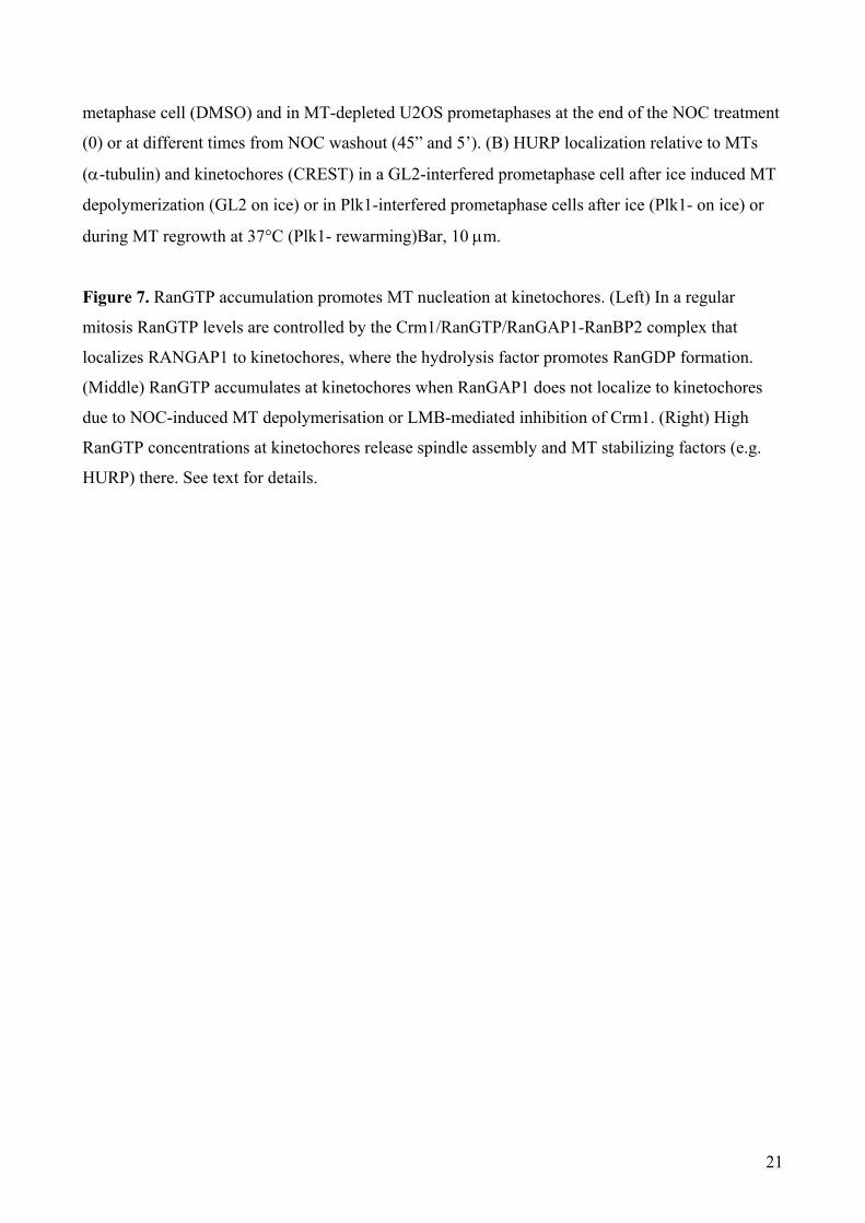
during MT regrowth at 37°C (Plk1- rewarming)Bar, 10 μm.
Figure 7. RanGTP accumulation promotes MT nucleation at kinetochores. (Left) In a regular
mitosis RanGTP levels are controlled by the Crm1/RanGTP/RanGAP1-RanBP2 complex that
localizes RANGAP1 to kinetochores, where the hydrolysis factor promotes RanGDP formation.
(Middle) RanGTP accumulates at kinetochores when RanGAP1 does not localize to kinetochores
due to NOC-induced MT depolymerisation or LMB-mediated inhibition of Crm1. (Right) High
RanGTP concentrations at kinetochores release spindle assembly and MT stabilizing factors (e.g.
HURP) there. See text for details.
21