Liposuction and Fat Graft to Enhance Facial Contour in Reconstructive Surgery - Nine Years...
14
2 Liposuction and Fat Graft to Enhance Facial Contour in Reconstructive Surgery - Nine Years Experience with the Use of Peridural Cannula Claudia Gutiérrez Gómez, Marcia Pérez Dosal and Alexander Cardenas Mejia Postgraduate Course in Plastic and Reconstructive Surgery Universidad Nacional Autonoma de México/General Hospital “Dr. Manuel Gea González “, México City Mexico 1. Introduction Correction of severe facial contour abnormalities still is a challenge to plastic surgeons. The aim of plastic surgical treatments is to restore a harmonious and symmetrical appearance. Some of the entities that cause this abnormalities include: Parry Romberg syndrome, lupus, Melkerson Rosenthal syndrome, Morphea, trauma’s sequel, embolization sequel, trauma, hemifacial microsomy, etc. (Gutiérrez “ et al”. 2007,2009). The free fat graft has been used since 1889, with the open ceiling technique, in 1893 Neuber recommended the use of fat grafts size lesser than an almond (Neuber 1893).In 1910 Lexer start the use of fat graft in aesthetic surgery and in 1925 reports the first case of facial contour reconstruction in a patient with Parry Romberg syndrome.(Lexer 1910) Peer reports lost of fat tissue as much as 50% (Peer 1950,1956) later it was used the fat obtained by liposuction ; absorption of the graft was the main problem and several different procedures have been described to minimize this phenomenon .Illouz in 1990 demonstrated that 80% of the injected fat graft was resorbed (Illouz 1990) .In 1994 it started the “ atraumatic purified” technique preconized by Coleman. Being the last one the one with better results in preserving volume because of a more viability of the adipose tissue and long lasting results. He recommends to avoid chopping, washing, manipulation, freezing, high negative pressure during extraction with a vacuum or high positive pressure during placement. Exposure to dry air will cause fat to desiccate rapidly.(Coleman 1995,1997,2002). Fat grafts collected by liposuction can be subcutaneously reinjected for correction of depressed or irregular areas. The live fat tissue is revascularized at the transplantation site within 48 hours,during which time it is fed by diffused materials from plasma. Explantation of adipose tissue as performed during the procedure of autologous fat transfer confers stress to preadipocytes and adipocytes. Disruption of blood supply during fat harvesting may result in hipoxia and apoptosis of the heterogeneous population of cells present in adipose tissue. Preadipocytes play an important role in soft tissue augmentation, because these adipocyte precursor cells have a higher survival rate under ischemic conditions than mature adipocytes and even have the ability to proliferate and differentiate into mature adipocytes. (Asken 1990;Guerrerosantos “et al”. 1996; Latoni “et al”.2000;Rieck & Schlaak 2003; Sadick &
-
Upload
independent -
Category
Documents
-
view
1 -
download
0
Transcript of Liposuction and Fat Graft to Enhance Facial Contour in Reconstructive Surgery - Nine Years...
2
Liposuction and Fat Graft to Enhance Facial Contour in Reconstructive Surgery - Nine Years
Experience with the Use of Peridural Cannula Claudia Gutiérrez Gómez,
Marcia Pérez Dosal and Alexander Cardenas Mejia Postgraduate Course in Plastic and Reconstructive Surgery Universidad Nacional Autonoma de México/General Hospital “Dr. Manuel Gea González “, México City
Mexico
1. Introduction Correction of severe facial contour abnormalities still is a challenge to plastic surgeons. The aim of plastic surgical treatments is to restore a harmonious and symmetrical appearance. Some of the entities that cause this abnormalities include: Parry Romberg syndrome, lupus, Melkerson Rosenthal syndrome, Morphea, trauma’s sequel, embolization sequel, trauma, hemifacial microsomy, etc. (Gutiérrez “ et al”. 2007,2009). The free fat graft has been used since 1889, with the open ceiling technique, in 1893 Neuber recommended the use of fat grafts size lesser than an almond (Neuber 1893).In 1910 Lexer start the use of fat graft in aesthetic surgery and in 1925 reports the first case of facial contour reconstruction in a patient with Parry Romberg syndrome.(Lexer 1910) Peer reports lost of fat tissue as much as 50% (Peer 1950,1956) later it was used the fat obtained by liposuction ; absorption of the graft was the main problem and several different procedures have been described to minimize this phenomenon .Illouz in 1990 demonstrated that 80% of the injected fat graft was resorbed (Illouz 1990) .In 1994 it started the “ atraumatic purified” technique preconized by Coleman. Being the last one the one with better results in preserving volume because of a more viability of the adipose tissue and long lasting results. He recommends to avoid chopping, washing, manipulation, freezing, high negative pressure during extraction with a vacuum or high positive pressure during placement. Exposure to dry air will cause fat to desiccate rapidly.(Coleman 1995,1997,2002). Fat grafts collected by liposuction can be subcutaneously reinjected for correction of depressed or irregular areas. The live fat tissue is revascularized at the transplantation site within 48 hours,during which time it is fed by diffused materials from plasma. Explantation of adipose tissue as performed during the procedure of autologous fat transfer confers stress to preadipocytes and adipocytes. Disruption of blood supply during fat harvesting may result in hipoxia and apoptosis of the heterogeneous population of cells present in adipose tissue. Preadipocytes play an important role in soft tissue augmentation, because these adipocyte precursor cells have a higher survival rate under ischemic conditions than mature adipocytes and even have the ability to proliferate and differentiate into mature adipocytes. (Asken 1990;Guerrerosantos “et al”. 1996; Latoni “et al”.2000;Rieck & Schlaak 2003; Sadick &
Advanced Techniques in Liposuction and Fat Transfer 36
Hudgins 2001.) Easy of technique, unlikelihood of scar formation, low morbidity, and low cost have increased the popularity of this operation. Fat grafts collected by liposuction can be subcutaneously reinjected for correction of depressed or irregular areas. Fat should be harvested as an intact tissue small enough to pass through a small- lumen cannula, eliminating the need to later reduce the size of the parcel of fat by washing, chopping or straining. To obtain predictable results harvested subcutaneous tissue should be refined so the material infiltrated is mainly viable fatty tissue; via centrifugation, oil, blood, water and extracellular components should be removed without causing significant damage to the fat to be transplanted (Coleman 2001).
2. Patients and methods; Patient data During the last nine years we have been injecting the fat graft with a peridural cannula in 73 patients for fat graft in the face.With ages from 5 to 61 years old, with a media of 28.3 years. They were females in 75.4% (55 cases) and male in 24.6% (18 cases). The ethiology of the deformities were Parry Romberg Syndrome 71.2% (52 cases), Morphea in 6 cases (8.2%), trauma sequel 4 cases (5.4%), hemifacial microsomia, lupus and post tumor resection sequel in 9 cases (3 each group; 4.1% each). Depression after embolization of vascular anomaly 1 case (1.3%), Number 7 facial cleft 1 case (1.3%).With a total of 132 procedures realized (about 1.8 per patient). Table 1. ETHIOOGYL # CASES % PARRY ROMBERG 52 71.2 MORPHEA 6 8.2 TRAUMA SEQUEL 4 5.4 HEMIFACIAL MICROSOMIA 3 4.1
TUMOR RESECTION SEQUEL 3 4.1 LUPUS 3 4.1 EMBOLIZATION SEQUEL 1 1.3 7 FACIAL CLEFT 1 1.3
Table 1. Etiology of facial contour deformity in 73 cases.
3. Surgical technique All the procedures were done under general anesthesia and meticulous sterile technique. The donor sites were abdomen and flanks in all patients.The donor sites were infiltrated with lidocaine 0.5% with 1:100,000 epinephrine in a Ringer’s lactate solution; in a ratio of 1 ml of solution for each cubic centimeter of fat harvested using a blunt cannula for infiltration. After 10 minutes we use a two holed cannula with blunt tip (shaped like a bucket handle), the other end of the cannula is attached to a 10cc syringe. The distal openings of the harvesting cannula are the same size as the entrance lumen of the syringe to avoid damage of the fatty tissue. The plunger of the syringe is gently manipulated to provide about 1 or 2 cc of negative pressure space in the barrel of the syringe while the cannula is pushed through the harvest site. After the fat has been harvested, the cannula is removed from the syringe and replaced with a plug which is twisted on to create a seal to
Liposuction and Fat Graft to Enhance Facial Contour in Reconstructive Surgery - Nine Years Experience with the Use of Peridural Cannula 37
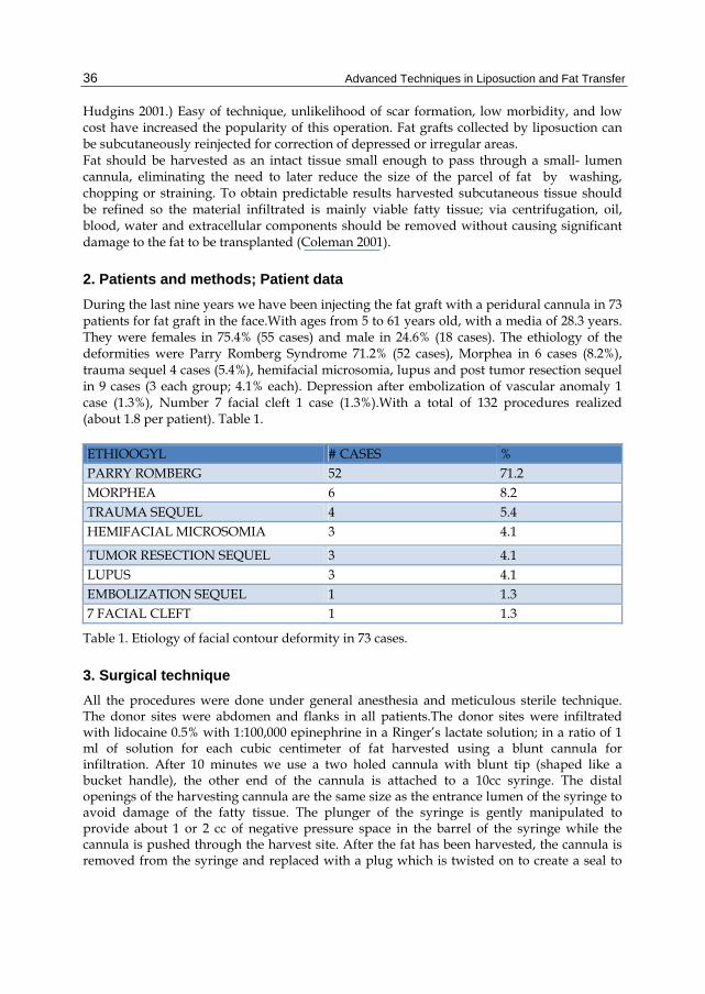
prevent spillage during the centrifuging process. The plunger is removed from the proximal end of the syringe. Then the syringes are centrifuged at 3000 rpm for 3 minutes. The upper oil is discharged, and also the lower portion (composed by blood, water and lidocaine). Then the middle portion of the syringe which is composed primarily of potentially viable parcels of fat tissue is transfered to 1cc syringes with a disposable three lines key , with a gentle aspiration from the 1 cc syringes. The recipient areas are not infiltrated to avoid deformity of the recipient areas. Only the sites were the peridural cannula will be placed are infiltrated with 0.5% lidocaine with 1:200,000 epinephrine with a 27 gauge needle, incisions 1 or 2 mm long are made with a No. 15 Bard Parker blade. The incisions will be placed depending of the areas to be injected 1 cm inside the scalp ( for forehead), in the external canthus, below the lobule, lip commissure, alar base ( for cheek, lip and chin), in nasion for the nose.The fat transfer is done with a peridural cannula ( 18G BD Tuohy 17g x 89mm). Although it has a blunt point (Huber-Tuohy-Hustead point). The bevel’s sharp point of the peridural cannula is unsharpened as shown in figure 1.
Fig. 1. Peridural cannula ( 18G BD Tuohy 17g x 89mm).Although it has a blunt point (Huber-Tuohy-Hustead point) The bevel’s sharp point of the peridural cannula is unsharpened on the lateral side of Adson forceps’ handle.
The adipocytes are deposited in crossing lines in the desired areas being left during the take out of the cannula. The patient is discharged from the hospital 24 hours later with ketorolac in case of pain, amoxicillin clavulanic acid for seven days and cold for 2-3 days. The patients come to control every three months the first year and new lipoinjection if needed is realized 12 months after de last procedure, if they do not need more volume they are seen every 6 months for 5 years.
4. Results Most of the patients had not had previous treatments 65 cases (89%), except 8 (10.9%).A total of 132 procedures realized in the first group about 1.8 per patient; in the second group (previous treated patients) 8 patients, had previous microsurgical corrections with 41 previous surgical procedures in this group about 5 procedures per patients . We injected 4755 cc fat tissue which represent about 65.4 cc per patient. The follow up was between 1 and 8 years. Complications the most common was under correction in 14 cases (19.1%), visible irregularities 5 (6.8%): oral mucosa perforation 2 (2.7%), granuloma 1 (1.3%) fat migration 2 (2.7%). We present some representative cases with long follow up.
Advanced Techniques in Liposuction and Fat Transfer 38
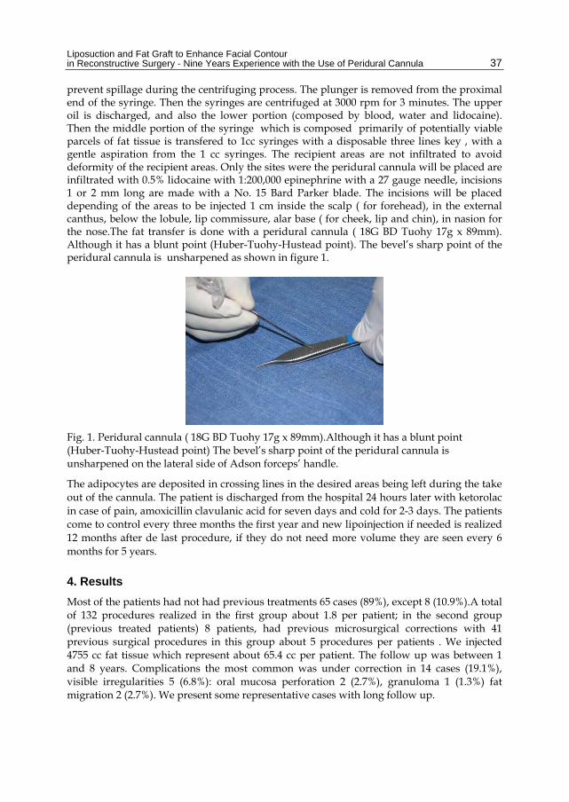
5. Case reports 5.1 Case 1
Fig. 2. 23 year-old girl had Morphea left side of the face, preoperative front, ¾ and lateral left views.
Fig. 3. Postoperative frontal ¾ and lateral left views after two lipoinjection procedures. Twelve months after the last one.

Liposuction and Fat Graft to Enhance Facial Contour in Reconstructive Surgery - Nine Years Experience with the Use of Peridural Cannula 39
Fig. 4. Postoperative frontal, left ¾ and lateral views after 6 lipoinjection with a total volume of 163 cc 3 years after the last one. We can see that she increased her body weight in last years with proportional increase of the transplanted fat tissue.
5.2 Case 2
Fig. 5. 30 year old woman severe bilateral cheek atrophy secondary to discoid Lupus. Preoperative front ¾ right ,3/4 left and basal views.
Advanced Techniques in Liposuction and Fat Transfer 40
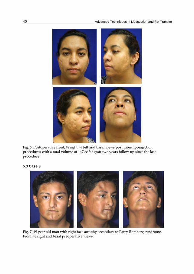
Fig. 6. Postoperative front, ¾ right, ¾ left and basal views post three lipoinjection procedures with a total volume of 147 cc fat graft two years follow up since the last procedure.
5.3 Case 3
Fig. 7. 19 year old man with right face atrophy secondary to Parry Romberg syndrome. Front, ¾ right and basal preoperative views.
Liposuction and Fat Graft to Enhance Facial Contour in Reconstructive Surgery - Nine Years Experience with the Use of Peridural Cannula 41
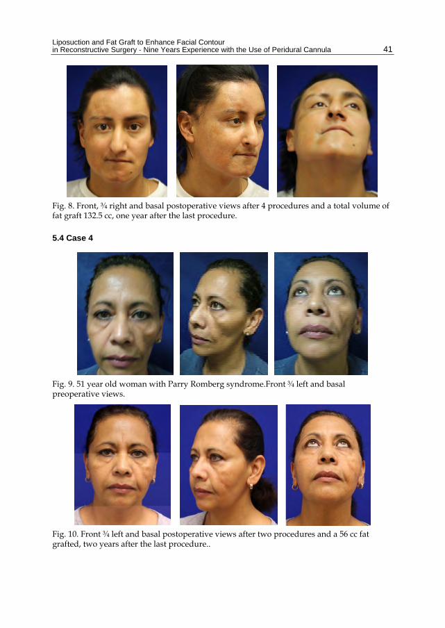
Fig. 8. Front, ¾ right and basal postoperative views after 4 procedures and a total volume of fat graft 132.5 cc, one year after the last procedure.
5.4 Case 4
Fig. 9. 51 year old woman with Parry Romberg syndrome.Front ¾ left and basal preoperative views.
Fig. 10. Front ¾ left and basal postoperative views after two procedures and a 56 cc fat grafted, two years after the last procedure..
Advanced Techniques in Liposuction and Fat Transfer 42
5.5 Case 5
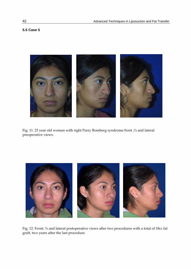
Fig. 11. 23 year old woman with right Parry Romberg syndrome front ,¾ and lateral preoperative views.
Fig. 12. Front, ¾ and lateral postoperative views after two procedures with a total of 18cc fat graft, two years after the last procedure.

Liposuction and Fat Graft to Enhance Facial Contour in Reconstructive Surgery - Nine Years Experience with the Use of Peridural Cannula 43
5.6 Case 6
Fig. 13. 6 year old girl with left Hemifacial microsomia , front , and ¾ preoperative views.
Fig. 14. Front and ¾ postoperative views after 3 lipoinjection procedures with a total 46 cc fat graft 3 years after the last lipoinjection.
Advanced Techniques in Liposuction and Fat Transfer 44
5.7 Case 7
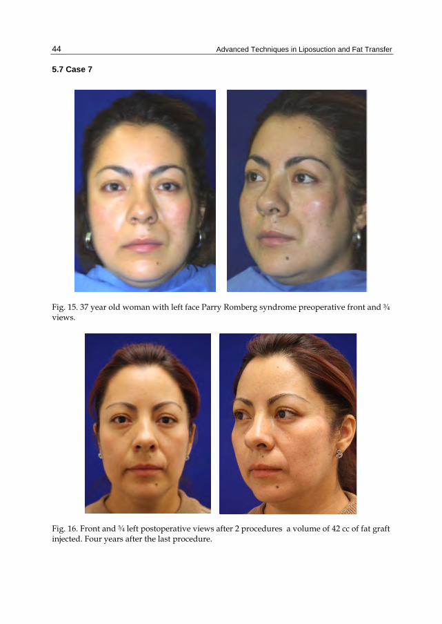
Fig. 15. 37 year old woman with left face Parry Romberg syndrome preoperative front and ¾ views.
Fig. 16. Front and ¾ left postoperative views after 2 procedures a volume of 42 cc of fat graft injected. Four years after the last procedure.
Liposuction and Fat Graft to Enhance Facial Contour in Reconstructive Surgery - Nine Years Experience with the Use of Peridural Cannula 45
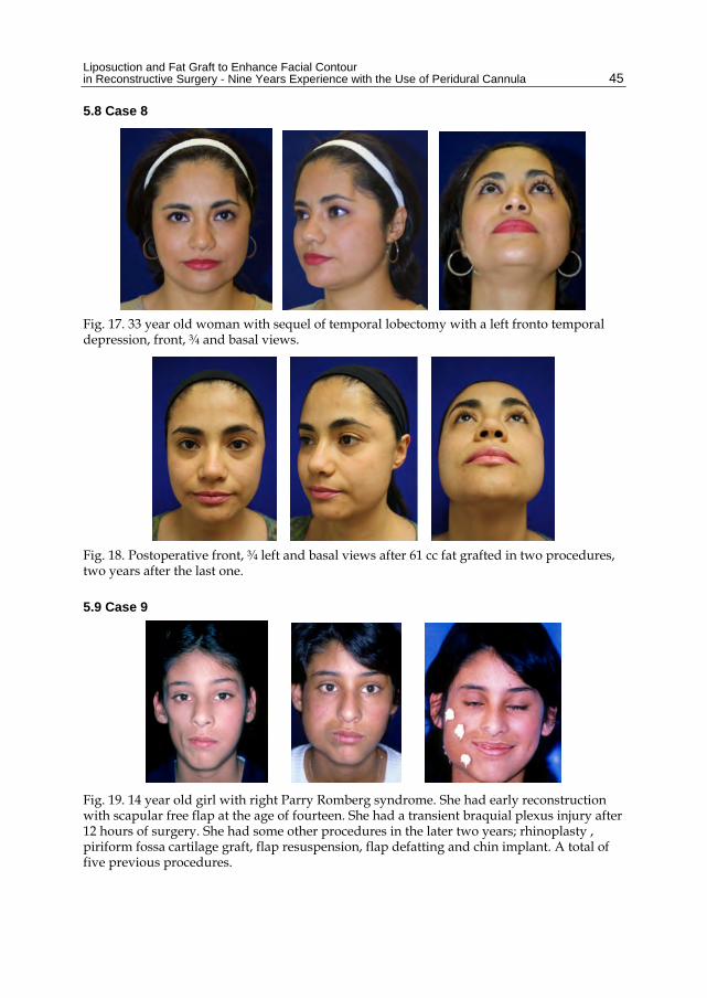
5.8 Case 8
Fig. 17. 33 year old woman with sequel of temporal lobectomy with a left fronto temporal depression, front, ¾ and basal views.
Fig. 18. Postoperative front, ¾ left and basal views after 61 cc fat grafted in two procedures, two years after the last one.
5.9 Case 9
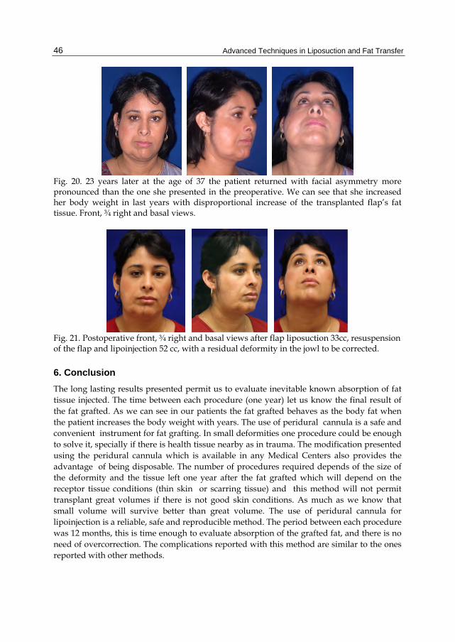
Fig. 19. 14 year old girl with right Parry Romberg syndrome. She had early reconstruction with scapular free flap at the age of fourteen. She had a transient braquial plexus injury after 12 hours of surgery. She had some other procedures in the later two years; rhinoplasty , piriform fossa cartilage graft, flap resuspension, flap defatting and chin implant. A total of five previous procedures.
Advanced Techniques in Liposuction and Fat Transfer 46
Fig. 20. 23 years later at the age of 37 the patient returned with facial asymmetry more pronounced than the one she presented in the preoperative. We can see that she increased her body weight in last years with disproportional increase of the transplanted flap’s fat tissue. Front, ¾ right and basal views.
Fig. 21. Postoperative front, ¾ right and basal views after flap liposuction 33cc, resuspension of the flap and lipoinjection 52 cc, with a residual deformity in the jowl to be corrected.
6. Conclusion The long lasting results presented permit us to evaluate inevitable known absorption of fat tissue injected. The time between each procedure (one year) let us know the final result of the fat grafted. As we can see in our patients the fat grafted behaves as the body fat when the patient increases the body weight with years. The use of peridural cannula is a safe and convenient instrument for fat grafting. In small deformities one procedure could be enough to solve it, specially if there is health tissue nearby as in trauma. The modification presented using the peridural cannula which is available in any Medical Centers also provides the advantage of being disposable. The number of procedures required depends of the size of the deformity and the tissue left one year after the fat grafted which will depend on the receptor tissue conditions (thin skin or scarring tissue) and this method will not permit transplant great volumes if there is not good skin conditions. As much as we know that small volume will survive better than great volume. The use of peridural cannula for lipoinjection is a reliable, safe and reproducible method. The period between each procedure was 12 months, this is time enough to evaluate absorption of the grafted fat, and there is no need of overcorrection. The complications reported with this method are similar to the ones reported with other methods.
Liposuction and Fat Graft to Enhance Facial Contour in Reconstructive Surgery - Nine Years Experience with the Use of Peridural Cannula 47
7. Discussion Free flaps has been considered as the gold standard for reconstruction in great defects (Inigo “et al”. 1993;Wojcicki & Zachara 2011;Yu-Feng “et al”. 2008). Unfortunately the long follow up as case 9 demonstrate that the behavior of the free flaps is more unpredictable than fat graft due to the gravitational force and the disproportional volume increase in relation to body weight changes. And the different match color when skin is required. Case one illustrates a great defect which was solved satisfactory with fat graft and when the patient increases her body weight the volumes is proportional to the normal side.
8. Acknowledgment Residents Xitlali Baron, Jordi Espel, Fernando Francis, Roberto López and Guillermo Sánchez for their help in taking care of the patients.
9. References Asken, S. (1990). Microliposuction and autologous fat transplantation for aesthetic
enhacement of the aging face. J. Dermatol Surg Oncol, Vol.16, pp. 965-972, ISBN 0148-0812
Coleman, SR. (1995). Long-term survival of fat transplants: controlled demonstrations. Aesthetic Plast Surg 1995, Vol.19, pp.421-425, ISSN 1432-5241
Coleman, SR. (1997). Facial recontourig with lipostructure. Clin Plast Surg, Vol. 24, pp. 347-367, ISSN 1558-0504
Coleman, SR. (2001). Structural fat grafts: the ideal filler. Clin Plast Surg, Vol.28, (January 2001), pp. 111-119, ISSN 1558-0504
Coleman, SR. (2002). Hand rejuvenation with structural fat grafting. Plast Reconstr Surg Vol.110, (December 2002), pp.1731-1745, ISSN 0032-1052
Guerrerosantos, J., González-Mendoza, A., & Masmela, Y. (1996). Long-term survival of free fat graft in muscle: an experimental study in rats. Aesthetic Plast Surg. Vol.20, (September 1996), pp. 403-408, ISSN 1432-5241
Gutiérrez C, Hayakawa V, Franco A, Reyes L.(2007)Lipoinyección para reconstrucción del contorno facial en S. de Parry Romberg, esclerodermia y secuelas de trauma. Una alternativa práctica utilizando cánulas para bloqueo peridural.Rev Cir Plást ; 17 ( 3): 168-175 ,ISSN 1405-0625
Gutierrez C y Cols. (2009)Corrección de deformidad en olán en S. Melkerson Rosenthal. Cirplástiberolatinoam Vol 35 No. 1 enero-marzo pp79-84,ISSN 1405-0625
Illouz, Y.G. (1990). Fat Injection: A four year clinical trial in lipoplasty, In: The theory and practice of blunt suction lipectomy, Brown, pp. 148-152, Little,Boston
Inigo, F., Rojo, P., & Ysunza, A. (1993). Aesthetic treatment of Romberg’s disease: experience with 35 cases. Br JPlastSurg, Vol.46, No.3, (April 1993), pp.194-200, ISSN 0007-1226
Latoni, J.D., Marshall, D.M., & Wolfe, S.A. (2000). Overgrowth of fat autotransplanted for correction of localized steroid-induced atrophy. Plast Reconstr Surg, Vol.106, (December 2000), pp. 1566-1569, ISSN 1529-4242
Peer, L.A.(1946) Plast Reconstr Surg 1955 Sep;16(3):161-8. Cell survival theory versus replacement theory. ISSN 1075-1270
Advanced Techniques in Liposuction and Fat Transfer 48
Peer, L.A. (1956). The neglected free fat graft its behavior and clinical use. Am J Surg Vol.92, No.1, (July 1956), pp.40-47, ISSN 1879-1883
Rieck, B., & Schlaak, S. (2003). Measurement in vivo of the survival rate in autologous adipocyte transplantation. Plast Reconstr Surg, Vol.11, (June 2003), pp.2315-2323, ISSN 1529-4242
Sadick, N.S., & Hudgins, L.C. (2001). Fatty acid analysis of transplanted adipose tissue. Arch Dermatol, Vol.137, (June 2001), pp. 723-727, ISSN1538-3652
Wojcicki, P., & Zachara, M. Treatment of Patients with Parry-Romberg Syndrome. Ann of Plast Surg, Vol.66, No.3, (March 2011), pp. 267-72, ISSN1536-3708
Yu-Feng, L., Lai, G., & Zhi-Yong, Z. (2008). Combined Treatments of Facial Contour Deformities Resulting from Parry –Romberg Syndrome, J Reconstr Microsurg, Vol.24, No.5, (July 2008), pp. 333-342, ISSSN 1098-8947