Ligand-induced folding of the guanine-sensing riboswitch is controlled by a combined predetermined...
11
Ligand-induced folding of the guanine-sensing riboswitch is controlled by a combined predetermined–induced fit mechanism OTMAR M. OTTINK, 1 SUMIENTRA M. RAMPERSAD, 1 MARCO TESSARI, 1 GUIDO J.R. ZAMAN, 2 HANS A. HEUS, 1 and SYBREN S. WIJMENGA 1 1 Institute for Molecules and Materials and Department of Biophysical Chemistry, University of Nijmegen, 6525 ED Nijmegen, The Netherlands 2 Molecular Pharmacology Unit, N.V. Organon, 5340 BH Oss, The Netherlands ABSTRACT All known guanine-sensing riboswitches regulate gene expression by specifically binding to guanine (G) or related analogs with high affinity to switch off transcription. The aptamers of this class of riboswitches are characterized by three helices (P1–P3), surrounding a central core of phylogenetically conserved nucleotides and a long-range loop–loop interaction. To gain more insight into the switching mechanism, we present here a comparison between the solution-state structures of the G-free and G-bound forms of the guanine aptamer from the xpt-pbuX operon of Bacillus subtilis, as derived from NMR chemical shifts and magnetic-field-induced residual dipolar couplings. The high-resolution NMR analysis shows the G-free aptamer is highly structured with parallel P2 and P3 helices and the long-range loop–loop interaction already present, implying that the structure is largely preformed to bind the ligand. Structural changes upon guanine binding are found to be localized to the central core. In the free state, the G-quadruple interaction and two base pairs of the P1 stem flanking the central core appear to be largely disordered. The ligand thus binds via a combined predetermined–induced fit mechanism, involving a previously unstructured five-residue loop of the J2–3 junction that folds over the ligand. These limited additional interactions within a preorganized setting possibly explain how the aptamer rapidly responds to ligand binding, which is necessary to switch the structural state of the expression platform within a narrow time frame before the RNA polymerase escapes the 59-UTR. Keywords: riboswitch; aptamer; RNA structure; RNA folding; NMR; mRNA; gene regulation INTRODUCTION Riboswitches are RNA control elements, found in the 59- or 39-untranslated regions (UTRs) and introns of many mRNAs of bacteria and some plant and fungi, which modulate gene expression without the need of protein cofactors (Mandal and Breaker 2004b; Nudler and Mironov 2004; Soukup and Soukup 2004). These riboswitches contain highly structured domains, which target specific metabolites, resulting in metabolite-induced conforma- tional transitions that ultimately regulate the cellular levels of the same metabolite. So far, riboswitches have been described that control the cellular levels of purines, vita- mins, coenzymes, and amino acids. Compounds for which riboswitches have been found are thiamin pyrophosphate (TPP) (Mironov et al. 2002; Winkler et al. 2002a), guanine (Mandal et al. 2003), adenine (Mandal et al. 2003; Mandal and Breaker 2004a), coenzyme B 12 (Nahvi et al. 2002), flavin mononucleotide (FMN) (Mironov et al. 2002; Winkler et al. 2002b), glucosamine-6-phosphate (GlcN6P) (Winkler et al. 2004), glycine (Mandal et al. 2004), lysine (Grundy et al. 2003; Rodionov et al. 2003; Sudarsan et al. 2003), and S-adenosylmethionine (SAM) (Epshtein et al. 2003; McDaniel et al. 2003; Winkler et al. 2003; Fuchs et al. 2006). Riboswitches consist of two modular domains: an aptamer domain, which contains the high-affinity binding site for a given metabolite or ligand, connected to a downstream region termed the expression platform, which can exist in two mutually exclusive conformational states. At sufficient concentrations, binding of the ligand stabilizes the structure of the aptamer domain, which pre- sumably results in switching the conformational state of the expression platform to regulate a certain gene expression system. While the aptamer sequences are largely conserved Reprint requests to: Hans A. Heus, Institute for Molecules and Materials and Department of Biophysical Chemistry, University of Nijmegen, Toernooiveld 1, 6525 ED Nijmegen, The Netherlands; e-mail: [email protected]; fax: 31-24-362112. Article published online ahead of print. Article and publication date are at http://www.rnajournal.org/cgi/doi/10.1261/rna.635307. 2202 RNA (2007), 13:2202–2212. Published by Cold Spring Harbor Laboratory Press. Copyright Ó 2007 RNA Society.
-
Upload
independent -
Category
Documents
-
view
1 -
download
0
Transcript of Ligand-induced folding of the guanine-sensing riboswitch is controlled by a combined predetermined...

Ligand-induced folding of the guanine-sensing riboswitch
is controlled by a combined predetermined–inducedfit mechanism
OTMAR M. OTTINK,1 SUMIENTRA M. RAMPERSAD,1 MARCO TESSARI,1 GUIDO J.R. ZAMAN,2
HANS A. HEUS,1 and SYBREN S. WIJMENGA1
1Institute for Molecules and Materials and Department of Biophysical Chemistry, University of Nijmegen, 6525 ED Nijmegen, The Netherlands2Molecular Pharmacology Unit, N.V. Organon, 5340 BH Oss, The Netherlands
ABSTRACT
All known guanine-sensing riboswitches regulate gene expression by specifically binding to guanine (G) or related analogs withhigh affinity to switch off transcription. The aptamers of this class of riboswitches are characterized by three helices (P1–P3),surrounding a central core of phylogenetically conserved nucleotides and a long-range loop–loop interaction. To gain moreinsight into the switching mechanism, we present here a comparison between the solution-state structures of the G-free andG-bound forms of the guanine aptamer from the xpt-pbuX operon of Bacillus subtilis, as derived from NMR chemical shiftsand magnetic-field-induced residual dipolar couplings. The high-resolution NMR analysis shows the G-free aptamer is highlystructured with parallel P2 and P3 helices and the long-range loop–loop interaction already present, implying that the structureis largely preformed to bind the ligand. Structural changes upon guanine binding are found to be localized to the central core. Inthe free state, the G-quadruple interaction and two base pairs of the P1 stem flanking the central core appear to be largelydisordered. The ligand thus binds via a combined predetermined–induced fit mechanism, involving a previously unstructuredfive-residue loop of the J2–3 junction that folds over the ligand. These limited additional interactions within a preorganizedsetting possibly explain how the aptamer rapidly responds to ligand binding, which is necessary to switch the structural state ofthe expression platform within a narrow time frame before the RNA polymerase escapes the 59-UTR.
Keywords: riboswitch; aptamer; RNA structure; RNA folding; NMR; mRNA; gene regulation
INTRODUCTION
Riboswitches are RNA control elements, found in the 59-or 39-untranslated regions (UTRs) and introns of manymRNAs of bacteria and some plant and fungi, whichmodulate gene expression without the need of proteincofactors (Mandal and Breaker 2004b; Nudler and Mironov2004; Soukup and Soukup 2004). These riboswitchescontain highly structured domains, which target specificmetabolites, resulting in metabolite-induced conforma-tional transitions that ultimately regulate the cellular levelsof the same metabolite. So far, riboswitches have beendescribed that control the cellular levels of purines, vita-mins, coenzymes, and amino acids. Compounds for which
riboswitches have been found are thiamin pyrophosphate(TPP) (Mironov et al. 2002; Winkler et al. 2002a), guanine(Mandal et al. 2003), adenine (Mandal et al. 2003; Mandaland Breaker 2004a), coenzyme B12 (Nahvi et al. 2002),flavin mononucleotide (FMN) (Mironov et al. 2002;Winkler et al. 2002b), glucosamine-6-phosphate (GlcN6P)(Winkler et al. 2004), glycine (Mandal et al. 2004), lysine(Grundy et al. 2003; Rodionov et al. 2003; Sudarsan et al.2003), and S-adenosylmethionine (SAM) (Epshtein et al.2003; McDaniel et al. 2003; Winkler et al. 2003; Fuchset al. 2006). Riboswitches consist of two modular domains:an aptamer domain, which contains the high-affinitybinding site for a given metabolite or ligand, connectedto a downstream region termed the expression platform,which can exist in two mutually exclusive conformationalstates. At sufficient concentrations, binding of the ligandstabilizes the structure of the aptamer domain, which pre-sumably results in switching the conformational state of theexpression platform to regulate a certain gene expressionsystem. While the aptamer sequences are largely conserved
Reprint requests to: Hans A. Heus, Institute for Molecules andMaterials and Department of Biophysical Chemistry, University ofNijmegen, Toernooiveld 1, 6525 ED Nijmegen, The Netherlands; e-mail:[email protected]; fax: 31-24-362112.
Article published online ahead of print. Article and publication date areat http://www.rnajournal.org/cgi/doi/10.1261/rna.635307.
2202 RNA (2007), 13:2202–2212. Published by Cold Spring Harbor Laboratory Press. Copyright � 2007 RNA Society.

to ensure specific binding to a given metabolite in variousorganisms, the expression platforms vary in sequence tocontrol gene expression using different mechanisms, mostlyby either modulating the formation of a transcription ter-minator or by sequestering a Shine–Dalgarno sequence toprevent translation initiation. Besides these two dominantregulating mechanisms, riboswitches have also been foundthat regulate through self-cleavage of the mRNA by ribo-zyme activity (Winkler et al. 2004).
The purine riboswitches are among the smallest ribo-switches known, contain one of the most phylogeneticallyconserved aptamers described to date, and hence are theones studied most extensively by structural biology andbiophysical means (Batey et al. 2004; Serganov et al. 2004;Noeske et al. 2005, 2007; Wickiser et al. 2005; Gilbert et al.2006; Lemay et al. 2006). The aptamer part of the Bacillussubtilis xpt-pbuX-mRNA guanine riboswitch, which is thesubject of this study and is representa-tive of all known purine riboswitches,folds into a three-way junction, thearms of which are designated P1, P2,and P3 connected by three interhelicalloops, J1–2, J2–3, and J3–1 (Fig. 1). Inthe crystal structures of the ligand–riboswitch complexes (Batey et al.2004; Serganov et al. 2004), the P2 andP3 helices are parallel and closelypacked together by two base quadruplesand a noncanonical base pair betweenthe apical P2 and P3 loops. The purineis buried within the central core byforming a base quadruple and is furtherclosed in by two base triples from aboveand two base triples from below. Thetwo upper base triples also help toanchor the P2 and P3 stems. The twolower base triples also pin down theJ2–3 loop to the top of P1, closing in theguanine at the N3/N9 side.
Biochemical studies provided evidencethat the long-range loop–loop interac-tion forms in the absence of ligand and isessential for ligand binding. In the in-line probing experiments, the apicalstem–loop regions of P2 and P3 wereprotected from cleavage and showed nodifference in the nucleotide protectionpatterns in the presence or absence ofligand (Mandal et al. 2003). Destroyingthe loop–loop interaction by replacingthe wild-type apical P2 and P3 loops byUUCG, which are incapable of interact-ing, abolishes the capacity of the aptamerto recognize the ligand (Batey et al.2004). NMR studies showed the pre-
existence of base pairs between P2 and P3 loop residueswhen no ligand is present (Noeske et al. 2007). These resultsare in accordance with the presence of a partially pre-organized aptamer structure in the absence of ligand.
On the other hand, it has also been noted that the ligand-binding site has to undergo a substantial conformationalchange upon binding because the ligand cannot access atightly preformed binding site as observed in the cocrystalstructures (Batey et al. 2004; Serganov et al. 2004). Thisimplies an induced-fit model, which is supported by in-lineprobing experiments, NMR studies, and thermodynamic data.In-line probing experiments of the unbound RNA showedseveral cleavage products mapping to the single-strandednucleotides of the three-way junction that are protectedin the presence of excess guanine, suggesting a locallydisordered binding pocket in the free state (Mandal et al.2003). NMR imino proton spectra showed additional peaks
FIGURE 1. (A) Secondary structure of the G-riboswitch aptamer (GRA) used for the NMRstudy. Highly conserved residues are shaded in gray. Deviations from the natural sequencefound in the Bacillus subtilis xpt-pbuX-mRNA gene, which have also been introduced forobtaining the crystal structure of the GRA in complex with guanine (Serganov et al. 2004), areshown in the boxed regions. C74, which confers specificity for guanine, is highlighted with acircle. (B) Gene regulation by the guanine riboswitch in the 59-UTR of mRNA. Initialtranscription generates the aptamer-binding domain, which can be preformed to bind guaninerapidly at sufficient concentration. In the absence of guanine, an antiterminator element isformed, which allows transcription to proceed (left). Binding of guanine is expected to stabilizethe guanine binding domain and P1 stem, which is believed to force formation of theterminator element that stops transcription (right).
Ligand-induced folding of a guanine riboswitch
www.rnajournal.org 2203

upon ligand binding without significant shifts of pre-existing peaks, indicative of a limited rearrangement ofthe global structure (Serganov et al. 2004). Thermodynamicmeasurements for ligand binding revealed unfavorableentropy values compensated by favorable enthalpy values,also in accordance with a local folding event (Gilbert et al.2006).
Thus, given the available data, the prevailing view is thatthe aptamer is partially preorganized by the stems andlong-range loop–loop interaction, while the junction andsurrounding regions have to be disordered to allow bindingof the ligand. This structural model forms the current basisfor some of the remaining key questions that cannot beanswered with the high-resolution crystal structures of theaptamer–ligand complexes alone: to what extent is theaptamer folded at the time of switching, and how is bindingof the ligand translated into a conformational switch of theexpression platform leading to transcription termination.An important aspect in evaluating this model is thatriboswitch control is tightly coupled to transcription (Fig.1B). In this process, the aptamer is synthesized first and iscapable of folding independently before the expressionplatform is generated. Ligand binding has to be transmittedto the downstream expression platform region while it isbeing synthesized by the elongating polymerase before itpasses through the 59-UTR. This leaves a very narrow timeframe of only a few seconds for the polymerase to choosebetween elongation and termination (Wickiser et al. 2005),depending on the structural state of the expression plat-form it encounters. For the xpt-pbuX G-riboswitch inparticular, this entails residues 78–82 (Fig. 1B), whicheither close the P1 stem, permitting formation of thetranscription terminator, or interact with a downstreamregion to form an antiterminator. Thus, ligand binding andsubsequent closure of the core region and P1 stem has to beconsiderably fast, otherwise the antiterminator stem–loopstructure is able to form, which is unlikely to switch backwithin the given time, since structural rearrangementsinvolving unfolding/refolding stretches of RNA helicescan be notably slow (e.g., Nagel et al. 2002). Overall, thismodel indicates the importance of a discrete thermody-namic and kinetic balance between formation of theremainder of the aptamer structure and the antiterminator.Therefore, the degree of structural organization of theaptamer is a very important consideration in understand-ing the mechanism of riboswitching.
To investigate the degree of structural organization, weobtained global and atomic detail structural information insolution on the G-riboswitch aptamer in its guanine boundand free states. We show that Mg2+ is required to drive thefree aptamer in a single conformational state. High-qualityNMR spectra were obtained for both states in the presenceof Mg2+, allowing complete assignment of the iminoprotons’ resonances, which are monitors of base hydrogenbond interactions and part of the non-exchangeable pro-
tons. In addition, by using magnetic-field-induced residualdipolar couplings (mRDCs) from imino N–H and adenineC2–H2 bonds, we show that there are no significant globaldifferences between the structures of both states. Moreover,the changes in base pairing and short NOE-derived dis-tances, together with the mRDCs, defined on a residue-specific basis could be used to propose a model for theextent and location of the conformational changes in theG-riboswitch aptamer upon guanine binding. This infor-mation can be used to provide further insight into themolecular mechanism of riboswitching.
RESULTS AND DISCUSSION
Mg2+ ions are required for proper folding of theguanine free G-riboswitch aptamer
The 73-mer G-riboswitch aptamer (GRA) sequence inves-tigated in the work presented here (Fig. 1) is derived fromthe B. subtilis xpt-pbuX operon (Mandal et al. 2003). A fewstructurally silent mutations were introduced to stabilizethe P2 helical region for the NMR studies (Noeske et al.2005), i.e., P2 mutations U25C, C26U, and A45G. Addi-tional guanosine residues were added to the 59 end (G12–G14) needed for efficient in vitro transcription and closedby additional cytosines (C82–C84) added at the 39 end toprevent possible RNA aggregation due to intermolecularinteractions of single-stranded G-stretches.
Initially the 1D imino proton spectra of the guanine freeGRA were recorded in 10 mM sodium phosphate buffer,at pH 6.7. However, under these buffer conditions, theresulting spectra were rather crowded, showing many res-onances with different linewidths (Fig. 2), suggestingmultiple conformations that prevented complete resonanceassignment. Therefore, we next tried to improve the spectraby dialyzing magnesium chloride into the sample. It is wellknown that magnesium ions are necessary for the tertiarystructure formation of many RNA sequences (Misra andDraper 1998). Furthermore, it has been shown that Mg2+
ions are required for guanine binding (Mandal et al. 2003).Also, in the crystal structures divalent ions seem to play astabilizing role in the interaction between the P2 and P3helices, by neutralizing unfavorable electrostatic interac-tions (Batey et al. 2004; Serganov et al. 2004). Indeed, thespectra were greatly simplified, as demonstrated by acomparison of the imino proton spectra without Mg2+
and with 5 mM Mg2+ presented in Figure 2.In the absence of Mg2+ ions, the Watson–Crick base pair
region between 12 and 15 ppm shows many overlappingresonances with different linewidths and intensities, whichchanges into a well-resolved spectrum with many fewerresonances and of similar width after dialysis into 5 mMMg2+. In the non-Watson–Crick base pair region of thespectrum, i.e., between 9 and 12 ppm, the difference is evenmore profound, showing at least six out of eight resonances to
Ottink et al.
2204 RNA, Vol. 13, No. 12

disappear between 10 and 11.5 ppm. The crowded spectrumof the free GRA in the absence of Mg2+ ions yielding manyadditional resonances with different linewidths is indicative ofmultiple species with different dynamic properties. This likelyrepresents a mixture between folded and unfolded states,lacking the long-range loop–loop interaction as demonstratedfor the adenine-sensing riboswitch aptamer by a single-molecule FRET study (Gilbert and Batey 2006; Lemay et al.2006). Apparently magnesium ions are required to force thefree GRA into a single stable conformation, illustrating theirstructural and biological importance.
Resonance assignment and solution structureof the GRA–guanine complex
We next recorded NMR spectra of the GRA–guaninecomplex in the presence of 5 mM Mg2+. The quality ofthe NMR spectra is excellent for a molecule of this size,showing many well-resolved imino proton resonances withsmall linewidths that allow for nearly complete resonanceassignment. Imino protons were assigned (Fig. 3) usingstandard assignment protocols (Wijmenga and van Buuren1998), involving sequential imino–imino NOE contactsassigned to residue type by separation of U and G iminoresonances in 15N HMQC spectra. To resolve some ambig-uous NOE contacts involving overlapping resonances, boththe P2 and P3 stem–loops were also synthesized and inves-tigated by NMR separately (O.M. Ottink, S.M. Rampersad,M. Tessari, H.A. Heus, and S.S. Wijmenga, unpubl.).
All stem imino protons could be assigned, indicatingformation of all base pairs in the three stems with theexception of the closing G12C84 pair, in which the iminoproton is not observable due to terminal fraying. The other
resonances thus originate from hydrogen-bonded iminoprotons outside the stem regions, of which several couldbe assigned to residues within the junction loops. U22 andG46, which form U22A52 and G46C53 base pairs in thecocrystal structure, are also base paired in the solutionstructure and form a continuous stack on P1, as shownby the chemical shift positions and sequential NOE walk.
FIGURE 3. NMR spectra of the GRA–guanine complex recorded in93% H2O/7% D2O solvent with 10 mM sodium phosphate, 5 mMMg2+Cl2, pH, 6.7 at 800 MHz. 1D imino proton spectrum of thecomplex (top), with resonance assignments indicated by residue typeand number. The corresponding 15N–1H HMQC (middle) spectrumand 2D 1H–1H NOESY spectrum (bottom), showing the same iminoproton region. Assignments of the crosspeaks in the HMQC anddiagonal peaks in the NOESY are indicated by residue type andnumber. Crosspeaks in the NOESY are indicated by residue numbersinvolved. Sequential NOE connectivities are indicated by lines.
FIGURE 2. One-dimensional 1H NMR spectra of the free GRArecorded in 93% H2O/7% D2O solvent with 10 mM sodium phosphate,pH 6.7, at 800 MHz, showing the imino proton region. The spectra weremeasured without magnesium (top) and with 5 mM Mg2+ (bottom).
Ligand-induced folding of a guanine riboswitch
www.rnajournal.org 2205

Using the crystal structure as basis (Batey et al. 2004;Serganov et al. 2004), which helps in achieving self-consistent assignment, the imino proton resonances ofthe J2–3 loop residues U47, U49, and U51, as well as theimino proton of the bound guanine could be assigned, andtogether with the observed NOEs is in accordance with afolded J2–3 loop structure enclosing the ligand. U47 andU51 are nearby, and crucial NOE contacts position theligand surrounded along its periphery by U51 and U75and the J2–3 loop close to the upper base pairs of P1.Interestingly, the observation of an NOE between the U47and U49 imino protons, which are 7.4 A apart in the crystalstructure, suggests enhanced stacking and a tighter fold ofthe central core in the solution structure. In support of thelong-range L2–L3 tertiary interaction, we were able toassign L2 residue G38 by an NOE contact with the G31imino proton. The chemical shift position of the G38 iminoproton agrees with formation of a Watson–Crick base pair,corresponding to the L2 G38–L3 C60 base pair, indicativeof the presence of the L2–L3 loop–loop interaction. Thenear-complete assignment leaves only a few imino protonsof loop nucleotides unassigned, i.e., the remainder of L2,J2–3 U48, and L3 G62 and U63. However, when we againtake the crystal structure (Batey et al. 2004; Serganov et al.2004) as basis, complete exposure to solvent of the bases ofL2 U36 and J2–3 U48 as well as the imino protons of G62and U63 is unlikely to give an imino proton signal for theseresidues due to exchange with water. The broad guanosineresonance at 11.4 ppm, which only shows up in the 1Dspectrum, can thus be assigned to either L2 G32 or G37.Judged from its resonance position, this could well be G32,which is unpaired but stacked within the L2–L3 kissingloop region in the crystal structure. In summary, the iminoproton assignment and observed NOE contacts show thepresence of the P1–P3 stems and also reveal many contactsinvolving interactions of junction loop residues and boundligand that compare to the tightly packed central coreobserved in the crystal structures. Besides these, we werealso able to identify a crucial contact that is consistent withformation of the long-range L2–L3 interaction. Thus theNMR structural data, inferred from the imino protonpatterns and the many NOEs involved, perfectly matchthe crystal structure of the bound GRA, indicating a highstructural homology with the solution structure.
The GRA–guanine complex in solution closelyresembles the crystal structure
To further define the resemblance of the solution andcrystal structures of the GRA-guanine complex, we alsomeasured magnetic-field-induced RDCs (mRDCs) andcompared those with calculated values using the crystalstructure as model. RDCs provide long-range spatialinformation that is orientational rather than distance-based, and in general they are used in NMR structure
determination for refinement and domain orientation byfitting RDC values to calculated structures. Thus the degreeof correlation between the experimental and calculatedmRDCs can be taken as a measure for the structuralcomparison of the solution and the target crystal structure.
This comparison is legitimate, provided structurallyequivalent N–H and C–H bonds are used, and the differ-ences between the sequence of the NMR construct and thenatural xpt-pbuX sequence (see Fig. 1) also relate to thecrystal structures. Thus, the H–N mRDC for C25G45 wascompared with the corresponding U25A45 base pair of thecrystal structure. Similarly, due to the insertion of the C16–G80 base pair, the value for C16–G80 was compared toA16U80 in the crystal structure and A15U81 to G15C81. TheG14C82 and U26A44 base pairs could not be used forcomparison since either the corresponding base pair is absentor does not contain an imino proton in the crystal structure.Finally, the mRDC for G56 could not reliably be derived dueto resonance overlap. Using the NOESY spectrum in H2O,the imino proton assignment was extended to theH2 protons of the eight AU base-paired adenosine residuesin the three stems, yielding a total of 31 mRDCs.
The asymmetric elongated shape of the GRA–ligandcomplex suggests susceptibility for magnetic field align-ment, and indeed the mRDCs could be derived bymeasuring one-bond N–H or C–H splittings at threedifferent field strengths (see Materials and Methods) orby using a J-coupling/chemical shift relationship (Grzesieket al. 2004). Figure 4A shows the comparison between the25 mRDCs measured for residues located in the three stemsand those predicted from the crystal structure. The stemmRDCs are well spread out over the three stems (P1: 10;P2: 8; P3: 7), providing a reliable comparison of the stemorientations as only a small set of (m)RDCs is required todetermine the global structure of branched nucleic acids(Mollova et al. 2000; van Buuren et al. 2004). The measuredmRDCs correlate quite well with the predicted values,calculated from the crystal structure model. The meandeviation (0.8 Hz), which is on the order of the experi-mental error, corresponds to a difference of z4°–8°,depending on the N–H or C–H bond orientation, andthe highest deviation of 3 Hz corresponds to a difference ofz11°–18°. Assuming no local differences in the helicalregions between the solution structure and the structure inthe crystal, these small deviations indicate that the stemorientations of the solution and crystal structures are highlysimilar and adopt the same fork-like structures, i.e., withparallel P2 and P3 stems pointing in one direction and P1pointing in the opposite direction.
To assess the structural comparison of the central core,six N–H and C2–H2 mRDCs, representing the majorityof junction nucleotides, could be used for the correlationshown in Figure 4B. The residual deviations of thesemRDCs are similar to those of the mRDCs of the stemresidues. The N–H mRDCs of U22, G46, and A52 match
Ottink et al.
2206 RNA, Vol. 13, No. 12
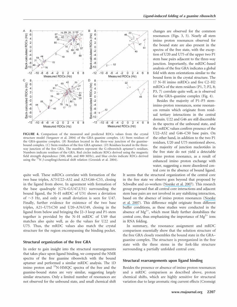
quite well. These mRDCs correlate with formation of thetwo base triples, A73:U22–A52 and A23:G46–C53, closingin the ligand from above. In agreement with formation ofthe base quadruple (C74–G:U47,U51) surrounding thebound ligand, the N–H mRDC of U51 shows a deviationof z3 Hz, and only a small deviation is seen for U47.Finally, further evidence for existence of the two basetriples, A21–U75:C50 and U20–A76:U49, closing in theligand from below and bringing the J2–3 loop and P1-stemtogether is provided by the N–H mRDC of U49 thatmatches also quite well, as do the values for U22 andU75. Thus, the mRDC values also match the crystalstructure for the region encompassing the binding pocket.
Structural organization of the free GRA
In order to gain insight into the structural rearrangementsthat takes place upon ligand binding, we compared the NMRspectra of the free guanine riboswitch with the boundaptamer and performed a similar mRDC analysis. The 1Dimino proton and 15N-HMQC spectra of the free and theguanine-bound states are very similar, suggesting largelysimilar structures. Only a limited number of resonances arenot observed for the unbound state, and small chemical shift
changes are observed for the commonresonances (Figs. 3, 5). Nearly all stemimino proton resonances observed forthe bound state are also present in thespectra of the free state, with the excep-tion of U20 and U75 of the upper two P1stem base pairs adjacent to the three-wayjunction. Importantly, the mRDC-basedanalysis of the free GRA indicates a globalfold with stem orientations similar to thebound form in the crystal structure. The17 N–H imino mRDCs and five C2–H2mRDCs of the stem residues (P1, 7; P2, 8;P3, 7) correlate quite well, as is observedfor the GRA–guanine complex (Fig. 4).
Besides the majority of P1–P3 stem-imino-proton resonances, some resonan-ces remain which originate from resid-ual tertiary interactions in the centraldomain. U22 and G46 are still discerniblein the spectra of the unbound state, andthe mRDC values confirm presence of theU22–A52 and G46–C50 base pairs. Onthe other hand, in addition to the two P1residues, U20 and U75 mentioned above,the majority of junction nucleotides inthe free state do not yield a detectableimino proton resonance, as a result ofenhanced imino proton exchange withwater, suggesting a more disordered cen-tral core in the absence of bound ligand.
It seems that the structural organization of the central corein the free state we observe goes beyond that proposed bySchwalbe and co-workers (Noeske et al. 2007). This researchgroup proposed that all central core interactions and adjacentstem base pairs are not involved in any stabilizing interaction,based on the absence of imino proton resonances (Noeskeet al. 2007). This difference might originate from differentbuffer conditions, as these studies were conducted in theabsence of Mg2+, which most likely further destabilizes thecentral core, thus emphasizing the importance of Mg2+ ionsin RNA folding.
In summary, the resonance assignment and mRDCcomparison essentially show that the solution structure ofthe free GRA closely resembles the bound state in the GRA–guanine complex. The structure is preorganized in the freestate with the three stems in the fork-like structuresurrounding a partially unfolded central core.
Structural rearrangements upon ligand binding
Besides the presence or absence of imino proton resonancesand a mRDC comparison as described above, protonchemical shifts, which are highly sensitive for structuralvariation due to large aromatic ring current effects (Cromsigt
FIGURE 4. Comparison of the measured and predicted RDCs values from the crystalstructure model (Serganov et al. 2004) of the GRA–guanine complex. (A) Stem residues ofthe GRA–guanine complex. (B) Residues located in the three-way junction of the guanine-bound complex. (C) Stem residues of the free GRA aptamer. (D) Residues located in the three-way junction of the free GRA. The numbers represent the G-riboswitch aptamer’s residues.Numbers indicate residues of the GRA. Red circles indicate RDCs derived using the magneticfield strength dependence (500, 600, and 800 MHz), and blue circles indicate RDCs derivedusing the 15N J-coupling/chemical shift relation (Grzesiek et al. 2004).
Ligand-induced folding of a guanine riboswitch
www.rnajournal.org 2207
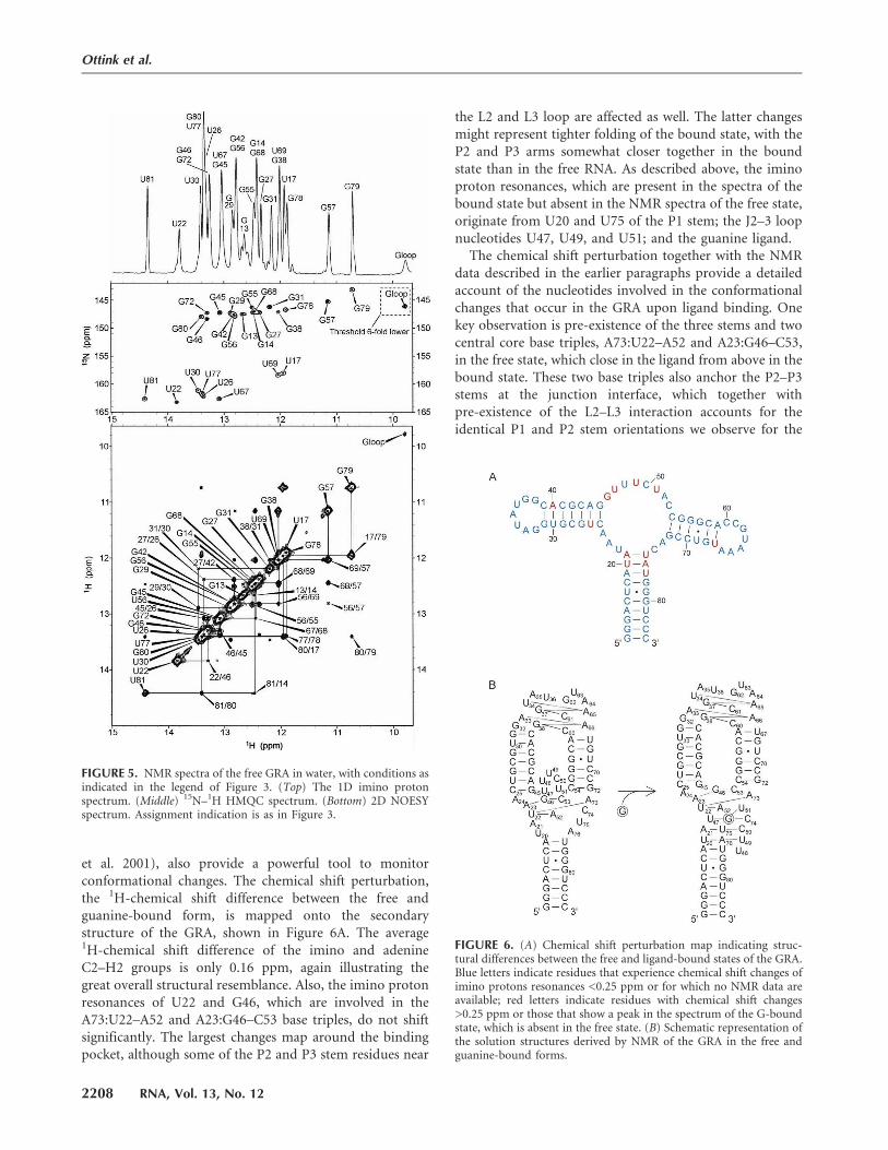
et al. 2001), also provide a powerful tool to monitorconformational changes. The chemical shift perturbation,the 1H-chemical shift difference between the free andguanine-bound form, is mapped onto the secondarystructure of the GRA, shown in Figure 6A. The average1H-chemical shift difference of the imino and adenineC2–H2 groups is only 0.16 ppm, again illustrating thegreat overall structural resemblance. Also, the imino protonresonances of U22 and G46, which are involved in theA73:U22–A52 and A23:G46–C53 base triples, do not shiftsignificantly. The largest changes map around the bindingpocket, although some of the P2 and P3 stem residues near
the L2 and L3 loop are affected as well. The latter changesmight represent tighter folding of the bound state, with theP2 and P3 arms somewhat closer together in the boundstate than in the free RNA. As described above, the iminoproton resonances, which are present in the spectra of thebound state but absent in the NMR spectra of the free state,originate from U20 and U75 of the P1 stem; the J2–3 loopnucleotides U47, U49, and U51; and the guanine ligand.
The chemical shift perturbation together with the NMRdata described in the earlier paragraphs provide a detailedaccount of the nucleotides involved in the conformationalchanges that occur in the GRA upon ligand binding. Onekey observation is pre-existence of the three stems and twocentral core base triples, A73:U22–A52 and A23:G46–C53,in the free state, which close in the ligand from above in thebound state. These two base triples also anchor the P2–P3stems at the junction interface, which together withpre-existence of the L2–L3 interaction accounts for theidentical P1 and P2 stem orientations we observe for the
FIGURE 5. NMR spectra of the free GRA in water, with conditions asindicated in the legend of Figure 3. (Top) The 1D imino protonspectrum. (Middle) 15N–1H HMQC spectrum. (Bottom) 2D NOESYspectrum. Assignment indication is as in Figure 3.
FIGURE 6. (A) Chemical shift perturbation map indicating struc-tural differences between the free and ligand-bound states of the GRA.Blue letters indicate residues that experience chemical shift changes ofimino protons resonances <0.25 ppm or for which no NMR data areavailable; red letters indicate residues with chemical shift changes>0.25 ppm or those that show a peak in the spectrum of the G-boundstate, which is absent in the free state. (B) Schematic representation ofthe solution structures derived by NMR of the GRA in the free andguanine-bound forms.
Ottink et al.
2208 RNA, Vol. 13, No. 12

free and bound states. Another key observation is theabsence of the other two base triples, A21–U75:C50 andU20–A76:U49,U47, closing in the ligand from below in thebound state and disordered nucleotides surrounding theguanine in the base quadruple (C74–G:U47,U51). Thus,prior to ligand binding, the free GRA appears to be largelyorganized, with preformation of the three stems and basetriples facing P2 and P3, but disordered in the lower part ofthe central core and the upper part of P1.
Mechanism of riboswitching
A wide body of structural and biophysical evidence isavailable suggesting the GRA is partially preorganized bythe stems and long-range loop–loop interaction, while thejunction and surrounding regions have to be partially dis-ordered to allow binding of the ligand. Our NMR studycan be used to evaluate this model and further specifythe conformational changes on a residue-specific basis. TheNMR structural data confirm existence of a preorganizedstructure in the free state containing the P1–P3 stem–loopsincluding the L2–L3 interaction surrounding a disorderedcentral core, as previously suggested by in-line probingexperiments and earlier NMR studies (Mandal et al. 2003;Serganov et al. 2004; Noeske et al. 2007). The in-lineprobing studies revealed that nearly all central core residuesand adjacent base pairs become protected from cleavageupon binding of the ligand, while nearly all P1–P3 stem aswell as the L2 and L3 residues are protected, whether theligand is present or not. Only U36, U48, and U63 aresusceptible for cleavage both in the free and bound states.A profound difference between our study and the in-lineprobing and earlier NMR studies is that we observe pre-existence of all base pairs facing the central core and thetwo base triples that assist in anchoring the P2 and P3helices. However, the in-line probing studies (Mandal et al.2003) were conducted in different buffer conditions (e.g.,an elevated pH of 8.5) to promote phosphodiester bondcleavage, and the earlier NMR studies (Noeske et al. 2007)were conducted in the absence of Mg2+, which, as wementioned above, might further destabilize the central coreand surrounding stem base pairs. We should also point outthat we modified the two base pairs of the P2 arm facingthe binding core, in order to stabilize this helix. These mayassist in preorganization of the adjacent core residues toan extent greater than for the natural RNA. There are,however, examples of guanine riboswitches where the P2arm has similar stabilizing base pairs adjacent to the core(Mandal et al. 2003).
Using the key observations described in the last para-graph, we can propose a multistep model for the sequentialfolding pathway induced by ligand binding (Fig. 6B). In thefirst step, the ligand is able to access the preorganized GRAand enters a partially disordered binding pocket by forminga base pair with C74, which in the initial binding event
can be further stabilized by stacking interactions with theexisting base triple above, i.e., A73:U22–A52. Subsequently,formation of the base quadruple surrounds the ligand, andfinally the ligand is closed in by formation of the two basetriples, which close in the ligand from below. Preformationof the two base triples implies that the site of entry of theligand is on the J2–3 side of the binding pocket. In the lastfolding step, after formation of the two upper Watson–Crick base pairs in P1, the J2–3 loop can thus be viewed asa lid that closes in the ligand on the N9/N3 side andsimultaneously fastens the J2–3 loop to the P1 stem byforming the tertiary interactions. A salient feature of thisproposed predetermined–induced fit model of ligandbinding is that only limited conformational changes inthe central core are required to drive the GRA to its stablebound-state configuration, which is an important aspectfor the mechanism of riboswitching. In the structure weobserve for the free GRA, only C50 and C74 are accessibleto form a Watson–Crick base pair with the incomingligand, which possibly promotes fast recognition withoutthe need for further proofreading. This suggests an addi-tional role for a preformed GRA in the free state, sincemore disorder would render more cytidines accessible,which might lead to unproductive binding. While thisconsideration seems less important for the GRA, with onlytwo accessible acceptors to form the Watson–Crick basepair, it might be more important for the structurally relatedadenine riboswitch where the many conserved uridines inthe central core might further enhance unproductivebinding. Besides the intrinsic property of a A–U base pairto form one less hydrogen bond, this might contributeto the apparent lower affinity of the adenine aptamer forits cognate ligand, with a dissociation constant (Kd) of0.5 mM, i.e., 100-fold higher than the Kd value reported forthe GRA–guanine interaction (5 nM) (Gilbert et al. 2006).
The limited structural rearrangement upon ligand bind-ing that we propose nicely corresponds with the thermo-dynamic data reported by Batey and co-workers (Gilbertet al. 2006). Assuming temperature independent enthalpyand entropy values, the reported values for guanine bindingcorrespond with a DG37 of �11.0 kcal/mol in accordancewith formation of roughly five to six Watson–Crick basepairs (Serra and Turner 1995), which matches well to for-mation of the remainder of the central core interactionsand the two upper base pairs in P1. Assuming that we arecorrect in modeling the upper two base pairs of P1 as dis-ordered in the unbound state, a total energy gain of �19.5kcal/mol at 37°C can be predicted for locking the aptamerstructure around guanine by adding the energy gain (Serraand Turner 1995) for formation of the lower six base pairsof P1, i.e., nucleotides 14–19 with 77–82 (Fig. 1B). This valueis comparable to the predicted thermodynamic stability ofthe antiterminator, i.e., the structure with which ligandbinding to the aptamer has to compete, which in case ofthe xpt-pbuX riboswitch amounts to �17.5 kcal/mol.
Ligand-induced folding of a guanine riboswitch
www.rnajournal.org 2209
Increasing evidence is emerging that RNA folding pro-ceeds via a hierarchic folding pathway in which highlystructured RNAs form through compaction of preformedsecondary-structure elements (Brion and Westhof 1997).In this view secondary structure elements, such as helicesor hairpin–loops are formed first, which subsequentlyinteract locally through coaxial stacking or pseudoknot ortriple-helix interactions. Finally, these independently foldedsubdomains interact cooperatively by loop–loop andnumerous contacts involving loops, bulges, and helices.Preorganization of the free GRA, followed by the multistepsequential folding pathway that we propose occurs uponligand binding, provides a classic example of such ahierarchic folding pathway on the one hand, but on theother hand shows how the unique folding capacity of RNAcan be adapted for a specific purpose. By making effectiveuse of the folding landscape of RNA, the aptamer is foldedto such an extent that binding specificity is secured, but atthe same time a sufficient stretch is left unstructured toensure quick response to ligand binding for transcriptioncontrol. We expect other riboswitches to operate via thiscombined predetermined–induced fit mechanism as well,and it will for instance be interesting to see how thismechanism is adapted by structurally dissimilar ribo-switches, such as the S-adenosylmethionine riboswitch,which also displays a high affinity for its cognate metabolite(Kd=4 nM) (Soukup and Soukup 2004).
RNAs that change their shape in response to externalstimuli, such as the riboswitch–metabolite interaction,are ideal tools for molecular design, for instance, in thedevelopment of nanomechanical devices and syntheticRNA circuits (e.g., Davidson and Ellington 2007), dueto its great predictive power in constructing secondary-and tertiary-structure elements in combination with func-tional diversity. To be able to build such devices, priorknowledge of the three-dimensional structures and con-comitant RNA folding processes remains a necessity.The detailed structural description of the GRA–guanineinteraction we have presented provides such a struc-tural framework for riboswitch utilization. It will, forinstance, be interesting to see if this framework can beused to engineer an adenine-responsive aptamer (ARA)with high adenine affinity by mutating the uridine-rich core while preserving the compact three-dimensionalstructure.
MATERIALS AND METHODS
RNA sample preparation
A double-stranded DNA fragment coding for the GRA (Fig. 1)and flanked by a T7 promoter sequence was cloned into a pUC18vector. Unlabeled and uniformly 13C/15N-labeled G-riboswitchaptamer samples were prepared by in vitro run-off transcriptionusing the linearized plasmid and T7 RNA polymerase (Milliganand Uhlenbeck 1989). The products were purified using denatur-ing PAGE, collected by electroelution, and dialyzed against NMRbuffer (10 mM sodium phosphate, pH 6.7). Magnesium wasadded to the solution by dialyzing the samples against the NMRbuffer containing the appropriate concentration of MgCl2. Thefinal concentrations of the NMR samples were z0.2 and 0.7 mMfor the unlabeled G-riboswitch and uniformly labeled G-riboswitch,respectively.
Complex formation
Because of the low solubility of guanine in H2O, the GRA–guaninecomplexes were prepared at low concentration. For the unlabeledGRA–guanine complex, this was achieved by mixing together1 mL of a 60 mM G-riboswitch solution with 3.5 mL of a 21 mMguanine solution, both dissolved in NMR buffer containing 5 mMMgCl2. The sample was subsequently concentrated to 300 mLusing a 10-kDa cut-off Centricon tube (Amicon). The finalconcentration of the NMR sample was 0.2 mM. Using the sameprotocol, the uniformly 13C/15N-labeled GRA-guanine complexwas prepared in a concentration of 0.7 mM.
NMR experiments
All spectra were recorded at 5 and 15°C on 500- and 800-MHzVarian Inova spectrometers equipped with cryoprobes and on a600-MHz Varian Inova spectrometer. NOESY spectra in water(93% H2O, 7% D2O) were recorded with 100-, 200-, and 300-msec mixing times. Nitrogen-decoupled 15N-HMQC spectrawere also recorded in 93% H2O/7% D2O. The water signal wassuppressed either by a selective 270° sine-shaped pulse in combi-nation with a Watergate water suppression scheme (Piotto et al.1992) or with a jump–return pulse (Plateau and Gueron 1982) incombination with a Watergate sequence. 15N-HMQC-IPAP spec-tra (Ottiger et al. 1998) were measured to derive the 1H–15Nsplittings. Adenine C2–H2 correlations were measured in waterby 13C-HSQC experiments using a constant time evolution periodto refocus carbon–carbon couplings. 1H–13C splittings were mea-sured by recording the 13C-HSQC spectra in an IPAP fashion. Allspectra were processed with NMRpipe (Delaglio et al. 1995) andassigned using the Sparky software (Kneller and Kuntz 1993).
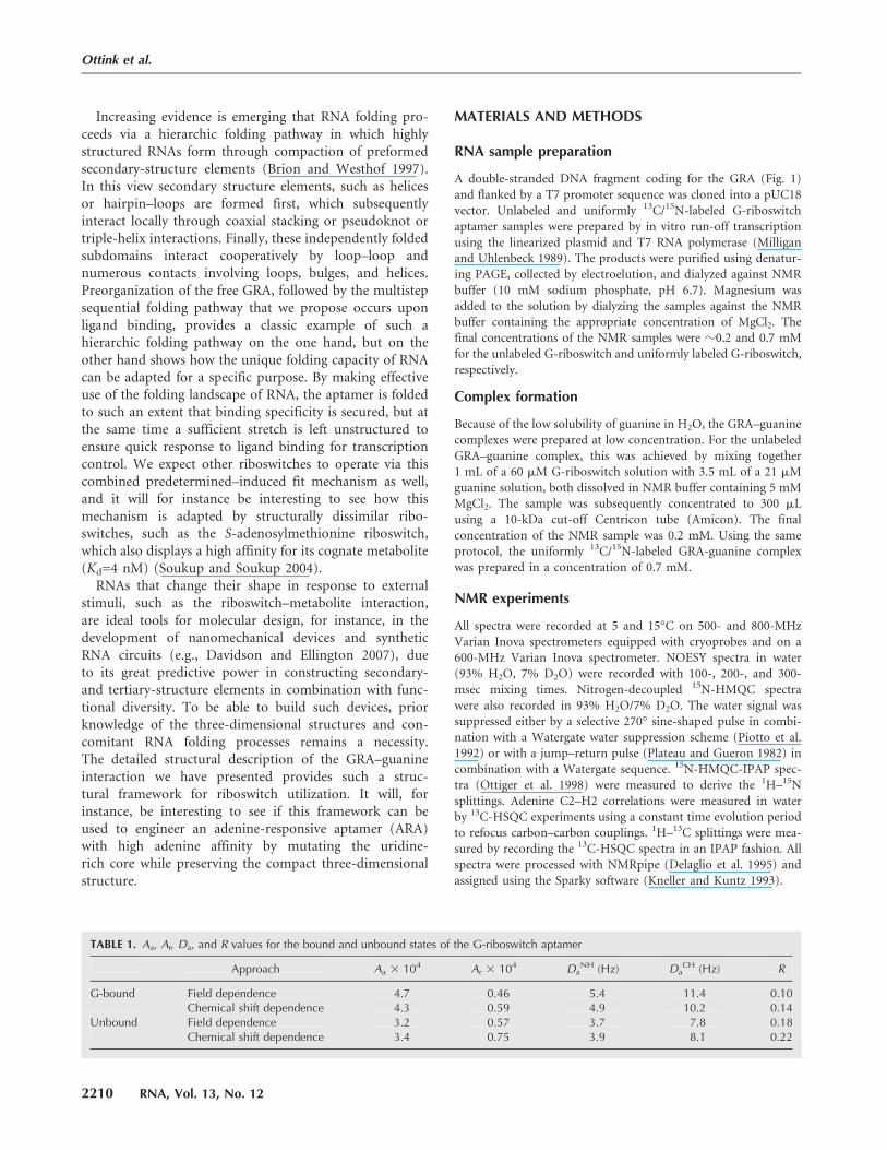
TABLE 1. Aa, Ar, Da, and R values for the bound and unbound states of the G-riboswitch aptamer
Approach Aa 3 104 Ar 3 104 DaNH (Hz) Da
CH (Hz) R
G-bound Field dependence �4.7 �0.46 �5.4 11.4 0.10Chemical shift dependence �4.3 �0.59 �4.9 10.2 0.14
Unbound Field dependence �3.2 �0.57 �3.7 7.8 0.18Chemical shift dependence �3.4 �0.75 �3.9 8.1 0.22
Ottink et al.
2210 RNA, Vol. 13, No. 12

RDC measurements
The asymmetric elongated shape of the 25-kDa GRA createdsufficient susceptibility for magnetic field alignment for derivationof mRDCs by measuring one-bond N–H or C–H splittings atthree different field strengths or by using a J-coupling/chemicalshift relationship (Grzesiek et al. 2004). In the first approach, theimino 15N–H RDCs of the aptamer are derived by measuring thesplittings at 500-, 600-, and 800-MHz proton frequencies. Themeasured splitting relates to the magnetic field strength, followingthe equation J(Hz) = J0(Hz)+k(1/MHz) 3B2(MHz), with J thetotal splitting, J0 the isotropic J coupling, B the magnetic fieldstrength, and k the RDC per magnetic-field-strength unit. Thus,by plotting the measured splittings against the square of themagnetic field strength, the RDC values can be derived from theslope of the fitted straight line. Multiplying the value for the slopewith the square of a certain magnetic field strength yields the RDCvalue at that field strength. The C2–H2 RDCs were derived bycalculating the difference between the measured splitting and thetheoretical value of 203.2 Hz for AMP. The other approach makesuse of the linear correlation between the strength of the imino15N–H scalar couplings involved in N–H–N hydrogen bonds andtheir chemical shift value following the equation 1JNH = (1.016Hz/ppm)dHN � 100.58 Hz (Grzesiek et al. 2004). Using thisequation, the theoretical J couplings are calculated. The RDC isthen the difference between the measured splitting and thecalculated scalar coupling.
Module software (Dosset et al. 2001) was used to derive thealignment tensor and back-calculated RDCs from the PDBcoordinate file (1Y27) of the guanine-bound GRA crystal struc-ture (Serganov et al. 2004) and our measured values. The entiresystem was considered as one structural entity. For obtaining Aa
and Ar values given by Module, only N–H RDCs from residueslocated in the three stem regions were used. From the Aa and Ar
values, the DaNH and Da
CH values were obtained by multiplyingAa with (m0hgHgN,C)/16(prNH,CH)3, assuming N–H and C–Hbond lengths of 1.02 and 1.09 A, respectively. The R values wereobtained by dividing Ar by Aa. Aa and Ar are the unitless axial andrhombic components of the molecular alignment tensor A,respectively. The back-calculated RDCs were directly given byModule. All values were obtained assuming an order parameter of1. The Da
NH,CH and R values, derived for the bound and unboundstates of the GRA are listed in Table 1.
SUPPLEMENTAL DATA
Figures showing the 1D imino proton, 2D NOESY spectra, andimino proton assignments of the P2 and P3 arms, and bond-angledeviation–RDC deviation correlation plots of the guanine-boundGRA and tables with measured and back-calculated mRDCsfor the guanine-bound and guanine-free GRA are available uponrequest from the authors (send an e-mail message to [email protected]).
Received May 18, 2007; accepted September 11, 2007.
REFERENCES
Batey, R.T., Gilbert, S.D., and Montange, R.K. 2004. Structure of anatural guanine-responsive riboswitch complexed with the metab-olite hypoxanthine. Nature 432: 411–415.
Brion, P. and Westhof, P. 1997. Hierarchy and dynamics of RNAfolding. Annu. Rev. Biophys. Biomol. Struct. 26: 113–137.
Cromsigt, J.A.M.T.C., Hilbers, C.W., and Wijmenga, S.S. 2001. Predic-tion of proton chemical shifts in RNA. Their use in structurerefinement and validation. J. Biomol. NMR 21: 11–29.
Davidson, E.A. and Ellington, A.D. 2007. Synthetic RNA circuits.Nat. Chem. Biol. 3: 23–28.
Delaglio, F., Grzesiek, S., Vuister, G.W., Zhu, G., Pfeifer, J., andBax, A. 1995. NMRpipe: A multidimensional spectral processingsystem based on Unix pipes. J. Biomol. NMR 6: 277–293.
Dosset, P., Hus, J.C., Marion, D., and Blackledge, M. 2001. A novelinteractive tool for rigid-body modeling of multidomain macro-molecules using residual dipolar couplings. J. Biomol. NMR 20:223–231.
Epshtein, V., Mironov, A.S., and Nudler, E. 2003. The riboswitch-mediated control of sulfur metabolism in bacteria. Proc. Natl.Acad. Sci. 100: 5052–5056.
Fuchs, R.T., Grundy, F.J., and Henkin, T.M. 2006. The S(MK) box isa new SAM- binding RNA for translational regulation of SAMsynthetase. Nat. Struct. Mol. Biol. 13: 226–233.
Gilbert, S.D. and Batey, R.T. 2006. Riboswitches: Fold and function.Chem. Biol. 13: 805–807.
Gilbert, S.D., Stoddard, C.D., Wise, S.J., and Batey, R.T. 2006.Thermodynamic and kinetic characterization of ligand bindingto the purine riboswitch aptamer domain. J. Mol. Biol. 359:754–768.
Grundy, F.J., Lehman, S.C., and Henkin, T.M. 2003. The L boxregulon: Lysine sensing by leader RNAs of bacterial lysine bio-synthesis genes. Proc. Natl. Acad. Sci. 100: 12057–12062.
Grzesiek, S., Cordier, F., Jaravine, V., and Barfield, M. 2004. Insightsinto biomolecular hydrogen bonds from hydrogen bond scalarcouplings. Prog. Nucl. Magn. Reson. Spectrosc. 45: 275–300.
Kneller, D.G. and Kuntz, I.D. 1993. UCSF Sparky: An NMR display,annotation and assignment tool. J. Cell. Biochem. 53: 254.
Lemay, J.F., Penedo, J.C., Tremblay, R., Lilley, D.M., andLafontaine, D.A. 2006. Folding of the adenine riboswitch. Chem.Biol. 13: 857–868.
Mandal, M. and Breaker, R.R. 2004a. Adenine riboswitches and geneactivation by disruption of a transcription terminator. Nat. Struct.Mol. Biol. 11: 29–35.
Mandal, M. and Breaker, R.R. 2004b. Gene regulation by riboswitches.Nat. Rev. Mol. Cell Biol. 5: 451–463.
Mandal, M., Boese, B., Barrick, J.E., Winkler, W.C., and Breaker, R.R.2003. Riboswitches control fundamental biochemical pathways inBacillus subtilis and other bacteria. Cell 113: 577–586.
Mandal, M., Boese, B., Barrick, J.E., Winkler, W.C., and Breaker, R.R.2004. A glycine-dependent riboswitch that uses cooperativebinding to control gene expression. Science 306: 275–279.
McDaniel, B.A., Grundy, F.J., Artsimovitch, I., and Henkin, T.M.2003. Transcription termination control of the S box system:Direct measurement of S-adenosylmethionine by the leader RNA.Proc. Natl. Acad. Sci. 100: 3083–3088.
Milligan, J.F. and Uhlenbeck, O.C. 1989. Synthesis of small RNAsusing T7 RNA-polymerase. Methods Enzymol. 180: 51–62.
Mironov, A.S., Gusarov, I., Rafikov, R., Lopez, L.E., Shatalin, K.,Kreneva, R.A., Perumov, D.A., and Nudler, E. 2002. Sensing smallmolecules by nascent RNA: A mechanism to control transcriptionin bacteria. Cell 111: 747–756.
Misra, V.K. and Draper, D.E. 1998. On the role of magnesium ions inRNA stability. Biopolymers 48: 113–135.
Mollova, E.T., Hansen, M.R., and Pardi, A. 2000. Global structure ofRNA determined with residual dipolar couplings. J. Am. Chem.Soc. 122: 11561–11562.
Nagel, J.H., Gultyaev, A.P., Oistamo, K.J., Gerdes, K., and Pleij, C.W.2002. A pH-jump approach for investigating secondary structurerefolding kinetics in RNA. Nucleic Acids Res. 30: e63. doi: 10.1093/nar/gnf057.
Ligand-induced folding of a guanine riboswitch
www.rnajournal.org 2211

Nahvi, A., Sudarsan, N., Ebert, M.S., Zou, X., Brown, K.L., andBreaker, R.R. 2002. Genetic control by a metabolite bindingmRNA. Chem. Biol. 9: 1043–1049.
Noeske, J., Richter, C., Grundl, M.A., Nasiri, H.R., Schwalbe, H., andWohnert, J. 2005. An intermolecular base triple as the basis ofligand specificity and affinity in the guanine- and adenine-sensingriboswitch RNAs. Proc. Natl. Acad. Sci. 102: 1372–1377.
Noeske, J., Buck, J., Furtig, B., Nasiri, H.R., Schwalbe, H., andWohnert, J. 2007. Interplay of ‘‘induced fit’’ and preorganizationin the ligand induced folding of the aptamer domain of theguanine binding riboswitch. Nucleic Acids Res. 35: 572–583. doi:10.1093/nar/gkl1094.
Nudler, E. and Mironov, A.S. 2004. The riboswitch control of bacterialmetabolism. Trends Biochem. Sci. 29: 11–17.
Ottiger, M., Delaglio, F., and Bax, A. 1998. Measurement of J anddipolar couplings from simplified two-dimensional NMR spectra.J. Magn. Reson. 131: 373–378.
Piotto, M., Saudek, V., and Sklenar, V. 1992. Gradient-tailoredexcitation for single-quantum NMR spectroscopy of aqueoussolutions. J. Biomol. NMR 2: 661–665.
Plateau, P. and Gueron, M. 1982. Exchangeable proton NMR withoutbase-line distortion, using new strong-pulse sequences. J. Am.Chem. Soc. 104: 7310–7311.
Rodionov, D.A., Vitreschak, A.G., Mironov, A.A., and Gelfand, M.S.2003. Regulation of lysine biosynthesis and transport genes inbacteria: Yet another RNA riboswitch? Nucleic Acids Res. 31: 6748–6757. doi: 10.1093/nar/gkg900.
Serganov, A., Yuan, Y.R., Pikovskaya, O., Polonskaia, A., Malinina, L.,Phan, A.T., Hobartner, C., Micura, R., Breaker, R.R., andPatel, D.J. 2004. Structural basis for discriminative regulation ofgene expression by adenine- and guanine-sensing mRNAs. Chem.Biol. 11: 1729–1741.
Serra, M.J. and Turner, D.H. 1995. Predicting thermodynamicproperties of RNA. Methods Enzymol. 259: 242–261.
Soukup, J.K. and Soukup, G.A. 2004. Riboswitches exert geneticcontrol through metabolite-induced conformational change. Curr.Opin. Struct. Biol. 14: 344–349.
Sudarsan, N., Nahvi, A., Roth, A., Collins, J.A., and Breaker, R.R.2003. An mRNA structure in bacteria that controls gene expres-sion by binding lysine. Genes & Dev. 17: 2688–2697.
van Buuren, B.N.M., Schleucher, J., Wittmann, V., Griesinger, C.,Schwalbe, H., and Wijmenga, S.S. 2004. NMR spectroscopicdetermination of the solution structure of a branched nucleic acidfrom residual dipolar couplings by using isotopically labelednucleotides. Angew. Chem. 43: 187–192.
Wickiser, J.K., Cheah, M.T., Breaker, R.R., and Crothers, D.M. 2005.The kinetics of ligand binding by an adenine-sensing riboswitch.Biochemistry 44: 13404–13414.
Wijmenga, S.S. and van Buuren, B.N.M. 1998. The use of NMRmethods for conformational studies of nucleic acids. Prog. Nucl.Magn. Reson. Spectrosc. 32: 287–387.
Winkler, W., Nahvi, A., and Breaker, R.R. 2002a. Thiamine derivativesbind messenger RNAs directly to regulate bacterial gene expres-sion. Nature 419: 952–956.
Winkler, W.C., Cohen-Chalamish, S., and Breaker, R.R. 2002b. AnmRNA structure that controls gene expression by binding FMN.Proc. Natl. Acad. Sci. 99: 15908–15913.
Winkler, W.C., Nahvi, A., Sudarsan, N., Barrick, J.E., andBreaker, R.R. 2003. An mRNA structure that controls geneexpression by binding S-adenosylmethionine. Nat. Struct. Mol.Biol. 10: 701–707.
Winkler, W.C., Nahvi, A., Roth, A., Collins, J.A., and Breaker, R.R.2004. Control of gene expression by a natural metabolite-responsiveribozyme. Nature 428: 281–286.
Ottink et al.
2212 RNA, Vol. 13, No. 12