
life - MDPI
34
Citation: Petroušková, P.; Hudáková, N.; Maloveská, M.; Humeník, F.; Cizkova, D. Non-Exosomal and Exosome-Derived miRNAs as Promising Biomarkers in Canine Mammary Cancer. Life 2022, 12, 524. https://doi.org/10.3390/ life12040524 Academic Editors: Lara Console and Mariafrancesca Scalise Received: 24 February 2022 Accepted: 30 March 2022 Published: 1 April 2022 Publisher’s Note: MDPI stays neutral with regard to jurisdictional claims in published maps and institutional affil- iations. Copyright: © 2022 by the authors. Licensee MDPI, Basel, Switzerland. This article is an open access article distributed under the terms and conditions of the Creative Commons Attribution (CC BY) license (https:// creativecommons.org/licenses/by/ 4.0/). life Review Non-Exosomal and Exosome-Derived miRNAs as Promising Biomarkers in Canine Mammary Cancer Patrícia Petroušková 1 , Nikola Hudáková 1 , Marcela Maloveská 1 , Filip Humeník 1 and Dasa Cizkova 1,2, * 1 Centre of Experimental and Clinical Regenerative Medicine, The University of Veterinary Medicine and Pharmacy, Komenského 73, 041 81 Košice, Slovakia; [email protected] (P.P.); [email protected] (N.H.); [email protected] (M.M.); fi[email protected] (F.H.) 2 Institute of Neuroimmunology, Slovak Academy of Sciences, Dúbravská Cesta 9, 845 10 Bratislava, Slovakia * Correspondence: [email protected]; Tel.: +421-918-752-157 Abstract: Canine mammary cancer (CMC), similar to human breast cancer (HBC) in many aspects, is the most common neoplasm associated with significant mortality in female dogs. Due to the limited therapy options, biomarkers are highly desirable for early clinical diagnosis or cancer progression monitoring. Since the discovery of microRNAs (miRNAs or miRs) as post-transcriptional gene regulators, they have become attractive biomarkers in oncological research. Except for intracellular miRNAs and cell-free miRNAs, exosome-derived miRNAs (exomiRs) have drawn much attention in recent years as biomarkers for cancer detection. Analysis of exosomes represents a non-invasive, pain- free, time- and money-saving alternative to conventional tissue biopsy. The purpose of this review is to provide a summary of miRNAs that come from non-exosomal sources (canine mammary tumor, mammary tumor cell lines or canine blood serum) and from exosomes as promising biomarkers of CMC based on the current literature. As is discussed, some of the miRNAs postulated as diagnostic or prognostic biomarkers in CMC were also altered in HBC (such as miR-21, miR-29b, miR-141, miR-429, miR-200c, miR-497, miR-210, miR-96, miR-18a, miR19b, miR-20b, miR-93, miR-101, miR- 105a, miR-130a, miR-200c, miR-340, miR-486), which may be considered as potential disease-specific biomarkers in both CMC and HBC. Keywords: canine; mammary cancer; biomarker; miRNA; exosome 1. Canine Mammary Cancer 1.1. General Information and Risk Factors In veterinary medicine, canine cancer represents a severe clinical problem [1]. Ap- proximately one in four dogs suffers cancer at some stage in their life and almost half of dogs over the age of 10 die due to neoplasia [2]. Tumors of the mammary glands are the second most commonly diagnosed type of cancer in dogs, with certain breeds, such as Labrador Retrievers, Cocker Spaniels, Irish Setters, German Shepherds, mixed-breed dogs, and miniature and Toy Poodles, and are over-represented in terms of high risk of neoplasia and mortality [3]. Mammary tumors typically develop in intact female dogs or elderly spayed (ovariohysterectomised) bitches, usually between 8 and 10 years old [4,5]. This problem is especially significant in Europe, where bitches are usually spayed at an older age [4]. However, the precise prevalence of canine mammary tumors differs from study to study due to the dog breeds and age and geographic location [6]. Biaoni et al. reported that canine mammary tumors were the most frequent tumor in Italy, wherein 476 cases per 100,000 dog-years at risk were malignant [7]. On the other hand, the incidence of mammary tumors in Sweden is higher and ranges from 111 to 154 per 10,000 dog-years at risk [8]. The incidence of canine mammary gland tumors is related to two main risk factors: age and time of exposure to ovarian hormones [9]. A study performed by Egenvall et al. de- scribes that in bitches at ages 6, 8, and 10 years, the incidence of mammary cancer increased Life 2022, 12, 524. https://doi.org/10.3390/life12040524 https://www.mdpi.com/journal/life
-
Upload
khangminh22 -
Category
Documents
-
view
2 -
download
0
Transcript of life - MDPI

�����������������
Citation: Petroušková, P.;
Hudáková, N.; Maloveská, M.;
Humeník, F.; Cizkova, D.
Non-Exosomal and
Exosome-Derived miRNAs as
Promising Biomarkers in Canine
Mammary Cancer. Life 2022, 12, 524.
https://doi.org/10.3390/
life12040524
Academic Editors: Lara Console and
Mariafrancesca Scalise
Received: 24 February 2022
Accepted: 30 March 2022
Published: 1 April 2022
Publisher’s Note: MDPI stays neutral
with regard to jurisdictional claims in
published maps and institutional affil-
iations.
Copyright: © 2022 by the authors.
Licensee MDPI, Basel, Switzerland.
This article is an open access article
distributed under the terms and
conditions of the Creative Commons
Attribution (CC BY) license (https://
creativecommons.org/licenses/by/
4.0/).
life
Review
Non-Exosomal and Exosome-Derived miRNAs as PromisingBiomarkers in Canine Mammary CancerPatrícia Petroušková 1 , Nikola Hudáková 1, Marcela Maloveská 1, Filip Humeník 1 and Dasa Cizkova 1,2,*
1 Centre of Experimental and Clinical Regenerative Medicine, The University of Veterinary Medicine andPharmacy, Komenského 73, 041 81 Košice, Slovakia; [email protected] (P.P.);[email protected] (N.H.); [email protected] (M.M.); [email protected] (F.H.)
2 Institute of Neuroimmunology, Slovak Academy of Sciences, Dúbravská Cesta 9, 845 10 Bratislava, Slovakia* Correspondence: [email protected]; Tel.: +421-918-752-157
Abstract: Canine mammary cancer (CMC), similar to human breast cancer (HBC) in many aspects, isthe most common neoplasm associated with significant mortality in female dogs. Due to the limitedtherapy options, biomarkers are highly desirable for early clinical diagnosis or cancer progressionmonitoring. Since the discovery of microRNAs (miRNAs or miRs) as post-transcriptional generegulators, they have become attractive biomarkers in oncological research. Except for intracellularmiRNAs and cell-free miRNAs, exosome-derived miRNAs (exomiRs) have drawn much attention inrecent years as biomarkers for cancer detection. Analysis of exosomes represents a non-invasive, pain-free, time- and money-saving alternative to conventional tissue biopsy. The purpose of this review isto provide a summary of miRNAs that come from non-exosomal sources (canine mammary tumor,mammary tumor cell lines or canine blood serum) and from exosomes as promising biomarkers ofCMC based on the current literature. As is discussed, some of the miRNAs postulated as diagnosticor prognostic biomarkers in CMC were also altered in HBC (such as miR-21, miR-29b, miR-141,miR-429, miR-200c, miR-497, miR-210, miR-96, miR-18a, miR19b, miR-20b, miR-93, miR-101, miR-105a, miR-130a, miR-200c, miR-340, miR-486), which may be considered as potential disease-specificbiomarkers in both CMC and HBC.
Keywords: canine; mammary cancer; biomarker; miRNA; exosome
1. Canine Mammary Cancer1.1. General Information and Risk Factors
In veterinary medicine, canine cancer represents a severe clinical problem [1]. Ap-proximately one in four dogs suffers cancer at some stage in their life and almost half ofdogs over the age of 10 die due to neoplasia [2]. Tumors of the mammary glands are thesecond most commonly diagnosed type of cancer in dogs, with certain breeds, such asLabrador Retrievers, Cocker Spaniels, Irish Setters, German Shepherds, mixed-breed dogs,and miniature and Toy Poodles, and are over-represented in terms of high risk of neoplasiaand mortality [3]. Mammary tumors typically develop in intact female dogs or elderlyspayed (ovariohysterectomised) bitches, usually between 8 and 10 years old [4,5]. Thisproblem is especially significant in Europe, where bitches are usually spayed at an olderage [4]. However, the precise prevalence of canine mammary tumors differs from study tostudy due to the dog breeds and age and geographic location [6]. Biaoni et al. reportedthat canine mammary tumors were the most frequent tumor in Italy, wherein 476 cases per100,000 dog-years at risk were malignant [7]. On the other hand, the incidence of mammarytumors in Sweden is higher and ranges from 111 to 154 per 10,000 dog-years at risk [8].
The incidence of canine mammary gland tumors is related to two main risk factors:age and time of exposure to ovarian hormones [9]. A study performed by Egenvall et al. de-scribes that in bitches at ages 6, 8, and 10 years, the incidence of mammary cancer increased
Life 2022, 12, 524. https://doi.org/10.3390/life12040524 https://www.mdpi.com/journal/life

Life 2022, 12, 524 2 of 34
from 1% to 6% and 13%, respectively [8]. Ovarian hormones (estrogen, progesterone) maylead to carcinogenesis and mammary hyperplasia. Thus, ovariohysterectomy is precaution-ary for tumor development, and its timing seems to be crucial [10,11]. A female dog of anybreed castrated before the first ovarian cycle has a 0.5% chance of developing a tumor. Ifthe bitch is spayed just after or at any subsequent ovarian cycle, the risk of mammary glandcancer increases from 8% to 26% [9,10]. Additionally, the study of Schneider et al. demon-strated that ovariohysterectomy of bitches after their second estrum had no preventiveimpact against the development of malignant tumors [10].
Canine studies also indicated that obesity is another major risk factor for mammarytumor development, especially if present early in a dog’s life [12,13]. The study of Sonnen-schein et al. demonstrated that a thin physique reduced the risk of mammary cancer amongspayed dogs by 99%, and non-spayed dogs by 40% [12]. The influence of the diet was alsostudied. Dogs on a homemade diet with high-red meat portions were at a higher risk ofdeveloping mammary dysplasia and tumors compared to a commercial diet [13]. Therefore,nutritional factors, operating especially early in life, also have etiological importance tocanine mammary cancer (CMC) development.
1.2. Classification System
Mammary glands are a frequent location for tumor development and, as in other typesof cancer, canine mammary tumors may be benign or malignant [14]. Since approximatelyhalf of mammary tumors in dogs are malignant with a high percentage of mortality if nottreated in time, there is no doubt that canine mammary neoplasia represents a seriousclinical issue [4,15,16]. Histopathology and biopsy remain the cornerstone and the goldstandards for the diagnosis and classification of canine mammary tumors [17]. However,the morphological heterogeneity of these tumors, with frequent presence of various cellpopulations, is challenging when providing an appropriate classification [17]. Nowadays,veterinary pathologists have available two systems of a histological classification schemefor canine mammary tumors: the official histological classification approved by the WorldHealth Organization (WHO) and the Armed Forces Institute of Pathology from 1999 [18]and the international consensus histological classification scheme based on 2011 updatesto the WHO HBC parameters proposed by Goldschmidt et al. from 2011 [19]. The latest2011 system combines various criteria for subtyping mammary tumors by separatingbenign forms from malignant lesions and determining the tissue of origin (epithelial,myoepithelial, mesenchymal). A comparison of the two classification systems by Canadaset al. demonstrated that the WHO and 2011 classification systems were very similar interms of the categorization of benign tumors, and both were prognostically relevant byidentifying malignant tumors [17]. Therefore, veterinary pathologists should include bothclassification systems in the diagnosis and classification of canine mammary tumors.
Based on the tissue of origin, mammary gland tumors of purely epithelial origin aremalignant carcinomas, such as carcinoma in situ, simple carcinoma (tubular, tubulopapil-lary, cystic-papillary, cybriform), solid carcinoma, anaplastic carcinoma, ductal carcinoma,complex and mixed type carcinoma [17,18]. However, there are also other special typesof malignant epithelial neoplasms (squamous cell carcinoma, adenosquamous carcinoma,mucinous carcinoma, lipid-rich carcinoma, spindle cell carcinoma, and inflammatory carci-noma) [18,19]. Tubular carcinoma (adenocarcinoma) is the most common type of mammarygland tumor in dogs [19,20]. Mesenchymal neoplasms are sarcomas (osteosarcoma, fibrosar-coma, chondrosarcoma, liposarcoma, hemangiosarcoma, and others), with osteosarcomaas the most frequent mesenchymal neoplasm of the canine mammary glands [19]. How-ever, some of them have mixed histology consisting of a combination of epithelial andmyoepithelial or mesenchymal tissue (complex carcinoma, carcinosarcoma, and benignmixed tumors) [19]. Benign mammary tumors are mostly simple and complex adenomas,fibroadenomas, myoepithelioma, ductal adenoma and ductal papilloma [18,19].
The cytology of canine mammary tumors can be another approach in diagnostics, butit should be taken into account carefully, because it offers many false results due to lesions

Life 2022, 12, 524 3 of 34
differentiation, atypic benign forms, and the presence of inflammation or necrosis in thetissue [21,22].
The Nottingham histological grade (NGS), described by Elston and Ellis in 1991 [23],is also used to provide prognostic information by assessing a malignancy score (nuclearpleomorphism, mitotic index, and tubule formation) [23]. In NHG, semi-quantitativeevaluation of tubule formation (≥75% of tumor area containing tubules—score I; 10–75% oftumor area containing tubules—score II;≤10% of tumor area containing tubules—score III),quantitative and qualitative judgement of nuclear polymorphism (small nuclei with regularoutlines—score I; visible nucleoli and mild nuclei variability in size and shape—score II;severe nuclei variability in size and shape—score III), and mitotic count using a minimum10 fields of tumor area (≤9 mitoses—score I; 10–19 mitoses—score II; ≥20 mitoses—scoreIII) [23]. After summing these three component scores, grades from I to III are generated,wherein scores 3–5 indicate a low-grade (I) tumor, scores 6–7 an intermediate (II) tumor,and scores 8–9 a high-grade (III) tumor [23].
1.3. Comparative Oncology
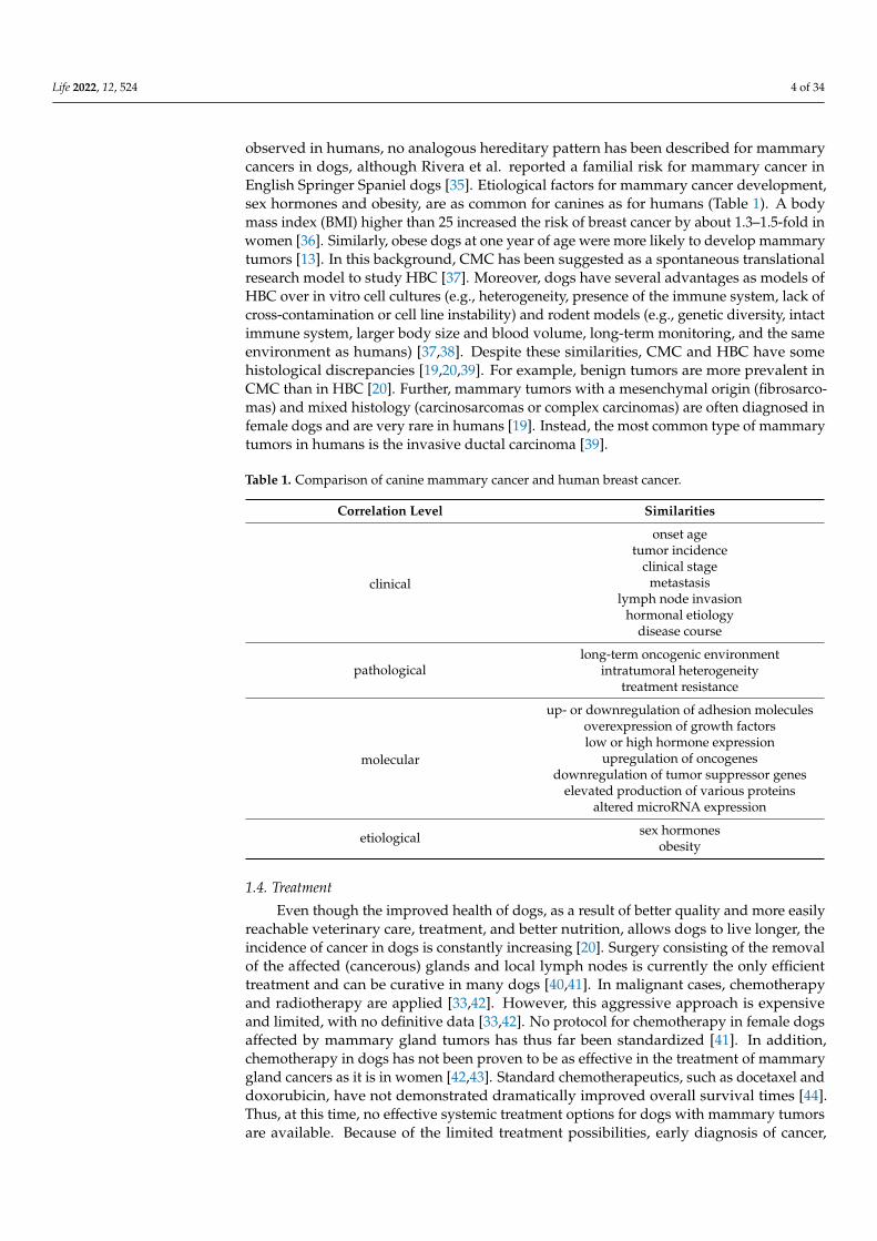
CMC exhibit several similarities with HBC, mainly at the clinical, genetic, molecular,pathological, and etiological levels which are summarized in Table 1 [24]. The clinicalcorrelation between CMC and HBC comprises the onset age, tumor incidence, hormonaletiology, and identical disease course [25]. Interestingly, the average onset age of mammarytumors in dogs (after 6 years) is comparable with the incidence of breast cancer in humans(after 40 years). In addition, the aspects affecting the clinical outcome, such as tumor size,clinical stage, metastasis, and lymph node invasion, are also comparable [26]. Approxi-mately 50% of canine mammary carcinomas are reported to metastasize to regional lymphnodes and lungs, and eventually bones [27,28]. In their human counterpart, almost 20%of HBCs have a prognosis to develop metastatic lesions [29]. Among pathologic charac-teristics, canine and human tumors share features in a long-term oncogenic environment,intratumoral heterogeneity, and acquired treatment resistance (Table 1) [30]. Markers havethe potential to predict the response to a certain anti-cancer treatment [e.g., estrogen andprogesterone receptors, cytochrome P450 or antigen Ki-67 for breast cancer; UGT1A1 geneencoding UDP-glucuronosyltransferase 1-1 enzyme or specific mutations of K-RAS (Kirstenrat sarcoma virus), for colorectal cancer; human epidermal growth factor receptor 2, HER2,for breast or gastric cancer; c-KIT gene encoding tyrosine-protein kinase KIT for gastriccancer; DNA excision repair protein ERCC1 or tumor protein p53 for lung cancer; reviewedin [31,32]] and provide cancer prognostic information [e.g., antigen Ki-67 or cyclin D1,cyclin E, matrix metalloproteinase-2 (MMP-2), protein p21, tumor protein p53, CD44 orE-cadherin for bladder cancer; beta tubulin for lung cancer; human epidermal growth factorreceptor 3 (HER3) or inhibitor of growth protein 3 (ING3) for melanoma; carcinoembryonicantigen (CEA) for colorectal cancer; reviewed in [32]]. Although molecular markers arenot routinely used in veterinary medicine, it is not surprising that biomarkers of HBCare also detectable in CMC (Table 1) [26,33]. Specifically, several molecular characteristics,such as up- or downregulation of adhesion molecules (E-cadherin; platelet endothelial celladhesion molecule-1, PECAM-1; carcinoembryonic antigen, CEA; mucin 1), overexpressionof growth factors (epidermal growth factor receptor, EGFR; vascular endothelial growthfactor, VEGF; epidermal growth factor, EGF; insulin-like growth factor-1, IGF-1), low orhigh hormone expression (estrogen, progesterone, prolactin), increased expression of en-zymes (metalloproteinase and cyclooxygenase), downregulation of tumor suppressor genes(cyclin-dependent kinase inhibitor 2A, CDKN2A; phosphatase and tensin homolog, PTEN;breast cancer gene 1, BRCA1; breast cancer gene 2, BRCA2; and tumor protein 53, TP53), up-regulation of oncogenes (K-RAS; or mitogen-activated protein kinase, MAPK), and elevatedproduction of various proteins (Ki-67 antigen; proliferating cell nuclear antigen, PCNA;von Willebrand factor VIII) in CMC mimic HBC (reviewed in [26,33]). A well-known fact isthat mutations of BRCA1 or BRCA2 tumor suppressor genes contribute to the formationof mammary gland tumors [34]. While the hereditary mutations of these genes have been
Life 2022, 12, 524 4 of 34
observed in humans, no analogous hereditary pattern has been described for mammarycancers in dogs, although Rivera et al. reported a familial risk for mammary cancer inEnglish Springer Spaniel dogs [35]. Etiological factors for mammary cancer development,sex hormones and obesity, are as common for canines as for humans (Table 1). A bodymass index (BMI) higher than 25 increased the risk of breast cancer by about 1.3–1.5-fold inwomen [36]. Similarly, obese dogs at one year of age were more likely to develop mammarytumors [13]. In this background, CMC has been suggested as a spontaneous translationalresearch model to study HBC [37]. Moreover, dogs have several advantages as models ofHBC over in vitro cell cultures (e.g., heterogeneity, presence of the immune system, lack ofcross-contamination or cell line instability) and rodent models (e.g., genetic diversity, intactimmune system, larger body size and blood volume, long-term monitoring, and the sameenvironment as humans) [37,38]. Despite these similarities, CMC and HBC have somehistological discrepancies [19,20,39]. For example, benign tumors are more prevalent inCMC than in HBC [20]. Further, mammary tumors with a mesenchymal origin (fibrosarco-mas) and mixed histology (carcinosarcomas or complex carcinomas) are often diagnosed infemale dogs and are very rare in humans [19]. Instead, the most common type of mammarytumors in humans is the invasive ductal carcinoma [39].
Table 1. Comparison of canine mammary cancer and human breast cancer.
Correlation Level Similarities
clinical
onset agetumor incidence
clinical stagemetastasis
lymph node invasionhormonal etiology
disease course
pathologicallong-term oncogenic environment
intratumoral heterogeneitytreatment resistance
molecular
up- or downregulation of adhesion moleculesoverexpression of growth factorslow or high hormone expression
upregulation of oncogenesdownregulation of tumor suppressor genes
elevated production of various proteinsaltered microRNA expression
etiological sex hormonesobesity
1.4. Treatment
Even though the improved health of dogs, as a result of better quality and more easilyreachable veterinary care, treatment, and better nutrition, allows dogs to live longer, theincidence of cancer in dogs is constantly increasing [20]. Surgery consisting of the removalof the affected (cancerous) glands and local lymph nodes is currently the only efficienttreatment and can be curative in many dogs [40,41]. In malignant cases, chemotherapyand radiotherapy are applied [33,42]. However, this aggressive approach is expensiveand limited, with no definitive data [33,42]. No protocol for chemotherapy in female dogsaffected by mammary gland tumors has thus far been standardized [41]. In addition,chemotherapy in dogs has not been proven to be as effective in the treatment of mammarygland cancers as it is in women [42,43]. Standard chemotherapeutics, such as docetaxel anddoxorubicin, have not demonstrated dramatically improved overall survival times [44].Thus, at this time, no effective systemic treatment options for dogs with mammary tumorsare available. Because of the limited treatment possibilities, early diagnosis of cancer,

Life 2022, 12, 524 5 of 34
evaluation of the cancer progression, and tumor response to chemotherapy can increase thesurvival of dog patients. Biomarkers represent a valuable tool in cancer research since theyoffer many applications, such as screening, differential diagnosis, prognosis determination,prediction to treatment, and disease progression monitoring [45,46].
2. MicroRNAs as Potent Biomarkers
A biomarker is generally defined as a quantifiable measure of a normal biologicalprocess or pathological process or as a response to a therapeutic administration [47]. Inother words, a biomarker offers information about the actual condition of a living organism.Changes in biomarkers expression levels, concentrations or structure may indicate theonset, progression or regression of some disorder in the body [48]. Biomarkers can berepresented by nucleic acids (DNA or RNA) [49], peptides [50], proteins [51], lipids [52] ormetabolites [53].
MicroRNAs (miRNAs or miRs) are becoming potential non-invasive cellular andmolecular biomarkers for the prediction, diagnosis, prognosis, and therapeutic targetsfor various types of cancers. Several studies have thus far confirmed the relevance ofmiRNAs in cancer-associated processes, including proliferation, differentiation, invasion,angiogenesis, metastasis, apoptosis, and drug resistance (reviewed in [54–57]).
2.1. Biogenesis and Function
miRNAs are short (18–22 nucleotides), highly evolutionary conserved members of smallnon-coding RNAs discovered in 1993 in a model organism Caenorhabditis elegans [58,59]. ThemiRNA arises as a transcription product of non-coding regions or introns by RNA poly-merase II [60]. Still in the nucleus, the resulting hundreds of nucleotides long primary miRNA(pri-miRNA) is subsequently cleaved by the endonuclease enzyme Drosha (RNAse III) andits cofactor microprocessor complex subunit DGCR8 (DiGeorge syndrome critical regiongene 8, also known as Pasha), giving the precursor miRNA (pre-miRNA) [61]. Pre-miRNAhas a hairpin and loop-shaped secondary structure with 80–100 nucleotides [62,63]. Thispre-miRNA is transported from the nucleus into the cytoplasm by the exportin-5 proteinand the Ran-GTP complex [64]. Here, the hairpin region of the pre-miRNA is processed bycytoplasmic ribonuclease Dicer into an 18 to 22 nucleotide long double-stranded miRNAduplex which contains two 5’ phosphorylated sequence strands with 3′ overhangs, namedthe mature miRNA guide strand and complementary passenger strand [65,66]. The miRNAduplex is unwinded into a single-stranded mature miRNA guide strand (depicted with blackcolor in Figure 1), while the second passenger strand is degraded (depicted with red colorin Figure 1) [67,68]. The mature miRNA strand binds to Argonaute 2 (Ago2) protein andother RNA-binding proteins (e.g., protein kinase RNA activator, PACT; trinucleotide repeat-containing gene 6A protein, TNRC6A; transactivation response RNA-binding protein, TRBP)to form an RNA-induced silencing complex (RISC) that regulates the translation of targetmessenger RNA (mRNA) [67]. In addition to the transcription repression within the cell, themature miRNA can be also secreted from the cell as free (circulating) miRNA or intracellularlypacked into the extracellular vesicles (EVs), such as exosomes (or small extracellular vesicles,sEVs) or microvesicles (or medium/large extracellular vesicles; m/lEVs) [69,70]. Mature miR-NAs are selectively incorporated into the sEVs (exosomes) or enwrapped with microvesiclesduring their biogenesis (in process of early endosome inner membrane budding), as explainedin Section 3.1, and subsequently, released to the extracellular milieu [70]. Such EV-packedmiRNAs are delivered, through the EVs, to other target cells, where the miRNAs regulate theircognate target genes at the transcriptional level [69,71–74]. Within the cell, mature miRNAsare associated with RNA-binding proteins, such as Ago2, which protect free miRNAs fromdegradation by RNases after their release from the cells to the extracellular environment [75].Free miRNAs are presented in different biofluids (such as blood plasma or serum [76,77],urine [78], breast milk [79], saliva [80], tears [81], or cerebrospinal fluid [82]) [75]. However,the precise mechanism of how free miRNA is released from cells is still not clear [69,70]. Theprocess of miRNA biogenesis is summarized in Figure 1.

Life 2022, 12, 524 6 of 34
The miRNAs play a key role as negative post-transcriptional gene regulators in thesafeguarding of all biological processes of multicellular organisms, including cell-cyclecontrol, cell proliferation, differentiation, migration, metabolism, and apoptosis [83]. Reg-ulatory action is mediated by the hybridization of miRNA to the 3′- or 5′-untranslatedregions (UTRs) [84,85], or the open reading frame (ORF) [86] of the target mRNAs, resultingin the suppression of the expression of the protein-coding genes either by translationalrepression, mRNA degradation or both [87,88]. More specifically, perfect base complemen-tary leads to mRNA degradation, while non-perfect (partial) base complementarity resultsin translation impairment [89].
Life 2022, 12, x FOR PEER REVIEW 6 of 36
incorporated into the sEVs (exosomes) or enwrapped with microvesicles during their bi-ogenesis (in process of early endosome inner membrane budding), as explained in Section 3.1, and subsequently, released to the extracellular milieu [70]. Such EV-packed miRNAs are delivered, through the EVs, to other target cells, where the miRNAs regulate their cognate target genes at the transcriptional level [69,71–74]. Within the cell, mature miR-NAs are associated with RNA-binding proteins, such as Ago2, which protect free miRNAs from degradation by RNases after their release from the cells to the extracellular environ-ment [75]. Free miRNAs are presented in different biofluids (such as blood plasma or se-rum [76,77], urine [78], breast milk [79], saliva [80], tears [81], or cerebrospinal fluid [82]) [75]. However, the precise mechanism of how free miRNA is released from cells is still not clear [69,70]. The process of miRNA biogenesis is summarized in Figure 1.
The miRNAs play a key role as negative post-transcriptional gene regulators in the safeguarding of all biological processes of multicellular organisms, including cell-cycle control, cell proliferation, differentiation, migration, metabolism, and apoptosis [83]. Reg-ulatory action is mediated by the hybridization of miRNA to the 3′- or 5′-untranslated regions (UTRs) [84,85], or the open reading frame (ORF) [86] of the target mRNAs, result-ing in the suppression of the expression of the protein-coding genes either by translational repression, mRNA degradation or both [87,88]. More specifically, perfect base comple-mentary leads to mRNA degradation, while non-perfect (partial) base complementarity results in translation impairment [89].
Figure 1. Biogenesis and release of microRNA (miRNA) and exosomes. The miRNA initially origi-nates as primary miRNA (Pri-miRNA). Pri-miRNA is cleaved into the precursor miRNA (Pre-miRNA) by the Drosha enzyme and its cofactor Pasha [61,62]. Exportin-5 protein and Ran-GTP com-plex transport the pre-miRNA into the cytoplasm, where it is processed into the double-strand miRNA duplex by the action of a Dicer endonuclease [64–66]. One of the strands is degraded (so-called passenger strand; depicted with red color) and the second, mature miRNA strand (also known as guide strand; depicted with black color) is loaded into the RNA-induced silencing com-plex (RISC) by the binding to RNA-binding proteins (Argonaute 2, Ago2; trinucleotide repeat-con-taining gene 6A protein, TNRC6A; transactivation response RNA-binding protein, TRBP) [67,68]. The mature miRNA strand is then guided to the target messenger RNA (mRNA) to either degrade
Figure 1. Biogenesis and release of microRNA (miRNA) and exosomes. The miRNA initially origi-nates as primary miRNA (Pri-miRNA). Pri-miRNA is cleaved into the precursor miRNA (Pre-miRNA)by the Drosha enzyme and its cofactor Pasha [61,62]. Exportin-5 protein and Ran-GTP complex trans-port the pre-miRNA into the cytoplasm, where it is processed into the double-strand miRNA duplexby the action of a Dicer endonuclease [64–66]. One of the strands is degraded (so-called passengerstrand; depicted with red color) and the second, mature miRNA strand (also known as guide strand;depicted with black color) is loaded into the RNA-induced silencing complex (RISC) by the bind-ing to RNA-binding proteins (Argonaute 2, Ago2; trinucleotide repeat-containing gene 6A protein,TNRC6A; transactivation response RNA-binding protein, TRBP) [67,68]. The mature miRNA strandis then guided to the target messenger RNA (mRNA) to either degrade (perfect base complementarity)or inhibit the mRNA translation (partial base complementarity) [89]. The mature miRNA can be alsosecreted from the cell as free miRNA bound to RNA-binding proteins or incorporated, within thecell, into the extracellular vesicles (EVs), specifically exosomes and microvesicles [69,70]. Exosomesor small extracellular vesicles (sEVs; <200 nm) [90] are produced within the cells starting with theformation of early endosomes by cell membrane invagination [91–93]. The inner membrane buddingof the early endosome leads to the maturation of the multivesicular bodies (MVBs) [91–93]. Someof MVBs are directed to lysosomes for degradation, while others are released to the extracellularspace as exosomes after fusion with the plasma membrane [94,95]. Microvesicles or medium/largeextracellular vesicles (m/lEVs; >200 nm–1000 nm) [90] are formed in the process of outward plasmamembrane budding [96,97]. Apoptotic bodies (>1000 nm), the largest group of EVs, are releasedfrom the cells undergoing apoptosis by plasma membrane blebbing [90,98,99]. An original figurewas created using Inkscape v1.1.2 software.

Life 2022, 12, 524 7 of 34
2.2. The Role of miRNAs in Cancer
Since gene regulation at the transcriptomic level does not require the high complemen-tarity of miRNA with the mRNA sequence, a single miRNA may target several mRNAs,and aberrant miRNA expression has the potential to considerably alter the expression levelof several hundred transcripts [100,101]. Dysregulation of miRNAs is particularly prevalentin cancer, where the genetic instability of tumors (such as amplifications, deletions, muta-tions, epigenetic changes or polymorphisms) leads to altered miRNA expression profilespromoting oncogenesis [102,103]. Downregulated and deleted miR-15a and miR-16-1 inpatients with chronic B-cell lymphocytic leukemia were firstly reported as altered miRNAs,leading to the onset, progression, and dissemination of cancer [104]. Subsequently, theinterface between overexpression or ablation of miRNA and cancer development wasexhibited in mouse models [102,105]. Nowadays, it is known that more than half of miR-NAs are located in cancer-associated genomic regions [106]. Generally, miRNAs involvedin cancer are either tumor suppressors or oncogenes, depending on the expression lev-els [107]. Overexpressed miRNAs, oncogenes, with a crucial role in the initiation andprogression of cancer, have been termed oncomiRs [108]. As of February 2022, more than40,000 free-full peer-reviewed articles dedicated to the investigation of the role of miRNAin cancer by diverse experimental approaches are available in the PubMed depository(https://pubmed.ncbi.nlm.nih.gov/?term=mirna+cancer&filter=simsearch2.ffrft (accessedon 1 February 2022)).
2.3. Non-Exosomal miRNA-Based Biomarkers of Canine Mammary Cancer
As of February 2022, 502 precursors and 453 mature miRNAs have been identifiedin the canine genome (miRBase database; https://www.mirbase.org/summary.shtml?org=cfa (accessed on 1 February 2022)) and most of them have been altered in CMC. Aswas discussed above, CMC and HBC demonstrate comparable clinical and pathologicalcharacteristics. Similarities in the miRNA expression pattern between canine mammary andhuman breast neoplasia have also been described [109] and several oncomiRs have beenfound to be highly conserved between dogs and humans [110,111]. These findings are notsurprising, since dogs and humans share not only the same environment but also analogousdiseases [112]. Moreover, considering the similarities between dogs and humans at thegenetic level, miRNAs may target genes conserved between both. Aberrant expressionof miRNAs implicated in cancer development, progression or metastasis may serve as auseful biomarker for diagnostic or prognostic purposes and, therefore, represent a targetfor therapy development [102].
Here, we review the most relevant miRNAs not packed into exosomes (hereinaftercalled non-exosomal) found in CMC studies in relation to the biomarkers for future clinicalapplications and compared their incidence in HBC. We found 11 articles related to the topic.According to the analysis carried out for this review, here the term “non-exosomal” is re-ferred to miRNAs which come from sources such as mammary tumors [110,111,113–115], tu-mor mammary cell lines [116–118] or canine blood serum [119–121] using commercial kits.
The first study of miRNAs expression in CMC from 2008 investigated the expressionlevels of HBC key miRNAs (miR-15a, miR-16, miR-17-5p, miR-21, miR-29b, miR-125b,miR-145, miR-155, miR-181b, let-7f) in relation to CMC. Boggs et al. revealed that, apartfrom miR-145, the monitored miRNAs proved to have the same expression pattern asobserved in humans [110]. The miR-15a and miR-16 show a significant downregulation incanine ductal carcinomas, while miR-21, miR-29b, miR-181b, and let-7f were upregulatedin tubular papillary carcinomas. Mainly, miR-21 and miR-29b demonstrated statisticallysignificant (p < 0.05) upregulation in canine tumor samples [110].
2.3.1. miR-21
It is assumed that overexpression of miR-21 is a hallmark of carcinogenic cells andmay serve as a common signal of pathological growth or cell stress [122]. The miR-21 ishighly conserved and one of the most abundant miRNAs expressed in multiple mammalian
Life 2022, 12, 524 8 of 34
cell types [122,123]. Physiologically, miR-21 regulates processes connected to cell growth,migration, and invasion [124]. In carcinogenesis, miR-21 acts as the oncomiR through theinhibition of tumor cell apoptosis [110,125,126]. Except in the study of Boggs et al. [110],the upregulated expression of miR-21 in canine benign or malignant tumors in comparisonto normal glands was observed in several canine mammary studies [113–115,119,120]. Theelevated expression of miR-21 in female dogs with mammary tumors is in correlation withprogressive clinical stage and poor prognosis [119]. Thus, the level of miR-21 expressionmay be useful for distinguishing between bitches with mammary tumors (benign or malig-nant) and healthy ones (without mammary tumors) [119]. Moreover, increased expressionof miR-21 in metastasis carcinoma (5.05-fold) compared to normal mammary gland makesit a good metastasis biomarker [114]. Regarding HBC, the altered expression of miR-21was associated with increased cell proliferation, colony formation, migration, invasion,metastasis, angiogenesis, advanced tumor stage, lymph node metastasis, and poor patientsurvival [127–132]. Blocking miR-21 expression inhibits tumor growth and metastasis [133].As miR-21 is one of the most upregulated miRNAs in HBC, it was postulated that targetingmiR-21 by miR-21 inhibitors (anti-miR-21) as post-transcriptional gene silencing agentsmay have a therapeutic potential [134–136]. It follows that miR-21 represents a sensitivenon-invasive biomarker for cancer screening, progression, and detection in CMC as well asin HBC.
2.3.2. miR-29b
Another non-invasive biomarker for diagnostic and prognostic purposes for varioustypes of cancer, including mammary cancer, can be miR-29b [137,138]. As a member of themiR-29 family together with miR-29a and miR-29c, miR-29b appears to have a crucial effecton mammary tumors by regulating multiple cancer-related processes essential for tumordevelopment, such as proliferation, apoptosis, metastasis, fibrosis, angiogenesis, proteolysis orcollagen remodeling [139]. However, the exact role of miR-29b in cancer remains controversial,as it has been declared as an oncomiR and tumor-suppressor [138–142]. The differential ex-pression of miR-29b has also been noted in CMC. Together with the study of Boggs et al. [110],the upregulated expression of miR-29b was observed in canine SNP cell line (4.0714-fold) [116]or serum samples from canine mammary carcinoma dogs (2.78-fold) [121]. In contrast, asignificant downregulation of miR-29b expression in metastasizing and non-metastasizingmammary tumors was observed in the studies of Jain et al. [119], Bulkowska et al. [113],and von Deetzen et al. [114]. Due to the altered expression of miR-29b in a metastatic groupin comparison with benign tumors, miR-29b may present another valuable biomarker formetastasis [113]. An inconsistent downregulated [143] or upregulated [144] expression pat-tern of miR-29b was also observed in HBC, wherein this was connected with proliferation,migration, impaired apoptosis, increased tumor cell migration, and invasion.
2.3.3. miR-141
The very first evidence of comprehensive expression profiles of the 277 investigatedmiRNAs from the canine genome, which were evaluated using a quantitative polymerasechain reaction strategy in cell lines derived from female dogs of different breeds withspontaneous mammary carcinomas or adenocarcinomas (CMT12, CMT27, and CMT28),revealed miR-141 to be a potent oncomiR [117]. In this study, miRNA-141, a member ofthe miR-200 family, was experimentally validated to target 3′-UTR of a tumor suppressorINK4 (inhibitor of CDK4), a member of the INK4/CDNK2 family of tumor suppressorgenes, through the direct correlation between the overexpression of miR-141 and the targetmRNA p16/INK4A in cell lines CMT12 and CMT27 [117]. Significant high expressionlevels of miR-141 are strongly associated with highly aggressive breast carcinomas (gradeIII) when compared to grade II breast cancer. ROC curve analysis revealed the diagnosticand prognostic utility of miR-141 in the discrimination of malignant from benign breasttissues (ROC-AUC = 0.97). Moreover, high expression of miR-141 is associated with worseoverall survival (OS) in breast cancer patients (HR = 1.43, 95% CI = 1.17–1.74, p = 0.00037;

Life 2022, 12, 524 9 of 34
among 1262 patients) [145]. Additionally, upregulation of miR-141 promotes the migratoryand invasive abilities of an aggressive triple-negative breast cancer cell line MDA-MB-231through regulation of the phosphatidylinositol-4,5-bisphosphate 3-kinase/protein kinaseB (PI3K/AKT) signaling pathway by increased secretion of vascular endothelial growthfactor A (VEGF-A) and expression of integrin-αV [146]. Together, all these data highlightthe role of miR-141 as a valuable biomarker with potential clinical applications in CMC aswell as HBC.
2.3.4. miR-429 and miR-200c
The study of Lutful Kabir et al. reported another group of miRNAs to be altered inboth canine mammary and human breast tumors [117]. The miR-9, miR-155, miR-200a/b,and miR-429 were overexpressed, whereas miR-1, miR-133a/b/c or miR-214 were foundto be downregulated in canine cell lines CMT12, CMT27, and CMT28 [117]. In particular,miR-429 and miR-200c were found to be highly upregulated (>1000 fold and 100–150 fold,respectively) and predicted to target the tumor suppressor ERBB receptor feedback inhibitor1 (ERRFI1) mRNA [117]. Thus, both miRNAs act as oncomiRs in CMC [117]. Comparableto HBC, miR-429 was also described as an oncomiR that affects the hypoxia-inducible factor1-alpha (HIF1α) pathway by targeting VHL mRNA [147]. The overexpressed miR-429 inbreast cancers with amplified human epidermal growth factor receptor 2 (HER2+) wasresponsible for the increased proliferation and migration of breast cancer cells, while thesilencing of miR-429 had an impact on tumor growth postponement [147]. In contrast,miR-200c was reported as a tumor suppressor in breast cancer tissue and cell lines wheresuppress the cell proliferation by targeting KRAS mRNA [148], contributes to the paclitaxelresistance by targeting (sex-determining region Y)-box 2 (SOX2) transcriptional factor [149],or inhibits the metastasis of triple-negative breast cancer [150]. Since both miRNAs areinvolved in the tumorigenesis and progression of a variety of cancers, they may representpotent biomarkers in CMC and HBC.
2.3.5. miR-497
Tumor-suppressor miR-497 family members (miR-497, miR-195, miR-15, and miR-16)were found to be downregulated in canine mammary cell lines [118]. Downregulation ofmiR-497 was also observed in the CMT1211 and CMT7364 cell lines compared to primarycanine mammary gland cells [118]. Transfection of miR-497 mimic and inhibitor into thecanine mammary tumor cells showed that overexpression of miR-497 significantly inhibitedcell proliferation and migration, and increased the apoptosis in the CMT1211 and CMT7364cell lines [118]. The observed negative correlation between miR-497 and the expression ofinterleukin-1 receptor-associated kinase-like 2 (IRAK2) suggested IRAK2 as a functionaltarget gene of miR-497. The suppression of IRAK2 mRNA by the overexpressed miR-497induced apoptosis by inhibiting the activation of the pro-survival NF-kB (nuclear factorkappa-light-chain-enhancer of activated B cells) pathway [118]. This study demonstratedthat miR-497 inhibits cancer cell growth, with the suggestion of the miR-497/IRAK2/NF-kB axis as a potential mechanism for CMC development [118]. Therefore, miR-497 wassuggested as a diagnostic biomarker and therapeutic target in CMC [118]. These findingsare consistent with HBC, where miR-497 was among the most prominently downregulatedmiRNAs [151]. Several studies have demonstrated that overexpression of miR-497 inhibitedthe proliferation, invasion, metastasis, angiogenesis or cell cycle of cancer cells, and inducedapoptosis in HBC by targeting Bcl-2-like protein 2 (Bcl-w) [152], B-cell lymphoma 2 protein(Bcl-2) [153], yes-associated protein 1 (YAP1) [154], HIF-1α [155], or cyclin E1 [156] mRNA.
2.3.6. miR-10b, miR-101, miR-125a/b, miR-136, miR-143, miR-145, let-7f, and miR-203
Several miRNAs demonstrated a more important role in the metastasis process thanin the malignant transformation. Downregulated miR-10b, miR-101, miR-125a/b, miR-136,miR-143, miR-145, and let-7f, as well as upregulated miR-203 were found in a metastaticgroup in comparison with non-metastasizing or benign canine mammary tumors [113,114].

Life 2022, 12, 524 10 of 34
The expression levels of miR-10b, miR-125b, miR-136, and let-7f in particular graduallydecreased from normal mammary tissue, through benign tumors and non-metastaticmalignant tumors, to metastatic tumors [113]. These findings are of great predictiveimportance for the course of a disease and, therefore, altered miRNAs may constitutemolecular markers of metastasis.
On the other hand, the expression level of miR-143 in non-metastasizing mammarycarcinoma [114] or the canine SNP cell line established by Osaki et al. [116] was higherin comparison to normal mammary gland tissue (2.70-fold and 1547.9-fold, respectively).Likewise, miR-203 expression was downregulated in benign tumors compared to a healthycontrol group [113]. Such discrepancies in the expression level of one particular miRNAmay be a result of changes in gene expression in the tumor, different tumor phenotypes oreven different data analyses used to evaluate miRNA expression [113].
2.3.7. miR-210
Some miRNAs are expressed at different stages of malignancy [114]. For example,miR-210 was found to be present in malignancies, such as adenoma, non-metastasizingcarcinoma, metastasizing carcinoma, and metastatic tissue with gradually increased expres-sion (7.01-fold, 10.41-fold, 10.72-fold, and 19.63-fold respectively) [114]. As explained bythe authors of the study, miR-210 has been termed a hypoxamir due to its upregulationas a result of hypoxia in tissues and it mediates the metabolic adaptation to anaerobicconditions [114,157]. Therefore, rising expression during the progression of malignancymay be a result of increased hypoxia in tumor growth. Since miR-210 is associated withthe formation of capillary-like structures [158], the author also hypothesized its role inmetastasis by enhanced angiogenesis. This makes miR-210 another potential diagnosticmarker in malignancies [114]. Higher expression of miR-210 in canine neoplasms than ina control group was also observed in the study of Bulkowska et al. [113]. In HBC tissue,overexpression of miR-210 correlates with lymph node metastasis, clinical staging, differen-tiation and poor prognosis in patients with breast cancer. Therefore, miR-210 was proposedas a potential prognostic biomarker of breast cancer [159,160].
2.3.8. miR-138a
Among 18 significantly decreased miRNAs in the canine SNP cell line, miR-138ashowed the greatest reduction in the expression (0.007-fold) [116]. As discussed in thisstudy, tumor-suppressive miRNA-138a represses the epithelial-mesenchymal transition(EMT), a process resulting in cancer aggressiveness and metastasis. Since this studyshowed that some SNP cells were positive for vimentin as an important EMT marker [161],the authors declared that SNP cells undergo the EMT process, which also confirms thesuppressive and biomarker role of miR-138a in CMC [116].
2.3.9. miR-8832, miR-96, and miR-149
Genome-wide methylation profiling in canine mammary tumors revealed miR-8832 asa new miRNA associated with both CMC and HBC [111]. Downregulated GNAO1 (guaninenucleotide-binding protein-alpha O1) in canine mammary tumors was predicted as itstarget gene. As discussed by the authors, this tumor suppressor gene is involved in thereduction of cell proliferation in some human cancers, and dysregulation of GNAO1 mRNAmay be involved in tumorigenesis. Thus, miR-8832 represents a potential biomarker inboth canines and humans [111].
The study also identified other miRNA candidates, upregulated miR-96 and downreg-ulated miR-149, reported as cancer-associated miRNAs in humans [111]. Oncogenic miR-96was found to be constantly upregulated in breast cancer tissues where it promotes prolifer-ation, migration, and the invasion of cancer cells through silencing the target gene PTPN9(gene for tyrosine-protein phosphatase non-receptor type 9) [162]. Tumor-suppressive miR-149 contributes to breast tumor progression by supporting aberrant Rac activation [163] andrecruitment of macrophages to the tumor [164]. Using the sequence-based target prediction

Life 2022, 12, 524 11 of 34
program TargetScan, the authors predicted BRPF3 (gene encoding a bromodomain andPHD finger containing 3), ADCY6 (gene encoding adenylyl cyclase type 6), and LRIG1(gene encoding leucine-rich repeats and immunoglobulin-like domains protein 1) as targetsfor miR-96, and RNF2 (gene encoding E3 ubiquitin-protein ligase RING2) as a target formiR-149, highly conserved genes in dogs and humans [111].
Generally, miRNAs are more stable (up to 10-times) than mRNAs [165,166] and easy todetect in samples, such as tissues obtained from biopsy or surgery or biological fluids (suchas serum, plasma, urine, saliva, seminal, ascites, amniotic pleural effusions, or cerebrospinalfluid) [167]. However, invasive procedures, such as tissue sample collection, are not verysuitable for diagnostic or screening purposes, as mammary biopsies may yield a very smallamount of RNA, with differences in quantified miRNAs at the level of one nucleotide [110].In this regard, feasible and relatively non-invasive biofluid-extracted circulating miRNAshave attracted interest in the term of biomarkers as novel diagnostic tools for cancer, as thiswould limit the need for the collection of tissue samples and other invasive procedures [168].Except for simple isolation, circulating miRNAs maintain stability under different condi-tions of sample processing and isolation [169]. Circulating miRNAs, as well as intracellularmiRNAs, are also involved in the regulation of several biological processes with abnormalexpression during pathological conditions [170]. Altered expression of circulating miRNAsis related to the initiation and progression of cancer [170]. Biofluid miRNAs show dynamicchanges in physiological and pathological states before the clinical signs appear [171].Furthermore, importantly, circulating miRNAs can be easily detected by basic moleculartechniques [170]. Several circulating miRNAs have been described as biomarkers in cancer,including HBC (reviewed in [170,172]). Based on a literature review, we found four studiesinvestigating levels of circulating miRNAs in plasma or serum samples in canine mammarytumors [113,119–121]. Nonetheless, the first study by Bulkowska et al., comparing differ-ences between metastatic and non-metastatic tumors, showed no significant differences inthe expression of selected metastasis-specific miRNAs (cfa-miR-144, cfa-miR-32, cfa-miR-374a, and hsa-miR-1246) by polymerase chain reaction (PCR) analysis [113]. On the otherhand, the recent study by Fish et al. revealed circulating miRNAs as biomarkers of caninemammary carcinoma [121]. In this work, serum miRNA from 10 healthy female dogs and10 bitches with histologically confirmed mammary carcinoma revealed 452 unique serummiRNAs by RNA deep-sequencing and 65 miRNAs differentially expressed (>±1.5-fold)and statistically significant between groups (carcinoma vs. healthy) by digital droplet PCR(dPCR). Although the expression of several miRNAs, such as miR-29b, miR-34c, miR-122,miR-125a, and miR-181a, was found to be upregulated, the authors suggested differentiallyexpressed circulating miR-18a and miR-19b as the most potential biomarkers.
2.3.10. Circulating miR-18a
Significantly upregulated serum miR-18a (1.94-fold by RNA sequencing; 1.24-fold bydPCR) was suggested as a candidate prognosis biomarker for CMC [121]. The authorsrevealed significantly higher levels of miR-18a in the group with histologic evidence oflymphatic metastasis invasion than without (2.82 versus 1.23 reads per million). Thus,miR-18a was proposed as a strong candidate prognostic biomarker also for HBC risk [121].Circulating miR-18a was also overexpressed in a set of 60 serum samples from women withearly-stage breast cancer compared to a sample of 51 healthy controls, suggesting miR-18aas a blood-based multi-marker for the early detection of HBC [173]. Generally, miR-18a, amember of the miR-17-92 cluster, suppresses the translation of estrogen receptor α (ERα),thus decreasing the protective effect of estrogen [174]. This finding was also observedin breast cancer-derived cell lines MCF-7 and MDA-MB-231, wherein not only the lowexpression of the ER, but also a decreased sensitivity to tamoxifen, and endocrine resistance,was associated with miR-18a high expression [175]. In another study, the overexpressionof miR-18a in breast cancer cell lines MCF7 and ZR-75-1 led to an increase in the cells’proliferation and migration, significant repression of E-cadherin, activation of genes ofthe Wnt (Wingless and Int-1) noncanonical pathway, PCP (planar cell polarity) pathway,

Life 2022, 12, 524 12 of 34
JNK (c-Jun N-terminal Kinase) pathway, and actin remodeling [176]. Furthermore, miR-18awas suggested as an early driver of tumorigenesis, since it was found to be upregulatedin contralateral unaffected breasts and benign biopsy samples before the development ofbreast cancer [177].
2.3.11. Circulating miR-19b
Another significantly upregulated (3.15-fold by RNA sequencing; 1.76-fold by dPCR)serum miR-19b was proposed as a candidate diagnostic biomarker [121]. The ability to dis-tinguish between mammary tumor-bearing dogs and dogs without neoplasia based on miR-19b was also revealed in this study with the ROC-AUC (receiver operator characteristic-areaunder the curve) and sensitivity/specificity analysis (ROC-AUC = 0.978) [121]. The miR-19b is a key molecule for cancer development, as it was found to be an active participantin the pathogenesis of various types of cancer, including HBC [178,179]. In breast cancerstudies, miR-19b has demonstrated tumor-promoting activities. The wound-healing assayand transwell invasion assay performed by Zhao et al. demonstrated that overexpressedmiR-19b facilitated the migration and metastasis of breast cancer cells by downregulation ofmyosin regulatory light chain interacting protein (MYLIP) involved in the regulation of cellmovement and migration [179]. In the same study, miR-19b promoted the downregulationof E-cadherin and upregulation of intercellular adhesion molecule 1 (ICAM-1), and Integrinβ1 in vitro and in vivo, leading to the activation of downstream signaling pathways (theRas-MAPK pathway and the PI3K/AKT pathway) and involved genes [179]. In anotherstudy, miR-19b was found in less invasive breast lines (MCF-7, T47D, and ZR-75-1 cells)as well as in invasive breast lines (MDA-MB-231 and BT-20 cells), wherein it regulated ata post-transcriptional level the expression of tissue factor, known as a regulator of tumorangiogenesis and metastasis [178]. Taking together the results of these studies, miR-19bserves as an oncomiR in the progression of breast cancer and could act as a biomarker.
2.3.12. Circulating miR-21 and miR-29b
The latest studies from 2021 investigated serum miRNA-based biomarkers, miR-21and miR-29b. Both miRNAs were also altered in tumor samples, as discussed above. In thestudy of Jain et al., serum samples of 60 female dogs (20 healthy/control, 20 with benigntumors, and 20 with malignant mammary tumors) were used [119]. Serum miR-21 wasupregulated in malignant (3.0-fold) and benign (1.8-fold) tumors compared to the controlsamples (1.1-fold), while the expression of serum miR-29b was significantly downregulatedin the malignant and benign group compared to the control samples (0.2-fold, 0.4-fold,and 1.1-fold, respectively). Interestingly, the expression was higher/lower in malignanttumors than in benign tumors. As suggested by the authors, circulating miR-21 could serveas a prognostic marker for the early detection of canine mammary tumors, and miR-29bcan add sensitivity and accuracy to a diagnosis if evaluated together with miR-21 [119].In the study of Ramadan et al., miR-21 was significantly upregulated (12.84-fold) in theserum samples of 10 female dogs with mammary tumors compared to the control groupof 7 healthy bitches. Thus, miR-21 was hypothesized as a more sensitive, non-invasiveindicator for CMC [120]. These observations are in accordance with other studies on tumorsamples [110,113,114].
Despite the above-mentioned advantages of circulating miRNAs as biomarkers (non-or minimally invasive availability and easy accessibility, stability or resistance towardsevere stressing conditions, such as high temperatures, repeated freeze–thaw cycles), theystill have several issues hindering their reliability for the clinical application [180]. One ofthe major limitations of circulating miRNAs as biomarkers is the inability to identify theirexact origin [181]. For example, most circulating miRNAs are obtained from blood usingplasma or serum as the source [181,182]. However, blood contains a variety of cell typesthat challenge the identification of the cell origin of a particular miRNA [181]. The majorityof the miRNAs in the blood are packaged in EVs like microvesicles (or m/lEVs) andexosomes (or sEVs) [180]. Exosomes and exosome-derived miRNAs have attracted great

Life 2022, 12, 524 13 of 34
attention in recent years in terms of biomarkers [183]. A literature review of miRNAs fromexosomal and non-exosomal sources showed that 71% of the selected articles concludedthat exosomes are the source of choice for miRNAs in biomarker studies. In addition, 75%of articles comparing both sources of miRNAs recommended exosome-derived miRNAsover non-exosomal miRNAs [181]. Thus, it can be assumed that exosomes can be a bettersource of miRNAs as biomarkers due to their benefits in terms of quantity, quality, andstability [181], as discussed below.
3. Exosomes3.1. Nomenclature
The International Society for Extracellular Vesicles (ISEV) approves the definitionof EVs as lipid bilayer-surrounded particles released from the cell without the ability toreplicate. Due to intersecting characteristics and the lack of consensus on specific markersof different EV subtypes (e.g., expression of CD9, probable marker of exosomes and ecto-somes; [184]), some authors suggested rather to consider the origin of EVs. Based on this,the term exosomes should refer to the intracellular compartment-originated EVs and ecto-somes (microparticles/microvesicles) as EVs derived from the plasma membrane [185,186].However, the EVs’ designation to a particular biogenesis pathway is challenging. Therefore,the ISEV proposed in 2018 “Minimal information for studies of extracellular vesicles 2018(MISEV2018): a position statement of the International Society for Extracellular Vesicles andupdate of the MISEV2014 guidelines” as recommendations for EVs nomenclature [90]. Intotal, 94% of MISEV2018 respondents affirm the classification of EVs subtypes according toeither (i) physical characteristics such as size (“small EVs”; sEVs (<100 nm or <200 nm) and“medium/large EVs”; m/lEVs (>200 nm)) or density (low, middle, high), (ii) biochemicalcomposition of surface markers (e.g., CD63+ EVs, CD81+ EVs, CD81− EVs, CD9+ EVs) or(iii) origin of parental cell or biological processes (e.g., tolerosomes, oncosomes, apoptoticbodies) [90]. However, the reviewed literature does not take into account the MISEV2018guidelines and keeps the term “exosomes”. To avoid misunderstanding in this review wedecided to keep both terms “exosomes” and “sEVs”.
3.2. Biogenesis
Since the identification of exosomes in sheep reticulocytes in the 1980s [187,188], thesesmall endosomal-derived membrane vesicles have gained high interest over the last decade.sEVs (exosomes) are a subset of EVs secreted into the extracellular space by prokaryotic andeukaryotic cells, as well as in physiological and pathological processes [189]. To distinguishthem from other EVs excluded from the body fluids, Rose Johnstone and colleagues gavethem the name exosomes [190], now called sEVs based on the MISEV2018 guidelines [90].As was described above, EVs are generally categorized based on their size into sEVs orm/lEVs [90]. Microvesicles (also known as ectosomes, microparticles or m/lEVs) havetypically a diameter of medium/large-sized EVs (>200 nm–1000 nm) and are formed inthe process of outward plasma membrane budding [90,96,97]. The suggested proteinmarkers are CD40, selectins, and integrins [191]. Whereas sEVs (exosomes) and m/lEVs(microvesicles) are secreted during normal cellular processes, apoptotic bodies (>1000 nm)are only formed and released from the cells undergoing programmed death by plasmamembrane blebbing [98,99] and express phosphatidylserine, the so-called “find-me, eat me”signal that triggers macrophage clearance [192–194]. Apoptotic bodies differ from the othertwo major EV groups by containing fragments of host DNA and cellular organelles [193].These EVs can be distinguished by protein markers, such as histones, thrombospondins,and C3b [195]. sEVs (exosomes) are nano-sized (<200 nm) EVs surrounded by a lipidbilayer membrane which is characteristic for all EVs and protects the encapsulated material,such as nucleic acids (DNA, mRNA, and non-coding RNAs), proteins, peptides, chaper-ons, lipids, metabolites, from the extracellular environment (Figure 2) [191,196]. Otherauthors subclassify the sEVs (exosomes) based on the size into exomeres (35 nm), smallexosomes (Exo-S) (60–80 nm), and large exosomes (Exo-L) (>90 nm) [197]. sEVs (exosomes)
Life 2022, 12, 524 14 of 34
are produced within the cells by an endocytic pathway regulated by proteins and lipidsin form of the multivesicular bodies (MVBs) and released to the intercellular space afterfusion with the cell membrane [94]. Shortly, sEV (exosome) maturation begins with theformation of the early secretory endosome mediated by clathrin- or caveolin-dependentor independent invagination of the cell membrane, together with the accumulation ofbioactive substances [91–93]. The budding of the inner membrane of early endosomes leadsto the maturation of the MVBs [91–93]. During this process, some proteins are incorporatedinto the invaginating membrane, while the cytosolic components (such as nucleic acids,protein, chaperones, peptides, metabolites, and lipids) are enclosed inside (Figure 2) [198].MVBs are late endosomes containing intraluminal vesicles. MVBs are of two destinies:(1) direction to the lysosome for degradation by enzymes in the lysosome lumen; or(2) fusion with the plasma membrane to release the content (i.e., intraluminal vesicles) intothe intercellular space (Figure 1) [94,95]. The factors determining the direction of MVBs arestill poorly known [199]. However, it was found that secreted MVBs contain an importantpool of cholesterol [200]. This observation raises the question of whether high levels ofcholesterol may be the determining parameter of MVBs’ destiny. Most of the releasedintraluminal vesicles are sEVs (exosomes). The biogenesis of MVBs, together with exosomeformation and release, is mediated by endosomal sorting complexes required for transport(ESCRT) mechanism and other ESCRT-associated proteins (vesicle trafficking 1, VTA1;apoptosis-linked gene 2-interacting protein X, ALIX; tumor susceptibility gene 101 protein,TSG101; or vacuolar protein sorting-associated protein; VPS4) [201–203]. The ESCRT iscomplex machinery that comprises four different types of multiprotein sub-unit complexes,named ESCRT-0 to III. ESCRT-0 is responsible for the recognition and recruitment of ubiq-uitinated cargo to the endosomal membrane, ESCRT-I and II for the membrane budding,and ESCRT-III mediates vesicle separation from the plasma membrane [204]. Additionally,recent evidence has demonstrated the effect of ESCRT-independent pathways on exosomeformation [205–207]. It can be assumed that exosome formation is controlled by factors inthe cell and tissue microenvironment [97,208–210]. On the one hand, the production of sEVs(exosomes) is cell-regulated, as needed [97]. On the other hand, cell stress factors (hypoxia,acidosis) [208,209] or stimulation by growth factors (epidermal growth factor) [210] werefound to induce exosome production and exocytosis. Several protein markers, includingtetraspanins (CD9, CD63, CD81, and CD82), ALIX, TSG101, flotillin, heat shock proteins(HSP70, HSC70, HSP90), and T-complex protein 1 subunit beta (CCT2), are suggested asmarkers to differentiate sEVs (exosomes) from other EVs [211,212], even though they arerecognizable by electron microscopy thanks to their typical biconcave or cup-like shape.
3.3. Function, Isolation, and Storage
Initially, sEVs (exosomes) were considered to be cellular waste released after cell damage oras unnecessary products of cell homeostasis, with no significant function and impact on neigh-boring cells [213]. The important role of sEVs (exosomes) as actual mediators of physiologicalpathways was revealed 10 years after their discovery by Raposo et al. [199] and, later, a plethoraof other studies. sEVs (exosomes) are ubiquitous in healthy or pathological conditions and foundto be secreted in biofluids, such as urine [214], blood plasma and serum [215], breast milk [216],colostrum [217], amniotic fluid [218], tears [219], vitreous humor [220], aqueous humor [221],synovial fluid [222], saliva [223], and tumor ascites [224]. sEVs (exosomes) represent a novel routeof cell-to-cell communication [225]. When reaching the target cells, sEVs (exosomes) release theircomplex cargo, represented by proteins, metabolites, lipids, DNA, RNA and small non-codingRNAs (including miRNAs) (Figure 2), which may eventually reprogram the recipient cells [71,199].Thus, sEVs (exosomes) and their biologically active cargo may be important in a variety of physio-logical or pathological processes, including immune response [99,226,227], inflammation [228–230],signal transduction [231–233], angiogenesis [234–236], antigen presentation [237–239], neurode-generative diseases [240–242], cardiovascular diseases [243–245], renal diseases [246–248], viralinfection [249–251], pregnancy [252–254], cancer progression [255–257], and cell death [98,258,259].

Life 2022, 12, 524 15 of 34
Life 2022, 12, x FOR PEER REVIEW 15 of 36
induce exosome production and exocytosis. Several protein markers, including tetraspan-ins (CD9, CD63, CD81, and CD82), ALIX, TSG101, flotillin, heat shock proteins (HSP70, HSC70, HSP90), and T-complex protein 1 subunit beta (CCT2), are suggested as markers to differentiate sEVs (exosomes) from other EVs [211,212], even though they are recog-nizable by electron microscopy thanks to their typical biconcave or cup-like shape.
Figure 2. Exosomal cargo. During the process of budding of the inner membrane of the early endo-some, some proteins are incorporated into the invaginating membrane, and the cytosolic compo-nents are enclosed inside [198]. sEVs (exosomes) contain selective repertoires of proteins, DNA, messenger RNA (mRNA), non-coding RNAs (miRNA), lipids, and metabolites that moderate sig-naling pathways in the recipient cells [71,199]. Tetraspanins (CD9, CD63, CD81, and CD82) or chap-erones (HSP70, HSC70, and HSP90) represent exosomal markers [211,212]. Biologically active cargo of sEVs (exosomes) participates in several physiological or pathological processes, including cancer. An original figure. The figure was created using Inkscape v1.1.2 software.
3.3. Function, Isolation, and Storage Initially, sEVs (exosomes) were considered to be cellular waste released after cell
damage or as unnecessary products of cell homeostasis, with no significant function and impact on neighboring cells [213]. The important role of sEVs (exosomes) as actual medi-ators of physiological pathways was revealed 10 years after their discovery by Raposo et al. [199] and, later, a plethora of other studies. sEVs (exosomes) are ubiquitous in healthy or pathological conditions and found to be secreted in biofluids, such as urine [214], blood plasma and serum [215], breast milk [216], colostrum [217], amniotic fluid [218], tears [219], vitreous humor [220], aqueous humor [221], synovial fluid [222], saliva [223], and tumor ascites [224]. sEVs (exosomes) represent a novel route of cell-to-cell communication [225]. When reaching the target cells, sEVs (exosomes) release their complex cargo, repre-sented by proteins, metabolites, lipids, DNA, RNA and small non-coding RNAs (includ-ing miRNAs) (Figure 2), which may eventually reprogram the recipient cells [71,199]. Thus, sEVs (exosomes) and their biologically active cargo may be important in a variety of physiological or pathological processes, including immune response [99,226,227], in-flammation [228–230], signal transduction [231–233], angiogenesis [234–236], antigen presentation [237–239], neurodegenerative diseases [240–242], cardiovascular diseases [243–245], renal diseases [246–248], viral infection [249–251], pregnancy [252–254], cancer progression [255–257], and cell death [98,258,259].
Figure 2. Exosomal cargo. During the process of budding of the inner membrane of the early endo-some, some proteins are incorporated into the invaginating membrane, and the cytosolic componentsare enclosed inside [198]. sEVs (exosomes) contain selective repertoires of proteins, DNA, messen-ger RNA (mRNA), non-coding RNAs (miRNA), lipids, and metabolites that moderate signalingpathways in the recipient cells [71,199]. Tetraspanins (CD9, CD63, CD81, and CD82) or chaperones(HSP70, HSC70, and HSP90) represent exosomal markers [211,212]. Biologically active cargo of sEVs(exosomes) participates in several physiological or pathological processes, including cancer. Anoriginal figure. The figure was created using Inkscape v1.1.2 software.
To allow the application of sEVs (exosomes) as biomarkers, effective isolation methodsand optimal storage conditions are crucial. The most commonly used method is ultra-centrifugation, followed by ultrafiltration, differential centrifugation, microfluid-basedtechniques, immunoaffinity chromatography, the polyethylene glycol-based precipitationmethod, and size-exclusion chromatography [260]. Each technique has its pros and consand differs in the processing of the sample and the purity and quality of the exosomesobtained (reviewed in [261,262]). Commercial kits are also available on the market, likeexoEasy Maxi kit (Qiagen, Hilden, Germany), Total Exosome Isolation Kit (Invitrogen™),ExoQuick® (System Biosciences, Palo Alto, CA, USA), MagCapture™ Exosome Isolation KitPS (Wako, Richmond, VA, USA), Exosome Isolation Kit Pan (Miltenyi Biotec Inc., Cologne,Germany), Intact Exosome Purification (Norgen Biotek Corp., Thorold, ON, Canada) orMinute™ Hi-Efficiency Exosome Precipitation Reagent (Invent, Plymouth, MN, USA).Commercial kits are time-saving and less laborious. At the same time, kits are expensive,and several studies have demonstrated that different kits may introduce variations inthe concentration, purity, and size of sEVs (exosomes) [263–265]. Thus, when evaluatingresults, it is necessary to take into account the advantages and disadvantages of individualisolation methods of EVs.
The great advantage of sEVs (exosomes) is the possibility of their long-term storage atlower temperatures before analysis, with no or minor impact on exosome yield or bioactiv-ity [171,266]. However, storage temperature depends on the sEVs (exosomes) source. Forexample, urine exosomes are sensitive to the storage temperature [267]. Zhou et al. showedthat storage of urine samples at −20 ◦C led to a significant loss of exosomes comparedto freshly collected urine. Preservation at −80 ◦C combined with extensive vortexingafter thawing maximized the efficiency of exosome recovery [267]. On the other hand,
Life 2022, 12, 524 16 of 34
multiple studies have shown that blood components, such as plasma or serum, can bestored long-term (for several years) either at 4 ◦C, −20 ◦C or −80 ◦C, and even at roomtemperature for short time (1–2 days), with no significant exosome or exosome-associatedRNA and proteins degradation [268–272]. However, the study of Dutta et al. showed adecrease in central nervous system-derived α-synuclein stability upon storing serum orplasma-originated exosomes after 5 years at −80 ◦C [273].
To summarize, the ability of exosomes to transfer regulatory messages to other cellsand their availability and stability make them a valuable source of biomarkers.
3.4. Exosome-Derived miRNAs as Biomarkers
Nowadays, sEVs (exosomes) are of interest in biomarker research. Naturally, this raisesthe question of why exactly sEVs (exosomes)? Exosome cargo (represented by nucleic acids,proteins, peptides, lipids, and metabolites; Figure 2) is specific and may vastly differ amongvarious cell types, even from the same primary cell [274], depending on their function andcurrent state (e.g., normal, transformed, differentiated, stimulated, and stressed). Thus, cell-or condition-specific sEV (exosome) content is something like a fingerprint of the donorcell reflecting the cellular processes and, therefore, may serve as biomarkers for variousdiseases [213]. Principally, the demonstration of miRNAs association with EVs by Valadiet al. in 2007 [71] open the way for a multitude of studies dealing with EV-associatedmiRNAs. Exosome-derived miRNAs have attracted considerable attention as non-invasivebiomarkers of various diseases with diagnostic and prognostic potential [183,275,276]. Todescribe selectively packaged, secreted, and transferred miRNAs between cells in sEVs(exosomes) and distinguish them from circulating miRNAs, Bhome et al. introducedthe term “exomiRs” [277]. These exomiRs offer some beneficial factors over circulatingmiRNAs that increased their importance as biomarkers. Except for the above-mentionedfact that the miRNA profile presents a signature of the parental cell, sEVs (exosomes)-packaged miRNAs are highly protected from degradation, even in non-optimal storageconditions and in the presence of RNases, hence conditions that normally degrade freemiRNAs [277–279]. Indeed, sEVs (exosomes) are considered to be a stable source ofmiRNAs, and exosomal miRNAs in biofluids are more stable in comparison to circulatingmiRNAs [280]. ExomiRs have been shown to maintain stability either for short-termstorage (2 weeks) at 4 ◦C or long-term storage (5 years) at −20 ◦C, as well as resistanceto freeze–thaw cycles [171]. Due to their ease of access and stability, exomiRs represent aminimally invasive tool for the diagnosis and prognosis of cancer. The fact that exomiRsare also secreted by other cell types and not only cancer cells could mask cancer-specificbiomarkers [278]. Profiling multiple exomiRs markers and isolating exosomes using tumor-specific protein markers could improve exosomal miRNAs sensitivity and specificity [278].
Today, research generally monitors and measures miRNAs, as well as exomiRs, usingmicroarrays and real-time PCR (RT-PCR) [275]. Microarrays can detect many aberrantmiRNAs with the entire genome expression profiling of miRNAs in the sample, but withoutdetermination of absolute quantification [275,281]. Being more sensitive and specific,RT-PCR allows the detection of low-level miRNAs with the determination of absolutequantification [275,281]. However, it cannot be used to identify novel miRNAs [281].Novel miRNAs and miRNAs distinguished only by one nucleotide can be detected bythe accurate and sensitive method of RNA sequencing because no primers or probes areneeded [275,281]. RNA sequencing was already applied in the detection of exosomalmiRNAs [282–284].
3.5. ExomiRs in Canine Mammary Cancer
Cancer cell-derived sEVs (exosomes) are not only inert cellular by-products but are,indeed, functionally and biologically important in neoplastic transformation [285] and/ortumor progression (reviewed in [205,286]). Cancer cells have been found to secrete moresEVs (exosomes) than non-malignant cells [287–291]. For instance, the concentration ofexosomes quantified by Exotest (author-designed ELISA) using CD63 and caveolin-1 as

Life 2022, 12, 524 17 of 34
detection antigens was significantly (p < 0.001) higher in melanoma patients with respect tohealthy [289]. More production of sEVs (exosomes) (quantified using nanoparticle trackinganalysis and expression of the suggested markers of sEVs (exosomes)—Alix and TSG101) byB42 clone 16 breast cancer cell line [(53.2 ± 1.6) × 108 exosomes per 106 cells] compared tonormal mammary epithelial cells HMEC B42 [(4.5 ± 2.3) × 108 exosomes per 106 cells] wasdemonstrated by Riches et al. [288]. A two-fold increase in the number of sEVs (exosomes)(with sizes in the range 85–150 nm confirmed using nanoparticle tracking analysis) fromplasma (13.3 × 10¹¹ particles/mL) and serum (9.9 × 10¹¹ particles/mL) of human prostatecancer patients in comparison to the healthy control (4.15 × 10¹¹ particles/mL) indicatethat tumor cells produce more sEVs (exosomes) than the normal cells [287]. The relativeconcentration of circulating sEVs (exosomes) with confirmed a lipid bilayer and CD9 posi-tivity was significantly higher (p < 0.05) also in the sera of patients with pancreatic ductaladenocarcinoma compared to healthy donors [290]. Additionally, significantly (p < 0.001)greater levels of sEVs (exosomes) with vesicular structures and size ≤ 100 nm confirmedby electron microscopy associated with three stages (I, II, and III) of ovarian cancer than inbenign disease or controls were observed [291]. These findings may indicate the potentialrole of tumor-derived sEVs (exosomes) in malignancy. sEVs (exosomes) released from thecancer cells have a strong capacity to affect cancer progression in several ways, includ-ing promotion of cancer cell migration [292], invasion [293], angiogenesis [234], vascularpermeability [294], drug resistance [295], or intracellular communication during tumor de-velopment by autocrine and paracrine secretion of exosomal cargo (represented by proteins,metabolites, DNAs, RNAs, and miRNAs; Figure 2) [296]. Since miRNAs are considered asthe major functional molecules of sEVs (exosomes) in intercellular communication, recruit-ment, and reprogramming important components of the tumor microenvironment [71],they are intensively studied among the exosomal RNA contents. Several studies indicatedthat sEVs (exosomes) contain high levels of miRNAs, which have been shown to con-tribute to metastasis [297], immunomodulation [298], chemoresistance [299], angiogenesisand vascular permeability [294] in multiple tumor types. Moreover, exosomal miRNAswere suggested as potential biomarkers for diagnosis and prognosis in various types ofcancer, such as miR-320d for metastatic colorectal cancer [300], miR-10-5p for early-stagehepatocellular carcinoma [301], miR-106b for lung cancer [302] or miR-34a for ovariancancer [303]. As sEVs’ (exosomes) isolation methods vary with respect to purity, a mixtureof sEVs (exosomes) and other vesicles may be found in the isolated fraction. The identityof sEVs (exosomes) mentioned in this review was confirmed either by transmission elec-tron microscopy [294,297–303] or by the presence of exosome-specific markers, such CD9,CD91, CD63, HSP70 or TSG101 [294,298,300,302,303], and size distribution (30–150 nm)was validated by nanoparticle tracking analysis [294,297,298,300].
sEVs (exosomes) research in veterinary medicine is still at an early stage, which isunderlined by the first articles from 2017 [304–307]. The majority of recently publishedstudies are mainly focused on canine and feline cancers [256,257,307–314]. RegardingexomiRs in canine cancer, our literature review revealed only a few studies analyzingmiRNAs in canine oral melanoma [314], multicentric lymphomas [312], and lymphoidtumor cell lines [310].
To the best of our knowledge, in 2018, Fish et al. published the first and so faronly study reporting the shedding of exosome-derived miRNA by canine mammary cellsin vitro [309]. In particular, cell-free conditioned media containing exosome-like vesiclesfrom three normal canine mammary epithelial cell cultures from canine patients withoutmammary pathology and five canine mammary tumor cell lines with histopathology-confirmed mammary carcinoma (CMT12, CMT27, CMT28, CMT47, CMT119) were used toyield a number of significantly upregulated and downregulated exomiRs that may representputative biomarkers of mammary neoplasia. This complex study detected 338 uniqueexomiRs with 145 differentially expressed exomiRs (118 upregulated and 27 downregulated)having >±1.5-fold difference between tumor and normal samples.
Life 2022, 12, 524 18 of 34
Two proposed circulating low-invasive biomarkers in canine neoplastic diseases,including mammary carcinoma [315], miR-126, and miR-214, were also monitored inmammary tumors-exosomes. Generally, both miRNAs demonstrated a broad influence oncancer pathogenesis through the regulation of angiogenesis, proliferation, migration, andcancer cell death [316,317]. Therefore, alteration in their expression has a critical impacton tumor progression. In this study, miR-126 was found to be upregulated (2.25-fold).Thus, miR-126 may represent a prospective exosomal miRNA-based biomarker in caninemammary tumors. However, the expression of miR-214 was strongly downregulated(−9.13-fold) in the exosomal RNA of canine mammary tumors. As explained by the authors,high levels of miR-214 monitored in canine neoplastic diseases, including mammary cancer,can be either a result of secretion of other than canine mammary tumors cells (i.e., cells ofthe immune system, stroma or other organs) or a mismatch between tumor cell, exosomaland circulating miRNA profiles [309].
The findings of this study correlate with previously published studies on miRNAsin CMC discussed above. Several miRNAs, including miR-18a, miR-19a, miR-29b/c,miR-181a/b, miR-215, miR-345, miR-371, and miR-1841, were found to be upregulated inboth canine mammary tumor cells and their exosomes [110,116,117,121]. However, somediscrepancies in exomiRs expression levels compared to miRNAs profiles of tumor cells inother studies were observed, such as miR-19a, miR-29b/c, miR-31, miR-34c, miR-181a/b,miR-155, and miR-495 [113,114,117]. As discussed by the authors, this inconsistency may bea result of the active selection or enrichment process of particular miRNAs within exosomesor as a consequence of dramatic changes in tumor cell phenotype and gene expression inmetastatic lesions [309].
In the same study, gene ontology enrichment analysis showed the cellular role ofexomiRs in the regulation of enriched biological processes, such as positive regulation ofcell proliferation, positive regulation of the apoptotic process, cell migration, response tohypoxia, regulation of gene expression, negative regulation of cell migration, or chromatinremodeling (histone ubiquitination or trimethylation) [309]. Target gene representationanalysis associated with enriched gene ontology terms in order to select suitable can-didates for clinical biomarker applications identified three miRNAs: miR-18a, miR-19a,and miR-181a [309]. These miRNAs were also the most significantly upregulated amongall exomiRs (10.34-fold, 3.84-fold, and 7.70-fold, respectively) [309]. Moreover, miR-18a,miR-19a, and miR-181a were predicted in silico to target the estrogen receptor (ESR1α),the expression of which is known to be lost in human and canine neoplasms along withincreasing grade and stage (miR-18a: miRDB target score = 99; miR-19a: miRDB targetscore = 71, and miR-181a: miRDB target score = 79) [309]. Based on these findings, theauthors assume that miR-18a, miR-19a, and miR-181a represent non-invasive markers ofhormone status and phenotype in CMC [309].
In contrast to CMC, there are several studies suggesting exosomal miRNAs as biomark-ers for HBC. In these studies, several exomiRs were proposed as diagnostic biomarkers(miR-10b, miR-21, miR-101-3p, miR-105-5p, miR-134, miR-200c, miR-372) [219,318–321],prognostic biomarkers (miR-17-5p, miR-18a-5p, miR-20b-5p, miR-21, miR-29b-3p, miR-93-5p, miR-105, miR-124-3p, miR-130a-3p, miR-195-5p, miR-200c-3p, miR-338-3p, miR-340-5p,miR-486-5p, miR-1246) [219,319,322,323] of breast cancer, or indicators of a triple-negativephenotype (miR-373) [318].
Some of these HBC exomiRs were also identified in the canine study of Fish et al. [309].In particular, cfa-miR-18a (hsa-mir-18a-5p), cfa-miR-20b (hsa-miR-20b-5p), cfa-miR-21 (hsa-miR-21), miR-29b (hsa-miR-29b-3p), miR-93 (hsa-miR-93-5p), cfa-miR-101 (hsa-miR-101-3p),cfa-miR-105a (hsa-miR-105-5p), cfa-miR-130a (hsa-miR-130a-3p), cfa-miR-200c (hsa-miR-200c-3p), cfa-miR-340 (hsa-340-5p), and cfa-miR-486 (hsa-miR-486-5p). These data showthat CMC and HBC cells exclude sEVs (exosomes) enriched in differentially expressedmiRNAs. The identification of exomiRs in exosomes excluded by cancer cells indicates thepossibility of also detecting them in biofluids, such as blood or urine. This allows their use

Life 2022, 12, 524 19 of 34
as non- or minimally invasive biomarkers. Furthermore, the observed similarity betweenCMC and HBC exomiRs profiles may have significance for translational research [309].
4. Conclusions
The non-exosomal and exosome-derived miRNAs identified in CMC as promisingbiomarkers reviewed in this study reveal heterogeneity in relation to expression level,potential use, and sampling (Table 2).
Table 2. Overview of non-exosomal and exosome-derived microRNAs (miRNAs) altered in caninemammary cancer with biomarker potential.
Non-Exosomal miRNAs
miRNA Expression Level Potential Use Sample Reference
miR-21
up diagnostic tumor [110,113,115]up prognostic blood serum [119]up metastatic tumor [114]up diagnostic blood serum [120]
miR-29b
up diagnostic tumor [110]up diagnostic blood serum [121]up diagnostic cell line [116]
down diagnostic tumor [113]down prognostic blood serum [119]down metastatic tumor [114]
miR-141 up diagnostic andprognostic cell line [117]
miR-429 up diagnostic cell line [117]miR-200c up diagnostic cell line [117]miR-497 down therapeutic cell line [118]miR-10b down metastatic tumor [113]miR-101 down metastatic tumor [113]
miR-125a/b down metastatic tumor [113,114]miR-136 down metastatic tumor [113]
miR-143down metastatic tumor [113]
up diagnostic tumor [114]up diagnostic cell line [116]
miR-145 down metastatic tumor [113]let-7f down metastatic tumor [113]
miR-203down diagnostic tumor [113]
up metastatic tumor [114]
miR-210 up diagnostic tumor [113,114]miR-138a down diagnostic cell line [116]miR-8832 down diagnostic tumor [111]
miR-96 up diagnostic tumor [111]miR-149 down diagnostic tumor [111]miR-18a up prognostic blood serum [121]miR-19b up diagnostic blood serum [121]
Exosome-derived miRNAs
miR-126 up diagnostic conditionedmedium [309]
miR-214 down diagnostic conditionedmedium [309]
miR-18a up diagnostic conditionedmedium [309]
miR-19a up diagnostic conditionedmedium [309]
miR-181a up diagnostic conditionedmedium [309]
Please note that miRNAs are listed based on their occurrence in the article.
However, CMC mimics human breast tumors in many aspects (histopathology, clinicaloutcome or molecular markers). The similarities in terms of function and dynamics ofmiRNAs in mammary/breast cancer point to the role of these small non-coding RNAs in
Life 2022, 12, 524 20 of 34
cancer mechanisms of both canine and human origin. MiRNAs are post-transcriptionalregulators of gene expression with an impact on practically all cellular physiological andpathological processes. Since miRNAs are involved in cancer-related processes (suchas carcinogenesis, cell proliferation, invasion, metastasis, apoptosis or chemoresistance),their diagnostic, prognostic, and therapeutic significance has been proposed. Over thepast few years, several studies regarding miRNA-based biomarkers of mammary cancerhave been carried out in canine patients. Most of these studies were focused on miRNAsderived from tumors or cancer cell lines. However, a traditional solid biopsy is graduallyreceding and more often is being replaced by liquid biopsy, wherein biofluid-extractedbiomarkers provide a platform for non-invasive or minimally invasive diagnosis andprognosis. sEVs (exosomes) are present in many biological fluids and can be used similarlyfor minimally invasive liquid biopsies in veterinary medicine. Furthermore, their cargoplays an important role in various physiological and pathological processes. In particular,exosome-derived miRNAs have been shown to have a complex role in tumorigenesis andtumor progression. However, utilizing sEVs (exosomes) and their exomiRs cargo as adiagnostic tool for CMC is still in its infancy and requires further investigation. Moreover,most of the presented studies were conducted on small groups of patients. Although all ofthe above-mentioned miRNAs-based biomarkers seem to have diagnostic or prognosticpotential in CMC, more detailed studies should be carried out in the near future.
Author Contributions: Conceptualization, P.P. and D.C.; writing—the literature review and originaldraft preparation, P.P.; writing—review and editing, D.C., N.H., M.M. and F.H.; figure preparation,N.H.; supervision, D.C. All authors have read and agreed to the published version of the manuscript.
Funding: This research was funded by APVV-19-0193 and VEGA 1/0376/20.
Institutional Review Board Statement: Not applicable.
Informed Consent Statement: Not applicable.
Data Availability Statement: No new data were created or analyzed in this study. Data sharing isnot applicable to this article.
Conflicts of Interest: The authors declare no conflict of interest.
Abbreviations
ADCY6 gene encoding adenylyl cyclase type 6Ago2 Argonaute 2ALIX apoptosis-linked gene 2-interacting protein XBMI body mass indexBRCA1 breast cancer gene 1BRCA2 breast cancer gene 2BRPF3 gene encoding bromodomain and PHD finger containing 3CCT2 T-complex protein 1 subunit betaCDKN2A gene encoding cyclin-dependent kinase inhibitor 2ACEA carcinoembryonic antigencfa Canis lupus familiarisc-KIT gene encoding tyrosine-protein kinase KITCMC canine mammary cancerDGCR8 DiGeorge syndrome critical region gene 8dPCR digital droplet PCREGF epidermal growth factorEGFR epidermal growth factor receptorEMT epithelial-mesenchymal transition

Life 2022, 12, 524 21 of 34
ERRFI1 ERBB receptor feedback inhibitor 1ERα estrogen receptor alfaERCC1 DNA excision repair proteinESCRT endosomal sorting complexes required for transportEVs extracellular vesiclesGNAO1 gene encoding guanine nucleotide-binding protein-alpha O1HBC human breast cancerHER2 human epidermal growth factor receptor 2HER3 human epidermal growth factor receptor 3HIF1α hypoxia-inducible factor 1-alphahsa Homo sapiensHSP heat shock proteinICAM-1 intercellular adhesion molecule 1IGF-1 insulin-like growth factor-1ING3 human epidermal growth factor receptor 3INK4 inhibitor of CDK4IRAK2 gene encoding interleukin-1 receptor-associated kinase-like 2ISEV The International Society for Extracellular VesiclesJNK c-Jun N-terminal KinaseK-RAS gene for Kirsten rat sarcoma virusLRIG1 gene encoding leucine-rich repeats and immunoglobulin-like domains 1m/lEVs medium/large extracellular vesiclesMAPK gene encoding mitogen-activated protein kinasemiRNA microRNAMMP2 matrix metalloproteinase-2mRNA messenger RNAMVB multivesicular bodiesMYLIP myosin regulatory light chain interacting proteinNF-kB nuclear factor kappa-light-chain-enhancer of activated B cellsNGS Nottingham histological gradePACT protein kinase RNA activatorPCNA proliferating cell nuclear antigenPCP planar cell polarityPCR polymerase chain reactionPECAM-1 platelet endothelial cell adhesion molecule-1PI3K/AKT phosphatidylinositol-4,5-bisphosphate 3-kinase/protein kinase BPre-miRNA precursor miRNAPri-miRNA primary miRNAPTEN gene encoding phosphatase and tensin homologRISC RNA-induced silencing complexRNF2 gene encoding E3 ubiquitin-protein ligase RING2ROC-AUC receiver operator characteristic-area under the curveRT-PCR real time-PCRsEVs small extracellular vesiclesSOX2 (sex determining region Y)-box 2TNRC6A trinucleotide repeat-containing gene 6A proteinTP53 gene encoding tumor protein 53TRBP transactivation response RNA binding proteinTSG101 tumor susceptibility gene 101 proteinUGT1A1UTR
gene encoding UDP-glucuronosyltransferase 1-1 enzymeuntranslated regions
VEGF vascular endothelial growth factorVEGF-A vascular endothelial growth factor AVPS4 vacuolar protein sorting-associated proteinVTA1 vesicle trafficking 1WHO World Health OrganisationWnt Wingless and Int-1YAP1 yes-associated protein 1

Life 2022, 12, 524 22 of 34
References1. Sultan, F.; Ganaie, B.A. Comparative oncology: Integrating human and veterinary medicine. Open Vet. J. 2018, 8, 25. [CrossRef]
[PubMed]2. Stephens, T. The Use of Chemotherapy to Prolong the Life of Dogs Suffering from Cancer: The Ethical Dilemma. Animal 2019,
9, 441. [CrossRef] [PubMed]3. Rutteman, G.R. Hormones and mammary tumour disease in the female dog: An update. In Vivo Athens Greece 1990, 4, 33–40.4. Sorenmo, K. Canine mammary gland tumors. Vet. Clin. N. Am. Small Anim. Pract. 2003, 33, 573–596. [CrossRef]5. Benavente, M.A.; Bianchi, C.P.; Aba, M.A. Canine Mammary Tumors: Risk Factors, Prognosis and Treatments. J. Vet. Adv. 2016.
[CrossRef]6. Priester, W.A. Occurrence of mammary neoplasms in bitches in relation to breed, age, tumour type, and geographical region from
which reported. J. Small Anim. Pract. 1979, 20, 1–11. [CrossRef]7. Baioni, E.; Scanziani, E.; Vincenti, M.C.; Leschiera, M.; Bozzetta, E.; Pezzolato, M.; Desiato, R.; Bertolini, S.; Maurella, C.; Ru, G.
Estimating canine cancer incidence: Findings from a population-based tumour registry in northwestern Italy. BMC Vet. Res. 2017,13, 203. [CrossRef]
8. Egenvall, A.; Bonnett, B.N.; Ohagen, P.; Olson, P.; Hedhammar, A.; von Euler, H. Incidence of and survival after mammary tumorsin a population of over 80,000 insured female dogs in Sweden from 1995 to 2002. Prev. Vet. Med. 2005, 69, 109–127. [CrossRef]
9. Beauvais, W.; Cardwell, J.M.; Brodbelt, D.C. The effect of neutering on the risk of mammary tumours in dogs—A systematicreview. J. Small Anim. Pract. 2012, 53, 314–322. [CrossRef]
10. Schneider, R.; Dorn, C.R.; Taylor, D.O.N. Factors Influencing Canine Mammary Cancer Development and Postsurgical Survival.JNCI J. Natl. Cancer Inst. 1969, 43, 1249–1261. [CrossRef]
11. Dorn, C.R.; Taylor, D.O.N.; Schneider, R.; Hibbard, H.H.; Klauber, M.R. Survey of Animal Neoplasms in Alameda and ContraCosta Counties, California. II. Cancer Morbidity in Dogs and Cats From Alameda County. JNCI J. Natl. Cancer Inst. 1968, 40,307–318. [CrossRef] [PubMed]
12. Sonnenschein, E.G.; Glickman, L.T.; Goldschmidt, M.H.; McKee, L.J. Body Conformation, Diet, and Risk of Breast Cancer in PetDogs: A Case-Control Study. Am. J. Epidemiol. 1991, 133, 694–703. [CrossRef] [PubMed]
13. Alenza, D.P.; Rutteman, G.R.; Peña, L.; Beynen, A.C.; Cuesta, P. Relation between Habitual Diet and Canine Mammary Tumors ina Case-Control Study. J. Vet. Intern. Med. 1998, 12, 132–139. [CrossRef] [PubMed]
14. Petrov, E.A.; Ilievska, K.; Trojacanec, P.; Celeska, I.; Nikolovski, G.; Gjurovski, I.; Dovenski, T. Canine Mammary Tumours-ClinicalSurvey. Maced. Vet. Rev. 2014, 37, 129–134. [CrossRef]
15. Sleeckx, N.; de Rooster, H.; Veldhuis Kroeze, E.; Van Ginneken, C.; Van Brantegem, L. Canine Mammary Tumours, an Overview.Reprod. Domest. Anim. 2011, 46, 1112–1131. [CrossRef]
16. Vail, D.M.; MacEwen, E.G. Spontaneously occurring tumors of companion animals as models for human cancer. Cancer Invest.2000, 18, 781–792. [CrossRef]
17. Canadas, A.; França, M.; Pereira, C.; Vilaça, R.; Vilhena, H.; Tinoco, F.; Silva, M.J.; Ribeiro, J.; Medeiros, R.; Oliveira, P.; et al.Canine Mammary Tumors: Comparison of Classification and Grading Methods in a Survival Study. Vet. Pathol. 2019, 56, 208–219.[CrossRef]
18. Wim, M.; Armed Forces Institute of Pathology (USA); American Registry of Pathology; WHO Collaborating Center for WorldwideReference on Comparative Oncology. Histological Classification of Mammary Tumors of the Dog and the Cat; Armed Forces Institute ofPathology in Cooperation with the American Registry of Pathology: Washington, DC, USA; The World Health OrganizationCollaborating Center for Worldwide Reference on Comparative Oncology: Washington, DC, USA, 1999, ISBN 978-1-881041-66-5.
19. Goldschmidt, M.; Peña, L.; Rasotto, R.; Zappulli, V. Classification and Grading of Canine Mammary Tumors. Vet. Pathol. 2011, 48,117–131. [CrossRef]
20. Salas, Y.; Márquez, A.; Diaz, D.; Romero, L. Epidemiological Study of Mammary Tumors in Female Dogs Diagnosed during thePeriod 2002–2012: A Growing Animal Health Problem. PLoS ONE 2015, 10, e0127381. [CrossRef]
21. Simon, D.; Schoenrock, D.; Nolte, I.; Baumgärtner, W.; Barron, R.; Mischke, R. Cytologic examination of fine-needle aspirates frommammary gland tumors in the dog: Diagnostic accuracy with comparison to histopathology and association with postoperativeoutcome. Vet. Clin. Pathol. 2009, 38, 521–528. [CrossRef]
22. Cassali, G.D.; Gobbi, H.; Malm, C.; Schmitt, F.C. Evaluation of accuracy of fine needle aspiration cytology for diagnosis of caninemammary tumours: Comparative features with human tumours. Cytopathol. Off. J. Br. Soc. Clin. Cytol. 2007, 18, 191–196.[CrossRef] [PubMed]
23. Elston, C.W.; Ellis, I.O. Pathological prognostic factors in breast cancer. I. The value of histological grade in breast cancer:Experience from a large study with long-term follow-up. Histopathology 1991, 19, 403–410. [CrossRef] [PubMed]
24. Frese, K. Comparative pathology of breast tumors in domestic animals. Verh. Dtsch. Ges. Pathol. 1985, 69, 152–170. [PubMed]25. Owen, L.N. A comparative study of canine and human breast cancer. Invest. Cell Pathol. 1979, 2, 257–275. [PubMed]26. Queiroga, F.L.; Raposo, T.; Carvalho, M.I.; Prada, J.; Pires, I. Canine Mammary Tumours as a Model to Study Human Breast
Cancer: Most Recent Findings. In Vivo 2011, 25, 455–465.27. Gilbertson, S.R.; Kurzman, I.D.; Zachrau, R.E.; Hurvitz, A.I.; Black, M.M. Canine Mammary Epithelial Neoplasms: Biologic
Implications of Morphologic Characteristics Assessed in 232 Dogs. Vet. Pathol. 1983, 20, 127–142. [CrossRef]

Life 2022, 12, 524 23 of 34
28. Rosol, T.J.; Tannehill-Gregg, S.H.; LeRoy, B.E.; Mandl, S.; Contag, C.H. Animal models of bone metastasis. Cancer 2003, 97,748–757. [CrossRef]
29. O’Shaughnessy, J. Extending Survival with Chemotherapy in Metastatic Breast Cancer. Oncologist 2005, 10, 20–29. [CrossRef]30. Ostrander, E.A. Both Ends of the Leash—The Human Links to Good Dogs with Bad Genes. N. Engl. J. Med. 2012, 367, 636–646.
[CrossRef]31. Duffy, M.J.; O’Donovan, N.; Crown, J. Use of molecular markers for predicting therapy response in cancer patients. Cancer Treat.
Rev. 2011, 37, 151–159. [CrossRef]32. Mehta, S.; Shelling, A.; Muthukaruppan, A.; Lasham, A.; Blenkiron, C.; Laking, G.; Print, C. Predictive and prognostic molecular
markers for cancer medicine. Ther. Adv. Med. Oncol. 2010, 2, 125–148. [CrossRef] [PubMed]33. Kaszak, I.; Ruszczak, A.; Kanafa, S.; Kacprzak, K.; Król, M.; Jurka, P. Current biomarkers of canine mammary tumors. Acta Vet.
Scand. 2018, 60, 66. [CrossRef] [PubMed]34. Mahdavi, M.; Nassiri, M.; Kooshyar, M.M.; Vakili-Azghandi, M.; Avan, A.; Sandry, R.; Pillai, S.; Lam, A.K.-Y.; Gopalan, V.
Hereditary breast cancer; Genetic penetrance and current status with BRCA. J. Cell. Physiol. 2019, 234, 5741–5750. [CrossRef][PubMed]
35. Rivera, P.; Melin, M.; Biagi, T.; Fall, T.; Häggström, J.; Lindblad-Toh, K.; von Euler, H. Mammary Tumor Development in Dogs IsAssociated with BRCA1 and BRCA2. Cancer Res. 2009, 69, 8770–8774. [CrossRef] [PubMed]
36. Calle, E.E.; Kaaks, R. Overweight, obesity and cancer: Epidemiological evidence and proposed mechanisms. Nat. Rev. Cancer2004, 4, 579–591. [CrossRef] [PubMed]
37. Abdelmegeed, S.M.; Mohammed, S. Canine mammary tumors as a model for human disease (Review). Oncol. Lett. 2018, 15,8195–8205. [CrossRef]
38. Khanna, C.; Lindblad-Toh, K.; Vail, D.; London, C.; Bergman, P.; Barber, L.; Breen, M.; Kitchell, B.; McNeil, E.; Modiano, J.F.; et al.The dog as a cancer model. Nat. Biotechnol. 2006, 24, 1065–1066. [CrossRef]
39. Weigelt, B.; Horlings, H.M.; Kreike, B.; Hayes, M.M.; Hauptmann, M.; Wessels, L.F.A.; de Jong, D.; Van de Vijver, M.J.;Van’t Veer, L.J.; Peterse, J.L. Refinement of breast cancer classification by molecular characterization of histological special types.J. Pathol. 2008, 216, 141–150. [CrossRef]
40. Zambrano-Estrada, X.; Landaverde-Quiroz, B.; Dueñas-Bocanegra, A.A.; De Paz-Campos, M.A.; Hernández-Alberto, G.;Solorio-Perusquia, B.; Trejo-Mandujano, M.; Pérez-Guerrero, L.; Delgado-González, E.; Anguiano, B.; et al. Molecular io-dine/doxorubicin neoadjuvant treatment impair invasive capacity and attenuate side effect in canine mammary cancer. BMC Vet.Res. 2018, 14, 87. [CrossRef]
41. Conforti, A.; Impellizeri, J.; Aurisicchio, L. Immunotherapy Applications (Telomerase and HER2) with Gene Electrotransfer. InElectroporation in Veterinary Oncology Practice: Electrochemotherapy and Gene Electrotransfer for Immunotherapy; Impellizeri, J.A., Ed.;Springer International Publishing: Cham, Switzerland, 2021; pp. 235–252, ISBN 978-3-030-80668-2.
42. Cassali, G.D.; Lavalle, G.E.; de Nardi, A.B.; Ferreira, E.; Bertagnolli, A.C.; Estrela-Lima, A.; Alessi, A.C.; Daleck, C.R.; Salgado, B.S.;Fernandes, C.G.; et al. Consensus for the diagnosis, prognosis and treatment of canine mammary tumors. Braz. J. Vet. Pathol.2011, 4, 153–180.
43. Levi, M.; Brunetti, B.; Sarli, G.; Benazzi, C. Immunohistochemical Expression of P-glycoprotein and Breast Cancer ResistanceProtein in Canine Mammary Hyperplasia, Neoplasia and Supporting Stroma. J. Comp. Pathol. 2016, 155, 277–285. [CrossRef][PubMed]
44. Simon, D.; Schoenrock, D.; Baumgärtner, W.; Nolte, I. Postoperative Adjuvant Treatment of Invasive Malignant Mammary GlandTumors in Dogs with Doxorubicin and Docetaxel. J. Vet. Intern. Med. 2006, 20, 1184–1190. [CrossRef] [PubMed]
45. Henry, N.L.; Hayes, D.F. Cancer biomarkers. Mol. Oncol. 2012, 6, 140–146. [CrossRef] [PubMed]46. Chatterjee, S.K.; Zetter, B.R. Cancer biomarkers: Knowing the present and predicting the future. Future Oncol. Lond. Engl. 2005, 1,
37–50. [CrossRef] [PubMed]47. Califf, R.M. Biomarker definitions and their applications. Exp. Biol. Med. 2018, 243, 213–221. [CrossRef]48. Wagner, P.D.; Verma, M.; Srivastava, S. Challenges for biomarkers in cancer detection. Ann. N. Y. Acad. Sci. 2004, 1022, 9–16.
[CrossRef] [PubMed]49. Zhang, X.; Sun, X.-F.; Shen, B.; Zhang, H. Potential Applications of DNA, RNA and Protein Biomarkers in Diagnosis, Therapy
and Prognosis for Colorectal Cancer: A Study from Databases to AI-Assisted Verification. Cancers 2019, 11, 172. [CrossRef]50. Mahendru, S.; Roy, K.; Kukreti, S. Peptide Biomarkers: Exploring the Diagnostic Aspect. Curr. Protein Pept. Sci. 2017, 18, 914–919.
[CrossRef]51. Boschetti, E.; D’Amato, A.; Candiano, G.; Righetti, P.G. Protein biomarkers for early detection of diseases: The decisive
contribution of combinatorial peptide ligand libraries. J. Proteom. 2018, 188, 1–14. [CrossRef]52. Liu, D.; Meister, M.; Zhang, S.; Vong, C.-I.; Wang, S.; Fang, R.; Li, L.; Wang, P.G.; Massion, P.; Ji, X. Identification of lipid biomarker
from serum in patients with chronic obstructive pulmonary disease. Respir. Res. 2020, 21, 242. [CrossRef]53. Long, J.; Yang, Z.; Wang, L.; Han, Y.; Peng, C.; Yan, C.; Yan, D. Metabolite biomarkers of type 2 diabetes mellitus and pre-diabetes:
A systematic review and meta-analysis. BMC Endocr. Disord. 2020, 20, 174. [CrossRef] [PubMed]54. Shah, V.; Shah, J. Recent trends in targeting miRNAs for cancer therapy. J. Pharm. Pharmacol. 2020, 72, 1732–1749. [CrossRef]
[PubMed]

Life 2022, 12, 524 24 of 34
55. Uzuner, E.; Ulu, G.T.; Gürler, S.B.; Baran, Y. The Role of MiRNA in Cancer: Pathogenesis, Diagnosis, and Treatment. In miRNomics:MicroRNA Biology and Computational Analysis; Methods in Molecular Biology; Allmer, J., Yousef, M., Eds.; Springer: New York,NY, USA, 2022; pp. 375–422. ISBN 978-1-07-161170-8.
56. Tan, W.; Liu, B.; Qu, S.; Liang, G.; Luo, W.; Gong, C. MicroRNAs and cancer: Key paradigms in molecular therapy. Oncol. Lett.2018, 15, 2735–2742. [CrossRef] [PubMed]
57. Peng, Y.; Croce, C.M. The role of MicroRNAs in human cancer. Signal Transduct. Target. Ther. 2016, 1, 15004. [CrossRef]58. Lee, R.C.; Feinbaum, R.L.; Ambros, V. The C. elegans heterochronic gene lin-4 encodes small RNAs with antisense complementarity
to lin-14. Cell 1993, 75, 843–854. [CrossRef]59. Wightman, B.; Ha, I.; Ruvkun, G. Posttranscriptional regulation of the heterochronic gene lin-14 by lin-4 mediates temporal
pattern formation in C. elegans. Cell 1993, 75, 855–862. [CrossRef]60. Kim, V.N.; Han, J.; Siomi, M.C. Biogenesis of small RNAs in animals. Nat. Rev. Mol. Cell Biol. 2009, 10, 126–139. [CrossRef]61. Han, J.; Lee, Y.; Yeom, K.-H.; Kim, Y.-K.; Jin, H.; Kim, V.N. The Drosha-DGCR8 complex in primary microRNA processing. Genes
Dev. 2004, 18, 3016–3027. [CrossRef]62. Lee, Y.; Ahn, C.; Han, J.; Choi, H.; Kim, J.; Yim, J.; Lee, J.; Provost, P.; Rådmark, O.; Kim, S.; et al. The nuclear RNase III Drosha
initiates microRNA processing. Nature 2003, 425, 415–419. [CrossRef]63. Denli, A.M.; Tops, B.B.J.; Plasterk, R.H.A.; Ketting, R.F.; Hannon, G.J. Processing of primary microRNAs by the Microprocessor
complex. Nature 2004, 432, 231–235. [CrossRef]64. Yi, R.; Qin, Y.; Macara, I.G.; Cullen, B.R. Exportin-5 mediates the nuclear export of pre-microRNAs and short hairpin RNAs. Genes
Dev. 2003, 17, 3011–3016. [CrossRef] [PubMed]65. Sheng, P.; Fields, C.; Aadland, K.; Wei, T.; Kolaczkowski, O.; Gu, T.; Kolaczkowski, B.; Xie, M. Dicer cleaves 5′-extended microRNA
precursors originating from RNA polymerase II transcription start sites. Nucleic Acids Res. 2018, 46, 5737–5752. [CrossRef][PubMed]
66. Ketting, R.F.; Fischer, S.E.J.; Bernstein, E.; Sijen, T.; Hannon, G.J.; Plasterk, R.H.A. Dicer functions in RNA interference and insynthesis of small RNA involved in developmental timing in C. elegans. Genes Dev. 2001, 15, 2654–2659. [CrossRef] [PubMed]
67. Bhaskaran, M.; Mohan, M. MicroRNAs: History, biogenesis, and their evolving role in animal development and disease. Vet.Pathol. 2014, 51, 759–774. [CrossRef] [PubMed]
68. Salzman, D.W.; Shubert-Coleman, J.; Furneaux, H. P68 RNA helicase unwinds the human let-7 microRNA precursor duplex andis required for let-7-directed silencing of gene expression. J. Biol. Chem. 2007, 282, 32773–32779. [CrossRef] [PubMed]
69. Albanese, M.; Chen, Y.-F.A.; Hüls, C.; Gärtner, K.; Tagawa, T.; Mejias-Perez, E.; Keppler, O.T.; Göbel, C.; Zeidler, R.;Shein, M.; et al. MicroRNAs are minor constituents of extracellular vesicles that are rarely delivered to target cells. PLoS Genet.2021, 17, e1009951. [CrossRef] [PubMed]
70. Sohel, M.H. Extracellular/Circulating MicroRNAs: Release Mechanisms, Functions and Challenges. Achiev. Life Sci. 2016, 10,175–186. [CrossRef]
71. Valadi, H.; Ekström, K.; Bossios, A.; Sjöstrand, M.; Lee, J.J.; Lötvall, J.O. Exosome-mediated transfer of mRNAs and microRNAs isa novel mechanism of genetic exchange between cells. Nat. Cell Biol. 2007, 9, 654–659. [CrossRef]
72. Pegtel, D.M.; Cosmopoulos, K.; Thorley-Lawson, D.A.; van Eijndhoven, M.A.J.; Hopmans, E.S.; Lindenberg, J.L.; de Gruijl, T.D.;Würdinger, T.; Middeldorp, J.M. Functional delivery of viral miRNAs via exosomes. Proc. Natl. Acad. Sci. USA 2010, 107,6328–6333. [CrossRef]
73. Montecalvo, A.; Larregina, A.T.; Shufesky, W.J.; Beer Stolz, D.; Sullivan, M.L.G.; Karlsson, J.M.; Baty, C.J.; Gibson, G.A.; Erdos, G.;Wang, Z.; et al. Mechanism of transfer of functional microRNAs between mouse dendritic cells via exosomes. Blood 2012, 119,756–766. [CrossRef]
74. Mittelbrunn, M.; Gutiérrez-Vázquez, C.; Villarroya-Beltri, C.; González, S.; Sánchez-Cabo, F.; González, M.Á.; Bernad, A.;Sánchez-Madrid, F. Unidirectional transfer of microRNA-loaded exosomes from T cells to antigen-presenting cells. Nat. Commun.2011, 2, 282. [CrossRef] [PubMed]
75. Arroyo, J.D.; Chevillet, J.R.; Kroh, E.M.; Ruf, I.K.; Pritchard, C.C.; Gibson, D.F.; Mitchell, P.S.; Bennett, C.F.;Pogosova-Agadjanyan, E.L.; Stirewalt, D.L.; et al. Argonaute2 complexes carry a population of circulating microRNAsindependent of vesicles in human plasma. Proc. Natl. Acad. Sci. USA 2011, 108, 5003–5008. [CrossRef] [PubMed]
76. Chen, X.; Ba, Y.; Ma, L.; Cai, X.; Yin, Y.; Wang, K.; Guo, J.; Zhang, Y.; Chen, J.; Guo, X.; et al. Characterization of microRNAs inserum: A novel class of biomarkers for diagnosis of cancer and other diseases. Cell Res. 2008, 18, 997–1006. [CrossRef] [PubMed]
77. Mompeón, A.; Ortega-Paz, L.; Vidal-Gómez, X.; Costa, T.J.; Pérez-Cremades, D.; Garcia-Blas, S.; Brugaletta, S.; Sanchis, J.;Sabate, M.; Novella, S.; et al. Disparate miRNA expression in serum and plasma of patients with acute myocardial infarction: Asystematic and paired comparative analysis. Sci. Rep. 2020, 10, 5373. [CrossRef] [PubMed]
78. Aftab, M.; Poojary, S.S.; Seshan, V.; Kumar, S.; Agarwal, P.; Tandon, S.; Zutshi, V.; Das, B.C. Urine miRNA signature as a potentialnon-invasive diagnostic and prognostic biomarker in cervical cancer. Sci. Rep. 2021, 11, 10323. [CrossRef]
79. Carrillo-Lozano, E.; Sebastián-Valles, F.; Knott-Torcal, C. Circulating microRNAs in Breast Milk and Their Potential Impact on theInfant. Nutrients 2020, 12, 3066. [CrossRef]
80. Setti, G.; Pezzi, M.E.; Viani, M.V.; Pertinhez, T.A.; Cassi, D.; Magnoni, C.; Bellini, P.; Musolino, A.; Vescovi, P.; Meleti, M. SalivaryMicroRNA for Diagnosis of Cancer and Systemic Diseases: A Systematic Review. Int. J. Mol. Sci. 2020, 21, 907. [CrossRef]

Life 2022, 12, 524 25 of 34
81. Kenny, A.; Jiménez-Mateos, E.M.; Zea-Sevilla, M.A.; Rábano, A.; Gili-Manzanaro, P.; Prehn, J.H.M.; Henshall, D.C.; Ávila, J.;Engel, T.; Hernández, F. Proteins and microRNAs are differentially expressed in tear fluid from patients with Alzheimer’s disease.Sci. Rep. 2019, 9, 15437. [CrossRef]
82. Pan, Z.; Yang, G.; He, H.; Gao, P.; Jiang, T.; Chen, Y.; Zhao, G. Identification of Cerebrospinal Fluid MicroRNAs Associated WithLeptomeningeal Metastasis From Lung Adenocarcinoma. Front. Oncol. 2020, 10, 387. [CrossRef] [PubMed]
83. Jansson, M.D.; Lund, A.H. MicroRNA and cancer. Mol. Oncol. 2012, 6, 590–610. [CrossRef]84. Lytle, J.R.; Yario, T.A.; Steitz, J.A. Target mRNAs are repressed as efficiently by microRNA-binding sites in the 5′ UTR as in the 3′
UTR. Proc. Natl. Acad. Sci. USA 2007, 104, 9667–9672. [CrossRef] [PubMed]85. Ørom, U.A.; Nielsen, F.C.; Lund, A.H. MicroRNA-10a Binds the 5′ UTR of Ribosomal Protein mRNAs and Enhances Their
Translation. Mol. Cell 2008, 30, 460–471. [CrossRef] [PubMed]86. Qin, W.; Shi, Y.; Zhao, B.; Yao, C.; Jin, L.; Ma, J.; Jin, Y. miR-24 Regulates Apoptosis by Targeting the Open Reading Frame (ORF)
Region of FAF1 in Cancer Cells. PLoS ONE 2010, 5, e9429. [CrossRef] [PubMed]87. Anglicheau, D.; Muthukumar, T.; Suthanthiran, M. MicroRNAs: Small RNAs with Big Effects. Transplantation 2010, 90, 105–112.
[CrossRef]88. Shyu, A.-B.; Wilkinson, M.F.; van Hoof, A. Messenger RNA regulation: To translate or to degrade. EMBO J. 2008, 27, 471–481.
[CrossRef]89. Bartel, D.P. MicroRNAs: Genomics, biogenesis, mechanism, and function. Cell 2004, 116, 281–297. [CrossRef]90. Théry, C.; Witwer, K.W.; Aikawa, E.; Alcaraz, M.J.; Anderson, J.D.; Andriantsitohaina, R.; Antoniou, A.; Arab, T.; Archer, F.;
Atkin-Smith, G.K.; et al. Minimal information for studies of extracellular vesicles 2018 (MISEV2018): A position statement ofthe International Society for Extracellular Vesicles and update of the MISEV2014 guidelines. J. Extracell. Ves. 2018, 7, 1535750.[CrossRef]
91. Mayor, S.; Pagano, R.E. Pathways of clathrin-independent endocytosis. Nat. Rev. Mol. Cell Biol. 2007, 8, 603–612. [CrossRef]92. Huotari, J.; Helenius, A. Endosome maturation. EMBO J. 2011, 30, 3481–3500. [CrossRef]93. Keller, S.; Sanderson, M.P.; Stoeck, A.; Altevogt, P. Exosomes: From biogenesis and secretion to biological function. Immunol. Lett.
2006, 107, 102–108. [CrossRef]94. Bebelman, M.P.; Smit, M.J.; Pegtel, D.M.; Baglio, S.R. Biogenesis and function of extracellular vesicles in cancer. Pharmacol. Ther.
2018, 188, 1–11. [CrossRef] [PubMed]95. Cocucci, E.; Racchetti, G.; Meldolesi, J. Shedding microvesicles: Artefacts no more. Trends Cell Biol. 2009, 19, 43–51. [CrossRef]
[PubMed]96. Heijnen, H.F.; Schiel, A.E.; Fijnheer, R.; Geuze, H.J.; Sixma, J.J. Activated platelets release two types of membrane vesicles:
Microvesicles by surface shedding and exosomes derived from exocytosis of multivesicular bodies and alpha-granules. Blood1999, 94, 3791–3799. [CrossRef] [PubMed]
97. Doyle, L.M.; Wang, M.Z. Overview of Extracellular Vesicles, Their Origin, Composition, Purpose, and Methods for ExosomeIsolation and Analysis. Cells 2019, 8, 727. [CrossRef]
98. Kakarla, R.; Hur, J.; Kim, Y.J.; Kim, J.; Chwae, Y.-J. Apoptotic cell-derived exosomes: Messages from dying cells. Exp. Mol. Med.2020, 52, 1–6. [CrossRef]
99. Barros, F.M.; Carneiro, F.; Machado, J.C.; Melo, S.A. Exosomes and Immune Response in Cancer: Friends or Foes? Front. Immunol.2018, 9, 730. [CrossRef]
100. Ye, P.; Shi, Y.; An, N.; Zhou, Q.; Guo, J.; Long, X. miR-145 overexpression triggers alteration of the whole transcriptome andinhibits breast cancer development. Biomed. Pharmacother. 2018, 100, 72–82. [CrossRef]
101. Tang, R.; Li, L.; Zhu, D.; Hou, D.; Cao, T.; Gu, H.; Zhang, J.; Chen, J.; Zhang, C.-Y.; Zen, K. Mouse miRNA-709 directly regulatesmiRNA-15a/16-1 biogenesis at the posttranscriptional level in the nucleus: Evidence for a microRNA hierarchy system. Cell Res.2012, 22, 504–515. [CrossRef]
102. Iorio, M.V.; Croce, C.M. MicroRNA dysregulation in cancer: Diagnostics, monitoring and therapeutics. A comprehensive review.EMBO Mol. Med. 2012, 4, 143–159. [CrossRef]
103. Calin, G.A.; Sevignani, C.; Dumitru, C.D.; Hyslop, T.; Noch, E.; Yendamuri, S.; Shimizu, M.; Rattan, S.; Bullrich, F.;Negrini, M.; et al. Human microRNA genes are frequently located at fragile sites and genomic regions involved in cancers. Proc.Natl. Acad. Sci. USA 2004, 101, 2999–3004. [CrossRef]
104. Calin, G.A.; Dumitru, C.D.; Shimizu, M.; Bichi, R.; Zupo, S.; Noch, E.; Aldler, H.; Rattan, S.; Keating, M.; Rai, K.; et al. Frequentdeletions and down-regulation of micro-RNA genes miR15 and miR16 at 13q14 in chronic lymphocytic leukemia. Proc. Natl.Acad. Sci. USA 2002, 99, 15524–15529. [CrossRef] [PubMed]
105. Izumchenko, E.; Chang, X.; Michailidi, C.; Kagohara, L.; Ravi, R.; Paz, K.; Brait, M.; Hoque, M.O.; Ling, S.; Bedi, A.; et al. TheTGFβ–miR200–MIG6 Pathway Orchestrates the EMT-Associated Kinase Switch That Induces Resistance to EGFR Inhibitors.Cancer Res. 2014, 74, 3995–4005. [CrossRef] [PubMed]
106. Di Leva, G.; Garofalo, M.; Croce, C.M. MicroRNAs in Cancer. Annu. Rev. Pathol. Mech. Dis. 2014, 9, 287–314. [CrossRef] [PubMed]107. Croce, C.M. Causes and consequences of microRNA dysregulation in cancer. Nat. Rev. Genet. 2009, 10, 704–714. [CrossRef]
[PubMed]108. PIVA, R.; SPANDIDOS, D.A.; GAMBARI, R. From microRNA functions to microRNA therapeutics: Novel targets and novel
drugs in breast cancer research and treatment. Int. J. Oncol. 2013, 43, 985–994. [CrossRef]

Life 2022, 12, 524 26 of 34
109. Wagner, S.; Willenbrock, S.; Nolte, I.; Murua Escobar, H. Comparison of non-coding RNAs in human and canine cancer. Front.Genet. 2013, 4, 46. [CrossRef]
110. Boggs, R.M.; Wright, Z.M.; Stickney, M.J.; Porter, W.W.; Murphy, K.E. MicroRNA expression in canine mammary cancer. Mamm.Genome 2008, 19, 561–569. [CrossRef]
111. Jeong, S.-J.; Lee, K.-H.; Nam, A.-R.; Cho, J.-Y. Genome-Wide Methylation Profiling in Canine Mammary Tumor Reveals miRNACandidates Associated with Human Breast Cancer. Cancers 2019, 11, 1466. [CrossRef]
112. Hytönen, M.K.; Lohi, H. Canine models of human rare disorders. Rare Dis. 2016, 4, e1241362. [CrossRef]113. Bulkowska, M.; Rybicka, A.; Senses, K.M.; Ulewicz, K.; Witt, K.; Szymanska, J.; Taciak, B.; Klopfleisch, R.; Hellmén, E.;
Dolka, I.; et al. MicroRNA expression patterns in canine mammary cancer show significant differences between metastatic andnon-metastatic tumours. BMC Cancer 2017, 17, 728. [CrossRef]
114. Von Deetzen, M.-C.; Schmeck, B.T.; Gruber, A.D.; Klopfleisch, R. Malignancy Associated MicroRNA Expression Changes inCanine Mammary Cancer of Different Malignancies. ISRN Vet. Sci. 2014, 2014, 148597. [CrossRef] [PubMed]
115. Łosiewicz, K.; Chmielewska-Krzesinska, M.; Socha, P.; Jakimiuk, A.; Wasowicz, K. MiRNA-21, miRNA-10b, and miRNA-34aExpression in Canine Mammary Gland Neoplasms. J. Vet. Res. 2014, 58, 447–451. [CrossRef]
116. Osaki, T.; Sunden, Y.; Sugiyama, A.; Azuma, K.; Murahata, Y.; Tsuka, T.; Ito, N.; Imagawa, T.; Okamoto, Y. Establishment of acanine mammary gland tumor cell line and characterization of its miRNA expression. J. Vet. Sci. 2016, 17, 385–390. [CrossRef][PubMed]
117. Lutful Kabir, F.M.; DeInnocentes, P.; Bird, R.C. Altered microRNA Expression Profiles and Regulation of INK4A/CDKN2ATumor Suppressor Genes in Canine Breast Cancer Models. J. Cell. Biochem. 2015, 116, 2956–2969. [CrossRef]
118. Zhang, T.; Feng, X.; Zhou, T.; Zhou, N.; Shi, X.; Zhu, X.; Qiu, J.; Deng, G.; Qiu, C. miR-497 induces apoptosis by the IRAK2/NF-κBaxis in the canine mammary tumour. Vet. Comp. Oncol. 2021, 19, 69–78. [CrossRef]
119. Jain, M.; Ingole, S.D.; Deshmukh, R.S.; Bharucha, S.V.; Nagvekar, A.S.; Gaikwad, R.V.; Kharde, S.D. CEA, CA 15-3, and miRNAexpression as potential biomarkers in canine mammary tumors. Chromosome Res. 2021, 29, 175–188. [CrossRef]
120. Ramadan, E.S.; Salem, N.Y.; Emam, I.A.; AbdElKader, N.A.; Farghali, H.A.; Khattab, M.S. MicroRNA-21 expression, serum tumormarkers, and immunohistochemistry in canine mammary tumors. Vet. Res. Commun. 2021. [CrossRef]
121. Fish, E.J.; Martinez-Romero, E.G.; DeInnocentes, P.; Koehler, J.W.; Prasad, N.; Smith, A.N.; Bird, R.C. Circulating microRNA asbiomarkers of canine mammary carcinoma in dogs. J. Vet. Intern. Med. 2020, 34, 1282–1290. [CrossRef]
122. Feng, Y.-H.; Tsao, C.-J. Emerging role of microRNA-21 in cancer. Biomed. Rep. 2016, 5, 395–402. [CrossRef]123. Krichevsky, A.M.; Gabriely, G. miR-21: A small multi-faceted RNA. J. Cell. Mol. Med. 2009, 13, 39–53. [CrossRef]124. Jenike, A.E.; Halushka, M.K. miR-21: A non-specific biomarker of all maladies. Biomark. Res. 2021, 9, 18. [CrossRef] [PubMed]125. Chan, J.A.; Krichevsky, A.M.; Kosik, K.S. MicroRNA-21 is an antiapoptotic factor in human glioblastoma cells. Cancer Res. 2005,
65, 6029–6033. [CrossRef] [PubMed]126. Medina, P.P.; Nolde, M.; Slack, F.J. OncomiR addiction in an in vivo model of microRNA-21-induced pre-B-cell lymphoma. Nature
2010, 467, 86–90. [CrossRef] [PubMed]127. Yan, L.-X.; Huang, X.-F.; Shao, Q.; Huang, M.-Y.; Deng, L.; Wu, Q.-L.; Zeng, Y.-X.; Shao, J.-Y. MicroRNA miR-21 overexpression in
human breast cancer is associated with advanced clinical stage, lymph node metastasis and patient poor prognosis. RNA 2008,14, 2348–2360. [CrossRef]
128. Iorio, M.V.; Ferracin, M.; Liu, C.-G.; Veronese, A.; Spizzo, R.; Sabbioni, S.; Magri, E.; Pedriali, M.; Fabbri, M.; Campiglio, M.; et al.MicroRNA Gene Expression Deregulation in Human Breast Cancer. Cancer Res. 2005, 65, 7065–7070. [CrossRef]
129. Zhang, C.; Liu, K.; Li, T.; Fang, J.; Ding, Y.; Sun, L.; Tu, T.; Jiang, X.; Du, S.; Hu, J.; et al. miR-21: A gene of dual regulation in breastcancer. Int. J. Oncol. 2016, 48, 161–172. [CrossRef]
130. Wang, H.; Tan, Z.; Hu, H.; Liu, H.; Wu, T.; Zheng, C.; Wang, X.; Luo, Z.; Wang, J.; Liu, S.; et al. microRNA-21 promotes breastcancer proliferation and metastasis by targeting LZTFL1. BMC Cancer 2019, 19, 738. [CrossRef]
131. Khalighfard, S.; Alizadeh, A.M.; Irani, S.; Omranipour, R. Plasma miR-21, miR-155, miR-10b, and Let-7a as the potentialbiomarkers for the monitoring of breast cancer patients. Sci. Rep. 2018, 8, 17981. [CrossRef]
132. Asaga, S.; Kuo, C.; Nguyen, T.; Terpenning, M.; Giuliano, A.E.; Hoon, D.S. Direct Serum Assay for MicroRNA-21 Concentrationsin Early and Advanced Breast Cancer. Clin. Chem. 2011, 57, 84–91. [CrossRef]
133. Yan, L.X.; Wu, Q.N.; Zhang, Y.; Li, Y.Y.; Liao, D.Z.; Hou, J.H.; Fu, J.; Zeng, M.S.; Yun, J.P.; Wu, Q.L.; et al. Knockdown of miR-21 inhuman breast cancer cell lines inhibits proliferation, in vitro migration and in vivo tumor growth. Breast Cancer Res. BCR 2011,13, R2. [CrossRef]
134. Dan, T.; Shastri, A.A.; Palagani, A.; Buraschi, S.; Neill, T.; Savage, J.E.; Kapoor, A.; DeAngelis, T.; Addya, S.;Camphausen, K.; et al. miR-21 Plays a Dual Role in Tumor Formation and Cytotoxic Response in Breast Tumors.Cancers 2021, 13, 888. [CrossRef] [PubMed]
135. Javanmard, S.H.; Vaseghi, G.; Ghasemi, A.; Rafiee, L.; Ferns, G.A.; Esfahani, H.N.; Nedaeinia, R. Therapeutic inhibition ofmicroRNA-21 (miR-21) using locked-nucleic acid (LNA)-anti-miR and its effects on the biological behaviors of melanoma cancercells in preclinical studies. Cancer Cell Int. 2020, 20, 384. [CrossRef] [PubMed]
136. Bonci, D. MicroRNA-21 as therapeutic target in cancer and cardiovascular disease. Recent Pat. Cardiovasc. Drug Discov. 2010, 5,156–161. [CrossRef] [PubMed]
Life 2022, 12, 524 27 of 34
137. Wu, Z.; Huang, X.; Huang, X.; Zou, Q.; Guo, Y. The inhibitory role of Mir-29 in growth of breast cancer cells. J. Exp. Clin. CancerRes. 2013, 32, 98. [CrossRef] [PubMed]
138. Kwon, J.J.; Factora, T.D.; Dey, S.; Kota, J. A Systematic Review of miR-29 in Cancer. Mol. Ther. Oncolytics 2018, 12, 173–194.[CrossRef] [PubMed]
139. Zhao, H.; Wilkie, T.; Deol, Y.; Sneh, A.; Ganju, A.; Basree, M.; Nasser, M.W.; Ganju, R.K. miR-29b defines the pro-/anti-proliferativeeffects of S100A7 in breast cancer. Mol. Cancer 2015, 14, 11. [CrossRef]
140. Chou, J.; Lin, J.H.; Brenot, A.; Kim, J.; Provot, S.; Werb, Z. GATA3 suppresses metastasis and modulates the tumour microenviron-ment by regulating microRNA-29b expression. Nat. Cell Biol. 2013, 15, 201–213. [CrossRef]
141. Jiang, H.; Zhang, G.; Wu, J.-H.; Jiang, C.-P. Diverse roles of miR-29 in cancer (Review). Oncol. Rep. 2014, 31, 1509–1516. [CrossRef]142. Yan, B.; Guo, Q.; Fu, F.; Wang, Z.; Yin, Z.; Wei, Y.; Yang, J. The role of miR-29b in cancer: Regulation, function, and signaling.
OncoTargets Ther. 2015, 8, 539–548. [CrossRef]143. Wang, H.; An, X.; Yu, H.; Zhang, S.; Tang, B.; Zhang, X.; Li, Z. MiR-29b/TET1/ZEB2 signaling axis regulates metastatic properties
and epithelial-mesenchymal transition in breast cancer cells. Oncotarget 2017, 8, 102119–102133. [CrossRef]144. Wang, C.; Bian, Z.; Wei, D.; Zhang, J. miR-29b regulates migration of human breast cancer cells. Mol. Cell. Biochem. 2011, 352,
197–207. [CrossRef] [PubMed]145. Taha, M.; Mitwally, N.; Soliman, A.S.; Yousef, E. Potential Diagnostic and Prognostic Utility of miR-141, miR-181b1, and miR-23b
in Breast Cancer. Int. J. Mol. Sci. 2020, 21, 8589. [CrossRef] [PubMed]146. Choi, S.K.; Kim, H.S.; Jin, T.; Hwang, E.H.; Jung, M.; Moon, W.K. Overexpression of the miR-141/200c cluster promotes the
migratory and invasive ability of triple-negative breast cancer cells through the activation of the FAK and PI3K/AKT signalingpathways by secreting VEGF-A. BMC Cancer 2016, 16, 570. [CrossRef]
147. Cava, C.; Novello, C.; Martelli, C.; Lo Dico, A.; Ottobrini, L.; Piccotti, F.; Truffi, M.; Corsi, F.; Bertoli, G.; Castiglioni, I. Theranosticapplication of miR-429 in HER2+ breast cancer. Theranostics 2020, 10, 50–61. [CrossRef] [PubMed]
148. Song, C.; Liu, L.-Z.; Pei, X.-Q.; Liu, X.; Yang, L.; Ye, F.; Xie, X.; Chen, J.; Tang, H.; Xie, X. miR-200c inhibits breast cancerproliferation by targeting KRAS. Oncotarget 2015, 6, 34968–34978. [CrossRef] [PubMed]
149. Chen, J.; Tian, W.; He, H.; Chen, F.; Huang, J.; Wang, X.; Chen, Z. Downregulation of miR-200c-3p contributes to the resistance ofbreast cancer cells to paclitaxel by targeting SOX2. Oncol. Rep. 2018, 40, 3821–3829. [CrossRef]
150. Chen, H.; Li, Z.; Zhang, L.; Zhang, L.; Zhang, Y.; Wang, Y.; Xu, M.; Zhong, Q. MicroRNA-200c Inhibits the Metastasis ofTriple-Negative Breast Cancer by Targeting ZEB2, an Epithelial-Mesenchymal Transition Regulator. Ann. Clin. Lab. Sci. 2020, 50,519–527. [PubMed]
151. Lehmann, U.; Streichert, T.; Otto, B.; Albat, C.; Hasemeier, B.; Christgen, H.; Schipper, E.; Hille, U.; Kreipe, H.H.; Länger, F.Identification of differentially expressed microRNAs in human male breast cancer. BMC Cancer 2010, 10, 109. [CrossRef]
152. Shen, L.; Li, J.; Xu, L.; Ma, J.; Li, H.; Xiao, X.; Zhao, J.; Fang, L. miR-497 induces apoptosis of breast cancer cells by targeting Bcl-w.Exp. Ther. Med. 2012, 3, 475–480. [CrossRef]
153. Wei, C.; Luo, Q.; Sun, X.; Li, D.; Song, H.; Li, X.; Song, J.; Hua, K.; Fang, L. microRNA-497 induces cell apoptosis by negativelyregulating Bcl-2 protein expression at the posttranscriptional level in human breast cancer. Int. J. Clin. Exp. Pathol. 2015, 8,7729–7739.
154. Li, Y.; Hua, K.; Jin, J.; Fang, L. miR-497 inhibits proliferation and invasion in triple-negative breast cancer cells via YAP1. Oncol.Lett. 2021, 22, 580. [CrossRef] [PubMed]
155. Wu, Z.; Cai, X.; Huang, C.; Xu, J.; Liu, A. miR-497 suppresses angiogenesis in breast carcinoma by targeting HIF-1α. Oncol. Rep.2016, 35, 1696–1702. [CrossRef] [PubMed]
156. Luo, Q.; Li, X.; Gao, Y.; Long, Y.; Chen, L.; Huang, Y.; Fang, L. MiRNA-497 regulates cell growth and invasion by targeting cyclinE1 in breast cancer. Cancer Cell Int. 2013, 13, 95. [CrossRef] [PubMed]
157. Kulshreshtha, R.; Ferracin, M.; Wojcik, S.E.; Garzon, R.; Alder, H.; Agosto-Perez, F.J.; Davuluri, R.; Liu, C.-G.; Croce, C.M.; Negrini,M.; et al. A microRNA signature of hypoxia. Mol. Cell. Biol. 2007, 27, 1859–1867. [CrossRef]
158. Fasanaro, P.; D’Alessandra, Y.; Di Stefano, V.; Melchionna, R.; Romani, S.; Pompilio, G.; Capogrossi, M.C.; Martelli, F. MicroRNA-210 modulates endothelial cell response to hypoxia and inhibits the receptor tyrosine kinase ligand Ephrin-A3. J. Biol. Chem. 2008,283, 15878–15883. [CrossRef]
159. WU, X. Expressions of miR-21 and miR-210 in Breast Cancer and Their Predictive Values for Prognosis. Iran. J. Public Health 2020,49, 21–29. [CrossRef]
160. Pasculli, B.; Barbano, R.; Rendina, M.; Fontana, A.; Copetti, M.; Mazza, T.; Valori, V.M.; Morritti, M.; Maiello, E.; Graziano, P.; et al.Hsa-miR-210-3p expression in breast cancer and its putative association with worse outcome in patients treated with Docetaxel.Sci. Rep. 2019, 9, 14913. [CrossRef]
161. Zhang, J.; Liu, D.; Feng, Z.; Mao, J.; Zhang, C.; Lu, Y.; Li, J.; Zhang, Q.; Li, Q.; Li, L. MicroRNA-138 modulates metastasis and EMTin breast cancer cells by targeting vimentin. Biomed. Pharmacother. 2016, 77, 135–141. [CrossRef]
162. Hong, Y.; Liang, H.; Uzair-ur-Rehman; Wang, Y.; Zhang, W.; Zhou, Y.; Chen, S.; Yu, M.; Cui, S.; Liu, M.; et al. miR-96 promotes cellproliferation, migration and invasion by targeting PTPN9 in breast cancer. Sci. Rep. 2016, 6, 37421. [CrossRef]
163. Bischoff, A.; Huck, B.; Keller, B.; Strotbek, M.; Schmid, S.; Boerries, M.; Busch, H.; Müller, D.; Olayioye, M.A. miR149 Functions asa Tumor Suppressor by Controlling Breast Epithelial Cell Migration and Invasion. Cancer Res. 2014, 74, 5256–5265. [CrossRef]

Life 2022, 12, 524 28 of 34
164. Sánchez-González, I.; Bobien, A.; Molnar, C.; Schmid, S.; Strotbek, M.; Boerries, M.; Busch, H.; Olayioye, M.A. miR-149 SuppressesBreast Cancer Metastasis by Blocking Paracrine Interactions with Macrophages. Cancer Res. 2020, 80, 1330–1341. [CrossRef][PubMed]
165. Gantier, M.P.; McCoy, C.E.; Rusinova, I.; Saulep, D.; Wang, D.; Xu, D.; Irving, A.T.; Behlke, M.A.; Hertzog, P.J.; Mackay, F.; et al.Analysis of microRNA turnover in mammalian cells following Dicer1 ablation. Nucleic Acids Res. 2011, 39, 5692–5703. [CrossRef][PubMed]
166. Minoura, K.; Sawada, Y.; Valer, M.; Wang, H. MicroRNA profiling is more stable than messenger RNA against RNA degradation.Cancer Res. 2008, 68, 5029.
167. Ye, J.; Xu, M.; Tian, X.; Cai, S.; Zeng, S. Research advances in the detection of miRNA. J. Pharm. Anal. 2019, 9, 217–226. [CrossRef][PubMed]
168. Malczewska, A.; Kidd, M.; Matar, S.; Kos-Kudla, B.; Modlin, I.M. A Comprehensive Assessment of the Role of miRNAs asBiomarkers in Gastroenteropancreatic Neuroendocrine Tumors. Neuroendocrinology 2018, 107, 73–90. [CrossRef] [PubMed]
169. Sourvinou, I.S.; Markou, A.; Lianidou, E.S. Quantification of Circulating miRNAs in Plasma: Effect of Preanalytical and AnalyticalParameters on Their Isolation and Stability. J. Mol. Diagn. 2013, 15, 827–834. [CrossRef]
170. Cui, M.; Wang, H.; Yao, X.; Zhang, D.; Xie, Y.; Cui, R.; Zhang, X. Circulating MicroRNAs in Cancer: Potential and Challenge.Front. Genet. 2019, 10, 626. [CrossRef]
171. Weber, J.A.; Baxter, D.H.; Zhang, S.; Huang, D.Y.; How Huang, K.; Jen Lee, M.; Galas, D.J.; Wang, K. The MicroRNA Spectrum in12 Body Fluids. Clin. Chem. 2010, 56, 1733–1741. [CrossRef]
172. Hamam, R.; Hamam, D.; Alsaleh, K.A.; Kassem, M.; Zaher, W.; Alfayez, M.; Aldahmash, A.; Alajez, N.M. Circulating microRNAsin breast cancer: Novel diagnostic and prognostic biomarkers. Cell Death Dis. 2017, 8, e3045. [CrossRef]
173. Kodahl, A.R.; Lyng, M.B.; Binder, H.; Cold, S.; Gravgaard, K.; Knoop, A.S.; Ditzel, H.J. Novel circulating microRNA signature asa potential non-invasive multi-marker test in ER-positive early-stage breast cancer: A case control study. Mol. Oncol. 2014, 8,874–883. [CrossRef]
174. Liu, W.; Yeh, S.; Lu, C.; Yu, S.; Chen, H.; Lin, C.; Chen, D.; Chen, P. MicroRNA-18a Prevents Estrogen Receptor-α Expression,Promoting Proliferation of Hepatocellular Carcinoma Cells. Gastroenterology 2009, 136, 683–693. [CrossRef] [PubMed]
175. Luengo-Gil, G.; García-Martínez, E.; Chaves-Benito, A.; Conesa-Zamora, P.; Navarro-Manzano, E.; González-Billalabeitia, E.;García-Garre, E.; Martínez-Carrasco, A.; Vicente, V.; de la Peña, F.A. Clinical and biological impact of miR-18a expression in breastcancer after neoadjuvant chemotherapy. Cell. Oncol. 2019, 42, 627–644. [CrossRef] [PubMed]
176. Nair, M.G.; Prabhu, J.S.; Korlimarla, A.; Rajarajan, S.; Hari, P.S.; Kaul, R.; Alexander, A.; Raghavan, R.; Srinath, B.S.; Sridhar,T.S. miR-18a activates Wnt pathway in ER-positive breast cancer and is associated with poor prognosis. Cancer Med. 2020, 9,5587–5597. [CrossRef] [PubMed]
177. Shidfar, A.; Costa, F.F.; Scholtens, D.; Bischof, J.M.; Sullivan, M.E.; Ivancic, D.Z.; Vanin, E.F.; Soares, M.B.; Wang, J.; Khan, S.A.Expression of miR-18a and miR-210 in Normal Breast Tissue as Candidate Biomarkers of Breast Cancer Risk. Cancer Prev. Res.2017, 10, 89–97. [CrossRef]
178. Zhang, X.; Yu, H.; Lou, J.R.; Zheng, J.; Zhu, H.; Popescu, N.-I.; Lupu, F.; Lind, S.E.; Ding, W.-Q. MicroRNA-19 (miR-19) RegulatesTissue Factor Expression in Breast Cancer Cells. J. Biol. Chem. 2011, 286, 1429–1435. [CrossRef]
179. Zhao, L.; Zhao, Y.; He, Y.; Mao, Y. miR-19b promotes breast cancer metastasis through targeting MYLIP and its related celladhesion molecules. Oncotarget 2017, 8, 64330–64343. [CrossRef]
180. Wang, H.; Peng, R.; Wang, J.; Qin, Z.; Xue, L. Circulating microRNAs as potential cancer biomarkers: The advantage anddisadvantage. Clin. Epigenet. 2018, 10, 59. [CrossRef]
181. Nik Mohamed Kamal, N.N.S.B.; Shahidan, W.N.S. Non-Exosomal and Exosomal Circulatory MicroRNAs: Which Are More Validas Biomarkers? Front. Pharmacol. 2020, 10, 1500. [CrossRef]
182. Dufourd, T.; Robil, N.; Mallet, D.; Carcenac, C.; Boulet, S.; Brishoual, S.; Rabois, E.; Houeto, J.-L.; de la Grange, P.; Carnicella, S.Plasma or serum? A qualitative study on rodents and humans using high-throughput microRNA sequencing for circulatingbiomarkers. Biol. Methods Protoc. 2019, 4, bpz006. [CrossRef]
183. Li, C.; Zhou, T.; Chen, J.; Li, R.; Chen, H.; Luo, S.; Chen, D.; Cai, C.; Li, W. The role of Exosomal miRNAs in cancer. J. Transl. Med.2022, 20, 6. [CrossRef]
184. Jackson, C.E.; Scruggs, B.S.; Schaffer, J.E.; Hanson, P.I. Effects of Inhibiting VPS4 Support a General Role for ESCRTs in ExtracellularVesicle Biogenesis. Biophys. J. 2017, 113, 1342–1352. [CrossRef] [PubMed]
185. Cocucci, E.; Meldolesi, J. Ectosomes and exosomes: Shedding the confusion between extracellular vesicles. Trends Cell Biol. 2015,25, 364–372. [CrossRef] [PubMed]
186. Stein, J.M.; Luzio, J.P. Ectocytosis caused by sublytic autologous complement attack on human neutrophils. The sorting ofendogenous plasma-membrane proteins and lipids into shed vesicles. Biochem. J. 1991, 274 Pt 2, 381–386. [CrossRef] [PubMed]
187. Pan, B.-T.; Johnstone, R.M. Fate of the transferrin receptor during maturation of sheep reticulocytes in vitro: Selective externaliza-tion of the receptor. Cell 1983, 33, 967–978. [CrossRef]
188. Harding, C.; Heuser, J.; Stahl, P. Receptor-mediated endocytosis of transferrin and recycling of the transferrin receptor in ratreticulocytes. J. Cell Biol. 1983, 97, 329–339. [CrossRef] [PubMed]

Life 2022, 12, 524 29 of 34
189. Yáñez-Mó, M.; Siljander, P.R.-M.; Andreu, Z.; Zavec, A.B.; Borràs, F.E.; Buzas, E.I.; Buzas, K.; Casal, E.; Cappello, F.; Carvalho, J.;et al. Biological properties of extracellular vesicles and their physiological functions. J. Extracell. Ves. 2015, 4, 27066. [CrossRef][PubMed]
190. Johnstone, R.M.; Adam, M.; Hammond, J.R.; Orr, L.; Turbide, C. Vesicle formation during reticulocyte maturation. Association ofplasma membrane activities with released vesicles (exosomes). J. Biol. Chem. 1987, 262, 9412–9420. [CrossRef]
191. Colombo, M.; Raposo, G.; Théry, C. Biogenesis, Secretion, and Intercellular Interactions of Exosomes and Other ExtracellularVesicles. Annu. Rev. Cell Dev. Biol. 2014, 30, 255–289. [CrossRef]
192. EL Andaloussi, S.; Mäger, I.; Breakefield, X.O.; Wood, M.J.A. Extracellular vesicles: Biology and emerging therapeutic opportuni-ties. Nat. Rev. Drug Discov. 2013, 12, 347–357. [CrossRef]
193. Akers, J.C.; Gonda, D.; Kim, R.; Carter, B.S.; Chen, C.C. Biogenesis of extracellular vesicles (EV): Exosomes, microvesicles,retrovirus-like vesicles, and apoptotic bodies. J. Neurooncol. 2013, 113, 1–11. [CrossRef]
194. Baxter, A.A.; Phan, T.K.; Hanssen, E.; Liem, M.; Hulett, M.D.; Mathivanan, S.; Poon, I.K.H. Analysis of extracellular vesiclesgenerated from monocytes under conditions of lytic cell death. Sci. Rep. 2019, 9, 7538. [CrossRef] [PubMed]
195. Konoshenko, M.Y.; Lekchnov, E.A.; Vlassov, A.V.; Laktionov, P.P. Isolation of Extracellular Vesicles: General Methodologies andLatest Trends. BioMed Res. Int. 2018, 2018, e8545347. [CrossRef] [PubMed]
196. Skotland, T.; Sagini, K.; Sandvig, K.; Llorente, A. An emerging focus on lipids in extracellular vesicles. Adv. Drug Deliv. Rev. 2020,159, 308–321. [CrossRef] [PubMed]
197. Zhang, H.; Freitas, D.; Kim, H.S.; Fabijanic, K.; Li, Z.; Chen, H.; Mark, M.T.; Molina, H.; Martin, A.B.; Bojmar, L.; et al. Identificationof distinct nanoparticles and subsets of extracellular vesicles by asymmetric flow field-flow fractionation. Nat. Cell Biol. 2018, 20,332–343. [CrossRef]
198. Sahu, R.; Kaushik, S.; Clement, C.C.; Cannizzo, E.S.; Scharf, B.; Follenzi, A.; Potolicchio, I.; Nieves, E.; Cuervo, A.M.; Santambrogio,L. Microautophagy of Cytosolic Proteins by Late Endosomes. Dev. Cell 2011, 20, 131–139. [CrossRef] [PubMed]
199. Raposo, G.; Stoorvogel, W. Extracellular vesicles: Exosomes, microvesicles, and friends. J. Cell Biol. 2013, 200, 373–383. [CrossRef]200. Möbius, W.; Ohno-Iwashita, Y.; van Donselaar, E.G.; Oorschot, V.M.J.; Shimada, Y.; Fujimoto, T.; Heijnen, H.F.G.; Geuze, H.J.;
Slot, J.W. Immunoelectron Microscopic Localization of Cholesterol Using Biotinylated and Non-cytolytic Perfringolysin O. J.Histochem. Cytochem. 2002, 50, 43–55. [CrossRef]
201. Wollert, T.; Hurley, J.H. Molecular mechanism of multivesicular body biogenesis by ESCRT complexes. Nature 2010, 464, 864–869.[CrossRef]
202. Hessvik, N.P.; Llorente, A. Current knowledge on exosome biogenesis and release. Cell. Mol. Life Sci. 2018, 75, 193–208. [CrossRef]203. Colombo, M.; Moita, C.; van Niel, G.; Kowal, J.; Vigneron, J.; Benaroch, P.; Manel, N.; Moita, L.F.; Théry, C.; Raposo, G. Analysis of
ESCRT functions in exosome biogenesis, composition and secretion highlights the heterogeneity of extracellular vesicles. J. CellSci. 2013, 126, 5553–5565. [CrossRef]
204. Hurley, J.H.; Hanson, P.I. Membrane budding and scission by the ESCRT machinery: It’s all in the neck. Nat. Rev. Mol. Cell Biol.2010, 11, 556–566. [CrossRef] [PubMed]
205. Tian, X.; Shen, H.; Li, Z.; Wang, T.; Wang, S. Tumor-derived exosomes, myeloid-derived suppressor cells, and tumor microenvi-ronment. J. Hematol. Oncol. 2019, 12, 84. [CrossRef] [PubMed]
206. Airola, M.V.; Hannun, Y.A. Sphingolipid Metabolism and Neutral Sphingomyelinases. In Sphingolipids: Basic Science and DrugDevelopment; Handbook of Experimental Pharmacology; Gulbins, E., Petrache, I., Eds.; Springer: Vienna, Austria, 2013; pp. 57–76,ISBN 978-3-7091-1368-4.
207. Trajkovic, K.; Hsu, C.; Chiantia, S.; Rajendran, L.; Wenzel, D.; Wieland, F.; Schwille, P.; Brügger, B.; Simons, M. Ceramide TriggersBudding of Exosome Vesicles into Multivesicular Endosomes. Science 2008, 319, 1244–1247. [CrossRef] [PubMed]
208. King, H.W.; Michael, M.Z.; Gleadle, J.M. Hypoxic enhancement of exosome release by breast cancer cells. BMC Cancer 2012,12, 421. [CrossRef]
209. Parolini, I.; Federici, C.; Raggi, C.; Lugini, L.; Palleschi, S.; De Milito, A.; Coscia, C.; Iessi, E.; Logozzi, M.; Molinari, A.; et al.Microenvironmental pH is a key factor for exosome traffic in tumor cells. J. Biol. Chem. 2009, 284, 34211–34222. [CrossRef]
210. Ciravolo, V.; Huber, V.; Ghedini, G.C.; Venturelli, E.; Bianchi, F.; Campiglio, M.; Morelli, D.; Villa, A.; Mina, P.D.; Menard, S.; et al.Potential role of HER2-overexpressing exosomes in countering trastuzumab-based therapy. J. Cell. Physiol. 2012, 227, 658–667.[CrossRef]
211. Théry, C.; Zitvogel, L.; Amigorena, S. Exosomes: Composition, biogenesis and function. Nat. Rev. Immunol. 2002, 2, 569–579.[CrossRef]
212. Andreu, Z.; Yáñez-Mó, M. Tetraspanins in Extracellular Vesicle Formation and Function. Front. Immunol. 2014, 5, 442. [CrossRef]213. Zhang, Y.; Liu, Y.; Liu, H.; Tang, W.H. Exosomes: Biogenesis, biologic function and clinical potential. Cell Biosci. 2019, 9, 19.
[CrossRef]214. Street, J.M.; Koritzinsky, E.H.; Glispie, D.M.; Star, R.A.; Yuen, P.S.T. Chapter Three-Urine Exosomes: An Emerging Trove of
Biomarkers. In Advances in Clinical Chemistry; Makowski, G.S., Ed.; Elsevier: Amsterdam, The Netherlands, 2017; Volume 78,pp. 103–122.
215. Ding, X.-Q.; Wang, Z.-Y.; Xia, D.; Wang, R.-X.; Pan, X.-R.; Tong, J.-H. Proteomic Profiling of Serum Exosomes From Patients WithMetastatic Gastric Cancer. Front. Oncol. 2020, 10, 1113. [CrossRef]

Life 2022, 12, 524 30 of 34
216. Qin, W.; Tsukasaki, Y.; Dasgupta, S.; Mukhopadhyay, N.; Ikebe, M.; Sauter, E.R. Exosomes in Human Breast Milk Promote EMT.Clin. Cancer Res. 2016, 22, 4517–4524. [CrossRef] [PubMed]
217. Villatoro, A.J.; del Carmen Martín-Astorga, M.; Alcoholado, C.; Becerra, J. Canine colostrum exosomes: Characterization andinfluence on the canine mesenchymal stem cell secretory profile and fibroblast anti-oxidative capacity. BMC Vet. Res. 2020, 16,417. [CrossRef] [PubMed]
218. Keller, S.; Rupp, C.; Stoeck, A.; Runz, S.; Fogel, M.; Lugert, S.; Hager, H.-D.; Abdel-Bakky, M.S.; Gutwein, P.; Altevogt, P. CD24 is amarker of exosomes secreted into urine and amniotic fluid. Kidney Int. 2007, 72, 1095–1102. [CrossRef] [PubMed]
219. Inubushi, S.; Kawaguchi, H.; Mizumoto, S.; Kunihisa, T.; Baba, M.; Kitayama, Y.; Takeuchi, T.; Hoffman, R.M.; Sasaki, R. OncogenicmiRNAs Identified in Tear Exosomes From Metastatic Breast Cancer Patients. Anticancer Res. 2020, 40, 3091–3096. [CrossRef][PubMed]
220. Zhao, Y.; Weber, S.R.; Lease, J.; Russo, M.; Siedlecki, C.A.; Xu, L.-C.; Chen, H.; Wang, W.; Ford, M.; Simó, R.; et al. Liquid Biopsyof Vitreous Reveals an Abundant Vesicle Population Consistent With the Size and Morphology of Exosomes. Transl. Vis. Sci.Technol. 2018, 7, 6. [CrossRef]
221. Dismuke, W.M.; Challa, P.; Navarro, I.; Stamer, W.D.; Liu, Y. Human aqueous humor exosomes. Exp. Eye Res. 2015, 132, 73–77.[CrossRef]
222. Gao, K.; Zhu, W.; Li, H.; Ma, D.; Liu, W.; Yu, W.; Wang, L.; Cao, Y.; Jiang, Y. Association between cytokines and exosomes insynovial fluid of individuals with knee osteoarthritis. Mod. Rheumatol. 2020, 30, 758–764. [CrossRef]
223. Machida, T.; Tomofuji, T.; Ekuni, D.; Maruyama, T.; Yoneda, T.; Kawabata, Y.; Mizuno, H.; Miyai, H.; Kunitomo, M.; Morita, M.MicroRNAs in Salivary Exosome as Potential Biomarkers of Aging. Int. J. Mol. Sci. 2015, 16, 21294–21309. [CrossRef]
224. Peng, P.; Yan, Y.; Keng, S. Exosomes in the ascites of ovarian cancer patients: Origin and effects on anti-tumor immunity. Oncol.Rep. 2011, 25, 749–762. [CrossRef]
225. Zhang, H.-G.; Grizzle, W.E. Exosomes: A novel pathway of local and distant intercellular communication that facilitates thegrowth and metastasis of neoplastic lesions. Am. J. Pathol. 2014, 184, 28–41. [CrossRef]
226. Zhou, X.; Xie, F.; Wang, L.; Zhang, L.; Zhang, S.; Fang, M.; Zhou, F. The function and clinical application of extracellular vesiclesin innate immune regulation. Cell. Mol. Immunol. 2020, 17, 323–334. [CrossRef] [PubMed]
227. Shenoda, B.B.; Ajit, S.K. Modulation of Immune Responses by Exosomes Derived from Antigen-Presenting Cells. Clin. Med.Insights Pathol. 2016, 9, CPath-S39925. [CrossRef] [PubMed]
228. Bd, C.; Wy, W.; Mm, L.; Wc, C.; Bk, Y.; Yw, K.; Wc, T. Exosomes in Inflammation and Inflammatory Disease. Proteomics 2019,19, 1800149. [CrossRef]
229. Zhang, H.; Lin, S.; McElroy, C.L.; Wang, B.; Jin, D.; Uteshev, V.V.; Jin, K. Circulating Pro-Inflammatory Exosomes Worsen StrokeOutcomes in Aging. Circ. Res. 2021, 129, e121–e140. [CrossRef] [PubMed]
230. Zou, Y.; Bhat, O.M.; Yuan, X.; Li, G.; Huang, D.; Guo, Y.; Zhou, D.; Li, P.-L. Release and Actions of Inflammatory Exosomes inPulmonary Emphysema: Potential Therapeutic Target of Acupuncture. J. Inflamm. Res. 2021, 14, 3501–3521. [CrossRef] [PubMed]
231. Bahrami, A.; Binabaj, M.M.; Ferns, G.A. Exosomes: Emerging modulators of signal transduction in colorectal cancer frommolecular understanding to clinical application. Biomed. Pharmacother. 2021, 141, 111882. [CrossRef]
232. Gangoda, L.; Boukouris, S.; Liem, M.; Kalra, H.; Mathivanan, S. Extracellular vesicles including exosomes are mediators of signaltransduction: Are they protective or pathogenic? Proteomics 2015, 15, 260–271. [CrossRef]
233. Kawahara, H.; Hanayama, R. The Role of Exosomes/Extracellular Vesicles in Neural Signal Transduction. Biol. Pharm. Bull. 2018,41, 1119–1125. [CrossRef]
234. Mao, Y.; Wang, Y.; Dong, L.; Zhang, Y.; Zhang, Y.; Wang, C.; Zhang, Q.; Yang, S.; Cao, L.; Zhang, X.; et al. Hypoxic exosomesfacilitate angiogenesis and metastasis in esophageal squamous cell carcinoma through altering the phenotype and transcriptomeof endothelial cells. J. Exp. Clin. Cancer Res. 2019, 38, 389. [CrossRef]
235. Olejarz, W.; Kubiak-Tomaszewska, G.; Chrzanowska, A.; Lorenc, T. Exosomes in Angiogenesis and Anti-angiogenic Therapy inCancers. Int. J. Mol. Sci. 2020, 21, 5840. [CrossRef]
236. Chen, S.; Chen, X.; Luo, Q.; Liu, X.; Wang, X.; Cui, Z.; He, A.; He, S.; Jiang, Z.; Wu, N.; et al. Retinoblastoma cell-derived exosomespromote angiogenesis of human vesicle endothelial cells through microRNA-92a-3p. Cell Death Dis. 2021, 12, 695. [CrossRef][PubMed]
237. Smith, V.L.; Cheng, Y.; Bryant, B.R.; Schorey, J.S. Exosomes function in antigen presentation during an in vivo Mycobacteriumtuberculosis infection. Sci. Rep. 2017, 7, 43578. [CrossRef] [PubMed]
238. Arima, Y.; Liu, W.; Takahashi, Y.; Nishikawa, M.; Takakura, Y. Effects of Localization of Antigen Proteins in Antigen-LoadedExosomes on Efficiency of Antigen Presentation. Mol. Pharm. 2019, 16, 2309–2314. [CrossRef] [PubMed]
239. Lindenbergh, M.F.S.; Wubbolts, R.; Borg, E.G.F.; Van’t Veld, E.M.; Boes, M.; Stoorvogel, W. Dendritic cells release exosomestogether with phagocytosed pathogen; potential implications for the role of exosomes in antigen presentation. J. Extracell. Ves.2020, 9, 1798606. [CrossRef] [PubMed]
240. Bonafede, R.; Turano, E.; Scambi, I.; Busato, A.; Bontempi, P.; Virla, F.; Schiaffino, L.; Marzola, P.; Bonetti, B.; Mariotti, R.ASC-Exosomes Ameliorate the Disease Progression in SOD1(G93A) Murine Model Underlining Their Potential Therapeutic Usein Human ALS. Int. J. Mol. Sci. 2020, 21, 3651. [CrossRef] [PubMed]
241. Sun, T.; Ding, Z.-X.; Luo, X.; Liu, Q.-S.; Cheng, Y. Blood Exosomes Have Neuroprotective Effects in a Mouse Model of Parkinson’sDisease. Oxid. Med. Cell. Longev. 2020, 2020, e3807476. [CrossRef] [PubMed]

Life 2022, 12, 524 31 of 34
242. Fayazi, N.; Sheykhhasan, M.; Soleimani Asl, S.; Najafi, R. Stem Cell-Derived Exosomes: A New Strategy of NeurodegenerativeDisease Treatment. Mol. Neurobiol. 2021, 58, 3494–3514. [CrossRef]
243. Jadli, A.S.; Parasor, A.; Gomes, K.P.; Shandilya, R.; Patel, V.B. Exosomes in Cardiovascular Diseases: Pathological Potential ofNano-Messenger. Front. Cardiovasc. Med. 2021, 8, 1483. [CrossRef]
244. Zeng, C.-Y.; Xu, J.; Liu, X.; Lu, Y.-Q. Cardioprotective Roles of Endothelial Progenitor Cell-Derived Exosomes. Front. Cardiovasc.Med. 2021, 8, 717536. [CrossRef]
245. Cao, C.; Wang, B.; Tang, J.; Zhao, J.; Guo, J.; Guo, Q.; Yue, X.; Zhang, Z.; Liu, G.; Zhang, H.; et al. Circulating exosomes repairendothelial cell damage by delivering miR-193a-5p. J. Cell. Mol. Med. 2021, 25, 2176–2189. [CrossRef]
246. Gang, D.; Yu, C.J.; Zhu, S.; Zhu, P.; Nasser, M.I. Application of mesenchymal stem cell-derived exosomes in kidney diseases. Cell.Immunol. 2021, 364, 104358. [CrossRef] [PubMed]
247. Lin, X.; Zhu, T.; Xu, F.; Zhong, J.-Y.; Li, F.; Shan, S.-K.; Wu, F.; Guo, B.; Zheng, M.-H.; Wang, Y.; et al. Plasma Exosomes DerivedFrom Patients With End-Stage Renal Disease and Renal Transplant Recipients Have Different Effects on Vascular Calcification.Front. Cell Dev. Biol. 2021, 8, 1821. [CrossRef] [PubMed]
248. Ji, C.; Zhang, J.; Zhu, Y.; Shi, H.; Yin, S.; Sun, F.; Wang, Q.; Zhang, L.; Yan, Y.; Zhang, X.; et al. Exosomes derived from hucMSCattenuate renal fibrosis through CK1δ/β-TRCP-mediated YAP degradation. Cell Death Dis. 2020, 11, 327. [CrossRef] [PubMed]
249. Saad, M.H.; Badierah, R.; Redwan, E.M.; El-Fakharany, E.M. A Comprehensive Insight into the Role of Exosomes in Viral Infection:Dual Faces Bearing Different Functions. Pharmaceutics 2021, 13, 1405. [CrossRef] [PubMed]
250. Xu, X.; Qian, J.; Ding, J.; Li, J.; Nan, F.; Wang, W.; Qin, Q.; Fei, Y.; Xue, C.; Wang, J.; et al. Detection of viral components inexosomes derived from NDV-infected DF-1 cells and their promoting ability in virus replication. Microb. Pathog. 2019, 128,414–422. [CrossRef]
251. De Toledo Martins, S.; Alves, L.R. Extracellular Vesicles in Viral Infections: Two Sides of the Same Coin? Front. Cell. Infect.Microbiol. 2020, 10, 737. [CrossRef]
252. Sheller-Miller, S.; Trivedi, J.; Yellon, S.M.; Menon, R. Exosomes Cause Preterm Birth in Mice: Evidence for Paracrine Signaling inPregnancy. Sci. Rep. 2019, 9, 608. [CrossRef]
253. Czernek, L.; Düchler, M. Exosomes as Messengers between Mother and Fetus in Pregnancy. Int. J. Mol. Sci. 2020, 21, 4264.[CrossRef]
254. Nair, S.; Salomon, C. Chapter 16—Potential role of exosomes in reproductive medicine and pregnancy. In Exosomes; Edelstein, L.,Smythies, J., Quesenberry, P., Noble, D., Eds.; Academic Press: Cambridge, MA, USA, 2020; pp. 357–381, ISBN 978-0-12-816053-4.
255. Liu, Y.; Shi, K.; Chen, Y.; Wu, X.; Chen, Z.; Cao, K.; Tao, Y.; Chen, X.; Liao, J.; Zhou, J. Exosomes and Their Role in CancerProgression. Front. Oncol. 2021, 11, 792. [CrossRef]
256. Lee, J.; Lee, S.-A.; Gu, N.-Y.; Jeong, S.Y.; Byeon, J.S.; Jeong, D.-U.; Ouh, I.-O.; Lee, Y.-H.; Hyun, B.-H. Canine Natural KillerCell-Derived Exosomes Exhibit Antitumor Activity in a Mouse Model of Canine Mammary Tumor. BioMed Res. Int. 2021,2021, e6690704. [CrossRef]
257. Brady, J.V.; Troyer, R.M.; Ramsey, S.A.; Leeper, H.; Yang, L.; Maier, C.S.; Goodall, C.P.; Ruby, C.E.; Albarqi, H.A.M.;Taratula, O.; et al. A Preliminary Proteomic Investigation of Circulating Exosomes and Discovery of Biomarkers Associated withthe Progression of Osteosarcoma in a Clinical Model of Spontaneous Disease. Transl. Oncol. 2018, 11, 1137–1146. [CrossRef]
258. Li, M.; Liao, L.; Tian, W. Extracellular Vesicles Derived From Apoptotic Cells: An Essential Link Between Death and Regeneration.Front. Cell Dev. Biol. 2020, 8, 1063. [CrossRef] [PubMed]
259. Zhao, G.; Liu, F.; Liu, Z.; Zuo, K.; Wang, B.; Zhang, Y.; Han, X.; Lian, A.; Wang, Y.; Liu, M.; et al. MSC-derived exosomes attenuatecell death through suppressing AIF nucleus translocation and enhance cutaneous wound healing. Stem Cell Res. Ther. 2020,11, 174. [CrossRef] [PubMed]
260. Zhou, M.; Weber, S.R.; Zhao, Y.; Chen, H.; Sundstrom, J.M. Chapter 2—Methods for exosome isolation and characterization. InExosomes; Edelstein, L., Smythies, J., Quesenberry, P., Noble, D., Eds.; Academic Press: Cambridge, MA, USA, 2020; pp. 23–38,ISBN 978-0-12-816053-4.
261. Yang, D.; Zhang, W.; Zhang, H.; Zhang, F.; Chen, L.; Ma, L.; Larcher, L.M.; Chen, S.; Liu, N.; Zhao, Q.; et al. Progress, opportunity,and perspective on exosome isolation-efforts for efficient exosome-based theranostics. Theranostics 2020, 10, 3684–3707. [CrossRef][PubMed]
262. Zhang, Y.; Bi, J.; Huang, J.; Tang, Y.; Du, S.; Li, P. Exosome: A Review of Its Classification, Isolation Techniques, Storage, Diagnosticand Targeted Therapy Applications. Int. J. Nanomed. 2020, 15, 6917–6934. [CrossRef]
263. Tang, Y.-T.; Huang, Y.-Y.; Zheng, L.; Qin, S.-H.; Xu, X.-P.; An, T.-X.; Xu, Y.; Wu, Y.-S.; Hu, X.-M.; Ping, B.-H.; et al. Comparison ofisolation methods of exosomes and exosomal RNA from cell culture medium and serum. Int. J. Mol. Med. 2017, 40, 834–844.[CrossRef]
264. Patel, G.K.; Khan, M.A.; Zubair, H.; Srivastava, S.K.; Khushman, M.; Singh, S.; Singh, A.P. Comparative analysis of exosomeisolation methods using culture supernatant for optimum yield, purity and downstream applications. Sci. Rep. 2019, 9, 5335.[CrossRef]
265. Skottvoll, F.S.; Berg, H.E.; Bjørseth, K.; Lund, K.; Roos, N.; Bekhradnia, S.; Thiede, B.; Sandberg, C.; Vik-Mo, E.O.;Roberg-Larsen, H.; et al. Ultracentrifugation versus kit exosome isolation: NanoLC–MS and other tools reveal similarperformance biomarkers, but also contaminations. Future Sci. OA 2018, 5, FSO359. [CrossRef]

Life 2022, 12, 524 32 of 34
266. Jeyaram, A.; Jay, S.M. Preservation and Storage Stability of Extracellular Vesicles for Therapeutic Applications. AAPS J. 2017,20, 1. [CrossRef]
267. Zhou, H.; Yuen, P.S.T.; Pisitkun, T.; Gonzales, P.A.; Yasuda, H.; Dear, J.W.; Gross, P.; Knepper, M.A.; Star, R.A. Collection, storage,preservation, and normalization of human urinary exosomes for biomarker discovery. Kidney Int. 2006, 69, 1471–1476. [CrossRef]
268. Ge, Q.; Zhou, Y.; Lu, J.; Bai, Y.; Xie, X.; Lu, Z. miRNA in Plasma Exosome is Stable under Different Storage Conditions. Molecules2014, 19, 1568–1575. [CrossRef] [PubMed]
269. Muller, L.; Hong, C.-S.; Stolz, D.B.; Watkins, S.C.; Whiteside, T.L. Isolation of biologically-active exosomes from human plasma. J.Immunol. Methods 2014, 411, 55–65. [CrossRef] [PubMed]
270. Sarker, S.; Scholz-Romero, K.; Perez, A.; Illanes, S.E.; Mitchell, M.D.; Rice, G.E.; Salomon, C. Placenta-derived exosomescontinuously increase in maternal circulation over the first trimester of pregnancy. J. Transl. Med. 2014, 12, 204. [CrossRef]
271. Cumba Garcia, L.M.; Peterson, T.E.; Cepeda, M.A.; Johnson, A.J.; Parney, I.F. Isolation and Analysis of Plasma-Derived Exosomesin Patients With Glioma. Front. Oncol. 2019, 9, 651. [CrossRef] [PubMed]
272. Enderle, D.; Spiel, A.; Coticchia, C.M.; Berghoff, E.; Mueller, R.; Schlumpberger, M.; Sprenger-Haussels, M.; Shaffer, J.M.; Lader, E.;Skog, J.; et al. Characterization of RNA from Exosomes and Other Extracellular Vesicles Isolated by a Novel Spin Column-BasedMethod. PLoS ONE 2015, 10, e0136133. [CrossRef] [PubMed]
273. Dutta, S.; Hornung, S.; Kruayatidee, A.; Maina, K.N.; del Rosario, I.; Paul, K.C.; Wong, D.Y.; Duarte Folle, A.; Markovic, D.;Palma, J.-A.; et al. α-Synuclein in blood exosomes immunoprecipitated using neuronal and oligodendroglial markers distinguishesParkinson’s disease from multiple system atrophy. Acta Neuropathol. 2021, 142, 495–511. [CrossRef]
274. Bazzan, E.; Tinè, M.; Casara, A.; Biondini, D.; Semenzato, U.; Cocconcelli, E.; Balestro, E.; Damin, M.; Radu, C.M.; Turato, G.;et al. Critical Review of the Evolution of Extracellular Vesicles’ Knowledge: From 1946 to Today. Int. J. Mol. Sci. 2021, 22, 6417.[CrossRef]
275. Wang, L.; Zhang, L. Circulating Exosomal miRNA as Diagnostic Biomarkers of Neurodegenerative Diseases. Front. Mol. Neurosci.2020, 13, 53. [CrossRef]
276. Jin, Y.; Ai, L.; Chai, X.; Tang, P.; Zhang, W.; Yang, L.; Hu, Y.; Xu, Y.; Li, S. Maternal Circulating Exosomal miRNAs as Non-invasiveBiomarkers for the Prediction of Fetal Ventricular Septal Defect. Front. Genet. 2021, 12, 1659. [CrossRef]
277. Bhome, R.; Del Vecchio, F.; Lee, G.-H.; Bullock, M.D.; Primrose, J.N.; Sayan, A.E.; Mirnezami, A.H. Exosomal microRNAs(exomiRs): Small molecules with a big role in cancer. Cancer Lett. 2018, 420, 228–235. [CrossRef]
278. Thind, A.; Wilson, C. Exosomal miRNAs as cancer biomarkers and therapeutic targets. J. Extracell. Ves. 2016, 5, 31292. [CrossRef][PubMed]
279. Koga, Y.; Yasunaga, M.; Moriya, Y.; Akasu, T.; Fujita, S.; Yamamoto, S.; Matsumura, Y. Exosome can prevent RNase from degradingmicroRNA in feces. J. Gastrointest. Oncol. 2011, 2, 215–222. [CrossRef] [PubMed]
280. Cheng, L.; Sun, X.; Scicluna, B.J.; Coleman, B.M.; Hill, A.F. Characterization and deep sequencing analysis of exosomal andnon-exosomal miRNA in human urine. Kidney Int. 2014, 86, 433–444. [CrossRef] [PubMed]
281. Pritchard, C.C.; Cheng, H.H.; Tewari, M. MicroRNA profiling: Approaches and considerations. Nat. Rev. Genet. 2012, 13, 358–369.[CrossRef] [PubMed]
282. Xiao, C.-T.; Lai, W.-J.; Zhu, W.-A.; Wang, H. MicroRNA Derived from Circulating Exosomes as Noninvasive Biomarkers forDiagnosing Renal Cell Carcinoma. OncoTargets Ther. 2020, 13, 10765–10774. [CrossRef]
283. Tang, B.; Wu, Y.; Fang, H.; Wu, Y.; Shi, K. Small RNA Sequencing Reveals Exosomal miRNAs Involved in the Treatment of Asthmaby Scorpio and Centipede. BioMed Res. Int. 2020, 2020, e1061407. [CrossRef]
284. Zhang, Y.; Han, T.; Feng, D.; Li, J.; Wu, M.; Peng, X.; Wang, B.; Zhan, X.; Fu, P. Screening of non-invasive miRNA biomarkercandidates for metastasis of gastric cancer by small RNA sequencing of plasma exosomes. Carcinogenesis 2020, 41, 582–590.[CrossRef]
285. Abd Elmageed, Z.Y.; Yang, Y.; Thomas, R.; Ranjan, M.; Mondal, D.; Moroz, K.; Fang, Z.; Rezk, B.M.; Moparty, K.; Sikka, S.C.; et al.Neoplastic Reprogramming of Patient-Derived Adipose Stem Cells by Prostate Cancer Cell-Associated Exosomes. Stem Cells2014, 32, 983–997. [CrossRef]
286. Whiteside, T.L. Tumor-derived exosomes and their role in cancer progression. Adv. Clin. Chem. 2016, 74, 103–141. [CrossRef]287. Kharmate, G.; Hosseini-Beheshti, E.; Caradec, J.; Chin, M.Y.; Guns, E.S.T. Epidermal Growth Factor Receptor in Prostate Cancer
Derived Exosomes. PLoS ONE 2016, 11, e0154967. [CrossRef]288. Riches, A.; Campbell, E.; Borger, E.; Powis, S. Regulation of exosome release from mammary epithelial and breast cancer cells—A
new regulatory pathway. Eur. J. Cancer 2014, 50, 1025–1034. [CrossRef] [PubMed]289. Logozzi, M.; Milito, A.D.; Lugini, L.; Borghi, M.; Calabrò, L.; Spada, M.; Perdicchio, M.; Marino, M.L.; Federici, C.; Iessi, E.; et al.
High Levels of Exosomes Expressing CD63 and Caveolin-1 in Plasma of Melanoma Patients. PLoS ONE 2009, 4, e5219. [CrossRef][PubMed]
290. Melo, S.A.; Luecke, L.B.; Kahlert, C.; Fernandez, A.F.; Gammon, S.T.; Kaye, J.; LeBleu, V.S.; Mittendorf, E.A.; Weitz, J.; Rahbari, N.;et al. Glypican-1 identifies cancer exosomes and detects early pancreatic cancer. Nature 2015, 523, 177–182. [CrossRef] [PubMed]
291. Taylor, D.D.; Gercel-Taylor, C. MicroRNA signatures of tumor-derived exosomes as diagnostic biomarkers of ovarian cancer.Gynecol. Oncol. 2008, 110, 13–21. [CrossRef]
292. Zhang, X.; Shi, H.; Yuan, X.; Jiang, P.; Qian, H.; Xu, W. Tumor-derived exosomes induce N2 polarization of neutrophils to promotegastric cancer cell migration. Mol. Cancer 2018, 17, 146. [CrossRef]

Life 2022, 12, 524 33 of 34
293. Yang, S.-S.; Ma, S.; Dou, H.; Liu, F.; Zhang, S.-Y.; Jiang, C.; Xiao, M.; Huang, Y.-X. Breast cancer-derived exosomes regulate cellinvasion and metastasis in breast cancer via miR-146a to activate cancer associated fibroblasts in tumor microenvironment. Exp.Cell Res. 2020, 391, 111983. [CrossRef]
294. Ma, Z.; Wei, K.; Yang, F.; Guo, Z.; Pan, C.; He, Y.; Wang, J.; Li, Z.; Chen, L.; Chen, Y.; et al. Tumor-derived exosomal miR-3157-3ppromotes angiogenesis, vascular permeability and metastasis by targeting TIMP/KLF2 in non-small cell lung cancer. Cell DeathDis. 2021, 12, 840. [CrossRef]
295. Yang, Z.; Zhao, N.; Cui, J.; Wu, H.; Xiong, J.; Peng, T. Exosomes derived from cancer stem cells of gemcitabine-resistant pancreaticcancer cells enhance drug resistance by delivering miR-210. Cell. Oncol. 2020, 43, 123–136. [CrossRef]
296. Wu, H.-J.; Hao, M.; Yeo, S.K.; Guan, J.-L. FAK signaling in cancer-associated fibroblasts promotes breast cancer cell migration andmetastasis by exosomal miRNAs-mediated intercellular communication. Oncogene 2020, 39, 2539–2549. [CrossRef]
297. Fang, T.; Lv, H.; Lv, G.; Li, T.; Wang, C.; Han, Q.; Yu, L.; Su, B.; Guo, L.; Huang, S.; et al. Tumor-derived exosomal miR-1247-3pinduces cancer-associated fibroblast activation to foster lung metastasis of liver cancer. Nat. Commun. 2018, 9, 191. [CrossRef]
298. Ding, G.; Zhou, L.; Qian, Y.; Fu, M.; Chen, J.; Chen, J.; Xiang, J.; Wu, Z.; Jiang, G.; Cao, L. Pancreatic cancer-derived exosomestransfer miRNAs to dendritic cells and inhibit RFXAP expression via miR-212-3p. Oncotarget 2015, 6, 29877–29888. [CrossRef][PubMed]
299. Fang, Y.; Zhou, W.; Rong, Y.; Kuang, T.; Xu, X.; Wu, W.; Wang, D.; Lou, W. Exosomal miRNA-106b from cancer-associatedfibroblast promotes gemcitabine resistance in pancreatic cancer. Exp. Cell Res. 2019, 383, 111543. [CrossRef] [PubMed]
300. Tang, Y.; Zhao, Y.; Song, X.; Song, X.; Niu, L.; Xie, L. Tumor-derived exosomal miRNA-320d as a biomarker for metastaticcolorectal cancer. J. Clin. Lab. Anal. 2019, 33, e23004. [CrossRef] [PubMed]
301. Cho, H.J.; Eun, J.W.; Baek, G.O.; Seo, C.W.; Ahn, H.R.; Kim, S.S.; Cho, S.W.; Cheong, J.Y. Serum Exosomal MicroRNA, miR-10b-5p,as a Potential Diagnostic Biomarker for Early-Stage Hepatocellular Carcinoma. J. Clin. Med. 2020, 9, 281. [CrossRef]
302. Sun, S.; Chen, H.; Xu, C.; Zhang, Y.; Zhang, Q.; Chen, L.; Ding, Q.; Deng, Z. Exosomal miR-106b serves as a novel marker for lungcancer and promotes cancer metastasis via targeting PTEN. Life Sci. 2020, 244, 117297. [CrossRef]
303. Maeda, K.; Sasaki, H.; Ueda, S.; Miyamoto, S.; Terada, S.; Konishi, H.; Kogata, Y.; Ashihara, K.; Fujiwara, S.; Tanaka, Y.; et al.Serum exosomal microRNA-34a as a potential biomarker in epithelial ovarian cancer. J. Ovarian Res. 2020, 13, 47. [CrossRef]
304. Ichii, O.; Ohta, H.; Horino, T.; Nakamura, T.; Hosotani, M.; Mizoguchi, T.; Morishita, K.; Nakamura, K.; Hoshino, Y.;Takagi, S.; et al. Urinary exosome-derived microRNAs reflecting the changes of renal function and histopathology in dogs. Sci.Rep. 2017, 7, 40340. [CrossRef]
305. Yang, V.K.; Loughran, K.A.; Meola, D.M.; Juhr, C.M.; Thane, K.E.; Davis, A.M.; Hoffman, A.M. Circulating exosome microRNAassociated with heart failure secondary to myxomatous mitral valve disease in a naturally occurring canine model. J. Extracell.Ves. 2017, 6, 1350088. [CrossRef]
306. El-Tookhy, O.S.; Shamaa, A.A.; Shehab, G.G.; Abdallah, A.N.; Azzam, O.M. Histological Evaluation of Experimentally InducedCritical Size Defect Skin Wounds Using Exosomal Solution of Mesenchymal Stem Cells Derived Microvesicles. Int. J. Stem Cells2017, 10, 144–153. [CrossRef]
307. Troyer, R.M.; Ruby, C.E.; Goodall, C.P.; Yang, L.; Maier, C.S.; Albarqi, H.A.; Brady, J.V.; Bathke, K.; Taratula, O.; Mourich, D.; et al.Exosomes from Osteosarcoma and normal osteoblast differ in proteomic cargo and immunomodulatory effects on T cells. Exp.Cell Res. 2017, 358, 369–376. [CrossRef]
308. Howard, J.; Wyse, C.; Argyle, D.; Quinn, C.; Kelly, P.; McCann, A. Exosomes as Biomarkers of Human and Feline MammaryTumours; A Comparative Medicine Approach to Unravelling the Aggressiveness of TNBC. Biochim. Biophys. Acta Rev. Cancer2020, 1874, 188431. [CrossRef] [PubMed]
309. Fish, E.J.; Irizarry, K.J.; DeInnocentes, P.; Ellis, C.J.; Prasad, N.; Moss, A.G.; Curt Bird, R. Malignant canine mammary epithelialcells shed exosomes containing differentially expressed microRNA that regulate oncogenic networks. BMC Cancer 2018, 18, 832.[CrossRef] [PubMed]
310. Asada, H.; Tomiyasu, H.; Uchikai, T.; Ishihara, G.; Goto-Koshino, Y.; Ohno, K.; Tsujimoto, H. Comprehensive analysis of miRNAand protein profiles within exosomes derived from canine lymphoid tumour cell lines. PLoS ONE 2019, 14, e0208567. [CrossRef][PubMed]
311. Agarwal, P.; Crepps, M.P.; Stahr, N.A.; Kretzschmar, W.P.; Harris, H.C.; Prasad, N.; Levy, S.E.; Smith, B.F. Identification of caninecirculating miRNAs as tumor biospecific markers using Next-Generation Sequencing and Q-RT-PCR. Biochem. Biophys. Rep. 2021,28, 101106. [CrossRef]
312. Garnica, T.K.; Lesbon, J.C.C.; Ávila, A.C.F.C.M.; Rochetti, A.L.; Matiz, O.R.S.; Ribeiro, R.C.S.; Zoppa, A.; Nishiya, A.T.; Costa, M.T.;de Nardi, A.B.; et al. Liquid biopsy based on small extracellular vesicles predicts chemotherapy response of canine multicentriclymphomas. Sci. Rep. 2020, 10, 20371. [CrossRef]
313. Ramos-Zayas, Y.; Franco-Molina, M.A.; Hernádez-Granados, A.J.; Zárate-Triviño, D.G.; Coronado-Cerda, E.E.; Mendoza-Gamboa,E.; Zapata-Benavides, P.; Ramírez-Romero, R.; Santana-Krymskaya, S.E.; Tamez-Guerra, R.; et al. Immunotherapy for thetreatment of canine transmissible venereal tumor based in dendritic cells pulsed with tumoral exosomes. Immunopharmacol.Immunotoxicol. 2019, 41, 48–54. [CrossRef]
314. Husna, A.A.; Rahman, M.M.; Lai, Y.-C.; Chen, H.-W.; Hasan, M.N.; Nakagawa, T.; Miura, N. Identification of melanoma-specificexosomal miRNAs as the potential biomarker for canine oral melanoma. Pigment Cell Melanoma Res. 2021, 34, 1062–1073.[CrossRef]

Life 2022, 12, 524 34 of 34
315. Heishima, K.; Ichikawa, Y.; Yoshida, K.; Iwasaki, R.; Sakai, H.; Nakagawa, T.; Tanaka, Y.; Hoshino, Y.; Okamura, Y.; Murakami, M.;et al. Circulating microRNA-214 and -126 as potential biomarkers for canine neoplastic disease. Sci. Rep. 2017, 7, 2301. [CrossRef]
316. Sharma, T.; Hamilton, R.; Mandal, C.C. miR-214: A potential biomarker and therapeutic for different cancers. Future Oncol. Lond.Engl. 2015, 11, 349–363. [CrossRef]
317. Meister, J.; Schmidt, M.H.H. miR-126 and miR-126*: New players in cancer. ScientificWorldJournal 2010, 10, 2090–2100. [CrossRef]318. Eichelser, C.; Stückrath, I.; Müller, V.; Milde-Langosch, K.; Wikman, H.; Pantel, K.; Schwarzenbach, H. Increased serum levels
of circulating exosomal microRNA-373 in receptor-negative breast cancer patients. Oncotarget 2014, 5, 9650–9663. [CrossRef][PubMed]
319. Hannafon, B.N.; Trigoso, Y.D.; Calloway, C.L.; Zhao, Y.D.; Lum, D.H.; Welm, A.L.; Zhao, Z.J.; Blick, K.E.; Dooley, W.C.; Ding, W.Q.Plasma exosome microRNAs are indicative of breast cancer. Breast Cancer Res. 2016, 18, 90. [CrossRef] [PubMed]
320. Singh, R.; Pochampally, R.; Watabe, K.; Lu, Z.; Mo, Y.-Y. Exosome-mediated transfer of miR-10b promotes cell invasion in breastcancer. Mol. Cancer 2014, 13, 256. [CrossRef] [PubMed]
321. O’Brien, K.; Lowry, M.C.; Corcoran, C.; Martinez, V.G.; Daly, M.; Rani, S.; Gallagher, W.M.; Radomski, M.W.; MacLeod, R.A.F.;O’Driscoll, L. miR-134 in extracellular vesicles reduces triple-negative breast cancer aggression and increases drug sensitivity.Oncotarget 2015, 6, 32774–32789. [CrossRef] [PubMed]
322. Li, X.J.; Ren, Z.J.; Tang, J.H.; Yu, Q. Exosomal MicroRNA MiR-1246 Promotes Cell Proliferation, Invasion and Drug Resistance byTargeting CCNG2 in Breast Cancer. Cell. Physiol. Biochem. 2017, 44, 1741–1748. [CrossRef]
323. Sueta, A.; Yamamoto, Y.; Tomiguchi, M.; Takeshita, T.; Yamamoto-Ibusuki, M.; Iwase, H. Differential expression of exosomalmiRNAs between breast cancer patients with and without recurrence. Oncotarget 2017, 8, 69934–69944. [CrossRef]