Parkinsons disease and dopamine transporter neuroimaging: a critical review
Upload
independentCategory
view
1download
0
Levodopa Challenge Neuroimaging of Levodopa-RelatedMood Fluctuations in Parkinson’s Disease
Kevin J Black*,1,2,3,4, Tamara Hershey1,2,4, Johanna M Hartlein1,4, Juanita L Carl2,4 andJoel S Perlmutter1,2,4,5,6
1Department of Psychiatry, Washington University School of Medicine, St Louis, MO, USA; 2Department of Neurology and Neurosurgery,
Washington University School of Medicine, St Louis, MO, USA; 3Department of Radiology and Mallinckrodt Institute of Radiology, Washington
University School of Medicine, St Louis, MO, USA; 4APDA Advanced Research Center for Parkinson Disease, Washington University School of
Medicine, St Louis, MO, USA; 5Department of Anatomy and Neurobiology, Washington University School of Medicine, St Louis, MO, USA;6Program in Physical Therapy, Washington University School of Medicine, St Louis, MO, USA
Some patients with advanced Parkinson’s disease (PD) develop dose-related fluctuations in mood. This may reflect alterations in
dopamine-influenced brain circuits that mediate emotion. However, there is no available information to localize which dopamine-
influenced neurons may be most affected. Eight patients with PD and clinically significant levodopa-related mood fluctuations (mania,
depression, or anxiety) were compared to 13 patients with similarly severe PD and fluctuations of motor function but not of mood.
Regional cerebral blood flow (rCBF) was measured with positron emission tomography before and after levodopa (in the presence of
carbidopa). The rCBF response to levodopa in medial frontal gyrus and posterior cingulate cortex (PCC) significantly differed between
mood fluctuators and control patients (corrected po0.02). Other regions with uncorrected po0.001 in this comparison were cortical
Brodmann areas 22, 40, 13, 11, and 28, hippocampus, and claustrum. The levodopa activation paradigm detected group differences not
evident in a comparison of resting rCBF. Abnormalities of dopamine innervation may produce mood fluctuations via effects on PCC, an
area strongly linked to mood and anxiety and with known rCBF responsiveness to levodopa or D2-like dopamine receptor agonists. We
speculate that mood fluctuations may arise in parkinsonian patients who have abnormal dopaminergic modulation of caudate nucleus,
anterior cingulate cortex, or orbital frontal cortex, all of which innervate PCC. The findings require confirmation in larger and better-
matched groups.
Neuropsychopharmacology (2005) 30, 590–601, advance online publication, 15 December 2004; doi:10.1038/sj.npp.1300632
Keywords: levodopa; positron emission tomography; mood disorders; Parkinson’s disease; cerebral blood flow; cingulate gyrus
���������������������������������������������������������
INTRODUCTION
‘I find my ‘offs’ are accompanied by a curiously deep andmalevolent depression.’
from a patient with Parkinson’s disease (Lees, 1989)
Mood and anxiety disorders comprise major public healthproblems. When mood symptoms arise in relation tospecific anatomic or pharmacologic insults, this relation-ship may lead to knowledge of how mood symptoms areproduced. One such experiment of nature occurs in somepatients with advanced Parkinson’s disease (PD).
Loss of dopamine-producing cells in the midbrainproduces the symptoms of PD (Hornykiewicz, 1963).Levodopa (L-dihydroxyphenylalanine, L-DOPA) is a naturalprecursor of dopamine that crosses the blood–brain barrierand is widely used to treat PD. Early in the disease course,levodopa commonly produces dramatic and sustainedsymptomatic relief (Cotzias et al, 1967). However, later inthe course of the illness, the benefit from a dose of levodopamay wane soon after it appears; this is referred to as ‘end-of-dose deterioration’ or ‘wearing off.’ Commonly patientsand physicians refer to ‘on’ periods, when the medication isproviding motor benefit, and ‘off’ periods, when theparkinsonian symptoms return. Interestingly, this nomen-clature derives from a patient ‘who likened the glow of thelevodopa awakening to the switching on of a light and theequally abrupt return of parkinsonian darkness to the lightgoing off’ (Lees, 1989; Duvoisin, 1974).
As this poetic description suggests, the motor fluctuationsthat define the on and off states are often accompanied byfluctuations of nonmotor CNS symptoms, including pain,autonomic dysfunction, cognitive problems, or emotional
Online publication: 2 November 2004 at http://www.acnp.org/citations/npp110204040079/default.pdf
Received 20 February 2004; revised 28 September 2004; accepted 26October 2004
*Correspondence: Dr KJ Black, Department of Psychiatry, WashingtonUniversity School of Medicine, Campus Box 8134, 660 S Euclid Ave.St Louis, MO 63110-1093, USA, Tel: þ 1 314 362 6281, Fax: þ 1 314362 0168, E-mail: [email protected]
Neuropsychopharmacology (2005) 30, 590–601& 2005 Nature Publishing Group All rights reserved 0893-133X/05 $30.00
www.neuropsychopharmacology.org

changes. Some sadness with increased disability is notsurprising, and mild mood fluctuations occur in almost allPD patients with motor fluctuations (Nissenbaum et al,1987; Hardie et al, 1984; Menza et al, 1990; Maricle et al,1995a, b). These fluctuations are not always tightly corre-lated to levodopa dosing (Menza et al, 1990; Richard et al,2001). However, a minority of patients develop severedepression, anxiety, or mania, usually with a morepredictable relationship to levodopa dosing (Damasio et al,1971; Hardie et al, 1984; Keshavan et al, 1986; Nissenbaumet al, 1987; Lees, 1989; Friedenberg and Cummings, 1989;Menza et al, 1990; Goodwin, 1990; Riley and Lang, 1993;Vazquez et al, 1993; Siemers et al, 1993; Maricle et al,1995a, b). The focus of this report is on these more clinicallysignificant symptoms, usually called ‘mood fluctuations’ inthe PD literature.
These symptoms are no mere academic curiosity. Patientsconsider mood symptoms more disabling than their motordeficits, and caregivers consider them more stressful(Witjas et al, 2002; Carter et al, 2002). Patients withclinically significant mood fluctuations tend to have severePD, with early onset, long duration of illness, and extremelyhigh rates of psychiatric comorbidity including dementia,prior (nonfluctuating) depression, and drug-induced psy-chosis (Racette et al, 2002).
There is some evidence that fluctuations of mood oranxiety in PD result from a direct effect of levodopa onselected brain pathways rather than a secondary psycholo-gical response to fluctuating disability. Supporting thisview, mood response to a dose of levodopa precedes theimprovement in motor function (Maricle et al, 1995a).Under blind conditions, a placebo causes no similar moodeffect (Maricle et al, 1995b). The extent of mood changedoes not correlate tightly with the extent of motorimprovement or with baseline severity of motor signs(Maricle et al, 1995a, 1998; Witjas et al, 2002). Finally,patients with rheumatoid arthritis and similar fluctuationsin motor disability have significantly less severe fluctuationsof mood (Cantello et al, 1986).
These observations suggest that PD patients withlevodopa-related emotional fluctuations may have dysfunc-tion of dopaminergic afferents to brain regions that mediateemotional responses. Unfortunately, existing data areinsufficient to clarify the pharmacology or functionalneuroanatomy of this fascinating dopamine-related moodsyndrome.
We used an extensively validated pharmacologic chal-lenge neuroimaging technique to identify brain structuresthat may be involved in the experience or provocation ofemotion. Specifically, we compared regional cerebral bloodflow (rCBF) responses to levodopa in PD patients withclinically significant mood fluctuations to responses inpatients with similarly severe PD and motor fluctuations butno mood fluctuations. Regional CBF responses to levodopachallenge are well characterized in normal and parkinsonianhumans and in other primates (Hershey et al, 1998, 2000,2003). Pretreatment with carbidopa blocks dopamineproduction outside the brain and consequently preventsindiscriminate vascular effects (Hershey et al, 1998, 2000,2003; Black et al, 2003). We hypothesized that moodfluctuators would show regional abnormalities in the rCBFresponse to levodopa, compared to similarly ill PD controls,
reflecting altered dopamine-mediated neuronal function inspecific neuroanatomical circuits that affect emotion.
METHODS
Clinical Methods
The Radioactive Drug Research Committee and the HumanStudies Committee of Washington University School ofMedicine (WUSM) approved this research, and all partici-pants gave informed consent to participation. Over a 5-yearperiod, patients with clinically diagnosed idiopathic PDwere recruited from the WUSM Movement DisordersCenter and through the American Parkinson DiseaseAssociation. All subjects underwent extensive psychiatricand neurological evaluation by a movement disordersspecialist who is board certified in psychiatry and geriatricpsychiatry. In addition, symptom severity at baseline wasrated using the Mini-Mental Status Exam, HamiltonDepression Rating Scale, Hamilton Anxiety Scale, Bechmania scale, Unified Parkinson Disease Rating Scale(UPDRS), and Barnes akathisia scale.
These scales are neither practical nor most appropriatefor repeated ratings of symptom severity while the patientwas in the positron emission tomography (PET) scanner.Instead, patients rated various symptoms using the visualanalog scale (VAS) (Folstein and Luria, 1973). The VASratings were recorded on 100 mm anchored scales andincluded sad–happy, akathisia, calm–anxious, pain, tremor,‘slowness and stiffness,’ dystonia, dyskinesias, disability–independence, and apathy–motivation. All patients prac-ticed the VAS ratings prior to the first scan and definitionswere clarified with each scan using written guidelines andappropriate follow-up.
All 21 subjects had motor fluctuations as defined by ahistory of marked fluctuations in motor symptoms inresponse to individual doses of PD medications, in spite ofappropriate pharmacological adjustments. There is noaccepted convention for diagnosis of mood fluctuations.We used the criteria listed in Appendix A1 to diagnose‘clinically significant levodopa-related mood fluctuations’ ineight patients and to exclude this diagnosis in 13 patients(‘motor fluctuator controls’). Dose-related depressive,anxious, or manic symptoms were all included in the moodfluctuation group, since most such patients have bothdepressive and anxious symptoms when off, and some haveon-period mania and off-period anxiety (Racette et al,2002). Appendix A1 also contains dialogue from arepresentative screening interview in one patient.
All patients were excluded for other neurological illness,other psychiatric illness (except remitted major depression),or concurrent systemic illness likely to affect the CNS ormake study participation unsafe. Demographic and diag-nostic information is provided in Table 1.
Protocol
Patients abstained from food and antiparkinsonian medica-tion for at least 8 h prior to the study. (A longer interruptionin treatment was not feasible given the marked severity ofmotor disability when ‘off’ in several patients.) They took200 mg carbidopa by mouth and baseline ratings of severity
Levodopa challenge PET in PD mood fluctuationsKJ Black et al
591
Neuropsychopharmacology

were recorded. Subjects then were placed in the scannerwith an individually molded polyform mask to helpminimize head movement. A 20-gauge catheter was insertedinto an antecubital vein to permit injection of H2
15O.We performed three baseline PET measurements of rCBF
at least 10 min apart. We then administered levodopa andrepeated three PET rCBF scans. During each PET scan, theroom was darkened and quiet, and subjects remained stillwith eyes closed. Just before or after each scan (whenpossible), patients recorded VAS symptom ratings and weperformed modified UPDRS ratings. We selected itemsfrom motor subscale 3 from UPDRS that could be assessedwith the patient in the scanner, including tremor, rigidity,and bradykinesia for upper extremities (16 total possiblepoints for each side, 32 points total) (Hershey et al, 1998).Initially, levodopa was administered orally as 150 mglevodopa/37.5 mg carbidopa, but after difficulties withvariable absorption in some patients in a parallel study weabandoned this in favor of an approximately bioequivalentdose given by the intravenous (i.v.) route (the intermediate-dose protocol in Black et al, 2003). By way of comparison,the average usual morning dose of antiparkinsonianmedication in these patients was equivalent to about250 mg levodopa using the formula of Hobson et al(2002), with no group difference (t-test, p40.30). Sevensubjects in each group received levodopa i.v. The on-levodopa scans were performed at B45�75 min after orallevodopa or B30–60 min after the start of the i.v. infusion;these times were chosen a priori to match the expected timeof peak blood levels (for oral dosing) and to approximatethese same blood levels (for i.v. dosing).
Levodopa/Carbidopa Quantification
In patients who received oral levodopa, blood samples fordetermination of plasma levodopa concentration were takenthrough the i.v. catheter. In patients who received i.v.levodopa, the sampling was performed through a second 20-gauge catheter placed in a vein in the contralateral upperextremity. Samples were taken before administration oflevodopa and again just after each post-levodopa PET scan.Concentrations were measured using high-performanceliquid chromatography with electrochemical detection(Baruzzi et al, 1986; Carl and Perlmutter, 1998).
PET Methods
PET studies were performed in 2D mode on a Siemens 953Bor 961 HR scanner (CTI, Knoxville, TN). Only one subject ineach patient group was studied on the 953B scanner. On the953B scanner, data were recorded simultaneously for 31slices with a center-to-center slice separation of 3.4 mm. Onthe 961 scanner, data were recorded simultaneously for 47slices with a 3.25 mm center-to-center slice separation. Aftersubjects were positioned, a transmission scan used forindividual attenuation correction was acquired with rotat-ing rod sources containing 68Ge/68Ga. Blood flow wasmeasured using a 40-s emission scan following the i.v. bolusinjection of 5–10 ml of saline containing 40–50 mCi of 15O-labeled water (Raichle et al, 1983; Herscovitch et al, 1983;Videen et al, 1987).
Image Processing
Data from both scanners were combined, given the similarimage resolution and the limited number of availablesubjects. Images were aligned within subject and non-linearly transformed to match the MNI instantiation ofTalairach and Tournoux atlas space, using the methods inthe SPM99 software package (http://www.fil.ion.ucl.ac.uk/spm/spm99.html) (Talairach and Tournoux, 1988; Mazziot-ta et al, 1995; Friston et al, 1995). The atlas-transformedimages were spatially filtered with an 18 mm (FWHM)Gaussian filter and intensity normalized.
Statistical Analysis
Clinical features were compared using a two-tailed t-test forquantitative data and the w2-test for categorical data.Changes with levodopa were tested using repeated measuresANOVA.
Only voxels present in all atlas-transformed images werefurther analyzed. (The most superior part of cortex was notcovered in the two 953b subjects.) Using SPM99, a generallinear model was computed at each voxel with variablescoding for diagnosis (mood fluctuations vs motor fluctua-tions only), drug status (baseline vs on-levodopa), andindividual (a Boolean variable for each subject). Theprimary contrast compared response to levodopa betweenthe two patient groups. Fixed effects analysis was performedgiven the small numbers of subjects in each group.Secondary analyses were (1) a between-group comparisonof baseline rCBF and (2) the response to levodopa across allsubjects. Using SPM99, the statistical significance was
Table 1 Demographic and Illness Information
Moodfluctuators
Motorfluctuatorcontrols
Number 8a 13
Age 59710 6178
Sex (F) 6 5
Handedness (RH) 7 10
Years PD 1276 1073
Severity (H&Y)b 2.270.3 2.570.7
Worst side (R4L) 2 3
Antidepressantc 6 5
Usual daily antiparkinsonian dose (mg)d 12177380 8627338*
aSee Table 5 for specific mood fluctuation types.bH&Y: Hoehn and Yahr scale.cCurrently taking an antidepressant at any dose. In mood fluctuators these were:sertraline in three subjects (25, 150, and 150 mg/day); mirtazapine in twosubjects (15 and 60 mg/day); amitriptyline in one subject (25 mg/day). Each ofthe following regimens was used by one motor fluctuator control: venlafaxine(225 mg/day) and trazodone (50 mg/day); paroxetine (20 mg/day) andimipramine (50 mg/day); sertraline 100 mg/day; trazodone (175 mg/day);amitriptyline (50 mg/day).dEquivalent dose of levodopa or dopamine agonists using the method ofHobson et al (2002). Other medications included amantadine (one mood, threemotor), selegiline (zero mood, two motor), and benztropine (zero mood, onemotor).*po0.05, uncorrected for multiple comparisons (all other table rows p40.10).
Levodopa challenge PET in PD mood fluctuationsKJ Black et al
592
Neuropsychopharmacology

corrected for multiple comparisons based on the size ofeach cluster of contiguous voxels having t43.17, corre-sponding to p¼ 0.001 uncorrected (df¼ 100). The a prioridecision was to consider significant only clusters with acorrected po0.05. However, this is a conservative choiceand may not detect all meaningful responses. Therefore, wealso report all other intracerebral clusters defined byvoxelwise po0.001. We also report an experimentwise (alsocalled ‘omnibus’ or ‘set-level’) statistic reflecting the like-lihood, computed by SPM99, of obtaining by chance theobserved number of suprathreshold clusters.
Relative CBF in regions identified by the statisticalanalysis was further examined as follows. Spherical volumesof interest (VOIs) with diameter 8 mm were centered onlocal maxima at least 8 mm apart (‘peaks’), and averagevoxel values from each VOI were obtained from eachatlas-transformed, smoothed rCBF image and expressed asthe ratio to the average voxel value in all analyzed voxels.
RESULTS
PET: Comparison of Baseline rCBF between MoodFluctuators and Controls
There were no statistically significant responses at correctedpo0.05. At an uncorrected threshold corresponding topo0.001, one small cluster of voxels, with local maximum(peak) at (�2,�54,20), showed lower baseline rCBF in themood fluctuator group (peak t¼ 3.76, 19 df, 0.2 ml). Thispeak lies in the posterior cingulate cortex (PCC) (Brodmannarea (BA) 23) near retrosplenial cortex, 32 mm posteriorand inferior to the PCC peak from the levodopa challengeanalysis.
PET: Levodopa Response across All Subjects
Responses to levodopa across both groups includedsignificant regional increases in blood flow (experimentwisepo0.0002), with the largest cluster encompassing mid-brain and pons and extending into temporal lobe and
thalamus (see Table 2; uncorrected p510�5; 73.5 ml).Regional CBF also increased significantly in three otherclusters, including lateral orbital cortex, bilateral insula, andmiddle and superior frontal gyrus (see Table 2). In theopposite direction, there was a significant pattern ofdecreased rCBF after levodopa (experimentwise p510�5),but there were no clusters with peaks in the brain for whichcorrected po0.05. At the uncorrected threshold of t43.17,levodopa decreased rCBF in two small intracerebral clusters,one in cerebellum (peak t¼ 3.54 at (18,�62,�20), 0.5 ml)and in medial motor cortex (BA4; peak t¼ 3.45 at(�2,�36,58), 0.3 ml).
PET: Comparison of Response to Levodopa betweenMood Fluctuators and Controls
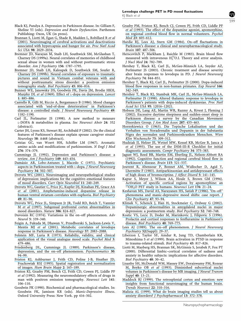
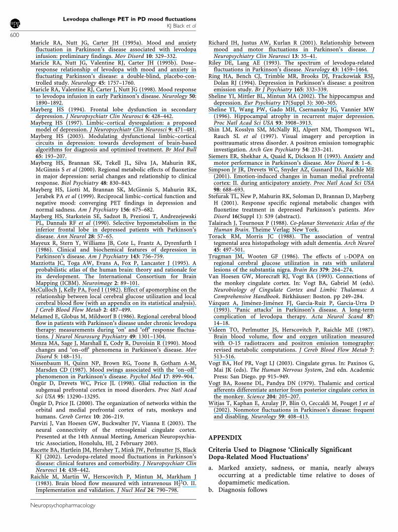
There was a significant difference between groups in theregional blood flow response to levodopa (mood4nonmood,experimentwise p¼ 0.002; moodononmood, experiment-wise po10�5). One cluster of voxels centered in the brainmet the predetermined criterion for significance after corre-ction for multiple comparisons. This cluster was identified inthe mood4nonmood comparison (corrected po0.01) andcontained two peaks centered at (�2,�26,54) and (�6,�26,36)(see Table 3 and Figure 1, top row). The first peak in thisregion is centered in medial frontal gyrus near the centralsulcus, and the second peak lies in PCC. Mean values fromspherical VOIs centered on these peaks show that PDcontrol patients have decreased rCBF after levodopa, whilemood fluctuators do not (repeated measures ANOVA,F1,19 ¼ 5.086, po0.04; post hoc Scheffe tests indicate a dec-rease with levodopa in the motor fluctuator group at po0.06but p40.25 for all other comparisons). Data from the PCCpeak are summarized in Figure 2. Other voxel clustersexceeding t¼ 3.17 are described in Table 3 and Figure 1.
No intracerebral cluster in the moodononmood compar-ison reached the specified significance criterion, but onecluster with corrected po0.08 had peak t¼ 4.37 at(20,�22,�16) in the body of the hippocampus (2.6 ml).The only other intracerebral voxel clusters surpassing
Table 2 Most Significant Regional Effects of Levodopa across All Subjects
Cluster p(cor) No. of voxels Voxel’s t x,y,z (mm) Description BA
510�5 9187 9.69 �4,�30,�26 Pons F
4.56 �24,�16,�12 Parahippocampal gyrus 28
4.00 �24,�10,�38 Parahippocampal gyrus/uncus 36/20
3.52 18,�26,16 Thalamus (pulvinar) F
0.002 949 6.47 36,42,�14 Middle frontal gyrus 11
0.001 1032 5.97 �42,�28,18 Insula 13
4.91 �32,�32,30 WM deep to inf. parietal lobule F
o10�5 2179 5.58 48,10,30 Middle frontal gyrus 9
4.45 26,22,36 WM deep to BA9 F
3.69 14,50,28 Superior frontal gyrus 9
0.089 304 4.38 �24,28,�20 Inferior frontal gyrus 11
0.082 316 4.29 50,�30,26 WM deep to inf. par. lobule 40
Most significant clusters of contiguous voxels with t43.17 (100 df), and local maxima within each cluster separated by more than 8 mm. See legend to Table 2 forabbreviations.
Levodopa challenge PET in PD mood fluctuationsKJ Black et al
593
Neuropsychopharmacology

t¼ 3.17 had peaks at (28,4,�20) (uncus, BA28, peak t¼ 4.01,1.1 ml), (�26,�22,18) (claustrum, peak t¼ 3.69, 0.3 ml), and(�24,�2,�40) (uncus, near BA20, peak t¼ 4.01, 0.2 ml).
Clinical Observations
There were no significant differences between groups on thebaseline characteristics listed in Table 1 (p40.10), exceptthat mood fluctuators were being treated with a higher totaldaily dose of levodopa or dopamine agonists (po0.05).
Clinical ratings of depression and anxiety, based onsymptoms during the week prior to the scan, showed amodest overall severity of mood and anxiety, with the meanin the nondepressed range (see Table 4). This is consistentwith the clinical description, since episodic, ultradiansymptoms define the group of interest, rather thansustained symptoms. Ratings of parkinsonian severity inboth groups diminished by roughly half following treatment(repeated measures ANOVA, drug effect po0.005; p40.20for both the diagnosis effect and the interaction), and mostsubjects had peak-dose dyskinesias (Table 4).
One mood fluctuator patient was crying in the waitingroom before the scan. Three mood fluctuators showedhypomanic behavior after levodopa, including euphoria,flirting or inappropriate sexual comments, silly or giddybehavior, joking, and talkativeness. Severity was mild, withBech mania scores of 1.5, 2, and 4. One patient showedsevere wearing-off bradyphrenia, trailing off to mutismwhile answering a question, only to spontaneously finish thesentence 10 min later during levodopa infusion. Oneadditional mood fluctuator patient said ‘I feel like I tookan upper.’ One control subject felt mood had improved andanother denied change in mood per se but said ‘mentally, Ifeel better.’
Surprisingly, several mood fluctuator patients did nothave marked sadness and anxiety during the baseline scans,and baseline mood ratings did not differ between patientgroups (Table 4). Patients told us they felt emotionallybetter during the study baseline than during their usual offperiods, and this was confirmed by their caregivers. Basedon their comments and our observations, we speculate thatthe constant personal attention we provided during thestudy somewhat reduced the severity of their off-periodanxiety and depression. To a small extent, the modestseverity of baseline symptoms on the study day may also
Figure 1 Top row: Mood fluctuators differed significantly from controlpatients with motor fluctuations only (Table 2). The t image is shown incolor superimposed on an averaged structural MR image in grayscale. Thecrosshairs are at the statistical peak in PCC (�6,�26,34). On coronalimages, the right side of the brain is shown on the right side of the figure.Other rows: Additional regions of possible group difference in levodoparesponse (Table 2). From top to bottom: Inferior parietal lobule, crosshairsat (68,�28,24), insula (48,�20,14), superior temporal gyrus (�52,�54,16),lateral orbital cortex (�24,50,�12).
Figure 2 The figures show mean7SD rCBF in an 8 mm spherical VOIcentered at the PCC peak shown in Figure 1, before and after levodopa, inmood fluctuators and in control patients with motor fluctuations only. Notethat each VOI includes data from 35 surrounding voxels in addition to thevoxel at the statistical peak.
Levodopa challenge PET in PD mood fluctuationsKJ Black et al
594
Neuropsychopharmacology

reflect a selection bias away from the most severely affectedpatients: two mood fluctuation patients who came to thePET suite to participate were unable to do so, one due tomarked off-period anxiety and the other due to severe off-period neck flexor dystonia. Both patient groups had higherin-scanner mood scores on levodopa (ANOVA, significanteffect of levodopa, po0.001), but the increase was twice asgreat in the mood fluctuators (interaction of diagnosis andlevodopa status, p¼ 0.12; post hoc two-tailed t-test, p¼ 0.02;see Table 4).
Levodopa Plasma Concentrations
Mean plasma concentrations increased from 1327177 ng/ml at baseline to 15907513 ng/ml after levodopa adminis-tration. There was no significant difference betweengroups in levodopa plasma concentrations either before orafter drug.
Subgroup Analyses
We examined whether it was likely that factors other thanthe mood fluctuation diagnosis affected the most significant
result in our primary PET analysis. First, some subjects ineach group received levodopa by mouth rather than i.v.Ignoring these subjects, the mean PCC rCBF response was�3.5% (controls) vs þ 1.3% (mood fluctuators), similar tothe means for all subjects at �3.6% (controls) and þ 0.8%(mood fluctuators). Thus the route of administration seemsto have had little effect on the main result. Second, onesubject in each group was scanned on the 953b scanner.Ignoring those two subjects, the mean PCC rCBF responsewas �3.9% (controls) and 1.3% (mood fluctuators), againsuggesting no meaningful difference. Third, the moodfluctuation group included patients with various combina-tions of off-period depression, off-period anxiety, and on-period mania or hypomania. The possible effect of moodfluctuation subtype on PCC rCBF response to levodopa isshown in Table 5; the numbers are too small and the patterntoo irregular for definitive conclusions, but the most‘typical’ response (ie the highest) for the mood fluctuatorgroup is in the only subject with both off-period depressionand on-period mania, while the most atypical (ie the mostnegative) response was in the only subject with neither.
Finally, some subjects in each group were taking anantidepressant (Table 1). Apparently antidepressants alonedo not explain the group difference in PCC response to
Table 4 Scan-Day Symptoms
Mood fluctuators Controls
Ham-D 8.374.9 5.074.6
Ham-A 8.974.8 5.872.6
Mood VAS ratings during scans (mm)
Sad (0)–happy (100), baseline 61723 58714
Sad (0)–happy (100), levodopa 86712 71713
Modified UPDRS (maximum¼ 32)
Baseline 7.173.7 11.879.3
Levodopa 4.171.7 6.573.7
Number with dyskinesias after levodopa 6 10
Table 3 Most Significant Regional Differences in Levodopa Response in the Mood Fluctuators4Motor Fluctuators Comparison
Cluster p(cor) No. of voxels Voxel’s t x,y,z (mm) Description BA
0.009 662 4.53 �2,�26,54 Medial frontal gyrus 6
3.79 �6,�26,34 Posterior cingulate 31
0.513 75 3.68 60,�54,10 Superior temporal gyrus 22
0.163 221 3.68 68,�28,24 Inferior parietal lobule 40
0.733 30 3.49 �50,14,�2 Inferior frontal gyrus 22
0.607 54 3.47 48,�20,14 Insula 13
0.803 18 3.42 �4,�38,10 Splenium/posterior cingulated F
0.647 46 3.40 �52,�54,16 WM deep to superior temporal gyrus 22
0.474 85 3.40 �24,50,�12 Middle frontal gyrus 11
0.797 19 3.35 �44,�20,10 WM deep to transverse temporal gyrus 13
Most significant clusters of contiguous voxels with t43.17 (100 df), and local maxima within each cluster separated by more than 8 mm (Figure 1).p(cor): p-value corrected for multiple comparisons; no. of voxels: number of contiguous voxels in each cluster; x,y,z: Talairach atlas coordinates; BA: Brodmann area.Voxel volume¼ 8 mm3.
Table 5 Mood Fluctuation Subtype and PCC Response toLevodopa
Off depression Off anxiety On mania PCC response (%)
N Y N �5.0
N N Ya �1.7
N N Y �.5
Y Y N �1.5
N Y Y +2.4
N Y Y +3.4
Y Y N +3.7
Y N Y +8.3
Additional diagnoses were off-period apathy (1) and off-periodbradyphrenia (1).aThis patient had on-period hypomania.
Levodopa challenge PET in PD mood fluctuationsKJ Black et al
595
Neuropsychopharmacology

levodopa, since within the motor fluctuator control groupthe response was similar regardless of antidepressantexposure (antidepressant, N¼ 5, �3.8%; none, N¼ 8,�3.4%). Mood fluctuators differed more, but neithersubgroup approached the responses of the controls (anti-depressant, N¼ 6, þ 1.4%; none, N¼ 2, �0.9%).
DISCUSSION
Baseline Differences in Resting rCBF in MoodFluctuators
To our knowledge, no other anatomic or functional imagingstudies have compared PD patients with and withoutlevodopa-related mood fluctuations. Published comparisonsof regional resting brain metabolism or rCBF between PDpatients with and without (nonfluctuating) major depres-sion have shown decreased activity of caudate nuclei andprefrontal cortex (Mayberg et al, 1990; Jagust et al, 1992;Mayberg, 1994; Ring et al, 1994). The decreased caudateactivity likely represents a true decrease per unit volumerather than a partial volume effect (Lisanby et al, 1993,p 18). In a before- and after-treatment FDG PET studyof depressed PD patients, an antidepressant response tofluoxetine was associated with a metabolic increase indorsal anterior cingulate regional metabolism and ametabolic decrease in ventral anterior cingulate (Stefuraket al, 2001; Mayberg, 2003). None of these differences weredetected in the mood fluctuators, in whom baseline rCBFdiffered only at a few voxels over 3 cm away.
Regional CBF Response to Levodopa in the WholeSample
The responses to levodopa in the combined patient samplelargely replicate those we previously observed in threeseparate samples of PD patients as well as in healthycontrols (Hershey et al, 1998, 2003). The large brainstemresponse to levodopa or dopamine agonists has beenreported in various species, and involves a diffuse midbrainarea even when assessed at much higher image resolutionusing [14C]2-deoxyglucose and ex vivo film autoradiogra-phy (Trugman and Wooten, 1986; Grasby et al, 1993; Kapuret al, 1994; Hershey et al, 2000; Black et al, 2000). In therodent studies, the response includes superior colliculus,midbrain reticular formation, and subthalamic nucleus.Other levodopa-responsive regions in Table 3 (eg para-hippocampal gyrus, insula, and lateral orbital cortex) werenot reported in our previous studies of levodopa activation,and may also mediate dopaminergic influences on mood orcognition. Together, these studies provide substantialinformation on the functional responses to levodopa innormal and parkinsonian humans.
Other groups have also reported levodopa activationimaging studies in PD, as reviewed in part by Hershey et al(2003). An early PET study found no rCBF differences afterlevodopa in 10 motor fluctuators (Melamed et al, 1986).Feigin et al (2001) report an FDG PET levodopa activationstudy in seven PD patients. As in the present study, theyfound a statistically modest decrease in activity incerebellum. Similar decreases occurred in putamen, thala-mus, and primary motor cortex; these likely would not have
been identified in our study due to differences in statisticalthreshold and axial field of view. Berding et al (2001) reporta similar study in 11 PD subjects; regional metabolismdecreased in orbital cortex (peak change was 12 mm fromthe peak in Table 2). Neither study detected regionalincreases, which may relate to sample size or differences inlevodopa challenge.
Regional CBF Response to Levodopa in MoodFluctuators vs Motor Fluctuator Controls
The primary PET analysis shows that the brain’s regionalresponse to levodopa is significantly different in PD patientswith levodopa-dose-related emotional changes, comparedto similar patients with fluctuations only in motorfunction. The rCBF response to levodopa detected groupdifferences not evident in the group comparison of baselineresting rCBF.
The most significant group difference in rCBF response tolevodopa occurred in a contiguous medial cortical regioncontaining a superior and an inferior peak. The superiorpeak (BA6) may reflect group differences in motor cortexinnervation by dopamine-influenced neurons, and may beimportant to mood fluctuations or may represent imperfectgroup matching on motor features of PD (although thegroups compared very closely on duration of illness, clinicalratings of parkinsonism, and prevalence of levodopa-induced dyskinesias). The other peak is centered in PCC,which has previously been shown to be abnormally active indepression or anxiety. This region probably includes BA31(Talairach and Tournoux, 1988) and retrosplenial cortex,which in primates extends this far anteriorly along thecorpus callosum (Parvizi et al, 2003).
Posterior Cingulate Cortex
In a PET study of major depression, metabolism in this partof PCC (peaks at (�8,�32,30) and (6,�26,32)) significantlyincreased in patients who responded to 6 weeks offluoxetine treatment, but significantly decreased in non-responders (Mayberg et al, 2000). Posterior cingulatemetabolism had decreased in both groups after 1 week offluoxetine treatment, before depression improved. In otherwords, activity of PCC was a state marker for remission ofmajor depression (increase), and for nontherapeuticexposure to fluoxetine (decrease). Similar results wereobserved over a much shorter time scale in an rCBF study ofinduced sadness or anxiety in normal volunteers (Liotti et al,2000). Decreased activity was observed during anxiousand depressed states in PCC, with peak differences at(6,�64,17) and (3,�40,20), somewhat posterior and inferiorto those seen here.
PCC activity is also linked to anxiety in different modelconditions. Breathlessness and air hunger induced byinhalation of 8% CO2 caused decreased PCC blood flowwith peak change at (0,�36,32) (Brannan et al, 2001), andthere was an B5% decrease in PCC rCBF in volunteers whowere anxious while awaiting a painful shock to the hand(did not reach statistical significance using conservativemethods) (Simpson et al, 2001). In studies of post-traumaticstress disorder that provoked anxiety by presenting trauma-related pictures and sounds, activations or deactivations
Levodopa challenge PET in PD mood fluctuationsKJ Black et al
596
Neuropsychopharmacology

were found in PCC (Shin et al, 1997; Bremner et al, 1999a, b;Liberzon et al, 1999). As in our study, Bremner et al (1999b)found that posterior cingulate activation in patients wassometimes of opposite sign than in healthy controls.
From such studies, Liotti et al (2000) concluded, ‘it isclear that the posterior cingulate plays a critical role in theregulation of both normal and pathologic negative emo-tions’ (p 36). In fact, Maddock (1999) noted that afterinferior prefrontal cortex, PCC (including retrosplenialcortex) was the brain region most consistently activated in51 functional neuroimaging studies of emotion. The specificrole PCC plays in emotion is not clear, but available datasuggest that it may encode the emotional significance ofstimuli, perhaps by mediating the comparison of presentpercepts to emotions associated with episodic (eg autobio-graphical) memory (Maddock, 1999).
The rCBF response in posterior cingulate may arise eitherfrom neurons intrinsic to PCC or from afferent projectionaxons terminating in PCC. Afferents to PCC includecontralateral PCC; ipsilateral anterior cingulate cortex(dorsal and ventral), posterior parietal cortex, superiortemporal sulcus (STS), parahippocampal gyrus, ventralclaustrum, and both orbital and dorsal prefrontal cortex;certain thalamic nuclei (pulvinar, lateral dorsal, anteriordorsal, anterior ventral); raphe nuclei and locus ceruleus;and the rostral medial anterior portion of caudate nucleus(Vogt et al, 1979, 2003; Baleydier and Mauguiere, 1980; VanHoesen et al, 1993; Parvizi et al, 2003). These regions mayhelp explain how PCC is modulated by dopamine or mood.
Dopaminergic modulation of PCC metabolism and bloodflow may arise directly from the caudate, which receives aheavy dopaminergic innervation. However, dopamine couldinfluence PCC indirectly via other brain regions. Forinstance, in a primate model, the most significanteffects on rCBF after administration of a dopamine agonistoccurred in STS after a D1 agonist and in orbital prefrontalcortex after a D3-preferring agonist (Black et al, 1997,2002b). Both regions project to PCC. In any case,levodopa and several dopamine agonists clearly affectPCC rCBF (Friston et al, 1992; Black et al, 2002a, b;Hershey et al, 2000).
Afferents to PCC may also contribute to the relationshipof emotion with PCC rCBF. For example, metabolism inboth rostral and ventral portions of anterior cingulatecortex is abnormal in major depression (Drevets, 2001;Mayberg, 1997). Patients with familial pure major depres-sion show decreased rCBF, metabolism, volume, and glialcell number in subgenual anterior cingulate cortex (Drevetset al, 1997; Ongur et al, 1998). Thus an altered PCC responseto dopamine in patients with mood fluctuations may reflectabnormal dopaminergic modulation of caudate, anteriorcingulate, or prefrontal cortex. One possible mechanismcould be differential loss of dopamine innervation to thesenuclei; no data directly address this possibility, but Torackand Morris (1988) did find greater cell loss in ventraltegmental area in PD patients with an antemortemdiagnosis of depression.
PCC: A Hypothesis
The motor fluctuator controls responded to levodopa with adecrease in PCC rCBF, an apparently normal response
replicated by D2-like agonists (Hershey et al 2000; Blacket al 2002a, b). Decreased PCC activity is often associatedwith anxiety or sadness (Mayberg, 2003), but must not besufficient to produce these emotions since levodopa andD2-like agonists do not usually cause anxiety or sadness.We speculate that dopaminergic pathways that affect PCCactivity may mitigate extremes of mood. The moodfluctuators’ loss of the PCC response to levodopa maythus be related to their clinical experience of emotionalextremes. The valence of the dopamine-related moodchanges may be determined by activity in other, anatomi-cally connected, areas of the brain such as anterior cingulateor orbital cortex.
Other Group Differences in rCBF Responses toLevodopa
Several other regions showed a more positive response tolevodopa in mood fluctuators. These included regions nearSTS, the inferior parietal lobule, the insula, and orbitalfrontal cortex (Table 2 and Figure 1). Lateral orbital cortex(BA11) is strongly implicated in regulation of mood andimpulsivity, receives striatal afferents, and is influenced bydopamine, suggesting that this region may be relevant to themood and anxiety fluctuations that differentiate our twogroups of patients (Ongur and Price, 2000; Black et al,2002b). A PET study of major depression identified rCBF inposterior inferior parietal lobule as correlated with anxiety(Grasby et al, 1993). In several studies, decreased metabolicactivity in inferior parietal cortex (BA40) correspondshighly with state sadness or with an anxiety disorder, butthe peak in Table 2 is at least 26 mm from these regions(Mayberg et al, 1999; Bremner et al, 1999a, b). Levodopadoes decrease inferior parietal blood flow in patients andnormal controls (Hershey et al, 2003). Several studies ofsadness or anxiety have detected abnormal activity ofinsula, primarily but not exclusively anterior insula (Mal-izia, 1999; Mayberg et al, 1999). There is no obviousrelationship of STS or surrounding cortex to moodregulation, but STS rCBF is affected by dopamine agonists(Black et al, 2000).
A cluster of voxels in hippocampus and parahippocampalgyrus reacted in the opposite direction (more negativeresponse to levodopa in mood fluctuators, po0.08). Thepotential role of the hippocampus in idiopathic majordepression has been reviewed recently (Sheline et al, 2002;Mayberg, 2003). Chronicity of major depression is asso-ciated with decreased hippocampal volume (Sheline et al,1996). Furthermore, in serotonergic treatment of majordepression, either with or without PD, hippocampal activitydecreases with treatment response but increases withtreatment failure (Mayberg, 2003). This pattern is not seenwith response to cognitive therapy or placebo, however.Conceivably, the greater decrease in hippocampal activitywith levodopa in mood fluctuators corresponds to thegreater improvement in mood in these subjects.
Pharmacology
The relationship of dopamine to (nonfluctuating) depres-sion in PD is unclear (Bunney et al, 1969; Jouvent et al,1983; Mayeux et al, 1986; Torack and Morris, 1988;
Levodopa challenge PET in PD mood fluctuationsKJ Black et al
597
Neuropsychopharmacology

Cummings, 1992; Kostic et al, 1996; Black and Pandya, inpress). By contrast, levodopa-related mood fluctuations inPD have a strongly face valid relationship to braindopamine concentrations. Dopaminergic effects on moodare well documented and have been reviewed elsewhere(Drevets et al, 2001; Klimek et al, 2002; Racette et al, 2002).However, there is little direct evidence as to what receptorsubtype(s) may mediate dopamine’s modulation of emo-tional symptoms or rCBF in these mood fluctuationpatients.
Recent pharmacologic activation PET studies in a primatemodel may shed light on this question. The activation ofPCC rCBF by levodopa is replicated by D2-like dopamineagonists, but not by a D1 agonist (Black et al, 2000,2002a, b). Similarly, lateral orbitofrontal cortex was stronglyactivated in normal baboons by the D3-preferring agonistpramipexole (Black et al, 2002b), and superior temporalgyrus is strongly activated by a D1 agonist (Black et al,2000). Further study will be required to confirm whether PDpatients with mood fluctuations have more pronouncedmood responses to D2-like than to D1-like dopamineagonists, or whether the imaging findings in this levodopaactivation study can be replicated with specific agonists.
Clinical Results, Limitations, and Future Directions
The two study groups were defined by a clinical history ofmood fluctuations. In the PET scanner, the two groups hadonly modest differences in mood ratings. In interpreting theimaging results, this observation reduces the possibleconfound of state-related group differences during the scansession. A similar dissociation between usual clinicalresponse outside the scanner and clinical observations inthe experimental setting benefited our prior levodopachallenge PET study of drug-induced dyskinesias (Hersheyet al, 1998). However, state-related changes in mood may beexamined if larger in-scanner fluctuations of mood areobserved in future patients.
The small number of subjects makes the study vulnerableto Type II errors. For instance, previous studies of restingregional brain metabolism in PD detected significantabnormalities in patients with major depression (Mayberget al, 1990). Mood fluctuators were taking more levodopaand dopamine agonists, but this likely reflects efforts totreat off-period psychiatric symptoms, given the compar-able off-period motor signs and duration of illness (Table 1).Additional limitations of this study include the potential forunidentified clinical differences between groups, and theheterogeneity in clinical and study procedures addressed inResults. Also, regional blood flow is an indirect measure ofregional metabolism and neuronal function, and itsinterpretation depends on the tight coupling between rCBFand regional metabolism. This coupling is preserved even inthe presence of non-ergot dopamine agonists (McCullochet al, 1982), and is likely even more stable in ourexperiment, since with levodopa/carbidopa, dopamineproduction outside the brain is blocked and levodopa hasno net effect on whole-brain blood flow (Hershey et al, 1998,2000, 2003).
Our results demonstrate the utility of the pharmacologicactivation approach in studying neuropsychiatric illness:although PCC has known dopamine-influenced afferents
and known association with emotional regulation, it has notpreviously been considered in discussions of levodopa-related mood fluctuations in PD. Also, in the absence ofpharmacologic stimulation, no baseline difference in PCCrCBF was identified. Future clinical, neuroimaging, andpathological studies may clarify the pathophysiology andpharmacology of PCC with respect to levodopa-mediatedeffects on mood.
ACKNOWLEDGEMENTS
The study was supported by Jonathan M Koller, Lennis Lich(Washington University), and Dr Kathryn Vehe (Barnes-Jewish Hospital). Patient self-ratings software was designedby Dr Robert J Feiwell. Patient referral was by Drs JonathanW Mink (now at University of Rochester Medical Center),Brad A Racette, and Fredy J Revilla (now at University ofCincinnati College of Medicine). This work was presented inpart at the Mental and Behavioral Dysfunction in MovementDisorders International Symposium, Montreal, Canada, 10–13 October 2001, at the Society for Neuroscience annualmeeting, San Diego, California, 14 November 2001, and atthe American College of Neuropsychopharmacology annualmeeting, 10 December 2001. Funding was provided byNINDS (NS01898), the American Parkinson Disease Asso-ciation (APDA) Advanced Research Center at WashingtonUniversity and the Greater St Louis chapter of the APDA,the Charles A Dana Foundation, and the McDonnell Centerfor Higher Brain Function. KJB was supported by a YoungInvestigator Award from the National Alliance for Researchon Schizophrenia and Depression (NARSAD).
REFERENCES
Baleydier C, Mauguiere F (1980). The duality of the cingulate gyrusin monkey. Neuroanatomical study and functional hypothesis.Brain 103: 525–554.
Baruzzi A, Contin M, Albani F, Riva R (1986). Simple and rapidmicromethod for the determination of levodopa and 3-O-methyldopa in human plasma by high-performance liquidchromotography with coulometric detection. J Chromatogr B375: 165–169.
Berding G, Odin P, Brooks DJ, Nikkhah G, Matthies C, Peschel Tet al (2001). Resting regional cerebral glucose metabolism inadvanced Parkinson’s disease studied in the off and onconditions with [(18)F]FDG-PET. Mov Disord 16: 1014–1022.
Black KJ, Carl JL, Hartlein JM, Warren SL, Hershey T, PerlmutterJS (2003). Rapid intravenous loading of levodopa for humanresearch: clinical results. J Neurosci Methods 127: 19–29.
Black KJ, Gado MH, Perlmutter JS (1997). PET measurement ofdopamine D2 receptor-mediated changes in striatopallidalfunction. J Neurosci 17: 3168–3177.
Black KJ, Hershey T, Gado MH, Perlmutter JS (2000). DopamineD1 agonist activates temporal lobe structures in primates. JNeurophysiol 84: 549–557.
Black KJ, Hershey T, Koller JM, Carl JL, Perlmutter JS (2002a).Mapping and quantification of dopamine D2 receptor activation.J Neuropsychiatr Clin Neurosci 14: 118–119 (abstract).
Black KJ, Hershey T, Koller JM, Videen TO, Mintun MA, Price JLet al (2002b). A possible substrate for dopamine-relatedchanges in mood and behavior: prefrontal and limbic effects ofa D3-preferring dopamine agonist. Proc Natl Acad Sci USA 99:17113–17118.
Levodopa challenge PET in PD mood fluctuationsKJ Black et al
598
Neuropsychopharmacology
Black KJ, Pandya A. Depression in Parkinson disease. In: Gilliam F,Sheline YI (eds). Depression and Brain Dysfunction. ParthenonPublishing: Oxon, UK (in press).
Brannan S, Liotti M, Egan G, Shade R, Madden L, Robillard R et al(2001). Neuroimaging of cerebral activations and deactivationsassociated with hypercapnia and hunger for air. Proc Natl AcadSci USA 98: 2029–2034.
Bremner JD, Narayan M, Staib LH, Southwick SM, McGlashan T,Charney DS (1999a). Neural correlates of memories of childhoodsexual abuse in women with and without posttraumatic stressdisorder. Am J Psychiatry 156: 1787–1795.
Bremner JD, Staib LH, Kaloupek D, Southwick SM, Soufer R,Charney DS (1999b). Neural correlates of exposure to traumaticpictures and sound in Vietnam combat veterans with andwithout posttraumatic stress disorder: a positron emissiontomography study. Biol Psychiatry 45: 806–816.
Bunney WE, Janowsky DS, Goodwin FK, Davis JM, Brodie HKH,Murphy DL et al (1969). Effect of L-dopa on depression. Lancet1: 885–886.
Cantello R, Gilli M, Riccio A, Bergamasco B (1986). Mood changesassociated with ‘end-of-dose deterioration’ in Parkinson’sdisease: a controlled study. J Neurol Neurosurg Psychiatry 49:1182–1190.
Carl JL, Perlmutter JS (1998). A new method to measureL-DOPA & metabolites in plasma. Soc Neurosci Abstr 24: 352(abstract).
Carter JH, Lyons KS, Stewart BJ, Archibald P (2002). Do the clinicalfeatures of Parkinson’s disease explain spouse caregiver strain?Neurology 58: A468 (abstract).
Cotzias GC, van Woert HH, Schiffer LM (1967). Aromaticamino acids and modifications of parkinsonism. N Engl J Med276: 374–379.
Cummings JL (1992). Depression and Parkinson’s disease: areview. Am J Psychiatry 149: 443–454.
Damasio AR, Lobo-Antunes J, Macedo C (1971). Psychiatricaspects in Parkinsonism treated with L-dopa. J Neurol NeurosurgPsychiatry 34: 502–507.
Drevets WC (2001). Neuroimaging and neuropathological studiesof depression: implications for the cognitive–emotional featuresof mood disorders. Curr Opin Neurobiol 11: 240–249.
Drevets WC, Gautier C, Price JC, Kupfer DJ, Kinahan PE, Grace AAet al (2001). Amphetamine-induced dopamine release inhuman ventral striatum correlates with euphoria. Biol Psychiatry49: 81–96.
Drevets WC, Price JL, Simpson Jr JR, Todd RD, Reich T, VannierM et al (1997). Subgenual prefrontal cortex abnormalities inmood disorders. Nature 386: 824–827.
Duvoisin RC (1974). Variations in the on–off phenomenon. AdvNeurol 5: 339–340.
Feigin A, Fukuda M, Dhawan V, Przedborski S, Jackson-Lewis V,Mentis MJ et al (2001). Metabolic correlates of levodoparesponse in Parkinson’s disease. Neurology 57: 2083–2088.
Folstein MF, Luria R (1973). Reliabiliy, validity, and clinicalapplication of the visual analogue mood scale. Psychol Med 3:479–486.
Friedenberg DL, Cummings JL (1989). Parkinson’s disease,depression, and the on–off phenomenon. Psychosomatics 30:94–99.
Friston KJ, Ashburner J, Frith CD, Poline J-B, Heather JD,Frackowiak RSJ (1995). Spatial registration and normalizationof images. Hum Brain Mapp 2: 165–189.
Friston KJ, Grasby PM, Bench CJ, Frith CD, Cowen PJ, Liddle PFet al (1992). Measuring the neuromodulatory effects of drugs inman with positron emission tomography. Neurosci Lett 141:106–110.
Goodwin FK (1990). Biochemical and pharmacological studies. In:Goodwin FK, Jamison KR (eds). Manic-Depressive Illness.Oxford University Press: New York. pp 416–502.
Grasby PM, Friston KJ, Bench CJ, Cowen PJ, Frith CD, Liddle PFet al (1993). The effect of the dopamine agonist, apomorphine,on regional cerebral blood flow in normal volunteers. PsycholMed 23: 605–612.
Hardie RJ, Lees AJ, Stern GM (1984). On–off fluctuations inParkinson’s disease: a clinical and neuropharmacological study.Brain 107: 487–506.
Herscovitch P, Markham J, Raichle M (1983). Brain blood flowmeasured with intravenous H2
15O.I. Theory and error analysis.J Nucl Med 24: 782–789.
Hershey T, Black KJ, Carl JL, McGee-Minnich LA, Snyder AZ,Perlmutter JS (2003). Chronic treatment and disease severityalter brain responses to levodopa in PD. J Neurol NeurosurgPsychiatry 74: 844–851.
Hershey T, Black KJ, Carl JL, Perlmutter JS (2000). Dopa-inducedblood flow responses in non-human primates. Exp Neurol 166:342–349.
Hershey T, Black KJ, Stambuk MK, Carl JL, McGee-Minnich LA,Perlmutter JS (1998). Altered thalamic response to levodopa inParkinson’s patients with dopa-induced dyskinesias. Proc NatlAcad Sci USA 95: 12016–12021.
Hobson DE, Lang AE, Martin WR, Razmy A, Rivest J, Fleming J(2002). Excessive daytime sleepiness and sudden-onset sleep inParkinson disease: a survey by the Canadian MovementDisorders Group. J Am Med Assoc 287: 455–463.
Hornykiewicz O (1963). Die Topische Lokalisation und dasVerhalten von Noradrenalin und Dopamin in der SubstantiaNigra des normalen und Parkinsonkranken Menschen. WienKlin Wochenschr 75: 309–312.
Hudziak JJ, Helzer JE, Wetzel MW, Kessel KB, McGee B, Janca Aet al (1993). The use of the DSM-III-R Checklist for initialdiagnostic assessments. Compr Psychiatry 34: 375–383.
Jagust WJ, Reed BR, Martin EM, Eberling JL, Nelson-Abbott RA(1992). Cognitive function and regional cerebral blood flow inParkinson’s disease. Brain 115: 521–537.
Jouvent R, Abensour P, Bonnet AM, Widlocher D, Agid Y,Lhermitte F (1983). Antiparkinsonian and antidepressant effectsof high doses of bromocriptine. J Affect Disord 5: 141–145.
Kapur S, Meyer J, Wilson AA, Houle S, Brown GM (1994).Activation of specific cortical regions by apomorphine: an15OH2O PET study in humans. Neurosci Lett 176: 21–24.
Keshavan MS, David AS, Narayanen HS, Satish P (1986). ‘On–off ’phenomena and manic-depressive mood shifts: case report. JClin Psychiatry 47: 93–94.
Klimek V, Schenck J, Han H, Stockmeier C, Ordway G (2002).Dopaminergic abnormalities in amygdaloid nuclei in majordepression: a postmortem study. Biol Psychiatry 52: 740–748.
Kostic VS, Lecic D, Doder M, Marinkovic J, Filipovic S (1996).Prolactin and cortisol responses to fenfluramine in Parkinson’sdisease. Biol Psychiatry 40: 769–775.
Lees AJ (1989). The on–off phenomenon. J Neurol NeurosurgPsychiatry 52(Suppl): 29–37.
Liberzon I, Taylor SF, Amdur R, Jung TD, Chamberlain KR,Minoshima S et al (1999). Brain activation in PTSD in responseto trauma-related stimuli. Biol Psychiatry 45: 817–826.
Liotti M, Mayberg HS, Brannan SK, McGinnis S, Jerabek P, Fox PT(2000). Differential limbic–cortical correlates of sadness andanxiety in healthy subjects: implications for affective disorders.Biol Psychiatry 48: 30–42.
Lisanby SH, McDonald WM, Massey EW, Doraiswamy PM, RozearM, Boyko OB et al (1993). Diminished subcortical nucleivolumes in Parkinson’s disease by MR imaging. J Neural TransmSuppl 40: 13–21.
Maddock RJ (1999). The retrosplenial cortex and emotion: newinsights from functional neuroimaging of the human brain.Trends Neurosci 22: 310–316.
Malizia AL (1999). What do brain imaging studies tell us aboutanxiety disorders? J Psychopharmacol 13: 372–378.
Levodopa challenge PET in PD mood fluctuationsKJ Black et al
599
Neuropsychopharmacology
Maricle RA, Nutt JG, Carter JH (1995a). Mood and anxietyfluctuation in Parkinson’s disease associated with levodopainfusion: preliminary findings. Mov Disord 10: 329–332.
Maricle RA, Nutt JG, Valentine RJ, Carter JH (1995b). Dose–response relationship of levodopa with mood and anxiety influctuating Parkinson’s disease: a double-blind, placebo-con-trolled study. Neurology 45: 1757–1760.
Maricle RA, Valentine RJ, Carter J, Nutt JG (1998). Mood responseto levodopa infusion in early Parkinson’s disease. Neurology 50:1890–1892.
Mayberg HS (1994). Frontal lobe dysfunction in secondarydepression. J Neuropsychiatr Clin Neurosci 6: 428–442.
Mayberg HS (1997). Limbic–cortical dysregulation: a proposedmodel of depression. J Neuropsychiatr Clin Neurosci 9: 471–481.
Mayberg HS (2003). Modulating dysfunctional limbic–corticalcircuits in depression: towards development of brain-basedalgorithms for diagnosis and optimised treatment. Br Med Bull65: 193–207.
Mayberg HS, Brannan SK, Tekell JL, Silva JA, Mahurin RK,McGinnis S et al (2000). Regional metabolic effects of fluoxetinein major depression: serial changes and relationship to clinicalresponse. Biol Psychiatry 48: 830–843.
Mayberg HS, Liotti M, Brannan SK, McGinnis S, Mahurin RK,Jerabek PA et al (1999). Reciprocal limbic–cortical function andnegative mood: converging PET findings in depression andnormal sadness. Am J Psychiatry 156: 675–682.
Mayberg HS, Starkstein SE, Sadzot B, Preziosi T, AndrezejewskiPL, Dannals RF et al (1990). Selective hypometabolism in theinferior frontal lobe in depressed patients with Parkinson’sdisease. Ann Neurol 28: 57–65.
Mayeux R, Stern Y, Williams JB, Cote L, Frantz A, Dyrenfurth I(1986). Clinical and biochemical features of depression inParkinson’s disease. Am J Psychiatry 143: 756–759.
Mazziotta JC, Toga AW, Evans A, Fox P, Lancaster J (1995). Aprobabilistic atlas of the human brain: theory and rationale forits development. The International Consortium for BrainMapping (ICBM). Neuroimage 2: 89–101.
McCulloch J, Kelly PA, Ford I (1982). Effect of apomorphine on therelationship between local cerebral glucose utilization and localcerebral blood flow (with an appendix on its statistical analysis).J Cereb Blood Flow Metab 2: 487–499.
Melamed E, Globus M, Mildworf B (1986). Regional cerebral bloodflow in patients with Parkinson’s disease under chronic levodopatherapy: measurements during ‘on’ and ‘off’ response fluctua-tions. J Neurol Neurosurg Psychiatry 49: 1301–1304.
Menza MA, Sage J, Marshall E, Cody R, Duvoisin R (1990). Moodchanges and ‘on–off’ phenomena in Parkinson’s disease. MovDisord 5: 148–151.
Nissenbaum H, Quinn NP, Brown RG, Toone B, Gotham A-M,Marsden CD (1987). Mood swings associated with the ‘on–off ’phenomenon in Parkinson’s disease. Psychol Med 17: 899–904.
Ongur D, Drevets WC, Price JL (1998). Glial reduction in thesubgenual prefrontal cortex in mood disorders. Proc Natl AcadSci USA 95: 13290–13295.
Ongur D, Price JL (2000). The organization of networks within theorbital and medial prefrontal cortex of rats, monkeys andhumans. Cereb Cortex 10: 206–219.
Parvizi J, Van Hoesen GW, Buckwalter JV, Vianna E (2003). Theneural connectivity of the retrosplenial cingulate cortex.Presented at the 14th Annual Meeting, American Neuropsychia-tric Association, Honolulu, HI, 2 February 2003.
Racette BA, Hartlein JM, Hershey T, Mink JW, Perlmutter JS, BlackKJ (2002). Levodopa-related mood fluctuations in Parkinson’sdisease: clinical features and comorbidity. J Neuropsychiatr ClinNeurosci 14: 438–442.
Raichle M, Martin W, Herscovitch P, Mintun M, Markham J(1983). Brain blood flow measured with intravenous H2
15O. II.Implementation and validation. J Nucl Med 24: 790–798.
Richard IH, Justus AW, Kurlan R (2001). Relationship betweenmood and motor fluctuations in Parkinson’s disease. JNeuropsychiatry Clin Neurosci 13: 35–41.
Riley DE, Lang AE (1993). The spectrum of levodopa-relatedfluctuations in Parkinson’s disease. Neurology 43: 1459–1464.
Ring HA, Bench CJ, Trimble MR, Brooks DJ, Frackowiak RSJ,Dolan RJ (1994). Depression in Parkinson’s disease: a positronemission study. Br J Psychiatry 165: 333–339.
Sheline YI, Mittler BL, Mintun MA (2002). The hippocampus anddepression. Eur Psychiatry 17(Suppl 3): 300–305.
Sheline YI, Wang PW, Gado MH, Csernansky JG, Vannier MW(1996). Hippocampal atrophy in recurrent major depression.Proc Natl Acad Sci USA 93: 3908–3913.
Shin LM, Kosslyn SM, McNally RJ, Alpert NM, Thompson WL,Rauch SL et al (1997). Visual imagery and perception inposttraumatic stress disorder. A positron emission tomographicinvestigation. Arch Gen Psychiatry 54: 233–241.
Siemers ER, Shekhar A, Quaid K, Dickson H (1993). Anxiety andmotor performance in Parkinson’s disease. Mov Disord 8: 1–6.
Simpson Jr JR, Drevets WC, Snyder AZ, Gusnard DA, Raichle ME(2001). Emotion-induced changes in human medial prefrontalcortex: II. during anticipatory anxiety. Proc Natl Acad Sci USA98: 688–693.
Stefurak TL, New P, Mahurin RK, Soloman D, Brannan D, MaybergH (2001). Response specific regional metabolic changes withfluoxetine treatment in depressed Parkinson’s patients. MovDisord 16(Suppl 1): S39 (abstract).
Talairach J, Tournoux P (1988). Co-planar Stereotaxic Atlas of theHuman Brain. Theime Verlag: New York.
Torack RM, Morris JC (1988). The association of ventraltegmental area histopathology with adult dementia. Arch Neurol45: 497–501.
Trugman JM, Wooten GF (1986). The effects of L-DOPA onregional cerebral glucose utilization in rats with unilaterallesions of the substantia nigra. Brain Res 379: 264–274.
Van Hoesen GW, Morecraft RJ, Vogt BA (1993). Connections ofthe monkey cingulate cortex. In: Vogt BA, Gabriel M (eds).Neurobiology of Cingulate Cortex and Limbic Thalamus: AComprehensive Handbook. Birkhauser: Boston. pp 249–284.
Vazquez A, Jimenez-Jimenez FJ, Garcia-Ruiz P, Garcia-Urra D(1993). ‘Panic attacks’ in Parkinson’s disease. A long-termcomplication of levodopa therapy. Acta Neurol Scand 87:14–18.
Videen TO, Perlmutter JS, Herscovitch P, Raichle ME (1987).Brain blood volume, flow and oxygen utilization measuredwith O-15 radiotracers and positron emission tomography:revised metabolic computations. J Cereb Blood Flow Metab 7:513–516.
Vogt BA, Hof PR, Vogt LJ (2003). Cingulate gyrus. In: Paxinos G,Mai JK (eds). The Human Nervous System, 2nd edn. AcademicPress: San Diego. pp 915–949.
Vogt BA, Rosene DL, Pandya DN (1979). Thalamic and corticalafferents differentiate anterior from posterior cingulate cortex inthe monkey. Science 204: 205–207.
Witjas T, Kaphan E, Azulay JP, Blin O, Ceccaldi M, Pouget J et al(2002). Nonmotor fluctuations in Parkinson’s disease: frequentand disabling. Neurology 59: 408–413.
APPENDIX
Criteria Used to Diagnose ‘Clinically SignificantDopa-Related Mood Fluctuations’
a. Marked anxiety, sadness, or mania, nearly alwaysoccurring at a predictable time relative to doses ofdopamimetic medication.
b. Diagnosis follows
Levodopa challenge PET in PD mood fluctuationsKJ Black et al
600
Neuropsychopharmacology

1. corroboration of history by spouse or other infor-mant, and
2. thorough neurological and psychiatric examinationby a clinician experienced in evaluating moodsymptoms in PD patients.
c. The symptoms cause clinically significant distress, orimpairment in social, occupational, or other importantareas of functioning.
d. The symptoms are not better accounted for by anothersyndrome, including major depression (ie while experi-encing above symptoms, patient must have normalmood and interests most of the time for at least amonth), pathological crying, or pure apathy; and are notmore parsimoniously attributable to PD without depres-sion (eg facial hypomimia, isolated fatigue, ‘internaltremor’ without other evidence of anxiety, or akathisiaor tremor that the patient misidentifies as ‘anxiety’).
A Screening Interview in a PD Patient Diagnosed withClinically Significant Mood Fluctuations (Summary,Based on Contemporaneous Written Notes)
First, the examiner established that the effect of each dose oflevodopa often wore off before the next dose was due,despite successive changes in the dosage of levodopa/carbidopa, switch to the sustained-release form, and
addition of ropinirole and later tolcapone. Then, theexaminer asked, ‘What symptoms do you notice when yourmedicine wears off?’ The patient replied, ‘I have somestiffness, [pause] but what markedly changes is my mentalattitude. I get depressed feeling and incapable of doinghardly anything.’ On follow-up questions, he describedfeeling at those moments sad, blue, tearful, tense, andworried, with a ‘negative attitude,’ low energy, and poorconcentration. At those times, ‘I question whether life isworth living.’ He was able occasionally to nap at these times.After taking his next dose of medication ‘it kicks in about30 min later and I feel good again.’ A semistructuredpsychiatric interview (Hudziak et al, 1993) revealed noadditional psychiatric symptoms except as follows. He twicehad developed transient hallucinations in late life, once aftersurgery and once after addition of an unknown antiparkin-sonian medication. He had a fear of heights starting in his40s without significant distress or impairment. He had hadno significant depressive symptoms until 10 years after thediagnosis of PD. At 3 years prior to study participation,after the death of his wife, he developed frequent sadnessand initial or middle insomnia but (except when ‘off’) hadnormal appetite, normal concentration, no excessive guilt,no serious thoughts of suicide, and generally intactinterests. He had never met research criteria for majordepression.
Levodopa challenge PET in PD mood fluctuationsKJ Black et al
601
Neuropsychopharmacology
Copyright © 2022 FDOKUMEN