Levels of arsenic, cadmium, lead and mercury in the branchial plate and muscle tissue of mobulid...
9
Levels of arsenic, cadmium, lead and mercury in the branchial plate and muscle tissue of mobulid rays Michelle S.M. Ooi a,⇑ , Kathy A. Townsend b,c , Michael B. Bennett d , Anthony J. Richardson e,f , Daniel Fernando g,h , Cesar A. Villa i , Caroline Gaus i a School of Veterinary Sciences, The University of Queensland, Gatton, QLD 4343, Australia b Moreton Bay Research Station, The University of Queensland, Dunwich, QLD 4183, Australia c School of Biological Sciences, The University of Queensland, St. Lucia, QLD 4072, Australia d School of Biomedical Sciences, The University of Queensland, St. Lucia, QLD 4072, Australia e Centre for Applications in Natural Resource Mathematics (CARM), The University of Queensland, St. Lucia, QLD 4072, Australia f CSIRO Wealth from Oceans Flagship, Division of Marine and Atmospheric Research, Dutton Park, QLD 4102, Australia g Department of Biology and Environmental Science, Linnaeus University, SE 39182 Kalmar, Sweden h The Manta Trust, Dorchester, United Kingdom i The National Research Centre for Environmental Toxicology, The University of Queensland, Coopers Plains, QLD 4108, Australia article info Article history: Available online xxxx Keywords: Mobula japanica Manta alfredi As Cd Pb Hg abstract Mobulid rays are targeted in fisheries for their branchial plates, for use in Chinese medicine. Branchial plate and muscle tissue from Mobula japanica were collected from fish markets in Sri Lanka, and muscle tissue biopsies from Manta alfredi in Australia. These were analysed for arsenic, cadmium, lead and mer- cury and compared to maximum levels (MLs) set by Food Standards Australia and New Zealand (FSANZ), European Commission (EC) and Codex Alimentarius Commission. The estimated intake for a vulnerable human age group was compared to minimal risk levels set by the Agency for Toxic Substances and Disease Registry. The mean inorganic arsenic concentration in M. japanica muscle was equivalent to the FSANZ ML while cadmium exceeded the EC ML. The mean concentration of lead in M. alfredi muscle tissue exceeded EC and Codex MLs. There were significant positive linear correlations between branchial plate and muscle tissue concentrations for arsenic, cadmium and lead. Ó 2015 The Authors. Published by Elsevier Ltd. This is an open access article under the CC BY-NC-ND license (http://creativecommons.org/licenses/by-nc-nd/4.0/). 1. Introduction Marine organisms, differentially exposed to metals, will accu- mulate the metals in their tissues to varying degrees as a result of differences in exposure routes and effects related to environ- mental chemistry (Adams et al., 2011). The primary routes of uptake by fish are via the gills and gut (McGeer et al., 2011; McIntyre and Linton, 2011). In response to public concern over exposure to metals through consumption of seafood, which caused incidents such as the outbreak of Minamata disease in Japan (Harada, 1995), international agencies (FAO/WHO and EU) have established limits for metals in various types of seafood (CODEX, 2012; Commission of the European Communities, 2006). Mobulid rays (Family Myliobatidae) belong to two distinctive genera, Manta and Mobula, consisting of eleven planktivorous fil- ter-feeding elasmobranchs that inhabit tropical, subtropical and temperate seas worldwide (Eschmeyer and Fong, 2014; Couturier et al., 2012). These mobulid species are under threat from anthro- pogenic activities such as targeted fishing, fisheries bycatch, boat strikes and marine litter (Ibid). Targeted fishing is a major threat that occurs in many regions around the world including The Democratic Socialist Republic of Sri Lanka (RSL) and the Republic of Indonesia (Fernando and Stevens, 2011; Dewar, 2002). In Lamakera, Indonesia, the number of mobulid rays caught increased from historical levels of 200–300 individuals per season to 1500 in 2002 (Ibid). This increase is attributed to the growing demand from the Chinese medicine market for dried mobulid branchial plates (Fernando and Stevens, 2011). At present, all mobulid spe- cies are classed as either Data Deficient, Near Threatened, Vulnerable or Endangered on the IUCN Red List of Threatened Species (International Union for Conservation of Nature, 2014), which makes the substantial market for branchial plates a concern for the long-term survival of the species. Sharks, rays, and skates are capable of accumulating non-essen- tial elements such as mercury (Hg) and lead (Pb) in their tissues http://dx.doi.org/10.1016/j.marpolbul.2015.02.005 0025-326X/Ó 2015 The Authors. Published by Elsevier Ltd. This is an open access article under the CC BY-NC-ND license (http://creativecommons.org/licenses/by-nc-nd/4.0/). ⇑ Corresponding author. E-mail address: [email protected] (M.S.M. Ooi). Marine Pollution Bulletin xxx (2015) xxx–xxx Contents lists available at ScienceDirect Marine Pollution Bulletin journal homepage: www.elsevier.com/locate/marpolbul Please cite this article in press as: Ooi, M.S.M., et al. Levels of arsenic, cadmium, lead and mercury in the branchial plate and muscle tissue of mobulid rays. Mar. Pollut. Bull. (2015), http://dx.doi.org/10.1016/j.marpolbul.2015.02.005
-
Upload
independent -
Category
Documents
-
view
2 -
download
0
Transcript of Levels of arsenic, cadmium, lead and mercury in the branchial plate and muscle tissue of mobulid...

Marine Pollution Bulletin xxx (2015) xxx–xxx
Contents lists available at ScienceDirect
Marine Pollution Bulletin
journal homepage: www.elsevier .com/locate /marpolbul
Levels of arsenic, cadmium, lead and mercury in the branchial plate andmuscle tissue of mobulid rays
http://dx.doi.org/10.1016/j.marpolbul.2015.02.0050025-326X/� 2015 The Authors. Published by Elsevier Ltd.This is an open access article under the CC BY-NC-ND license (http://creativecommons.org/licenses/by-nc-nd/4.0/).
⇑ Corresponding author.E-mail address: [email protected] (M.S.M. Ooi).
Please cite this article in press as: Ooi, M.S.M., et al. Levels of arsenic, cadmium, lead and mercury in the branchial plate and muscle tissue of mobulMar. Pollut. Bull. (2015), http://dx.doi.org/10.1016/j.marpolbul.2015.02.005
Michelle S.M. Ooi a,⇑, Kathy A. Townsend b,c, Michael B. Bennett d, Anthony J. Richardson e,f,Daniel Fernando g,h, Cesar A. Villa i, Caroline Gaus i
a School of Veterinary Sciences, The University of Queensland, Gatton, QLD 4343, Australiab Moreton Bay Research Station, The University of Queensland, Dunwich, QLD 4183, Australiac School of Biological Sciences, The University of Queensland, St. Lucia, QLD 4072, Australiad School of Biomedical Sciences, The University of Queensland, St. Lucia, QLD 4072, Australiae Centre for Applications in Natural Resource Mathematics (CARM), The University of Queensland, St. Lucia, QLD 4072, Australiaf CSIRO Wealth from Oceans Flagship, Division of Marine and Atmospheric Research, Dutton Park, QLD 4102, Australiag Department of Biology and Environmental Science, Linnaeus University, SE 39182 Kalmar, Swedenh The Manta Trust, Dorchester, United Kingdomi The National Research Centre for Environmental Toxicology, The University of Queensland, Coopers Plains, QLD 4108, Australia
a r t i c l e i n f o
Article history:Available online xxxx
Keywords:Mobula japanicaManta alfrediAsCdPbHg
a b s t r a c t
Mobulid rays are targeted in fisheries for their branchial plates, for use in Chinese medicine. Branchialplate and muscle tissue from Mobula japanica were collected from fish markets in Sri Lanka, and muscletissue biopsies from Manta alfredi in Australia. These were analysed for arsenic, cadmium, lead and mer-cury and compared to maximum levels (MLs) set by Food Standards Australia and New Zealand (FSANZ),European Commission (EC) and Codex Alimentarius Commission. The estimated intake for a vulnerablehuman age group was compared to minimal risk levels set by the Agency for Toxic Substances andDisease Registry. The mean inorganic arsenic concentration in M. japanica muscle was equivalent tothe FSANZ ML while cadmium exceeded the EC ML. The mean concentration of lead in M. alfredi muscletissue exceeded EC and Codex MLs. There were significant positive linear correlations between branchialplate and muscle tissue concentrations for arsenic, cadmium and lead.� 2015 The Authors. Published by Elsevier Ltd. This is an open access article under the CC BY-NC-ND license
(http://creativecommons.org/licenses/by-nc-nd/4.0/).
1. Introduction
Marine organisms, differentially exposed to metals, will accu-mulate the metals in their tissues to varying degrees as a resultof differences in exposure routes and effects related to environ-mental chemistry (Adams et al., 2011). The primary routes ofuptake by fish are via the gills and gut (McGeer et al., 2011;McIntyre and Linton, 2011). In response to public concern overexposure to metals through consumption of seafood, which causedincidents such as the outbreak of Minamata disease in Japan(Harada, 1995), international agencies (FAO/WHO and EU) haveestablished limits for metals in various types of seafood (CODEX,2012; Commission of the European Communities, 2006).
Mobulid rays (Family Myliobatidae) belong to two distinctivegenera, Manta and Mobula, consisting of eleven planktivorous fil-ter-feeding elasmobranchs that inhabit tropical, subtropical and
temperate seas worldwide (Eschmeyer and Fong, 2014; Couturieret al., 2012). These mobulid species are under threat from anthro-pogenic activities such as targeted fishing, fisheries bycatch, boatstrikes and marine litter (Ibid). Targeted fishing is a major threatthat occurs in many regions around the world including TheDemocratic Socialist Republic of Sri Lanka (RSL) and the Republicof Indonesia (Fernando and Stevens, 2011; Dewar, 2002). InLamakera, Indonesia, the number of mobulid rays caught increasedfrom historical levels of 200–300 individuals per season to �1500in 2002 (Ibid). This increase is attributed to the growing demandfrom the Chinese medicine market for dried mobulid branchialplates (Fernando and Stevens, 2011). At present, all mobulid spe-cies are classed as either Data Deficient, Near Threatened,Vulnerable or Endangered on the IUCN Red List of ThreatenedSpecies (International Union for Conservation of Nature, 2014),which makes the substantial market for branchial plates a concernfor the long-term survival of the species.
Sharks, rays, and skates are capable of accumulating non-essen-tial elements such as mercury (Hg) and lead (Pb) in their tissues
id rays.

2 M.S.M. Ooi et al. / Marine Pollution Bulletin xxx (2015) xxx–xxx
(Lopez et al., 2013). While several studies have examined metalaccumulation in some elasmobranchs (De Boeck et al., 2010;Marcovecchio et al., 1991; Storelli and Marcotrigiano, 2004), onlytwo have reported metal concentrations from the genus Manta(Essumang, 2009, 2010). These two investigations reported elevat-ed arsenic (As), cadmium (Cd), mercury (Hg) and platinum (Pt)concentrations in edible tissues and concluded that they pose apotential risk for people who consume Manta birostris meat on adaily basis. Unfortunately, analyses of the highly sought afterbranchial plates were not included in their assessment.
According to Jarup (2003), As, Cd, Pb and Hg are the mainnon-essential elements that contribute to human health risks viathe consumption of food. Pb, Cd and Hg are not required by thehuman body and toxic effects have been recorded at extremelylow concentrations (Goyer, 1995).
The aim of the present study was to determine the concentra-tions of As, Cd, Pb and Hg in the branchial plate and muscle tissueof Manta alfredi and Mobula japanica and identify whether theymay be of potential concern to public health when consumed,based on established MLs. In addition, we investigated the poten-tial for predicting branchial plate concentrations from biopsiedmuscle tissue, which are less invasive and simpler to obtain fromwild animals than branchial plate samples.
2. Materials and methods
2.1. Sample collection
Tissue samples from dead specimens of M. japanica were col-lected at Republic of Sri Lanka (RSL) markets located in Negombo(7�120N 79�500E) and Mirissa (5�560N 80�280E). These specimenswere caught 100–500 nautical miles offshore. The specimensmay have been dead for �1 week but carcasses were evaluatedto be in good condition (condition code D2; adapted from Haineset al., 1999), with no significant decomposition of tissue as theywere stored on ice. Disc width (wing-tip to wing-tip), disc length(from anterior margin of head, excluding cephalic lobes, to poste-rior margin of pectoral fin), gender and maturity of specimensare presented in Table 1. Maturity of male rays was determinedby the size and extent of calcification of claspers (White et al.,2006). Evaluation of the maturity of female rays requires an assess-ment of internal organs (Ibid), which was not possible for thesespecimens.
A total of 15 muscle and 14 branchial plate tissue samples werecollected from 15 M. japanica, following the method from Fernando(2012). Each animal was identified using the codes RSL 1–RSL 15.Specimens were cut in half along the midline and 20–30 g of mus-cle tissue taken from the region just posterior to the gills on theventral side of the body. Paired branchial plate samples were takenfrom the ventral side antero-dorsal to the muscle sample site.
In addition, 12 muscle samples (0.02–0.3 g) were collected fromM. alfredi by biopsy of free-ranging animals in February and June2013 at Lady Elliot Island (LEI) reef, Australia, (24�070S 152�420E)following the method from Couturier et al. (2013). The correspond-ing samples were identified as LEI 1–LEI 12 and the data are pre-sented in Table 1. Manta ray size and maturity could not bereliably assessed in these free-ranging animals.
2.2. Metal analysis
Upon collection at the fish markets, the 29 RSL samples weredehydrated within 2 h of collection, or frozen at �20 �C prior todehydration. Dehydration was performed with a conventionalkitchen food dehydrator for 5–10 h (depending on the size of thesample and the relative humidity) at 60 �C. Prior to digestion and
Please cite this article in press as: Ooi, M.S.M., et al. Levels of arsenic, cadmium,Mar. Pollut. Bull. (2015), http://dx.doi.org/10.1016/j.marpolbul.2015.02.005
analysis, the 29 dehydrated M. japanica samples were freeze-dried(Christ, Alpha 2-4D) for 18 h to ensure uniform dehydration.
After collection at LEI, the fresh muscle samples were storedindividually at �18 �C before being transported on ice off theisland. These samples were not freeze-dried as their individualmasses were too low. Tissue metal analyses were carried out atthe Queensland Department of Health, Forensic and ScientificServices, following methods modified from Tinggi et al. (2004) asdescribed below.
4 mL of 67–69% w/w HNO3 (Australian Chemical Reagents,Australia) was added to �0.1 mg of each sample. These wereallowed to stand in a fume cupboard overnight at room tem-perature for an initial digestion phase, after which they weremicrowave digested (CEM MarsXpress, Mathews, NC) using athree-stage program (85 �C at 400 W for 14 min, followed by110 �C at 800 W for 20 min and 160 �C at 1600 W for 10 min).The cooled solutions were washed into separate polypropylenetubes with Milli-Q water and made up to 20 mL.
Inductively coupled plasma mass spectrometry (ICP-MS) usingan Agilent 7700 Series was used to determine total concentrationsof As, Cd, Pb and Hg in the sample digest. The instrument wascalibrated using a serially diluted multi-element solution; from0.1 to 200 lg/L for As, Pb, and Cd, and from 0.1 to 10 lg/L for Hg(High Purity Standards, HPS-Q19283). An internal standard solu-tion, composed of iridium, scandium and rhodium, was added toeach sample via the CETAC ASX-520 AutoSampler online injectionsystem. To minimize polyatomic interferences, a collision cellusing helium gas was used during data acquisition for each targetelement.
Because RSL samples were dried in the field for their preserva-tion, wet mass was not obtained. The average water content ofmuscle and branchial plate tissue (70% and 80% respectively) wasused for conversion of dry weight concentrations to wet weightconcentrations. These averages were based on the muscle massbefore and after dehydration of further RSL samples, and are con-sistent with reported tissue water content from other studies(Food Standards Australia and New Zealand (FSANZ), 2013a; TheUniversity of Western Australia (UWA), 2009).
2.3. Quality assurance/quality control
Four method blanks (one per ten samples) consisting of 16 mLof Milli-Q water were prepared identically to the samples. Fourindividual sets of certified reference material (CRM: dogfish liver,NSERC, Canada) were analysed with each batch of samples tomonitor for instrument accuracy and method extraction efficiency.Average recoveries for Cd and Hg in the CRM were 112% and 93%respectively. The average recovery for As was 114%. All As resultswere re-sloped down by 10% because results from both the digest-ed CRM and two independent QC solutions made from single ele-ment stocks indicated that the slope of the calibration curveresulted in a 10% overestimation of As concentrations. Initial aver-age recovery of Pb was 67% but improved to 93% after sample dilu-tion (factor 2.16 for the samples and 2.42 for DORM) and re-injection. The limit of detection (LOD) for each element wasdefined as three times the standard deviation of the average resultfor four blank replicates.
2.4. Statistical analysis
To test for significant differences in the suite of metal concentra-tions simultaneously in muscle tissue between location/species (M.japanica from RSL and M. alfredi from LEI), we conducted a MANOVAusing a Pillai test. Only if there was a significant difference in metalconcentration between locations/species in the MANOVA, did wethen perform individual ANOVAs for each metal. This two-step
lead and mercury in the branchial plate and muscle tissue of mobulid rays.
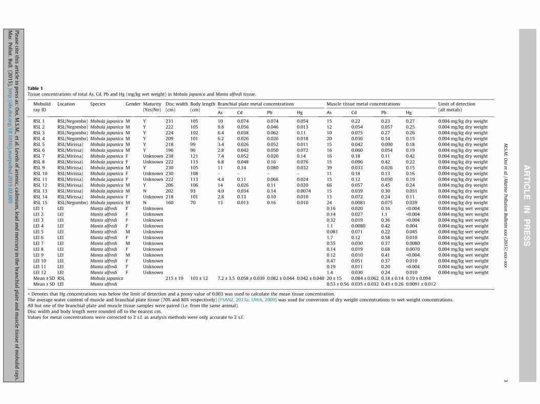
Table 1Tissue concentrations of total As, Cd, Pb and Hg (mg/kg wet weight) in Mobula japanica and Manta alfredi tissue.
Mobulidray ID
Location Species Gender Maturity(Yes/No)
Disc width(cm)
Body length(cm)
Branchial plate metal concentrations Muscle tissue metal concentrations Limit of detection(all metals)
As Cd Pb Hg As Cd Pb Hg
RSL 1 RSL(Negombo) Mobula japanica M Y 231 105 10 0.074 0.074 0.054 15 0.22 0.23 0.27 0.004 mg/kg dry weightRSL 2 RSL(Negombo) Mobula japanica M Y 222 105 9.8 0.056 0.046 0.013 12 0.054 0.057 0.25 0.004 mg/kg dry weightRSL 3 RSL(Negombo) Mobula japanica M Y 224 102 6.4 0.038 0.062 0.11 10 0.075 0.27 0.26 0.004 mg/kg dry weightRSL 4 RSL(Negombo) Mobula japanica M Y 209 101 6.2 0.026 0.026 0.018 20 0.036 0.14 0.15 0.004 mg/kg dry weightRSL 5 RSL(Mirissa) Mobula japanica M Y 218 99 3.4 0.026 0.052 0.011 15 0.042 0.090 0.18 0.004 mg/kg dry weightRSL 6 RSL(Mirissa) Mobula japanica M Y 196 96 2.8 0.042 0.050 0.072 16 0.060 0.054 0.19 0.004 mg/kg dry weightRSL 7 RSL(Mirissa) Mobula japanica F Unknown 238 121 7.4 0.052 0.020 0.14 16 0.18 0.11 0.42 0.004 mg/kg dry weightRSL 8 RSL(Mirissa) Mobula japanica F Unknown 222 115 6.8 0.048 0.16 0.076 15 0.096 0.42 0.22 0.004 mg/kg dry weightRSL 9 RSL(Mirissa) Mobula japanica M Y 230 105 11 0.14 0.080 0.032 39 0.033 0.026 0.15 0.004 mg/kg dry weightRSL 10 RSL(Mirissa) Mobula japanica F Unknown 230 108 – – – – 11 0.18 0.13 0.16 0.004 mg/kg dry weightRSL 11 RSL(Mirissa) Mobula japanica F Unknown 222 113 4.4 0.11 0.066 0.024 15 0.12 0.030 0.19 0.004 mg/kg dry weightRSL 12 RSL(Mirissa) Mobula japanica M Y 206 106 14 0.026 0.11 0.020 66 0.057 0.45 0.24 0.004 mg/kg dry weightRSL 13 RSL(Mirissa) Mobula japanica M N 202 93 4.0 0.034 0.14 0.0074 15 0.039 0.30 0.051 0.004 mg/kg dry weightRSL 14 RSL(Mirissa) Mobula japanica F Unknown 218 101 2.8 0.13 0.10 0.010 13 0.072 0.24 0.11 0.004 mg/kg dry weightRSL 15 RSL(Negombo) Mobula japanica M N 160 70 13 0.013 0.16 0.010 24 0.0081 0.075 0.039 0.004 mg/kg dry weightLEI 1 LEI Manta alfredi F Unknown 0.16 0.020 0.16 <0.004 0.004 mg/kg wet weightLEI 2 LEI Manta alfredi F Unknown 0.14 0.027 1.1 <0.004 0.004 mg/kg wet weightLEI 3 LEI Manta alfredi F Unknown 0.32 0.019 0.36 <0.004 0.004 mg/kg wet weightLEI 4 LEI Manta alfredi F Unknown 1.1 0.0080 0.42 0.004 0.004 mg/kg wet weightLEI 5 LEI Manta alfredi M Unknown 0.081 0.071 0.22 0.045 0.004 mg/kg wet weightLEI 6 LEI Manta alfredi F Unknown 1.7 0.12 0.58 0.010 0.004 mg/kg wet weightLEI 7 LEI Manta alfredi M Unknown 0.55 0.030 0.37 0.0080 0.004 mg/kg wet weightLEI 8 LEI Manta alfredi F Unknown 0.14 0.019 0.68 0.0070 0.004 mg/kg wet weightLEI 9 LEI Manta alfredi M Unknown 0.12 0.010 0.41 <0.004 0.004 mg/kg wet weightLEI 10 LEI Manta alfredi F Unknown 0.47 0.051 0.37 0.010 0.004 mg/kg wet weightLEI 11 LEI Manta alfredi F Unknown 0.19 0.011 0.20 <0.004 0.004 mg/kg wet weightLEI 12 LEI Manta alfredi F Unknown 1.4 0.030 0.24 0.010 0.004 mg/kg wet weightMean ± SD RSL Mobula japanica 215 ± 19 103 ± 12 7.2 ± 3.5 0.058 ± 0.039 0.082 ± 0.044 0.042 ± 0.040 20 ± 15 0.084 ± 0.062 0.18 ± 0.14 0.19 ± 0.094Mean ± SD LEI Manta alfredi 0.53 ± 0.56 0.035 ± 0.032 0.43 ± 0.26 0.0091 ± 0.012
< Denotes that Hg concentrations was below the limit of detection and a proxy value of 0.003 was used to calculate the mean tissue concentration.The average water content of muscle and branchial plate tissue (70% and 80% respectively) (FSANZ, 2013a; UWA, 2009) was used for conversion of dry weight concentrations to wet weight concentrations.All but one of the branchial plate and muscle tissue samples were paired (i.e. from the same animal).Disc width and body length were rounded off to the nearest cm.Values for metal concentrations were corrected to 2 s.f. as analysis methods were only accurate to 2 s.f.
M.S.M
.Ooi
etal./M
arinePollution
Bulletinxxx
(2015)xxx–
xxx3
Pleasecite
thisarticle
inpress
as:O
oi,M.S.M
.,et
al.Levelsofarsenic,cadm
ium
,leadand
mercu
ryin
thebranchialplate
andm
uscletissue
ofmobulid
rays.M
ar.Pollut.Bull.(2015),http://dx.doi.org/10.1016/j.marpolbu
l.2015.02.005

4 M.S.M. Ooi et al. / Marine Pollution Bulletin xxx (2015) xxx–xxx
procedure protected against automatically conducting multiplestatistical tests and compounding the experiment-wise type I errorrate in our analyses. To test for significant differences in metal con-centrations between paired branchial plate and muscle tissue, weperformed a paired MANOVA by differencing the concentrationsof each metal in M. japanica samples, and tested if the interceptwas different from zero using a Pillai test. If the difference was sig-nificant, we then performed individual paired ANOVAs on eachmetal. The relationship between muscle and branchial plate tissuewas further represented through linear regression.
Model residuals were visually assessed for normality andhomogeneity of variance and an untransformed response with anormal error structure was found to be appropriate. All data wereanalysed in R version 3.0.2.
2.5. Comparison to maximum levels in food standards and minimalrisk levels (MRLs)
Average metal concentrations for each tissue type were com-pared with the maximum allowable levels (MLs) recommendedfor fish consumption per the FSANZ, European Commission (EC)and the Codex Alimentarius Commission (WHO/FAO). (CODEX,2012; Commission of the European Communities, 2006; FSANZ,2013). These comparisons were made to provide a frame of refer-ence for the reported metal concentrations from these tissues.
Given the use of mobulid ray branchial plates in Chinese med-icine as a cure for chicken pox, the highest non-outlier concentra-tion of each metal from the samples of branchial plates was used toestimate the intake of each metal for a vulnerable age group. Thiswas compared to the minimal risk levels (MRLs) set by the Agency
As
muscle (mg/kg ww)200 40 60
Bran
chia
l pla
te (m
g/kg
ww
)
-5
0
5
10
15
20
25
y = 3.844 + 1.633 x (n = 14, R2= 0.44, P = 0.01)
Pb
muscle (mg/kg ww)0.0 0.1 0.2 0.3 0.4 0.5
Bran
chia
l pla
te (m
g/kg
ww
)
-0.10
-0.05
0.00
0.05
0.10
0.15
0.20
0.25
y = 0.051 + 0.173 x (n = 14, R2= 0.29, P = 0.01)
Fig. 1. Correlation between metal concentratio
Please cite this article in press as: Ooi, M.S.M., et al. Levels of arsenic, cadmium,Mar. Pollut. Bull. (2015), http://dx.doi.org/10.1016/j.marpolbul.2015.02.005
for Toxic Substances and Disease Registry (ATSDR) for an acuteduration (1–14 days) of oral exposure.
3. Results
Table 1 presents the results of metal analyses of branchial plateand muscle tissue from 15 specimens of M. japanica from SriLankan markets (RSL) and muscle tissue samples from 12 speci-mens of M. alfredi from Lady Elliot Island reef (LEI). The sampleswere compared with each other, with Food Standards (Fig. 2) andwith MRLs set by the ATSDR.
3.1. Comparison between samples
Significant differences were seen between the two muscle sam-ple sets (MANOVA, Pillai = 0.768, F = 18.2, df1 = 4, df2 = 22,p < 0.001), with mean As, Cd and Hg significantly higher in theRSL samples and Pb higher in those from LEI (ANOVA for As, Cd,Hg and Pb respectively were: F = 21.7, df = 1, p < 0.001; F = 6.31,df = 1, p = 0.018; F = 44.1, df = 1, p < 0.001; F = 10.3, df = 1,p = 0.004). Metal concentrations in matched muscle and branchialtissue from the RSL samples differed significantly (PairedMANOVA, Pillai = 0.87, F = 16.9, dfl = 4, df2 = 10, p < 0.001), withmean As, Pb, and Hg much higher in the muscle (ANOVA for As,Pb and Hg respectively were: F = 16.0, df = 1, p < 0.001; F = 8.50,df = 1, p = 0.012, F = 63.2, df = 1, p < 0.001), while there was no sig-nificant difference between Cd in the two (F = 1.32, df = 1,p = 0.271). A significant linear correlation between metal concen-trations of the two tissue types can be seen in Fig. 1.
Hg
muscle (mg/kg ww)0.0 0.1 0.2 0.3 0.4 0.5
Bran
chia
l pla
te (m
g/kg
ww
)
-0.10
-0.05
0.00
0.05
0.10
0.15
0.20
0.25
y = - 0.021 + 0.326 x (n = 14, R2= 0.58, P = 0.259)
Cd
muscle (log10mg/kg ww)-2.2 -2.0 -1.8 -1.6 -1.4 -1.2 -1.0 -0.8 -0.6 -0.4
Bran
chia
l pla
te (l
og10
mg/
kg w
w)
-3.0
-2.5
-2.0
-1.5
-1.0
-0.5
0.0
Log10
(y) = - 0.753 + 0.470 log10
(x) ( n = 14, R2= 0.31, P = 0.01)
ns in muscle tissue and branchial plates.
lead and mercury in the branchial plate and muscle tissue of mobulid rays.

Table 2Calculated intake of metals via consumption of branchial plates for a vulnerable agegroup.
Metal InorganicAs
Cd Pb Hg
M.S.M. Ooi et al. / Marine Pollution Bulletin xxx (2015) xxx–xxx 5
3.2. Comparison to maximum levels in food standards
Comparison of metal concentrations in each tissue type withthe MLs set by FSANZ, EC and Codex Alimentarius are representedin Fig. 2.
Amount in one serving ofbranchial plate
57.0 lg 5.9 lg 7.0 lg 0.80 lg
Calculated intake per dayof a 5-year old Chinesegirl weighing 15.9 kg(Marshall, 1981)
3.6 lg/kg/day
0.37 lg/kg/day
0.44 lg/kg/day
0.050 lg/kg/day
ATSDR oral exposure MRLfor acute durationexposure
5 lg/kg/day
Noneavailabledue toinsufficientdata
Noneavailabledue toinsufficientdata
Noneavailabledue toinsufficientdata
3.2.1. ArsenicAmong the three organisations, only FSANZ has established an
ML for arsenic in fishes, which is based on its inorganic form. Inthe edible portion of marine fishes, �10% of arsenic is generallypresent in inorganic forms (Rahman et al., 2012). Under theassumption that this is also valid for rays, the concentration ofinorganic arsenic was estimated for each tissue sample, as onlytotal arsenic was measured. Mean inorganic As concentrations inLEI and RSL branchial plate tissues were below the FSANZ ML,whereas the mean RSL muscle concentration (2 mg/kg wet weight)was equivalent.
3.2.2. CadmiumAmong the three organisations, only EC has established an ML
for Cd in fishes. Mean Cd concentrations in RSL branchial plateand muscle samples were above the ML set by EC. Although themean level of Cd in LEI muscle tissue did not exceed this limit,25% of the LEI muscle samples (ranging from 0.05 to 0.12 mg/kgwet weight) exceeded this ML.
As
Con
cent
ratio
n m
g/kg
ww
0
2
4
6
8
FSANZ - 2mg/kg ww
Pb
Con
cent
ratio
n (m
g/kg
ww
)
0.0
0.2
0.4
0.6
0.8
1.0
1.2
FSANZ - 0.5 mg/kg ww
Codex and EC - 0.3mg/kg ww
RSL - branchial plate RSL - muscle LEI - muscle
RSL - branchial plate RSL - muscle LEI - muscle
Fig. 2. Comparison of metal concentrat
Please cite this article in press as: Ooi, M.S.M., et al. Levels of arsenic, cadmium,Mar. Pollut. Bull. (2015), http://dx.doi.org/10.1016/j.marpolbul.2015.02.005
3.2.3. LeadMLs for Pb set by EC and Codex are 0.3 mg/kg wet weight and
0.5 mg/kg wet weight for FSANZ. The RSL branchial plate and mus-cle samples had mean levels of Pb which were below both MLs.However, 13% of the RSL muscle samples had Pb concentrationsthat were >0.3 mg/kg wet weight. As for the muscle samples fromLEI, the mean Pb concentration was above the EC and Codex limit.Although the mean level of Pb for LEI muscle samples did notexceed the FSANZ limit, 25% of the samples were above it.
Hg
Con
cent
ratio
n m
g/kg
ww
0.0
0.2
0.4
0.6
0.8
1.0
1.2
FSANZ - 1mg/kg ww
EC - 0.5mg/kg ww
median
mean
Cd
0.00
0.05
0.10
0.15
0.20
0.25
RSL - branchial plate RSL - muscle LEI - muscle
RSL - branchial plate RSL - muscle LEI - muscle
EC - 0.05mg/kgww
ions in samples to food standards.
lead and mercury in the branchial plate and muscle tissue of mobulid rays.

6 M.S.M. Ooi et al. / Marine Pollution Bulletin xxx (2015) xxx–xxx
3.2.4. MercuryMLs for total mercury in fish set by FSANZ and EC are 1 mg/kg
and 0.5 mg/kg wet weight respectively. All tissue samples had con-centrations below these MLs.
Codex provided a guideline level (GL) for methyl mercury,defined as ‘‘the ML of a substance in a food or feed commoditywhich is recommended by the Codex Alimentarius Commissionto be acceptable for commodities moving in international trade’’,of 0.5 mg/kg wet weight instead of a ML for total Hg. Based onthe assumption that methyl mercury is �83% of total mercury(Kannan et al., 1998), the concentration of methyl mercury in oursamples was calculated and compared with the Codex GL. Methylmercury concentrations in all tissue samples had concentrationsbelow the Codex GL of 0.5 mg/kg wet weight.
3.3. Comparison to ATSDR minimal risk levels
In Chinese medicine, the medicinal dose rate of mobulid raybranchial plates is 4.5–9 g (dried) once a month to stay healthy,and one dose daily for measles and chicken pox until the virus sub-sides (Paul Hilton, pers. comm., March 2014). In the case of chickenpox, the illness usually lasts from 5 to 10 days (Centers for DiseaseControl and Prevention, 2011), with the age group most likely tocontract chickenpox being 3–8 year olds (Ma and Fontaine, 2006).Using this information and the highest non-outlier concentrationof As out of all the RSL branchial plate samples, we calculated thatthere can be up to 57 lg of inorganic As in a single serving of bran-chial plates (assuming inorganic As is �10% of total As (Rahmanet al., 2012)). The calculations we made for a 5-year old Chinese girlof 15.9 kg body mass (Marshall, 1981) shows that her intake in asingle serving could be 3.6 lg As/kg/day of inorganic As.
Using the same principles as above, a single serving of branchialplates could have 5.9 lg of Cd, 7.0 lg of Pb and 0.80 lg of Hg. Thisequates to intake of 0.37 lg/kg/day of Cd, 0.44 lg/kg/day of Pb and0.050 lg/kg/day of Hg respectively. These calculations are tabulat-ed in Table 2.
4. Discussion
Overall, our findings show that consumption of M. japanicabranchial plates and muscle tissue from RSL and M. alfredi muscletissue from LEI need further investigation to determine if they posea risk to human health when consumed, especially to children.
4.1. Risks related to human consumption
4.1.1. ArsenicConcentrations of inorganic As in all RSL branchial plate sam-
ples were lower than the ML set by FSANZ (Fig. 2), and the calcu-lated intake of inorganic As by a vulnerable age group is lowerthan the oral exposure MRL set by the ATSDR (Table 2). However,the concentration of As contained varies with each individual sam-ple. Furthermore, ingested As is readily absorbed (between 30%and 95%) by humans, and children are more susceptible thanadults to toxicity due to the lack of hepatic detoxification enzymes(Fowler et al., 2011; ATSDR, 2007a). Anti-asthma preparations of25–107,000 lg/g As prescribed in Chinese medicine have reported-ly resulted in acute poisoning in children and adults (Ibid). A healthrisk assessment carried out by Essumang (2009) found that con-suming As-containing M. birostris meat in Ghana poses some can-cer risk. There is also potential for adverse, non-cancerous healtheffects for humans consuming As-contaminated meat and liver(Ibid). Therefore, although our results were lower than the stan-dards set, further investigation is needed to evaluate the potential
Please cite this article in press as: Ooi, M.S.M., et al. Levels of arsenic, cadmium,Mar. Pollut. Bull. (2015), http://dx.doi.org/10.1016/j.marpolbul.2015.02.005
health risk from consuming mobulid ray branchial plates contain-ing inorganic As.
4.1.2. CadmiumEssumang (2009) found that there is potential for adverse, non-
cancerous health effects for humans consuming Cd-contaminatedliver from manta rays. Although Cd levels in liver were notassessed in this study, some Cd concentrations in branchial tissuesfrom RSL mobulid rays were over the ML set by EC, and a youngchild could have an intake of 0.37 lg/kg/day (Table 2) from a serv-ing of mobulid branchial plates.
Horiguchi et al. (2004) found that higher rates of absorption andlower rates of excretion of Cd in humans were negatively correlat-ed with age. Similarly, dietary toxicity experiments found thatimmature female rats had much lower calculated LD50 values(ATSDR, 2012). Therefore, younger age groups may be at higherrisk of Cd toxicity. Consequences of Cd exposure to humans includerenal disease and skeletal damage, with higher risk of osteoporosisand fractures (Hellström et al., 2001; Engstrom et al., 2011). Inaddition, Cd is classed as a Group 1 human carcinogen by theInternational Agency for Research on Cancer (IARC, 1993).
Although the ATSDR does not have an acute duration MRL fororal exposure to Cd due to insufficient data (ATSDR, 2012), giventhat these tissues are used in Chinese medicine to treat feversand chicken-pox in children (Zhongguo ben cao tu lu, 1988), adetailed risk assessment is required to determine the potentialhealth risk from Cd via consumption of mobulid branchial plates,particularly for young girls.
4.1.3. LeadWe found that the mean Pb concentration from LEI muscle sam-
ples as well as the concentration in several of the RSL muscle sam-ples exceeded the limit of 0.3 mg/kg wet weight proposed byCodex and EC. Similarly, Lopez et al. (2013) found that the meanPb concentration in muscle, liver and stomach samples taken fromblue sharks (Prionace glauca), and in muscle and liver samples frommako short fin sharks (Isurus oxyrinchus) exceeded this limit. Lopezet al. (2013) concluded that their findings indicated a risk forhuman health from consumption of shark tissues by humans.
Lead toxicity has several negative effects on humans. Exposureto Pb in pre- and postnatal development periods cause delayed orreduced neurological and sexual development (ATSDR, 2007b).Short-term Pb exposure can cause gastrointestinal distress, anae-mia, encephalopathy, and death (CODEX, 2004). Children are moresusceptible than adults to effects of Pb toxicity (Jarup, 2003). Someof the health effects of Pb may occur at blood lead levels so low asto be essentially without a threshold (United States EnvironmentalProtection Agency, 1988a). The IARC has classified inorganic Pbcompounds as probably carcinogenic to humans (Group 2A), anddetermined organic Pb compounds to be non-classifiable as to theircarcinogenicity to humans (IARC, 2006).
We calculated that a child of a certain age group (Table 2) mayhave an intake of 0.44 lg/kg/day of Pb when consuming mobulidbrachial plates. Although the ATSDR does not have an acute dura-tion MRL for oral exposure Pb due to insufficient data (ATSDR,2007b), because of the potentially severe health effects, the con-centrations reported here from mobulid ray tissues warrants fur-ther investigation into potential human health risks.
4.1.4. MercuryOur comparison to the food standards had similar findings to
Lopez et al., 2013: Hg concentrations in stomach, muscle and livertissue samples from P. glauca and I. oxyrinchus did not exceed thelimit proposed by Codex. However, it was mentioned that Hg levelsmay have been masked by another metal hence there could havebeen more Hg in the samples than what was detected (Ibid).
lead and mercury in the branchial plate and muscle tissue of mobulid rays.

M.S.M. Ooi et al. / Marine Pollution Bulletin xxx (2015) xxx–xxx 7
According to the study done by Essumang (2009), there is nopotential health risk from Hg for human consumers of M. birostrismeat in Ghana.
Concentrations of total Hg reported from fish tissues are expect-ed to be mostly composed of methyl mercury (Commission of theEuropean Communities, 2006). Since methyl mercury is capable ofbioaccumulating in fish, larger long-lived species, such as sharksand swordfish, tend to be the focus for dietary restrictions(FSANZ, 2013).
The effects of oral exposure to methyl mercury can be severe. Ahuman epidemiological study found that daily oral intake ofmethyl mercury concentrations as low as 0.86 mg/kg/day (fromfish) in expectant mothers corresponded with neurophysiologicaldisorders in their children by the age of 7 (USEPA, 1988b).
The intake of Hg by a child consuming mobulid branchial platesas a cure for chicken pox could be 0.050 lg/kg/day (Table 2). Aswith Cd and Pb, the ATSDR does not have an acute duration MRLfor oral exposure to Hg (ATSDR, 1999). Although our reported con-centration of total Hg and methyl mercury in mobulid rays doesnot generally exceed regulatory guidelines, due to the severity ofthe potential adverse health effects, a review of the use of mobulidtissues warrants further investigation into the potential humanhealth risks.
4.2. Comparing paired branchial plate and muscle concentrations fromthe same ray
We found that for As, Pb and Hg, there was significantly more ofeach heavy metal accumulated in the muscle than branchial plates,while Cd had statistically similar levels in both tissues. Our find-ings for As were similar to Allen et al. (2004)’s who found thatmuscle levels of As exceeded gill levels in snakehead (Channa punc-tatus). However, concentrations of As, Cd and Pb were found to behigher in the gills as compared to muscle tissue in zebrafish (Daniorerio) and spotted dogfish (Scyliorhinus canicula) (Hamdi et al.,2009; De Boeck et al., 2010). The higher accumulation of metalsin some tissues over others could be because ionoregulatory organshave high accumulation rates (De Boeck et al., 2010) and the inter-action between metals and cellular constituents, for As in par-ticular (Allen et al., 2004).
There were significant positive correlations between branchialplate and muscle tissue concentrations for As, Cd and Pb in M. japa-nica tissue (Fig. 1). Therefore, it may be possible to estimate theconcentrations of certain metals in branchial plates using musclesamples, which are less invasive to obtain. However, as the numberof samples in this study was relatively low and the results showedsome variability, these equations should be used with some cau-tion until more samples can be collected and analysed.
4.3. Differences in metal concentration from two different species inseparate locations
When we looked at the difference in metal concentrations inmuscle tissue between M. japanica from RSL and M. alfredi fromLEI, we found that As, Cd and Hg were significantly higher in sam-ples from RSL than LEI. However, there were two different speciesbeing investigated at separate locations. Therefore, these differ-ences may be due to species differences rather than differencesin locality.
These differences between species could be due to differencesin diet or inherent factors. Marcovecchio et al. (1991) found thatthe most important factor among 3 shark species analysed for tis-sue metal concentrations was variation in their feed, rather thanlocation. Storelli and Marcotrigiano (2004) stated that differentlevels of As accumulation may differ between species of sharks
Please cite this article in press as: Ooi, M.S.M., et al. Levels of arsenic, cadmium,Mar. Pollut. Bull. (2015), http://dx.doi.org/10.1016/j.marpolbul.2015.02.005
due to species-specific accumulation, or intrinsic factors such asuptake rate.
Besides natural sources, domestic waste, coal-burning powerplants, metal smelters and sewage are sources of heavy metals inthe marine environment (Nriagu and Pacyna, 1988). A possible rea-son for the relatively higher levels of As, Cd and Hg in RSL samplesis the lack of proper waste management in Sri Lanka (Pernetta,1993).
It is possible that the significantly higher Pb levels found inmuscle samples from LEI than those from RSL could be linked tothe large-scale mining activities in North-west Queensland, inlandfrom the Great Barrier Reef (Australian atlas of minerals resources,2012). According to Mager (2011), Pb mining is a major cause ofconcern for environmental contamination. Pb mining is a majorindustry in Australia (Australian atlas of minerals resources,2012), with Australia being the second largest producer of Pbworldwide in 2008 (Mager, 2011).
Other marine organisms in the Great Barrier Reef have beenfound to be similarly affected by high Pb loads. For example, inthe Townsville harbour, adjacent to the central Great BarrierReef, concentrations of Pb in unfiltered seawater were found byEsslemont (2000) to exceed the recommended limits set byAustralian and New Zealand Environmental and ConservationCouncil (ANZECC). The coral Goniastrea aspera sourced from theTownsville harbour also had high levels of Pb (Esslemont, 2000).These elevated levels were due to suspended, metal-bearing finesediment present in the unfiltered seawater (Ibid). Reichelt andJones (1994), who studied trace metals in Cleveland Bay, close toTownsville, similarly found that the range of metals (includingPb) present were mainly from detrital particles originating fromre-suspended sediments or river discharge. In addition, concentra-tions of Pb in the livers of dugong (Dugong dugon) from theSouthern Great Barrier Reef have increased from 20 years ago(Haynes et al., 2005).
In conclusion, mean As in M. japanica muscle tissue, mean Cdconcentration in M. japanica muscle tissue and branchial plates,and concentrations of Cd and Pb in some individual samples ofM. alfredi muscle were above the set MLs for food safety, with Hgconcentrations in all tissues below the set MLs. The calculatedintake of inorganic As for a vulnerable age group consuming bran-chial plates is below the acute duration MRL for oral exposure setby the ATSDR. There are no acute duration MRLs set by the ATSDRfor oral exposure to Cd, Pb or Hg. Given that some metal concentra-tions exceeded set MLs and that children are especially vulnerableto the toxic effects of some metals, further investigation and anassessment of human health risk from consumption of mobulidbranchial plates should be carried out.
Acknowledgements
We thank the staff at Queensland Department of Health,Forensic and Scientific Services for assistance with laboratory tech-niques and equipment. We thank Lydie Couturier, Fabrice Jaine,Katherine Burgess, Peran Bray and Julie Vercelloni for their assis-tance with this study. This study was supported by the GoodmanFoundation, ARC Linkage Grant LP110100712, EarthwatchInstitute Australia and Sibelco Pty Ltd. Field work was supportedby Lady Elliot Island Eco Resort and was conducted underFisheries Permit (165491), Great Barrier Reef Marine Park Permit(G09/2985.1) and Ethics approval (SBMS/071/08/SEAWORLD).
References
Adams, W.J., Blust, R., Borgmann, U., Brix, K.V., DeForest, D.K., Green, A.S., Meyer, J.S.,McGeer, J.C., Paquin, P.R., Rainbow, P.S., Wood, C.M., 2011. Utility of tissue
lead and mercury in the branchial plate and muscle tissue of mobulid rays.

8 M.S.M. Ooi et al. / Marine Pollution Bulletin xxx (2015) xxx–xxx
residues for predicting effects of metals on aquatic organisms. IntegratedEnviron. Assess. Manage. 7, 75–98. http://dx.doi.org/10.1002/ieam.108.
Allen, T., Singhal, R., Rana, S., 2004. Resistance to oxidative stress in a freshwater fishChanna punctatus after exposure to inorganic arsenic. Biol. Trace Elem. Res. 98,63–72, <http://link.springer.com/article/10.1385/BTER:98:1:63> (accessed4.10.13).
Agency for Toxic Substances and Disease Registry, 1999. Toxicological profile forMercury. U.S. Department of Health and Human Services, Public Health Service,Atlanta, GA. <http://www.atsdr.cdc.gov/toxprofiles/tp46.pdf> (accessed6.12.13).
Agency for Toxic Substances and Disease Registry, 2007a. Toxicological profile forArsenic. U.S. Department of Health and Human Services, Public Health Service.,Atlanta, GA. <http://www.atsdr.cdc.gov/toxprofiles/tp2.pdf> (accessed 5.12.13).
Agency for Toxic Substances and Disease Registry, 2007b. Toxicological profile forLead. U.S. Department of Health and Human Services, Public Health Service.,Atlanta, GA. <http://www.atsdr.cdc.gov/toxprofiles/tp13.pdf> (accessed8.12.13).
Agency for Toxic Substances and Disease Registry, 2012. Toxicological profile forCadmium. U.S. Department of Health and Human Services, Public HealthService., Atlanta, GA. <http://www.atsdr.cdc.gov/toxprofiles/tp5.pdf> (accessed8.12.13).
Australian Atlas of Minerals Resources, m.p.c., 2012. Lead – Fact Sheet. <http://www.australianminesatlas.gov.au/education/fact_sheets/lead.html> (accessed4.10.13).
Centers for Disease Control and Prevention, 2011. Chickenpox (Varicella): Signs andSymptoms, Atlanta, USA. <http://www.cdc.gov/chickenpox/about/symptoms.html> (accessed 28.09.13).
CODEX, 2004. Code of Practice for the Prevention and Reduction of LeadContamination in Foods. CAC/RCP 56–2004, Introduction. Codex AlimentariusCommission. <file:///C:/Users/Admin/Downloads/CXP_056e.pdf> (accessed29.09.13).
CODEX, 2012. Codex General Standard for Contaminants and Toxins in Food andFeed. CODEX STAN 193-1995, WHO Food Standards Programme. Joint FAO/WHO Expert Committee on Food Additives/JECFA. <http://www.fao.org/fileadmin/user_upload/livestockgov/documents/1_CXS_193e.pdf> (accessed6.03.13).
Commission of the European Communities, 2006. Commission Regulation (EC) No1881/2006 of 19 December 2006: setting maximum levels for certaincontaminants in foodstuffs. Official Journal of the European Union Legislation364. <https://www.fsai.ie/uploadedFiles/Consol_Reg1881_2006.pdf> (accessed6.03.13).
Couturier, L.I.E., Marshall, A.D., Jaine, F.R.A., Kashiwagi, T., Pierce, S.J., Townsend,K.A., Weeks, S.J., Bennett, M.B., Richardson, A.J., 2012. Biology, ecology andconservation of the Mobulidae. J. Fish Biol. 80, 1075–1119. http://dx.doi.org/10.1111/j.1095-8649.2012.03264.x.
Couturier, L., Rohner, C., Richardson, A., Pierce, S., Marshall, A., Jaine, F., Townsend,K., Bennett, M., Weeks, S., Nichols, P., 2013. Unusually high levels of n-6polyunsaturated fatty acids in whale sharks and reef manta rays. Lipids 48,1029–1034. http://dx.doi.org/10.1007/s11745-013-3829-8.
De Boeck, G., Eyckmans, M., Lardon, I., Bobbaers, R., Sinha, A.K., Blust, R., 2010. Metalaccumulation and metallothionein induction in the spotted dogfish Scyliorhinuscanicula. Comp. Biochem. Physiol. – Part A: Mol. Integrative Physiol. 155, 503–508. http://dx.doi.org/10.1016/j.cbpa.2009.12.014.
Dewar, H., 2002. Preliminary report: Manta harvest in Lamakera. Report from thePfleger Institute of Environmental Research and the Nature Conservancy, 3.<http://www.equilibrioazul.org/documentos/Dewar_Report.pdf> (accessed3.04.12).
Engstrom, A., Michaelsson, K., Suwazono, Y., Wolk, A., Vahter, M., Akesson, A., 2011.Long-term cadmium exposure and the association with bone mineral densityand fractures in a population-based study among women. J. Bone Miner. Res.26, 486–495. http://dx.doi.org/10.1002/jbmr.224.
Eschmeyer, W.N., Fong, J.D., 2014. Catalog of Fishes. <http://research.calacademy.org/research/ichthyology/catalog/fishcatmain.asp> (accessed 27.09.13).
Esslemont, G., 2000. Heavy metals in seawater, marine sediments and corals fromthe Townsville section, Great Barrier Reef Marine Park, Queensland. Mar. Chem.71, 215–231. http://dx.doi.org/10.1016/S0304-4203(00)00050-5.
Essumang, D.K., 2009. Analysis and Human Health Risk Assessment of Arsenic,Cadmium, and Mercury in Manta birostris (Manta Ray) Caught Along theGhanaian Coastline. Human and Ecological Risk Assessment 15, pp. 985–998,2012, doi: http://dx.doi.org/10.1080/10807030903153451.
Essumang, D., 2010. First determination of the levels of platinum group metals inManta birostris (Manta Ray) caught along the Ghanaian coastline. Bull.Environ. Contam. Toxicol. 84, 720–725. http://dx.doi.org/10.1007/s00128-010-0019-8.
Fernando, D., 2012. Instructions for collecting and storing mobulid ray samplesfrom dead specimens. The Manta Trust.
Fernando, D., Stevens, G., 2011. A study of Sri Lanka’s manta and mobula ray fishery.The Manta Trust. <http://www.mantatrust.org/wp-content/uploads/2011/09/Sri-Lankas-Manta-Mobula-Ray-Fishery.pdf> (accessed 4.04.12).
Fowler, B.A., Nordberg, G.F., Nordberg, M., Friberg, L., 2011. Handbook on theToxicology of Metals. Academic Press, Chapter 19, p. 373.
Food Standards Australia and New Zealand (FSANZ), 2013. Contaminants andnatural toxicants, Standard 1.4.1, Australia and New Zealand. <http://www.comlaw.gov.au/Details/F2013C00140> (accessed 6.03.13).
Goyer, R.A., 1995. Nutrition and metal toxicity. Am. J. Clin. Nutr. 61, 646S–650S,<http://ajcn.nutrition.org/content/61/3/646S.full.pdf> (accessed 28.03.14).
Please cite this article in press as: Ooi, M.S.M., et al. Levels of arsenic, cadmium,Mar. Pollut. Bull. (2015), http://dx.doi.org/10.1016/j.marpolbul.2015.02.005
Haines, J., Limpus, C., Flakus, S., 1999. Marine Wildlife Stranding and MortalityDatabase annual report 1999 – III. Marine Turtles. (Service, Queensland Parksand Wildlife Services).
Hamdi, M., Sanchez, M., Beene, L., Liu, Q., Landfear, S., Rosen, B., Liu, Z., 2009. Arsenictransport by zebrafish aquaglyceroporins. BMC Mol. Biol. 10, 104. http://dx.doi.org/10.1186/1471-2199-10-104.
Harada, M., 1995. Minamata disease: methylmercury poisoning in japan caused byenvironmental pollution. Crit. Rev. Toxicol. 25, 1–24. http://dx.doi.org/10.3109/10408449509089885.
Haynes, D., Carter, S., Gaus, C., Müller, J., Dennison, W., 2005. Organochlorine andheavy metal concentrations in blubber and liver tissue collected fromQueensland (Australia) dugong (Dugong dugon). Mar. Pollut. Bull. 51, 361–369. http://dx.doi.org/10.1016/j.marpolbul.2004.10.020.
Hellström, L., Elinder, C.-G., Dahlberg, B., Lundberg, M., Järup, L., Persson, B., Axelson,O., 2001. Cadmium exposure and end-stage renal disease. Am. J. Kidney Dis. 38,1001–1008. http://dx.doi.org/10.1053/ajkd.2001.28589.
Horiguchi, H., Oguma, E., Sasaki, S., Miyamoto, K., Ikeda, Y., Machida, M., Kayama, F.,2004. Comprehensive study of the effects of age, iron deficiency, diabetesmellitus, and cadmium burden on dietary cadmium absorption in cadmium-exposed female Japanese farmers. Toxicol. Appl. Pharmacol. 196, 114–123.http://dx.doi.org/10.1016/j.taap.2003.11.024.
International Agency for Research on Cancer (IARC), 1993. IARC Monographs on theevaluation of carcinogenic risks to humans: Beryllium, Cadmium, Mercury, andExposures in the Glass Manufacturing Industry. World Health Organization.<http://monographs.iarc.fr/ENG/Monographs/vol58/mono58.pdf> (accessed7.04.13).
International Agency for Research on Cancer (IARC), 2006. IARC Monographs on theevaluation of carcinogenic risks to humans. Inorganic and organic leadcompounds. World Health Organization. <http://monographs.iarc.fr/ENG/Monographs/vol87/mono87.pdf> (accessed 22.03.13).
International Union for Conservation of Nature, 2014. The IUCN Red List ofThreatened Species. Version 2014.3. <http://www.iucnredlist.org/> (accessed25.02.14).
Jarup, L., 2003. Hazards of heavy metal contamination. Brit. Med. Bull. 68, 167–182.http://dx.doi.org/10.1093/bmb/ldg032.
Kannan, K., Smith Jr., R., Lee, R., Windom, H., Heitmuller, P., Macauley, J., Summers,J., 1998. Distribution of total mercury and methyl mercury in water, sediment,and fish from south Florida estuaries. Arch. Environ. Contam. Toxicol. 34, 109–118. http://dx.doi.org/10.1007/s002449900294.
Lopez, S.A., Abarca, N.L., Meléndez, R., 2013. Heavy metal concentrations of twohighly migratory sharks (Prionace glauca and Isurus oxyrinchus) in thesoutheastern Pacific waters: comments on public health and conservation.Tropical Conservation Sci. 6, 126–137, <http://tropicalconservationscience.mongabay.com/content/v6/TCS-2013_Vol_6(1)_126-137_Lopez_et_al.pdf>(accessed 19.03.13).
Ma, H., Fontaine, R., 2006. Varicella outbreak among primary school students—Beijing, China, 2004. Morbidity and Mortality Weekly Report 55, 39–43. <http://origin.glb.cdc.gov/mmwr/preview/mmwrhtml/su5501a10.htm> (accessed27.08.13).
Mager, E.M., 2011. 4 – Lead. In: Chris, M., Wood, A.P.F., Colin, J.B. (Eds.), FishPhysiology. Academic Press, pp. 185–236. doi: http://dx.doi.org/10.1016/S1546-5098(11)31026-6.
Marcovecchio, J.E., Moreno, V.J., Pérez, A., 1991. Metal accumulation in tissues ofsharks from the Bahía Blanca estuary, Argentina. Mar. Environ. Res. 31, 263–274. http://dx.doi.org/10.1016/0141-1136(91)90016-2.
Marshall, W.A., 1981. Body Weights and Heights by Countries. FAO/WHO/UNU EPR/81/8, Joint FAO/WHO/UNU Expert Consultation on Energy and ProteinRequirements. Food and Agriculture Organization of the United Nations.<http://www.fao.org/3/contents/6599c1e3-8e06-59ed-8a0b-980140c81ce3/M2846E00.HTM> (accessed 9.09.14).
McGeer, J.C., Niyogi, S., Scott Smith, D., 2011. 3 – Cadmium. In: Chris, M., Wood,A.P.F., Colin, J.B. (Eds.), Fish Physiology. Academic Press, pp. 125–184. doi:http://dx.doi.org/10.1016/S1546-5098(11)31025-4.
McIntyre, D.O., Linton, T.K., 2011. 6 – Arsenic. In: Chris, M., Wood, A.P.F., Colin, J.B.(Eds.), Fish Physiology. Academic Press, pp. 297–349. doi: http://dx.doi.org/10.1016/S1546-5098(11)31028-X.
Nriagu, J.O., Pacyna, J.M., 1988. Quantitative assessment of worldwidecontamination of air, water and soils by trace metals. Nature 333, 134–139,<http://lrg.elte.hu/oktatas/Elemek%20korforgasa%20PhD/Baricza%20Agnes%20Elemek_korforgasa_PbHgCd/Nriagu-JO-and-Pacyna-JM-1988.pdf> (accessed3.09.13).
Pernetta, J., 1993. Marine Protected Area Needs in the South Asian Seas Region.Volume 5: Sri Lanka. A Marine Conservation and Development Report. IUCN,Gland, Switzerland, p. vii + 67pp, Chapter 4, pp. 28–29.
Rahman, M.A., Hasegawa, H., Lim, R.P., 2012. Bioaccumulation, biotransformationand trophic transfer of arsenic in the aquatic food chain. Environ. Res. 116, 118–135. http://dx.doi.org/10.1016/j.envres.2012.03.014.
Reichelt, A.J., Jones, G.B., 1994. Trace metals as tracers of dredging activity inCleveland Bay—field and laboratory studies. Mar. Freshw. Res. 45, 1237–1257.http://dx.doi.org/10.1071/MF9941237.
Storelli, M.M., Marcotrigiano, G.O., 2004. Interspecific variation in total arsenic bodyconcentrations in elasmobranch fish from the Mediterranean Sea. Mar. Pollut.Bull. 48, 1145–1149. http://dx.doi.org/10.1016/j.marpolbul.2004.03.005.
The University of Western Australia (UWA), 2009. Blue Histology – Skeletal Tissue –Cartilage. <http://www.lab.anhb.uwa.edu.au/mb140/CorePages/Cartilage/Cartil.htm> (accessed 26.08.13).
lead and mercury in the branchial plate and muscle tissue of mobulid rays.
http://tropicalconservationscience.mongabay.com/content/v6/TCS-2013_Vol_6(1)_126-137_Lopez_et_al.pdf

M.S.M. Ooi et al. / Marine Pollution Bulletin xxx (2015) xxx–xxx 9
Tinggi, U., Gianduzzo, T., Francis, R., Nicol, D., Shahin, M., Scheelings, P., 2004.Determination of selenium in red blood cells by inductively coupledplasma mass spectrometry (ICP-MS) after microwave digestion. J. Radioanal.Nucl. Chem. 259, 469–472. http://dx.doi.org/10.1023/B:JRNC.0000020920.28409.30.
United States Environmental Protection Agency, 1988a. Lead and compounds(inorganic) (CASRN 7439-92-1), Integrated Risk Information System. <http://www.epa.gov/iris/subst/0277.htm> (accessed 2.03.14).
Please cite this article in press as: Ooi, M.S.M., et al. Levels of arsenic, cadmium,Mar. Pollut. Bull. (2015), http://dx.doi.org/10.1016/j.marpolbul.2015.02.005
United States Environmental Protection Agency, 1988b. Methylmercury (MeHg)(CASRN 22967-92-6), Integrated Risk Information System. <http://www.epa.gov/iris/subst/0073.htm> (accessed 3.03.14).
White, W.T., Giles, J., Potter, I.C., 2006. Data on the bycatch fishery and reproductivebiology of mobulid rays (Myliobatiformes) in Indonesia. Fish. Res. 82, 65–73.http://dx.doi.org/10.1016/j.fishres.2006.08.008.
Zhongguo ben cao tu lu, 1988. Ren min wei sheng chu ban she, Beijing, p. 213.
lead and mercury in the branchial plate and muscle tissue of mobulid rays.