Letters to the Editor - Journal of Medical Genetics
31
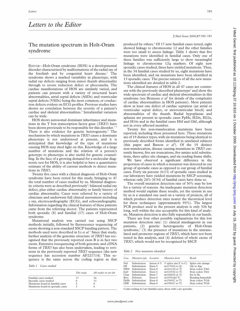
Letters to the Editor J Med Genet 2000;37:785–787 The mutation spectrum in Holt-Oram syndrome EDITOR—Holt-Oram syndrome (HOS) is a developmental disorder characterised by malformations of the radial ray of the forelimb and by congenital heart disease. 1 The syndrome shows a marked variability in phenotype, with radial ray defects ranging from minor thumb abnormality through to severe reduction defect or phocomelia. The cardiac manifestations of HOS are similarly varied, and patients can present with a variety of structural heart abnormalities, atrial septal defects (ASDs) and ventricular septal defects (VSDs) being the most common, or conduc- tion defects evident on ECG profiles. Previous studies have shown no correlation between the severity of a patient’s cardiac and skeletal abnormalities. 2 Intrafamilial variation can be wide. HOS shows autosomal dominant inheritance and muta- tions in the T box transcription factor gene (TBX5) have been shown previously to be responsible for this disorder. 34 There is also evidence for genetic heterogeneity. 5 The mechanism by which mutations in TBX5 cause a dominant phenotype is not understood at present, and it is anticipated that knowledge of the type of mutations causing HOS may shed light on this. Knowledge of a large number of mutations and the relation of a person’s genotype to phenotype is also useful for genetic counsel- ling. In the face of a growing demand for a molecular diag- nostic test for HOS, it is also helpful to have a quantitative estimate of the ability of current methods to detect muta- tions in TBX5. Twenty five cases with a clinical diagnosis of Holt-Oram syndrome have been tested for this study, bringing to 47 the total number of cases studied by us. Minimal diagnos- tic criteria were as described previously 2 : bilateral radial ray defect, plus either cardiac abnormality or family history of cardiac abnormality. Cases were referred by a variety of clinicians and underwent full clinical assessment including x ray, electrocardiography (ECG), and echocardiography. Information regarding the clinical features of these patients came from the referring doctor. The patients represented both sporadic (8) and familial (17) cases of Holt-Oram syndrome. Mutational analysis was carried out using SSCP methods initially, followed by fluorescence sequencing of exons showing a non-standard SSCP banding pattern. The methods used were described in Li et al. 3 Since that study, further analysis of the genomic structure of TBX5 has rec- ognised that the previously reported exon B is in fact two exons. Extensive resequencing of both genomic and cDNA forms of TBX5 has also been undertaken, leading to revi- sions in the previously reported TBX5 sequence (the new sequence has accession number AF221714). This se- quence is the same across the coding region as that produced by others. 6 Of 17 new familial cases tested, eight showed linkage to chromosome 12 and the other families were too small to assess linkage. Table 1 shows that five mutations were identified in familial cases. Only one of these families was suYciently large to show meaningful linkage to chromosome 12q markers. Of eight new sporadic cases studied, three have yielded mutations. Thus, in the 34 familial cases studied by us, eight mutations have been identified, and six mutations have been identified in 13 sporadic cases. The precise natures of all the new muta- tions identified are detailed in table 2. The clinical features of HOS in all 47 cases are consist- ent with the previously described phenotype 2 and show the wide spectrum of cardiac and skeletal abnormalities in this syndrome (see Bruneau et al 7 for details of the complexity of cardiac abnormalities in HOS patients). Most patients show at least one defect of cardiac septation (an atrial or ventricular septal defect, or atrioventricular block) and abnormalities of the thumb. Radial hypoplasias and aplasias are present in sporadic cases PpHs, H20s, H22s, and H16s and in the familial cases H6f and Ghf, although not in every aVected member. Twenty five non-translocation mutations have been reported, including those presented here. These mutations are of 19 distinct types, with six mutations being identical to previously described forms identified in unrelated subjects (this paper and Basson et al 6 ). Of the 19 distinct non-translocation, disease causing mutations in TBX5 cur- rently known, five are truncations, five amino acid substitu- tions, three splice site changes, and six reading frame shifts. We have observed a significant diVerence in the proportion of cases in which a mutation was detected in our group of sporadic cases as opposed to our group of familial cases. Forty six percent (6/13) of sporadic cases studied in our laboratory have yielded mutations by SSCP screening, whereas only 24% (8/34) of familial cases have done so. The overall mutation detection rate of 30% may be low for a variety of reasons. An inadequate mutation detection method would explain these results, yet the system in use by us is a standard one used on a variety of projects, all of which produce detection rates nearer the theoretical level for these techniques (approximately 95%). The largest PCR product used in the present analysis is only 326 bp long, well within the size acceptable for this kind of analy- sis. Mutation detection is also fully repeatable in our hands. There are four other possible explanations for this low mutation detection rate: (1) clinical misdiagnosis in our patients, (2) genetic heterogeneity of Holt-Oram syndrome, 5 (3) the presence of mutations in the untrans- lated and promoter regions of TBX5, which have not been tested in this analysis, and (4) deletion of whole exons of TBX5, which would not be recognised by SSCP. Table 1 Cases studied This study All studies in our lab Familial cases studied 17 34 Sporadic cases studied 8 13 Mutations found in familial cases 5 8 Mutations found in sporadic cases 3 6 Table 2 New mutations identified Case Mutation type Location Mutation form Result Nef Substitution Intron 4–5 5' splice site(T to C) Splice site change FIf Substitution Exon 5 nt1170(G to A) Gly to Arg FR9f Substitution Exon 8 nt1611(G to T) Stop codon TAG Jonf Substitution Exon 6 nt1233(G to T) Stop codon TAA Chas Substitution Exon 7 nt1420(G to T) Ser to Ile Bers Deletion Exon 8 nt1470(del T) Reading frameshift PpHs Substitution Exon 8 nt1500(C to T) Stop codon TGA GHf Substitution Exon 8 nt1500(C to T) Stop codon TGA Codes ending in f are familial cases, those with s are sporadic. Letters 785 www.jmedgenet.com on July 19, 2022 by guest. Protected by copyright. http://jmg.bmj.com/ J Med Genet: first published as 10.1136/jmg.37.10.810 on 1 October 2000. Downloaded from
-
Upload
khangminh22 -
Category
Documents
-
view
2 -
download
0
Transcript of Letters to the Editor - Journal of Medical Genetics
Letters to the Editor
J Med Genet 2000;37:785–787
The mutation spectrum in Holt-Oramsyndrome
EDITOR—Holt-Oram syndrome (HOS) is a developmentaldisorder characterised by malformations of the radial ray ofthe forelimb and by congenital heart disease.1 Thesyndrome shows a marked variability in phenotype, withradial ray defects ranging from minor thumb abnormalitythrough to severe reduction defect or phocomelia. Thecardiac manifestations of HOS are similarly varied, andpatients can present with a variety of structural heartabnormalities, atrial septal defects (ASDs) and ventricularseptal defects (VSDs) being the most common, or conduc-tion defects evident on ECG profiles. Previous studies haveshown no correlation between the severity of a patient’scardiac and skeletal abnormalities.2 Intrafamilial variationcan be wide.
HOS shows autosomal dominant inheritance and muta-tions in the T box transcription factor gene (TBX5) havebeen shown previously to be responsible for this disorder.3 4
There is also evidence for genetic heterogeneity.5 Themechanism by which mutations in TBX5 cause a dominantphenotype is not understood at present, and it isanticipated that knowledge of the type of mutationscausing HOS may shed light on this. Knowledge of a largenumber of mutations and the relation of a person’sgenotype to phenotype is also useful for genetic counsel-ling. In the face of a growing demand for a molecular diag-nostic test for HOS, it is also helpful to have a quantitativeestimate of the ability of current methods to detect muta-tions in TBX5.
Twenty five cases with a clinical diagnosis of Holt-Oramsyndrome have been tested for this study, bringing to 47the total number of cases studied by us. Minimal diagnos-tic criteria were as described previously2: bilateral radial raydefect, plus either cardiac abnormality or family history ofcardiac abnormality. Cases were referred by a variety ofclinicians and underwent full clinical assessment includingx ray, electrocardiography (ECG), and echocardiography.Information regarding the clinical features of these patientscame from the referring doctor. The patients representedboth sporadic (8) and familial (17) cases of Holt-Oramsyndrome.
Mutational analysis was carried out using SSCPmethods initially, followed by fluorescence sequencing ofexons showing a non-standard SSCP banding pattern. Themethods used were described in Li et al.3 Since that study,further analysis of the genomic structure of TBX5 has rec-ognised that the previously reported exon B is in fact twoexons. Extensive resequencing of both genomic and cDNAforms of TBX5 has also been undertaken, leading to revi-sions in the previously reported TBX5 sequence (the newsequence has accession number AF221714). This se-quence is the same across the coding region as that
produced by others.6 Of 17 new familial cases tested, eightshowed linkage to chromosome 12 and the other familieswere too small to assess linkage. Table 1 shows that fivemutations were identified in familial cases. Only one ofthese families was suYciently large to show meaningfullinkage to chromosome 12q markers. Of eight newsporadic cases studied, three have yielded mutations. Thus,in the 34 familial cases studied by us, eight mutations havebeen identified, and six mutations have been identified in13 sporadic cases. The precise natures of all the new muta-tions identified are detailed in table 2.
The clinical features of HOS in all 47 cases are consist-ent with the previously described phenotype2 and show thewide spectrum of cardiac and skeletal abnormalities in thissyndrome (see Bruneau et al7 for details of the complexityof cardiac abnormalities in HOS patients). Most patientsshow at least one defect of cardiac septation (an atrial orventricular septal defect, or atrioventricular block) andabnormalities of the thumb. Radial hypoplasias andaplasias are present in sporadic cases PpHs, H20s, H22s,and H16s and in the familial cases H6f and Ghf, althoughnot in every aVected member.
Twenty five non-translocation mutations have beenreported, including those presented here. These mutationsare of 19 distinct types, with six mutations being identical topreviously described forms identified in unrelated subjects(this paper and Basson et al6). Of the 19 distinctnon-translocation, disease causing mutations in TBX5 cur-rently known, five are truncations, five amino acid substitu-tions, three splice site changes, and six reading frame shifts.
We have observed a significant diVerence in theproportion of cases in which a mutation was detected in ourgroup of sporadic cases as opposed to our group of familialcases. Forty six percent (6/13) of sporadic cases studied inour laboratory have yielded mutations by SSCP screening,whereas only 24% (8/34) of familial cases have done so.
The overall mutation detection rate of 30% may be lowfor a variety of reasons. An inadequate mutation detectionmethod would explain these results, yet the system in useby us is a standard one used on a variety of projects, all ofwhich produce detection rates nearer the theoretical levelfor these techniques (approximately 95%). The largestPCR product used in the present analysis is only 326 bplong, well within the size acceptable for this kind of analy-sis. Mutation detection is also fully repeatable in our hands.
There are four other possible explanations for this lowmutation detection rate: (1) clinical misdiagnosis in ourpatients, (2) genetic heterogeneity of Holt-Oramsyndrome,5 (3) the presence of mutations in the untrans-lated and promoter regions of TBX5, which have not beentested in this analysis, and (4) deletion of whole exons ofTBX5, which would not be recognised by SSCP.
Table 1 Cases studied
This study All studies in our lab
Familial cases studied 17 34Sporadic cases studied 8 13Mutations found in familial cases 5 8Mutations found in sporadic cases 3 6
Table 2 New mutations identified
Case Mutation type Location Mutation form Result
Nef Substitution Intron 4–5 5' splice site(T to C) Splice site changeFIf Substitution Exon 5 nt1170(G to A) Gly to ArgFR9f Substitution Exon 8 nt1611(G to T) Stop codon TAGJonf Substitution Exon 6 nt1233(G to T) Stop codon TAAChas Substitution Exon 7 nt1420(G to T) Ser to IleBers Deletion Exon 8 nt1470(del T) Reading frameshiftPpHs Substitution Exon 8 nt1500(C to T) Stop codon TGAGHf Substitution Exon 8 nt1500(C to T) Stop codon TGA
Codes ending in f are familial cases, those with s are sporadic.
Letters 785
www.jmedgenet.com
on July 19, 2022 by guest. Protected by copyright.
http://jmg.bm
j.com/
J Med G
enet: first published as 10.1136/jmg.37.10.810 on 1 O
ctober 2000. Dow
nloaded from

Other studies of HOS have not published mutationdetection rates. We can only speculate as to why the rate ofmutation detection in sporadic cases should be higher thanthat in familial cases (particularly in those familial caseswhose disease loci are known to be present on 12q). Anobvious explanation for such a discrepancy is ascertain-ment bias. A sporadic case must have both detectable heartand limb symptoms to be diagnosed as having HOS,whereas a family need only show both these eVects acrossthe pedigree (rather than in one subject) along with anautosomal dominant inheritance pattern.
A group of five of the 25 mutations published so far arethe same, a C to T transition at position 1500, generating astop codon. This mutation is seen in both sporadic andfamilial HOS cases among patients who do not share com-mon alleles at microsatellite loci closely linked to TBX5.This site is likely to represent, therefore, a true “mutationhotspot”. The residue is part of a CG duplet, and thereforeis likely to be methylated, with a high frequency ofmutation to a thymine residue. Studies across a variety ofhuman diseases have found distributions of mutationsskewed towards the mutation of CpG sites. The retino-blastoma gene, RB1, shows a distribution of mutationsseverely skewed towards a few C→T transition hotspots(see the RB1 mutation database8 for details).
Basson et al6 argue, based on a collection of HOS fami-lies of varying symptom severity, that there is a relationshipbetween the phenotype of patients and their specific muta-tion. It is proposed that truncation mutations in TBX5 leadto both limb and cardiac malformation, whereas singleamino acid changes have diVerent eVects depending ontheir position in the T box. The set of HOS cases we haveexamined contains no large families of the type studied inthis earlier analysis.
The five known cases (two familial and three sporadic)with the same mutation, a C→T transition at nucleotide1500 in exon 8, present an opportunity to examine moreclosely the possibility of mutation specific genotype-phenotype correlation in cases with a truncation in TBX5.The clinical phenotypes of these cases are presented intable 3. Cardiac defects presented include isolated ASDand isolated VSD but no case with this mutation has acomplex cardiac lesion. Syndactyly of the thumb and firstfinger is common within this group. There is muchvariation in symptom severity and the group as a wholeshows no bias towards a particular severity of either cardiacor skeletal symptoms, in agreement with Basson et al.6
Along with the truncation forms already described, wehave identified two new mutations which each result in asingle amino acid substitution. The change in family Fifinserts an arginine in place of a glycine at position 169,
which is conserved across T box genes such as Xbra (Xeno-pus), T (mouse), TbxT (chick), and omb (Drosophila).9 Thisintroduces a strongly basic residue into a non-polar regionin the DNA binding T domain. Family Fif comprises eightaVected subjects in two generations, who show significantcardiac involvement compared with very mild skeletalfindings. One case has a complex lesion, ASD with VSD,and another case has pulmonary stenosis, a conotruncalmalformation which is not typical in HOS. This is consist-ent with a role for TBX5 which extends beyond cardiacseptation.10 Only one subject in family Fif has ademonstrable limb abnormality and this is stiVening of thethumbs. The phenotype of this family is therefore consist-ent with the suggestion that substitution mutationsproduce predominantly limb or predominantly cardiacfeatures, depending upon their location within TBX5.6
The change of a serine to an isoleucine in patient Chasis outside the T domain of the protein and its biochemicaleVects are not known. The phenotype of this patientincluded a spinal scoliosis, which has not previously beenobserved in Holt-Oram syndrome, together with bilateralhypoplastic thumbs, syndactyly, and a ventricular septaldefect. Currently available details on expression of TBX5during development of the mouse and chick give noevidence of expression in the developing spine which wouldaccount for such a phenotype being the result of mutationof TBX5.12 13
In summary, these new data expand our knowledge ofthe spectrum of mutations that cause Holt-Oram syn-drome and also raise interesting questions about thegenetic heterogeneity of this disease and its mutations.Clearly there is a need to improve the frequency of muta-tion detection in HOS and current analysis of untranslatedand promoter regions, and screens for whole exondeletions should prove useful. A diagnostic service forTBX5 mutations is being set up.13
The first three authors contributed equally to this work. This work was fundedby the British Heart Foundation. Ethical approval for the study was obtainedfrom the Nottingham City Hospital Research Ethics Committee.
STEPHEN J CROSS*YUNG-HAO CHING*
QUAN YI LI*¥LINDSAY ARMSTRONG-BUISSERET*
STEPHANIE SPRANGER†STANISLAW LYONNET‡
DAMIEN BONNET§MAILA PENTTINEN¶
PHILIPPE JONVEAUX**BRUNO LEHEUP††
GEERT MORTIER‡‡CONNY VAN RAVENSWAAIJ§§
CAROL-ANNE GARDINER¶¶J DAVID BROOK*
RUTH NEWBURY-ECOB***
Table 3 Clinical phenotypes
Family Pedigree No Mutation Skeletal abnormality Cardiac abnormality
H8f I.1 Exon 8 Bilateral hypoplastic thumbs AV blocknt1500 C→T Syndactyly 1/2
II.2 Right triphalangeal thumb ASDLeft absent thumbBilateral radial hypoplasia
H12f I.1 Exon 8 Bilateral hypoplastic thumbs AV blocknt1500 C→T Syndactyly 1/2
II.1 Bilateral hypoplastic thumbs AV blockSyndactyly 1/2
II.3 Absent thumbs VSDBilateral radial hypoplasia
H22s Exon 8 Bilateral triphalangeal thumbs ASDnt1500 C→T Bilateral radial hypoplasia
PpHs Exon 8 Bilateral absent thumbs AV blocknt1500 C→T Left radial aplasia
Right radial hypoplasiaGhf Exon 8 Bilateral absent thumbs ASD
nt1500 C→T Right radial aplasiaLeft radial hypoplasia
786 Letters
www.jmedgenet.com
on July 19, 2022 by guest. Protected by copyright.
http://jmg.bm
j.com/
J Med G
enet: first published as 10.1136/jmg.37.10.810 on 1 O
ctober 2000. Dow
nloaded from

*Institute of Genetics, University of Nottingham, Queen’s Medical Centre,Nottingham NG7 2UH, UK†Centre of Human Genetics and Genetic Counseling, University ofBremen, Leobener Street, D-28359 Bremen, Germany‡Unité de Recherches sur les Handicaps Génétiques de l’Enfant, GroupeHospitalier Necker-Enfants Malades, 149 Rue de Sevres, 75743 ParisCedex 15, France§Service de Cardiologie Pédiatrique, Groupe Hospitalier Necker-EnfantsMalades, 149 Rue de Sevres, 75743 PARIS Cedex 15, France¶Turku University Central Hospital, Clinical Genetics Unit, PL 52, 20521Turku, Finland**Service de Génétique, Centre Hospitalier, Universitaire de Nancy, ruedu Morvan, 54511 Vandoeuvre les Nancy, France††Médecine Infantile et Génétique Clinique, Hôpital d’Enfants, CentreHospitalier Universitaire de Nancy, rue du Morvan, 54511 Vandoeuvreles Nancy, France‡‡Department of Medical Genetics, University Hospital Gent, DePintelaan 185, B-9000 Gent, Belgium§§Department of Human Genetics, University Hospital Nijmegen, POBox 9101, 6500 HB Nijmegen, The Netherlands¶¶Yorkshire Regional Genetic Service, Department of Clinical Genetics, StJames’s University Hospital, Ashley Wing, Beckett Street, Leeds LS9 7TF,UK***Clinical Genetic Service, Institute of Child Health, Bristol RoyalHospital for Sick Children, St Michael’s Hill, Bristol BS2 8BJ, UK
Correspondence to: Professor Brook, [email protected]
¥Present address: Department of Pediatric Cardiology, The Children’s Hospi-tal of Philadelphia, Philadelphia, PA 19104, USA
1 Holt M, Oram S. Familial heart disease with skeletal malformations. BrHeart J 1960;22:236-42.
2 Newbury-Ecob R, Leanage R, Raeburn JA, Young ID. The Holt-Oramsyndrome: a clinical genetic study. J Med Genet 1996;33:300-7.
3 Li QY, Newbury-Ecob RA, Terrett JA, Wilson DI, Curtis ARJ, Yi CH,Gebuhr T, Bullen PJ, Robson SC, Strachan T, Bonnet D, Lyonnet S, YoungID, Raeburn JA, Buckler AJ, Law DJ, Brook JD. Holt-Oram syndrome iscaused by mutations in TBX5, a member of the Brachyury(T) gene. NatGenet 1997;15:21-9.
4 Basson CT, Bachinsky DR, Lin RC, Levi T, Elkins JA, Soults J, Grayzel D,Kroumpouzou E, Traill TA, Leblanc-Straceski J, Renault B, KucherlapatiR, Seidman JG, Seidman CE. Mutations in human TBX5 cause limb andcardiac malformation in Holt-Oram syndrome. Nat Genet 1997;15:30-5.
5 Terrett JA, Newbury-Ecob R, Cross GS, Fenton I, Raeburn JA, Young ID,Brook JD. Holt-Oram syndrome is a genetically heterogeneous disease withone locus mapping to human chromosome 12q. Nat Genet 1994;6:401-4.
6 Basson CT, Huang T, Lin RC, Bachinsky DR, Weremowicz S, Vaglio A,Bruzzone R, Quadrelli R, Lerone M, Romeo G, Silengo M, Pereira A,Krieger J, Mesquita SF, Kamisago M, Morton CC, Pierpont MEM, MullerCW, Seidman JG, Seidman CE. DiVerent TBX5 interactions in heart andlimb defined by Holt-Oram syndrome mutations. Proc Natl Acad Sci USA1999;96:2919-24.
7 Bruneau BG, Logan M, Davis N, Levi T, Tabin CJ, Seidman JG, SeidmanCE. Chamber-specific cardiac expression of Tbx5 and heart defects inHolt-Oram syndrome. Dev Biol 1999;211:100-8.
8 http://www.d-lohmann.de/Rb9 Muller CW, Herrmann BG. Crystallographic structure of the T domain -
DNA complex of the Brachyury transcription factor. Nature 1997;389:884-8.10 Horb ME, Thomsen GH. Tbx5 is essential for heart development. Develop-
ment 1999;126:1739-51.11 Chapman DL, Garvey N, Hancock S, Alexiou M, Agulnik S, Gibson-Brown
JJ, Cebra-Thomas J, Bollag RJ, Silver LM, Papaioannou VE. Expression ofthe T-box family genes, Tbx1-Tbx5, during early mouse development. DevDynamics 1996;206:379-90.
12 Gibson-Brown JJ, Agulnik SI, Silver LM, Papaioannou VE. Expression ofT-box genes Tbx2-Tbx5 during chick organogenesis. Mech Dev 1998;74:165-9.
13 Contact Dr Gareth Cross; [email protected]
J Med Genet 2000;37:787–789
Hemiplegic cerebral palsy and thefactor V Leiden mutation
EDITOR—Two mutations have been identified in recentyears that predispose a heterozygous carrier to venousthrombosis. One is a mutation localised to the factor Vgene, Arg 506 to Gln (factor V Leiden mutation, FVL),which has been shown to be the most common cause offamilial thrombosis1 2 through resistance to activatedprotein C (APC), which is an inhibitor of activated factorsV and VIII.3 The second is in the gene for the coagulationfactor prothrombin (factor II), the G20210A mutation.4
There are two published estimates of prevalence of theFVL mutation in the Australian population, the first in astudy of recurrent miscarriage,5 where 3.5% of the controlshad FVL. The other is a recent study of blood donorswhere 3.6% were found to be heterozygous.6
Fewer studies have been done on the prothrombin (PT)mutation which is not as prevalent, thought to occur inapproximately 2% of healthy, normal controls.1 4
Of particular relevance to this study are the findings ofchildren with ischaemic strokes7–9 or thromboembolism,10 11
who have been reported as having a high prevalence of theFVL mutation. A more recent study12 suggests that neitherFVL nor PT is a risk factor for childhood stroke and that “alarge prospective multicentre study is required” to investi-gate this further. There has also been a report of three babieswith hemiplegic cerebral palsy (CP) who were heterozygousfor the FVL mutation.13 In the three cases, there was a sug-gestion that placental infarction/thrombosis or neonatalstroke may have occurred and resulted in the hemiplegia.Other relevant studies have also been done, suggesting thatplacental infarction and late fetal loss14 15 may occur morefrequently in women who are carriers of FVL.
Cerebral palsy (CP) is a common physical disability inchildhood occurring in 2.0-2.5 per 1000 live births. In a sig-
nificant proportion, no cause can be established. Currentresearch suggests that between 6 and 8% of cases are theresult of birth asphyxia16–18 and postnatal problems accountfor a further 10%.19 20 Prenatal events are now thought to beresponsible for approximately 75% of all cases of cerebralpalsy, although it is usually impossible to determine thenature and exact timing of the damaging event. Given thelifetime impact that cerebral palsy has on the person and thefamily, a search for further causative factors is essential, asnew information regarding aetiology may provide the firststep to instituting preventive strategies.
In Victoria, there is a CP Register containing demo-graphic and clinical details of 2093 babies born between1970 and 1995. A letter was sent to the parents of 69selected patients requesting permission to access the bloodspot collected routinely at birth (Guthrie card) fornewborn screening purposes. These 69 were chosenbecause they were private patients of one of the authors(DR) and could therefore be approached directly. Theidentifiable blood spots are held at The MurdochChildren’s Research Institute and are not available forresearch or other purposes unless permission is obtainedfrom the parents. All had a diagnosis of cerebral palsy witha specific motor diagnosis of spastic hemiplegia.
We estimated that a sample size of 50 would detect a sig-nificant increase in prevalence of the FVL mutation from abackground prevalence of 4% to one of 15%, with a powerof 80% and alpha of 0.05.
Two 3 mm2 sections of Guthrie blood spots were cut outand placed in 50 µl of PCR buVer. They were initiallyincubated at 95°C for 30 minutes, then kept at 37°C over-night, after which they were pulse centrifuged. The super-natant was then transferred to a sterile 500 µl tube and keptat 4°C until the day of analysis.
DNA encompassing the FVL codon 506 mutation wasamplified by PCR. The resulting product was checked ona 1% agarose gel stained with ethidium bromide beforerestriction enzyme digestion. DNA was digested overnightat 37°C using the enzyme MnlI. Fragments were then
Letters 787
www.jmedgenet.com
on July 19, 2022 by guest. Protected by copyright.
http://jmg.bm
j.com/
J Med G
enet: first published as 10.1136/jmg.37.10.810 on 1 O
ctober 2000. Dow
nloaded from

separated on a 6% polyacrylamide gel and subsequentlystained with ethidium bromide. Both normal and heterozy-gote samples were run as controls in each assay.
The same methodology was applied for detection of theprothrombin 20 210 polymorphism, but with HindIIIrestriction enzyme used, and with digest fragments beingseparated on a 3% agarose gel.
Power calculations were done in SPSS for Windows,version 8.0, SAMPLE POWER, and the binomialcomparison of the sample prevalence with the estimatedpopulation prevalence was also done in SPSS version 8.0.
Of the 69 parents contacted, 58 responded (84%). Allgave permission for use of the blood spot, except for onewhere the child was in the care of foster parents who did notfeel that it was appropriate to give consent. Two Guthriecards were unable to be found leaving a sample of 55 cases.
Of the 55 subjects recruited, 54 and 52 could be ampli-fied successfully for the FVL and PT mutations, respec-tively. Of the 54 samples screened for FVL, one was foundto be homozygous while three were found to beheterozygous. Therefore, the frequency of cases with atleast one mutation of FVL was 7.4% which does not diVersignificantly from the average Australian populationheterozygote prevalence of 3.6% (binomial test, one tailedp value=0.13). Only one of the 52 samples analysed for PTwas found to be heterozygous (1.9%).
The clinical features of the five subjects with positivefindings are as follows.
(1) Homozygous for FVL mutation. This child was bornby caesarean section at 32 weeks following a pregnancycomplicated by pre-eclamptic toxaemia. His birth weightwas 1815 g. Hemiplegic cerebral palsy was diagnosed at theage of 4 months. A CT brain scan showed appearances con-sistent with infarction. There was no family history of notebut testing showed that his mother is a heterozygote for FVL.
(2) Heterozygous for FVL mutation. These three childrenwere all normal birth weight, term infants. One childrequired some resuscitation at birth and meconium hadbeen passed before delivery. Her MRI showed middle andanterior cerebral atrophy infarction. The family history wasunremarkable apart from the unexpected death of the pater-nal grandfather, from a myocardial infarct, at 50 years of age.The other two children had uneventful perinatal periods andhemiplegia became apparent during the first year of life.Both had CT scans which showed loss of cerebralhemisphere substance in both grey and white matter withenlarged ventricles, not necessarily typical of major vesselinfarction. While one child had a negative family history, theother child’s grandmother had sustained a deep venousthrombosis following a hysterectomy at the age of 50 years.
(3) Heterozygous for the prothrombin mutation. Thischild had an uneventful perinatal period. A diagnosis ofhemiplegic cerebral palsy was made at the age of 8 months.A CT brain scan showed signs of infarction with almostcomplete absence of the left frontal temporal and parietallobes, with appearances typical of congenital occlusion of theleft anterior and middle cerebral arteries and subsequentporencephaly. There was no family history of note.
All available radiological findings were assessed and aresummarised in table 1.
If only cases with radiological evidence of ischaemia (14cases) are used in the calculations, the frequency of eitherof the thrombophilia mutations (three cases) is 21%. Thisis significantly higher than the population prevalence(binomial test, one tailed p value=0.013).
In summary, evidence of the influence of the FVL muta-tion on thrombosis led us to consider whether a significantattributable factor to CP may be the presence of this muta-tion, which could cause an adverse vascular event such asplacental infarcts or stroke in utero or early in postnatallife. To test this hypothesis, we obtained the frequency ofthe mutation in blood spots from a sample of children, allof whom had hemiplegic CP. This group was chosenbecause it was thought to be relatively homogeneous and itseemed that if a vascular event were causative, then spastichemiplegia could be the most likely outcome. However, anexamination of their radiological findings, after themutation detection had been undertaken, showed quitemarked heterogeneity. It was among the 14 cases with aknown ischaemic event that three mutations were found,giving a significantly higher prevalence of 21%.
From these results, we conclude that there may be a rela-tionship between carrier status for mutations predisposingan infant to thrombophilia and cerebral vascular events inutero (or neonatally) that lead to CP. To confirm such arelationship it will be necessary to study a larger sample ofCP patients with a vascular basis, such as venous or arterialocclusion, and compare mutation frequencies with childrenwith CP who do not have a vascular basis for CP.
In addition, investigation of maternal mutation statuswould be useful, particularly because the frequency ofmaternal pre-eclampsia in this sample was higher thanexpected (data not shown), 11% compared with thereported population frequency of approximately 5%21 22 andit is not known whether these mothers had FVL or PTmutations.
We report our findings at this stage to illustrate howDNA testing for common polymorphisms, which are ofunknown importance in the general population, may be ofimportance to subjects within subgroups of the population.Primarily this would be for clinical management, but per-haps when potentially interactive aetiological factors, suchas smoking in pregnancy, are better defined, this geneticinformation could be used for preventive measures.
We would like to thank Nick Tzanakos in the newborn screening laboratory ofthe Victorian Clinical Genetics Service, Murdoch Institute, for his technicalassistance and Carole Webley for help in preparation of the manuscript.
J L HALLIDAY*D REDDIHOUGH†
K BYRON‡H EKERT§
M DITCHFIELD¶*Epidemiology Unit, The Murdoch Children’s Research Institute,Parkville, Victoria 3052, Australia†Child Development and Rehabilitation, Royal Children’s Hospital,Parkville, Victoria, Australia‡Centre for Medical Research, Royal Melbourne Hospital, Parkville,Victoria, Australia
Table 1 Radiological findings
Radiology NormalEvidence ofischaemic lesion Hydrocephalus PFVL IVH
Non-specificlesion Total
Ultrasound 3 — 3 1 2 1 10CT scan 1 10 (2) 2 1 — 3 (2) 17MRI — 4 (1) 1 1 — — 6Not available/not done 22
Number in brackets refers to number of patients with mutation.PFVL: periventricular leucomalacia.IVH: intraventricular haemorrhage.
788 Letters
www.jmedgenet.com
on July 19, 2022 by guest. Protected by copyright.
http://jmg.bm
j.com/
J Med G
enet: first published as 10.1136/jmg.37.10.810 on 1 O
ctober 2000. Dow
nloaded from

§Haematology and Oncology, Royal Children’s Hospital, Parkville,Victoria, Australia¶Department of Radiology, Royal Children’s Hospital, Parkville, Victoria,Australia
Correspondence to: Dr Halliday, [email protected]
1 Bertina RM, Koeleman BP, Koster T, Rosendaal FR, Dirven RJ, Ronde Hd.Mutation in blood coagulation factor V associated with resistance toactivated protein C. Nature 1994;369:64-7.
2 Zoller B, Dahlback B. Linkage between inherited resistance to activatedprotein C and factor V gene mutation in venous thrombosis. Lancet 1994;343:1536-8.
3 Dahlback B, Carlsson M, Svensson PJ. Familial thrombophilia due to a pre-viously unrecognised mechanism characterised by poor anticoagulantresponse to activated protein C: prediction of a cofactor to activated proteinC. Proc Natl Acad Sci USA 1993;90:1004-8.
4 Poort SR, Rosendaal FR, Reitsma PH, Bertina RM. A common geneticvariation in the 3'-untranslated region of the prothrombin gene isassociated with elevated plasma prothrombin levels and an increase invenous thrombosis. Blood 1996;88:3698-703.
5 Metz J, Kloss M, O’Malley CJ, Rockman SP, De Rosa L, Doig RG,McGrath KM. Prevalence of factor V Leiden is not increased in womenwith recurrent miscarriage. Clin Appl Thromb/Hemo 1997;3:137-40.
6 Pecheniuk NM, Marsh NA, Walsh TP, Dale JL. Use of first nucleotidechange technology to determine the frequency of factor V Leiden in apopulation of Australian blood donors. Blood Coag Fibrin 1997;8:491-5.
7 Nowak-Gottl U, Strater R, Dubbers A, Oleszuk-Raschke K, Vielhaber H.Ischaemic stroke in infancy and childhood: role of the Arg506 to Glnmutation in the factor V gene. Blood Coag Fibrin 1996;7:684-8.
8 Ganesan V, Kelsey H, Cookson J, Osborn A, Kirkham FJ. Activated proteinC resistance in childhood stroke. Lancet 1996;347:260.
9 Zenz W, Bodo Z, Plotho J, Streif W, Male Ch, Bernert G, Rauter L, Ebets-berger G, Kaltenbrunner K, Kurnik P, Lischka A, Paky F, Ploier R, HoflerG, Mannhalter C, Muntean W. Factor V Leiden and prothrombin gene G20210 a variant in children with ischemic stroke. Thromb Haemostas 1998;80:763-6.
10 Aschka I, Aumann V, Bergmann F, Budde E, Eberl W, Eckhof-Donovan S,Krey S, Nowak-Gottl U, Schobess R, Sutor AH, Wendisch J, Schneppen-heim R. Prevalence of factor V Leiden in children with thrombo-embolism.Eur J Pediatr 1996;155:1009-14.
11 Vielhaber H, Ehrenforth S, Koch H, Scharrer I, Werf Nvd, Nowak-Gottl U.Cerebral venous sinus thrombosis in infancy and childhood: role of geneticand acquired risk factors of thrombophilia. Eur J Pediatr 1998;157:555-60.
12 McColl MD, Chalmers EA, Thomas A, Sproul A, Healey C, RaVerty I,McWilliam R, Eunson P. Factor V Leiden, prothrombin 20210 G→A andthe MTHFR C677T mutations in childhood stroke. Thromb Haemostas1999;81:690-4.
13 Thorarensen O, Ryan S, Hunter J, Younkin DP. Factor V Leiden mutation:an unrecognized cause of hemiplegic cerebral palsy, neonatal stroke andplacental thrombosis. Ann Neurol 1997;42:372-5.
14 Rai R, Regan L, Hadley E, Dave M, Cohen H. Second-trimester pregnancyloss with activated protein C resistance. Br J Haematol 1996;92:489-90.
15 Grandone E, Margaglione M, Colaizzo D, d’Addedda M, Cappucci G, Vec-chione G, Scianname N, Pavone G, Di Minno G. Factor V Leiden is asso-ciated with repeated and recurrent unexplained fetal losses. ThrombHaemostas 1997;77:822-4.
16 Blair E, Stanley F. Intrapartum asphyxia: a rare cause of cerebral palsy. JPediatr 1988;112:515-19.
17 Naeye RL, Peters EC, Bartholomew M, Landis JR. Origins of cerebral palsy.Am J Dis Child 1989;143:1154-61.
18 Yudkin PL, Johnson A, Clover LM, Murphy KW. Assessing the contributionof birth asphyxia to cerebral palsy in term singletons. Paediatr Perinat Epi-demiol 1995;9:156-70.
19 Holm VA. The causes of cerebral palsy. A contemporary perspective. JAMA1982;247:1473-7.
20 Pharoah P, Cooke T, Rosenbloom L. Acquired cerebral palsy. Arch Dis Child1989;64:1013-16.
21 Dizon-Townson DS, Nelson LM, Easton K, Ward K. The factor V Leidenmutation may predispose women to severe preeclampsia. Am J ObstetGynecol 1996;175:902-5.
22 Ros HS, Cnattingius S, Lipworth L. Comparison of risk factors forpreeclampsia and gestational hypertension in a population-based cohortstudy. Am J Epidemiol 1998;147:1062-70.
J Med Genet 2000;37:789–791
Analysis of the human tumour necrosisfactor-alpha (TNFá) gene promoterpolymorphisms in children with bonecancer
EDITOR—TNFá (tumour necrosis factor-alpha) is a cytokineproduced by macrophages and monocytes with a wide rangeof activities, and polymorphisms within this gene have beenpostulated to contribute to MHC associations with autoim-mune and infectious diseases.1 The role of TNFá in canceris a controversial matter, because while it plays a key role inthe “in vitro” killing of tumour cells by macrophages andlymphocytes, it has also been found in high concentrations inpatients with cancer, suggesting that it may be anendogenous tumour promoter “in vivo”.2 3 DiVerent resultswith several tumour types show that TNFá may have bothtumour necrotic and tumour promoting activities.
Recently, several genetic polymorphisms have beendescribed in the human TNFá promoter.4–6 Among them,the rare allele at position −308 (TNF308.2) has beenproven to be part of a complex haplotype that is involved inhigher TNFá levels and has been related to poor prognosisin several diseases.7 The existence of diVerent TNFáalleles, related to diVerent levels of TNFá, raises the possi-bility that tumour development is somewhat related to thegenetic propensity of the person to produce higher levels ofTNFá and, therefore, with the presence of genetic variantsin this gene. In fact, an increase in the TNF308.1/TNF308.2 genotype has been reported in diVerent tumourtypes, with a significantly increased frequency of theTNF308.2 allele in patients with malignant tumours.8
Wilson et al7 have shown that the polymorphism at −308has a significant eVect on the transcriptional activity of thehuman TNFá gene, either because the interaction of thetranscription enhancers is increased owing to the diVerent
DNA conformations, or because the TNF2 variant is thetarget for novel binding proteins.
The G to A transition at position –238 (TNF238.2) isalso suspected to influence the expression of TNFá, andalthough its clinical and functional consequences are notclear so far,9 it has been associated with development andprognosis of diVerent diseases as well.10 11
The eVects of TNFá on osteoblast diVerentiation andproliferation are complex but it is generally assumed that itinhibits bone formation and stimulates bone resorption.12
The activity of TNFá in osteosarcoma, Ewing’s sarcoma,and primary human osteoblast cultures has been widelystudied showing that it has an antiproliferative andcytotoxic role which usually depends on the type of cell lineunder analysis.13 14
Synergistic cytotoxicity has been described betweenTNFá and other cytokines, most frequently IFNã(interferon gamma) and also with certain drugs that arecommonly used in the treatment of osteosarcoma andEwing’s sarcoma like the topoisomerase II inhibitors.13 Thedata available indicate that TNFá and agents that stimulateits production by host macrophages may have a role in thetreatment of osteosarcoma and Ewing’s sarcoma.
Based on the role of TNFá in bone biology and thegrowing evidence of the relationship existing betweenTNFá genetics and cancer, we tried to test the hypothesisof whether genetic polymorphisms of the TNFá promotercontribute to the pathogenesis or prognosis of paediatricbone tumours.
DNA was extracted following standard procedures15
from peripheral blood lymphocytes of 110 paediatricpatients (52 females, 58 males) with bone tumours (63osteosarcomas and 47 Ewing’s sarcomas) and 111 healthychildren (53 females, 58 males). All the subjects includedin the analysis were white, most of them from the region ofNavarre (Spain), and the age distribution was very similarin the tumour and control groups (mean (SD): osteo-sarcoma 13.5 years (SD 3.3), Ewing’s sarcoma 12 years(SD 3.7), controls 11.1 years (SD 5.1)).
Letters 789
www.jmedgenet.com
on July 19, 2022 by guest. Protected by copyright.
http://jmg.bm
j.com/
J Med G
enet: first published as 10.1136/jmg.37.10.810 on 1 O
ctober 2000. Dow
nloaded from

The human TNFá promoter region between nucleotides−398 and −103 was analysed by PCR-DGGE (polymerasechain reaction coupled to denaturing gradient gel electro-phoresis) as previously published16 17 (fig 1).
Statistical analyses were performed with the StatisticalPackage for the Social Sciences (SPSS, version 9.0). Thechi-square contingency test, with Yates’s modification forsmall numbers if needed, was used to test for a significantassociation between disease and TNFá genotypes orhaplotypes. Crude odds ratios (ORs) were calculated andgiven with 95% confidence intervals (CI). To test forassociation between clinical parameters and genotypes orhaplotypes, t tests or one way analysis of variance(ANOVA) were performed. Statistical significance wasassumed if p<0.05 and highly statistical diVerence ifp<0.01; diVerences between variables with p>0.05 wereconsidered not statistically diVerent. Osteosarcomas andEwing’s sarcomas were treated independently in the statis-tical analyses given that they are diVerent clinical entities,both in genetic background and in the clinical andhistological aspects.
We have analysed the distribution of the TNFá genepromoter polymorphisms at positions –376, −308, −238,and –163 in 110 bone sarcoma paediatric patients and in
111 paired healthy children, to search for the putativeassociation between the TNFá gene and tumour develop-ment or prognosis.
The overall distribution of TNFá polymorphic alleles inbone cancer paediatric patients and control subjects isshown in table 1. As in previous reports, polymorphisms at−376 and −238 were found to be in linkagedisequilibrium.5 To date, there are no consistent data onthe frequency of the polymorphic alleles at –376 and –163in white populations, and larger series should be screenedto establish the frequencies for these alleles in both normaland disease related populations.
The frequencies of the alleles at −308 and –238 in theSpanish population (0.87 and 0.92, respectively, deducedfrom table 2) were found to be very similar to thosedescribed for European white populations.10 18
Tables 2 and 3 show the allele and genotype frequenciesof the genetic variants of the TNFá gene promoter at –308and –238 in both cancer patients and healthy subjects. Thefrequency of the TNF238.2 allele (adenine at position–238) and TNF238.1/TNF238.2 heterozygote genotypewas significantly lower in the osteosarcoma group, but notamong Ewing’s sarcomas. We did not find any outstandingdiVerence in the distribution of the TNF308.2 allele(adenine at position –308) between bone sarcomas andcontrols.
No relationship was found between the presence ofgenotypes for the TNFá gene promoter and any of theclinical parameters tested: tumour stage, tumour locationor size, development of metastasis or relapse, and age atdiagnosis (data not shown).
Surprisingly, the percentage of males carrying polymor-phisms of the TNFá promoter was statistically higher thanin females (p=0.025, OR=4.05, CI=1.13-14.43). We didnot detect this diVerence in the group of healthy controls,in which the distribution of polymorphic patterns wassimilar in both sexes.
Several reports have indicated that diVerent HLA(human leucocyte antigen) products and related genes maybe risk factors for and also protective factors againstcancer. The TNFá gene is of particular interest because ofits involvement in tumour immunity and cancer pathogene-sis and the relationship existing between certain TNFágenetic variants and human tumours.
Although the exact regulatory mechanisms altered bythe TNFá promoter polymorphisms are not completelydelineated, studies on these polymorphisms have shownthat those at −308 and −238 are associated with the devel-opment and even prognosis of certain types of cancer.Chouchane et al8 detected a marked decrease of theTNF308.1 homozygous genotype in patients with non-Hodgkin’s lymphoma, breast carcinoma, and in a group ofdiVerent malignant tumours. Nevertheless, it must be takeninto account that the results of this study were obtained with
Figure 1 DGGE gel showing polymorphic and normalband patterns. Lanes 1 to 3: genotype normal/−376−238.Lane 4: genotype normal/−308. Lanes 5 and 6: normal,non-polymorphic patterns.
1
P
2
P
3
P
4
P
5
N
6
N
30%
80%
Table 1 Genotypes for the TNFá gene promoter in the control subjectsand the bone tumour groups
Genotypes
Controls (n=111)* Osteosarcoma (n=63) ES† (n=47)
No % No % No %
Normal/normal 67 60.4 47 74.6 35 74.5Normal/−308 26 23.4 13 20.6 6 12.8Normal/−238−376 11 9.9 2 3.2 4 8.5−308/−238−376 2 1.8 0 0 0 0Normal/−238 5 4.5 0 0 2 4.3−308/−308 0 0 1 1.6 0 0
*Number of subjects analysed.†ES, Ewing’s sarcoma.
Table 2 Frequencies of the TNFá promoter alleles –238 and –308 in the paediatric bone sarcoma groups compared tocontrol children
Allele
Controls (n=222)* Osteosarcoma (n=126) ES† (n=94)
No % No % No %
Locus TNFá -308G (TNF308.1) 194 87.4 111 88.1 88 93.6A (TNF308.2) 28 12.6 15 11.9 6 6.4
p=0.847, NS, OR=0.94 (CI 0.48, 1.83) p=0.102, NS, OR=0.47 (CI 0.19, 1.18)Locus TNFá -238G (TNF238.1) 204 91.9 124 98.4 88 93.6A (TNF238.2) 18 8.1 2 1.6 6 6.4
p=0.012, ‡, OR=0.18 (CI 0.042, 0.8) p=0.6, NS, OR=0.77 (CI 0.29, 2.01)
*Number of alleles analysed.†ES, Ewing’s sarcoma.‡Statistically significant.
790 Letters
www.jmedgenet.com
on July 19, 2022 by guest. Protected by copyright.
http://jmg.bm
j.com/
J Med G
enet: first published as 10.1136/jmg.37.10.810 on 1 O
ctober 2000. Dow
nloaded from

adult patients in the Tunisian population and the polymor-phisms of TNFá are dependent on ethnicity.
In the present study, we report a decrease in theTNF238.2 rare allele among osteosarcoma paediatricpatients and no diVerence from the control population inthe distribution of the TNF308.2 variant in either of thetumour groups analysed, while other authors havedescribed a decreased representation of the TNF308.2allele in cancer populations (for example, in chroniclymphocytic leukaemia).19
Although the exact influence of the TNF238.2 varianton TNFá function and expression is not fully understoodto date, the localisation of this polymorphism in the regu-latory Y box20 suggests that it may contribute to the optimalfunction and regulation of the TNFá promoter. However, ithas been proven, in transfection assays, that there are nodiVerences in TNFá production after stimulation ofTNF238.2 heterozygous or normal homozygous cells;therefore TNF238.2 is not likely to be of functionalrelevance for transcriptional activation, and the actualmeaning of the –238 promoter polymorphism remains acontroversial matter.
There is evidence that the −308 TNF2 allele is over-expressed in diseases where TNFá levels are associated withpoor prognosis. In our bone sarcoma series, we did not finda relationship between this or any other polymorphic variantof the TNFá promoter and tumour prognosis, disease freesurvival or development of metastasis or relapse (data notshown). One hypothesis is that these polymorphisms mayserve as markers for additional polymorphisms or mutationsin neighbouring genes that may be involved in the disease.
With regard to the increased number of TNFá polymor-phic alleles in male osteosarcoma patients, several otherauthors have reported gender diVerences and increasedTNFá levels in male patients aVected by type II diabetesmellitus.21 A possible explanation for this finding is that theincreased plasma levels of TNFá in males are theconsequence of the presence of an increased number of theTNF308.2 allele, which has been associated with higherexpression of the TNFá gene. In fact, in our series ofosteosarcoma, there is a tendency for a higher number ofTNF308.2 alleles in male patients, without reaching, how-ever, statistical significance (p=0.079).
Computer DGGE programs Melt87 and SQHTX were kindly provided by DrL Lerman. We are also grateful to Dr H Holden from the Laboratory ofMolecular Medicine of the University of SheYeld for providing us with somepolymorphic samples and to Dr Reyes López de Mesa for statistical assistance.This work was supported by a PIUNA grant from the University of Navarra.
ANA PATIÑO-GARCÍA*ELENA SOTILLO-PIÑEIRO*
CONSUELO MODESTO†LUIS SIERRASESÚMAGA*
*Laboratory of Paediatrics, University of Navarra and University Clinic,CIFA Building, Irunlarrea SN, E31080 Pamplona, Spain†Department of Internal Medicine, Hospital Val D’Hebrón, Barcelona, SpainCorrespondence to: Dr Patiño-García, [email protected] Rink L, Kirchner H. Recent progress in the tumor necrosis factor-á field. Int
Arch Allergy Immunol 1996;111:199-209.2 Roessler K, Suchanek G, Breitschopf H, Kitz K, Matula C, Lassmann H, Koos
WT. Detection of tumor necrosis factor-alpha protein and messenger RNA inhuman glial brain tumors. Comparison of immunohistochemistry with in situhybridization using molecular probes. J Neurosurg 1995;83:291-7.
3 Lejeune F, Lienard D, Eggermont A, Koops HS, Kroon B, Gerain J,Rosenkainer F, Schmitz P. Clinical experience with high-dose tumor necro-sis factor alpha in regional therapy of advanced melanoma. Circ Shock1994;43:191-7.
4 Hermann SM, Ricard S, Nicaud V, Mallet C, Arveiler D, Evans A, Ruidav-ets JB, Luc G, Bara L, Parra HJ, Poirier O, Cambien F. Polymorphisms ofthe tumor necrosis factor-alpha gene, coronary heart disease and obesity.Eur J Clin Invest 1998;28:59-66.
5 Hammann A, Mantzoros C, Vidal-Puig A, Flier JS. Genetic variability in theTNFá promoter is not associated with type II diabetes mellitus (NIDDM).Biochem Biophys Res Commun 1995;211:833-9.
6 Uglialoro AM, Turbay D, Pesavento PA, Delgado JC, McKenzie FE, Grib-ben JG, Hartl D, Yunis EJ, Goldfeld AE. Identification of three new singlenucleotide polymorphisms in the human tumor necrosis factor-alpha genepromoter. Tissue Antigens 1998;52:359-67.
7 Wilson AG, Symons JA, McDowell TL, McDevitt HO, DuV GW. EVects ofa polymorphism in the human tumor necrosis factor á promoter ontranscriptional activation. Proc Natl Acad Sci USA 1997;94:3195-9.
8 Chouchane L, Ben Ahmed S, Baccouche S, Remadi S. Polymorphism in thetumor necrosis factor-á promoter region and in the heat shock protein 70genes associated with malignant tumors. Cancer 1997;80:1489-96.
9 Pociot F, D’Alfonso S, Compasso S, Scorza R, Richiardi PM. Functionalanalysis of a new polymorphism in the human TNF alpha gene promoter.Scand J Immunol 1995;42:501-4.
10 Höhler T, Kruger A, Gerken G, Schneider PM, Meyer zum BüschenfeldeKH, Rittner C. Tumor necrosis factor alpha promoter polymorphism atposition –238 is associated with chronic active hepatitis C infection. J MedVirol 1998;54:173-7.
11 McGuire W, Knight JC, Hill AV, Allsopp CE, Greenwood BM, KwiatkowskiD. Severe malarial anemia and cerebral malaria are associated with diVer-ent tumor necrosis factor promoter alleles. J Infect Dis 1999;179:287-90.
12 Zheng MH, Wood DJ, Papadimitriou JM. What’s new in the role ofcytokines on osteoblast proliferation and diVerentiation? Path Res Pract1992;188:1104-21.
13 Jia SF, Kleinermann ES. Antitumor activity of TNFá, IL-1 and IFNãagainst three human osteosarcoma cell lines. Lymphokine Cytokine Res1991;10:281-4.
14 Van Valen F, Winkelmann W, Burdach S, Göbel U, Jürgens H. Interferon ãand tumor necrosis factor á induce a synergistic antiproliferative responsein Ewing’s sarcoma cells in vitro. J Cancer Res Clin Oncol 1993;119:615-21.
15 Sambrook J, Fritsch EF, Maniatis T. Molecular cloning: a laboratory manual.Cold Spring Harbor, NY: Cold Spring Harbor Laboratory Press, 1989.
16 Patiño-García A, Sotillo-Piñeiro E, Modesto C, Sierrasesúmaga L. Screen-ing of the tumor necrosis factor-alpha (TNFá) gene promoter polymor-phisms by PCR-DGGE analysis. Mutat Res Genomics 1999;406:121-5.
17 Wilson AG, di Giovane FS, Blakemore AF, DuV GW. Single base polymor-phism in the human tumor necrosis factor alpha (TNFá) gene detectableby NcoI restriction of PCR product. Hum Mol Genet 1992;1:353.
18 Verjans GM, Brinkmann BNM, Van Doornik CEM, Kijlstra A, Verweij CL.Polymorphism of tumor necrosis factor-alpha (TNFá) at position -308 inrelation with ankylosing spondylitis. Clin Exp Immunol 1994;97:45-7.
19 Demeter J, Porzsolt F, Rämisch S, Schmidt D, Schmid M, Messer G. Poly-morphism of the tumor necrosis factor-alpha and lymphotoxin-alpha genesin chronic lymphocytic leukaemia. Br J Haematol 1997;97:107-12.
20 D’Alfonso S, Richiardi PM. A polymorphic variation in a putative regulationbox of the TNFá promoter region. Immunogenetics 1994,39:150-4.
21 PfeiVer A, Janott J, Möhlig M, Ristow M, Rochlitz H, Busch K, Schatz H,SchiVerdecker E. Circulating tumor necrosis factor-alpha is elevated inmale but not in female patients with type II diabetes mellitus. Horm MetabRes 1997;29:111-14.
Table 3 Genotypes for the TNFá promoter variants at –238 and –308 in bone sarcoma patients and in healthy controls
Genotype
Controls (n=111)* Osteosarcoma (n=63) ES† (n=47)
No % No % No %
Locus TNFá -308‡GG 83 74.8 49 77.8 41 87.2GA 28 25.2 14§ 22.2 6 12.8
p=0.65, NS, OR=0.85 (CI 0.41, 1.76) p=0.08, NS, OR=0.43 (CI 0.17, 1.13)Locus TNFá -238GG 93 83.8 61 96.8 41 87.2GA 18 16.2 2 3.2 6 12.8
p=0.0095, ¶, OR=0.17 (CI 0.04, 0.76) p=0.58, NS, OR=0.76 (CI 0.28, 2.04)
*Number of subjects analysed.†ES, Ewing’s sarcoma.‡In each polymorphism G is the normal allele and A is the polymorphic one.§13 patients are GA and one is AA.¶Statistically significant.
Letters 791
www.jmedgenet.com
on July 19, 2022 by guest. Protected by copyright.
http://jmg.bm
j.com/
J Med G
enet: first published as 10.1136/jmg.37.10.810 on 1 O
ctober 2000. Dow
nloaded from
J Med Genet 2000;37:792–794
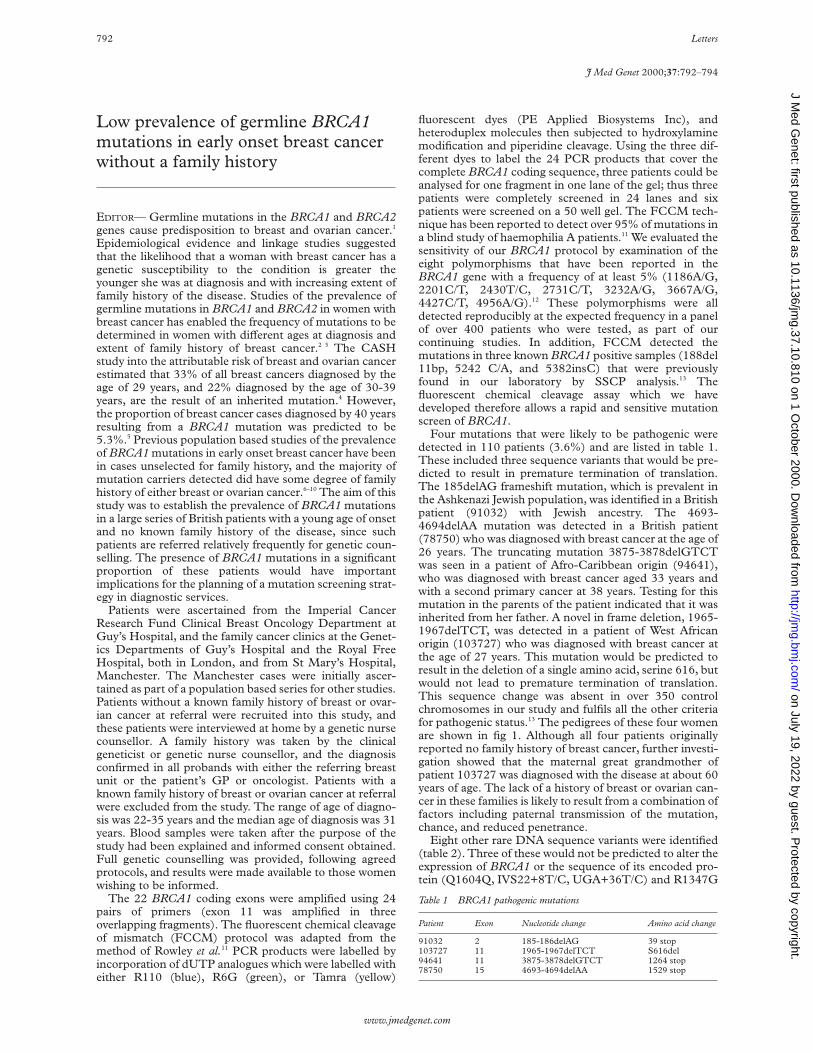
Low prevalence of germline BRCA1mutations in early onset breast cancerwithout a family history
EDITOR— Germline mutations in the BRCA1 and BRCA2genes cause predisposition to breast and ovarian cancer.1
Epidemiological evidence and linkage studies suggestedthat the likelihood that a woman with breast cancer has agenetic susceptibility to the condition is greater theyounger she was at diagnosis and with increasing extent offamily history of the disease. Studies of the prevalence ofgermline mutations in BRCA1 and BRCA2 in women withbreast cancer has enabled the frequency of mutations to bedetermined in women with diVerent ages at diagnosis andextent of family history of breast cancer.2 3 The CASHstudy into the attributable risk of breast and ovarian cancerestimated that 33% of all breast cancers diagnosed by theage of 29 years, and 22% diagnosed by the age of 30-39years, are the result of an inherited mutation.4 However,the proportion of breast cancer cases diagnosed by 40 yearsresulting from a BRCA1 mutation was predicted to be5.3%.5 Previous population based studies of the prevalenceof BRCA1 mutations in early onset breast cancer have beenin cases unselected for family history, and the majority ofmutation carriers detected did have some degree of familyhistory of either breast or ovarian cancer.6–10 The aim of thisstudy was to establish the prevalence of BRCA1 mutationsin a large series of British patients with a young age of onsetand no known family history of the disease, since suchpatients are referred relatively frequently for genetic coun-selling. The presence of BRCA1 mutations in a significantproportion of these patients would have importantimplications for the planning of a mutation screening strat-egy in diagnostic services.
Patients were ascertained from the Imperial CancerResearch Fund Clinical Breast Oncology Department atGuy’s Hospital, and the family cancer clinics at the Genet-ics Departments of Guy’s Hospital and the Royal FreeHospital, both in London, and from St Mary’s Hospital,Manchester. The Manchester cases were initially ascer-tained as part of a population based series for other studies.Patients without a known family history of breast or ovar-ian cancer at referral were recruited into this study, andthese patients were interviewed at home by a genetic nursecounsellor. A family history was taken by the clinicalgeneticist or genetic nurse counsellor, and the diagnosisconfirmed in all probands with either the referring breastunit or the patient’s GP or oncologist. Patients with aknown family history of breast or ovarian cancer at referralwere excluded from the study. The range of age of diagno-sis was 22-35 years and the median age of diagnosis was 31years. Blood samples were taken after the purpose of thestudy had been explained and informed consent obtained.Full genetic counselling was provided, following agreedprotocols, and results were made available to those womenwishing to be informed.
The 22 BRCA1 coding exons were amplified using 24pairs of primers (exon 11 was amplified in threeoverlapping fragments). The fluorescent chemical cleavageof mismatch (FCCM) protocol was adapted from themethod of Rowley et al.11 PCR products were labelled byincorporation of dUTP analogues which were labelled witheither R110 (blue), R6G (green), or Tamra (yellow)
fluorescent dyes (PE Applied Biosystems Inc), andheteroduplex molecules then subjected to hydroxylaminemodification and piperidine cleavage. Using the three dif-ferent dyes to label the 24 PCR products that cover thecomplete BRCA1 coding sequence, three patients could beanalysed for one fragment in one lane of the gel; thus threepatients were completely screened in 24 lanes and sixpatients were screened on a 50 well gel. The FCCM tech-nique has been reported to detect over 95% of mutations ina blind study of haemophilia A patients.11 We evaluated thesensitivity of our BRCA1 protocol by examination of theeight polymorphisms that have been reported in theBRCA1 gene with a frequency of at least 5% (1186A/G,2201C/T, 2430T/C, 2731C/T, 3232A/G, 3667A/G,4427C/T, 4956A/G).12 These polymorphisms were alldetected reproducibly at the expected frequency in a panelof over 400 patients who were tested, as part of ourcontinuing studies. In addition, FCCM detected themutations in three known BRCA1 positive samples (188del11bp, 5242 C/A, and 5382insC) that were previouslyfound in our laboratory by SSCP analysis.13 Thefluorescent chemical cleavage assay which we havedeveloped therefore allows a rapid and sensitive mutationscreen of BRCA1.
Four mutations that were likely to be pathogenic weredetected in 110 patients (3.6%) and are listed in table 1.These included three sequence variants that would be pre-dicted to result in premature termination of translation.The 185delAG frameshift mutation, which is prevalent inthe Ashkenazi Jewish population, was identified in a Britishpatient (91032) with Jewish ancestry. The 4693-4694delAA mutation was detected in a British patient(78750) who was diagnosed with breast cancer at the age of26 years. The truncating mutation 3875-3878delGTCTwas seen in a patient of Afro-Caribbean origin (94641),who was diagnosed with breast cancer aged 33 years andwith a second primary cancer at 38 years. Testing for thismutation in the parents of the patient indicated that it wasinherited from her father. A novel in frame deletion, 1965-1967delTCT, was detected in a patient of West Africanorigin (103727) who was diagnosed with breast cancer atthe age of 27 years. This mutation would be predicted toresult in the deletion of a single amino acid, serine 616, butwould not lead to premature termination of translation.This sequence change was absent in over 350 controlchromosomes in our study and fulfils all the other criteriafor pathogenic status.13 The pedigrees of these four womenare shown in fig 1. Although all four patients originallyreported no family history of breast cancer, further investi-gation showed that the maternal great grandmother ofpatient 103727 was diagnosed with the disease at about 60years of age. The lack of a history of breast or ovarian can-cer in these families is likely to result from a combination offactors including paternal transmission of the mutation,chance, and reduced penetrance.
Eight other rare DNA sequence variants were identified(table 2). Three of these would not be predicted to alter theexpression of BRCA1 or the sequence of its encoded pro-tein (Q1604Q, IVS22+8T/C, UGA+36T/C) and R1347G
Table 1 BRCA1 pathogenic mutations
Patient Exon Nucleotide change Amino acid change
91032 2 185-186delAG 39 stop103727 11 1965-1967delTCT S616del94641 11 3875-3878delGTCT 1264 stop78750 15 4693-4694delAA 1529 stop
792 Letters
www.jmedgenet.com
on July 19, 2022 by guest. Protected by copyright.
http://jmg.bm
j.com/
J Med G
enet: first published as 10.1136/jmg.37.10.810 on 1 O
ctober 2000. Dow
nloaded from

was present in a patient with a frameshift mutation.12 Thepathogenic status of the other four (Q804H, S1140G,P1637L, and M1652I) remains inconclusive in the absenceof a functional assay for the BRCA1 protein. At present,screening panels of ethnically matched controls is a usefulmeans of excluding missense mutations as pathogenicmutations of high penetrance, and it would be helpful ifthis information was provided in the Breast Cancerdatabase (www.nhgri.nih.gov/Intramural_research/Lab_transfer/Bic/index.html).
Our detection of pathogenic BRCA1 mutations in 3.6%of young breast cancer patients without a family history ofbreast or ovarian cancer is consistent with the estimate ofFord et al5 that the proportion of breast cancer cases in thegeneral population resulting from BRCA1 is 5.3% belowthe age of 40 years. A recent population based study ofyoung British breast cancer patients unselected for familyhistory found BRCA1 mutations in nine of 254 (3.5%)women diagnosed by the age of 36 years.8 In North Ameri-can studies of BRCA1 mutations in women unselected forfamily history and diagnosed below the age of 35 years,Langston et al7 detected mutations in 7.5%, Malone et al9 in6.2%, and Struewing et al10 in 5.7%. Fitzgerald et al6
detected BRCA1 mutations in 13% of women diagnosedbefore the age of 30 years, but this included Ashkenazi
Jewish patients who have founder mutations and somepatients with a family history. Since we did not screen thepromoter region of BRCA1 or for deletions of entireexons,14 we cannot exclude the possibility that some muta-tions were missed, and the pathogenic status of severalsequence variants remains unresolved. However, theimportant practical implication of our study is that, giventhe time and expense involved, it would be reasonable toattach a low priority to BRCA1 mutation screening ofyoung isolated cases of breast cancer in the context of theprovision of a publicly funded and cost eVective diagnosticservice. A screen of this cohort of patients for BRCA2mutations is in progress.
This study was supported by the NHS National Cancer Research and Develop-ment Programme (UK) (project No NCP/B11/34).
DAVID ELLIS*JILL GREENMAN*
SHIRLEY HODGSON*SAM McCALL*
FIONA LALLOO†JUNE CAMERON*
LOUISE IZATT*GILLIAN SCOTT*
CHRIS JACOBS*SALLY WATTS*
WENDY CHORLEY‡CHRIS PERRETT‡
Figure 1 Pedigrees of cases in whom a pathogenic BRCA1 mutation was identified.
94641
103727
9103278750
no data
Table 2 BRCA1 unclassified variants and polymorphisms
Exon Nucleotide changeAmino acidchange
Freq incontrols
Conserved inmouse/dog
BICentries
11 2531 G/C Q804H 0.0 +/− 011 3537 A/G S1140G 0.0 −/+ 1511 4158 A/G R1347G 0.01* −/+ 7516 4931 A/G Q1604Q ND +/+† 1616 5029 C/T P1637L 0.0 +/+ 1118 5075 G/A M1652I 0.01 +/+ 8intron 22 IVS22+8 T/C Unknown ND ND 23' untrans UGA+36 C/G Unknown ND ND 4
*As reported by Langston et al.7
†Nucleotide sequence is conserved in mouse and dog BRCA1 genes.ND = not determined.
Letters 793
www.jmedgenet.com
on July 19, 2022 by guest. Protected by copyright.
http://jmg.bm
j.com/
J Med G
enet: first published as 10.1136/jmg.37.10.810 on 1 O
ctober 2000. Dow
nloaded from

KAY MACDERMOT§SHEHLA MOHAMMED*
GARETH EVANS†CHRISTOPHER G MATHEW*
*Division of Medical and Molecular Genetics, GKT School of Medicine,8th Floor, Guy’s Tower, Guy’s Hospital, London SE1 9RT, UK†Department of Medical Genetics, St. Mary’s Hospital, Hathersage Road,Manchester M13 0JH, UK‡Department of Obstetrics and Gynaecology, Royal Free Hospital, PondStreet, London NW3 2PF, UK§Department of Clinical Genetics, Royal Free Hospital, Pond Street,London NW3 2PF, UK
Correspondence to: Professor Mathew, [email protected]
1 Rahman N, Stratton MR. The genetics of breast cancer susceptibility. AnnuRev Genet 1998;32:95-121.
2 Stratton MR. Recent advances in understanding of genetic susceptibility tobreast cancer. Hum Mol Genet 1996;5:1515-19.
3 Neuhausen SL, Ostrander EA. Mutation testing of early-onset breast cancergenes BRCA1 and BRCA2. Genet Test 1997;1:75-82.
4 Claus EB, Schildkraut JM, Thompson WD, Risch NJ. The genetic attribut-able risk of breast and ovarian cancer. Cancer 1996;77:2318-24.
5 Ford D, Easton DF, Peto J. Estimates of the gene frequency of BRCA1 andits contribution to breast and ovarian cancer incidence. Am J Hum Genet1995;57:1457-62.
6 Fitzgerald MG, MacDonald DJ, Krainer M, Hoover I, O’Neil E, Unsal H,Silva-Arrieto S, Finkelstein DM, Beer-Romero P, Englert C, Sgroi DC,
Smith BL, Younger JW, Garber JE, Duda RB, Mayzel KA, Isselbacher KJ,Friend SH, Haber DA. Germline BRCA1 mutations in Jewish andnon-Jewish women with early-onset breast cancer. N Engl J Med 1996;334:143-9.
7 Langston AA, Malone KE, Thompson JD, Daling JR, Ostrander EA.BRCA1 mutations in a population-based sample of young women withbreast cancer N Engl J Med 1996;334:136.
8 Peto J, Collins N, Barfoot R, Seal S, Warren W, Rahman N, Easton DF,Evans C, Deacon J, Stratton MR. Prevalence of BRCA1 and BRCA2 genemutations in patients with early-onset breast cancer. J Natl Cancer Inst1999;91:943-9.
9 Malone KE, Daling JR, Thompson JD, O’Brien CA, Francisco LV,Ostrander EA. BRCA1 mutations and breast cancer in the general popula-tion. Analyses in women before age 35 years and in women before age 45years with first-degree family history. JAMA 1998;279:922-9.
10 Struewing JP, Tarone RE, Brody LC, Li FP, Boice JD. BRCA1 mutations inyoung women with breast cancer. Lancet 1996;347:1493.
11 Rowley G, Saad S, Gianelli F, Green P. Ultrarapid mutation detection bymultiplex, solid-phase chemical cleavage. Genomics 1995;30:574-82.
12 Durocher F, Shattuck-Eidens D, McClure M, Labrie F, Skolnick MH,Goldgar DE, Simard J. Comparison of BRCA1 polymorphisms, raresequence variants and/or missense mutations in unaVected and breast/ovarian cancer populations. Hum Mol Genet 1996;5:835-42.
13 Greenman J, Mohammed S, Ellis D, Watt S, Scott G, Izatt L, Barnes D,Solomon E, Hodgson S, Mathew C. Identification of missense andtruncating mutations in the BRCA1 gene in sporadic and familial breastand ovarian cancer. Genes Chrom Cancer 1998;21:244-9.
14 Puget N, Stoppa-Lyonnet D, Sinilnikova OM, Pages S, Lynch HT, LenoirGM , Mazoyer S. Screening for germ-line rearrangements and regulatorymutations in BRCA1 led to the identification of four new deletions. CancerRes 1999;59:455-61.
J Med Genet 2000;37:794–796
Delineation of a new syndrome:clustering of pyloric stenosis,endometriosis, and breast cancer intwo families
EDITOR—Familial tendencies have previously been ob-served for congenital pyloric stenosis, endometriosis, andbreast cancer. These conditions have never been consid-ered to have shared aetiological origins and consequentlyno previous attempts have been made to investigate anassociation. For example, when obtaining family historyinformation for a child with pyloric stenosis, one would notroutinely request a description of adult onset conditionssuch as endometriosis or breast cancer. Two families shar-ing an unusual clustering of these three conditions (pyloricstenosis, endometriosis, and breast cancer) were ascer-tained at the familial cancer clinics of the Women’s Collegeand Princess Margaret Hospitals in Toronto.
Family 1 (fig 1) contains four confirmed cases of breastcancer (below age 60), seven cases of endometriosis, fivecases of congenital pyloric stenosis, nine cases of polycysticovaries, and four cases of non-insulin dependent diabetes.In a second unrelated family, a woman previouslydiagnosed with premenopausal breast cancer, endometrio-sis, and pyloric stenosis reported one other case ofcongenital pyloric stenosis and four other cases ofendometriosis in her family (fig 1). It is the similar andunusual presentation in these two families which suggeststhat the clustering of pyloric stenosis, endometriosis, andbreast cancer may not be the result of chance.
A family history of breast cancer is known to be the mostsignificant risk factor for developing the disease. Approxi-mately 5-10% of all cases are hereditary and accounted forby mutations in cancer susceptibility genes BRCA1 andBRCA2.1 2 Family 1 met our criteria for BRCA1 andBRCA2 testing, with four known cases of breast cancerdiagnosed below the age of 60. Mutation analysis by directsequencing of the coding regions of BRCA1 and BRCA2 aswell as 1700 adjacent non-coding intronic base pairs wasperformed by Myriad Genetic Laboratories (http://
www.myriad.com). No BRCA mutation was identified forfamily 1. Family 2 was not tested for BRCA1 or BRCA2mutations.
The aetiology of endometriosis remains uncertain,although familial trends have been described.3 Four studiesfound that there is an increased risk among first degreerelatives.4–7 A small twin study found 6/8 monozygotic and0/2 dizygotic twin pairs had endometriosis.7 In family 2,five women in three generations had endometriosis and infamily 1 seven women in three generations were given thediagnosis. The pattern of inheritance is consistent with sexlimited, autosomal dominant inheritance.
A multifactorial genetic contribution for pyloric stenosishas been well established, although its pathological basisremains unknown. Twin studies have shown that there is a25-40% concordance rate in monozygotic twins.8 9 Basedon pooled data from several family studies and assuming apopulation prevalence of 0.3%, a relative risk for firstdegree relatives compared to the general population was1810; however, population based studies (unselectedpatients) have not been done. Pyloric stenosis has recentlybeen linked to the locus of the neuronal nitric oxidesynthase (NOS1) gene, based on 27 families.11 The NOS1locus was also examined for other multifactorial conditionssuch as asthma (candidate gene)13 14 and multiple sclerosis(no association).15 Family 1 has five documented cases ofsurgically corrected pyloric stenosis in three males and twofemales. Family 2 has a parent and child with PS, bothfemale.
In addition to endometriosis, family 1 contains ninewomen with polycystic ovary syndrome (PCOS), includingone woman with non-insulin dependent diabetes mellitus(NIDDM). PCOS and NIDDM have been shown to havea shared aetiology.12 Women with PCOS have a unique dis-order of insulin action and are at increased risk of develop-ing NIDDM, which occurs substantially younger (in thethird to fourth decades) than it does in the general popula-tion.
Breast cancer, endometriosis, and pyloric stenosis infamilies 1 and 2 may be explained by separate genetic pre-dispositions; however, the possibility that there is acommon genetic basis exists. It is the complex interplaybetween environmental, hormonal, and genetic factorswhich poses a challenge to understanding the aetiology of
794 Letters
www.jmedgenet.com
on July 19, 2022 by guest. Protected by copyright.
http://jmg.bm
j.com/
J Med G
enet: first published as 10.1136/jmg.37.10.810 on 1 O
ctober 2000. Dow
nloaded from

each condition. A future study of pyloric stenosis in a case-control design may investigate any association with breastcancer or endometriosis.
ALEXANDER LIEDETUYA PAL
MARGOT MITCHELLSTEVEN A NAROD
Breast Cancer Research, University of Toronto, 790 Bay Street, 750A,Toronto, ON M5G 1N8, Canada
Correspondence to: Professor Narod, [email protected]
1 Wooster R, Bignell G, Lancaster J. Identification of the breast cancersusceptibility gene BRCA2. Nature 1995;378:789-92.
2 Miki Y, Swensen J, Shattuck-Eidens D. A strong candidate for thebreast and ovarian cancer susceptibility gene BRCA1. Science 1994;266:66-71.
3 Kennedy S, Mardon H, Barlow D. Familial endometriosis. J Assist ReprodGenet 1995;12:32-4.
4 Simpson JL, Elias S, Malinak LR, Buttram VC. Heritable aspects ofendometriosis. I. Genetic studies. Am J Obstet Gynecol 1980;137:327-31.
5 Coxhead D, Thomas EJ. Familial inheritance of endometriosis in a Britishpopulation. A case control study. J Obstet Gynaecol 1993;13:42-4.
6 Moen MH, Magnus P. The familial risk of endometriosis. Acta ObstetGynaecol Scand 1993;73:59-62.
7 Moen MH. Endometriosis in monozygotic twins. Acta Obstet GynaecolScand 1994;73:59-62.
8 Metrakos JD. Congenital hypertrophic pyloric stenosis in twins. Arch DisChild 1953;27:351-8.
Figure 1 Pedigrees of families 1 and 2. Proband is indicated by an arrow. The age of the subjects appears directly below symbol. CA = breast cancerfollowed by age of diagnosis (dx); Psu = primary site of cancer was not known; Pr CA = prostate cancer; Cx CA = cervical cancer; TAH = totalabdominal hysterectomy; BSO = bilateral salpingo-oophorectomy; NIDDM = non-insulin dependent diabetes mellitus; CHD = congenital heart disease;D&C = dilatation and curettage; SA = spontaneous abortion; SB = stillbirth.
Cx CAd 75
d 64d stroke d 66 d infant 48 50
4042
dx 66 dx 25
Br CAdx 42
8 6 2
d 67 d 50 70Kidney CA Crohn's
Kidney CA
d old age d 32 SA
PsuPsu Pr CA Colon CA96 89
dx 90
Br CA60
dx 45
D&C49
CHD44 40 33
SB17 49
22 6CHD
40 34Cx CA
33
Infertility
Br CA65
dx 52TAH-BSO
Br CA59
dx 59TAH
Br CAd 54
TAH57
TAH-BSO50
dx 50TAH-BSO
D&C70 68 d 54
dx 89
Crohn's Skin CATAH
Colitis Melanoma Ileitis
Family 2
Family 1
Pyloric Stenosis
Non-InsulinDependentDiabetesMellitus
Polycystic OvarySyndrome
Endometriosis
3
2
2 2 2 4 2 23
Letters 795
www.jmedgenet.com
on July 19, 2022 by guest. Protected by copyright.
http://jmg.bm
j.com/
J Med G
enet: first published as 10.1136/jmg.37.10.810 on 1 O
ctober 2000. Dow
nloaded from

9 MacMahon B, McKeown T. Infantile hypertrophic pyloric stenosis: data on81 pairs of twins. Acta Genet Med Gemellol 1955;4:320-5.
10 Mitchell LE, Risch N. The genetics of infantile hypertrophic pyloricstenosis: a reanalysis. Am J Dis Child 1993;147:1203-11.
11 Chung E, Curtis D, Chen G, Marsden PA, Twells R, Xu W, Gardiner M.Genetic evidence for the neuronal nitric oxide synthase gene (NOS1) as asusceptibility locus for infantile pyloric stenosis. Am J Hum Genet 1996;58:363-70.
12 Dunaif A. Hyperandrogenic anovulation (PCOS): a unique disorder ofinsulin action associated with an increased risk of non-insulin-dependentdiabetes mellitus. Am J Med 1995;98:33-9S.
13 Grasemann H, Yandava CN, Drazen JM. Neuronal NO synthase (NOS1) isa major candidate gene for asthma. Clin Exp Allergy 1999;29(suppl 4):39–41.
14 Gao PS, Kawada H, Kasamatsu T, Mao XQ, Roberts MH, Miyamoto Y,Yoshimura M, Saitoh Y, Yasue H, Nakao K, Adra CN, Kun JF, Moro-okaS, Inoko H, Ho LP, Shirakawa T, Hopkin JM. Variants of NOS1, NOS2,and NOS3 genes in asthmatics. Biochem Biophys Res Commun 2000;267:761–3.
15 Xu C, Hillert J. Absence of linkage with the neuronal nitric oxide synthase(NOS1) gene in 41 multiplex Swedish MS families. Eur J Neurol1998;5:393–6.
J Med Genet 2000;37:796–798
Punctate calcification of the epiphyses,visceral malformations, andcraniofacial dysmorphism in a femalebaby
EDITOR—We report a fetus with striking craniofacialdysmorphism, brachydactyly, and cerebral and cardiacmalformations in addition to punctate calcification of theepiphyses.
The mother was treated for tuberculosis seven yearsbefore the pregnancy but there were no known systemicillnesses or teratogenic influences during this pregnancy.
The mother’s first pregnancy resulted in a termination at22 weeks of gestation for multiple congenital abnormali-ties, but further details are not known.
The baby was the second child born to a 21 year oldmother. A termination was performed at 21 weeks of ges-tation because of multiple anomalies seen on antenatalscanning. Necropsy showed a female fetus (fig 1) with aweight of 1544 g, consistent with 17 weeks’ gestation. Thecrown-heel length was 16.4 cm and right foot length was18 mm. Facial examination showed an open right eye withexophthalmos, hypertelorism, a flat nasal bridge withhypoplasia of the alae nasi, flattening of the midface, ashort philtrum with a well defined philtral groove, largelips, and a wide mouth with micrognathia. The right earwas simple and low set and the left ear was rudimentary
Figure 1 (A) Front view of fetus. (B) Hand of fetus. (C) Foot of fetus.
796 Letters
www.jmedgenet.com
on July 19, 2022 by guest. Protected by copyright.
http://jmg.bm
j.com/
J Med G
enet: first published as 10.1136/jmg.37.10.810 on 1 O
ctober 2000. Dow
nloaded from

and poorly formed. The palate was normal. Both handshad brachydactyly involving all digits with short thumbs.There were bilateral varus foot deformities and both feetwere rockerbottom in shape. The remainder of theexamination showed nuchal thickening, a short, broadchest, and a straight spine. The genitalia were female witha vaginal opening but there was clitoral hypertrophy andprominence of the labial folds.
Internal examination of the brain showed enlargement ofboth lateral cerebral ventricles, absent olfactory bulbs, andan absent pituitary gland. The heart had a common trun-cus arising from the right ventricle with a high ventricularseptal defect. The right adrenal gland was absent.Histological sections showed that one umbilical artery wasrudimentary without a lumen. Examination of the placentashowed no inflammatory changes. Unfortunately, nohistological studies were performed on bone.
Chromosome analysis showed an apparently normalfemale karyotype but the quality of the preparation wassuYcient only to exclude numerical and large structuralabnormalities. Culture of the fetal cells failed andperoxisomal studies and a 7-dehydrocholesterol level werenot performed.
Radiographs (fig 2) showed extensive calcification andstippling in all epiphyseal areas. There was undermodellingof the long bones with shortening of the humeri and rela-tive widening of the diaphyses. There was poor calcificationand sagittal clefting of the vertebral bodies. Radiographs ofthe hands and feet showed stippling in the carpal and tar-
sal bones and the fingers and toes were extremely shortwith a single phalanx in the second to fifth digits andalmost absent ossification of the phalanges. The thorax wasshort and broad with 11 pairs of ribs and there was poste-rior dislocation of the hips. The couple chose not to attendgenetic counselling clinic.
This baby had an unusual pattern of malformationsincluding right exophthalmos, midface hypoplasia, rudi-mentary ears, cerebral ventriculomegaly, a commontruncus arteriosus and ventricular septal defect, andextreme brachydactyly in addition to calcific stippling ofthe epiphyses. Epiphyseal stippling can be found in the pri-mary chondrodysplasias,1 but the visceral abnormalities inthis baby are not typical of chondrodysplasia and the pres-entation is neither consistent with brachytelephalangicchondrodysplasia punctata (CP)2 3 or humerometacarpalCP.4 5 One plausible diagnosis is Pacman dysplasia, a rarecondition in which stippling of the lower spine and epiphy-ses has been found with bowing of the femora, spinal clef-ting, and giant, multinucleated, osteoclast-like cells.6 7
Although a patent ductus was described in one child, Pac-man dysplasia has not been reported with severe visceralabnormalities or craniofacial dysmorphism as found in thisbaby.
Stippling of the epiphyses is well known to be causallyheterogeneous,1 8 but syndromes with punctate calcifica-tion and visceral malformations are rare. Two sibs weredescribed with short stature, ocular colobomata, midfacehypoplasia with a small nose, low set ears, and dysplasticdistal phalanges in addition to stippled epiphyses.9 Asecond report of a girl with similar facial dysmorphism, aright retinal coloboma and an incomplete atrioventricularcanal, coarctation of the aorta, enlargement of the anteriorhorns of the lateral ventricles, and partial agenesis of thecorpus callosum concluded that these anomalies com-prised a separate syndrome.10 Recently, a male baby withpunctate epiphyses and coronal clefting of the spine wasdescribed with a Dandy-Walker malformation, hypogen-esis of the corpus callosum and falx cerebri, a ventricularseptal defect, and bilateral cleft lip and palate.11 This childalso had craniofacial dysmorphism with telecanthus,clouding of the corneas, a high nasal bridge andretromicrognathia, and brachydactyly.
These cases show a degree of similarity to this baby withcalcific stippling in association with midface and nasalhypoplasia, dysplastic ears, brachydactyly, and cranial andcerebral malformations. However, the variable nature ofthe accompanying anomalies makes it diYcult to be confi-dent that these patients represent heterogeneity of the samecondition. Despite the incomplete clinical information inour case, we believe that documentation of theseabnormalities will prove useful in view of the rarity of thecombination of features and the paucity of reports describ-ing epiphyseal stippling and malformations.
ANNE SLAVOTINEKHELEN KINGSTON
University Department of Medical Genetics and Regional GeneticService, St Mary’s Hospital, Hathersage Road, Manchester M13 0JH,UK
Correspondence to: Dr Kingston, [email protected]
1 Wulfsberg EA, Curtis J, Jayne CH. Chondrodysplasia punctata: a boy withX-linked recessive chondrodysplasia punctata due to an inherited X-Ytranslocation with a current classification of these disorders. Am J MedGenet 1992;43:823-8.
2 Maroteaux P. Brachytelephalangic chondrodysplasia punctata: a possibleX-linked recessive form. Hum Genet 1989;82:167-70.
3 Zizka J, Charvat J, Baxova A, Balicek P, Kozlowski K. Brachytelephalangicchondrodysplasia punctata with distinctive phenotype and normalkaryotype. Am J Med Genet 1998;76:213-16.
4 Borochowitz Z. Generalized chondrodysplasia punctata with shortness ofhumeri and brachymetacarpy: humero-metacarpal (HM) type: variation orheterogeneity? Am J Med Genet 1991;41:417-22.
5 Fryburg JS, Kelly TE. Chondrodysplasia punctata, humero-metacarpaltype: a second case. Am J Med Genet 1996;64:493-6.
Figure 2 Radiograph of fetus showing punctatecalcification of epiphyseal areas and stippling of carpal andtarsal bones.
Letters 797
www.jmedgenet.com
on July 19, 2022 by guest. Protected by copyright.
http://jmg.bm
j.com/
J Med G
enet: first published as 10.1136/jmg.37.10.810 on 1 O
ctober 2000. Dow
nloaded from

6 Shohat M, Rimoin DL, Gruber HE, Lachman R. New epiphyseal stipplingsyndrome with osteoclastic hyperplasia. Am J Med Genet 1993;45:558-61.
7 Wilcox WR, Lucas BC, Loebel B, Bachman RP, Lachman RS, Rimoin DL.Pacman dysplasia: report of two aVected sibs. Am J Med Genet 1998;77:272-6.
8 Bennett CP, Berry AC, Maxwell DJ, Seller MJ. Chondrodysplasia punctata:another possible X-linked recessive case. Am J Med Genet 1992;44:795-9.
9 Toriello HV, Higgins JV, Miller T. Provisionally unique autosomal recessivechondrodysplasia punctata syndrome. Am J Med Genet 1998;77:272-6.
10 Ciske DJ, Waggoner DJ, Dowton SB. Unique cardiac and cerebral anoma-lies with chondrodysplasia punctata. Am J Med Genet 1993;47:797-9.
11 Mortier GR, Messiaen LM, Espeel M, Smets KJ, Vazieleghem BD, Roels F,De Paepe AM. Chondrodysplasia punctata with multiple congenitalanomalies: a new syndrome? Pediatr Radiol 1998;28:790-3.
J Med Genet 2000;37:798–804
Unexpected high frequency of de novounbalanced translocations in patientswith Wolf-Hirschhorn syndrome(WHS)
EDITOR—Wolf-Hirschhorn syndrome (WHS), first de-scribed independently in 1965 by Wolf et al1 andHirschhorn et al,2 is a well defined multiple congenitalanomalies/mental retardation syndrome resulting fromdeletion involving chromosomal band 4p16.3 with a mini-mal critical region of 165 kb.3
DiVerent mechanisms are responsible for WHS, includ-ing terminal 4p deletions, familial translocations, and denovo complex chromosomal rearrangements such asunbalanced translocations. The frequency of transloca-tions in WHS as a result of parental chromosomal translo-cations is estimated to be 5 to 13%,4 5 whereas the rate ofsporadic translocations in WHS was suggested to be 1.6%(2/120).5 A few isolated cases with de novotranslocations5–12 resulting in WHS have been describedrecently. The trisomic segment, however, often could notbe defined owing to the lack of specific cytogenetictechniques.5 6 8 13–15
Here we report six patients with unbalanced transloca-tions, t(4p;8p) and t(4p;7p), respectively, and discuss theirphenotypic abnormalities.
Since 1996 we have performed clinical, molecular,cytogenetic, and molecular-cytogenetic investigations in a
total of 22 patients with clinical signs of Wolf-Hirschhornsyndrome. These patients were all seen by one of theauthors. In five of them (22%), the combination ofcytogenetic and molecular investigations showed a de novounbalanced translocation; in a sixth patient the de novooccurrence of the translocation could not be confirmedbecause no blood sample from the father was available.
The clinical findings of patients 1-5, who all presentedwith characteristic features of WHS, are summarised intable 1. Patient 6 (CL220585/87E1624) had someadditional findings listed in table 1.
The combination of Hirschsprung disease and hydro-cephalus was suggestive of CRASH syndrome in patient 6,but SSCP analysis of the L1CAM gene was normal.
Chromosome studies including GTG banding were per-formed on peripheral blood lymphocytes according toslightly modified standard techniques.16
DNA from cosmids pC847.351,17 L21f12 (D4S180), andL228a7 (D4S81)18 and inter-Alu PCR products19 from YAC877G6, 405D10, 794D12, 225D2, and 435A11 (CEPH)were labelled with digoxigenin using a BRL nick translationkit (Gibco, Life Technologies Inc, Gaithersburg, MD, USA).Labelled cosmid and YAC DNA was separated fromunincorporated nucleotides by using 1800 ml Centricon 30(Amicon Inc, Beverly, MA 01915, USA) filters. FISH wasperformed as described by Lichter et al.20 Labelled cosmidDNA (50 ng) or YAC DNA (100 ng) was mixed with humanCot-1 fraction DNA to suppress repetitive sequences orblock non-specific hybridisation. Hybridisation was detectedusing monoclonal anti-digoxin, anti-mouse IgG FITC con-jugate and anti-rabbit IgG FITC conjugate (Sigma Immuno
Table 1 Clinical findings in patients 1-5 with t(4;8) and patient 6 with t(4;7)
Patient 1(CG/96E0600)
Patient 2(DT/92E1657)
Patient 3(LF/96E0551)
Patient 4(NR/97E877)
Patient 5(AS/94E1416)
Patient 6(CL/87E1624)
Anthropometric dataBirth (gestational weeks) 39 36 38 40 40 41Weight (g) 2900 (−1.0 SD) 2120 (−1.2 SD) 1800 (−3.4 SD) 2240 (−3.6 SD) 2200 (−3.7 SD) 2950 (−1.6 SD)Length (cm) NR 46 (−0.4 SD) 42 (−2.9 SD) 47 (−3.2 SD) 45 (−4.3 SD) 48 (−2.5 SD)OFC (cm) 31.5 (−2.4 SD) 30.5 (−1.9 SD) NR 31.5 (−2.7 SD) 32 (−2.3 SD) 34 (−1.6 SD)Age at examination (y) 11⁄12 35⁄12 36⁄12 2 510⁄12 12Weight (g) 5240 (−3.8 SD) 9060 (−3.2 SD) NR NR NR 29 000 (−2 SD)Height (cm) 64.2 (−4.4 SD) 89 (−2.7 SD) 84.5 (−6.7 SD) 72 (−5 SD) 91 (−4.4 SD) 133 (−2.4 SD)OFC (cm) 41.3 (−3.5 SD) 46.5 (−2.2 SD) 44 (−5.0 SD) 42 (−4.6 SD) 46 (−3.5 SD) 54 (mean)Dysmorphic featuresHypertelorism + + + + + +Strabismus + − + + + +Broad, large mouth − + − − + +Downturned corners of mouth + + + + + +Micrognathia + + + + +Cleft lip/palate − − + + + −Dysplastic ears + + + + + +Preauricular tag − − + − − −Internal findingsHeart defect − − ASD ASD − −Genital anomalies − + NR − − +Neurological developmentSitting age (mth) 13 20 60 − 24 36Walking age (y) 2 35⁄12 69⁄12 − 5 ?Simple words (y) 26⁄12 − − − − ?Understanding of speech ++ ++ (+) − − ++Seizures − + + + + +Additional findings Oligohydramnios
Bilateral pneumothoraxFlexion contractures of large jointsHypoplasia of corpus callosumHirschsprung disease
798 Letters
www.jmedgenet.com
on July 19, 2022 by guest. Protected by copyright.
http://jmg.bm
j.com/
J Med G
enet: first published as 10.1136/jmg.37.10.810 on 1 O
ctober 2000. Dow
nloaded from

Chemicals, St Louis, USA) and counterstained usingpropidium iodide or DAPI.
A cosmid derived from D4S96, the 8p telomere probeand 7p distal probe (Oncor, Gaithersburg, VA, USA) andthe whole chromosome paints 4 and 8 (AGS, Heidelberg,Germany/Oncor, Gaithersburg, VA, USA) were hybridisedaccording to the manufacturer’s instructions. For thehybridisations, clone pGXba11/34021 was used as centro-meric control for human chromosome 4.
Genomic DNA was prepared from peripheral bloodlymphocytes or EBV transformed lymphoblastoid cell linesfrom patients and their parents according to previouslydescribed techniques.22 23
Genotyping of chromosome 4p was performed using thefollowing previously described microsatellite markers:D4S1182 (Acc ID GDB 19723924), D4S43 (Acc ID GDB12436024), ADRA2C (Acc ID GDB 37083425), HOX7(Acc ID GDB 15699126 27), D4S403 (Acc ID GDB18810628), D4S2366 (Acc ID GDB 684453), D4S2639(Acc ID GDB 685881), and D4S2397 (Acc ID GDB683982). A new microsatellite, 75B9Rep, was generated byone of the authors based on sequence information of cos-mid 75B9A (Acc No Z69651) which is derived from thedistal end of the Huntington’s disease cosmid contig.29 Oli-
gomers were designed from sequences flanking thecompound dinucleotide repeat (CT)2G(CT)26(CA)14. Thefollowing primer sequences were used for the amplificationof the repeat, forward primer 5' CTGAACTTTATC-CAATTAGTCTTG 3' and reverse primer 5' GAATCTT-TCTGTCCCACGAT 3'. This primer set amplifies a 227bp DNA fragment product. PCR reactions were performedin 10 µl volumes containing 100 ng DNA, 0.5 U GenecraftR
Taq polymerase, 1 × GenecraftR PCR buVer, 2 µmol/ldNTP, 4 pmol/l of each primer, 1 µCi 32P-dCTP (3000Ci/mmol/lol, Amersham), and 2 mmol/l MgCl2. Forma-mide (2%) was added to increase stringency of thereaction. After an initial denaturation of one minute, 30cycles of one minute at 94°C, 30 seconds at 57°C, and 30seconds at 72°C were carried out in a Crocodile IIIR (App-ligene) thermocycler. The PCR products were resolved on6% polyacrylamide gels flanked by a sequence ladder(M13, −40) as size marker. The Hu4/Hu5 (Acc ID GDB249651) primer pair amplifies the CAG repeat of thehuntingtin gene. Primer Hu4 has been published by Riesset al25 (primer sequence: Hu4 5' ATGGCGACCCT-GGAAAAGCTGATGAA 3'), and primer Hu5 has beenpublished by Rubinsztein et al30 (primer sequence: Hu5 5'GGCGGTGGCGGCTGTTGCTGCTGCTGC 3').
Figure 1 (A) Chromosomes 4 of patient 2 showing an additional G positive band in 4pter. (B) Chromosomes 4 of patient 4 showing same length of 4pbut diVerent G banded pattern.
Letters 799
www.jmedgenet.com
on July 19, 2022 by guest. Protected by copyright.
http://jmg.bm
j.com/
J Med G
enet: first published as 10.1136/jmg.37.10.810 on 1 O
ctober 2000. Dow
nloaded from

At the beginning of our study of 22 WHS patients, wereanalysed the chromosomes in lymphocytes by standardGiemsa banding techniques. At a banding resolution of atleast 450 bands per haploid genome,31 an additional smallG positive band was observed on the distal short arm ofone chromosome 4 in patients 1 and 2 (fig 1A) and anunbalanced translocation was suspected in patients 3, 4,and 5 (fig 1B). In patient 6, a deletion of 4p withbreakpoints in 4p16.2 was suggested. Thus, we suspectedan unbalanced translocation between the short arm of oneof the chromosomes 4 and another unidentified chromo-some in patients 1-5, while the cytogenetic aberration inpatient 6 appeared to be a simple deletion.
Chromosomal analysis in all participating parentsshowed a normal karyotype. In patient 5 the father was not
available. The remaining 16 patients of this study aredescribed elsewhere (D Wieczorek, manuscript in prepara-tion).
DiVerent cosmids were used to confirm the clinicaldiagnosis of WHS in all patients. In patients 1, 2, and 5, adeletion of D4S96 (Oncor) and of cosmids pC847.351 andL228a7 on one homologue of chromosome 4 was noted(fig 2A). In patients 3 and 4, YAC 405D10 was also deletedon one homologue. In patient 5, the size of the deletion wasdetermined by FISH only. Cosmids pC847.351, L21f12,and L228a7 and YACs 877G6 and 405D10 were deletedon one chromosome 4, whereas YACs 794D12, 225D2,and 435A11 were present on both chromosomes 4 (table2).
Table 2 Extent and origin of the 4p deletion in patients 1–6
Distance from4pter
Microsatellitemarker Cosmid YAC Patient 1 Patient 2 Patient 3 Patient 4 Patient 5 Patient 6
∼ 200 kb pC847.351 (D4F26) del del del del del del∼ 1.2 Mb pC678 (D4S96) del del del del del del∼ 1.9 Mb L75B9 Rep M M M∼ 2.0 Mb D4S1182 NI M NI∼ 2.2 Mb D4S43 33c6 (D4S43) M NI NI M∼ 3.0 Mb Hu4/Hu5 M M NI NI∼ 3.3 Mb L21f12 (D4S180) del del del∼ 3.8 Mb ADRA2C NI M M∼ 3.8 Mb L228a7 (D4S81) del del del del del del∼ 5.5 Mb Hox7 ND ND NI NI∼ 7.0 Mb 877G6 del ND∼ 8.8 Mb D4S2366 ND ND M∼ 13.0 Mb 405D10 del del del ND∼ 14.0 Mb 794D12 ND ND ND ND∼ 16.5 Mb D4S403 ND ND ND∼ 19.8 Mb 225D2 ND ND ND ND∼ 21.5 Mb 435A11 ND ND∼ 23.0 Mb D4S2639 ND ND ND ND∼ 34.0 Mb D4S2397 ND ND ND ND
del: deleted; ND: not deleted; NI: not informative; M: maternally deleted.
Figure 2 FISH results in patient 4. (A) FISH with cosmid pC847.351 showing lack of signal in one chromosome 4p. (B) FISH with wcp(4) showingfluorescent signals on the entire length of both chromosomes 4 except for the terminal region of one chromosome 4. (C) FISH with distal 8p probe showingtwo signals in the distal region of 8p and an additional signal in the distal short arm of one chromosome 4.
800 Letters
www.jmedgenet.com
on July 19, 2022 by guest. Protected by copyright.
http://jmg.bm
j.com/
J Med G
enet: first published as 10.1136/jmg.37.10.810 on 1 O
ctober 2000. Dow
nloaded from

FISH staining with a whole chromosome 4 paint (AGS/Oncor) in all six patients gave hybridisation signals alongthe entire length of both chromosomes 4 except for thedistal part of the aberrant chromosome 4p (fig 2B). Bothchromosomes 8 showed hybridisation signals along theentire length with whole chromosome paint 8 (AGS), butalso on one chromosome 4 in patients 1-5. FISH with an8p telomere probe (Oncor, Gaithersburg, VA, USA)showed signals on both chromosomes 8 and an additionalsignal on one chromosome 4 (fig 2C).
Based on these findings we determined the karyotype ofpatients 1 and 2 as 46,XX,der(4),t(4;8)(p16.3;p23.1).isht(4;8)(wcp4−, wcp8+, 8ptel+, pC847.351−, D4S96−,228a7−; wcp4−, wcp8+, 8ptel+) and in patients 3, 4, and5 as 46,XX,der(4),t(4;8)(p16.2;p21).ish t(4;8)(wcp4−,wcp8+, 8ptel+, pC847.351−, D4S96−, 228a7−,405D10−; wcp4−, wcp8+, 8ptel+).
FISH was also performed in the parents of patients 1-4and the mother of patient 5. Normal results were detectedwith D4S96 (Oncor) and with the 8p telomere probe(Oncor). We conclude that the translocation t(4;8)occurred de novo in all patients. However, in patient 4 onecould not exclude that the translocation may be inheritedfrom the father.
To identify the chromosome involved in the transloca-tion of patient 6, we used diVerent paints and telomericprobes of G negative bands. Additional signals on chromo-some 4 were noted with distal 7p probe (Oncor) (fig 3). Wedetermined the karyotype in patient 6 as46,XY,der(4),t(4;7)(p16;p22).ish t(4;8)(wcp4−, 7ptel+,pC847.351−, D4S96−, 228a7−; wcp4−, 7ptel+). Balancedtranslocations in his parents were excluded with FISHusing D4S96 and 7ptel in combination with pGXba11/340.
The results of the analyses of polymorphic markers arelisted in table 2. Blood samples from the father of patient 5were not available. Therefore, the size of the deletion wasdetermined by FISH only. DNA from patient 4 was notavailable to identify the origin of the deletion.
In patients 1, 2, 3, and 6, the maternal allele was absentand in patients 4 and 5 the origin of the deletion remainsundetermined. In patients 1, 2, and 6, the deletion 4p wasat least 3.8 Mb in size and in patients 3-5 the size of thedeletion can be estimated to be 13 Mb.
In our study of 22 patients with Wolf-Hirschhornsyndrome, a de novo translocation was present in 5/22(22.7%) and a familial translocation in 3/22 patients(13.6%) (manuscript in preparation). In one additional
patient (patient 5) a de novo translocation was suspected,but could not be confirmed. To our knowledge, such a highrate of proven de novo translocations in WHS has not pre-viously been reported. This could be explained by therefined molecular-cytogenetic techniques such as FISHanalysis and microsatellite analysis used here. In particular,de novo unbalanced translocations between the short armsof chromosomes 4 and 8 seem to be not uncommon inWHS. Müller-Navia et al10 described one fetus and Petit etal11 12 another male patient with this chromosomal aberra-tion.
The breakpoints of 4p and 8p diVer in our patients,which makes sequence homologies between these chromo-somal segments less likely. Thus, it is of great interest thatKogi et al32 and Gondo et al33 described a new class of tan-demly repeated satellite DNA elements on chromosome 4pand 8p. They identified a tandem array of several RS447sequence copies in 4p15 and distal 8p. This repeat wassuggested to be responsible for formation of inversionduplication 8p34 and might probably explain the higher fre-quency of translocations between 4p and 8p.
Moreover, other chromosomes also, for example chro-mosome 7 in patient 6, occur in de novo unbalanced trans-locations involving the short arm of chromosome 4. Ourfindings support the hypothesis of Reid et al35 that crypticcomplex chromosomal rearrangements are more commonthan usually disclosed by light microscopy of convention-ally stained chromosomes. Selection bias was unlikely, aswe investigated blood specimens of all children with knownWHS and with a tentative diagnosis of WHS.
Determination of the origin of the deletion showed thatin all four patients in whom this was possible the maternalallele was absent, which is in contrast to published data. Toavoid bias owing to familial translocations, which aremostly inherited maternally in a 2:1 ratio,36 the patients’parents were also investigated by conventional karyotypingand FISH analysis. We reviewed cases in which the originof the 4p deletion was determined and found 27 paternallyinherited and six maternally inherited deletions.7 15 37–45
Thus, 82% of reported patients with WHS show apaternally derived deletion.
As to the published patients with maternally inheriteddeletions, the patient Val described by Anvret et al39 had amore complex chromosomal rearrangement causing WHS.Further cytogenetic investigations were not possible.Patients 3 and 6 from the report of Dallapiccola et al44 andcase 2 of Thies et al42 presented with a de novo, maternallyinherited deletion with breakpoints in 4p16.1, 4p16.3, or4p13, respectively. However, FISH analysis with wholechromosome 4 paint was not performed to exclude a cryp-tic translocation. In the patient described by Greenberg etal,38 cryptic translocation in the mother was discussed, butnot investigated. Patient 3 in the report of Kant et al37 withmaternal origin of the deletion had an unbalancedkaryotype based on a cryptic translocation t(4;8) in themother. Thies et al42 described a third patient with de novodeletion and also a maternally inherited deletion. On theother hand, in two WHS patients with unidentified de novotranslocations40 the deletion was of paternal origin.
DiVerent explanations for the preponderance of pater-nally inherited deletions are possible, for example, statisti-cal deviation or real biological phenomena such asincreased mutation rates in sperm or genomic imprinting.41
An excess of rearrangements in male meiosis, related todiVerences between the mechanisms of sperm and eggproduction, have also been discussed.44
One aim of our study was to determine whether theWHS phenotype is influenced by the trisomic autosomalsegment. The clinical findings in our patients 1-5, in previ-ously published patients with familial translocations
Figure 3 FISH results in patient 6. FISH with distal 7p probe showingtwo signals in the distal region of 7p and an additional signal in the distalshort arm of one chromosome 4.
Letters 801
www.jmedgenet.com
on July 19, 2022 by guest. Protected by copyright.
http://jmg.bm
j.com/
J Med G
enet: first published as 10.1136/jmg.37.10.810 on 1 O
ctober 2000. Dow
nloaded from

t(4;8),37 46–52 in patients with partial trisomy 8 owing toinversion duplication53–59 or direct duplication,60 61 respec-tively, and in patients with WHS6 62 are compared in table3. There are several clinical signs which are usually presentin patients with partial monosomy 4p as well as in thosewith partial trisomy 8p, such as mental retardation,muscular hypotonia, prominent forehead, broad nasalbridge, large mouth, dysplastic ears, and congenital heartdefects. Thus, these features are by no means specificenough to distinguish between the phenotypes.
The face in WHS is much more characteristic than inpartial trisomy 8p and is not significantly influenced by thepresence of this trisomic segment. All in all, it seems to usimpossible to diVerentiate clinically between patients withWHS resulting from monosomy 4p and WHS resultingfrom unbalanced translocation 4p;8p. This agrees with theconclusions of other authors.37 47–51
As to patient 6, to our knowledge, no other patient witha de novo translocation t(4p;7p) has been describedbefore.63 64 However, many patients with trisomy 7p result-ing from familial translocations have been reported. These
patients are not listed in table 3 because a considerableinfluence on the phenotype by monosomic autosomal seg-ments cannot be excluded. Only those patients with distalduplication 7p are included.65–67 The characteristic, clini-cally recognisable phenotype in trisomy 7p encompassesdelayed closure of fontanelles, sparse or even lack ofeyebrows, a short nose with a low and broad nasal bridge,small upper and prominent, full lower lip, micrognathia,hypotonia, congenital heart defect, and delayed speechdevelopment.65 Macrocephaly and enlarged cerebral ven-tricles were only described in one patient.66 One mightspeculate that relative macrocephaly in patient 6, which ishighly unusual in patients with WHS, may be the result ofthe duplicated segment in 7p. Delicado et al66 noted intes-tinal malrotation with dilatation of the sigmoid and leftcolon in their patient, but excluded Hirschsprung’s disease.Our patient presented with similar anomalies of the intes-tines. We assume that this clinical sign, which has not beendescribed in WHS before, may be caused by theduplication 7p. In conclusion, the overall phenotype in
Table 3 Clinical signs of patients with duplication 8p, with unbalanced translocation t(4p;8p), with monosomy 4p, with trisomy 7p, and reported patients
inv dup 8p* dir dup 8p† t (4p;8p)‡Ourpatients 1–5 WHS§
Ourpatient 6 Trisomy 7p¶
Duplicated segment At least 8p21.1-p21.3 8p22-p23.1 or 8p21.3-p22 8p21-pter 8p 7/24 7p 7p11.2-pter−8p23.1-pter
Monosomic segment 8p23.1-pter in 6 cases none 4p15.3-pter 4p 4p14-16 4p 7q36-qter−4p16.3-pter
De novo 14/15 − 0/8 4/5 8/11 + 2/3Sex 12 F; 8 M 3 F; 3 M 2 F; 6 M 5 F 14 F; 10 M M 2 M; 1 FMental retardation 20/20 5/6 8/8 5/5 22/23 + 3/3Muscular hypotonia 13/13 0/6 4/4 4/5 20/24 + 2/2Seizures/EEG anomaly 1/2 0/6 5/8 4/5 18/24 + NRNormal birth weight 7/8 4/4 0/6 0/5 0/13 − 2/3Low birth weight (<3rd centile) 1/17 0/4 6/6 5/5 13/13 + 1/3Postnatal normal stature 3/6 3/5 0/6 0/5 4/23 − 0/2Postnatal short stature 2/6 2/5 6/6 5/5 19/23 + 2/2Absence of dysmorphic signs 10/10 2/3 0/8 0/5 0/23 − 0/3Craniofacial anomaliesMicrocephaly 3/14 0/6 5/5 5/5 20/24 − 2/3Macrocephaly 2/14 0/6 0/5 0/5 0/13 − 1/3Wide open fontanelles 0/1 NR NR 3/5 NR + 3/3Flat occiput 8/13 0/3 1/1 1/2 NR − NRProminent forehead 13/18 3/6 5/5 5/5 8/11 + 2/3Prominent glabella 0/5 0/3 4/5 3/5 18/24 − 0/3Hypertelorism 3/15 1/3 7/7 5/5 20/24 + 3/3Hypotelorism 3/15 0/3 0/7 0/5 0/13 − 0/3Strabismus 1/1 0/3 3/4 4/5 13/23 + NRBroad nasal bridge 11/17 3/6 6/6 5/5 20/24 + 1/3Narrow nasal bridge 2/6 0/3 0/6 0/5 0/13 − 1/3Anteverted nares 7/18 0/3 1/4 0/5 NR − 2/3Beaked nose 3/6 0/3 2/6 0/5 6/11 + 1/2Short philtrum 1/6 1/6 7/7 3/5 13/13 + 1/2Long philtrum 2/6 0/6 0/7 0/5 0/13 − 2/3Large mouth 14/19 0/6 4/5 2/5 10/13 + 1/3Downturned corners of mouth 1/5 0/3 3/4 5/5 13/13 1/3Thin upper lip 9/17 2/6 2/5 4/5 6/13 + 3/3High arched palate 10/13 0/3 1/3 0/2 2/11 + NRCleft lip/palate NR 0/3 4/5 3/5 8/24 − NRAbnormal dentition 12/13 1/5 3/3 1/1 1/11 − 1/1Micrognathia 6/8 0/6 6/8 5/5 18/24 + 3/3Large chin 2/8 1/6 1/5 0/5 0/13 − 1/3Small ears 0/9 0/3 0/3 0/5 1/11 + NRLarge ears 8/9 0/3 0/3 0/5 2/11 − NRDysplastic ears 15/18 0/3 6/7 5/5 18/24 + 3/3Brain anomaliesEnlarged ventricles 2/6 0/3 1/2 0/2 NR + 1/1Agenesis of corpus callosum 4/6 0/3 0/2 0/2 NR + NRCortical atrophy 2/6 0/3 0/2 0/2 NR + NRCongenital heart defect 5/12 0/6 3/5 2/5 6/24 2/3Scoliosis 6/13 0/3 1/3 0/5 3/13 − NRHypoplastic external genitalia 1/2 0/5 2/2 1/4 3/11 + 2/2Hypospadias NR 0/5 5/5 / 7/10 + 0/2Renal anomalies 1/2 0/3 2/5 1/3 2/11 + NRInguinal/umbilical hernia 3/13 0/3 2/2 0/3 NR − NRMegacolon NR 0/3 0/2 0/3 NR + 1/1
*Refs 53–58, 61.†Refs 60 and 61.‡Refs 37, 46–51; 10 was not included because of lack of clinical description.§Refs 6 and 62.¶Refs 65–67.
802 Letters
www.jmedgenet.com
on July 19, 2022 by guest. Protected by copyright.
http://jmg.bm
j.com/
J Med G
enet: first published as 10.1136/jmg.37.10.810 on 1 O
ctober 2000. Dow
nloaded from

patient 6, in contrast to patients 1-5, appears to be consid-erably influenced by the trisomic region of 7p (table 3).
In summary, we show that de novo translocations caus-ing WHS are more frequent than previously estimated. Werecommend performing FISH analysis with wcp(4) inevery WHS patient to exclude or confirm a de novo trans-location as a common mechanism causing WHS. Alsopatients with cytogenetically visible deletions should beinvestigated by molecular-cytogenetic techniques to ex-clude cryptic rearrangements. Moreover, de novo translo-cations t(4p;8p) in WHS patients are more frequent thanpreviously suspected.
Note added in proofSince submission of the final version of this paper, 13patients of this study have been published elsewhere.
We thank Professor Passarge for continous support and for reading the manu-script, and Professor Horsthemke for helpful discussion. In addition, we thankBarbara Henke, Barbara Ulrich, Elke Jürgens, and Gudrun Rodepeter for excel-lent technical assistance and the parents for taking part in this study. We thankDr Herdit Schüler for the results of previous chromosomal analysis in patient 1,Dr A J H Hamers for performing FISH with D4S10 in patient 2, Dr J J MEngelen for FISH in patient 4, and Dr Schröder and Professor A Gal for SSCPanalysis of the L1CAM gene in patient 6. We also thank Tracy Wright for thecosmids pC847.351, 33c6, 21f12, and 228a7. In addition, we thank CEPH forproviding the YACs. DW and GG-K were supported in part by a young investi-gators grant from the Medical Faculty University of Essen (IFORES 107 402.0)and by the Deutsche Forschungsgemeinschaft (Wi1440/4-1).
DAGMAR WIECZOREK*MARIO KRAUSE†
FRANK MAJEWSKI‡BEATE ALBRECHT*PETER MEINECKE§
OLAF RIESS†¶GABRIELE GILLESSEN-KAESBACH*
*Institut für Humangenetik, Universitätsklinikum Essen, Germany†Molekulare Humangenetik, Ruhr-Universität Bochum, Germany‡Institut für Humangenetik und Anthropologie,Heinrich-Heine-Universität Düsseldorf, Germany§Abteilung Medizinische Genetik, Altonaer Kinderkrankenhaus,Hamburg, Germany¶Abteilung für Medizinische Genetik, Universität Rostock, Germany
Correspondence to: Dr Wieczorek, Institut für Humangenetik, Universitäts-klinikum Essen, Hufelandstrasse 55, D-45122 Essen, Germany,[email protected]
1 Wolf U, Reinwein H, Porsch R, Schröter R, Baitsch H. Defizienz an denkurzen Armen eines Chromosoms Nr 4. Humangenetik 1965;1:397-413.
2 Hirschhorn K, Cooper HL, Firschein IL. Deletion of short arms of chromo-some 4-5 in a child with defects of midline fusion. Humangenetik1965;1:479-82.
3 Wright TJ, Ricke DO, Denison K, Abmayr S, Cotter PD, Hirschhorn K,Keinänen M, McDonald-McGinn D, Somer M, Spinner N, Yang-Feng T,Zackai E, Altherr MR. A transcript map of the newly defined 165 kb Wolf-Hirschhorn syndrome critical region. Hum Mol Genet 1997;6:317-24.
4 Centerwall WR, Thompson WP, Irving EA, Fobes CD. Translocation 4p-syndrome. Am J Dis Child 1975;129:366-70.
5 Lurie IW, Lazjuk GI, Ussova YI, Presman EB, Gurevich DB. TheWolf-Hirschhorn syndrome. I. Genetics. Clin Genet 1980;17:375-84.
6 Wilson MG, Towner JW, CoYn GS, Ebbin AJ, Siris E, Brager P. Geneticand clinical studies in 13 patients with the Wolf-Hirschhorn syndrome[del(4p)]. Hum Genet 1981;59:297-307.
7 Gusella JF, Tanzi RE, Bader PI, Phelan MC, Stevenson R, Hayden MR,Hofman KJ, Faryniarz AG, Gibbons K. Deletion of Huntington’s disease-linked G8 (D4S10) locus in Wolf-Hirschhorn syndrome. Nature 1985;7:75-8.
8 Cannizzaro LA, Hecht BK, Bixeman HA, Pazdziorko S, Tamis R, Hecht F.Prenatal diagnosis of a de novo unbalanced translocation (4p+) following invitro fertilization. Prenat Diagn 1988;8:297-301.
9 Gandelmann KY, Gibson L, Meyn MS, Yang-Feng TL. Moleculardefinition of the smallest region of deletion overlap in the Wolf-Hirschhornsyndrome. Am J Hum Genet 1992;51:571-8.
10 Müller-Navia J, Nebel A, Oehler D, Theile U, Zabel B, Schleiermacher E.Microdissection and DOP-PCR-based reverse chromosome painting as afast and reliable strategy in the analysis of various structural chromosomeabnormalities. Prenat Diagn 1996;16:915-22.
11 Petit P, Schmit J, van den Berghe H, Fryns JP. On two patients with andwithout the classical Wolf-Hirschhorn syndrome (WHS) sharing the samechromosome 4p16.3 specific probe deletion: evidence of a contiguous genedeletion syndrome. Clin Genet 1996;50:19-22.
12 Petit P, Vermeersch JR, Fryns JP. Identification of a de novo 46,XY, 4p+with incomplete Wolf-Hirschhorn syndrome as 46,XY, der(4)t(4;8). GenetCouns 1998;9:157-8.
13 Rivas F, Hernandez A, Nazara Z, Fragoso R, Olivares N, Rolon A, CantuJM. On the deletion 4p16 Wolf-Hirschhorn syndrome. Ann Genet 1979;22:228-31.
14 Ivanov VI, Levina LY, Konstantinova LM, Zayarny AN, Kuleshov NP, Kau-rov BA. Phenotype and crucial chromosomal segments in partialaneuploids of chromosomal segments in partial aneuploids of chromosome4 in man. Genetika 1990;26:912-24.
15 Estabrooks LL, Lamb AN, Aylsworth AS, Callanan P, Rao KW. Molecularcharacterisation of chromosome 4p deletions resulting in Wolf-Hirschhornsyndrome. J Med Genet 1994;31:103-7.
16 Seabright M. A rapid banding technique for human chromosomes. Lancet1971;2:971-2.
17 Altherr MR, Smith B, MacDonald ME, Wasmuth JJ. Isolation of a novelmildly repetitive DNA sequence that is predominantly located at the termi-nus of the short arm of chromosome 4 near the Huntington disease gene.Genomics 1989;5:581-8.
18 Riess O, Siedlaczck I, Kredtke S, Melmer G, Epplen JT, Deaven LL. Char-acterization of a human chromosome 4 flow-sorted cosmid library.Cytogenet Cell Genet 1994;65:238-42.
19 Lengauer C, Green ED, Cremer T. Fluorescence in situ hybridization ofYAC clones after Alu-PCR amplification. Genomics 1992;13:826-8.
20 Lichter P, Chang CJ, Call K, Hermanson G, Evans GA, Housman D, WardDC. High resolution mapping of human chromosome 11 by in situhybridization with cosmid clones. Science 1990;247:64-7.
21 Hulsebos T, Schonk D, Schepens J, Coerwinkel-Driessen M, van Dalen I,Ropers HH, Wieringa B. Isolation and characterization of repetitivealphoid sequences specific for the centromeres of chromosomes 4 and 19.Cytogenetic Cell Genet 1987;46:632.
22 Neitzel H. A routine method for the establishment of permanent growinglymphoblastoid cell lines. Hum Genet 1986;73:320-6.
23 Miller SA, Dykes DD, Polesky HF. A simple salting out procedure forextracting DNA from human nucleated cells. Nucleic Acids Res 1988;6:1215.
24 Goold RD, diSibio GL, Xu H, Lang DB, Dadgar J, Magrane GG, DuaiczykA, Smith KA, Cox DR, Masters SB, Myers RM. The development ofsequence-tagged sites for human chromosome 4. Hum Mol Genet1993;2:1271-88.
25 Riess O, Thies U, Siedlaczck I, Potisek S, Graham R, Theilmann J, GrimmT, Epplen JT, Hayden MR. Precise mapping of the brain 2-adrenergicreceptor gene within chromosome 4p16. Genomics 1994;19:298-302.
26 Padanilam BJ, Stadler HS, Mills KA, Mc Leod LB, Solursh M, Lee B,Ramirez F, Buetow KH, Murray JC. Characterization of the human HOX7 cDNA and identification of polymorphic markers. Hum Mol Genet 1992;1:407-10.
27 Cooperative Human Linkage Centre (CHLC). A comprehensive geneticlinkage map with centimorgan density. Science 1994;265:2049-54.
28 Gyapay G, Morissette J, Vignal A, Dib C, Fizames C, Millasseau P, Marc S,Bernardi G, Lathrop M, Weissenbach J. The 1993-94 Genethon humangenetic linkage map. Nat Genet 1994;7:246-339.
29 Baxendale S, MacDonald ME, Mott R, Francis F, Lin C, Kirby SF, JamesM, Zehetner G, Hummerich H, Valdes J, Collins FS, Deaven LJ, GusellaJF, Lehrach H, Bates GP. A cosmid contig and high resolution restrictionmap of the 2 megabase region containing the Huntington’s disease gene.Nat Genet 1993;4:181-6.
30 Rubinsztein DC, Barton DE, Davison BC, Ferguson-Smith MA. Analysis ofthe huntingtin gene reveals a trinucleotide-length polymorphism in theregion of the gene that contains two CCG-rich stretches and a correlationbetween decreased age of onset of Huntington’s disease and CAG repeatnumber. Hum Mol Genet 1993;2:1713-15.
31 Mitelman F, ed. ISCN 1995. An international system for human cytogeneticnomenclature. Basel: S Karger, 1995.
32 Kogi M, Fukushige S, Lefevre C, Hadano S, Ikeda JE. Novel tandem repeatsequence located on human chromosome 4p: isolation and characteriza-tion. Genomics 1997;42:278-83.
33 Gondo Y, Okada T, Matsuyama N, Saitoh Y, Yanagisawa Y, Ikeda JE.Human megasatellite DNA RS447: copy-number polymorphisms andinterspecies conservation. Genomics 1998;54:39-49.
34 Nelson TN, Dill FJ, Wood S. A megasatellite repeat may be involved in theformation of inversion duplication 8p. Am J Hum Genet Suppl1998;63:A212.
35 Reid E, Morrison N, Barron L, Boyd E, Cooke A, Fielding D, Tolmie JL.Familial Wolf-Hirschhorn syndrome resulting from a cryptic translocation:a clinical and molecular study. J Med Genet 1996;33:197-202.
36 Nahara K, Himoto Y, Yokoyama Y, Kasai R, Hata A, Kikkawa K, TakahashiY, Wkita Y, Kimura S, Kimoto H. The critical monosomic segment in 4p-syndrome: a high-resolution banding study on five inherited cases. Jpn JHum Genet 1984;29:403-13.
37 Kant SG, Van Haeringen A, Bakker E, Stec I, Donnai D, Mollevanger P,Beverstock GC, Lindeman-Kusse MC, Van Ommen GJB. Pitt-Rogers-Danks syndrome and Wolf-Hirschhorn syndrome are caused by a deletionin the same region on chromosome 4p16.3. J Med Genet 1997;34:569-72.
38 Greenberg F, Elder FFB, Ledbetter DH, Altherr M, Wasmuth JJ. Molecularconfirmation of Wolf-Hirschhorn syndrome with apparently normal chro-mosomes. Pediatr Res 1989;25(suppl):76A.
39 Anvret M, Nordenskjöld M, Stolpe L, Johansson L, Bröndum-Nielsen K.Molecular analysis of 4p deletion associated with Wolf-Hirschhornsyndrome moving the “critical segment” towards the telomere. Hum Genet1991;86:481-3.
40 Quarrell OWJ, Snell RG, Curtis MA, Roberts SH, Harper PS, Shaw DJ.Paternal origin of the chromosomal deletion resulting in Wolf-Hirschhornsyndrome. J Med Genet 1991;28:256-9.
41 Altherr MR, Gusella JF, Wasmuth JJ, Kummer MA, McKercher SW, John-son VP. Molecular detection of a 4p deletion using PCR-basedpolymorphisms; a technique for the rapid detection of the Wolf-Hirschhornsyndrome. Am J Med Genet 1992;44:449-54.
42 Thies U, Back E, WolV G, Schroeder-Kurth T, Hager H-D, Schröder K.Clinical, cytogenetic and molecular investigations in three patients withWolf-Hirschhorn syndrome. Clin Genet 1992;42:201-5.
43 Tupler R, Bortotto L, Bühler EM, Alkan M, Malik NJ, Bösch-Al Jadooa N,Memo L, Maraschio P. Paternal origin of the de novo deleted chromosome4 in Wolf-Hirschhorn syndrome. J Med Genet 1992;29:53-5.
44 Dallapiccola B, Mandich P, Bellone E, Selicorni A, Mokin V, Ajmar F,Novelli G. Parental origin of chromosome 4p deletion in Wolf-Hirschhornsyndrome. Am J Med Genet 1993;47:921-4.
45 Dean JCS, Davidson M, Kelly K, Stephen GS. Wolf-Hirschhorn syndrome:paternal origin of a submicroscopic deletion of chromosome 4. J Med Genet1993;30:348.
Letters 803
www.jmedgenet.com
on July 19, 2022 by guest. Protected by copyright.
http://jmg.bm
j.com/
J Med G
enet: first published as 10.1136/jmg.37.10.810 on 1 O
ctober 2000. Dow
nloaded from

46 Oorthuys JWE, Bleeker-Wagemakers EM. A girl with the Pitt-Rogers-Danks syndrome: further delineation. Am J Med Genet 1989;55:420-2.
47 Tranebjaerg L, Petersen A, Hove K, Rehder H, Mikkelsen M. Clinical andcytogenetic studies in a large (4;8) translocation family with pre- and post-natal Wolf syndrome. Ann Genet 1984;27:224-9.
48 Stengel-Rutkowski S, Warkotsch A, Schimanek P, Stene J. Familial Wolf ’ssyndrome with a hidden 4p deletion by translocation of an 8p segment.Unbalanced inheritance from a maternal translocation (4;8)(p15.3;p22).Case report, review and risk estimates. Clin Genet 1984;25:500-21.
49 Martsolf JT, Chase TR, Jalal SM, Wasdahl WA. Familial transmission ofWolf syndrome resulting from specific deletion 4p16 from t(4;8)(p16;p21)mat. Clin Genet 1987;31:366-9.
50 Wheeler PG, Weaver DD, Palmer CG. Familial translocation resulting inWolf-Hirschhorn syndrome in two related unbalanced individuals: clinicalevaluation of a 39-year-old man with Wolf-Hirschhorn syndrome. Am JMed Genet 1995;55:462-5.
51 Partington MW, Fagan K, Soubjaki V, Turner G. Translocation involving4p16.3 in three families: deletion causing the Pitt-Rogers-Danks syndromeand duplication resulting in a new overgrowth syndrome. J Med Genet1997;34:719-28.
52 Bar-El H, Ziv M, Yehudai I, Borochowitz Z, Dar H. Familial translocationt(4;8)(p15;p23) resulting in Wolf-Hirschhorn syndrome: a recurrentnon-random rearrangement? Am J Hum Genet Suppl 1998;63:712.
53 Fryns JP, Klezkowsky A, Dereymaker AM, Hoefnagels M, Heremans G,Marien J, van den Berghe H. Partial trisomy due to interstitial duplication:karyotype: 46,XX,inv dup(8)(p21.1→p22). Clin Genet 1985;28:546-9.
54 Dill FJ, Schertzer M, Sandercock J, Tischler B, Wood S. Inverted tandemduplication generates a duplication deficiency of chromosome 8p. ClinGenet 1987;32:109-13.
55 Kleczkowska A, Fryns JP, D’Hondt F, Jaeken J, van den Berghe H. Partialduplication 8p due to interstitial duplication: inv dup (8)(p21.1→p22).Ann Genet 1987;30:47-51.
56 Nevin NC, Morrison PJ, Jones J, Mac Reid C. Inverted tandem duplicationof 8p12→p23.1 in a child with increased activity of glutathione reductase.J Med Genet 1990;27:135-6.
57 Gorinati M, Caufin D, Minelli A, Memo L, Gaspardo G, Dodero A. Inv dup(8) (p21.1→22.1): further case report and a new hypothesis on the originof the chromosome abnormality. Clin Genet 1991;39:55-9.
58 Feldmann GL, Weiss L, Phelan MC, Schroer RJ, Van Dyke DL. Invertedduplication of 8p: ten new patients and review of the literature. Am J MedGenet 1993;47:482-6.
59 Engelen JJM, de Die-Smulders CEM, Fryns JP, Hoovers JMN, AlbrechtsJCM, Loots WJG, Jacobs ME, Hamers AJH. Partial trisomy andmonosomy 8p due to inversion duplication. Clin Genet 1994;45:203-7.
60 Dhooge C, Van Roy N, Crae M, Speleman F. Direct transmission of a tan-dem duplication in the short arm of chromosome 8. Clin Genet1994;45:36-9.
61 Engelen JJM, de Die-Smulders CEM, Sijstermans JMJ, Meers LEC, Albre-chts JCM, Hamers AJH. Familial partial trisomy 8p without dysmorphicfeatures and only mild mental retardation. J Med Genet 1995;32:792-5.
62 Estabrooks LL, Rao KW, Driscoll DA, Crandall BF, Dean JCS, Ikonen E,Korf B, Aylsworth AS. Preliminary phenotypic map of chromsome 4p16based on 4p deletions. Am J Med Genet 1995;57:581-6.
63 Human cytogenetics database. Oxford: Oxford University Press, 1994.64 Borgaonkar DS. Chromosomal variation in man. 7th ed. New York:
Wiley-Liss, 1994.65 Zerres K, Schwanitz G, Gellissen K, Schroers L, Sohler R. Duplication 7p
de novo and literature review. Ann Genet 1989;32:225-9.66 Delicado A, Escribano E, Pajares IL, de Bustamante AD, Carrasco S. A
malformed child with a recombinant chromosome 7, rec(7) dup p, derivedfrom a maternal pericentric inversion inv(7)(p15q36). J Med Genet1991;28:126-7.
67 Redha MA, Krishna Murty DS, Al-Awadi SA, Al-Sulaiman IS, Sabry MA,El-Bahey SA, Farag TI. De novo direct duplication 7p (p11.2→pter) in anarab child with MCA/MR syndrome. Trisomy 7p - a delineated syndrome?Ann Genet 1996;39:5-9.
68 Wieczorek D, Krause M, Majewski F, Albrecht B, Horn D, Riess O,Gillessen-Kaesbach G. EVect of the size of the deletion and clinical mani-festation in Wolf-Hirschhorn syndrome: analysis of 13 patients with a denovo deletion. Eur J Hum Genet 2000;8:515–26.
J Med Genet 2000;37:804–806
Mosaicism for a dup(12)(q22q13) in apatient with hypomelanosis of Ito andasymmetry
EDITOR—Hypomelanosis of Ito (HI) (MIM 146150) is anaetiologically heterogeneous physical finding characterisedby a swirling pattern of hypopigmentation of the skin, typi-cally distributed along the lines of Blaschko, reflecting pig-mentary mosaicism. Chromosomal abnormalities arecommon and very heterogeneous. They include X;auto-somal translocations and mosaicism of aneuploidy, seg-mental trisomies, or monosomies.1–5 As HI is not a specificdisorder, it has also been referred to as pigmentarymosaicism.6 This report adds a further chromosomalanomaly which has not been described in pigmentarymosaicism previously.
The proband was the first son born to a 29 year oldmother and a 30 year old father. There was no family his-tory of recurrent abortions, consanguinity, or mental retar-dation. He was born by caesarian section at term after anuncomplicated pregnancy with a weight of 3560 g (50thcentile), a length of 50 cm (50th centile), and an occipito-frontal circumference (OFC) of 32 cm (<3rd centile).Apgar scores were 8, 9, and 10. No complications werereported in the perinatal period. Despite delayed psycho-motor milestones (sitting at 9 months, walking at 2 years,first words at 3 years), the first evaluation was only done atthe age of 5 years. At the age of 10 years he was referred toour endocrinological outpatient clinic because of crypt-orchidism and glandular hypospadias. At this time he wasfound to be a shy and uncommunicative boy. He showedmoderate mental retardation and attended a school forhandicapped children. He had a height of 1.3 m (3rd cen-tile), a weight of 26 kg (3rd centile), and an OFC of 50 cm(<3rd centile). On physical examination, mild facial asym-
Figure 1 Patient at the age of 10 years.
804 Letters
www.jmedgenet.com
on July 19, 2022 by guest. Protected by copyright.
http://jmg.bm
j.com/
J Med G
enet: first published as 10.1136/jmg.37.10.810 on 1 O
ctober 2000. Dow
nloaded from

metry, epicanthic folds, a broad nose with a broad nasal tip,retrognathia, dysplastic ears, and a low set posterior hairline were noted (fig 1). Areas of linear hypopigmentationwere distributed along the lines of Blaschko and were moreevident on the legs. He also showed hemihypertrophy ofthe right leg, bilateral pes equinovarus, and pectus excava-tum. There were no organic defects.
Chromosome analysis of peripheral lymphocytes showedthe karyotype 46,XY[91]/46,XY,add(12q)[9]. The GTGbanding pattern was suggestive of an interstitial duplica-tion involving chromosomal segment q13-q21 or q13-q22(fig 2). Parental karyotypes were normal. Fibroblastcultures were obtained from a normal and a hypopig-mented skin area on the left arm. Fifty cells from each firstsubculture were analysed and identified 70% abnormalcells in the normal skin and 15% abnormal cells in thehypopigmented skin fibroblast culture. In addition, 2/50cells from the normally pigmented skin showed the karyo-type 46,XY,del(7)(q31).
Multiplex-FISH (M-FISH) allows the identification ofchromosomes with distinct colours and was performed asdescribed elsewhere.7 The M-FISH analysis showed thatthe der(12) consisted of chromosome 12 material only,excluding an interchromosomal rearrangement. No otherabnormalities were noted. In order to characterise theder(12) in more detail a chromosome 12 specific multicol-our bar code was constructed with YAC clones from theCEPH library, as previously described8 (table 1). On the
der(12), two signals (blue) were observed for the 12q13specific YAC 926h3 (fig 2). These signals were locatedclose to each other indicating a duplication of therespective region. The 12q21-22 specific YAC 923a3displayed a normal signal (red) distal and a weak signalproximal to the 926h3 double spot, indicating that thisparticular YAC is partially duplicated and spans abreakpoint of the duplication. The multicolour bandingpattern clearly showed an inverted duplication involvingthe segment 12q13-q21/22 and allowed the diVerentiationbetween the two possibilities of inverted duplications(pter-q13::q22-q13::q22-qter versus pter-q21/22::q22-q13::q22-qter). Using the GTG banding pattern inaddition to the FISH results, the structure of the der(12)could be designated as dup(12)(pter-q13::q22-q13::q22-qter) (fig 2). The patient was thus mosaic for a puretrisomy of the segment 12q13-q21.
Figure 2 FISH and GTG banding. (A) M-FISH painted the elongated long arm of chromosome 12 homogeneously, indicating a duplication ofchromosome 12 material. (B) Application of a multicolour chromosome 12 specific bar code allowed the identification and mapping of the duplicatedsegment. On the derivative chromosome 12, two signals corresponding to YAC 926h3 (blue) are located close to each other. The 12q21-22 specific YAC923a3 displayed a weak red signal proximal to the 926h3 double spot in addition to a normal red signal distal to the blue double spot. Thus, therearrangement represents an inverted duplication of the segment 12q13-q21/22 with upward direction, dup(12)(pter-q13::q22-q13::q22-qter).(C) Breakpoint analysis using GTG banding of chromosome 12, which is in concordance with the results of FISH analysis.
Table 1 Barcode set 12-2
YAC Band Fluor Pseudocolour
924h12 p12 Cy3.5, FITC Lilac952a6 p11.2-p12 Cy3 Green (dark)754a1 12q12 Cy5.5 Yellow926h3 12q13 Cy3.5, Cy3 Blue923a3 12q21/q22 FITC Red908g3 12q24.1-q24.2 Cy5 Green (light)751b8 12q24.3 Cy3, FITC Green
Letters 805
www.jmedgenet.com
on July 19, 2022 by guest. Protected by copyright.
http://jmg.bm
j.com/
J Med G
enet: first published as 10.1136/jmg.37.10.810 on 1 O
ctober 2000. Dow
nloaded from

In many kinds of de novo chromosomal aberrations, it isimpossible to determine the chromosomal origin of theextra material by banding patterns alone and characterisa-tion of the rearrangement often needs FISH analysis withsingle copy probes. In the present case, a partial trisomy12q was suspected from the G banding pattern and wasconfirmed by M-FISH within a single hybridisationexperiment. Chromosome specific bar codes obtainedfrom well characterised YAC clones have been shown to beuseful for characterising chromosomal rearrangements.8
By performing multicolour FISH using a set of chromo-some 12 specific YAC clones, the type of duplication in theproband was identified as inverted. Moreover, we couldalso diVerentiate between the two possibilities of invertedduplication occurring upwards or downwards on the chro-mosome arm. Using YAC mapping data and GTGbanding pattern analysis, the rearrangement in theproband was defined as an inverted duplication in anupward direction involving the segment 12q13-q22. Thiscase illustrates the potential of multicolour chromosomespecific bar codes for the characterisation of intrachromo-somal rearrangements by a single hybridisation experi-ment. Other methods, such as CGH and reverse painting,in general allow the accurate mapping of over-representedregions. However, CGH analysis would have failed in thiscase because of the mosaicism with a high percentage ofnormal cells. At best, reverse FISH could only allow thediVerentiation between direct and the two types of invertedduplication. In contrast, the fortuitous determination of abreakpoint YAC from the bar code enables mapping of theduplication at a molecular level, which would be impossibleby CGH or reverse painting.
The trisomy 12q13-q22 was detected in 10% of periph-eral lymphocytes, in 70% of fibroblasts obtained from thenormally pigmented skin, and in 15% of fibroblasts fromthe hypopigmented skin. Thus, the proportion of abnormalcells in the two diVerent skin areas did not correlate withthe skin pigmentation in the patient. This is not surprisingas fibroblasts were investigated and it is expected thatmelanocytes, as the carriers of the biochemical defect, havea diVerent distribution, because the two cell types have dif-ferent embryological derivations. As a consequence, theselection of the skin biopsy area should not be guided bythe localisation of skin pigmentation abnormality, andbiopsy of more than one site may be required to identify thechromosomal anomaly in fibroblasts. A deletion 7q wasfound in two fibroblast cells of the normally pigmentedskin, but not in other cells. We do not know if deletion 7qis the result of a cultural artefact or a third cell line in skinfibroblasts. The low proportion of abnormal lymphocytesshows the importance of analysis of a large number of cells.Moreover, a normal karyotype in lymphocytes of HIpatients does not rule out a chromosomal defect, as hasbeen shown for many HI associated chromosomal anoma-lies, which were detected only in fibroblasts but not inblood cultures.5 Like the proband described here, morethan 70% of HI patients have one or more abnormalities ofthe central nervous system, the musculoskeletal system,facies, epidermal structures, and inner organs. Thevariability of the features associated with HI can beinterpreted as a consequence of diVerent chromosomalimbalances. Our proband also showed asymmetrical clini-cal features, which can also be considered as a non-specificsign of chromosomal mosaicism and as an indication forrepeated chromosomal analysis of diVerent tissues, espe-cially if it is associated with mental retardation or dysmor-phic features or both.9
The proband’s phenotype is diVerent from other patientswith trisomy of a more distal part of 12q. Trisomy for thesegment 12q13-q22 has not been described previously anda characteristic phenotypic pattern cannot be determined.To our knowledge, only three other patients have beenreported with an interstitial duplication 12q.10 11 All ofthem showed mosaicism suggesting that non-mosaic inter-stitial trisomy 12q may be lethal.
Cytogenetic diagnosis of structural mosaicism enablesaccurate genetic counselling of families. Given that theparents have normal karyotypes, as in the present family,the recurrence risk is very small. Mosaicism of structuralabnormalities with a 46 chromosome complement is rare,and a significant proportion of such cases are direct orinverted duplications.12 If chimerism were excluded, apostzygotic origin of the rearrangement in a chromosoma-lly normal conceptus can be postulated for intrachromo-somal duplications, which would rule out the small risk ofparental gonadal mosaicism.
In summary, the present case stresses the importance ofcareful chromosomal analysis of diVerent tissues inpatients with pigmentary anomalies or asymmetrical clini-cal findings or both, and has shown the usefulness of mul-ticolour FISH with single copy probes resulting inchromosome specific bar codes to characterise intrachro-mosomal rearrangements.
This study was supported by the Deutsche Forschungsgemeinschaft (Sp460/3-1) and the Friedrich-Baur-Stiftung.
HEINRICH SCHMIDT*SABINE UHRIG†
GABY LEDERER‡JAN MURKEN‡
MICHAEL R SPEICHER†SIMONE SCHUFFENHAUER‡
*Children’s Hospital, Ludwig-Maximilians-University Munich,Goethestrasse 29, D-80336 Munich, Germany†Institute of Anthropology and Human Genetics,Ludwig-Maximilians-University Munich, Goethestrasse 31, D-80336Munich, Germany‡Department of Medical Genetics, Children’s Hospital,Ludwig-Maximilians-University Munich, Goethestrasse 29, D-80336Munich, Germany
Correspondence to: Dr SchuVenhauer, Abteilung Medizinische Genetik,Kinderpoliklinik, Ludwig-Maximilians-Universität München, Goethestrasse29, D-80336 München, Germany, [email protected]
1 Ritter CL, Steele MW, Wenger SL, Cohen BA. Chromosome mosaicism inhypomelanosis of Ito. Am J Med Genet 1990; 35:14-17.
2 Chitayat D, Friedman JM, Johnston MM. Hypomelanosis of Ito - a nonspe-cific marker of somatic mosaicism: report of case with trisomy 18mosaicism. Am J Med Genet 1990; 35:422-4.
3 Fritz B, Kuster W, Orstavik KH, Naumova A, Spranger J, Rehder H.Pigmentary mosaicism in hypomelanosis of Ito. Further evidence for func-tional disomy of Xp. Hum Genet 1998;103:441-9.
4 Hatchwell E. Hypomelanosis of Ito and X;autosome translocations: a unify-ing hypothesis. J Med Genet 1996; 33:177-83.
5 Lenzini E, Bertoli P, Artifoni L, Battistella PA, Baccichetti C, Peserico A.Hypomelanosis of Ito: involvement of chromosome aberrations in this syn-drome. Ann Genet 1991;34:30-2.
6 Küster W, König A. Hypomelanosis of Ito: no entity, but a cutaneous sign ofmosaicism. Am J Med Genet 1999; 85:346-50.
7 Eils R, Uhrig S, Saracoglu K, Sätzler K, Bolzer A, Chassery JM, Ganser M,Speicher M. An optimized, fully automated system for fast and accurateidentification of chromosomal rearrangements by multiplex-FISH (M-FISH). Cytogenet Cell Genet 1998; 82:160-71.
8 Uhrig S, SchuVenhauer S, Fauth C, Wirtz A, Daumer-Haas C, Apacik C,Cohen M, Müller-Navia J, Cremer T, Murken J, Speicher MR. Multiplex-FISH for pre- and postnatal applications. Am J Hum Genet 1999;65:448-62.
9 Woods CG, Bankier A, Curry J, SheYeld LJ, Slaney SF, Smith K, VoullaireL, Wellesley D. Asymmetry and skin pigmentary anomalies in chromosomemosaicism. J Med Genet 1994;31:694-701.
10 Dixon JW, Costa T, Teshima IE. Mosaicism for duplication 12q(12q13→q24.2) in a dysmorphic male infant. J Med Genet 1993; 30:70-2.
11 Harrod MJ, Byrne JB, Dev VG, Francke U. Duplication 12q mosaicism intwo unrelated patients with a similar syndrome. Am J Med Genet1980;7:123-9.
12 Gardner RJM, Dockery HE, Fitzgerald PH, Parfitt RG, Romain DR, ScobieN, Shaw RL, Tumewu P, Watt AJ. Mosaicism with a normal cell line anautosomal structural rearrangement. J Med Genet 1994;31:108-14.
806 Letters
www.jmedgenet.com
on July 19, 2022 by guest. Protected by copyright.
http://jmg.bm
j.com/
J Med G
enet: first published as 10.1136/jmg.37.10.810 on 1 O
ctober 2000. Dow
nloaded from

J Med Genet 2000;37:807–809
An analphoid supernumerary markerchromosome derived fromchromosome 3 ascertained in a fetuswith multiple malformations
EDITOR—We report a case in which a termination of preg-nancy for fetal abnormality at 18 weeks’ gestation showeda supernumerary marker chromosome. This extra chromo-some did not hybridise to any alphoid probes and wasfound to have a chromosome 3 origin when investigated byM-FISH.
An anomaly ultrasound scan was performed because ofraised alphafetoprotein and beta HCG levels at 17 weeks’gestation in a 32 year old, primigravida mother. The scanshowed a large and cystic left kidney, banana sign, andabsent cisterna magna, and signs of an open sacral spinabifida. The pregnancy was terminated and necropsyshowed a male fetus consistent with 18 weeks’ gestationwith no dysmorphic facial features. A high arched palatewith a small amount of postnuchal oedema was noted aswell as a single transverse palmar crease on the right hand.Inspection of the back showed a 1.3 cm long lumbosacralmyelomeningocele with protruding lower lumbar spinalcord. On internal examination the cerebral hemisphereswere fully cleaved and appeared fluctuant suggesting thepossibility of internal hydrocephalus. The posterior fossa ofthe brain was reduced in anteroposterior diameter as wellas appearing deep and funnel shaped, and the extension ofthe cerebellar tonsils was below the level of the foramenmagnum. These findings are consistent with Arnold-Chiarimalformation. There was marked asymmetry of thekidneys; the right kidney showed normal fetal lobation andshape but the left kidney was very large and had thin,translucent, subcapsular cysts, especially at the lower pole.The cut surface showed a poor demarcation between thecortex and medulla and the presence of cysts in most of therenal parenchyma. These findings are consistent withcystic renal dysplasia. The placenta was unremarkable andthe cord had three normal blood vessels.
The chromosomes of the abortus were examined fromfetal skin fibroblasts derived using the method of Fisher etal.1 The metaphases from the fetal fibroblasts and parentalblood were GTL banded using a modification of themethod of Seabright.2 The abortus showed a malekaryotype with a metacentric supernumerary marker chro-mosome approximately the size of a G group chromosomein 17 out of 30 (57%) metaphases examined in primarycultures. In subsequent passaging of the cultures, the pro-portion of the cells with the marker rapidly diminished.Both parents had apparently normal karyotypes. Afibroblast cell line (DD3329) and lymphoblastoid cell lines(DD3389 father, DD3390 mother) from both parents areavailable from ECACC, Porton Down, Salisbury, Wilts,SP4 0JG, UK.
Fluorescence in situ hybridisation (FISH) with nicktranslated biotin or digoxigenin (Boehringer-MannheimUK) labelled centromere specific alpha satellite probeswere used based on a technique by Pinkel et al.3 The in situhybridisation was detected using one layer of FITC conju-gated anti-avidin for biotin labelled probes or TRITC con-jugated anti-digoxigenin for digoxigenin labelled probes.Diamino-2-phenylindole (DAPI) at the rate of 0.05 mg/mlsuspended in an antifade solution (Vectashield, Vector
Labs, UK) was used to counterstain the chromosomes. ACarl Zeiss Axioskop epifluorescent microscope fitted witha Pinkel Fluorescent No 83 filter series (Chroma Technol-ogy) was used to examine the hybridisation, while a cooledcharged couple device camera captured the images. Smart-capture software (Digital Scientific, Cambridge, UK) wasused to analyse and visualise the digitised data. The normalhomologues acted as internal controls for the FISH. Themarker was screened with a library of alphoid centromerespecific probes at 1 × SSC in 50% formamide stringency,but failed to hybridise to any of the probes, suggesting thatwhat appeared to be the marker’s primary constriction didnot contain alphoid repeats. This was confirmed when anall centromere alphoid mixture used at low stringency (2 ×SCC at room temperature) showed strong signal at all cen-tromeres except for the marker (fig 1a).
Multiplex fluorescence in situ hybridisation (M-FISH)4
was performed on the marker using the Spectra VisionAssay™ (Vysis). The protocol and probe set was as speci-fied in the SpectraVision™ Assay protocol. The imageswere captured on a Provis microscope (Olympus)equipped with a motorised eight position turret with anepifluorescence filter set designed for the fluors used.Analysis was performed using M-FISH software suppliedby Perceptive Scientific International Ltd (PSI). UsingM-FISH, the marker was identified as being from chromo-some 3.
FISH with the 3p and 3q subtelomere probes showedhybridisation to the 3q subtelomere probe on the ends ofboth arms (fig 1b), and wcp3 hybridised to the whole of themarker (fig 1c). Subsequently CGH5 6 was applied usingDNA extracted from fetal skin and testis. The CGHprofiles were analysed using Vysis Quips CGH softwarefollowing hybridisations to 10 metaphases from eachtissue. The CGH profiles showed a significant gain ofmaterial in distal 3q26 in fetal skin and in DNA extractedfrom testis, a tissue not cultured in vitro. These profilessuggested that the tetrasomy may not include the most dis-tal 3q bands (q28 and q29); however, CGH profiles at theextreme ends of chromosomes are known to be problem-atical because of variable repeat sequences. ConventionalFISH with YAC 919f12 (3q29) confirmed that the markercontained two copies of this sequence (fig 1d). Owing tothe instability of the marker in culture, we were unable toperform any investigations with constitutive centromerebinding proteins. The conventional cytogenetics wasre-evaluated and suggested that the marker was an invertedduplication from chromosome region 3q26.2→qter.
Molecular analysis was undertaken to check for thebiparental inheritance of the two normal chromosome 3homologues and to find the parental origin of the markerchromosome. DNA was extracted from fetal tissue andperipheral blood from the parents. Primer sets were used todetect polymorphic microsatellite repeat sequences alongthe length of chromosome 3 and it was found that themarker was maternal in origin and that the fetus hadinherited one normal chromosome 3 from each of his par-ents and so excluded uniparental disomy 3.
As far as we are aware, the marker described here is thefirst instance of an inverted duplication causing tetrasomyfor chromosome region 3q26.2→qter. Our patient had aprenatally detected lumbosacral myelomeningocele,Arnold-Chiari malformation with possible hydrocephalus,and cystic renal dysplasia, and as a result was terminated at18 weeks’ gestation. Arnold-Chiari malformation is seen inapproximately 1 in 1000 livebirths7 and is often associated
Letters 807
www.jmedgenet.com
on July 19, 2022 by guest. Protected by copyright.
http://jmg.bm
j.com/
J Med G
enet: first published as 10.1136/jmg.37.10.810 on 1 O
ctober 2000. Dow
nloaded from

with spina bifida with myelomeningocele and hydrocepha-lus. Schinzel8 observed lumbosacral myelomeningoceleand Arnold-Chiari malformation in single incidences ofdup 3q23→25→q27 to qter. The marker breakpoint isthought to be at 3q26.2, so the marker contains two copiesof 3q26.3, which Ireland et al9 considered to be the locationof the Cornelia de Lange syndrome gene and the duplica-tion 3q syndrome critical region.10 The only features seenwhich may be associated with de Lange or duplication 3qsyndromes, and may also be coincidental, were high archedpalate, a transverse crease on the right hand, and left cysticrenal aplasia.11 However, our case does prove the need todo a detailed karyotype where upper and mid neural tubedefects are associated with other abnormalities.
Portnoï et al12 reported a similar supernumerary markerchromosome in a healthy 22 year old male of normal intel-ligence. He was not dysmorphic, but was referred becauseof skin pigmentary anomalies showing hyperpigmentedbrown macular streaks following the lines of Blaschko, theonset of which occurred aged 10 to 12 years. The normalskin fibroblasts showed no evidence of the marker, butblood and hyperpigmented fibroblasts showed 30% and6% cells respectively with the marker. Their marker wasanalphoid, acrocentric with a breakpoint 3q27.1. Thelower level of mosaicism in vivo, proven tissue specificity,and the smaller size of the marker may account for theameliorated phenotype in this patient compared with ourfetus. Another neocentromere located at 3q26 wasreported by Wandell et al13 and was observed in a father and
daughter, ascertained because of developmental delay inthe child along with hypertelorism, epicanthus, and a largehead. The father had borderline mental retardation. In thiscase the normal centromeric region was deleted from thechromosome 3 and had formed a small linear markerchromosome. The two distal portions of the deleted 3 hadrejoined and a neocentromere was present at 3q26.Interestingly, the neocentromere formed microtubuleassociated kinetochores of the same size as other largechromosome kinetochores, but was found to be weaklypositive with anticentromere (CREST) antibodies,whereas the normal centromere on the small marker chro-mosome showed a reduced kinetochore size but a strongCREST antibody signal.
Our marker increases the haploid autosomal length ofthe cell by about 1.5%, but is mosaic (57%) in primarycultures. Other analphoid markers which give rise to tetra-somies of the duplicated regions are also found to beunstable in long term or fibroblasts cultures and are oftenlost altogether.14 It seems remarkable that these markersseem to be stable for many cell generations in vivo only tobe lost so rapidly in culture. This instability in vitro makesit diYcult to judge how much eVect our marker had on thephenotype, although we know that it was present in tissuesfrom two diVerent embryonic lineages (fetal skin andtestis).
The centromere is an essential structure of the chromo-some and chromosomes lacking an active centromere willeventually be lost during subsequent cell divisions. The
Figure 1 Molecular cytogenetic characterisation of the marker chromosome. Arrow indicates marker chromosome. (a) Allcentromere alphoid mix. (b) 3q subtelomere probe (196f4) green, 3p subtelomere probe (dJ11286B18) red. (c) Wholechromosome paint 3. (d) 3q29 probe (YAC 919f12).
808 Letters
www.jmedgenet.com
on July 19, 2022 by guest. Protected by copyright.
http://jmg.bm
j.com/
J Med G
enet: first published as 10.1136/jmg.37.10.810 on 1 O
ctober 2000. Dow
nloaded from

centromeric DNA is composed of highly repetitive A+Trich sequences. The most investigated is alpha satelliteDNA which in humans is a 171 bp sequence tandemlyrepeated many times such that between 2 and 4 Mb may bepresent in a typical centromere.15 There seems to be nosimilarity in the primary DNA sequence between speciesand a lack of centromeric DNA conservation throughoutevolution makes it diYcult to equate its sequence tofunction.16 Nonetheless, the repetitive nature of the DNAand its A+T content appears to be a consistent feature ofmany organisms and suggests that it is significant incentromere function.17
Supernumerary marker chromosomes (SMCs) have aprevalence of less than 1 in 1000 in the general population18
and in recent years in situ hybridisation using alpha satel-lite probes allows the origin of most of the SMCs to beidentified. However, a minority do not hybridise to any ofthe alphoid probes,19 but nevertheless these analphoidmarkers are more or less stable in vivo and in vitro,14 20 sug-gesting the presence of some centromeric properties,unlike a true acentric chromosome. Two main explanationshave been suggested. Firstly, a complex rearrangement hasdeleted the normal centromere to such an extent that,although it can still function, the highly repetitive alphasatellite probes cannot hybridise to it. Secondly, when thenormal centromere was lost, a latent centromere (orneocentromere) was activated in a region not normallyassociated with centromeric function.16 21 22 This latterexplanation is currently more favoured. Unfortunately, asmarker chromosomes tend to be found by chance, only theendpoint is seen, never the intermediate steps nor themechanism in action by which the neocentromere may beformed.23
Recent sequencing of the centromeric region of a chro-mosome 10 derived analphoid marker has shown thatcompared with the sequence of a normal centromere themarker centromere is lacking in repetitive sequences. Theevidence from this neocentromere, and that from the deac-tivation of centromeres in dicentric chromosomes, is moreproof that repetitive sequences per se do not dictatecentromere function. In sequencing the chromosome 10neocentromere, it was found that although the A-T contentwas no diVerent from that of the rest of the genome, therewas evidence of A-T rich islands but the significance of thisremains unknown. Nor did the neocentromere sequencediVer significantly from the homologous region in the nor-mal chromosome 10 and there was no major featurepresent similar to known centromeric DNA. The complexrearrangement hypothesis seems unlikely because so muchof the material that makes up a normal centromere is miss-ing. So it does seem that a neocentromere forms from alatent centromere activating in a region not known to becentromeric.
Fewer than 35 neocentromeric markers have beenreported to date, but they are probably more frequent thanthis figure suggests because of past diYculties in identifica-tion, and it is likely more will be recognised and character-ised in the future.14 Although at least 11 diVerent chromo-somes have been implicated in the formation ofneocentromeres, it is interesting that our case is the third tobe reported in which a neocentromere has been activatedin the region near 3q26. Alphoid SMCs have duplicatedmaterial from around the centromere, whereas neocentro-meric markers will allow us to investigate the eVects ofduplicated genetic material from other chromosomalregions.
We are grateful to Miss Sarah Beal for excellent technical assistance and wewould like to acknowledge Andrew Sharp for the parental origin studies. Wewish to thank Dr L Kearney and the National Institutes of Health and Molecu-lar Medicine Collaboration for the subtelomeric probes and also wish to thankthe Wellcome Trust for their financial assistance in the purchase of the imageenhancement equipment. The CGH profiles were obtained during the“Advanced Molecular Cytogenetics” course held at Cold Spring Harbor Labo-ratory, March 1999. JAC would like to thank the course tutors, Dr Thomas Riedand Dr Evelin Schröck, for their expert help during the course, and theWellcome Trust for financial assistance to attend Cold Spring Harbor.
ANNETTE E COCKWELL*BARBARA GIBBONS†ISABELLA E MOORE‡
JOHN A CROLLA*
*Wessex Regional Genetics Laboratory, Salisbury District Hospital,Salisbury, Wiltshire SP2 8BJ, UK
†NE Thames Regional Cytogenetics Laboratories, 2nd Floor QueenSquare House, Institute of Neurology, Queen Square, LondonWC1N 3BG, UK
‡Histopathology Department, Level E, Southampton General Hospital,Tremona Road, Southampton SO9 4XY, UK
Correspondence to: Dr Cockwell, [email protected]
1 Fisher AM, Cockwell AE, Moore KJ, Gregson NM, Campbell PL, CampbellCM, Herbert A, Barber JCK, Crolla JA. Rapid in situ harvesting and cyto-genetic analysis of perinatal tissue samples. Prenat Diagn 1996;16:615-21.
2 Seabright M. A rapid banding technique for human chromosomes. Lancet1971;ii:971-2.
3 Pinkel D, Landegant J, Collins C, Fuscoe J, Segraves R, Lucas J, Gray J.Fluorescence in situ hybridization with human chromosome-specificlibraries: detection of trisomy 21 and translocations of chromosome 4. ProcNatl Acad Sci USA 1988;85:9138-42.
4 Speicher MR, Ballard SG, Ward DC. Karyotyping human chromosomes bycombinational multi-fluor FISH. Nat Genet 1996;12:368-75.
5 Kallioniemi A, Kallioniemi OP, Sudar D, Rutovitz D, Gray JW, Waldman F,Pinkel D. Comparative genomic hybridization for molecular cytogeneticanalysis of solid tumours. Science 1992;258:818-21.
6 Schöck E, Thiel G, Lozanova T, du Manoir S, MeVert MC, Jauch A,Speicher MR, Nürnberg P, Vogel S, Jänisch W, Donis-Keller H, Ried T,Witkowski R, Cremer T. Comparative genomic hybridization of humanglioma reveals consistent genetic imbalances and multiple amplicationsites. Am J Pathol 1994;144:1203-18.
7 Moore KL, Persaud TVN. The developing human, clinically orientated embry-ology. 5th ed. Philadelphia: Saunders, 1993.
8 Schinzel A. Catalogue of unbalanced chromosome aberrations in man. Berlin:Walter de Gruyter, 1983.
9 Ireland M, English C, Cross I, Houlsby WT, Burn J. A de novo translocationt(3;17)(q26.3;q23.1) in a child with Cornelia de Lange syndrome. J MedGenet 1991;28:639-40.
10 Ireland M, English C, Cross I, Lindsay S, Strachan T. Partial trisomy 3q andthe mild Cornelia de Lange syndrome phenotype. J Med Genet1995;32:837-8.
11 de Grouchy J, Turleau C. Clinical atlas of human chromosomes. 2nd ed. NewYork: John Wiley, 1984.
12 Portnoï MF, Boutchneï S, Bouscarat F, Morlier G, Nizard S, DersarkissianH, Crickx B, Nouchy M, Taillemite JL, Belaich S. Skin pigmentary anoma-lies and mosaicism for an acentric marker chromosome originating from3q. J Med Genet 1999;36:246-50.
13 Wandell A, Tranebjaerg L, Tommerup N. A neocentromere on humanchromosome 3 without detectable alpha satellite DNA forms morphologi-cally normal kinetochores. Chromosoma 1998;107:359-65.
14 Choo KHA. Centromere DNA dynamics: latent centromeres andneocentromere formation. Am J Hum Genet 1997;61:1225-33.
15 Trowell HE, Nagy A, Vissel B, Choo KHA. Long-range analyses of the cen-tromeric regions of human chromosomes 13,14 and 21: identification of anarrow domain containing two key centromeric DNA elements. Hum MolGenet 1993;2:1639-49.
16 Choo KHA. Turning on the centromere. Nat Genet 1998;18:3-4.17 Barry AE, Howman EV, Cancilla MR, SaVery R, Choo KHA. Sequence
analysis of an 80 kb human neocentromere. Hum Mol Genet 1999;8:217-27.
18 Buckton KE, Spowart G, Newton MS, Evans HJ. Forty four probands withan additional “marker” chromosome. Hum Genet 1985;69:353-70.
19 Crolla JA. FISH and molecular studies of autosomal supernumerary markerchromosomes excluding those derived from chromosome 15. II. A reviewof the literature. Am J Med Genet 1998;75:367-81.
20 Depinet TW, Zackowski JL, Earnshaw WC, KaVe S, Sekhon GS, Stallard R,Sullivan BA, Vance GH, Van Dyke DL, Willard HF, Zinn AB, Schwartz S.Characterization of neo-centromeres in marker chromosomes lackingdetectable alpha-satellite DNA. Hum Mol Genet 1997;6:1195-204.
21 Brown W, Tyler-Smith C. Centromere inactivation. Trends Genet 1995;11:337-9.
22 du Sart D, Cancilla MR, Earle E, Mao J, SaVrey R, Tainton KM, Kalitsis P,Martyn J, Barry AE, Choo KHA. A functional neo-centromere formedthrough activation of a latent human centromere and consisting ofnon-alpha-satellite DNA. Nat Genet 1997;16:144-53.
23 Williams BC, Murphy TD, Goldberg ML, Karpen GH. Neocentromereactivity of structurally acentric mini-chromosomes in Drosophila. Nat Genet1998;18:30-7.
Letters 809
www.jmedgenet.com
on July 19, 2022 by guest. Protected by copyright.
http://jmg.bm
j.com/
J Med G
enet: first published as 10.1136/jmg.37.10.810 on 1 O
ctober 2000. Dow
nloaded from

J Med Genet 2000;37:810–815
Why patients do not attend for theirappointments at a genetics clinic
EDITOR—When a patient does not attend a scheduledappointment, or cancels so late that a replacement cannotbe found, there is a cost to the health care system in termsof personnel time, extended waiting lists, and the loss ofpotentially beneficial services to patients who miss theirvisit. These costs are particularly important for geneticsclinics because a great deal of preparation is often requiredbefore a clinic visit. Preparation may include sending out afamily history questionnaire from which a pedigreediagram is constructed, and a review of the medical reportsand charts of the patient and other family members. In thecase of rare genetic conditions, a preliminary review ofpublications/computer database search may be conductedand research laboratories may be sought which would bewilling to receive patient samples. Furthermore, geneticsdepartments typically set aside at least an hour for eachnew patient visit.
Failed appointment rates at community and universitymedical clinics have been reported to range between 10and 30%.1 2 Studies involving hospital clinics set in lowsocioeconomic status populations have shown no showrates in the upper end of this range, whereas family practiceclinics have reported fail rates as low as 5%.3
There is some evidence to suggest that missed appoint-ments may be more likely among certain demographicgroups, such as young adults and adults with youngchildren,3 patients with lower socioeconomic and edu-cational status, and those with larger families.1 Moreover,geographical distance from the clinic or the inability toobtain transport or both have been found to impedeappointment keeping.1 Sex and race have not been associ-ated with compliance.1
Problems with communicating to patients about thetiming or nature of an appointment and in providing themwith information about their diagnosis may lead to missedappointments,1 4 and a strong recommendation by thereferring physician has been shown to have a major benefiton compliance.5–7
There may be a relationship between clinic attendancerates and certain attitudinal factors. There is evidence thatpatients are more likely to miss their appointments if theyperceive the appointment as less urgent1 3 or less helpful.4
Other potential psychological determinants of health careuse are variables from the Health Belief Model (HBM),8
including people’s perceived risk of developing a particularhealth condition, perceived severity of the health condition,and the perceived benefits, weighed against the costs, of anassociated health behaviour. The HBM has been applied toa variety of health behaviours, such as breast cancerscreening practices.5 6 9 Patients’ beliefs about the personalcosts of medical clinic visits have also been shown to aVectappointment keeping rates.10
The Children’s Hospital of Eastern Ontario (CHEO)genetics clinic provides diagnostic and counselling servicesto patients of all ages, including routine advanced maternalage (AMA) counselling, personal or family history ofknown genetic disease, and the assessment of subjectswhose condition is of unknown cause. At a time whenclinical demands on our programme are increasing, webecame concerned about the negative impact of no showson our ability to deliver eYcient and timely services. On
that basis we undertook a study in order to determine theapproximate rates of appointment cancellations and noshows at diVerent Canadian genetics clinics, and to identifyfactors that may be associated with missing clinic appoint-ments. It was hoped that some associated variables mightbe amenable to modification and lead to improved attend-ance rates.
Twenty genetics clinics across Canada responded to asurvey regarding the frequency of broken appointments(no shows and cancellations). The centres provide geneticservices free of charge as part of their respective provincialhealth services. The non-attendance rate at the CHEOgenetics clinic was also determined. The clinics were sepa-rated into three groups according to number of patientsseen per year; eight clinics had fewer than 500 patient visitsper year (small), eight saw between 500 and 2000 patientsper year (medium), and five clinics saw more than 2000patients per year (large). Representatives of each geneticsclinic, usually a medical geneticist or clinic administrator,completed a single page postal questionnaire designed toassess their estimated rates of missed appointments, theextent to which they considered these rates to be aproblem, and the strategies they used to reduce non-attendance.
The CHEO genetics clinic operates according to the fol-lowing pre-appointment procedure. Patients are referred tothe clinic by their physician. The clinic receptionist sched-ules the appointment and, for non-AMA cases, sends thepatient a family history questionnaire and consent form forrelease of medical information. Before the clinic appoint-ment, the patient’s case is reviewed with relevantdocuments and, for non-AMA patients, a family pedigreeis drawn. Non-AMA patients are contacted by telephone24-48 hours before the scheduled visit in order to confirmtheir attendance (AMA patients do not receive a remindertelephone call). At all stages, patients are asked to cancel ifthey do not plan to attend their clinic visit.
Data were collected by telephone from two groups ofpatients originally scheduled for clinic between 1 February1998 and 30 April 1999: 75 who attended theirappointments at the CHEO genetics clinic and 62 whoeither did not show up for their appointments or who can-celled with less than 12 hours notice. It should be notedthat late cancellations (less than 12 hours notice) werecounted as no shows because the ensuing consequenceswere considered to be equivalent. The other surveyedgenetics clinics provided separate rates for cancellations ingeneral and for “pure no shows”.
A parent was interviewed if the index patient was under18 years of age. All participants (total n=137) were Englishor French speaking and lived in the Ottawa-Carletonregional catchment area of approximately 1 million.
Two slightly diVerent versions of the survey instrumentwere used, one for each group of participants. The instru-ment was developed by the authors to assess information infour main content areas: (1) demographics (age, maritalstatus, children, education, family income, languagespoken at home); (2) referral and genetic serviceinformation (reason for referral, the degree to whichpatients understood these reasons, the quality of explana-tions provided by referring physicians regarding thesereasons, whether or not patients were referred at their ownrequest, and the degree to which referring physiciansrecommended the genetics appointment); (3) environmen-tal factors (transport, distance from home to the clinic, andarrangements for child care and taking time oV work); and
810 Letters
www.jmedgenet.com
on July 19, 2022 by guest. Protected by copyright.
http://jmg.bm
j.com/
J Med G
enet: first published as 10.1136/jmg.37.10.810 on 1 O
ctober 2000. Dow
nloaded from
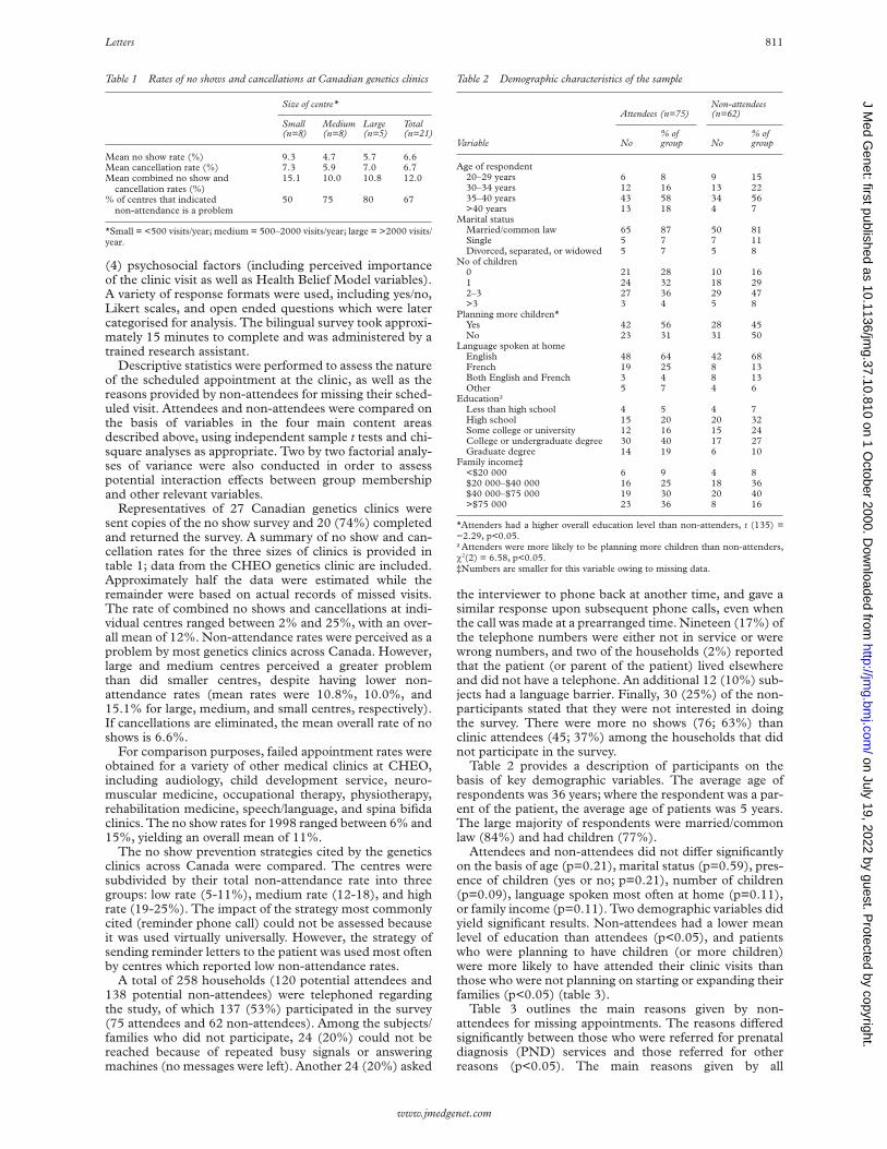
(4) psychosocial factors (including perceived importanceof the clinic visit as well as Health Belief Model variables).A variety of response formats were used, including yes/no,Likert scales, and open ended questions which were latercategorised for analysis. The bilingual survey took approxi-mately 15 minutes to complete and was administered by atrained research assistant.
Descriptive statistics were performed to assess the natureof the scheduled appointment at the clinic, as well as thereasons provided by non-attendees for missing their sched-uled visit. Attendees and non-attendees were compared onthe basis of variables in the four main content areasdescribed above, using independent sample t tests and chi-square analyses as appropriate. Two by two factorial analy-ses of variance were also conducted in order to assesspotential interaction eVects between group membershipand other relevant variables.
Representatives of 27 Canadian genetics clinics weresent copies of the no show survey and 20 (74%) completedand returned the survey. A summary of no show and can-cellation rates for the three sizes of clinics is provided intable 1; data from the CHEO genetics clinic are included.Approximately half the data were estimated while theremainder were based on actual records of missed visits.The rate of combined no shows and cancellations at indi-vidual centres ranged between 2% and 25%, with an over-all mean of 12%. Non-attendance rates were perceived as aproblem by most genetics clinics across Canada. However,large and medium centres perceived a greater problemthan did smaller centres, despite having lower non-attendance rates (mean rates were 10.8%, 10.0%, and15.1% for large, medium, and small centres, respectively).If cancellations are eliminated, the mean overall rate of noshows is 6.6%.
For comparison purposes, failed appointment rates wereobtained for a variety of other medical clinics at CHEO,including audiology, child development service, neuro-muscular medicine, occupational therapy, physiotherapy,rehabilitation medicine, speech/language, and spina bifidaclinics. The no show rates for 1998 ranged between 6% and15%, yielding an overall mean of 11%.
The no show prevention strategies cited by the geneticsclinics across Canada were compared. The centres weresubdivided by their total non-attendance rate into threegroups: low rate (5-11%), medium rate (12-18), and highrate (19-25%). The impact of the strategy most commonlycited (reminder phone call) could not be assessed becauseit was used virtually universally. However, the strategy ofsending reminder letters to the patient was used most oftenby centres which reported low non-attendance rates.
A total of 258 households (120 potential attendees and138 potential non-attendees) were telephoned regardingthe study, of which 137 (53%) participated in the survey(75 attendees and 62 non-attendees). Among the subjects/families who did not participate, 24 (20%) could not bereached because of repeated busy signals or answeringmachines (no messages were left). Another 24 (20%) asked
the interviewer to phone back at another time, and gave asimilar response upon subsequent phone calls, even whenthe call was made at a prearranged time. Nineteen (17%) ofthe telephone numbers were either not in service or werewrong numbers, and two of the households (2%) reportedthat the patient (or parent of the patient) lived elsewhereand did not have a telephone. An additional 12 (10%) sub-jects had a language barrier. Finally, 30 (25%) of the non-participants stated that they were not interested in doingthe survey. There were more no shows (76; 63%) thanclinic attendees (45; 37%) among the households that didnot participate in the survey.
Table 2 provides a description of participants on thebasis of key demographic variables. The average age ofrespondents was 36 years; where the respondent was a par-ent of the patient, the average age of patients was 5 years.The large majority of respondents were married/commonlaw (84%) and had children (77%).
Attendees and non-attendees did not diVer significantlyon the basis of age (p=0.21), marital status (p=0.59), pres-ence of children (yes or no; p=0.21), number of children(p=0.09), language spoken most often at home (p=0.11),or family income (p=0.11). Two demographic variables didyield significant results. Non-attendees had a lower meanlevel of education than attendees (p<0.05), and patientswho were planning to have children (or more children)were more likely to have attended their clinic visits thanthose who were not planning on starting or expanding theirfamilies (p<0.05) (table 3).
Table 3 outlines the main reasons given by non-attendees for missing appointments. The reasons diVeredsignificantly between those who were referred for prenataldiagnosis (PND) services and those referred for otherreasons (p<0.05). The main reasons given by all
Table 1 Rates of no shows and cancellations at Canadian genetics clinics
Size of centre*
Small(n=8)
Medium(n=8)
Large(n=5)
Total(n=21)
Mean no show rate (%) 9.3 4.7 5.7 6.6Mean cancellation rate (%) 7.3 5.9 7.0 6.7Mean combined no show and
cancellation rates (%)15.1 10.0 10.8 12.0
% of centres that indicatednon-attendance is a problem
50 75 80 67
*Small = <500 visits/year; medium = 500–2000 visits/year; large = >2000 visits/year.
Table 2 Demographic characteristics of the sample
Variable
Attendees (n=75)Non-attendees(n=62)
No% ofgroup No
% ofgroup
Age of respondent20–29 years 6 8 9 1530–34 years 12 16 13 2235–40 years 43 58 34 56>40 years 13 18 4 7
Marital statusMarried/common law 65 87 50 81Single 5 7 7 11Divorced, separated, or widowed 5 7 5 8
No of children0 21 28 10 161 24 32 18 292–3 27 36 29 47>3 3 4 5 8
Planning more children*Yes 42 56 28 45No 23 31 31 50
Language spoken at homeEnglish 48 64 42 68French 19 25 8 13Both English and French 3 4 8 13Other 5 7 4 6
Education†Less than high school 4 5 4 7High school 15 20 20 32Some college or university 12 16 15 24College or undergraduate degree 30 40 17 27Graduate degree 14 19 6 10
Family income‡<$20 000 6 9 4 8$20 000–$40 000 16 25 18 36$40 000–$75 000 19 30 20 40>$75 000 23 36 8 16
*Attenders had a higher overall education level than non-attenders, t (135) =−2.29, p<0.05.†Attenders were more likely to be planning more children than non-attenders,÷2(2) = 6.58, p<0.05.‡Numbers are smaller for this variable owing to missing data.
Letters 811
www.jmedgenet.com
on July 19, 2022 by guest. Protected by copyright.
http://jmg.bm
j.com/
J Med G
enet: first published as 10.1136/jmg.37.10.810 on 1 O
ctober 2000. Dow
nloaded from

non-attendees for missing appointments were being “toobusy” or unable to get time oV work, forgetting theappointment or having a scheduling conflict, and believingthe appointment to be non-mandatory or unimportant.Reasons associated with the PND group included beingunsure or afraid of the risks associated with amniocentesis,having recently suVered a miscarriage, experiencing morn-ing sickness on the day of the appointment, and wanting towait for laboratory test results (such as maternal serumscreening) before deciding whether to be seen at the genet-ics clinic.
Slightly less than half (46%) of all participants werescheduled to meet a medical geneticist at the clinic, and theremainder were scheduled to meet a genetic counsellor.There was no significant diVerence between attendees andnon-attendees with respect to the type of health profes-sional they were scheduled to see at the clinic (p=0.86).Approximately half (51%) of all participants were sched-uled for appointments associated with advanced maternalage prenatal screening. Another 42 patients (31%) werebeing seen in order to seek a specific diagnosis; 23 patients(17%) were scheduled to discuss genetic risk information/carrier screening, and only two respondents (1.5%)indicated that they did not know the reason for their refer-ral to the genetics clinic. Attendees and non-attendees didnot diVer on the basis of their reasons for referral to theclinic (p=0.46). Moreover, attendees were no more or lesslikely than non-attendees to have been referred to the clinicat their own request (p= 0.92), to understand well thereasons for their referral (p=0.17), or to indicate that theirphysicians had highly recommended the appointment(p=0.15) or had explained the reason for referral well(p=0.13).
It was hypothesised that some of the referral relatedvariables (table 4) may have been confounded withpatients’ education level, and so additional analyses wereperformed in order to separate these eVects. A dichoto-mous version of the education variable (originally in a Lik-ert scale format) was created by cutting scores at themedian, yielding the two categories of “high” (at least acollege diploma or university undergraduate degree) and“low” (high school education or less). Two by two analysesof variance showed a significant group by education levelinteraction with regard to patients’ reported degree ofunderstanding of the reasons for their referral (p<0.05)and the reported quality of the explanation of these reasonsby their referring physicians (p<0.05). An examination ofcell means indicated that among patients with a highereducation level, attendees understood their reasons forreferral better and claimed their physicians had explainedthese reasons better than did non-attendees. No such
group diVerence existed among patients with a lower edu-cation level.
We hypothesised that patients with a higher educationlevel would more likely have been referred to the geneticsclinic at their own request, because of their presumedgreater knowledge about genetic health services. Achi-square analysis showed this not to be the case(0.55<p<0.60).
The relationship between non-attendance and environ-mental variables is shown in table 5. Patients who reportedhaving to arrange for child care in order to attend a clinicvisit were more likely to have missed their scheduledappointments than those who indicated no such require-ment (p<0.05). Although having to take time oV work toattend a clinic visit was not associated with a greater likeli-hood of non-attendance (0.70<p<0.75), those respond-ents who were not paid for such time oV were more likelyto have missed their appointments than those who wereable to take paid leave from work to attend the clinic(p<0.05). No significant diVerences between attendees andnon-attendees were found in methods of transport to theclinic (0.20<p<0.25) or travel time to the clinic(0.10<p<0.15). Failure to make contact by telephone toremind the patient of the clinic visit occurred in 10 of theno shows and none of the attendees.
The relationship between non-attendance and psychoso-cial variables is shown in table 6. Respondents whoattended their appointments perceived the clinic visit to bemore important than respondents who did not attend(p<0.01). With regard to the Health Belief Modelvariables, no significant group diVerences were found interms of perceived severity of the health condition (orpotential health conditions) which were to be discussedduring the appointment (0.65<p<0.70). Moreover, thetwo groups did not diVer significantly with respect to theirperceived risk of having (or eventually developing) thesehealth conditions (0.60<p<0.65).
Non-attendance was significantly related to perceivedbenefits and disadvantages of the genetics appointment.Specifically, patients who missed their appointmentsperceived the potential benefits of the clinic visit to be less
Table 3 Reasons given by non-attendees for missing genetics clinicappointments
Reason given for missing appointment
Reason for referral to genetics
PND* (n=31)Non-PND*(n=31)
No % No %
Too busy/no time oV work 8 26 14 45Forgot/scheduling conflict 4 13 7 23Believed appointment was
non-mandatory or unimportant4 13 1 3
Wanted to wait for lab results 3 10 1 3Other 1 3 6 19Unsure/afraid re risks of amniocentesis 4 13 1 3Recent miscarriage 4 13 0 0Morning sickness on day of
appointment3 10 1 3
PND = prenatal diagnosis.*Reasons diVered between PND and non-PND patients: ÷2(7) = 15.64, p<0.05.
Table 4 Comparison of attendees and non-attendees on referral/geneticservice variables
Variable
Attendees (n=75)Non-attendees(n=62)
No% ofgroup No
% ofgroup
Reason for referral to geneticsPrenatal diagnosis 39 52 31 50Carrier testing/risk information 12 16 11 18Diagnostic evaluation 24 32 18 29Other 0 0 2 3
How well was reason explained?*Very poorly 0 0 1 2Poorly 2 3 3 6Adequately 8 14 10 20Well 14 25 12 25Very well 33 58 23 47
How well did you understand the reason?*Not at all 0 0 2 3Poorly 2 3 2 3Adequately 3 5 4 7Well 9 15 12 22Very well 48 77 35 65
Referred at own requestYes 26 35 22 36No 49 65 40 64
How strongly referring physician recommended the genetics appointment*Not strongly 5 8 7 13Somewhat strongly 20 30 22 40Quite strongly 15 23 9 16Very strongly 25 38 16 29Recommended against 1 1 1 2
*Numbers are smaller for these variables owing to missing data.
812 Letters
www.jmedgenet.com
on July 19, 2022 by guest. Protected by copyright.
http://jmg.bm
j.com/
J Med G
enet: first published as 10.1136/jmg.37.10.810 on 1 O
ctober 2000. Dow
nloaded from

important (p<0.01) and the potential disadvantages to bemore important (p<0.01) than patients who kept theirappointments.
The results of this study help to quantify the problem ofmissed appointments at genetics clinics across Canada.Over a one year period, approximately 11% of patientsscheduled for visits at genetics clinics either cancelled ordid not show up for their appointments. When cancella-tions are eliminated, this number decreases to 6%,representing the rate of “pure no shows”. In comparison,11% of patients failed to show up for their appointments atother non-genetics outpatient clinics at the Children’sHospital of Eastern Ontario (CHEO). These values arerelatively low compared with the 10-30% range frequentlyreported.1 2 Family practice centres have fewer broken
appointments than adult medical centres.3 Perhaps thefamily orientation of paediatric and genetics clinics is moreakin to family practice and reduces the likelihood thatappointments will not be kept.
It should be noted that in the survey of the CHEOgenetics clinic, the non-attendees group included cancella-tions with less than 12 hours notice which, owing to theirnegative consequences, were considered equivalent to noshows. This distinction between early and late cancella-tions was not requested from the other genetics clinics sur-veyed in this study (they were simply asked to report twoseparate numbers for “pure” no shows and cancellations,respectively). If late cancellations had been included in theno show rates provided by the other clinics, the resulting noshow rates would probably have been somewhat higherthan reported.
Although an 11% rate of failed appointments atCanadian genetics clinics is comparatively low, it is still asizeable barrier to the optimal provision of genetic healthservices. Unlike many clinics, where multiple bookings arecommon and visits are usually short, genetics clinicsexpend extensive resources for pre-appointment planningand generally allocate an hour for a clinic visit.
This study suggests a number of factors which may con-tribute to the problem of missed appointments. The mostcommonly stated reasons for missed appointments at theCHEO genetics clinic were scheduling conflicts andinability to get time oV work. As these are predictable fac-tors, it is surprising that more than half of respondentscited them, despite being called 24-48 hours before theappointment. The question arises as to whether theanswers provided by patients over the telephone concealedunderlying reasons such as anxiety, confusion, or personalbelief systems, which lead to last minute decisions not toattend, or were too personal to admit to the secretary or theresearch assistant.
Some psychosocial variables appeared to contribute tothe non-attendance rate. Patients who attended theirscheduled appointments thought their visit to the geneticsclinic to be more important than those who did not attend.This is similar to previous research which has foundperceived urgency of medical clinic visits to be significantlyrelated to compliance,1 3 though urgency and importancemay represent relatively distinct constructs.
Patients who attended the clinic also perceived morebenefits and fewer barriers associated with their appoint-ments than did non-attendees. This supports the HealthBelief Model8 and previous research, which has indicatedsuch health beliefs to be important predictors of healthbehaviours.6 9 10 Apparently, for non-attendees, the poten-tial costs of the clinic visit (such as the anxiety or guilt thatmay be experienced after learning of one’s carrier status)outweighed the possible benefits (such as relief fromuncertainty or clarification of one’s options). In contrast,the perception of the benefits of attending the geneticsclinic appeared to outweigh the perceived disadvantagesfor those who kept their appointments.
We found that certain psychosocial factors may play arole in non-attendance, but not all of the anticipated eVectswere found. For example, perceived severity of the healthcondition (or potential health condition) for which patientswere referred to the clinic, along with the perceived risk ofhaving or eventually facing that health condition, wereunrelated to clinic attendance. The former result is notsurprising, given that the “perceived severity” componentof the Health Belief Model has been shown to have limitedvalue in predicting a variety of health behaviours.11
However, a high perceived risk has been found to predict anumber of health behaviours, including mammography,5 12
genetic testing for susceptibility to breast cancer,13 14 and
Table 5 Comparison of attendees and non-attendees on environmentalvariables
Variable
Attendees (n=75) Non-attendees (n=62)
No% ofgroup No
% ofgroup
Have to arrange for child care to attend the clinic?*Yes 12 16 19 31No 63 84 43 69
Have to take time oV work to attend the clinic?Yes 39 52 30 49No 36 48 31 51
If yes, paid for this time oV?*Yes 23 60 10 33No 15 40 20 67
Method of transport to clinicOwn vehicle 65 87 48 77Get a ride 7 9 6 10Bus 3 4 6 10Walk 0 0 2 3
Travel time to clinic<15 minutes 4 5 4 715–30 minutes 35 47 24 3930–45 minutes 18 24 10 1645–60 minutes 7 9 7 11>60 minutes 11 15 17 27
*p<0.05.
Table 6 Comparison of attendees and non-attendees on psychosocialvariables
Variable
Attendees (n=75)Non-attendees(n=62)
No% ofgroup No
% ofgroup
Perceived importance of clinic visit*Not at all important 1 1 6 10Of little importance 4 5 5 8Neutral 8 11 10 16Somewhat important 16 21 2 3Very important 46 62 27 43
Perceived severity of genetic conditionNot at all serious 8 11 11 18Of little severity 14 19 13 21Neutral 18 24 5 8Somewhat serious 12 16 14 23Very serious 22 30 18 30
Perceived risk of genetic conditionVery low 16 21 19 32Somewhat low 13 17 11 18Medium 26 35 12 20Somewhat high 11 15 7 12Very high 9 12 11 18
Perceived benefits of clinic visit*No benefits 1 1 7 12Few benefits 4 5 4 7Neutral 10 13 10 17Some benefits 14 19 13 22Many benefits 46 62 25 42
Perceived importance of disadvantages of clinic visit*Not at all important 56 74 30 52Of little importance 6 8 5 8Neutral 8 11 8 14Somewhat important 2 3 3 5Very important 3 4 12 21
*p<0.01.
Letters 813
www.jmedgenet.com
on July 19, 2022 by guest. Protected by copyright.
http://jmg.bm
j.com/
J Med G
enet: first published as 10.1136/jmg.37.10.810 on 1 O
ctober 2000. Dow
nloaded from

cystic fibrosis carrier screening.15 One possible explana-tion for the absence of such an eVect in this study lies inthe nature of the “health condition” in question. Previousresearch has examined patients’ health beliefs with respectto a single, specific illness or procedure. In the presentstudy, patients were referred to the genetics clinic for avariety of reasons; consequently, a more general questionregarding perceived risk was included in the survey whichpermitted patients to respond according to their ownsituation. This lack of specificity may have led to avaried interpretation of the question, thus reducing thestrength of the “perceived risk” and “perceived severity”variables.
If the above health beliefs are the true reasons thatunderlie the stated explanations (for example, could notget time oV work) given by some patients for missing theirappointments, this holds important implications for theprovision of adequate public education regarding geneticsrelated health issues. It is widely accepted that a thoroughexplanation of the risks and benefits associated with agiven medical intervention is required in order for patientsto provide informed consent. It is not clear how suchstandards extend to diVerent contexts, such as a geneticsclinic appointment. Greater public awareness of the serv-ices provided by genetics professionals will providepatients with a balanced and accurate understanding ofthe importance, risks, and benefits of a scheduledappointment. The fact that patients with a higher educa-tion level (at least an undergraduate university degree)were more likely to attend their appointments than thosewith less education reinforces this need for greater publiceducation. Studies have found that people of lowereducational status are more likely to miss their medicalappointments.1 People with a university educationprobably have greater access to (and experience with)health related information found in scientific journals,books, and news magazines. Delivery of health infor-mation to those who have less formal education mayfacilitate the use of health services by more subjects andfamilies. An alternative interpretation of the educationeVect is equally possible: those working in higher levelpositions may simply have greater job flexibility and maythus have had less diYculty attending their clinic appoint-ments.
Other demographic variables, including age, maritalstatus, number of children, language spoken most often athome, and family income, did not diVerentiate betweenattendees and non-attendees. This is encouraging, in thatmost of these variables are not easily targeted by clinicbased interventions to improve attendance rates. Oneexception may be language, which has been subject tolimited previous research and is an important variable toassess in a country with two oYcial languages; in ourgenetics clinic services are regularly provided inFrench and English. Our finding that language does notappear to be a barrier to attendance is encouraging, butthe question may need to be addressed for patients who donot speak either oYcial language. People not fluent inEnglish or French were excluded from the sample. Therewere 12 people who did not participate in the surveybecause of a language barrier. Nine of these were in thegroup who missed their appointments, so this is anissue that may merit further investigation, as alanguage barrier would represent a potential target forintervention.
It should be noted that the lack of significant results formany demographic variables may be in part because of thenarrow response ranges associated with the correspondingsurvey items. For example, the mean age of surveyrespondents was quite young (36 years) and people over
the age of 45 were virtually absent from the sample. Stud-ies of adult non-genetics clinics would encompass a widerrange of patient ages, leading to a greater likelihood of sig-nificant age eVects. Moreover, the majority of participantswere married (84%) and had children (77%). Only 10 par-ticipants (7%) reported a family income of less than$20 000, indicating a clear under-representation of lowsocioeconomic status families. The lack of any associationwith family income may also reflect universal access underthe Canadian health care system. Previous studies showingsignificant eVects of socioeconomic status have generallybeen conducted in the United States, a country which lacksuniversal health care.
The quality of information given to patients by their refer-ring physicians about the referral to genetics was not associ-ated with the overall rate of missed appointments. However,it did play a role in the specific subgroup of patients with ahigher education level. Among those participants, theattendees reported that they understood the reasons for theirreferral better, and that their physicians had explained thesereasons better, than those who did not attend. It appears thatwhile patients with a higher education level are more likely toattend their appointments, they are also more likely torequire thorough explanations of the rationale for theappointment before deciding whether or not to attend. Thusthere may be some argument for improving the quality ofpre-appointment information provided to patients by healthcare professionals.
These results lend some support to previous researchindicating that patients who understand the reasons fortheir referral relatively well are more likely to attend theirclinic appointments.4 Contrary to previous studies,5 7 how-ever, the degree to which the referring physician recom-mended the genetics clinic appointment did not influencethe likelihood of attendance.
Finally, although diYculties with scheduling conflicts,transport, and inability to take time oV work may often atleast in part conceal more important underlying reasons fornon-attendance, they may sometimes compromise ap-pointment keeping. Patients who had to arrange for childcare in order to attend the clinic were more likely to misstheir appointments, and this confirms previous resultsindicating that broken medical appointments are morelikely among larger families and those with youngerchildren.1 3 The need for child care is an important barrierto the provision of health services at a genetics clinicbecause families comprise most of the patient population.Provision of day care facilities within medical institutionsmight prove cost eVective, given that all clinics experiencea similar, if not higher, no show rate.
The need to take time oV work for a clinic appointmentdoes not appear to present a significant barrier to appoint-ment keeping, except for those who are required to taketime oV without pay. This problem could be solved if clin-ics extended their hours of operation, so as to accommo-date work schedules.
Contrary to previous research,1 other practical matterssuch as distance from the clinic and available method oftransport were unrelated to rates of failed appointments atthe CHEO genetics clinic. Such eVects were lacking,despite the fact that patients reported a wide range of traveltimes, including 28 patients (20%) who indicated that ittook them more than an hour to get to the hospital. It maybe that once the patient has made an assessment that theappointment is important, travel time may be irrelevant.We cannot draw any conclusions regarding any eVects oftransport method, because the vast majority (83%) ofrespondents drove their own vehicles to the clinic. Thisprobably reflects the high mean socioeconomic status ofthe current sample; studies involving less privileged people
814 Letters
www.jmedgenet.com
on July 19, 2022 by guest. Protected by copyright.
http://jmg.bm
j.com/
J Med G
enet: first published as 10.1136/jmg.37.10.810 on 1 O
ctober 2000. Dow
nloaded from

might show that lack of a convenient mode of transport is asignificant obstacle in appointment keeping.
Our patient sample does not reflect the generalpopulation and is a limitation of the present study.Participants were highly educated and reported relativelyhigh family incomes, which may account for the slightlylower rate of non-attendance at this clinic, as comparedwith the rates seen in previous studies which have focusedmainly on low socioeconomic status populations. It ispossible that factors other than those we have evaluated, orthat we have found negative in our sample, may contributeto non-attendance rates in patients with a lowereducational and socioeconomic status. Nevertheless, ourresults support some findings of previous studies whichincluded low socioeconomic status populations.3 4 10
Moreover, the fact that our non-attendance rate was con-sistent with those reported by genetics clinics acrossCanada suggests that these rates reflect an accurateportrayal of the problem in the context of genetics healthcare in Canada.
Other limitations of this study include the limited gener-alisability of results to the United States health care system,as well as the telephone interview methodology. Fornon-attendees in particular, the latter approach may haverestricted the degree of candour in participants’ surveyresponses; future studies may benefit from the use of amore anonymous data collection format. Finally, approxi-mately half of the surveyed genetics clinics providedestimated as opposed to actual rates of non-attendance.Estimation may not accurately reflect no show rates; how-ever, a comparison of means indicated that the actual andestimated numbers were similar.
Results of this study suggest a number of potentialtargets for improving attendance rates at genetics and otheroutpatient medical clinics. Better education of patientsabout their medical condition, the nature and purpose ofspecific options available to them, and the costs andbenefits associated with such options, is clearly indicated.To be maximally eVective, such education should comefrom a variety of sources, including the mass media, pam-phlets distributed to pharmacies and medical clinics of alltypes, and, most importantly, open and detailed communi-cation between patients and physicians. Some clinics couldalso attempt to extend or modify their hours of operationand on site child care initiatives could be explored. Theselarger scale approaches, in combination with moretraditional methods such as telephone and mailed appoint-ment reminders, may help to replace lengthy waiting listsand wasted physician time with more eYcient and farreaching health care services.
Support for this project was provided by the Children’s Hospital of EasternOntario (CHEO) Research Institute. We thank the Canadian genetics centresfor their contribution to data collection in the first phase of this study. We alsothank Heather Tindell and Laura Van Houten for their role in collecting data atCHEO, as well as the participants for their time and cooperation.
L HUMPHREYS*A G W HUNTER†
A ZIMAK†A O’BRIEN*
Y KORNELUK*M CAPPELLI*
*Mental Health Program, Children’s Hospital of Eastern Ontario, 401Smyth Road, Ottawa, Ontario, Canada K1H 8L1†Genetics Program, Children’s Hospital of Eastern Ontario, Ottawa,Ontario, CanadaCorrespondence to: Dr Cappelli, [email protected]
1 Barron WM. Failed appointments: who misses them, why they are missed,and what can be done. Primary Care 1980;7:563-73.
2 Deyo RA, Inui TS. Dropouts and broken appointments. Med Care 1980;18:1146-57.
3 Oppenheim GL, Bergman JJ, English EC. Failed appointments: a review. JFamily Pract 1979;8:789-96.
4 Blankson ML, Goldenberg RL, Keith B. Noncompliance of high-risk preg-nant women in keeping appointments at an obstetric complications clinic.Southern Med J 1994;87:634-8.
5 Aiken LS, West SG, Woodward CK, Reno RR. Health beliefs andcompliance with mammography: screening recommendations in asympto-matic women. Health Psychol 1994;13:122-9.
6 Fulton JP, Buechner JS, Scott HD, DeBuono BA, Feldman JP, Smith RA,Kovenock D. A study guided by the Health Belief Model of the predictorsof breast cancer screening of women ages 40 and older. Public Health Rep1991;106:410-19.
7 Lerman C, Rimer B, Trock B, Balshem A, Engstrom PF. Factors associatedwith repeat adherence to breast cancer screening. Prev Med 1990;19:279-90.
8 Rosenstock IM. The Health Belief Model and preventive health behavior.Health Educ Monogr 1974;2:354-86.
9 Yeomans-Kinney C, Vernon SW, Frankowski RF, Weber DM, Bitsura JM,Vogel VG. Factors related to enrollment in the breast cancer preventiontrial at a comprehensive cancer center during the first year of recruitment.Cancer 1995;76:46-56.
10 Mirotznik J, Ginzler E, Zagon G, Baptiste A. Using the Health Belief Modelto explain clinic appointment-keeping for the management of a chronicdisease condition. J Commun Health 1998;23:195-209.
11 Janz NZ, Becker MH. The Health Belief Model: a decade later. Health EducQuart 1984;11:1-47.
12 Calnan M. The Health Belief Model and participation in programmes forthe early detection of breast cancer: a comparative analysis. Soc Sci Med1984;19:823-30.
13 Chaliki H, Loader S, Levenkron JC. Women’s receptivity to testing for agenetic susceptibility to breast cancer. Am J Public Health 1995;85:1133-5.
14 Lerman C, Daly M, Balshem A. Attitudes about genetic testing for breast-ovarian cancer susceptibility. J Clin Oncol 1994;12:843-50.
15 Tambor ES, Bernhardt BA, Chase GA. OVering cystic fibrosis carrierscreening to an HMO population: factors associated with utilization. Am JHum Genet 1994;55:626-37.
CORRECTION
In the April 2000 issue of the journal, in the paper byMortier et al on “Report of five novel and one recurrentCOL2A1 mutations with analysis of genotype-phenotypecorrelation in patients with a lethal type II collagen disor-der”, the mutation T1191N should have been T1190Nthroughout.
Letters 815
www.jmedgenet.com
on July 19, 2022 by guest. Protected by copyright.
http://jmg.bm
j.com/
J Med G
enet: first published as 10.1136/jmg.37.10.810 on 1 O
ctober 2000. Dow
nloaded from