Leishmania cytosolic silent information regulatory protein 2 deacetylase induces murine B-cell...
12
Leishmania cytosolic silent information regulatory protein 2 deacetylase induces murine B-cell differentiation and in vivo production of specific antibodies Introduction Protozoans of the genus Leishmania are obligate intracel- lular parasites causing a wide spectrum of human and veterinary diseases known as leishmaniasis. The disease causes a broad range of clinical symptoms, from a self- healing localized cutaneous form to the disseminated potential lethal infection, depending on the complex interaction between the infecting Leishmania species and the host immune response. 1 Active visceral leishmaniasis (VL), caused by members of Leishmania donovani complex (L. donovani in the Old World, L. infantum in the South-east of Europe and Mediterranean area and L. chagasi in the New World) is a progressive infection characterized by fever, cachexia, hepatosplenomegaly and hypergammaglobulinaemia, symptoms that develop in both humans and dogs. 2,3 The interaction between the parasite and its host led to a variety of disturbances in the immune system. One of the immunological hallmarks of VL is a profound suppression of parasite-specific T-cell mediated immune responses 4 associated with a remark- able increase in the serum immunoglobulin levels as a Ricardo Silvestre, 1,2,3 Anabela Cordeiro-da-Silva, 1,2 Joana Tavares, 1,2,3 Denis Sereno 3 and Ali Ouaissi 3 1 Faculdade de Farma ´cia da Universidade do Porto, Portugal, 2 Instituto de Biologia Molecu- lar e Celular da Universidade do Porto, Portugal and 3 Institut de Recherche pour le De ´veloppement, Montpellier, France doi:10.1111/j.1365-2567.2006.02468.x Received 20 April 2006; revised 2 August 2006; accepted 9 August 2006. Correspondence: Dr A. Ouaissi, Institut de Recherche pour le De ´veloppement, UR008, Montpellier, France. Email: [email protected] Senior author: Ali Ouaissi Summary In previous studies, we identified a gene product belonging to the silent information regulatory 2 protein (SIR2) family. This protein is expressed by all Leishmania species so far examined (L. major, L. infantum, L. ama- zonensis, L. mexicana) and found to be crucial for parasite survival and virulence. In the present study, we investigated whether a Leishmania SIR2 recombinant protein (LmSIR2) would affect T- and B-cell functions in a murine model. In vitro treatment of spleen cells from normal BALB/c mice with LmSIR2 showed increased expression of CD69 on B cells. This effect was not abolished by the addition of polymyxin B. Intravenous injection of LmSIR2 into BALB/c mice induced increased spleen B cell number by a factor of about 16, whereas no modification occurred at the level of CD4 + and CD8 + cells. Furthermore, intraperitoneal injection of LmSIR2 alone without adjuvant into BALB/c mice or nude mice trig- gered the production of elevated levels of LmSIR2-specific antibodies. The analysis of specific isotype profiles showed a predominance of immuno- globulin G1 (IgG1) and IgG2a antibody responses in BALB/c mice, and IgM in nude mice. Moreover, the anti-LmSIR2 mouse antibodies in the presence of complement induced the in vitro lysis of L. infantum amasti- gotes. In the absence of complement, the antibodies induced significant inhibition of amastigotes developpement inside macrophages. Together, the current study provides the first evidence that a Leishmania protein belonging to the SIR2 family may play a role in the regulation of immune response through its capacity to trigger B-cell effector function. Keywords: Leishmania; SIR2; B cells; specific antibodies; leishmaniasis Abbreviations: VL, visceral leishmaniasis; CML, complement-mediated lysis; FCS, fetal calf serum; LPS, lipopolysaccharide; mAb, monoclonal antibody; SDS–PAGE, sodium dodecyl sulphate–polyacrylamide gel electrophoresis; PBS, phosphate-buffered saline; i.p., intraperitoneally; i.v., intravenously; MFI, mean fluorescence intensity; ELISA, enzyme-linked immunosorbent assays; RLU, relative light units; WT, wild type; FACS, fluorescence-activated cell sorter. Ó 2006 Blackwell Publishing Ltd, Immunology, 119, 529–540 529 IMMUNOLOGY ORIGINAL ARTICLE
-
Upload
independent -
Category
Documents
-
view
4 -
download
0
Transcript of Leishmania cytosolic silent information regulatory protein 2 deacetylase induces murine B-cell...
Leishmania cytosolic silent information regulatory protein 2
deacetylase induces murine B-cell differentiation and in vivo
production of specific antibodies
Introduction
Protozoans of the genus Leishmania are obligate intracel-
lular parasites causing a wide spectrum of human and
veterinary diseases known as leishmaniasis. The disease
causes a broad range of clinical symptoms, from a self-
healing localized cutaneous form to the disseminated
potential lethal infection, depending on the complex
interaction between the infecting Leishmania species and
the host immune response.1 Active visceral leishmaniasis
(VL), caused by members of Leishmania donovani
complex (L. donovani in the Old World, L. infantum in
the South-east of Europe and Mediterranean area and
L. chagasi in the New World) is a progressive infection
characterized by fever, cachexia, hepatosplenomegaly and
hypergammaglobulinaemia, symptoms that develop in
both humans and dogs.2,3 The interaction between the
parasite and its host led to a variety of disturbances in
the immune system. One of the immunological hallmarks
of VL is a profound suppression of parasite-specific T-cell
mediated immune responses4 associated with a remark-
able increase in the serum immunoglobulin levels as a
Ricardo Silvestre,1,2,3 Anabela
Cordeiro-da-Silva,1,2 Joana
Tavares,1,2,3 Denis Sereno3 and
Ali Ouaissi3
1Faculdade de Farmacia da Universidade do
Porto, Portugal, 2Instituto de Biologia Molecu-
lar e Celular da Universidade do Porto,
Portugal and 3Institut de Recherche pour le
Developpement, Montpellier, France
doi:10.1111/j.1365-2567.2006.02468.x
Received 20 April 2006; revised 2 August
2006; accepted 9 August 2006.
Correspondence: Dr A. Ouaissi, Institut de
Recherche pour le Developpement,
UR008, Montpellier, France.
Email: [email protected]
Senior author: Ali Ouaissi
Summary
In previous studies, we identified a gene product belonging to the silent
information regulatory 2 protein (SIR2) family. This protein is expressed
by all Leishmania species so far examined (L. major, L. infantum, L. ama-
zonensis, L. mexicana) and found to be crucial for parasite survival and
virulence. In the present study, we investigated whether a Leishmania
SIR2 recombinant protein (LmSIR2) would affect T- and B-cell functions
in a murine model. In vitro treatment of spleen cells from normal BALB/c
mice with LmSIR2 showed increased expression of CD69 on B cells. This
effect was not abolished by the addition of polymyxin B. Intravenous
injection of LmSIR2 into BALB/c mice induced increased spleen B cell
number by a factor of about �1�6, whereas no modification occurred at
the level of CD4+ and CD8+ cells. Furthermore, intraperitoneal injection
of LmSIR2 alone without adjuvant into BALB/c mice or nude mice trig-
gered the production of elevated levels of LmSIR2-specific antibodies. The
analysis of specific isotype profiles showed a predominance of immuno-
globulin G1 (IgG1) and IgG2a antibody responses in BALB/c mice, and
IgM in nude mice. Moreover, the anti-LmSIR2 mouse antibodies in the
presence of complement induced the in vitro lysis of L. infantum amasti-
gotes. In the absence of complement, the antibodies induced significant
inhibition of amastigotes developpement inside macrophages. Together,
the current study provides the first evidence that a Leishmania protein
belonging to the SIR2 family may play a role in the regulation of immune
response through its capacity to trigger B-cell effector function.
Keywords: Leishmania; SIR2; B cells; specific antibodies; leishmaniasis
Abbreviations: VL, visceral leishmaniasis; CML, complement-mediated lysis; FCS, fetal calf serum; LPS, lipopolysaccharide;mAb, monoclonal antibody; SDS–PAGE, sodium dodecyl sulphate–polyacrylamide gel electrophoresis; PBS, phosphate-bufferedsaline; i.p., intraperitoneally; i.v., intravenously; MFI, mean fluorescence intensity; ELISA, enzyme-linked immunosorbent assays;RLU, relative light units; WT, wild type; FACS, fluorescence-activated cell sorter.
� 2006 Blackwell Publishing Ltd, Immunology, 119, 529–540 529
I M M U N O L O G Y O R I G I N A L A R T I C L E
result of polyclonal B-cell activation, which invariably
produce large amounts of parasite non-specific antibodies
with self autoreactivity particularly the immunoglobulin
M (IgM) and IgG isotypes.5 Using a mechanism common
to other intracellular pathogens, Leishmania spp. displays
mitogenic-like activities that produce a non-specific gen-
eral activation of lymphocytes. Thus, previous reports
have characterized immunosupressor factors and mito-
genic molecules within the parasite antigens.6,7 Thus, it
seems that the persistence of infection could be related
somehow to the immunosuppression phenomenon, the
non-specific responses, and the development of auto-
immune processes in relation to defined parasite mole-
cules. Therefore, it seems reasonable to assume that
induction of specific responses against ‘immuno-
dominant’, ‘immunopathological’ or ‘protective’ antigens,
which could interfere with some of the biological activit-
ies, might be among the most effective means to block
the development of pathogenic processes.8
It is generally accepted that protective immunity against
leishmaniasis is associated with a classical cell-mediated
immune response, rather than a humoral one. This view
has been interpreted in context of the T helper 1 (Th1)/
Th2-biased responses in the mouse model of L. major.
However, in visceral leishmaniasis, while a interleukin-12
(IL-12)-driven type 1 response is imperative for protec-
tion, type 2 responses (that support humoral immunity)
also play a role, although minor, in promoting resist-
ance.9 Although the real contribution of antibodies is still
under debate, studies in different intracellular pathogens
have shown that antibodies can also have a function in
restricting the infection when the parasite is exposed to
the extracellular milieu.10 In leishmaniasis, interaction of
antibodies with the parasite could occur when the pro-
mastigote infects an individual or when the amastigotes
are released from the infected macrophages.11 Moreover,
their possible role in protection was shown by passive
in vivo transfer of monoclonal antibodies.12
In previous reports we have characterized silent infor-
mation regulatory 2 protein (SIR2) proteins from
L. major (LmSIR2)13 and L. infantum (LiSIR2)14 while
two other related protein sequences can be found in
the Leishmania genome database (L. major sirtuin
(CAB55543), L. major cobB (LmjF34.2140). Molecular
and biological approaches allowed us to show the poten-
tial role of LmSIR2 and LiSIR2 genes in parasite sur-
vival.14,15 Furthermore, using LmSIR2 as a probe,
immunological investigations demonstrated that LmSIR2
was highly immunogenic during natural human and can-
ine infections.16
In the current study, we investigated whether LmSIR2
would trigger the activation of murine lymphocytes, and
surprisingly murine B cells, but not T cells, respond
strongly and directly to stimulation with LmSIR2. The B
cells underwent differentiation in vivo as evidenced by the
production of significant levels of specific antibodies. Our
data suggest an immunoregulatory role of leishmania
SIR2 protein during leishmaniasis.
Materials and methods
Leishmania strain and mice
A cloned line of L. infantum (MHOM/MA/67/ITMAP-
263, wild type (WT)) was used for the infectious chal-
lenge. A cloned line of L. infantum expressing the
luciferase (LUC) gene derived from WT17 was used in all
in vitro experiments. Six to seven-week-old BALB/c male
mice were obtained from Harlem Iberica (Spain). Seven-
week-old BALB/c athymic (nude) mice were obtained
from Charles River (L’Arbresle, France).
Purification of LmSIR2
The L. major SIR2 was obtained as recombinant protein
containing six histidine residues at its N-terminal. The
construction of plasmid and purification of protein have
been described elsewhere.18 The purity of the recombin-
ant protein (LmSIR2) was analysed by sodium dodecyl
sulphate–polyacrylamide gel electrophoresis (SDS–PAGE)
stained with Coomassie blue. For biological assays, the
protein was dialysed against phosphate-buffered saline
(PBS) in decreasing concentrations of urea. The final
dialysis was performed against PBS. The protein concen-
tration was determined using the Folin procedure.19 To
eliminate endotoxins, the recombinant protein was passed
through an EndoTrap�red column (Profos, Germany)
following manufacturer’s instructions.
Mouse injections
In the case of BALB/c mouse experiments two different
routes of administration were followed. One group of
BALB/c mice was injected three times intraperitoneally
(i.p.) at a 7-day interval with 50 lg of LmSIR2 in 300 ll
of PBS, or 300 ll of PBS (control group). Two weeks
after final immunization, spleens and sera were collected.
Another group of BALB/c mice received one intravenous
(i.v.) injection of 20 lg of LmSIR2 in 150 ll of PBS, or
150 ll of PBS (control group). After 72 hr, mice were
killed and spleens collected. In the case of BALB.nude
mice, two groups of five mice each were injected three
times i.p. at a 7-day interval with either 50 lg of endo-
toxin-free LmSIR2 in 400 ll of PBS, or 400 ll of PBS
(control group).
BALB/c mice immunization and infection
For immunization, one group of BALB/c mice was injec-
ted three times i.p. at a 7-day interval with 50 lg of
530 � 2006 Blackwell Publishing Ltd, Immunology, 119, 529–540
R. Silvestre et al.
LmSIR2 in 200 ll of sterile PBS. A control group was
injected with 200 ll of sterile PBS in a similar schedule.
Fifteen days after the last immunization both groups were
challenged with 108 stationary-phase WT promastigotes
ressuspended in 200 ll of sterile PBS.
Spleen B-cell isolation
After cervical dislocation, the spleens were removed and
homogenized in a Petri dish to obtain a single-cell sus-
pension. After two washes in RPMI-1640 culture medium
(Cambrex, Saint Beauzine, France), the cells were adjusted
to 107/ml in RPMI-1640 culture medium supplemen-
ted with 10% fetal calf serum (FCS), 2 mM glutamine,
100 U/ml penicillin, 100 lg/ml streptomycin and 20 mM
HEPES. Spleen B-cell isolation was carried out using a B
Cell Isolation Kit, MACS� (Miltenyi Biotec, Auburn, CA)
as described by the manufacturer. The effectiveness of
B-cell purification was determined by double labelling
with specific monoclonal antibodies (mAb; anti-l+, anti-
CD4+ and anti-CD8+) and further fluorescence-activated
cell sorting (FACS) analysis. After purification, we
obtained over 97% B-cell purity.
Flow cytometry determinations
Spleen cells or isolated B cells from normal or LmSIR2
immunized mice and their respective controls were pre-
pared in order to obtain single-cell suspensions. The cells
were washed by centrifugation and ressuspended in PBS
supplemented with 2% FCS. A total of 106 viable cells
were incubated for 30 min at 4� with saturating concen-
trations of phycoerythrin (PE)-conjugated mAb to CD69
plus fluorescein isothiocyanate (FITC)-conjugated mAbs
to either CD4, CD8a or IgM (anti-l) from BD Pharmin-
gen (San Diego, CA). After two washing steps with
PBS)2% FCS, the cells were analysed by flow cytometry
in a FACS Scan equipped with CellQuest Pro software
(Becton Dickinson, San Jose, CA). Lymphocytes were
selected on the basis of forward scatter/side scatter values
and dead cells were excluded from all samples by propi-
dium iodide labelling.
To evaluate the binding of antibodies to LUC-recom-
binant WT amastigotes, 2�5 · 105 parasites were washed
twice in PBS containing 0�5% of bovine serum albumin
(PBS–BSA) and incubated for 30 min at 4� with
mouse anti-SIR2 immune serum diluted 1 : 10 in PBS–
BSA. After two washes with PBS–BSA, the parasites
were incubated for 30 min at 4� with FITC-conjugated
goat anti-mouse IgG diluted 1 : 5000 in PBS–BSA.
Labelled amastigotes were washed twice and resuspend-
ed in 500 ll of PBS–BSA followed by flow cytometry
analysis. A preimmune serum was used as control.
Results were expressed as mean fluorescent intensity
(MFI).
Enzyme-linked immunosorbent assays (ELISA) forimmunoglobulins
Ninety-six-well flat-bottomed microtitre plates (Greiner,
Laborchnik, Solingen, Germany) were coated overnight at
4� with one of the following reagents in 0�01 M carbonate/
bicarbonate buffer pH 8�5: unlabelled goat anti-mouse
immunoglobulin (5 lg/ml), total L. infantum protein
extract (10 lg/ml), LmSIR2 (5 lg/ml), whale skeletal mus-
cle type II myoglobulin (MYO, 5 lg/ml), keyhole limpet
haemocyanin (KLH, 5 lg/ml), LmS3a (5 lg/ml). The
plates were washed with PBS containing 0�1% Tween-20
(PBS-T) and blocked with PBS 1% gelatine (200 ll/well)
for 1 hr at 37�. The plates were incubated at 37� with
serial dilutions (for total titres) or 1 : 100 dilutions in
triplicate (for specific antibody reactivity) of each serum
for 2 hr. After washing with PBS-T, the plates were incu-
bated for 30 min at 37� with peroxidase-labelled goat anti-
mouse immunoglobulin isotypes (anti-IgM, anti-IgG,
anti-IgG1, anti-IgG2a) and developed with 0�5 mg/ml of
o-phenylenediamine dihydrochloride (OPD, Sigma, St
Louis, MO) with 10 ll of H2O2 in citrate buffer. The reac-
tions were stopped by the addition of 50 ll of 3 M HCl to
each well. The concentration of non-specific antibody was
determined by comparison to a standard curve generated
with unlabelled purified isotypes. Absorbance values were
read at 492 nm in an automatic ELISA reader.
Western blot
The SDS–PAGE (12%) was run with 50 lg of LUC-
L. infantum amastigotes total protein extract in each
lane. After migration proteins were transferred onto a
nitrocellulose membrane, using transfer buffer (10% of
Tris-glycine 10·, 20% of ethanol 100%). Transfer was
performed at 80 mA for 75 min at room temperature
(RT). Membranes were then saturated for 1 hr with a
solution of PBS (0�01 M pH 7�4) complemented with
5% fat dehydrated milk, during 1 hr at RT. The first
antibody (sera from LmSIR2-treated mice, sera from
PBS-treated mice or preimmune sera) was incubated
overnight at 4�, with agitation, and then washed three
times in 0�05% PBS–Tween for 15 min at RT and two
times in PBS for 10 min. Horseradish peroxidase
(HRP)-labelled goat antibodies to mouse IgG were then
added at a dilution of 1 : 5000 and membranes were
incubated for 1 hr at RT with agitation, followed by
washing procedures as above. At last, it was revealed
with ECL Western blotting analysis system.
Immunofluorescence assays
L. infantum axenic amastigotes were fixed with 4% para-
formaldehyde in PBS for 1 hr at room temperature. After
several washes, the parasites were permeabilized with
� 2006 Blackwell Publishing Ltd, Immunology, 119, 529–540 531
Leishmania LmSIR2 induces in vivo B-cell differentiation
0�1% (v/v) Triton-X-100 in PBS. Parasites were then
incubated with a mouse immune serum to LmSIR2 or
preimmune serum (control) diluted 1 : 100 in PBS and
containing 1% bovine serum albumin (PBS–1% BSA).
The secondary antibody used was Alexa Fluor 488 goat
anti-mouse IgG diluted 1 : 100 in PBS–1% BSA (Molecu-
lar Probes, Eugene, OR). Washed parasites were mounted
in Vectashield with DAPI (Vector Laboratories, Burlin-
game, CA) and analysed with a fluorescent microscope
(Axioskop-Carl Zeiss, Jena, Germany) at 1000· magnifi-
cation and images captured with a digital camera (Spot
2-Diagnostic Instruments, Sterling Heights, MI, USA) and
the software Spot 3.1 (Diagnostic Instruments, USA).
Complement mediated lysis assay (CML)
CML assay was done according to Norris et al.20 with
some modifications. Briefly, cultured LUC-recombinant
promastigotes or amastigotes were washed and resus-
pended in RPMI-1640 culture medium with 10% of
BSA at 107/ml. A 50-ll portion of this suspension was
incubated with a mouse anti-LmSIR2 immune serum for
1 hr, washed and incubated with rabbit complement
(1 : 10) for an additional hour. All incubations were car-
ried out at 37�. The complement lysis was determined
by measuring the luciferase activity of the remaining
intact parasites after washing. The percentage of lysis
was determined as follows: killing percentage ¼100 ) [(luciferase activity after antibodies plus comple-
ment/luciferase activity after antibody plus inactivated
complement) · 100].
In vitro macrophage infections
Peritoneal macrophages obtained from 6–8-week-old
healthy male BALB/c mice were washed with prewarmed
RPMI-1640 medium supplemented with 10% FCS, 2 mM
glutamine, 100 U/ml penicillin and 100 lg/ml strepto-
mycin and cultured at 2 · 104 macrophages/well in
96-well test plates (TPP, Trasadingen, Switzerland).
Non-adherent cells were removed by washing twice with
prewarmed RPMI medium and macrophages were infec-
ted with cultured LUC-recombinant amastigotes (pre-
treated or not with sera for 1 hr at 37� with 5% CO2)
at a ratio of 5 : 1 amastigotes per macrophage for 2 hr
at 37� with 5% CO2. Non-internalized parasites were
removed by gently washing, and culture was maintained
for 24 hr. The sera were decomplemented by incubation
at 56� for 1 hr in a water bath.
Luciferase activity
The luciferase activity of the LUC-recombinant parasites
in the CML assay or in intracellular amastigotes isolated
from infected macrophages was determined essentially as
described elsewhere.21 Values were expressed as relative
light units (RLU).
Statistical analysis
The data were analysed using Student’s t-test.
Results
B cells, but not T cells, express CD69 in responseto LmSIR2 treatment
In previous studies we have examined the antibody
response during human and canine L. infantum infections
using defined recombinant Leishmania proteins (LmS3a,
LmSIR2 and LimTXNPx16). The LmSIR2 was found to be
among highly immunoreactive antigens. Thus, we though
it was reasonable to examine the effect of LmSIR2 on the
cells of the immune system using a murine model.
To identify the cell populations responding to LmSIR2,
we analysed the in vitro membrane expression of CD69,
an early activation marker, by CD4+, CD8+ and B cells.
As shown in Fig. 1(a), normal spleen cells from BALB/c
mice, cultured in the presence of LmSIR2 for 20 hr,
showed increased expression of CD69 marker on B cells
compared to the unstimulated cells. Interestingly, no sig-
nificant increase in CD69 surface expression was observed
in the case of CD4+ and CD8+ T-cell populations when
subjected to the same treatment. As a positive control,
lipopolysaccharide (LPS) induced high expression of
CD69 by B cells (Fig. 1a). Therefore, in order to rule out
the possibility that LmSIR2 biological activity could be
linked to endotoxin contaminants, additional experiments
where polymyxin B was added to the culture were con-
ducted. As shown in Fig. 1(b), the presence of polymyxin
B was able to significantly abrogate the LPS activity
whereas no effect on LmSIR2-induced up-regulation of
CD69 expression by spleen cells could be demonstrated.
To further explore whether the activation of B cells by
LmSIR2 requires T-cell dependent mechanisms, comple-
mentary experiments were done using BALB/c purified B
splenocytes. LmSIR2-treated purified B cells showed signi-
ficant increase of CD69 surface expression (95% CD69
positive B cells) when compared to the control non-
treated cells (10% CD69 positive B cells). Positive control
test using LPS as a triggering agent showed increased
expression of CD69 on B cells. Interestingly, polymyxin B
partially prevents LPS but not LmSIR2-induced CD69
expression (Fig. 1c). These data allow excluding the
potential involvement of contaminating endotoxin in
LmSIR2-induced up-regulation of CD69 expression by B
cells. Similar results were obtained when using spleen cells
from athymic-BALB/c mice (nude mice; data not shown).
It is thus reasonable to assume that the LmSIR2 preferen-
tially triggers the direct activation of B cells.
532 � 2006 Blackwell Publishing Ltd, Immunology, 119, 529–540
R. Silvestre et al.
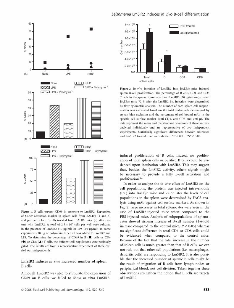
LmSIR2 induces in vivo increased number of spleenB cells
Although LmSIR2 was able to stimulate the expression of
CD69 on B cells, we failed to show in vitro LmSIR2-
induced proliferation of B cells. Indeed, no prolifer-
ation of total spleen cells or purified B cells could be evi-
denced upon incubation with LmSIR2. This may suggest
that, besides the LmSIR2 activity, others signals might
be necessary to provide a fully B-cell activation and
proliferation.22
In order to analyse the in vivo effect of LmSIR2 on the
cell populations, the protein was injected intravenously
(i.v.) into BALB/c mice and 72 hr later the levels of cell
populations in the spleen were determined by FACS ana-
lysis using mAb against cell surface markers. As shown in
Fig. 2, large increases in total splenocytes were seen in the
case of LmSIR2-injected mice when compared to the
PBS-injected mice. Analysis of subpopulations of spleno-
cytes showed striking increase of B-cell number (1�6-fold
increase compared to the control mice, P < 0�05) whereas
no significant difference in total CD4 or CD8 cells could
be evidenced when compared to the control mice.
Because of the fact that the total increase in the number
of spleen cells is much greater than that of B cells, we can
not rule out that other cell populations (i.e. macrophages,
dendritic cells) are responding to LmSIR2. It is also possi-
ble that the increased number of splenic B cells might be
the result of migration of B cells from lymph nodes or
peripherical blood, not cell division. Taken together these
observations strengthen the notion that B cells are targets
of LmSIR2.
1·4×1008
1·2×1008
1·0×1008
8·0×1007
6·0×1007
4·0×1007
2·0×1007
0·0
Cel
l num
ber
Total B
**
*
spleen cells
PBS treated
LmSIR2 treated
CD4 CD8
Figure 2. In vivo injection of LmSIR2 into BALB/c mice induced
spleen B-cell proliferation. The percentage of B cells, CD4 and CD8
T cells in the spleen of untreated and LmSIR2 (20 lg/mouse)-treated
BALB/c mice 72 h after the LmSIR2 i.v. injection were determined
by flow cytometric analysis. The number of each spleen cell subpop-
ulation was calculated based on the total viable cells determined by
trypan blue exclusion and the percentage of cell bound mAb to the
specific cell surface marker (anti-CD4, anti-CD8 and anti-l). The
data represent the mean and the standard deviations of three animals
analysed individually and are representative of two independent
experiments. Statistically significant differences between untreated
and LmSIR2 treated mice are indicated: *P < 0�01; **P < 0�05.
60
50
40
30
20
10
0
(a)
(b)
(c)
60
50
40
30
20
10
0
100
75
50
25
0
% C
D69
% C
D69
% C
D69
None
None
LPS
LPS
LPS + Polymyxin B
SIR2 + Polymyxin B
SIR2
SIR2
None
LPS
LPS + Polymyxin B
SIR2 + Polymyxin B
SIR2
Figure 1. B cells express CD69 in response to LmSIR2. Expression
of CD69 activation marker in spleen cells from BALB/c (a and b)
and purified spleen B cells isolated from BALB/c mice (c) after cul-
ture with LmSIR2. A total of 2�5 · 105 cells per well were cultured
in the presence of LmSIR2 (10 lg/ml) or LPS (10 lg/ml). In some
experiments 10 lg of polymixin B per ml was added to LmSIR2 and
LPS. To determine the percentage of CD69 in B (j) cells or CD4
(d) or CD8 (m) T cells, the different cell populations were positively
gated. The results are from a representative experiment of three car-
ried out independently.
� 2006 Blackwell Publishing Ltd, Immunology, 119, 529–540 533
Leishmania LmSIR2 induces in vivo B-cell differentiation
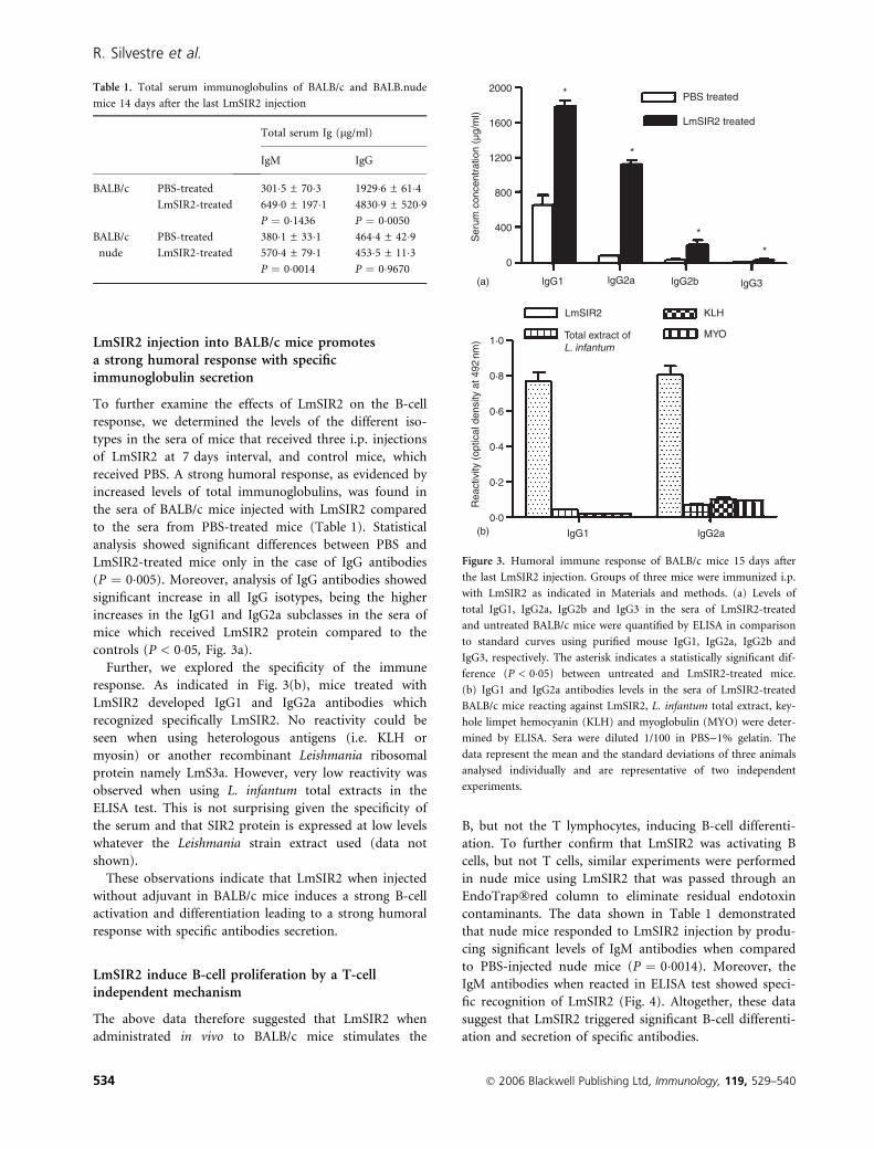
LmSIR2 injection into BALB/c mice promotesa strong humoral response with specificimmunoglobulin secretion
To further examine the effects of LmSIR2 on the B-cell
response, we determined the levels of the different iso-
types in the sera of mice that received three i.p. injections
of LmSIR2 at 7 days interval, and control mice, which
received PBS. A strong humoral response, as evidenced by
increased levels of total immunoglobulins, was found in
the sera of BALB/c mice injected with LmSIR2 compared
to the sera from PBS-treated mice (Table 1). Statistical
analysis showed significant differences between PBS and
LmSIR2-treated mice only in the case of IgG antibodies
(P ¼ 0�005). Moreover, analysis of IgG antibodies showed
significant increase in all IgG isotypes, being the higher
increases in the IgG1 and IgG2a subclasses in the sera of
mice which received LmSIR2 protein compared to the
controls (P < 0�05, Fig. 3a).
Further, we explored the specificity of the immune
response. As indicated in Fig. 3(b), mice treated with
LmSIR2 developed IgG1 and IgG2a antibodies which
recognized specifically LmSIR2. No reactivity could be
seen when using heterologous antigens (i.e. KLH or
myosin) or another recombinant Leishmania ribosomal
protein namely LmS3a. However, very low reactivity was
observed when using L. infantum total extracts in the
ELISA test. This is not surprising given the specificity of
the serum and that SIR2 protein is expressed at low levels
whatever the Leishmania strain extract used (data not
shown).
These observations indicate that LmSIR2 when injected
without adjuvant in BALB/c mice induces a strong B-cell
activation and differentiation leading to a strong humoral
response with specific antibodies secretion.
LmSIR2 induce B-cell proliferation by a T-cellindependent mechanism
The above data therefore suggested that LmSIR2 when
administrated in vivo to BALB/c mice stimulates the
B, but not the T lymphocytes, inducing B-cell differenti-
ation. To further confirm that LmSIR2 was activating B
cells, but not T cells, similar experiments were performed
in nude mice using LmSIR2 that was passed through an
EndoTrap�red column to eliminate residual endotoxin
contaminants. The data shown in Table 1 demonstrated
that nude mice responded to LmSIR2 injection by produ-
cing significant levels of IgM antibodies when compared
to PBS-injected nude mice (P ¼ 0�0014). Moreover, the
IgM antibodies when reacted in ELISA test showed speci-
fic recognition of LmSIR2 (Fig. 4). Altogether, these data
suggest that LmSIR2 triggered significant B-cell differenti-
ation and secretion of specific antibodies.
Table 1. Total serum immunoglobulins of BALB/c and BALB.nude
mice 14 days after the last LmSIR2 injection
Total serum Ig (lg/ml)
IgM IgG
BALB/c PBS-treated 301�5 ± 70�3 1929�6 ± 61�4LmSIR2-treated 649�0 ± 197�1 4830�9 ± 520�9
P ¼ 0�1436 P ¼ 0�0050
BALB/c
nude
PBS-treated 380�1 ± 33�1 464�4 ± 42�9LmSIR2-treated 570�4 ± 79�1 453�5 ± 11�3
P ¼ 0�0014 P ¼ 0�9670
2000
1600
1200
800
400
0
(a)
(b)
*
*
*
*
1·0
0·8
0·6
0·4
0·2
0·0
lgG1
Total extract of L. infantum
KLH
MYO
Rea
ctiv
ity (
optic
al d
ensi
ty a
t 492
nm)
Ser
um c
once
ntra
tion
(µg/
ml)
lgG2a
lgG2b lgG3 lgG1 lgG2a
LmSIR2
PBS treated
LmSIR2 treated
Figure 3. Humoral immune response of BALB/c mice 15 days after
the last LmSIR2 injection. Groups of three mice were immunized i.p.
with LmSIR2 as indicated in Materials and methods. (a) Levels of
total IgG1, IgG2a, IgG2b and IgG3 in the sera of LmSIR2-treated
and untreated BALB/c mice were quantified by ELISA in comparison
to standard curves using purified mouse IgG1, IgG2a, IgG2b and
IgG3, respectively. The asterisk indicates a statistically significant dif-
ference (P < 0�05) between untreated and LmSIR2-treated mice.
(b) IgG1 and IgG2a antibodies levels in the sera of LmSIR2-treated
BALB/c mice reacting against LmSIR2, L. infantum total extract, key-
hole limpet hemocyanin (KLH) and myoglobulin (MYO) were deter-
mined by ELISA. Sera were diluted 1/100 in PBS)1% gelatin. The
data represent the mean and the standard deviations of three animals
analysed individually and are representative of two independent
experiments.
534 � 2006 Blackwell Publishing Ltd, Immunology, 119, 529–540
R. Silvestre et al.

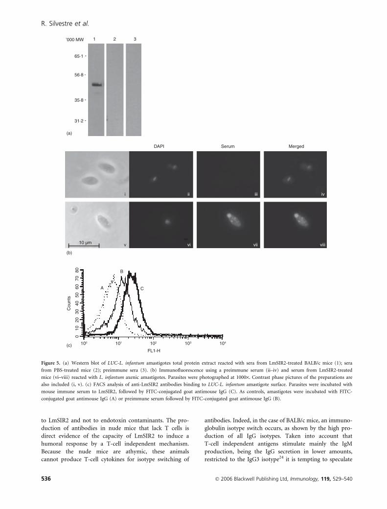
Anti-LmSIR2 antibodies bind to the amastigotesurface
The above observations demonstrated that LmSIR2
immunization of mice induced a strong specific antibody
response. Thus, we examined the reactivity of anti-
LmSIR2 mice immune serum against L. infantum axenic
amastigotes carrying a luciferase encoding gene (LUC-
L. infantum). As shown in Fig. 5(a), sera from mice
immunized with LmSIR2 recognized the corresponding
antigen (MW � 52 000) in LUC-L.infantum extracts
whereas no reactivity could be seen when using pre-
immune sera or the sera from PBS-injected mice. More-
over, indirect immunofluorescence assays showed a
positive labelling in vesicles and in the flagellar pocket
zone (Fig. 5b), that may indicate protein secretion from
the parasite.23
We then analysed by FACS whether a binding of the
anti-LmSIR2 antibodies was detectable. The results shown
in Fig. 5(c) indicated that indeed, anti-LmSIR2 bound to
the amastigote. The level of binding was approximately
three times higher when using sera from LmSIR2-immun-
ized mice compared to preimmune sera (MFI ¼ 45 and
15, respectively).
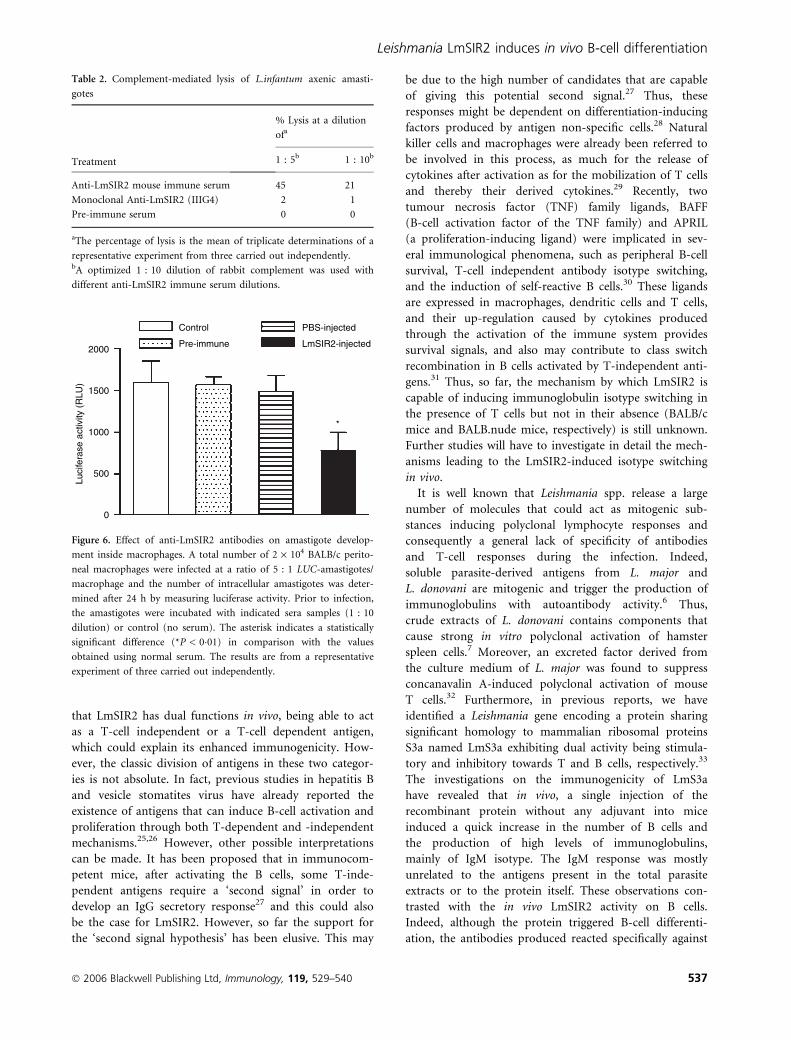
Anti-LmSIR2 antibodies induce complementmediated lysis and inhibit amastigote developpementinside macrophages in vitro
The above data indicate that LmSIR2 epitopes are
expressed on the parasite surface and are accessible to the
antibodies. Therefore, we thought it was reasonable to
determine the ability of sera from LmSIR2-immunized
mice to lyse in vitro LUC-L. infantum axenic amastigotes.
As shown in Table 2, the percentage of killing of amasti-
gotes was between 21 and 45% depending on the concen-
tration of serum used. As a control, preimmune serum
was unable to support lysis of the parasites even at high
concentration. This was also the case for a IIIG4 mAb
against LmSIR2,14 which showed no capacity to induce
parasite lysis. This might be the result of the fact that
IIIG4 mAb, being of the IgG1 subclass, fixes complement
poorly. Furthermore, the anti-LmSIR2 immune serum
was also found to be able to induce the lysis of LUC-
L. infantum promastigotes (data not shown).
We also tested whether the anti-LmSIR2 immune sera
were able to modulate in vitro amastigote–macrophage
interaction. Mouse peritoneal macrophages were used as
in vitro model to measure infection with LUC-L.infantum
axenic amastigotes in the presence of heat-inactivated
anti-LmSIR2 immune serum. Thus, peritoneal macroph-
ages were incubated with LUC-L. infantum axenic amasti-
gotes at a ratio of five parasites per macrophage in the
presence of 1 : 10 dilution of preimmune serum or sera
from LmSIR2 and PBS-injected mice. Twenty-four hr
later, the level of infection was determined. As shown in
Fig. 6 there was a significant inhibition of amastigote
development inside macrophages in the presence of heat-
inactivated anti-LmSIR2 immune serum when compared
to the control preimmune sera or sera from PBS-injected
mice. These data suggest that anti-LmSIR2 sera in the
absence of complement can reduce intracellular parasite
development.
In preliminary immunization experiments, two groups
of four mice each were injected i.p. with or without
50 lg of LmSIR2 in 200 ll of PBS three times at 7 day
intervals. Fifteen days later, they were infected i.p. with
108 WT promastigotes. At 15 days postinfection, a signifi-
cant decrease (�1�5-log reduction; P ¼ 0�03) of parasite
load was observed in the spleen of LmSIR2-immunized
mice in comparison to the control (PBS-injected mice).
Discussion
In the current study, we demonstrated that the Leis-
hmania cytosolic nicotinamide adenine dinucleotide-
dependent deacetylase, LmSIR2, could directly activate
B lymphocytes (but not T cells) from normal mice.
However, although we were unable, though, to show the
in vitro proliferation of spleen cells or purified B cells
upon incubation with LmSIR2, in vivo administration of
LmSIR2 i.v. into BALB/c mice triggered the differenti-
ation of B cells, demonstrating therefore that LmSIR2
could serve as an important activation signal in vivo.
Indeed, in vivo injection of LmSIR2 alone without adju-
vant into BALB/c mice or nude mice induced the synthe-
sis of IgG and IgM antibodies, respectively, thereby
indicating that B cells were able to undergo differentiation
into antibody-secreting cells. Several experiments demon-
strated that the B lymphocytes were responding directly
Rea
ctiv
ity (
optic
al d
ensi
ty a
t 492
nm) 1·0
0·8
0·6
0·4
0·2
0·0
Total extract of LmSIR2 LmS3a L. infantum
PBS treated
LmSIR2 treated *
Figure 4. Levels of specific IgM anti-LmSIR2 in the sera of BALB.-
nude mice. Sera from LmSIR2 and PBS injected mice were reacted
in ELISA assay against L. infantum total extract, LmS3a or LmSIR2.
Statistically significant differences between sera from LmSIR2-treated
and control mice were observed: *P < 0�005.
� 2006 Blackwell Publishing Ltd, Immunology, 119, 529–540 535
Leishmania LmSIR2 induces in vivo B-cell differentiation
to LmSIR2 and not to endotoxin contaminants. The pro-
duction of antibodies in nude mice that lack T cells is
direct evidence of the capacity of LmSIR2 to induce a
humoral response by a T-cell independent mechanism.
Because the nude mice are athymic, these animals
cannot produce T-cell cytokines for isotype switching of
antibodies. Indeed, in the case of BALB/c mice, an immuno-
globulin isotype switch occurs, as shown by the high pro-
duction of all IgG isotypes. Taken into account that
T-cell independent antigens stimulate mainly the IgM
production, being the IgG secretion in lower amounts,
restricted to the IgG3 isotype24 it is tempting to speculate
’000 MW 1
65·1
56·8
35·8
31·2
DAPI
(a)
(b)
(c)
Serum
i
A
10 µm
FL1-H
0
100 101 102 103 104
Cou
nts
1020
3040
5060
7080
ii
B
iii
C
iv
v vi vii viii
Merged
-
-
-
-
2 3
Figure 5. (a) Western blot of LUC-L. infantum amastigotes total protein extract reacted with sera from LmSIR2-treated BALB/c mice (1); sera
from PBS-treated mice (2); preimmune sera (3). (b) Immunofluorescence using a preimmune serum (ii–iv) and serum from LmSIR2-treated
mice (vi–viii) reacted with L. infantum axenic amastigotes. Parasites were photographed at 1000·. Contrast phase pictures of the preparations are
also included (i, v). (c) FACS analysis of anti-LmSIR2 antibodies binding to LUC-L. infantum amastigote surface. Parasites were incubated with
mouse immune serum to LmSIR2, followed by FITC-conjugated goat antimouse IgG (C). As controls, amastigotes were incubated with FITC-
conjugated goat antimouse IgG (A) or preimmune serum followed by FITC-conjugated goat antimouse IgG (B).
536 � 2006 Blackwell Publishing Ltd, Immunology, 119, 529–540
R. Silvestre et al.
that LmSIR2 has dual functions in vivo, being able to act
as a T-cell independent or a T-cell dependent antigen,
which could explain its enhanced immunogenicity. How-
ever, the classic division of antigens in these two categor-
ies is not absolute. In fact, previous studies in hepatitis B
and vesicle stomatites virus have already reported the
existence of antigens that can induce B-cell activation and
proliferation through both T-dependent and -independent
mechanisms.25,26 However, other possible interpretations
can be made. It has been proposed that in immunocom-
petent mice, after activating the B cells, some T-inde-
pendent antigens require a ‘second signal’ in order to
develop an IgG secretory response27 and this could also
be the case for LmSIR2. However, so far the support for
the ‘second signal hypothesis’ has been elusive. This may
be due to the high number of candidates that are capable
of giving this potential second signal.27 Thus, these
responses might be dependent on differentiation-inducing
factors produced by antigen non-specific cells.28 Natural
killer cells and macrophages were already been referred to
be involved in this process, as much for the release of
cytokines after activation as for the mobilization of T cells
and thereby their derived cytokines.29 Recently, two
tumour necrosis factor (TNF) family ligands, BAFF
(B-cell activation factor of the TNF family) and APRIL
(a proliferation-inducing ligand) were implicated in sev-
eral immunological phenomena, such as peripheral B-cell
survival, T-cell independent antibody isotype switching,
and the induction of self-reactive B cells.30 These ligands
are expressed in macrophages, dendritic cells and T cells,
and their up-regulation caused by cytokines produced
through the activation of the immune system provides
survival signals, and also may contribute to class switch
recombination in B cells activated by T-independent anti-
gens.31 Thus, so far, the mechanism by which LmSIR2 is
capable of inducing immunoglobulin isotype switching in
the presence of T cells but not in their absence (BALB/c
mice and BALB.nude mice, respectively) is still unknown.
Further studies will have to investigate in detail the mech-
anisms leading to the LmSIR2-induced isotype switching
in vivo.
It is well known that Leishmania spp. release a large
number of molecules that could act as mitogenic sub-
stances inducing polyclonal lymphocyte responses and
consequently a general lack of specificity of antibodies
and T-cell responses during the infection. Indeed,
soluble parasite-derived antigens from L. major and
L. donovani are mitogenic and trigger the production of
immunoglobulins with autoantibody activity.6 Thus,
crude extracts of L. donovani contains components that
cause strong in vitro polyclonal activation of hamster
spleen cells.7 Moreover, an excreted factor derived from
the culture medium of L. major was found to suppress
concanavalin A-induced polyclonal activation of mouse
T cells.32 Furthermore, in previous reports, we have
identified a Leishmania gene encoding a protein sharing
significant homology to mammalian ribosomal proteins
S3a named LmS3a exhibiting dual activity being stimula-
tory and inhibitory towards T and B cells, respectively.33
The investigations on the immunogenicity of LmS3a
have revealed that in vivo, a single injection of the
recombinant protein without any adjuvant into mice
induced a quick increase in the number of B cells and
the production of high levels of immunoglobulins,
mainly of IgM isotype. The IgM response was mostly
unrelated to the antigens present in the total parasite
extracts or to the protein itself. These observations con-
trasted with the in vivo LmSIR2 activity on B cells.
Indeed, although the protein triggered B-cell differenti-
ation, the antibodies produced reacted specifically against
2000
1500
1000
500
0
Luci
fera
se a
ctiv
ity (
RLU
)
Control
Pre-immune
PBS-injected
LmSIR2-injected
*
Figure 6. Effect of anti-LmSIR2 antibodies on amastigote develop-
ment inside macrophages. A total number of 2 · 104 BALB/c perito-
neal macrophages were infected at a ratio of 5 : 1 LUC-amastigotes/
macrophage and the number of intracellular amastigotes was deter-
mined after 24 h by measuring luciferase activity. Prior to infection,
the amastigotes were incubated with indicated sera samples (1 : 10
dilution) or control (no serum). The asterisk indicates a statistically
significant difference (*P < 0�01) in comparison with the values
obtained using normal serum. The results are from a representative
experiment of three carried out independently.
Table 2. Complement-mediated lysis of L.infantum axenic amasti-
gotes
Treatment
% Lysis at a dilution
ofa
1 : 5b 1 : 10b
Anti-LmSIR2 mouse immune serum 45 21
Monoclonal Anti-LmSIR2 (IIIG4) 2 1
Pre-immune serum 0 0
aThe percentage of lysis is the mean of triplicate determinations of a
representative experiment from three carried out independently.bA optimized 1 : 10 dilution of rabbit complement was used with
different anti-LmSIR2 immune serum dilutions.
� 2006 Blackwell Publishing Ltd, Immunology, 119, 529–540 537
Leishmania LmSIR2 induces in vivo B-cell differentiation
SIR2 and not other heterologous antigens such as myo-
sin or KLH, and were able to induce the complement-
mediated killing of amastigotes and inhibition of their
multiplication inside macrophages. Indirect immunofluo-
rescence assays of L. infantum axenic amastigotes with
sera from LmSIR2-immunized mice showed the presence
of SIR2 protein in vesicles and in the flagellar pocket
zone, already known to be filled with secretory mater-
ial;23 this being therefore in agreement with our
previous observations.18 Moreover, using a highly sensi-
tive radiolabelled immunoprecipitation technique,
we observed that SIR2 is among the parasite excreted-
secreted antigens (unpublished data). Results from the
in vitro macrophage infections with LUC-L. infantum
amastigotes, the presence of anti-LmSIR2 sera may indi-
cate a potential role of SIR2 in the binding, internali-
zation and/or multiplication of the parasite in the
macrophage.
Studies on the molecular mechanisms of parasite entry
into macrophages have led to the identification of several
candidate receptors facilitating multiple routes of entry.34
Indeed, internalization of promastigotes into macrophages
has been shown to be mediated by macrophage mem-
brane proteins such as the mannose receptor,35 the fibro-
nectin receptor,36 the Fc receptor (FcR),37 and the
complement receptors such as CR1 (CD35) and CR3
(CD11b/CD18).35,38 Thus, the in vitro modulation of
macrophage infection by anti-LmSIR2 antibodies may
suggest possible roles of SIR2 in the internalization pro-
cess of the amastigote and/or further multiplication of the
parasite.
Evasion from the host complement system is one of the
strategies used by Leishmania parasites to avoid host
immune defence.39 Indeed, metacylic promastigotes and
amastigotes are relatively resistant to direct serum kill-
ing.40 However, previous studies have shown that anti-
bodies that were able to bind to living parasites and lyse
them in conjunction with complement were associated
with host protection.20 Thus, one can assume that, the
production of antibodies capable of enhancing comple-
ment cytotoxicity towards the amastigote stage might be,
working in co-operation with the cellular immunity, an
important requirement for effective antiparasitic immu-
nity.41 Given that the anti-LmSIR2 sera induced comple-
ment-mediated lysis of amastigotes and promastigotes,
one may speculate that a LmSIR2 immunization can be
seen as capable of protecting the host against infection
and disease progression.
Although it has been reported that the antibodies may
play a role in the host protection mechanisms against
experimental leishmaniasis12 a more recent study has
shown that antibodies could exert deleterious effects
on the host. In fact, by using B cell-mutant mice and
genetically modified mice lacking circulating antibodies
infected with L. amazonensis and L. pifanoi, the authors
reported that these mice developed barely detectable
lesions compared to control BALB/c mice.42 Reconstitu-
tion of the B cell-mutant with the immune anti-Leishma-
nia serum increased the pathological processes in the
otherwise non-susceptible mice. Therefore, preliminary
BALB/c immunization experiments were conducted using
LmSIR2 protein. A significant decrease of parasite load in
the spleen of LmSIR2-immunized mice was observed in
comparison to non-immunized control mice at 2 weeks
postchallenge infection. Thus, it is reasonable to suggest
that anti-LmSIR2 antibodies may in part play a role in
protective immune mechanisms rather than exacerbating
the disease. Further studies using different doses and
routes of immunization are needed to explore further the
protective role of the LmSIR2 molecule.
The LmSIR2 protein belongs to a highly conserved
family of closely related proteins in both prokaryotic and
eukaryotic species.43 Historically, the biological signifi-
cance of SIR2-like proteins was attributed to the histones’
deacetylation, leading to chromatin condensation and
transcriptional silencing.44 However, diverse cellular local-
izations were found among SIR2 homologues, suggesting
that these enzymes have other physiological substrates
than histones and thus several biological functions inside
the different organisms. Indeed, several roles have been
attributed to SIR2-related family of proteins including
cell cycle progression and chromosome stability,45 DNA-
damage repair,46 life span extension in yeast47 and in
Caenorhabditis elegans.48 To our knowledge, this report is
the first description of a protein belonging to this large
family, which is among the Leishmania cytosolic and
secretory products that proved to have a role in the regu-
lation of the immune response through its capacity to
trigger preferentially B-cell effector functions. The high
degree of homology within this family of proteins, and
the fact that homologues have been found in mouse49
and human50 has led us to perform additional experi-
ments in order to verify the possibility of the existence of
SIR2 cross-reactive epitopes on the mouse and human
cells that could be recognized by the sera from LmSIR2-
immunized mice. Using different approaches (Western
blot, ELISA, immunoprecipitation of [35S]methionine-
labelled mouse spleen cells) no such cross-reactivity was
found, arguing for the Leishmania specificity of the
antibodies produced by LmSIR2 immunization (data not
shown).
In summary, our present results show that LmSIR2 is a
potent B-cell modulatory factor both in vitro and in vivo.
Following the injections of LmSIR2 without adjuvant a
selective B-cell response was induced resulting in a sur-
prisingly parasite-specific production of antibodies, which
were lytic in co-operation with the complement, and
restrain the capacity of the parasites to infect macro-
phages. There is strong evidence that an ideal vaccine,
especially against visceral leishmaniasis, may require the
538 � 2006 Blackwell Publishing Ltd, Immunology, 119, 529–540
R. Silvestre et al.
induction of both humoral and cellular branches of the
immune system, for maximum efficiency.11 In that view,
one can consider LmSIR2 to be among vaccination can-
didate, taking into account the safety and the strong
response when delivered in a low dose. Overall, these data
add LmSIR2 to the list of Leishmania antigens that could
specifically stimulate the immune system of the host and
suggest that LmSIR2 could be among the parasite mole-
cules to be used to design an optimal multicomponent
vaccine to control Leishmania infection.
Acknowledgements
This work was supported by Fundacao para a Ciencia e
Tecnologia (FCT) and FEDER, grant POCTI/CVT/39257/
2001 and POCI/CVT/59840/2004, INSERM and IRD
UR008. R.S. and J.T. are supported by fellowships from
FCT and FEDER number SFRH/BD/13120/2003 and
SFRH/BD/18137/2004, respectively.
References
1 Badaro R, Jones TC, Carvalho EM, Sampaio D, Reed SG, Barral
A, Teixeira R, Johnson WD. New perspectives on a subclinical
form of visceral leishmaniasis. J Infect Dis 1986; 154:1003–11.
2 Ashford RW. Leishmaniasis reservoirs and their siginificance in
control. Clin Dermatol 1996; 14:523–32.
3 Desjeux P. Leishmaniasis: public health aspects and control.
Clin Dermatol 1996; 14:417–23.
4 Carvalho EM, Teixeira RS, Johnson WD. Cell-mediated immu-
nity in American visceral leishmaniasis: reversible immuno-
supression during acute infection. Infect Immun 1981; 33:498–502.
5 Galvao-Castro B, Ferreira JAS, Marzochi KF, Marzochi MC,
Coutinho SG, Lambert. PH. Polyclonal B cell activation, circula-
ting immune complexes and autoimmunity in American visceral
leishmaniasis. Clin Exp Immunol 1984; 56:58–66.
6 Bohme MW, Evans DA, Miles MA, Holborow EJ. Occurrence of
autoantibodies to intermediate filament proteins in human vis-
ceral leishmaniasis and their induction by experimental poly-
clonal B-cell activation. Immunology 1986; 59 (4):583–8.
7 Bunn-Moreno MM, Madeira ED, Miller K, Menezes JA, Cam-
pos-Neto A. Hypergammaglobulinaemia in Leishmania donovani
infected hamsters. possible association with a polyclonal activa-
tor of B cells and with supression of T cell function. Clin Exp
Immunol 1985; 59 (2):427–34.
8 Reina-San-Martin B, Cosson A, Minoprio P. Lymphocyte poly-
clonal activation: a pitfall for vaccine design against infectious
agents. Parasitol Today 2000; 16:62–7.
9 McMahon-Pratt D, Alexander J. Does the Leishmania major
paradigm of pathogenesis and protection hold for New World
cutaneous leishmaniasis or the visceral disease? Immun Rev 2004;
201:206–24.
10 Casadevall A. Antibody-mediated protection against intracellular
pathogens. Trends Microbiol 1998; 6:102–7.
11 Ravindran R, Nahid A. Progress in vaccine research and possible
effector mechanism in visceral leishmaniasis. Curr Mol Med
2004; 4:697–709.
12 Monjour L, Berneman A, Vouldoukis I, Domurado M, Guille-
min MC, Chopin C, Alfred C, Roseto A. Monoclonal antibodies
against defined Leishmania antigens protect mice against infec-
tion by different species of Leishmania. CR Acad Sci III 1985;
300 (9):395–8.
13 Yahiaoui B, Taibi A, Ouaissi A. A Leishmania major protein with
extensive homology to silent information regulator 2 of
Saccharomyces cerevisae. Gene 1996; 169:115–8.
14 Vergnes B, Sereno D, Madjidian-Sereno N, Lemesre JL, Ouaissi
A. Cytoplasmic SIR2 homologue overexpression promotes survi-
val of Leishmania parasites by preventing programmed cell
death. Gene 2002; 296:139–50.
15 Vergnes B, Sereno D, Tavares J et al. Targeted disruption of cyto-
solic SIR2 deacetylase discloses its essential role in Leishmania
survival and proliferation. Gene 2005; 363:85–96.
16 Cordeiro-da-Silva A, Cardoso L, Araujo N et al. Identification of
antibodies to Leishmania silent information regulatory 2 (SIR2)
protein homologue during canine natural infections: pathologi-
cal implications. Immunol Lett 2003; 68:155–62.
17 Sereno D, Roy G, Lemesre JL, Papadopoulou B, Ouellette M.
DNA transformation of Leishmania infantum axenic amastigotes
and their use in drug screening. Antmicrob Agents Chemother
2001; 45:1168–73.
18 Zemzoumi K, Sereno D, Francis C, Guilvard E, Lemesre JL,
Ouaissi A. Leishmania major. Cell type dependent distribution of
a 43 kDa antigen related to silent information regulatory-2 pro-
tein family. Biol Cell 1998; 90:239–45.
19 Lowry OH, Rosebrough NJ, Farr L, Randall RJ. Protein meas-
urement with the Folin phenol reagent. J Biol Chem 1951;
193:267–9.
20 Norris KA, Hearth G, So M. Purification of a Trypanosoma cruzi
membrane glycoprotein which elicits lytic antibody. Infect Immun
1989; 57:2372–7.
21 Roy G, Dumas C, Sereno D et al. Episomal and stable expres-
sion of the luciferase reporter gene for quantifying Leishmania
spp. infections in macrophages and in animal models. Mol Bio-
chem Parasitol 2000; 110:195–206.
22 Baumgarth N. A two-phase of B-cell activation. Immunol Rev
2000; 176:171–80.
23 Pimenta PFP, Saraiva EMB, Sacks D. The comparative fine
structure and surface glycoconjugate expression of three life
stages of Leishmania major. Exp Parasitol 1991; 72:191–204.
24 Bjorklund M, Coutinho A. Isotype commitment in the in vivo
immune responses. II. Polyclonal plaque-forming cell responses
to lipopolysaccharide in the spleen and bone marrow. Euro J
Immunol 1983; 13:44–50.
25 Freer G, Burkhart C, Ciernik I, Bachmann MF, Hengartner H,
Zinkernagel RM. Vesicular stomatitis virus Indiana glycoprotein
as a T-cell-dependent and – independent antigen. J Virol 1994;
68:3650–5.
26 Milich DR, McLachlan A. The nucleocapsid of hepatitis B virus
is both a T-cell-independent and a T-cell-dependent antigen.
Science 1986; 234:1398–401.
27 Vos Q, Lees A, Wu Z, Snapper CM, Mond JJ. B-cell activa-
tion by a T-cell independent type 2 antigens as an integral part
of the humoral immune response to pathogenic microorganism.
Immunol Rev 2000; 176:154–70.
28 Snapper CM, Mond JJ. Towards a comprehensive view of
immunoglobulin class switching. Immunol Today 1993; 14:15–7.
� 2006 Blackwell Publishing Ltd, Immunology, 119, 529–540 539
Leishmania LmSIR2 induces in vivo B-cell differentiation
29 Mond JJ, Vos Q, Lees A, Snapper CM. T cell independent anti-
gens. Curr Opin Immunol 1995; 7:349–54.
30 Schneider P. The role of APRIL and BAFF in lymphocyte activa-
tion. Curr Opin Immunol 2005; 17:282–9.
31 Castigli E, Wilson SA, Scott S et al. TACI and BAFF-R mediate
isotype switching in B cells. J Exp Med 2005; 201:35–9.
32 Grimaldi GJ, Tesh RB. Leishmaniases of the New World: current
concepts and implications for future research. Clin Microbial Rev
1993; 6 (3):230–50.
33 Cordeiro-da-Silva A, Borges MC, Guilvard E, Ouaissi A. Dual
role of the Leishmania major ribosomal protein S3a homologue
in regulation of T- and B-cell activation. Infect Immun 2001;
69:6588–96.
34 Alexander J, Satoskar AR, Russell DG. Leishmania species: mod-
els of intracellular parasitism. J Cell Sci 1999; 112:2993–3002.
35 Wilson ME, Pearson RD. Roles of CR3 and mannose receptors
in the attachment and ingestion of Leishmania donovani by
human mononuclear phagocytes. Infect Immun 1988; 56:363–9.
36 Brittingham A, Chen G, McGwire BS, Chang KP, Mosser DM.
Interaction of Leishmania gp63 with cellular receptors for fibro-
nectin. Infect Immun 1999; 67:4477–84.
37 Love DC, Kane MM, Mosser DM. Leishmania amazonensis: the
phagocytosis of amastigotes by macrophages. Exp Parasitol 1998;
88 (3):161–71.
38 Dominguez M, Torano A. Immune adherence-mediated opsono-
phagocytosis. mechanism of Leishmania infection. J Exp Med
1999; 189:25–35.
39 Bogdan C, Rollinghoff M. The immune response to Leishmania:
mechanism of parasite control and evasion. Int J Parasitol 1998;
28:121–34.
40 Hall BF, Joiner KA. Strategies of obligate intracellular parasites
for evading host defenses. Parasitol Today 1991; 12:A22–A7.
41 Hoover DL, Berger M, Oppenheim MH, Hockmeyer WT,
Meltzer MS. Cytotoxicity of human serum for Leishmania
donovani amastigotes: antibody facilitation of alternate comple-
ment pathway-mediated killing. Infect Immun 1985; 47 (1):247–
52.
42 Kima PE, Constant SL, Hannum L, Colmenares M, Lee KS,
Haberman AM, Shlomchik MJ, McMahon-Pratt D. Internaliza-
tion of Leishmania mexicana complex amastigotes via the Fc
receptor is required to sustain infection in murine cutaneous
leishmaniasis. J Exp Med 2000; 191 (6):1063–7.
43 Frye RA. Phylogenetic classification of prokaryotic and eukaryot-
ic Sir2-like proteins. Biochem Biophys Res Commun 2000;
273:793–8.
44 Gasser SM, Cockell MM. The molecular biology of the SIR pro-
teins. Gene 2001; 279:1–16.
45 Brachmann CB, Sherman JM, Devine SE, Cameron EE, Pillus L,
Boeke JD. The SIR2 gene family, conserved from bacteria to
humans, functions in silencing, cell cycle progression, and
chromosome stability. Genes Dev 1995; 9:2888–902.
46 Tsukamoto Y, Kato J, Ikeda H. Silencing factors participate
in DNA repair and recombination in Saccharomyces cerevisiae.
Nature 1997; 388:900–3.
47 Kaeberlein M, McVey M, Guarente L. The SIR2/3/4 complex
and SIR2 alone promote longevity in Saccharomyces cerevisiae by
two different mechanisms. Genes Dev 1999; 13:2570–80.
48 Tissenbaum HA, Guarente L. Increased dosage of a Sir-2 gene
extends lifespan in Caenorhabditis elegans. Nature 2001; 410:227–
30.
49 Yang YH, Chen YH, Zhang CY, Nimmakayalu MA, Ward DC,
Weissman S. Cloning and characterization of two mouse genes
with homology to the yeast Sir2 gene. Genomics 2000; 69:355–
69.
50 Frye RA. Characterization of five human cDNAs with homology
to the yeast SIR2 gene: SIR2-like proteins (sirtuins) metabolize
NAD and may have protein ADP-ribosyltransferase activity.
Biochem Biophys Res Commun 1999; 260:273–9.
540 � 2006 Blackwell Publishing Ltd, Immunology, 119, 529–540
R. Silvestre et al.