Laser Direct Writing of Silver Clusters-Based Subwavelength ...
14
HAL Id: hal-03682520 https://hal.archives-ouvertes.fr/hal-03682520 Submitted on 31 May 2022 HAL is a multi-disciplinary open access archive for the deposit and dissemination of sci- entific research documents, whether they are pub- lished or not. The documents may come from teaching and research institutions in France or abroad, or from public or private research centers. L’archive ouverte pluridisciplinaire HAL, est destinée au dépôt et à la diffusion de documents scientifiques de niveau recherche, publiés ou non, émanant des établissements d’enseignement et de recherche français ou étrangers, des laboratoires publics ou privés. Laser Direct Writing of Silver Clusters-Based Subwavelength Periodic Structures Embedded in Mid-Infrared Gallo-Germanate Glass Théo Guérineau, Alexandre Fargues, Jerome Lapointe, Réal Vallée, younès Messaddeq, Lionel Canioni, yannick Petit, Thierry Cardinal To cite this version: Théo Guérineau, Alexandre Fargues, Jerome Lapointe, Réal Vallée, younès Messaddeq, et al.. Laser Direct Writing of Silver Clusters-Based Subwavelength Periodic Structures Embed- ded in Mid-Infrared Gallo-Germanate Glass. Advanced Photonics Research, inPress, pp.2200032. 10.1002/adpr.202200032. hal-03682520
-
Upload
khangminh22 -
Category
Documents
-
view
1 -
download
0
Transcript of Laser Direct Writing of Silver Clusters-Based Subwavelength ...
HAL Id: hal-03682520https://hal.archives-ouvertes.fr/hal-03682520
Submitted on 31 May 2022
HAL is a multi-disciplinary open accessarchive for the deposit and dissemination of sci-entific research documents, whether they are pub-lished or not. The documents may come fromteaching and research institutions in France orabroad, or from public or private research centers.
L’archive ouverte pluridisciplinaire HAL, estdestinée au dépôt et à la diffusion de documentsscientifiques de niveau recherche, publiés ou non,émanant des établissements d’enseignement et derecherche français ou étrangers, des laboratoirespublics ou privés.
Laser Direct Writing of Silver Clusters-BasedSubwavelength Periodic Structures Embedded in
Mid-Infrared Gallo-Germanate GlassThéo Guérineau, Alexandre Fargues, Jerome Lapointe, Réal Vallée, younès
Messaddeq, Lionel Canioni, yannick Petit, Thierry Cardinal
To cite this version:Théo Guérineau, Alexandre Fargues, Jerome Lapointe, Réal Vallée, younès Messaddeq, etal.. Laser Direct Writing of Silver Clusters-Based Subwavelength Periodic Structures Embed-ded in Mid-Infrared Gallo-Germanate Glass. Advanced Photonics Research, inPress, pp.2200032.�10.1002/adpr.202200032�. �hal-03682520�
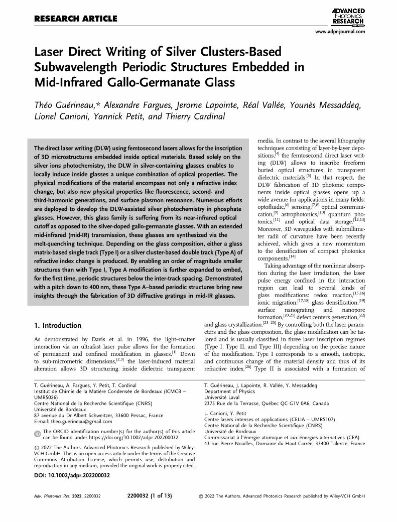
Laser Direct Writing of Silver Clusters-BasedSubwavelength Periodic Structures Embedded inMid-Infrared Gallo-Germanate Glass
Théo Guérineau,* Alexandre Fargues, Jerome Lapointe, Réal Vallée, Younès Messaddeq,Lionel Canioni, Yannick Petit, and Thierry Cardinal
1. Introduction
As demonstrated by Davis et al. in 1996, the light–matterinteraction via an ultrafast laser pulse allows for the formationof permanent and confined modification in glasses.[1] Downto sub-micrometric dimensions,[2,3] the laser-induced materialalteration allows 3D structuring inside dielectric transparent
media. In contrast to the several lithographytechniques consisting of layer-by-layer depo-sitions,[4] the femtosecond direct laser writ-ing (DLW) allows to inscribe freeformburied optical structures in transparentdielectric materials.[5] In that respect, theDLW fabrication of 3D photonic compo-nents inside optical glasses opens up awide avenue for applications in many fields:optofluidic,[6] sensing,[7,8] optical communi-cation,[9] astrophotonics,[10] quantum pho-tonics,[11] and optical data storage.[12,13]
Moreover, 3D waveguides with submillime-ter radii of curvature have been recentlyachieved, which gives a new momentumto the densification of compact photonicscomponents.[14]
Taking advantage of the nonlinear absorp-tion during the laser irradiation, the laserpulse energy confined in the interactionregion can lead to several kinds ofglass modifications: redox reaction,[15,16]
ionic migration,[17,18] glass densification,[19]
surface nanograting and nanoporeformation,[20,21] defect centers generation,[22]
and glass crystallization.[23–25] By controlling both the laser param-eters and the glass composition, the glass modification can be tai-lored and is usually classified in three laser inscription regimes(Type I, Type II, and Type III) depending on the precise natureof the modification. Type I corresponds to a smooth, isotropic,and continuous change of the material density and thus of itsrefractive index,[26] Type II is associated with a formation of
T. Guérineau, A. Fargues, Y. Petit, T. CardinalInstitut de Chimie de la Matière Condensée de Bordeaux (ICMCB –UMR5026)Centre National de la Recherche Scientifique (CNRS)Université de Bordeaux87 avenue du Dr Albert Schweitzer, 33600 Pessac, FranceE-mail: [email protected]
The ORCID identification number(s) for the author(s) of this articlecan be found under https://doi.org/10.1002/adpr.202200032.
© 2022 The Authors. Advanced Photonics Research published by Wiley-VCH GmbH. This is an open access article under the terms of the CreativeCommons Attribution License, which permits use, distribution andreproduction in any medium, provided the original work is properly cited.
DOI: 10.1002/adpr.202200032
T. Guérineau, J. Lapointe, R. Vallée, Y. MessaddeqDepartment of PhysicsUniversité Laval2375 Rue de la Terrasse, Québec QC G1V 0A6, Canada
L. Canioni, Y. PetitCentre lasers intenses et applications (CELIA – UMR5107)Centre National de la Recherche Scientifique (CNRS)Université de BordeauxCommissariat à l’énergie atomique et aux énergies alternatives (CEA)43 rue Pierre Noailles, Domaine du Haut Carrée, 33400 Talence, France
The direct laser writing (DLW) using femtosecond lasers allows for the inscriptionof 3D microstructures embedded inside optical materials. Based solely on thesilver ions photochemistry, the DLW in silver-containing glasses enables tolocally induce inside glasses a unique combination of optical properties. Thephysical modifications of the material encompass not only a refractive indexchange, but also new physical properties like fluorescence, second- andthird-harmonic generations, and surface plasmon resonance. Numerous effortsare deployed to develop the DLW-assisted silver photochemistry in phosphateglasses. However, this glass family is suffering from its near-infrared opticalcutoff as opposed to the silver-doped gallo-germanate glasses. With an extendedmid-infrared (mid-IR) transmission, these glasses are synthesized via themelt-quenching technique. Depending on the glass composition, either a glassmatrix-based single track (Type I) or a silver cluster-based double track (Type A) ofrefractive index change is produced. By enabling an order of magnitude smallerstructures than with Type I, Type A modification is further expanded to embed,for the first time, periodic structures below the inter-track spacing. Demonstratedwith a pitch down to 400 nm, these Type A–based periodic structures bring newinsights through the fabrication of 3D diffractive gratings in mid-IR glasses.
RESEARCH ARTICLEwww.adpr-journal.com
Adv. Photonics Res. 2022, 2200032 2200032 (1 of 13) © 2022 The Authors. Advanced Photonics Research published by Wiley-VCH GmbH

birefringent periodic nanostructures,[20] while Type III relates tovoid-like structures arising from Coulombic explosions.[27] Overtime, researchers have extensively explored the femtosecond directlaser writing in a wide range of glass families, that is, silicate,borate, fluoride, chalcogenide, phosphate, germanate, etc.,[1,28–31]
revealing the universality of this laser-induced glass processing.In silver-containing phosphate glasses, the silver ions act as
photosensitive agents and trigger an exotic material modificationbased solely on the silver ion photochemistry.[32] As enlightenedby Bellec et al.,[33] the laser irradiation generates free electronsinside the glass that recombine with Agþ silver ions to formAg0 silver atoms. By the accumulation of the femtosecond laserpulses, the migration of silver atoms and ions is initiated fromthe inner modification region to the outer one.[32] In the mean-time, the clusterization of the silver atoms and ions occurs, lead-ing to the creation of various silver entities. However, thecompetition between both the photodiffusion and photodissoci-ation phenomena allows stabilizing silver clusters only at theperiphery of the laser irradiation zone. The final silver clusterscan have various nuclearities and electrical charges from thesmallest one, that is, Ag2
þ, to the more complex Agmxþ with
x<m. After stationary laser irradiation of at least 104 pulses,the persistent modification consists only of a silver cluster-formed cylinder elongated along the laser beam propagationaxis.[34] This micrometric elongated distribution possesses adiffraction-limit-beating wall, reaching an 80 nm thickness.[33]
When the laser irradiation is no longer stationary but rather fol-lows a perpendicular translation to the propagation beam axis,the resulted modification is a double plane with two roundedendings.[34] At the exact location of each plane, a refractive indexchange appears due to the formation of new silver–silver chemi-cal bonds.[35] This exotic refractive index change, specificallyresulting from the laser-induced photochemistry of silver ionswithout significantly affecting the glass matrix, is categorizedas the Argentum Type (Type A). The formation of laser-generatedsilver clusters also gives rise to other remarkable optical proper-ties such as photoluminescence, second- and third-harmonicgeneration, and the creation of silver plasmonic nanopar-ticles.[16,33,36,37] Finally, the influence of the silver-doped phos-phate glass structure under different types of irradiations(DLW and X-Ray) on the glass photosensitivity has been recentlypublished elsewhere.[38–40] It has been reported that the increaseof non-bridging oxygens (NBOs, oxygens linked only to oneglass-former chemical element) via the change of glass structureleads to a significant increase of the glass photosensivity. Thephotosensitivity enhancement is notably observed through theincrease of several photoinduced silver entities, like electron–hole silver defects Ag2þ/Ag0 andmolecular silver clusters Agm
xþ.Several applications have already emerged from the localized
change of the refractive index induced by laser in silver-contain-ing phosphate glasses, such as single-mode waveguiding or ultra-sensitive refractive index sensor.[41] However, the main drawbackof the phosphate glass family is its restricted mid-infrared (mid-IR) optical transmission window ending at 3 μm, limiting theirpotential of applications. On the one hand, the atmospherictransparency window between 3 and 5 μm allows for the devel-opment of military counter measurement system and the astro-nomical observations performed on Earth, while on the otherhand, the strong absorption of water at 3 μm allows for the
elaboration of Erbium-doped optical fiber for medical surgery.Hence, the need of glass composition with a UV-to-mid-IR trans-parency up to 5 μm is requested. Originally developed by the U.S.Naval Laboratory, germanium–gallium–barium (BGG) oxideglasses possess tremendous properties, including mid-IR trans-mission up to 6microns, strong mechanical properties, and highglass transition temperature.[42,43] In this last decade, severalresearch groups have already performed laser inscription inBGG glasses, proving the 3D fabrication of optical couplers orwaveguides.[29,31,44] However, none of them have explored thepotential of silver-doped BGG glasses for DLW.
In this article, we report the fabrication of silver-doped BGGglass samples and their subsequent irradiation leading to either ahybrid combination of both Type A and Type I modifications, orsolely to a Type A modification. We show that by precisely tailor-ing the glass compositions and structures, the type of laser-induced glass modification can be selected. Through the increaseof barium ion concentration, the silver solubility in glass isenhanced. At low barium concentration, the hybrid Type A/Imodification is favored, while at high barium ion concentration,only the Type A is promoted. We suggest a mechanism explain-ing the hybrid Type A/I modification based on silver-assistedType A modification through a decrease of the laser pulse energythreshold. In a high-barium-containing BGG glass and at lowlaser fluence, a single track of refractive index change and fluo-rescence is reported, suggesting the observation of the very earlystages of Type A formation. At higher laser fluence, Type A mod-ifications are observed with the typical double track of refractiveindex change and fluorescence. The refractive index changereaches 2� 10�3 and 1� 10�4 for the double (Type A) and single(Type-A early stages) track, respectively. In the high-barium-containing BGG glasses, embedded periodic structures with apitch down to 400 nm have been successfully fabricated bydecreasing the distance between two laser paths below theType-A double-track spacing. This work provides an effectiveand simple method to control the type of laser-induced glassmodification in mid-infrared dielectric transparent media,simply by tailoring the glass composition and the DLW param-eters. Hence, it opens up a new way to fabricate subwavelengthintegrated components in mid-infrared devices.
2. Glass Preparation and Basic Characterizations
From our previous work on the quaternary glass systemGa2O3–GeO2–BaO–K2O,
[45] it has been highlighted that the pres-ence of alkali and alkali-earth ions allows for the generation ofNBOs, while alkali ions help significantly to increase the cationmobility in the glass. As for silver-doped phosphateglasses,[32,38,39] the amount of NBOs does also significantlyinfluence the photosensitivity of BGG glasses. Thus, twoglasses have been prepared from the quaternary systemGa2O3–GeO2–BaO–K2O with a significant difference in termsof NBOs quantity while maintaining the ability of crack-free glassmodifications during the irradiation process. These two glasseshave been doped with their highest solubility concentration ofsilver ions, determined from an unpublished preliminary workwhere the glass synthesis of several silver concentrations hasbeen performed: the first one with a large amount of gallium
www.advancedsciencenews.com www.adpr-journal.com
Adv. Photonics Res. 2022, 2200032 2200032 (2 of 13) © 2022 The Authors. Advanced Photonics Research published by Wiley-VCH GmbH

and germanium, namely GGBK, and the second one with a largeamount of barium and germanium, namely BGGK. GGBK glassis experimentally made of 32.9 mol% of GaO3/2, 35.1 mol% ofGeO2, 14.8 mol% of BaO, 16.6 mol% of KO1/2, and 0.6mol%of AgO1/2, while BGGK glass is made of 15.4 mol% ofGaO3/2, 40.5 mol% of GeO2, 37.5 mol% of BaO, 5.3mol% ofKO1/2, and 1.3mol% of AgO1/2. Glass precursors are firstweighed and then introduced in a platinum crucible to be meltedfor 15 h at 1400 and 1350 °C for GGBK and BGGK, respectively.The long melting duration is rather important for the homoge-neous dispersion of silver ions in the glass network. To freeze themixture without disturbing its homogeneity, a quick quenchingis conducted by immersing the crucible bottom in water at roomtemperature. Performed on ground glass chunks, X-Ray diffrac-tion experiments have revealed no crystallization peaks, confirm-ing full vitrification of both glasses (see Experimental Section).The detection limit specified by the manufacturer is about 1%by volume. However, the exact detection limit depends on the
density, Z number, and crystal structure of the studied com-pound. Hence, a degree of crystallization below 5% is consideredas a safe value for the limit of detection. By determination of theexperimental density evaluated at 4.86 and 4.25 g cm�3 forBGGK and GGBK, respectively (see Experimental Section), theirconcentration of silver ions per cubic centimeter has been calcu-lated at 3.2� 1020 and 1.6� 1020 cm�3, respectively. A differen-tial scanning calorimetry (DSC) measurement has beenperformed (see Experimental Section), which allows for deter-mining the glass transition temperature Tg. For GGBK glasscomposition, the Tg is measured at 642 °C, whereas forBGGK, Tg is 624 °C. After thermal annealing at 30 °C belowthe glass transition temperature for 4 h, glass chunks from bothcompositions are cut and polished on two parallel faces so as toobtain optical grade 1mm thick samples. Table 1 summarizesthe physical and chemical properties of the two glasses presentedin this work.
3. Results
3.1. Pristine Glass Properties
To characterize the glass structure and the optical transmissionwindow of both pristine gallo-germanate glasses, the polishedglass samples have been subjected to Raman and absorptionspectroscopies. In Figure 1a are depicted the normalizedRaman spectra of both GGBK and BGGK glasses under a532 nm laser excitation. Both glasses can be separated in threeregions of low (200–400 cm�1), intermediate (400–650 cm�1),and high (650–1000 cm�1) frequencies. The lowest spectraldomain can be assigned to either out-of-plane oxygen motionsin bent T–O–T bridge (T¼Ge or Ga in tetrahedral coordina-tion)[46] or network-modifying cations vibrating in large intersti-tial sites.[47] The intermediate spectral range can be assigned toseveral vibrational contributions of T–O–T bending with in-the-plane T–O–T oxygen motions.[46,47] Finally, the highest spectraldomain can be attributed to symmetric and antisymmetric
Table 1. Physical chemical properties of GGBK and BGGK glasses.
Physical and chemical properties GGBK sample BGGK sample
Experimental composition [mol%] 32.9 mol% GaO3/2
35.1 mol% GeO2
14.8 mol% BaO16.6 mol% KO1/2
0.6 mol% AgO1/2
15.4 mol% GaO3/2
40.5 mol% GeO2
37.5 mol% BaO5.3 mol% KO1/2
1.3 mol% AgO1/2
Glass transition temperature (�3 °C) 642 624
Density 4.25 4.86
Concentration of silver ions (1020 cm�3) 1.6 3.2
Main Raman contribution position(cm�1) & assignment
510/T–O–Ta) 800/[GeØ3O�]b)
Optical window transparencyc) [μm] 0.31–5.7 0.34–5.8
a)T¼Ge or Ga in tetrahedral coordination; b)germanium tetrahedral unit with onenon-bridging oxygen; c)defined for an absorption coefficient value of 10 cm�1.
200 400 600 800 1000
Raman shift (cm-1)
1000 2000 3000 4000 5000 6000 70000
10
20
30
40
50
60
70
Wavelength (nm)
250
280 320 360 4000
10
20
30
40
50
60
a (
cm-1
)
Wavelength (nm)
GGBKBGGK
GGBKBGGK
Abs
orpt
ion
coef
ficie
nt (
cm-1
)
Nor
mal
ized
inte
nsity
(a) (b)
Figure 1. a) Raw data Raman spectra normalized at their maximum intensity and b) linear absorption coefficient in the UV-visible-to-mid-IR wavelengthrange for both GGBK and BGGK. Inset: Magnification of the UV–blue wavelength domain.
www.advancedsciencenews.com www.adpr-journal.com
Adv. Photonics Res. 2022, 2200032 2200032 (3 of 13) © 2022 The Authors. Advanced Photonics Research published by Wiley-VCH GmbH
stretching modes of gallium and germanium tetrahedral units[TO4].
[46,48] In this high-frequency region, the well-known contri-bution at the vicinity of 800 cm�1 is assigned to the germaniumtetrahedral unit with an NBO denoted [GeØ3O
�].[46,49] In GGBKglass, the Raman spectrum reveals an important Raman signalresulting from the intermediate-frequency region peaking at510 cm�1 with a significant shoulder at about 450 cm�1, whereasin BGGK glass, the most important signal results from the high-frequency domain peaking at 800 cm�1 followed by a tail around700 cm�1 and the vanishing of the Raman contributionat 900 cm�1.
The linear absorption coefficient between 250 and 7000 nmhas been assessed for both GGBK and BGGK. As presentedin Figure 1b, the transmission window is extending from310 nm (UV) to 5.7 μm (mid-IR) for GGBK, and from 335 nmto 5.8 μm for the BGGK, corresponding to linear absorption coef-ficients below the limit of 10 cm�1. Since gallo-germanate glasseswith close compositions but without silver normally present a UVcutoff near 270 nm,[42,50] the upshifted UV absorption edges ofour silver-doped glasses are attributed to presence of the silverions. This assignment is also supported by the location in thesame wavelength range as the silver ion absorption in phosphate
glasses,[38,51] and also by the UV absorption edge undergoing aredshift from the GGBK to BGGK while the silver concentrationincreases, that is, with 1.6� 1020 and 3.2� 1020 cm�3 for GGBKand BGGK, respectively. Conjointly, the IR absorption edge isattributed to the multiphonon vibration modes. Between bothgallo-germanate glasses, the IR absorption edge is affected bythe glass composition revealing a 100 nm bathochromic shiftfrom GGBK to BGGK and an increase of the 6.3 μm contributionband, as well. As no special care was taken during glass synthe-sis, OH impurities are detected around 3.2 μm.
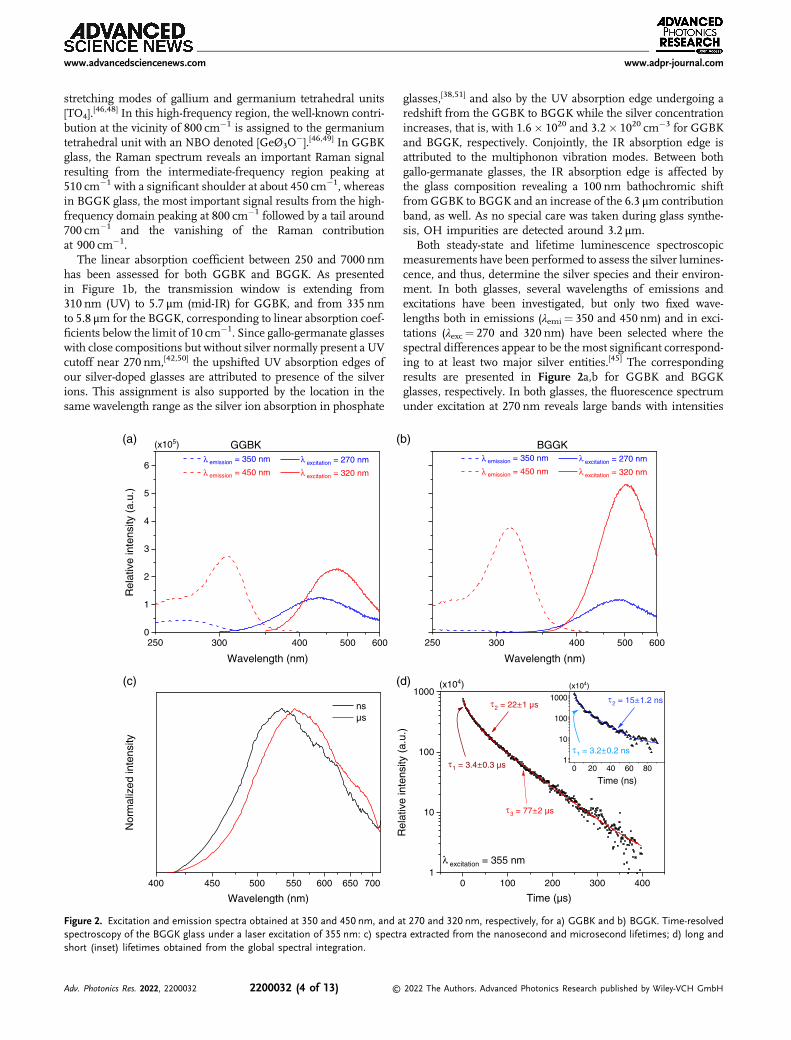
Both steady-state and lifetime luminescence spectroscopicmeasurements have been performed to assess the silver lumines-cence, and thus, determine the silver species and their environ-ment. In both glasses, several wavelengths of emissions andexcitations have been investigated, but only two fixed wave-lengths both in emissions (λemi¼ 350 and 450 nm) and in exci-tations (λexc¼ 270 and 320 nm) have been selected where thespectral differences appear to be the most significant correspond-ing to at least two major silver entities.[45] The correspondingresults are presented in Figure 2a,b for GGBK and BGGKglasses, respectively. In both glasses, the fluorescence spectrumunder excitation at 270 nm reveals large bands with intensities
300 400 500 6000
1
2
3
4
5
6 emission = 350 nm
emission = 450 nmexcitation = 270 nm
excitation = 320 nm
250
(x105) GGBK
400 450 500 550 600 650 700
nsµs
Wavelength (nm)
Wavelength (nm)
Rel
ativ
e in
tens
ity (
a.u.
)
(a)
(c)
Nor
mal
ized
inte
nsity
300 400 500 600
emission = 350 nm
emission = 450 nmexcitation = 270 nm
excitation = 320 nm
250
BGGK
Wavelength (nm)
(b)
0 100 200 300 4001
10
100
1000
Time (µs)
Time (ns)
1 = 3.4 0.3 µs
2 = 22 1 µs
3 = 77 2 µs
2 = 15 1.2 ns
1 = 3.2 0.2 ns
excitation = 355 nm
0 20 40 60 801
10
100
1000
(x104) (x10 )(d)
Rel
ativ
e in
tens
ity (
a.u.
)
Figure 2. Excitation and emission spectra obtained at 350 and 450 nm, and at 270 and 320 nm, respectively, for a) GGBK and b) BGGK. Time-resolvedspectroscopy of the BGGK glass under a laser excitation of 355 nm: c) spectra extracted from the nanosecond and microsecond lifetimes; d) long andshort (inset) lifetimes obtained from the global spectral integration.
www.advancedsciencenews.com www.adpr-journal.com
Adv. Photonics Res. 2022, 2200032 2200032 (4 of 13) © 2022 The Authors. Advanced Photonics Research published by Wiley-VCH GmbH

and locations that are composition dependent. In the barium-poor glass, the maximal intensity is reached at 440 nm withthe presence of a contribution at 350 nm, whereas in thebarium-rich glass, the maximal intensity peaks at 490 nm withoutany contribution at 350 nm. In the meantime, the associated exci-tation spectrum at this emission luminescence (λexc¼ 270 nm)has been measured. The corresponding spectrum has beenrecorded for an emission wavelength of 350 nm and highlightsan excitation band at 280 nm for GGBK, while in BGGK, the exci-tation band is barely visible. By photoexciting both glasses at320 nm, it gives rise to the emission of broadband from350 nm to beyond 600 nm. Themaximal intensity is denoted peak-ing at 470 and 500 nm for GGBK and BGGK, respectively.Conjointly, the associated excitation spectrum with an emissionwavelength at 450 nm has been conducted. In this excitation spec-trum, a significant band is observed at 307 and 315 nm for barium-poor and barium-rich glasses, respectively, going with a shoulderat 270 nm in both glasses.
To study the lifetime spectroscopy of BGGK glass, time-resolved spectroscopy has been performed with a laser excitationat 355 nm. Figure 2c shows the emission spectra acquired from along (microsecond scale—red curve) and short (nanosecondscale—black curve) decay time. In both spectra, a wide emissionband is observed in the visible range. From the nanosecond time-scale to the microsecond timescale, a bathochromic shift of themaximal intensity is noticed from 525 to 550 nm, while the spec-tral distribution remains mostly unchanged. After a full spectralintegration from 425 to 700 nm, the temporal evolution of theintegrated fluorescence intensity is monitored. As presentedin Figure 2d, the nanosecond timescale reveals two decay timesof 3.2 and 15 ns, while the microsecond timescale shows threedecay times of 3.4, 22, and 77 μs. However, concerning the short-est decay time of 3.2 ns and a laser pulse with a temporal halfwidth at half maximum (FWHM) of 3–5 ns, this decay time couldnot be experimentally resolved with the used time-resolved appa-ratus, highlighting a decay time inferior at 3.2 ns. For the GGBKglass composition (results not shown in Figure 2), nanosecondand microsecond timescales are also detected. Three long decaytimes of 0.9, 16, and 47 μs, and one short decay time of 5.6 ns areextracted. Moreover, a noteworthy difference in terms of lumi-nescence amplitude is noted. In both glasses, the luminescenceamplitude is strongly dominated by the microsecond decaysrather than by the nanosecond ones.
The steady-state and time-resolved spectrofluorometries of sil-ver doped-gallo-germanate glasses with close composition up to15mol% of barium, such as in the GGBK glass, have alreadybeen published elsewhere.[45] In this latter paper, the reportedluminescence has been assigned not only to silver entities in dif-ferent composition sites, such as [GaO4]
� and [GeØ3O�], but also
with different nuclearities, such as isolated and aggregated silverspecies. In BGGK, the spectral distribution is highly similar tothat of GGBK glass, with the existence of nanosecond and micro-seconds decay times. Hence, the presence of isolated and aggre-gated silver entities is expected. Nevertheless, from GGBK toBGGK glass compositions, bathochromic shifts are observedin the excitation and emission contribution bands. These bath-ochromic shifts highlight a strong difference in terms of ratios ofsilver entity populations, compared to the ones observed inbarium-poor glass compositions, as in GGBK.
3.2. Direct Infrared Femtosecond Laser Writing andCharacterizations
A scanning speed versus irradiance photo-inscription matrixwas obtained for both GGBK and BGGK glasses. Due to thedifference of photosensitivity between GGBK and BGGK, theirradiance range has been voluntarily adapted with nine irradi-ances (6.4/7.8/9.0/10.4/11.7/12.8/13.7/14.3/14.7 TW cm�2) forGGBK and six irradiances (6.3/6.8/7.3/7.9/8.4/8.9 TW cm�2)for BGGK. For both glasses, the same seven translation speeds(50/100/200/350/550/800/1100 μm s�1) were employed. Eachscanning speed versus irradiance data corresponds to a squarestructure that has been photo-inscribed from continuous laserirradiation. As shown, a back-and-forth inscription procedureis resulting in a 50� 50 μm2 square pattern with a 5 μm interlinespacing. This kind of square structure was already performed inprevious works.[38] Both glasses have been subjected to IR fem-tosecond direct laser writing and show the capacity to beinscribed (see Experimental Section). For each glass composi-tion, three DLW regimes were found: no fluorescence underUV excitation, fluorescence without Type III modification, andfluorescence with Type III. In the GGBK glass composition, onlythe 7.8 TW cm�2, 50 μm s�1 DLW structure reveals a regime offluorescence without matrix explosion, while in the BGGK glass,many DLW structures show such a regime. Moreover, in BGGK,two sub-schemes at this regime were identified: strong fluores-cence with a double-track fluorescent line or weak fluorescencewith a single-track fluorescent line. As a result, only undamagedstructures producing fluorescence are considered for furtherinvestigations. Hence, the characterizations have been con-ducted on the 7.8 TW cm�2, 50 μm s�1 DLW structure forGGBK, whereas on the 8.4 TW cm�2, 50 μm s�1 with adouble-track fluorescent line sub-scheme and 6.8 TW cm�2,50 μm s�1 with a single-track fluorescent line sub-schemeDLW structures for BGGK. To characterize these DLW squarestructures, microfluorescence and microabsorption spectroscop-ies, as well as phase-contrast and fluorescence confocal imager-ies, have been carried out.
The DLW photosensitivity in BGGK is defined by a large rangeof DLW parameters, certainly facilitated by a better silver solubilitythan in GGBK glass composition. This DLW range has beenimaged under confocal fluorescence microscopy, as presentedin Figure 3a, expanding from 7.3 to 8.4 TW cm�2 and from50 to 350 μms�1. To highlight the fluorescence intensity evolu-tion, integration of the fluorescence intensity has been performedon each DLW square structure with ImageJ software, as presentedin Figure 3b. On the intensity diagram of Figure 3b, the increaseof the fluorescence intensity with respect to the diminution of thescanning speed is observed and is remarkably similar to what wasreported for silver-doped sodo-gallo-phosphates glasses.[38]
From the DLW regime classification given by Abou Khalilet al., there is no doubt that at high irradiance, the DLW mod-ifications are of the Argentum Type. Indeed, the Type A regime isa regime where the DLWmodifications are only silver supported.Considering both superimposed double-track fluorescent andrefractive index change lines with an inter-track spacing of2 μm, as compared to the 1.8 μm observed in phosphateglasses,[35] this confirms DLW Type A regime. At low irradiance,
www.advancedsciencenews.com www.adpr-journal.com
Adv. Photonics Res. 2022, 2200032 2200032 (5 of 13) © 2022 The Authors. Advanced Photonics Research published by Wiley-VCH GmbH
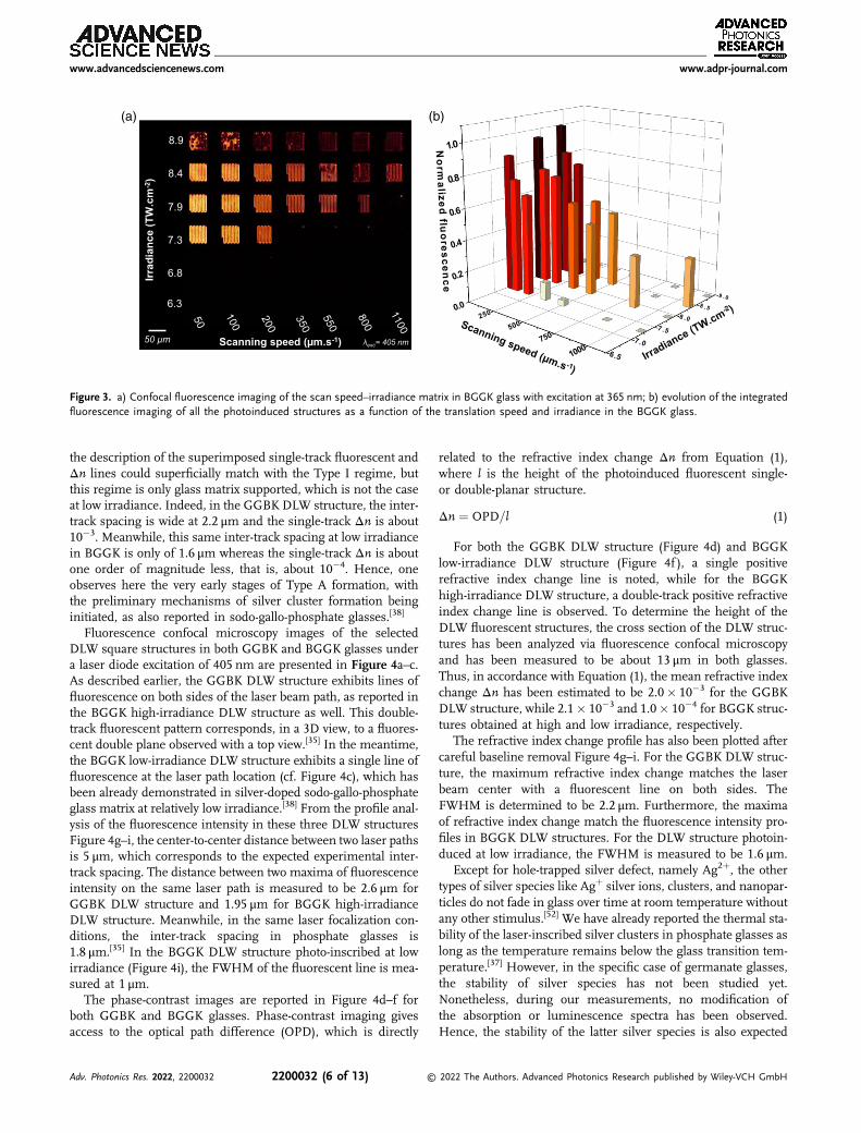
the description of the superimposed single-track fluorescent andΔn lines could superficially match with the Type I regime, butthis regime is only glass matrix supported, which is not the caseat low irradiance. Indeed, in the GGBK DLW structure, the inter-track spacing is wide at 2.2 μm and the single-track Δn is about10�3. Meanwhile, this same inter-track spacing at low irradiancein BGGK is only of 1.6 μm whereas the single-track Δn is aboutone order of magnitude less, that is, about 10�4. Hence, oneobserves here the very early stages of Type A formation, withthe preliminary mechanisms of silver cluster formation beinginitiated, as also reported in sodo-gallo-phosphate glasses.[38]
Fluorescence confocal microscopy images of the selectedDLW square structures in both GGBK and BGGK glasses undera laser diode excitation of 405 nm are presented in Figure 4a–c.As described earlier, the GGBK DLW structure exhibits lines offluorescence on both sides of the laser beam path, as reported inthe BGGK high-irradiance DLW structure as well. This double-track fluorescent pattern corresponds, in a 3D view, to a fluores-cent double plane observed with a top view.[35] In the meantime,the BGGK low-irradiance DLW structure exhibits a single line offluorescence at the laser path location (cf. Figure 4c), which hasbeen already demonstrated in silver-doped sodo-gallo-phosphateglass matrix at relatively low irradiance.[38] From the profile anal-ysis of the fluorescence intensity in these three DLW structuresFigure 4g–i, the center-to-center distance between two laser pathsis 5 μm, which corresponds to the expected experimental inter-track spacing. The distance between two maxima of fluorescenceintensity on the same laser path is measured to be 2.6 μm forGGBK DLW structure and 1.95 μm for BGGK high-irradianceDLW structure. Meanwhile, in the same laser focalization con-ditions, the inter-track spacing in phosphate glasses is1.8 μm.[35] In the BGGK DLW structure photo-inscribed at lowirradiance (Figure 4i), the FWHM of the fluorescent line is mea-sured at 1 μm.
The phase-contrast images are reported in Figure 4d–f forboth GGBK and BGGK glasses. Phase-contrast imaging givesaccess to the optical path difference (OPD), which is directly
related to the refractive index change Δn from Equation (1),where l is the height of the photoinduced fluorescent single-or double-planar structure.
Δn ¼ OPD=l (1)
For both the GGBK DLW structure (Figure 4d) and BGGKlow-irradiance DLW structure (Figure 4f ), a single positiverefractive index change line is noted, while for the BGGKhigh-irradiance DLW structure, a double-track positive refractiveindex change line is observed. To determine the height of theDLW fluorescent structures, the cross section of the DLW struc-tures has been analyzed via fluorescence confocal microscopyand has been measured to be about 13 μm in both glasses.Thus, in accordance with Equation (1), the mean refractive indexchange Δn has been estimated to be 2.0� 10�3 for the GGBKDLW structure, while 2.1� 10�3 and 1.0� 10�4 for BGGK struc-tures obtained at high and low irradiance, respectively.
The refractive index change profile has also been plotted aftercareful baseline removal Figure 4g–i. For the GGBK DLW struc-ture, the maximum refractive index change matches the laserbeam center with a fluorescent line on both sides. TheFWHM is determined to be 2.2 μm. Furthermore, the maximaof refractive index change match the fluorescence intensity pro-files in BGGK DLW structures. For the DLW structure photoin-duced at low irradiance, the FWHM is measured to be 1.6 μm.
Except for hole-trapped silver defect, namely Ag2þ, the othertypes of silver species like Agþ silver ions, clusters, and nanopar-ticles do not fade in glass over time at room temperature withoutany other stimulus.[52] We have already reported the thermal sta-bility of the laser-inscribed silver clusters in phosphate glasses aslong as the temperature remains below the glass transition tem-perature.[37] However, in the specific case of germanate glasses,the stability of silver species has not been studied yet.Nonetheless, during our measurements, no modification ofthe absorption or luminescence spectra has been observed.Hence, the stability of the latter silver species is also expected
(a) (b)
Figure 3. a) Confocal fluorescence imaging of the scan speed–irradiance matrix in BGGK glass with excitation at 365 nm; b) evolution of the integratedfluorescence imaging of all the photoinduced structures as a function of the translation speed and irradiance in the BGGK glass.
www.advancedsciencenews.com www.adpr-journal.com
Adv. Photonics Res. 2022, 2200032 2200032 (6 of 13) © 2022 The Authors. Advanced Photonics Research published by Wiley-VCH GmbH

to be long lasting or even permanent in BGGK and GGBK glassesas some fluorescent patterns inscribed months ago are stillobservable. To identify the long-term stable photoinduced silverspecies, microabsorption and microfluorescence spectroscopicmeasurements were conducted. As depicted in Figure 5a, theDLW structures in both GGBK and BGGK glasses present novelabsorption contributions, which strongly overlap between 300and 550 nm. For the barium-poor glass composition, a predomi-nant contribution peaking at 435 nm is observed with a narrowprofile and a shoulder centered at about 315 nm. Conjointly, theabsorption spectra are different depending on the irradianceintensity in the barium-rich glass composition. For the DLWstructure inscribed at high irradiance in BGGK, no contributionclearly arises, highlighting a wide hump with a maximum near330 nm, whereas for the DLW structures inscribed at low irradi-ance, a clearly defined peak at 330 nm is observed with a long tailup to 475 nm. Despite the microabsorption spectrum differen-ces, DLW structures emit in the visible range with a large emis-sion spectrum peaking at 580 nm under a laser diode excitationof 405 nm (Figure 5b). For information, the maximal fluores-cence intensity in BGGK is about 100 times more intense forthe high-irradiance DLW structure than for the low-irradiance
DLW structure (as also visible with the very distinct signal-to-noise ratios for BGGK in Figure 5b).
3.3. Fabrication of Embedded Periodic Structure
The inter-track spacing in BGGK is about 1.95 μm at high irra-diance of DLW. By decreasing the spacing between to laser paths(interline spacing) below the inter-track spacing, the silver clustersphotodissociation and photodiffusion processes jointly contributeto decrease the actual spacing between the two remnant modifica-tion tracks. Hence, DLW of structures with a smaller spacing thanthe actual interline spacing of 5 μmhave been performed in BGGKglass with an irradiance of 7.3 TW cm�2 and a translation speed of100 μms�1. Three structures have been inscribed with a pitch of2 μm, 1 μm, and 400 nm. Their analyses by both fluorescence andphase-contrast imaging are highlighted in Figure 6.
To characterize the periodicity of the structures, the recordedphase-contrast and high-resolution fluorescence images havebeen analyzed. Note however that with a 300 nm resolutionper pixel for the phase-contrast imaging, only the 2 μmpitch peri-odic structure can be resolved whereas the fluorescence imaging
0 10 20 30 40 50
0 10 20 30 40 50
0 10 20 30 40 50
Fluorescence
n
5 µm
GG
BK
7.8 TW.cm- – 50 µm.s-
Fluorescence
Fluorescence
Distance (µm)
Nor
mal
ized
inte
nsity
Nor
mal
ized
inte
nsity
Nor
mal
ized
inte
nsity
8.4 TW.cm- – 50 µm.s-
6.8 TW.cm- – 50 µm.s-
BG
GK
(H
igh
DLW
)B
GG
K (
Low
DLW
)
n
n
(a) (d) (g)
(b) (e) (h)
(c) (f) (i)
Figure 4. High-resolution imaging of both confocal fluorescence and phase contrast microscopies for photo-inscribed structures at a,d) 7.8 TW cm�2,50 μms�1 in GGBK, and at b,e) 8.4 TW cm�2, 50 μm s�1, and c,f ) 6.8 TW cm�2, 50 μms�1 in BGGK; superposition of both fluorescence intensity andrefractive index variation profiles for g) 7.8 TW cm�2, 50 μms�1 direct laser writing (DLW) structure in GGBK, and h) 8.4 TW cm�2, 50 μms�1 DLWstructure and f ) 6.8 TW cm�2, 50 μms�1 DLW structure in BGGK, extracted from images (a–f ) at the location of the gray dashed lines.
www.advancedsciencenews.com www.adpr-journal.com
Adv. Photonics Res. 2022, 2200032 2200032 (7 of 13) © 2022 The Authors. Advanced Photonics Research published by Wiley-VCH GmbH

enables a theoretical lateral resolution up to 120 nm thanks to the1.4 NA micro-objective (see Experimental Section). In Figure 7are presented the periodicity analyses for both 2 μm and 400 nmpitch periodic structures. To determine the experimental period-icity, a periodical test function has been simulated. For the 2 μmpitch structure, the refractive index change simulation gives amean periodicity of 2.0 μm, while for the 400 nm pitch structure,the fluorescence intensity variation simulation highlights a peri-odicity of 397 nm.
4. Discussion
4.1. Pristine Glass Properties
When the (2Ba2þ þ Kþ)/Ga3þ ratio equals or is superior to 1, thegallo-germanate glass structure is composed of germanium andgallium tetrahedra, that is, [GeO4] as well as [GaO4]
�, thanks to
enough positive-charge compensators (Kþ and/or Ba2þ) to thenegatively charged gallium tetrahedra. Since the germanium tet-rahedron is chemically neutral, it is not considered in the previ-ous ratio. From the Raman spectroscopy study (Figure 1a),substantial structural differences have been highlighted betweenboth GGBK and BGGK glass compositions. In GGBK glass, theRaman spectrum has evidenced a predominant Raman responsefrom [GaO4]
� and [GeO4] tetrahedra, while the Raman signaloriginating from [GeØ3O
�] entities is weak. In comparison withthis later glass, the BGGK Raman spectrum shows a clear inver-sion of the Raman response intensity coming from both galliumor germanium tetrahedra without NBOs and the germanium tet-rahedra with one single NBO. Considering that from GGBK toBGGK glasses, the (2Ba2þþ Kþ)/Ga3þ ratio rises from 1.3 to 5.1,clear network depolymerization of the gallo-germanate glassstructure is occurring in BGGK glass, being induced by the for-mation of [GeØ3O
�] tetrahedra with barium and potassium ascharge compensators. Such kind of glass structure interpretationis highly supported by the previous work of McKeown andMerzbacher, where the BGGK counterpart of their study withoutpotassium and silver has presented the relatively same Ramanspectrum and interpretation.[47] Moreover, considering our pre-vious work on silver-doped GGBK glasses with the substitutionof four different barium contents (i.e., 0, 5, 10, and 15mol%)[45]
and as long as the (2Ba2þþ Kþ)/Ga3þ ratio is superior to 1, onecan clearly attribute to the barium ion the role of network depo-lymerizing agent. Thus, regarding the drastic change of the cat-ionic composition of both glasses, a gallo-germanate polymerizedglass network and a bario-germanate depolymerized glass net-work are considered for the GGBK and BGGK glasses, respec-tively. Such kind of glass network difference could likelyexplain the observed modification of the IR absorption cutoff.
From GGBK to BGGK glasses, the glass transition tempera-ture decreases by 18 °C along with an increase of the molar vol-ume from 23.2 to 24.4 cm3mol�1. These physical–chemicalproperty changes highlight a less reticulated and constrainedglass network, which is consistent with the strong glass structuredepolymerization reported in BGGK glass composition. While inGGBK glass, the silver ion introduction seems limited below
300 350 400 450 500 550
Wavelength (nm)
GGBK BGGK at high irradiance BGGK at low irradiance
450 500 550 600 650 700 750Wavelength (nm)
GGBK BGGK at high irradiance BGGK at low irradiance
exc = 405 nm
Nor
mal
ized
fluo
resc
ence
Nor
mal
ized
abs
orpt
ion
(a) (b)
Figure 5. Normalized a) microabsorption and b) fluorescence spectra of DLW structures inscribed at 7.8 TW cm�2, 50 μm s�1 for GGBK glass, and at8.4 TW cm�2, 50 μm s�1 and 6.8 TW cm�2, 50 μm s�1 for BGGK glass.
Figure 6. Structures photo-inscribed at 7.3 TW cm�2 and 100 μms�1 witha pitch of 2 μm, 1 μm, and 400 nm in BGGK glass. High-resolution confo-cal fluorescence imaging on the top under laser excitation wavelength of405 nm and phase-contrast microscopy at the bottom. Each square struc-ture has a length of 50 μm.
www.advancedsciencenews.com www.adpr-journal.com
Adv. Photonics Res. 2022, 2200032 2200032 (8 of 13) © 2022 The Authors. Advanced Photonics Research published by Wiley-VCH GmbH

1mol% using the classical melt-quenching glass synthesis, andin BGGK glass, the silver ion introduction can be greater than1mol%. Hence, it appears that the strong change in the glassnetwork due to the increase of barium ions contributes tothe solubilization and stabilization of silver ions in the glassmatrix. One possible explanation would be that instead of com-pensating the negatively charged gallium tetrahedron, silver ionsmay create stable ionic bonds with NBOs mostly coming from the[GeØ3O
�] entities which are in a great amount in BGGK glass.The silver luminescence measurement reveals few spectral
distributions and decay times change, evidencing highly similarsilver entities in both GGBK and BGGK glasses. The presence ofnanosecond decay time with a broad emission in the visiblerange is very characteristic of molecular silver entities like silverclusters as demonstrated in liquid solution[53–55] and silver-dopedphosphate glasses,[56] while microsecond decay times are typicalof isolated Agþ and/or paired Agþ–Agþ silver species.[45,57,58]
However, comparing BGGK glass to its counterpart, the strongincrease of the 330 nm excitation and 480 nm emission bands,as well as the vanishing of the 270 nm excitation contribution,strongly demonstrated that the silver entities, that is, isolatedand aggregated, are not in the same proportion in both glasses.The major contribution in terms of amplitude of the microseconddecays against the nanosecond ones highlights a greater amountof isolated Agþ or paired Agþ–Agþ rather than silver clusters.Hence, it is believed that silver ions are stabilized preferentiallyin another structural site for BGGK glass. Indeed, rather thanbeing involved in the [GaO4]
� delocalized charge compensatorsite, silver ions are more likely engaged in the stable ionic bond-ing involving the NBO of the [GeØ3O
�] germanium tetrahedra.
4.2. Direct Infrared Femtosecond Laser Writing andCharacterizations
In silver-containing phosphate glasses supporting Type-A IRfemtosecond DLW, the laser-induced novel fluorescence and
absorptions are due to the generation of silver clusters.[12,38,56,59]
With a similar broad emission band and absorption band local-ized in the same wavelength range as in phosphate glasses, it islikely that the photoinduced absorptions and fluorescence inboth GGBK and BGGK glasses are also attributed to silver clus-ters. However, as previously described by Marquestaut et al.,[37]
the strong overlap of the novel absorption contributions makesdifficult the band attributions and thus their assignment to pre-cise silver entities. Hence, considering a strong similarity ofphoto-generated species between phosphate and gallo-germanatematrices, the 435 nm band clear contribution with a thin profilein GGBK is the only absorption band that can be assigned. It isattributed to a surface plasmon resonance (SPR) band, caused byreduced silver species, such as metallic nanoparticles of silver Agn,as reported elsewhere in phosphate glasses,[37,60] and in a germa-nate glass as well.[61] The SPR bandwidth must be compared withthe volume concentration of nanoparticles and the filling factorwith respect to the mean radius. From a Maxwell–Garnett simu-lation, as performed by Vangheluwe et al., and considering amonodisperse size distribution,[61] the Agn mean radius is esti-mated at about 1.13 nm with a filling factor of 0.385� 10�3
(Figure S1, Supporting Information). However, the presence ofa high concentration of metallic silver nanoparticles, known tobe nonluminescent, is surprising since a strong silver cluster fluo-rescence is denoted under a 405 nm excitation. Indeed,Marquestaut et al. had reported the cancelling of silver cluster fluo-rescence emission while these silvers clusters were grown intoplasmonic metallic silver nanoparticles in a phosphate glass,thanks to a post-DLW thermal treatment.[37] Hence, in the presentcase, it is proposed that the DLW species may be inhomogene-ously localized in the GGBK sample. Considering the profile ofluminescence intensity in this latter composition, the nonlumi-nescent species would be in the center or the very close peripheryof the laser beam path, while the concentration of silver clusterswould be mostly at a certain distance away from the center of thelaser path as directly observed by fluorescence imaging.
0 10 20 30 400.0
0.5
1.0
1.5
2.0
2.5
3.0
3.5
ExperimentalSimulation
Distance (µm)
(x10-4) 2 µm-pitch structure 400 nm-pitch structureΔn
(a) (b)
Figure 7. Periodicity analyses of the a) 2 μm pitch structure recorded by phase-contrast imaging and b) 400 nm pitch structure recorded with the high-resolution fluorescence imaging. Both structures have been photoinscribed at 7.3 TW cm�2 and 100 μm s�1 in the BGGK glass.
www.advancedsciencenews.com www.adpr-journal.com
Adv. Photonics Res. 2022, 2200032 2200032 (9 of 13) © 2022 The Authors. Advanced Photonics Research published by Wiley-VCH GmbH
One last interesting point concerns the difference of microab-sorption spectral distribution between both low- and high-irradiance DLW structures, while their microfluorescence spectraare strongly similar. Indeed, considering the excitation wavelengthfor the microfluorescence is at 405 nm (where the absorption isstronger in the high-irradiance DLW structures) and the silverclusters fluorescence is stronger when the DLW is at higher irra-diance, it is suggested that at lowDLW irradiance, the formation ofnonluminescent silver entities, such as Ag0-reduced species, or/and colored silver centers, that is, Ag2þ may be favored. Thishypothesis is highly supported by the appearance of the strongabsorption band at 330 nm, being concomitant with the presenceof both electron and hole silver defects, that is, Ag0 and Ag2þ, aspreviously observed in sodo-gallo-phosphate glasses.[39]
4.2.1. Hybrid Type A/I Modification
As reported by Abou Khalil et al. in silver-containing phosphateglasses subjected to IR femtosecond DLW,[62] two types of DLWregimes can be observed, depending on the laser irradiation con-ditions. First, the Type A regime is associated with a refractiveindex change underlain by silver photochemistry, with a doubleline of fluorescence and refractive index. Second, the Type Iregime is related to a smooth local modification of the glass den-sity often driven by thermal effects, along with a single line ofboth fluorescence.
In the GGBK glass, the observed modifications present a sin-gle refractive index line with a double fluorescence line, posi-tioned on either side of the inscription line. Recently, Bérubéet al. have reported the inscription of a single refractive indexline in a nearly similar gallo-germanate glass matrix.[29] In theirwork, in absence of silver, a Type I inscription regime has beenevidenced by a local densification of the glass. In our study,Raman spectroscopy at 532 nm has been conducted on GGBKglass (Figure S2, Supporting Information) but modification ofthe glass density could not be detected. For silica glasses, theRaman spectrum changes (i.e., band positions and intensities)are progressive and appear strongly after a certain irradi-ance.[63,64] Hence, it is supposed that in the BGGK glass, thedeposited energy was not high enough to initiate strong glassstructure modification, as monitored by Raman spectroscopy.
In light of Bérubé et al. study and our results, we propose thefollowing explanations for the new regime observed in GGBKglass: 1) During laser irradiation, the energy transfer and electronthermalization phenomena followed by the silver photodissocia-tion and diffusion processes are considered as the initiators ofthe laser modification. This leads to the first steps of the silverclusters creation. 2) The generation of new silver entities, thatis, hole–electron trap silver centers and silver clusters, contributessignificantly to the local temperature elevation (below the glasstransition temperature) through the generation of new absorptionbands in the visible spectral region leading to a more efficientabsorption of the laser. Coupled with the thermally activated cat-ionic migrations, it allows for material densification, especially inthe laser path center, where temperature elevation is the highest,as for classic Type I modification.[65] 3) Such a temperature eleva-tion strongly favors nucleation/growth processes of silver nanopar-ticles in the center of the laser beam. 4) This temperature
elevation, however, decreases the amount of elementary compo-nent Ag0 essential in the early stages of silver cluster formation.Once the Ag0 species no longer exist, either by thermal destructionof these electron trap centers Ag0 ! Agþ þ e� or through therapid formation of silver nanoparticles, the creation of silver clus-ters Agm
xþ with high nuclearity is stopped. Such a kind of Ag0
inhibition has been already highlighted by Petit et al. during anIR femtosecond DLW coupled with a UV co-illumination.[66]
4.2.2. Type A Transposition in Mid-Infrared Glass
Unlike GGBK, the DLW of Type A in BGGK glass leads to theformation of a double track of both the refractive index changeand the fluorescence where both properties are as expected spa-tially superimposed since they both originate from the formationof silver clusters. Until now, this remnantmodification of the glasswas only reported in silver-doped phosphate glasses. Developed inseveral types of phosphate glass compositions, such as zinc phos-phate,[12] sodium magnesium phosphate, sodium gallium phos-phate,[38] and the DLW in silver-containing phosphate matricespromote the Type A modification by means of the glass redoxpotential favorable to stabilize in a great quantity the silver ions.During the DLW process, the silver ions act as a fuse, like othertransition metals,[67] capable of restraining the laser modificationon itself through photochemical reactions leading to the formationof stable photoinduced silver clusters.[32] Consequently, when theconcentration of fuse-acting chemical elements is not highenough, the laser pulse energy is only partially restrained onthe doping element, and severe glass matrix modifications couldbe generated. In BGGK, the silver ion solubility is better than inGGBK by a factor of two, which could explain the difference ofbehavior in both glasses during the DLW. Moreover, the discrep-ancies in terms of silver species types and proportions stabilized inboth pristine glasses could also have significant impact on theirphotosensitivity (Figure 2). Finally, compared to the silver concen-tration in phosphate glasses,[38] the silver quantity in BGGK stillremains lower and could justify the restricted range of laserparameters observed in Figure 3. Indeed, while in BGGK,this range is extending from 7.3 up to 8.4 TW cm�2 and 50 upto 350 μms�1, it reaches 16.3 TW cm�2 and 500 μms�1 for asilver-doped gallo-phosphate of sodium.[38]
4.2.3. Embedment of Subwavelength Periodic Structures inMid-Infrared Glass
The formation of the silver cluster-based double track of the TypeA modification is originated from both the photodiffusion andphotodissociation of the silver entities during the laser pulsesaccumulation.[33] As reported by Desmoulin et al.,[34] the compe-tition of these two phenomena results in a partial depletion of thesilver reservoir up to 20% at the remnant double-track center.During the formation of glass-embedded DLW structures witha spacing between to laser paths smaller than the inter-trackone (Figure 6 and 7), the laser beam successively irradiates a par-tially silver-depleted glass region because of the previous laserpass. While no fading of both fluorescence intensity and refrac-tive index change is noted over the entire DLW structure, it isbelieved that the entire silver reservoir is remobilized and still
www.advancedsciencenews.com www.adpr-journal.com
Adv. Photonics Res. 2022, 2200032 2200032 (10 of 13) © 2022 The Authors. Advanced Photonics Research published by Wiley-VCH GmbH
contributes to the formation of perennial silver clusters after suc-cessive laser passes. To the best of our knowledge, this is the firsttime that remobilization of the silver reservoir is used to developperiodic structures. As discussed by de Castro et al.,[68] the capa-bility to remobilize the silver reservoir was believed to originatefrom the presence of highly co-mobile ions such as fluorine andalkaline ions in silver-doped oxyfluoride phosphate glasses. Inour work, we have shown that the presence of barium ions, inmajority in contrast to the potassium, promotes the remobiliza-tion of the silver reservoir. While the barium ionmobility in BGGglass is lower than that of potassium,[45] it seems that either thehigh-quantity of low-mobility barium ions and/or the low-quantity of high-mobility potassium ions contributes beneficiallyto manage the silver reservoir in BGG glasses. Hence, theseresults extend the understanding of the silver reservoir remobi-lization in silver-doped glasses, which is promising for the futuredevelopment of novel optical diffractive elements.
5. Conclusion
We successfully synthesized silver-doped gallo-germanate oxideglasses of barium and potassium via the classical melt-quenchingmethod. Two BGG glasses were fabricated with an extendedinfrared optical transmission up to 5.7 μm, either with a lowor high content of barium cations. By increasing the ratio of bar-ium ions, the silver solubility inside the glass has been improvedby a factor larger than 2. In the meantime, the barium concen-tration increase affects the glass network by depolymerizing theglass network and thus promoting new structural sites forthe silver species. After the DLW process, at low Ba2þ content, theglass modification consisted of a hybrid combination of the Type Iand Type A modifications. A bright luminescence from the silverclusters is reported between 450 and 750 nm, as well as followedby a refractive index change up to 2.0� 10�3. At a high concen-tration of Ba2þ, the DLW-induced refractive index modificationsare entirely supported by the silver species, namely correspondingto the Argentum Type. At low laser fluence, a single track of fluo-rescence and refractive index change (up to 1� 10�4) is reported,corresponding to the very preliminary stages of Type A formation.At high laser fluence, the more usual double track of fluorescenceand refractive index change (up to 2.1� 10�3) is demonstrated.For the first time, by taking advantage of the double-trackDLW modification, embedded periodic structures with a pitchdown to 400 nm have been successfully fabricated by narrowingartificially the Type A inter-track spacing. This work provides newinsights through the fabrication of freeform photonic compo-nents buried inside mid-IR glasses.
6. Experimental Section
Glass Characterizations: Chemical analyses were conducted thanks toboth inductively coupled plasma optical emission spectroscopy (ICP-OES) with an Agilent 5110 VDV apparatus, and electron-probed microanal-ysis (EPMA) using an electron microprobe (Wavelength-DispersiveSpectroscopy [WDS]) CAMECA SX 100. The density, ρ, was determinedby Archimedes method using diethyl phthalate as an immersion liquidat room temperature. The measurement precision is estimated to be�0.01 g cm�3. DSC measurements were made on a single 70mg glasschunk in platinum crucible using a Netzsch DSC Pegasus 404 F3 from
500 and 1100 °C. Thanks to DSC measurements, the glass transition tem-perature defined as the intersection of the base line with the inflexion tan-gent of the glass transition domain was extracted.
Raman spectra were recorded at room temperature from 200 to1000 cm�1 with a resolution of 2.5 cm�1 using a LABRAM 800-HRRaman spectrometer (Horiba Jobin Yvon) and a microscope objective50�, NA 0.75. The excitation source is a single longitudinal mode laserat 532 nm. The UV–visible–near-IR transmission spectra from 200 to2500 nm were recorded on a Cary 5000 (Varian) spectrometer by stepsof 1 nm, while the near-IR-mid-IR transmission spectra were obtainedfrom 2.5 to 7 μm using a Fourier-transform infrared spectrometer withan accumulation of 200 scans and a resolution of 4 cm�1.
The steady-state excitation and emission spectra were recorded with aSPEX Fluorolog-2 spectrofluorometer (Horiba Jobin Yvon) on glass pow-der. Each spectrum was conducted with a step and resolution of 1 nm, andat room temperature. The excitation source was a 450W xenon lamp hav-ing a continuous excitation from 200 to 800 nm. To detect and amplify theluminescence signal, a Hamamatsu R298 photomultiplier was employed.The decay time results were extracted from time-resolved spectroscopy. AContinuum Surelite SL II-10 laser was used as a pulsed 355 nm excitationsource (10 Hz, 70mJ, 4–6 ns) followed by a half-wave plate and a polarizedbeam splitter to control the laser fluence, ensuring that the measurementbehaves only as an excitation regime in a lifetime-probing regime (and notin an irradiance regime that would lead to glass modifications). Emissionspectra were recorded using a monochromator and a gated intensifiedcharge-coupled device (ICCD) camera (Andor) being optically triggeredby the pulsed UV laser.
Direct Infrared Femtosecond Laser Writing: Direct laser writings were per-formed on 1mm thick glass samples using a ytterbium dopedpotassium–gadolinium tungstate crystal (KGW:Yb) femtosecond oscillator(up to 2.6 W, 10MHz, 390 fs FWHM at 1030 nm) combined with anacousto-optic modulator to control the number and the energy of pulses,thus enabling several irradiances. Spherical aberrations were mostly cor-rected using a spatial light modulator (LCOS; X10468-03, HamamatsuPhotonics). A high-precision 3D translation stage XMS-50 (better than50 nm resolution) was used to perform the sample positioning and dis-placements with various velocities from 50 to 1100 μm s�1. An Olympusmicroscope objective (20�, NA 0.75) was used to produce the photoin-duced structures, 160 μm below the sample surface.
Analysis of the Photoinduced Direct Laser Writing Structures:Microluminescence was conducted with a LABRAM 800-HR spectropho-tometer (Horiba Jobin-Yvon) and an Olympus microscope objective(50�, NA 0.75) using an excitation laser diode at 405 nm (100mW,TEM00, OBIS, COHERENT). Microluminescence spectra were recordedthanks to a thermoelectric cooled charge-coupled device (CCD) Camera(Synapse Model 354 308). The experimental spectra were corrected fromthe detection arm spectral response by a correction function determinedusing reference samples with broad spectral emission, while the pristine glassluminescence was subtracted from the recorded spectra. Microabsorptionspectra were recorded using a “CRAIC Technologies”microspectrophotome-ter equipped with a Xenon lamp, a condenser, and a 10� microscope objec-tive. Using this appliance, the obtained results consist directly of differentialabsorption spectra with a subtraction of the pristine glass absorption.
Confocal fluorescence imaging was performed using a Leica DM6 CFSTCS SP8 confocal microscope equipped with a 405 nm laser diode.Microscope objectives 10�, NA 0.3 DRY and 63�, NA 1.4 OIL were used.Confocal fluorescence imaging was also performed using a Leica SP2 con-focal microscope using a 405 nm laser diode to determine DLW structureheight of the glasses, using a 100�, NA 1.4 OIL microscope objective. Therefractive index modifications of the DLW structures were recorded using aphase-contrast microscopy equipped with a commercial SID4Bio Phasicscamera and a microscope objective (100�, NA 1.3 OIL).
Supporting InformationSupporting Information is available from the Wiley Online Library or fromthe author.
www.advancedsciencenews.com www.adpr-journal.com
Adv. Photonics Res. 2022, 2200032 2200032 (11 of 13) © 2022 The Authors. Advanced Photonics Research published by Wiley-VCH GmbH
AcknowledgementsThis work has received funding from the French Government, managed bythe French National Research Agency (ANR-17-CE08-0042-01 and ANR-19-CE08-0021) and the Nouvelle Aquitaine Region (APPR2020-2019-8193110), and from the Canadian Government, managed by SentinelNorth program of University Laval and the Canadian Research Chair pro-gram (CERC). This project has also received funding from the innovationprogram under the Marie-Skłodowska-Curie grant agreement N°823941(FUNGLASS). The corresponding author, T.G., holds a Sentinel NorthExcellence Postdoctoral Fellowship and held an Excellence doctoral schol-arship from the French Ministry of Higher Education and Research duringthe preparation of this work.
Conflict of InterestThe authors declare no conflict of interest.
Data Availability StatementThe data that support the findings of this study are available from thecorresponding author upon reasonable request.
Keywordsdirect laser writing, glass, grating, mid-infrared, periodic structure, silvercluster
Received: February 10, 2022Revised: April 19, 2022
Published online:
[1] K. M. Davis, K. Miura, N. Sugimoto, K. Hirao, Opt. Lett. 1996, 21,1729.
[2] R. R. Gattass, E. Mazur, Nat. Photonics 2008, 2, 219.[3] Y. Shimotsuma, P. G. Kazansky, J. Qiu, K. Hirao, Phys. Rev. Lett. 2003,
91, 247405.[4] M. Campbell, D. N. Sharp, M. T. Harrison, R. G. Denning,
A. J. Turberfield, Nature 2000, 404, 53.[5] S. Gross, M. J. Withford, Nanophotonics 2015, 4, 332.[6] A. Crespi, R. Osellame, F. Bragheri, Opt. Mater. X 2019, 4, 100042.[7] M. Haque, K. K. C. Lee, S. Ho, L. A. Fernandes, P. R. Herman, Lab
Chip 2014, 14, 3817.[8] J. Lapointe, M. Gagné, M.-J. Li, R. Kashyap, Opt. Express 2014, 22,
15473.[9] M. Ams, G. D. Marshall, P. Dekker, J. A. Piper, M. J. Withford, Laser
Photonics Rev. 2009, 3, 535.[10] A. Arriola, S. Gross, M. Ams, T. Gretzinger, D. Le Coq, R. P. Wang,
H. Ebendorff-Heidepriem, J. Sanghera, S. Bayya, L. B. Shaw,M. Ireland, P. Tuthill, M. J. Withford,Opt. Mater. Express 2017, 7, 698.
[11] T. Meany, M. Gräfe, R. Heilmann, A. Perez-Leija, S. Gross, M. J. Steel,M. J. Withford, A. Szameit, Laser Photonics Rev. 2015, 9, 363.
[12] A. Royon, K. Bourhis, M. Bellec, G. Papon, B. Bousquet, Y. Deshayes,T. Cardinal, L. Canioni, Adv. Mater. 2010, 22, 5282.
[13] J. Zhang, M. Gecevicius, M. Beresna, P. G. Kazansky, Phys. Rev. Lett.2014, 112, 033901.
[14] J. Lapointe, J.-P. Bérubé, Y. Ledemi, A. Dupont, V. Fortin,Y. Messaddeq, R. Vallée, Light Sci. Appl. 2020, 9, 64.
[15] K. Bourhis, A. Royon, G. Papon, M. Bellec, Y. Petit, L. Canioni,M. Dussauze, V. Rodriguez, L. Binet, D. Caurant, M. Treguer,J.-J. Videau, T. Cardinal, Mater. Res. Bull. 2013, 48, 1637.
[16] G. Papon, Y. Petit, N. Marquestaut, A. Royon, M. Dussauze,V. Rodriguez, T. Cardinal, L. Canioni, Opt. Mater. Express 2013, 3,1855.
[17] S. Kanehira, K. Miura, K. Hirao, Appl. Phys. Lett. 2008, 93,023112.
[18] T. T. Fernandez, B. Sotillo, J. Del Hoyo, J.-A. Valles, R. MartinezVazquez, P. Fernandez, J. Solis, IEEE Photonics Technol. Lett. 2015,27, 1068.
[19] D. M. Krol, J. Non-Cryst. Solids 2008, 354, 416.[20] M. Lancry, B. Poumellec, J. Canning, K. Cook, J.-C. Poulin, F. Brisset,
Laser Photonics Rev. 2013, 7, 953.[21] F. Liang, R. Vallée, S. L. Chin, Opt. Express 2012, 20, 4389.[22] R. Stoian, Appl. Phys. A 2020, 126, 438.[23] A. Stone, M. Sakakura, Y. Shimotsuma, K. Miura, K. Hirao, V. Dierolf,
H. Jain, Mater. Des. 2018, 146, 228.[24] J. Cao, B. Poumellec, L. Mazerolles, F. Brisset, A.-L. Helbert, S. Surble,
X. He, M. Lancry, J. Am. Ceram. Soc. 2017, 100, 115.[25] X. Huang, Q. Guo, D. Yang, X. Xiao, X. Liu, Z. Xia, F. Fan, J. Qiu,
G. Dong, Nat. Photonics 2019, 14, 82.[26] T. T. Fernandez, S. Gross, K. Privat, B. Johnston, M. Withford, Adv.
Funct. Mater. 2021, 2103103.[27] E. N. Glezer, E. Mazur, Appl. Phys. Lett. 1997, 71, 882.[28] L. B. Fletcher, J. J. Witcher, N. Troy, S. T. Reis, R. K. Brow, D. M. Krol,
Opt. Express 2011, 19, 7929.[29] J.-P. Bérubé, A. Le Camus, S. H. Messaddeq, Y. Petit, Y. Messaddeq,
L. Canioni, R. Vallée, Opt. Mater. Express 2017, 7, 3124.[30] S. Wong, M. Deubel, F. Pérez-Willard, S. John, G. A. Ozin,
M. Wegener, G. von Freymann, Adv. Mater. 2006, 18, 265.[31] H. Yao, R. Zaiter, M. Cavillon, B. Sapaly, F. Calzavara, P. Delullier,
T. Cardinal, Y. Dai, B. Poumellec, M. Lancry, Ceram. Int. 2021, 47,34235.
[32] Y. Petit, S. Danto, T. Guérineau, A. Abou Khalil, A. Le Camus,E. Fargin, G. Duchateau, J.-P. Bérubé, R. Vallée, Y. Messaddeq,T. Cardinal, L. Canioni, Adv. Opt. Technol. 2018, 7, 291.
[33] M. Bellec, A. Royon, B. Bousquet, K. Bourhis, M. Treguer, T. Cardinal,M. Richardson, L. Canioni, Opt. Express 2009, 17, 10304.
[34] J.-C. Desmoulin, Y. Petit, L. Canioni, M. Dussauze, M. Lahaye,H. M. Gonzalez, E. Brasselet, T. Cardinal, J. Appl. Phys. 2015, 118,213104.
[35] A. Abou Khalil, J.-P. Bérubé, S. Danto, J.-C. Desmoulin, T. Cardinal,Y. Petit, R. Vallée, L. Canioni, Sci. Rep. 2017, 7, 11124.
[36] L. Canioni, M. Bellec, A. Royon, B. Bousquet, T. Cardinal, Opt. Lett.2008, 33, 360.
[37] N. Marquestaut, Y. Petit, A. Royon, P. Mounaix, T. Cardinal,L. Canioni, Adv. Funct. Mater. 2014, 24, 5824.
[38] T. Guérineau, L. Loi, Y. Petit, S. Danto, A. Fargues, L. Canioni,T. Cardinal, Opt. Mater. Express 2018, 8, 3748.
[39] T. Guérineau, F. Cova, Y. Petit, A. Abou Khalil, A. Fargues,M. Dussauze, S. Danto, A. Vedda, L. Canioni, T. Cardinal, Int. J.Appl. Glass Sci. 2020, 11, 15.
[40] J. Harb, T. Guérineau, A. Morana, A. Meyer, G. Raffy, A. D. Guerzo,Y. Ouerdane, A. Boukenter, S. Girard, T. Cardinal, Y. Petit, L. Canioni,Chemosensors 2022, 10, 110.
[41] A. Abou Khalil, P. Lalanne, J.-P. Bérubé, Y. Petit, R. Vallée, L. Canioni,Opt. Express 2019, 27, 31130.
[42] S. S. Bayya, G. D. Chin, J. S. Sanghera, I. D. Aggarwal, Opt. Express2006, 14, 11687.
[43] F. Calzavara, M. Allix, M. Dussauze, V. Jubera, M. Nalin, T. Cardinal,E. Fargin, J. Non-Cryst. Solids 2021, 571, 121064.
[44] A. Le Camus, Y. Petit, J.-P. Bérubé, M. Bellec, L. Canioni, R. Vallée,Opt. Express 2021, 29, 8531.
[45] T. Guérineau, A. Fargues, Y. Petit, E. Fargin, T. Cardinal, J. Non-Cryst.Solids 2021, 566, 120889.
www.advancedsciencenews.com www.adpr-journal.com
Adv. Photonics Res. 2022, 2200032 2200032 (12 of 13) © 2022 The Authors. Advanced Photonics Research published by Wiley-VCH GmbH
[46] T. Skopak, S. Kroeker, K. Levin, M. Dussauze, R. Méreau, Y. Ledemi,T. Cardinal, E. Fargin, Y. Messaddeq, J. Phys. Chem. C 2019, 123, 1370.
[47] D. A. McKeown, C. I. Merzbacher, J. Non-Cryst. Solids 1995, 183, 61.[48] T. Guérineau, C. Strutynski, T. Skopak, S. Morency, A. Hanafi,
F. Calzavara, Y. Ledemi, S. Danto, T. Cardinal, Y. Messaddeq,E. Fargin, Opt. Mater. Express 2019, 9, 2437.
[49] T. Skopak, P. Hee, Y. Ledemi, M. Dussauze, S. Kroeker, T. Cardinal,E. Fargin, Y. Messaddeq, J. Non-Cryst. Solids 2017, 455, 83.
[50] T. Skopak, F. Calzavara, Y. Ledemi, F. Célarié, M. Allix, E. Véron,M. Dussauze, T. Cardinal, E. Fargin, Y. Messaddeq, J. Non-Cryst.Solids 2019, 514, 98.
[51] K. Bourhis, Photostructuration par laser infrarouge femtoseconde deverres photosensibles de phosphates de zinc, d’argent et de gallium, 2011.
[52] A. Royon, K. Bourhis, L. Béchou, T. Cardinal, L. Canioni, Y. Deshayes,Microelectron. Reliab. 2013, 53, 1514.
[53] B. G. Ershov, N. L. Sukhov, A. V. Kiseleva, G. V. Ionova, Russ. Chem.Bull. 1996, 45, 545.
[54] B. G. Ershov, G. V. Ionova, A. A. Kiseleva, Russ. J. Phys. Chem. 1995,689, 239.
[55] M. Treguer, F. Rocco, G. Lelong, A. Le Nestour, T. Cardinal, A. Maali,B. Lounis, Solid State Sci. 2005, 7, 812.
[56] M. Bellec, A. Royon, K. Bourhis, J. Choi, B. Bousquet, M. Treguer,T. Cardinal, J.-J. Videau, M. Richardson, L. Canioni, J. Phys. Chem.C 2010, 114, 15584.
[57] I. Belharouak, C. Parent, B. Tanguy, G. Le Flem, M. Couzi, J. Non-Cryst. Solids 1999, 244, 238.
[58] I. Belharouak, C. Parent, P. Gravereau, J. P. Chaminade, G. Le Flem,B. Moine, J. Solid State Chem. 2000, 149, 284.
[59] K. Bourhis, A. Royon, M. Bellec, J. Choi, A. Fargues, M. Treguer,J.-J. Videau, D. Talaga, M. Richardson, T. Cardinal, L. Canioni, J.Non-Cryst. Solids 2010, 356, 2658.
[60] G. Y. Shakhgildyan, A. S. Lipatiev, M. P. Vetchinnikov, V. V. Popova,S. V. Lotarev, N. V. Golubev, E. S. Ignat’eva, M. M. Presniakov,V. N. Sigaev, J. Non-Cryst. Solids 2018, 481, 634.
[61] M. Vangheluwe, Y. Petit, N. Marquestaut, A. Corcoran, E. Fargin,R. Vallée, T. Cardinal, L. Canioni, Opt. Mater. Express 2016, 6, 743.
[62] A. Abou Khalil, J.-P. Bérubé, S. Danto, T. Cardinal, Y. Petit, L. Canioni,R. Vallée, Opt. Mater. Express 2019, 9, 2640.
[63] T. Seuthe, M. Grehn, A. Mermillod-Blondin, H. J. Eichler, J. Bonse,M. Eberstein, Opt. Mater. Express 2013, 3, 755.
[64] N. Varkentina, M. Dussauze, A. Royon, M. Ramme, Y. Petit,L. Canioni, Opt. Mater. Express 2016, 6, 79.
[65] T. T. Fernandez, M. Sakakura, S. M. Eaton, B. Sotillo, J. Siegel, J. Solis,Y. Shimotsuma, K. Miura, Prog. Mater. Sci. 2018, 94, 68.
[66] Y. Petit, K. Mishchik, N. Varkentina, N. Marquestaut, A. Royon,I. Manek-Hönninger, T. Cardinal, L. Canioni, Opt. Lett. 2015, 40,4134.
[67] B. Boizot, N. Ollier, F. Olivier, G. Petite, D. Ghaleb, E. Malchukova,Nucl. Instrum. Methods Phys. Res., Sect. B 2005, 240, 146.
[68] T. de Castro, H. Fares, A. A. Khalil, R. Laberdesque, Y. Petit,C. Strutinski, S. Danto, V. Jubera, S. J. L. Ribeiro, M. Nalin,T. Cardinal, L. Canioni, J. Non-Cryst. Solids 2019, 517, 51.
www.advancedsciencenews.com www.adpr-journal.com
Adv. Photonics Res. 2022, 2200032 2200032 (13 of 13) © 2022 The Authors. Advanced Photonics Research published by Wiley-VCH GmbH