Lanthanide(III) Complexes of Novel Mixed Carboxylic-Phosphorus Acid Derivatives of...
17
Lanthanide(iii) Complexes of Novel Mixed Carboxylic-Phosphorus Acid Derivatives of Diethylenetriamine: A Step towards More Efficient MRI Contrast Agents Jan Kotek, [a, b] Petra Lebdus œkovµ, [a, b] Petr Hermann, [b] Luce Vander Elst, [c] Robert N. Muller, [c] Carlos F. G. C. Geraldes, [d] Thomas Maschmeyer, [a] Ivan Lukes œ,* [b] and Joop A. Peters* [a] Introduction Metal chelates of the polyaminocarboxylates DTPA 5 (DTPA 5 = diethylenetriamine-N,N,N’,N’’,N’’-pentaacetate), DOTA 4 (DOTA 4 = 1,4,7,10-tetraazacyclododecane- 1,4,7,10-tetraacetate) and derivatives thereof have found widespread use in medical diagnosis (e.g. Magnetic Reso- nance Imaging, MRI; Positron Emission Tomography, PET; or Single-Photon Emission Computed Tomography, SPECT) and in radiotherapy. [1±3] These complexes have high thermo- dynamic and kinetic stabilities, essential features for in vivo applications, since the metal aqua ions as well as their li- gands are toxic, whereas the complexes are not. The applica- bility of radioactive complexes also requires that complexa- tion should be rapid enough to allow radiolabelling by a simple procedure just prior to the diagnostic procedure or the treatment. Complexes of DTPA 5 meet this require- ment, while the formation of complexes of DOTA 4 is usu- ally very slow. [a] Dr. J.A. Peters, J. Kotek, P. Lebdus œkovµ, Prof. Dr. T. Maschmeyer Laboratory for Applied Organic Chemistry and Catalysis Delft University of Technology Julianalaan 136, 2628BL Delft (The Netherlands) Fax: (+ 31) 152-784-289 E-mail: [email protected] [b] Prof. Dr. I. Lukes œ, J. Kotek, P. Lebdus œkovµ, Dr. P. Hermann Department of Inorganic Chemistry, Charles University Hlavova 2030, 12840 Prague (Czech Republic) E-mail : [email protected] [c] Prof. Dr L. Vander Elst, Prof. Dr. R. N. Muller NMR Laboratory, Department of Organic Chemistry University of Mons±Hainaut, 7000 Mons (Belgium) [d] Prof. Dr. C. F. G.C. Geraldes Departamento de BioquÌmica Faculdade de CiÜncias e Tecnologica, e Centro de NeurociÜncias Universidade de Coimbra, 3049 Coimbra (Portugal) Supporting information for this article is available on the WWW under http://www.chemeurj.org/ or from the author. Abstract: Three novel phosphorus-con- taining analogues of H 5 DTPA (DTPA = diethylenetriaminepentaacetate) were synthesised (H 6 L 1 ,H 5 L 2 ,H 5 L 3 ). These compounds have a -CH 2 - P(O)(OH)-R function (R = OH, Ph, CH 2 NBn 2 ) attached to the central ni- trogen atom of the diethylenetriamine backbone. An NMR study reveals that these ligands bind to lanthanide(iii ) ions in an octadentate fashion through the three nitrogen atoms, a P O oxygen atom and four carboxylate oxygen atoms. The complexed ligand occurs in several enantiomeric forms due to the chirality of the central nitro- gen atom and the phosphorus atom upon coordination. All lanthanide com- plexes studied have one coordinated water molecule. The residence times (t 298 M ) of the coordinated water mole- cules in the gadolinium(iii ) complexes of H 6 L 1 and H 5 L 2 are 88 and 92 ns, re- spectively, which are close to the opti- mum. This is particularly important upon covalent and noncovalent attach- ment of these Gd 3 + chelates to poly- mers. The relaxivity of the complexes studied is further enhanced by the presence of at least two water mole- cules in the second coordination sphere of the Gd 3 + ion, which are probably bound to the phosphonate/phosphinate moiety by hydrogen bonds. The com- plex [Gd(L 3 )(H 2 O)] 2 shows strong binding ability to HSA, and the adduct has a relaxivity comparable to MS-325 (40 s 1 mm 1 at 40 MHz, 37 8C) even though it has a less favourable t M value (685 ns). Transmetallation experiments with Zn 2 + indicate that the complexes have a kinetic stability that is compara- ble to–or better than–those of [Gd(dtpa)(H 2 O)] 2 and [Gd(dtpa- bma)(H 2 O)]. Keywords: chelates ¥ imaging agents ¥ lanthanides ¥ NMR spectroscopy ¥ phosphinate complexes ¥ phosphonate com- plexes Chem. Eur. J. 2003, 9, 5899 ± 5915 DOI: 10.1002/chem.200305155 ¹ 2003 Wiley-VCH Verlag GmbH&Co. KGaA, Weinheim 5899 FULL PAPER
-
Upload
independent -
Category
Documents
-
view
0 -
download
0
Transcript of Lanthanide(III) Complexes of Novel Mixed Carboxylic-Phosphorus Acid Derivatives of...

Lanthanide(iii) Complexes of Novel Mixed Carboxylic-Phosphorus AcidDerivatives of Diethylenetriamine: A Step towards More EfficientMRI Contrast Agents
Jan Kotek,[a, b] Petra Lebdusœkovµ,[a, b] Petr Hermann,[b] Luce Vander Elst,[c] RobertN. Muller,[c] Carlos F. G. C. Geraldes,[d] Thomas Maschmeyer,[a] Ivan Lukesœ,*[b] andJoop A. Peters*[a]
Introduction
Metal chelates of the polyaminocarboxylates DTPA5�
(DTPA5� = diethylenetriamine-N,N,N’,N’’,N’’-pentaacetate),DOTA4� (DOTA4� = 1,4,7,10-tetraazacyclododecane-1,4,7,10-tetraacetate) and derivatives thereof have foundwidespread use in medical diagnosis (e.g. Magnetic Reso-nance Imaging, MRI; Positron Emission Tomography, PET;or Single-Photon Emission Computed Tomography, SPECT)and in radiotherapy.[1±3] These complexes have high thermo-dynamic and kinetic stabilities, essential features for in vivoapplications, since the metal aqua ions as well as their li-gands are toxic, whereas the complexes are not. The applica-bility of radioactive complexes also requires that complexa-tion should be rapid enough to allow radiolabelling by asimple procedure just prior to the diagnostic procedure orthe treatment. Complexes of DTPA5� meet this require-ment, while the formation of complexes of DOTA4� is usu-ally very slow.
[a] Dr. J. A. Peters, J. Kotek, P. Lebdusœkovµ, Prof. Dr. T. MaschmeyerLaboratory for Applied Organic Chemistry and CatalysisDelft University of TechnologyJulianalaan 136, 2628BL Delft (The Netherlands)Fax: (+31)152-784-289E-mail : [email protected]
[b] Prof. Dr. I. Lukesœ, J. Kotek, P. Lebdusœkovµ, Dr. P. HermannDepartment of Inorganic Chemistry, Charles UniversityHlavova 2030, 12840 Prague (Czech Republic)E-mail : [email protected]
[c] Prof. Dr L. Vander Elst, Prof. Dr. R. N. MullerNMR Laboratory, Department of Organic ChemistryUniversity of Mons±Hainaut, 7000 Mons (Belgium)
[d] Prof. Dr. C. F. G. C. GeraldesDepartamento de BioquÌmicaFaculdade de CiÜncias e Tecnologica, e Centro de NeurociÜnciasUniversidade de Coimbra, 3049 Coimbra (Portugal)
Supporting information for this article is available on the WWWunder http://www.chemeurj.org/ or from the author.
Abstract: Three novel phosphorus-con-taining analogues of H5DTPA (DTPA= diethylenetriaminepentaacetate)were synthesised (H6L
1, H5L2, H5L
3).These compounds have a -CH2-P(O)(OH)-R function (R = OH, Ph,CH2NBn2) attached to the central ni-trogen atom of the diethylenetriaminebackbone. An NMR study reveals thatthese ligands bind to lanthanide(iii)ions in an octadentate fashion throughthe three nitrogen atoms, a P�Ooxygen atom and four carboxylateoxygen atoms. The complexed ligandoccurs in several enantiomeric formsdue to the chirality of the central nitro-gen atom and the phosphorus atomupon coordination. All lanthanide com-
plexes studied have one coordinatedwater molecule. The residence times(t298M ) of the coordinated water mole-cules in the gadolinium(iii) complexesof H6L
1 and H5L2 are 88 and 92 ns, re-
spectively, which are close to the opti-mum. This is particularly importantupon covalent and noncovalent attach-ment of these Gd3+ chelates to poly-mers. The relaxivity of the complexes
studied is further enhanced by thepresence of at least two water mole-cules in the second coordination sphereof the Gd3+ ion, which are probablybound to the phosphonate/phosphinatemoiety by hydrogen bonds. The com-plex [Gd(L3)(H2O)]2� shows strongbinding ability to HSA, and the adducthas a relaxivity comparable to MS-325(40 s�1mm
�1 at 40 MHz, 37 8C) eventhough it has a less favourable tM value(685 ns). Transmetallation experimentswith Zn2+ indicate that the complexeshave a kinetic stability that is compara-ble to–or better than–those of[Gd(dtpa)(H2O)]2� and [Gd(dtpa-bma)(H2O)].
Keywords: chelates ¥ imagingagents ¥ lanthanides ¥ NMRspectroscopy ¥ phosphinatecomplexes ¥ phosphonate com-plexes
Chem. Eur. J. 2003, 9, 5899 ± 5915 DOI: 10.1002/chem.200305155 ¹ 2003 Wiley-VCH Verlag GmbH&Co. KGaA, Weinheim 5899
FULL PAPER

MRI contrast agents are mostly Gd3+ complexes, as thisparamagnetic ion has a relatively long electronic relaxationtime, which leads to high nuclear relaxation efficiency. Thisis usually expressed as the relaxivity, r1, which is the en-hancement of the water proton relaxation rate in s�1mm
�1.Other important parameters governing the relaxivity are therotational correlation time (tR), the number of Gd3+-boundwater molecules (q), their residence time (tM) and the elec-tron spin relaxation times (Tie, i = 1,2). Theory predicts op-timal efficiency for high-molecular-weight gadolinium(iii)chelates if the residence time, tM, is in the range of 20±50ns.[4] All current commercial Gd3+-based contrast agentshave low molecular weights and are hydrophilic. Conse-quently, these compounds are distributed rather unselective-ly over the extracellular fluids. More efficient contrastagents are being developed that may be directed to targetsof interest, thereby achieving higher local concentrations atlower dosages.[5] These agents usually are conjugates of oneor more Gd3+ chelates and a targeting vector. The criterionregarding the water exchange rate is particularly critical toachieve optimal efficiency for this new class of compounds.The current commercial Gd3+ chelates show water exchangerates that are an order of magnitude lower than the optimalvalue.[1,2, 4,6] Recently, it was shown that phosphorus-contain-ing analogues of the commercially used [Gd(dota)(H2O)]�
complex have faster water exchange than the parent system.[7] Similar results were observed on pyridine-containing mac-rocyclic ligands with phosphonic acid pendant arms.[8,9]
Moreover, these compounds show rapid complex formation,which makes them suitable for radiodiagnostic and radio-therapeutic applications. The phosphorus-containing armcan be functionalised (e.g. with an ester moiety or somealkyl or aryl group) to afford bifunctional ligands that canbe easily linked to a biologically active compound that de-termines the biodistribution of the final complex. The inter-action of a paramagnetic Gd3+ complex with a macromole-cule results in an increase in relaxivity due to the elongationof tR.
We have extended these studies to phosphorus-containinganalogues of open-chain DTPA5� complexes. In this paper,we describe the synthesis and physicochemical characterisa-tion of lanthanide complexes of three novel DTPA5� deriva-tives with a phosphorus acid pendant arm on the central ni-trogen atom of the diethylenetriamine backbone, H6L
1, H5L2
and H5L3 (see Scheme 1). Ligand H6L
1 is the parent struc-ture, while H5L
2 is a ligand that, after appropriate substitu-tion of the phenyl group, can be linked covalently to a poly-mer. Ligand H5L
3 has a dibenzylamino moiety attached tothe phosphorus function, a structural motif that has somesimilarity with that occurring in MS-325.[10] The Gd3+ com-plex of the latter ligand is known to be a very efficientblood-pool contrast agent due to its ability to bind noncova-lently to human serum albumin (HSA).
Results and Discussion
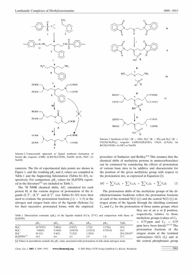
Synthesis of the ligands : Attempts to build up the ligandsfrom benzylamine (2) by treatment with tosylaziridine (1),
followed by deprotection of the tosyl groups and alkylationwith ethyl bromoacetate to give a H5DTPA analogue with aN-benzyl-protected central amino group of the skeleton (5)were not successful due to the formation of a very stablelactam (6) after debenzylation (Scheme 2). This lactam wasfound to be extremely stable towards hydrolysis; it could behydrolysed only under very harsh conditions (i.e., 20%NaOH, 90 8C, overnight), affording the tetraacetic derivative(7). Therefore, it was decided first to attach the phosphorus-containing moiety to the diethylenetriamine backbone. Thiswas achieved by a Mannich-type reaction between N,N’’-bis(phthaloyl)diethylenetriamine (8), paraformaldehyde andthe appropriate phosphorus derivative, followed by depro-tection of phthaloyl moieties with hydrazine. Then, alkyla-tion of intermediate (10) with ethyl bromoacetate and hy-drolysis of the ester groups afforded the desired compoundsH6L
1, H5L2 and H5L
3 in overall isolated yields of 50±80%(Scheme 3).
Determination of the ligand-protonation constants by using1H and 31P NMR chemical-shift titrations : Since the thermo-dynamic stability of the Ln3+ complexes of aminocarboxy-lates is related to the summed protonation constants of thefree ligand,[11±13] insight into the structural effects on theseconstants is desirable. Therefore, the protonation constantsof all the ligands were determined by using the pH depend-ence of 1H and 31P NMR chemical shifts. The chemical shiftcurves (see Figure 1) display sharp changes at several rangesof pH values; they may be ascribed to the shift dependenceon the changes of the protonation state of the ligand con-cerned.
Since the protonation equilibria are fast on the NMRtimescale, the chemical shift of each signal can be given as aweighted average of the shifts of the various protonated spe-cies (see [Eq. (1)]).[14]
dobs ¼X
Xi � di ð1Þ
Here dobs is the observed chemical shift of a given signal,Xi is the molar fraction of species i and di is its chemicalshift. The observed 1H and 31P chemical shifts were fitted si-multaneously according to Equation (1) by using the dissoci-ation constants (pKai) and the values of di as adjustable pa-
Scheme 1. Molecular structures of a) the ligands discussed and of b)H5DTPA, c) atom-labelling scheme for NMR assignment.
¹ 2003 Wiley-VCH Verlag GmbH&Co. KGaA, Weinheim www.chemeurj.org Chem. Eur. J. 2003, 9, 5899 ± 59155900
FULL PAPER
rameters. The fits of experimental data points are shown inFigure 1, and the resulting pKa and di values are compiled inTable 1 and the Supporting Information (Tables S1±S3), re-spectively. For comparison, pKa values for H5DTPA report-ed in the literature[15] are included in Table 1.
The 1H NMR chemical shifts, Ddni , calculated for each
proton Hi at the various degrees of protonation of the li-gands (L1)6�, (L2)5� and (L3)5� (see Tables S1±S3) were thenused to evaluate the protonation fractions fi (i = 1±5) at thenitrogen and oxygen basic sites of the ligands (Scheme 1c)for their successive protonated forms, with the empirical
procedure of Sudmeier and Reilley.[14] This assumes that thechemical shifts of methylene protons in aminocarboxylatescan be estimated by considering the effects of protonationof various basic sites to be additive and characteristic forthe position of the given methylene group with respect tothe protonation site, as expressed in Equation (2).
Ddni ¼X
CNfN þX
CN0 fN þX
COfO þX
CPfP ð2Þ
The protonation shifts of the methylene groups of the di-ethylenetriamine backbone reflect the protonation fractionsof each of the terminal N(1) (f1) and the central N(2) (f2) ni-trogen atoms of the ligands through the shielding constantsCN and CN’ for the protonation of those amino groups, when
they are at an a or b position,respectively, relative to thosemethylene groups (values of CN
= 0.75 ppm and CN’ = 0.35ppm have been listed).[14,15] Theprotonation fractions of theoxygen atoms at the terminalcarboxylates O(3) (f3) and atthe central phosphonate group
Scheme 2. Unsuccessful approach to ligand synthesis–formation oflactam (6), reagents: i) HBr; ii) BrCH2COOEt, NaOH; iii) H2, Pd/C; iv)NaOH.
Scheme 3. Synthesis of H6L1 (R = OH), H5L
2 (R = Ph) and H5L3 (R =
CH2N(CH2Ph)2); reagents: i) HP(O)(R)(OEt), CH2O; ii) N2H4; iii)BrCH2COOEt; iv) HCl or NaOH.
Table 1. Dissociation constants (pKa) of the ligands studied (0.1m, 25 8C) and comparison with those ofH5DTPA.
pKa1 pKa2 pKa3 pKa4 pKa5 �pKa
H6L1 10.747(5) 7.88(2) 6.92(7) 2.7(2) 2.17(6) 30.4
H5L2 9.60(5) 9.10(4) 2.63(19) 2.15(12) 0.72(14) 24.2
H5L3 10.1(1) 9.4(2) 7.13(2) 1.32(5) 28.0 (20.8)[a]
H5DTPA[15] 10.2 8.6 4.2 2.9 1.8 27.7
[a] Values in parenthesis exclude the pKa value associated with protonation of side-chain nitrogen atom.
Chem. Eur. J. 2003, 9, 5899 ± 5915 www.chemeurj.org ¹ 2003 Wiley-VCH Verlag GmbH&Co. KGaA, Weinheim 5901
Lanthanide Complexes of Diethylenetriamine 5899 ± 5915

O(4) (f4) were also calculated by using shielding constantsCO = 0.20 ppm for a-carboxylate protonation[15] and CP =
0.20 ppm for a-phosphonate/phosphinate protonation.[16,17]
The results are given in Table 2 together with data forDTPA5� reported previously.[15] It can be seen that the firsttwo protons bind exclusively to the backbone nitrogens inthe three cases. The first protonation of the phosphonateligand (HL1)5� takes place mainly on the central nitrogenatom of the backbone (f2), whereas in HDTPA4� and(HL3)4�, the preference for the central nitrogen is somewhatless, so that the central nitrogen atom is protonated to aboutthe same extent as the sum of the two terminal ones. This isin agreement with the basicity of the nitrogen atom in ami-nomethylphosphonates, which is generally higher than that
in aminomethylcarboxylates.[13] However, aminomethylphos-phinates are less basic than the corresponding aminomethyl-carboxylates; this explains why the central nitrogen atom(f2) of the phenylphosphinic derivative (L2)5� is only poorlyprotonated in the monoprotonated species (HL2)4�. In allcases, the protons of the (H2L)
x� species are located mainlyon the outer nitrogen atoms (f1), this can be rationalised bythe electrostatic repulsion between the two incoming protons.
For n>2, the protonation also involves the basic atomslocated at the pendant arms of the ligands. For the ligand(L1)6�, the fi values show that the third protonation stepmainly occurs at the phosphonate moiety; this is in agree-ment with its value of pKa3 (6.92) being close to the pKa
values commonly observed for phosphonates.[18] The next
Figure 1. 1H and 31P NMR chemical-shift titration curves of 0.1m solutions of H6L1 (a,b), H5L
2 (c,d) and H5L3 (e,f) in H2O/D2O (9:1 v/v) at 25 8C and 7 T.
Vertical lines mark the dissociation constants (for values, see Table 1). For labelling of hydrogen atoms see Scheme 1c: a (^), b (&), c (*), d (~), e (^), f(~).
¹ 2003 Wiley-VCH Verlag GmbH&Co. KGaA, Weinheim www.chemeurj.org Chem. Eur. J. 2003, 9, 5899 ± 59155902
FULL PAPER I. Lukesœ, J. A. Peters et al.
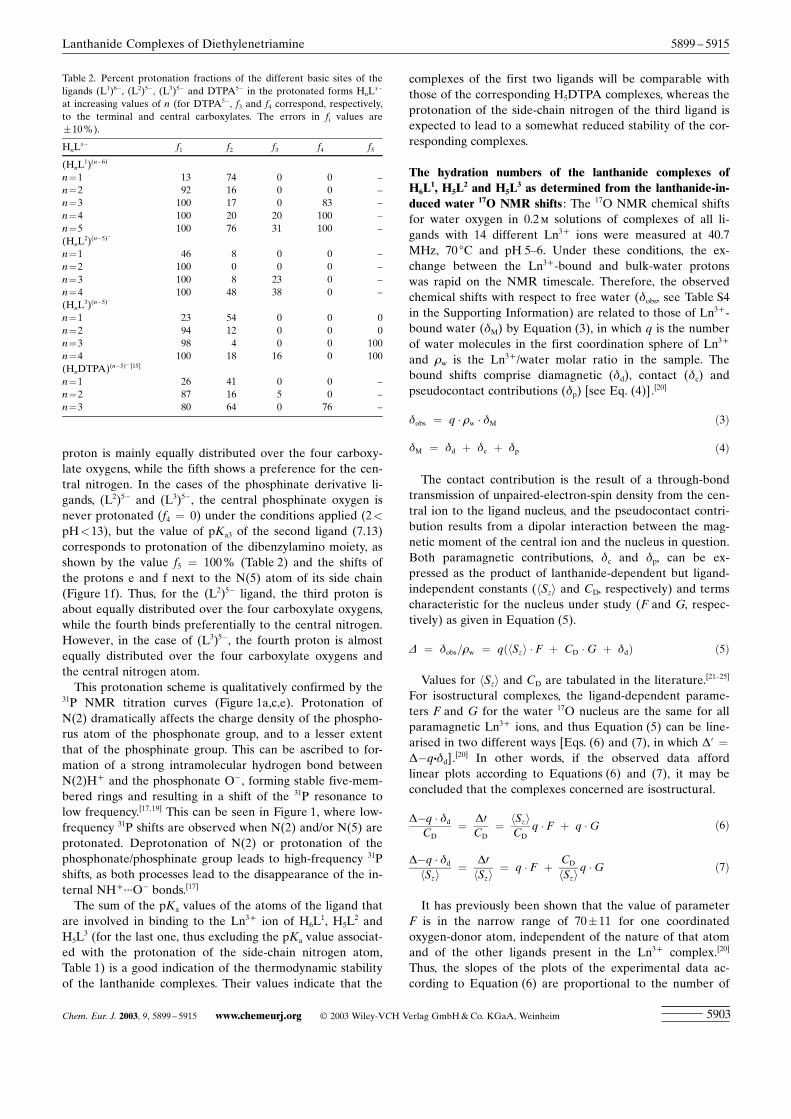
proton is mainly equally distributed over the four carboxy-late oxygens, while the fifth shows a preference for the cen-tral nitrogen. In the cases of the phosphinate derivative li-gands, (L2)5� and (L3)5�, the central phosphinate oxygen isnever protonated (f4 = 0) under the conditions applied (2<pH<13), but the value of pKa3 of the second ligand (7.13)corresponds to protonation of the dibenzylamino moiety, asshown by the value f5 = 100% (Table 2) and the shifts ofthe protons e and f next to the N(5) atom of its side chain(Figure 1f). Thus, for the (L2)5� ligand, the third proton isabout equally distributed over the four carboxylate oxygens,while the fourth binds preferentially to the central nitrogen.However, in the case of (L3)5�, the fourth proton is almostequally distributed over the four carboxylate oxygens andthe central nitrogen atom.
This protonation scheme is qualitatively confirmed by the31P NMR titration curves (Figure 1a,c,e). Protonation ofN(2) dramatically affects the charge density of the phospho-rus atom of the phosphonate group, and to a lesser extentthat of the phosphinate group. This can be ascribed to for-mation of a strong intramolecular hydrogen bond betweenN(2)H+ and the phosphonate O� , forming stable five-mem-bered rings and resulting in a shift of the 31P resonance tolow frequency.[17,19] This can be seen in Figure 1, where low-frequency 31P shifts are observed when N(2) and/or N(5) areprotonated. Deprotonation of N(2) or protonation of thephosphonate/phosphinate group leads to high-frequency 31Pshifts, as both processes lead to the disappearance of the in-ternal NH+ ¥¥¥O� bonds.[17]
The sum of the pKa values of the atoms of the ligand thatare involved in binding to the Ln3+ ion of H6L
1, H5L2 and
H5L3 (for the last one, thus excluding the pKa value associat-
ed with the protonation of the side-chain nitrogen atom,Table 1) is a good indication of the thermodynamic stabilityof the lanthanide complexes. Their values indicate that the
complexes of the first two ligands will be comparable withthose of the corresponding H5DTPA complexes, whereas theprotonation of the side-chain nitrogen of the third ligand isexpected to lead to a somewhat reduced stability of the cor-responding complexes.
The hydration numbers of the lanthanide complexes ofH6L
1, H5L2 and H5L
3 as determined from the lanthanide-in-duced water 17O NMR shifts : The 17O NMR chemical shiftsfor water oxygen in 0.2m solutions of complexes of all li-gands with 14 different Ln3+ ions were measured at 40.7MHz, 70 8C and pH 5±6. Under these conditions, the ex-change between the Ln3+-bound and bulk-water protonswas rapid on the NMR timescale. Therefore, the observedchemical shifts with respect to free water (dobs, see Table S4in the Supporting Information) are related to those of Ln3+-bound water (dM) by Equation (3), in which q is the numberof water molecules in the first coordination sphere of Ln3+
and 1w is the Ln3+/water molar ratio in the sample. Thebound shifts comprise diamagnetic (dd), contact (dc) andpseudocontact contributions (dp) [see Eq. (4)].[20]
dobs ¼ q � 1w � dM ð3Þ
dM ¼ dd þ dc þ dp ð4Þ
The contact contribution is the result of a through-bondtransmission of unpaired-electron-spin density from the cen-tral ion to the ligand nucleus, and the pseudocontact contri-bution results from a dipolar interaction between the mag-netic moment of the central ion and the nucleus in question.Both paramagnetic contributions, dc and dp, can be ex-pressed as the product of lanthanide-dependent but ligand-independent constants (hSzi and CD, respectively) and termscharacteristic for the nucleus under study (F and G, respec-tively) as given in Equation (5).
D ¼ dobs=1w ¼ qðhSzi � F þ CD �G þ ddÞ ð5Þ
Values for hSzi and CD are tabulated in the literature.[21±25]
For isostructural complexes, the ligand-dependent parame-ters F and G for the water 17O nucleus are the same for allparamagnetic Ln3+ ions, and thus Equation (5) can be line-arised in two different ways [Eqs. (6) and (7), in which D’ =D�q¥dd].
[20] In other words, if the observed data affordlinear plots according to Equations (6) and (7), it may beconcluded that the complexes concerned are isostructural.
D�q � ddCD
¼ D0CD
¼ hSziCD
q � F þ q �G ð6Þ
D�q � ddhSzi
¼ D0hSzi
¼ q � F þ CDhSzi
q �G ð7Þ
It has previously been shown that the value of parameterF is in the narrow range of 7011 for one coordinatedoxygen-donor atom, independent of the nature of that atomand of the other ligands present in the Ln3+ complex.[20]
Thus, the slopes of the plots of the experimental data ac-cording to Equation (6) are proportional to the number of
Table 2. Percent protonation fractions of the different basic sites of theligands (L1)6�, (L2)5�, (L3)5� and DTPA5� in the protonated forms HnL
x�
at increasing values of n (for DTPA5�, f3 and f4 correspond, respectively,to the terminal and central carboxylates. The errors in fi values are10%).
HnLx� f1 f2 f3 f4 f5
(HnL1)(n�6)�
n=1 13 74 0 0 ±n=2 92 16 0 0 ±n=3 100 17 0 83 ±n=4 100 20 20 100 ±n=5 100 76 31 100 ±(HnL
2)(n�5)�
n=1 46 8 0 0 ±n=2 100 0 0 0 ±n=3 100 8 23 0 ±n=4 100 48 38 0 ±(HnL
3)(n�5)�
n=1 23 54 0 0 0n=2 94 12 0 0 0n=3 98 4 0 0 100n=4 100 18 16 0 100(HnDTPA)(n�5)� [15]
n=1 26 41 0 0 ±n=2 87 16 5 0 ±n=3 80 64 0 76 ±
Chem. Eur. J. 2003, 9, 5899 ± 5915 www.chemeurj.org ¹ 2003 Wiley-VCH Verlag GmbH&Co. KGaA, Weinheim 5903
Lanthanide Complexes of Diethylenetriamine 5899 ± 5915
water molecules coordinated in the inner sphere of the Ln3+
ion.The observed chemical shifts for the diamagnetic La3+
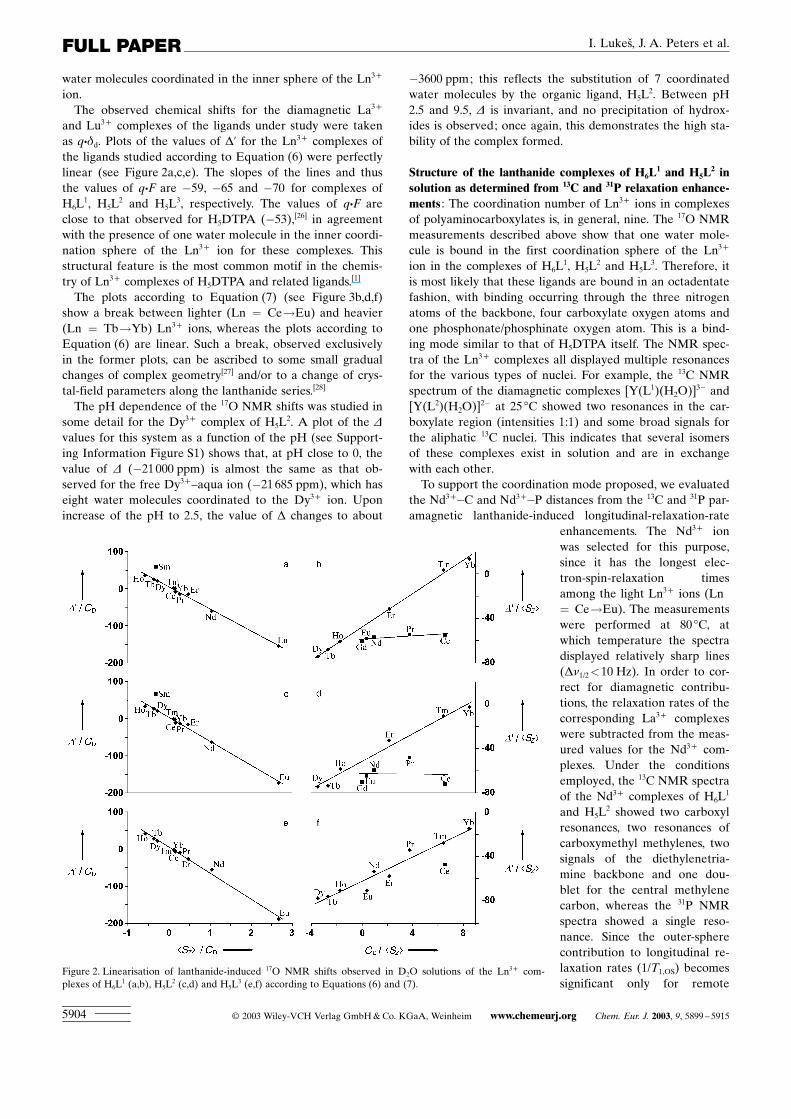
and Lu3+ complexes of the ligands under study were takenas q¥dd. Plots of the values of D’ for the Ln3+ complexes ofthe ligands studied according to Equation (6) were perfectlylinear (see Figure 2a,c,e). The slopes of the lines and thusthe values of q¥F are �59, �65 and �70 for complexes ofH6L
1, H5L2 and H5L
3, respectively. The values of q¥F areclose to that observed for H5DTPA (�53),[26] in agreementwith the presence of one water molecule in the inner coordi-nation sphere of the Ln3+ ion for these complexes. Thisstructural feature is the most common motif in the chemis-try of Ln3+ complexes of H5DTPA and related ligands.[1]
The plots according to Equation (7) (see Figure 3b,d,f)show a break between lighter (Ln = Ce!Eu) and heavier(Ln = Tb!Yb) Ln3+ ions, whereas the plots according toEquation (6) are linear. Such a break, observed exclusivelyin the former plots, can be ascribed to some small gradualchanges of complex geometry[27] and/or to a change of crys-tal-field parameters along the lanthanide series.[28]
The pH dependence of the 17O NMR shifts was studied insome detail for the Dy3+ complex of H5L
2. A plot of the D
values for this system as a function of the pH (see Support-ing Information Figure S1) shows that, at pH close to 0, thevalue of D (�21000 ppm) is almost the same as that ob-served for the free Dy3+±aqua ion (�21685 ppm), which haseight water molecules coordinated to the Dy3+ ion. Uponincrease of the pH to 2.5, the value of D changes to about
�3600 ppm; this reflects the substitution of 7 coordinatedwater molecules by the organic ligand, H5L
2. Between pH2.5 and 9.5, D is invariant, and no precipitation of hydrox-ides is observed; once again, this demonstrates the high sta-bility of the complex formed.
Structure of the lanthanide complexes of H6L1 and H5L
2 insolution as determined from 13C and 31P relaxation enhance-ments : The coordination number of Ln3+ ions in complexesof polyaminocarboxylates is, in general, nine. The 17O NMRmeasurements described above show that one water mole-cule is bound in the first coordination sphere of the Ln3+
ion in the complexes of H6L1, H5L
2 and H5L3. Therefore, it
is most likely that these ligands are bound in an octadentatefashion, with binding occurring through the three nitrogenatoms of the backbone, four carboxylate oxygen atoms andone phosphonate/phosphinate oxygen atom. This is a bind-ing mode similar to that of H5DTPA itself. The NMR spec-tra of the Ln3+ complexes all displayed multiple resonancesfor the various types of nuclei. For example, the 13C NMRspectrum of the diamagnetic complexes [Y(L1)(H2O)]3� and[Y(L2)(H2O)]2� at 25 8C showed two resonances in the car-boxylate region (intensities 1:1) and some broad signals forthe aliphatic 13C nuclei. This indicates that several isomersof these complexes exist in solution and are in exchangewith each other.
To support the coordination mode proposed, we evaluatedthe Nd3+�C and Nd3+�P distances from the 13C and 31P par-amagnetic lanthanide-induced longitudinal-relaxation-rate
enhancements. The Nd3+ ionwas selected for this purpose,since it has the longest elec-tron-spin-relaxation timesamong the light Ln3+ ions (Ln= Ce!Eu). The measurementswere performed at 80 8C, atwhich temperature the spectradisplayed relatively sharp lines(Dn1/2<10 Hz). In order to cor-rect for diamagnetic contribu-tions, the relaxation rates of thecorresponding La3+ complexeswere subtracted from the meas-ured values for the Nd3+ com-plexes. Under the conditionsemployed, the 13C NMR spectraof the Nd3+ complexes of H6L
1
and H5L2 showed two carboxyl
resonances, two resonances ofcarboxymethyl methylenes, twosignals of the diethylenetria-mine backbone and one dou-blet for the central methylenecarbon, whereas the 31P NMRspectra showed a single reso-nance. Since the outer-spherecontribution to longitudinal re-laxation rates (1/T1,OS) becomessignificant only for remote
Figure 2. Linearisation of lanthanide-induced 17O NMR shifts observed in D2O solutions of the Ln3+ com-plexes of H6L
1 (a,b), H5L2 (c,d) and H5L
3 (e,f) according to Equations (6) and (7).
¹ 2003 Wiley-VCH Verlag GmbH&Co. KGaA, Weinheim www.chemeurj.org Chem. Eur. J. 2003, 9, 5899 ± 59155904
FULL PAPER I. Lukesœ, J. A. Peters et al.

nuclei, this was neglected. From the electron-spin relaxationfor Nd3+ (T1e�10�13 s),[29] it can be estimated that the con-tact contribution to the paramagnetic relaxation is negligi-ble. Therefore, two contributions are of importance: the di-polar relaxation and the Curie relaxation. These are repre-sented by a combination of a simplified Solomon±Bloember-gen equation with one for Curie relaxation, giving Equation(8):[20]
1T1
¼�43
�m04p
�2� m2 � g2I � b2 � T1e
þ�65
�m04p
�2 g2I �H20 � m4 � b4
ð3kBTÞ2�� tR
�1r6
ð8Þ
Here m0/4p is the permeability of a vacuum, m is the effec-tive magnetic moment of Nd3+ , gI is the gyromagnetic ratioof the nucleus under study (13C and 31P), b is the Bohr mag-neton, T1e is electronic spin relaxation time for Nd3+ ,[29] H0
is the strength of the magnetic field, kB is the Boltzmannconstant, T is the temperature, tR is the rotational correla-tion time for the complex species and r is the distance ofNd3+ to the nuclei in the complex. This equation can beused to calculate the Nd3+�C and Nd3+�P distances r. Forthese calculations, values of tR at 80 8C of the correspondingGd3+ complexes (23.1 ps for [Nd(L1)(H2O)]3� and 33.2 psfor [Nd(L2)(H2O)]2�) were employed. These values wereevaluated from variable-concentration 2H NMR data per-formed on the deuterated La3+ complexes by using the acti-vation energy for tR as obtained from the fitting of the 17Oand nuclear magnetic relaxation dispersion (NMRD) data(see below).Table 3 lists the experimental values of longitu-dinal relaxation rates observed for Nd3+ and La3+ com-plexes, together with the Nd3+�C and Nd3+�P distances cal-culated from them by using Equation (8). For comparison,results obtained for complexes of H5DTPA and its bis(ami-de)derivatives reported previously[26,30,31] have been includedin the Table. The similarity of these values confirms the pro-posed octadentate binding mode (similar to structures ofwell-known complexes of H5DTPA) of the new ligands intheir lanthanide complexes.
Interconversion between isomers of the Ln3+ complexes ofthe ligands under study : Upon binding of the ligands H6L
1,H5L
2 and H5L3 to a Ln3+ ion in an octadentate fashion
through the diethylenetriamine nitrogen atoms, a phospho-nate/phosphinate oxygen atom and four carboxylate oxygenatoms, the central nitrogen atom and the phosphorus atombecome chiral. An inspection of crystal structures of Ln3+
complexes of DTPA derivatives has shown that the two eth-ylene moieties can adopt either a ll or a dd conforma-tion.[32,33] Therefore, this is most likely to also be the casefor the presently studied ligands. Then, four enantiomers(two diastereomeric pairs) are possible: llR, llS, ddR andddS, where R and S denote the chirality of the phosphorusatom. In a static situation, all 13C nuclei in an isomer arechemically different. Therefore, for example four carboxy-late resonances should be expected for each diastereomericpair, leading to an expected total number of eight carboxy-late resonances.
The variable-temperature behaviour of the 13C NMRspectrum of the diamagnetic [Y(L2)(H2O)]2� complex wasstudied in some detail. At 0.5 8C, four carboxylate resonan-ces of about equal intensity were observed. Upon increasingthe temperature, these resonances broadened and coalescedto two resonances at about 9 8C, and sharpened again uponfurther temperature increase. Similar behaviour was ob-served for the other 13C resonances; this indicates that a rac-emisation process becomes rapid on the NMR timescale.Racemisation of the central nitrogen atom can be achievedby a wagging motion of the diethylenetriamine moiety,which interconverts its ll and dd conformations. The phos-phorus atom can racemise by decoordination of the phosphi-nate moiety followed by ™inversion∫ of the phosphorusatom, that is, rotation around the CH2�P bond and recoordi-nation. Apparently, one of these two racemisation processesis already rapid on the NMR timescale at 0.5 8C, whereasthe other becomes fast above 9 8C. From the coalescencetemperature of the carboxylate resonances (9 8C), the freeenthalpy of activation of the exchange process concerned(DG282) can be estimated to be 563 kJmol�1. The value isin the range of DG values generally found for the racemisa-
Table 3. Observed longitudinal relaxation rates in La3+ and Nd3+ complexes of ligands H6L1 and H5L
2 and calculated nonbonding distances r(Nd-P) andr(Nd-C).
longitudinal relaxation rates 1/T1 [s�1] distances from Nd3+ [ä]
atom La�H6L1 Nd�H6L
1 La�H5L2 Nd�H5L
2 H6L1 H5L
2 H5DTPA[a] H3DTPA-bis(amides)[b]
P 0.356 10.06 0.291 9.73 3.49 3.52CO 0.17[c] 6.02±6.25[c] 0.10[c] 6.29±8.93[c] 3.22±3.24[c] 3.03±3.22[c] 3.15±3.20 3.14±3.30N-CH2-CO 2.56[c] 7.04±7.46[c] 2.62[c] 7.52±7.69[c] 3.33±3.38[c] 3.33±3.35[c] [d] 3.20±3.59N-CH2-P 3.00[e] 5.39[e] 1.72 8.71 3.76[e] 3.15 3.17[f] 3.14±3.30[f]
CH2-N-CH2-P 3.11 6.99 3.10 7.52 3.47 3.41 3.48[g] 3.04±3.48[g]
CH2-N-CH2-CO 2.82 7.09 3.11 8.40 3.41 3.30 3.21 3.04±3.48P-C(arom) ± ± 0.11 1.96 ± 3.94 ± ±C(arom-o) ± ± 0.72 1.29 ± 4.79 ± ±C(arom-m) ± ± 0.70 0.89 ± 5.75 ± ±C(arom-p) ± ± 1.28 1.41 ± 6.13 ± ±
[a] Taken from ref. [26]. [b] Taken from ref. [31]. [c] Two different signals of equal intensity are found in 13C NMR spectra. [d] Not determined. [e] Thesignal has low intensity and overlaps others; this makes it unsuitable for the determination of relaxation rates. [f] Value corresponding to the signal of anacetate pendant moiety bound to the central nitrogen atom. [g] Value corresponding to the signal of a backbone carbon atom bound to the central nitro-gen atom.
Chem. Eur. J. 2003, 9, 5899 ± 5915 www.chemeurj.org ¹ 2003 Wiley-VCH Verlag GmbH&Co. KGaA, Weinheim 5905
Lanthanide Complexes of Diethylenetriamine 5899 ± 5915
tion of the central nitrogen atom in Ln3+ complexes ofH5DTPA and its derivatives;[32,33] this suggests that the ex-change process observed here can be assigned to such a rac-emisation. The racemisation at the phosphorus atom has aconsiderably lower barrier, as at 0 8C the exchange betweenthe corresponding two enantiomeric forms is already rapidon the NMR timescale.
These results were confirmed by a variable-temperatureand variable-pH study of the 1H and 31P NMR spectra of a0.1m aqueous solution of the diamagnetic [La(L3)(H2O)]2�
complex. The 31P and 1H NMR signals of the complex werequite broad at 25 8C, but considerably sharpened at 60 8C.The proton resonances were assigned with the aid of aCOSY spectrum at 60 8C, pH 6.4. A value of pKa = 6.58(4)was obtained from fitting the pH dependence of the 31P andHe and Hf resonances of the [La(L3)(H2O)]2� complex, withthe protonation shift values indicating that the processoccurs at the side-chain N(5) atom. At 60 8C, two resonancesof about equal intensity were observed for each of the back-bone (Hc,c’, Hd,d’) and acetate (Ha,a’, giving two AB patternswith 2JHH values of 16.6 Hz) protons of the complex, whileonly one sharp resonance was observed for each of the side-chain protons (singlet Hf, doublets He and Hb with 2JPHvalues of 9.8 and 8.0 Hz, respectively). This again indicatesthat at 60 8C the racemisation processes for the central nitro-gen and the phosphorus atom are rapid on the NMR time-scale.
Evaluation of rotational correlation times by 2H NMR : Therotational correlation time, tR, is one of the parameters gov-erning the relaxivity of a Gd3+ complex. Usually, a relativelylarge discrepancy exists between the t298R values evaluatedfrom the 1H and 17O NMR data. Therefore, we decided todetermine the rotational correlation times independentlyusing the deuterium longitudinal relaxation rates of the deu-terated ligands [D8]H6L
1, [D8]H5L2 and [D8]H5L
3 in their dia-magnetic La3+ complexes.[34] In such a diamagnetic system,the deuterium relaxation depends only on quadrupolar in-teractions and is given by Equation (9):
R1 ¼ 1T1
¼ 38
�e2qQ�h
�2tR ð9Þ
The quadrupolar coupling constant (e2qQ/�h) has a valueof 170î2p kHz for an sp3-hybridised C�2H bond. It hasbeen demonstrated that tR values obtained in this way agreewell with those obtained from 1H NMRD measurements.[34]
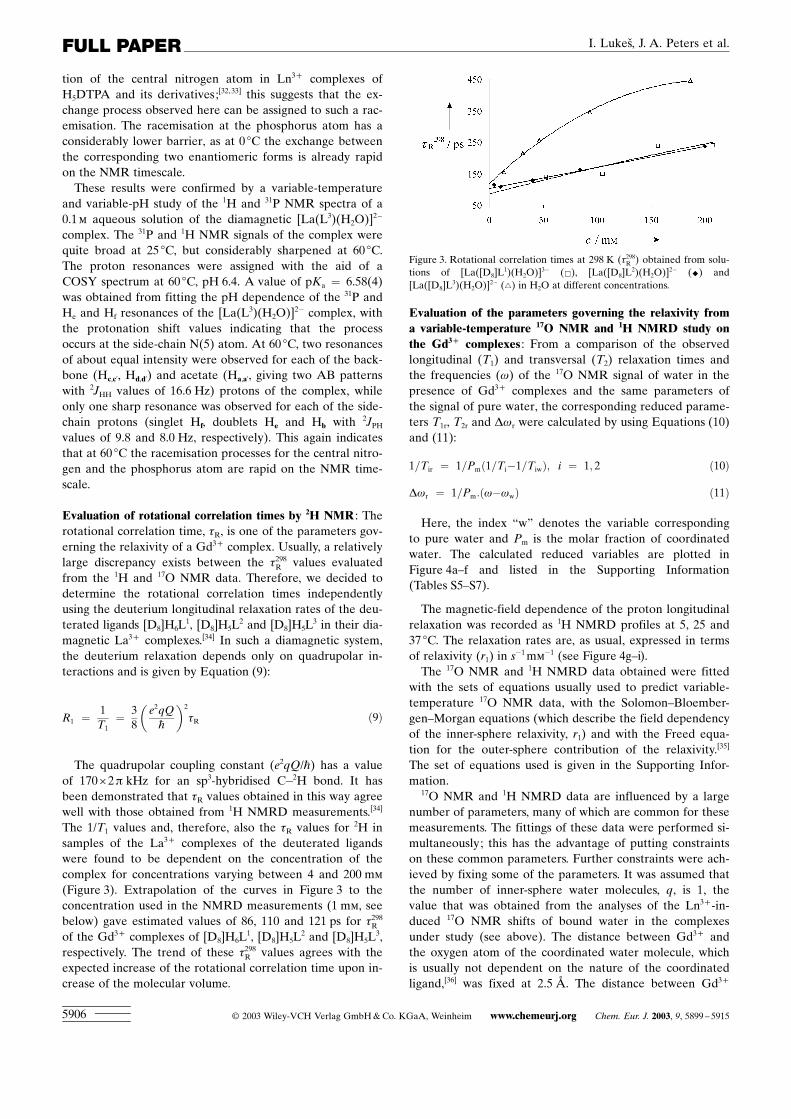
The 1/T1 values and, therefore, also the tR values for 2H insamples of the La3+ complexes of the deuterated ligandswere found to be dependent on the concentration of thecomplex for concentrations varying between 4 and 200 mm
(Figure 3). Extrapolation of the curves in Figure 3 to theconcentration used in the NMRD measurements (1 mm, seebelow) gave estimated values of 86, 110 and 121 ps for t298R
of the Gd3+ complexes of [D8]H6L1, [D8]H5L
2 and [D8]H5L3,
respectively. The trend of these t298R values agrees with theexpected increase of the rotational correlation time upon in-crease of the molecular volume.
Evaluation of the parameters governing the relaxivity froma variable-temperature 17O NMR and 1H NMRD study onthe Gd3+ complexes : From a comparison of the observedlongitudinal (T1) and transversal (T2) relaxation times andthe frequencies (w) of the 17O NMR signal of water in thepresence of Gd3+ complexes and the same parameters ofthe signal of pure water, the corresponding reduced parame-ters T1r, T2r and Dwr were calculated by using Equations (10)and (11):
1=Tir ¼ 1=Pmð1=Ti�1=TiwÞ; i ¼ 1; 2 ð10Þ
Dwr ¼ 1=Pm:ðw�wwÞ ð11Þ
Here, the index ™w∫ denotes the variable correspondingto pure water and Pm is the molar fraction of coordinatedwater. The calculated reduced variables are plotted inFigure 4a±f and listed in the Supporting Information(Tables S5±S7).
The magnetic-field dependence of the proton longitudinalrelaxation was recorded as 1H NMRD profiles at 5, 25 and37 8C. The relaxation rates are, as usual, expressed in termsof relaxivity (r1) in s
�1mm�1 (see Figure 4g±i).
The 17O NMR and 1H NMRD data obtained were fittedwith the sets of equations usually used to predict variable-temperature 17O NMR data, with the Solomon±Bloember-gen±Morgan equations (which describe the field dependencyof the inner-sphere relaxivity, r1) and with the Freed equa-tion for the outer-sphere contribution of the relaxivity.[35]
The set of equations used is given in the Supporting Infor-mation.
17O NMR and 1H NMRD data are influenced by a largenumber of parameters, many of which are common for thesemeasurements. The fittings of these data were performed si-multaneously; this has the advantage of putting constraintson these common parameters. Further constraints were ach-ieved by fixing some of the parameters. It was assumed thatthe number of inner-sphere water molecules, q, is 1, thevalue that was obtained from the analyses of the Ln3+-in-duced 17O NMR shifts of bound water in the complexesunder study (see above). The distance between Gd3+ andthe oxygen atom of the coordinated water molecule, whichis usually not dependent on the nature of the coordinatedligand,[36] was fixed at 2.5 ä. The distance between Gd3+
Figure 3. Rotational correlation times at 298 K (t298R ) obtained from solu-tions of [La([D8]L
1)(H2O)]3� (&), [La([D8]L2)(H2O)]2� (^) and
[La([D8]L3)(H2O)]2� (~) in H2O at different concentrations.
¹ 2003 Wiley-VCH Verlag GmbH&Co. KGaA, Weinheim www.chemeurj.org Chem. Eur. J. 2003, 9, 5899 ± 59155906
FULL PAPER I. Lukesœ, J. A. Peters et al.

and a water proton was fixed at 3.1 ä, whereas the distanceof closest approach of a water molecule to Gd3+ , aH, wasfixed at 3.5 ä. The value of EV, the activation energy of thecorrelation time tv, was fixed at 1 kJmol�1. Attempts tounfix this parameter led to negative values of the activationenergy. The hyperfine coupling constants A/�h were fixed atthe values calculated from the F values obtained from the17O NMR studies described above and by using Equation(12), in which b is the Bohr magneton, k is the Boltzmannconstant and gI is the 17O magnetogyric ratio. Furthermore,the quadrupolar coupling constant of the bound water,c(1+h2)1/2, was taken equal to that determined recently forthe complex [Gd(dota)(H2O)]� of 5.2 MHz.[37]
F ¼ b
3kTgI
A�h106 ð12Þ
Fitting of the data with a single rotational correlationtime resulted in bad fits, whereas separate fitting of the 17Oand NMRD data resulted in good fits, but with different tRvalues. This may be ascribed to the large difference in con-centration at which the 17O (200 mm) and the NMRD meas-
urements (1 mm) were carried out (see above).[34] Further-more, the 17O and 1H relaxation rates are modulated by ro-tation of the Gd�O and the Gd�H vectors, respectively. Itmay be expected that these rotations have different correla-tion times.[37] Therefore, two rotational correlation timeswere taken into consideration, tHR and tOR. The parameter tHRwas fixed at the values obtained from the 2H NMR meas-urements (see above).
A comparison of the values of the fitted parameters ofthe complexes under study with those of [Gd(dtpa)(H2O)]2�
(see Table 4) reveals significant differences in the parame-ters related to the electronic relaxation (the square of thezero-field-splitting tensor, D2, and the corresponding correla-tion time, tv and, particularly, in the diffusion coefficient,DGdH, (see Table 4), which is unexpectedly low. DGdH de-pends on the self-diffusion coefficients of the Gd3+ complexconcerned, Dcomplex, and that of water, Dwater [Eq. (13)].
DGdH ¼ Dcomplex þ Dwater ð13Þ
Since Dwater at 298 K is 2.23î10�9 m2s�1,[38] it should be ex-pected that D298
GdH is larger than this value. The relatively low
Figure 4. Simultaneously fitted data from 17O NMR and 1H NMRD measurements of [Gd(L1)(H2O)]3� (a,d,g), [Gd(L2)(H2O)]2� (b,e,h) and[Gd(L3)(H2O)]2� (c,f,i). First row (a,b,c): the temperature dependence of logarithms of the reduced relaxation rates (upper line (~) corresponds to T2r,lower line (&) corresponds to T1r). Second row (d,e,f): the temperature dependence of the reduced frequency (^). Third row (g,h,i): proton-relaxivity de-pendence on the magnetic field at 37 8C (^), 25 8C (~) and 5 8C (&).
Chem. Eur. J. 2003, 9, 5899 ± 5915 www.chemeurj.org ¹ 2003 Wiley-VCH Verlag GmbH&Co. KGaA, Weinheim 5907
Lanthanide Complexes of Diethylenetriamine 5899 ± 5915

values obtained for D298GdH indicate that the outer-sphere con-
tribution to the total relaxivity is overestimated in the calcu-lations. Most likely, this is due to an unaccounted contribu-tion of water molecules in the second coordination sphereof Gd3+ , which may be bound to the ligand through hydro-gen bonds to, for example, the negatively charged phosphi-nate/phosphonate group.[39] To account for such a contribu-tion of second-sphere water molecules, we included a seriesof equations that are similar to those for the inner-spherecontribution (see Supporting Information). It should benoted, however, that it is very difficult to evaluate thesecond-sphere parameters because strong correlations existamong some of them. Moreover, the second-sphere waterprotons probably do not occupy a unique location but mayexchange among various sites. We fixed the distance be-tween the Gd3+ ion and the protons of the second-spherewater molecules, rH2s, at 3.5 ä in the fitting procedure. Fur-thermore, the values of DT
water at various temperatures werecalculated with the semiempirical relationship proposed byHindman [Eq. (14)].[38] The size of the Gd3+ complexes ofH6L
1, H5L2 and H5L
3 is much larger than that of water and,consequently, the self-diffusion constants of these complexeswill be much smaller than that of water. In the present fit-tings Dcomplex was, therefore, neglected.
�lnDTwater ¼ ln ½3:11815� 10�4 eð5:06258�103=TÞ
þ 1:54792� 102 eð1:62931�103=TÞ�ð14Þ
A good fit was obtained by assuming about two second-sphere water molecules (q2s�2). The resulting optimised pa-rameters are included in Table 4, and the results are alsoshown as curves in Figure 4g±i. Now, the optimised parame-ters obtained all compare well with those previously report-
ed for [Gd(dtpa)(H2O)]2�.[36] The residence times of thesesecond-sphere water molecules (t298M2s = 35±60 ps) are of thesame magnitude as those obtained for other systems.[39±41]
Although the accuracy of the second-sphere parameters ob-tained may be low due to the many assumptions made, it isclear that at least two second-sphere water molecules, with aresidence time that is sufficiently long to be detected, haveto be included in the model to adequately explain theNMRD profile. Aime et al.[42] have reported that twosecond-sphere water molecules are present in [Gd(pcp2a)-(H2O)2]
� , a complex of a pyridine containing macrocyclebearing one methylenephosphonic and two acetate arms.Apparently, phosphonate/phosphinate groups are capable offorming hydrogen bonds to two water molecules. Previously,it has been shown that second-sphere water molecules con-tribute to the relaxivity of several other phosphonate-bear-ing ligands including [Gd(dotp)]5� (H8DOTP = 1,4,7,10-tet-raazacyclododecane-1,4,7,10-tetrakis(methylphosphonicacid)).[42]
While the results of simultaneous fits of the present datagave a t289R O/t
289R H ratio of 2.7±3.0, the concentration depend-
ence of tR as determined from 2H NMR (see above) gavean estimate of this ratio of about 1.4 for equal concentra-tions. The latter value (1.4) is in agreement with that report-ed by Dunand et al. for the [Gd(dota)(H2O)]� complex.[37]
The differences between t289R H and t289R O may be ascribed todifferences in the rotation rates of the Gd3+�H andGd3+�O vectors.
The residence time of the Gd3+-bound water molecule,tM, is an important parameter with regard to the efficiencyof an MRI contrast agent. The theoretical curve of the re-laxivity as a function of tM has a sharp maximum between20 and 50 ns.[1,2,4] The values of t298M for the [Gd(L1)(H2O)]3�
and [Gd(L2)(H2O)]2� systems (88 and 92 ns, respectively)
Table 4. Parameters for Gd3+ complexes as obtained from the simultaneous fitting of 17O NMR and 1H NMRD data by using a model including second-sphere water molecules and a model without second-sphere water molecules (see text), compared with literature values for [Gd(dtpa)(H2O)]2� complex.
Parameter [Gd(L1)(H2O)]3� [Gd(L2)(H2O)]2� [Gd(L3)(H2O)]2� [Gd(dtpa)(H2O)]2� [a]
no 2nd sphere 2nd sphere no 2nd sphere 2nd sphere no 2nd sphere 2nd sphere no 2nd sphere
t298M [ns] 6220 8826 7425 9229 543120 685297 303DH# [kJmol�1] 3810 418 369 378 2910 379 51.6t298R H [ps] 86.4[b] 86.4[b] 109.5[b] 109.5[b] 121.0[b] 121.0[b] 58ER [kJmol�1] 142 213 153 193 203 233 17.3t298R O/t
298R H 2.30.4 2.70.4 2.50.4 2.70.4 3.50.4 3.70.4 ±
t298V [ps] 303 223 343 263 313 253 25EV [kJmol�1] 1[b] 1[b] 1[b] 1[b] 1[b] 1[b] 1.6D2 [1020 s�2] 0.290.03 0.450.09 0.220.02 0.320.06 0.260.03 0.360.02 0.46dgL
2 [10�2] 52 62 82 83 122 112 1.2Ah�1 [106 rads�1] �3.28[b] �3.28[b] �4.2 �3.61[b] �3.69[b] �3.69[b] �3.8D298
GdH [10�10 m2 s�1] 14.30.7 22.75[c] 13.20.5 22.75[c] 12.40.6 22.75[c] 20EDGdH [kJ mol�1] 28.73 ± 39.63 ± 253 ± 19.4rGdO [ä] 2.50[b] 2.50[b] 2.50[b] 2.50[b] 2.50[b] 2.50[b] 2.20c(1+h2/3)1/2 [MHz] 5.2[b] 5.2[b] 5.2[b] 5.2[b] 5.2[b] 5.2[b] 14q2s 0 2.20.4 0 2.00.3 0 1.90.3 ±rGdH2s [ä] ± 3.5[b] ± 3.5[b] ± 3.5[b] ±t298M2s [ps] ± 358 ± 509 ± 6010 ±DH2s [kJ mol�1] ± 3611 ± 4812 ± 3510
ln(1/T2e) exp[d] 23.65 23.47 23.25
ln(1/T2e) calcd[e] 22.53 22.24 22.35
[a] Taken from ref. [36]. [b] Parameters were fixed during the fitting. [c] Calculated with Equation (14), [d] Determined from EPR line widths; [e] Calcu-lated by using fitted parameters.
¹ 2003 Wiley-VCH Verlag GmbH&Co. KGaA, Weinheim www.chemeurj.org Chem. Eur. J. 2003, 9, 5899 ± 59155908
FULL PAPER I. Lukesœ, J. A. Peters et al.

are close to the optimum value. These tM values are lowerthan those of the current commercial contrast agents. Forexample, [Gd(dtpa)(H2O)]2� and [Gd(dota)(H2O)]� havet298M values of 303 and 243 ns, respectively.[36] Consequently, itmay be expected that very efficient MRI contrast agents canbe obtained from [Gd(L2)(H2O)]2� by increasing its tR valuethrough covalent or noncovalent binding to macromolecules.Unfortunately, the t298M value of the [Gd(L3)(H2O)]2� com-plex is considerably higher (685 ns) and, thus, it may be ex-pected that for this complex tM is limiting the relaxivityupon binding to a high-molecular-weight compound.
It has been shown that the water exchange in Gd3+-polya-minocarboxylates with one Gd3+-bound water generallytakes place through a dissociative mechanism.[35] Then,steric strain at the water site may increase the energy of theinitial state and, therefore, decrease the activation energy.The decrease in t298M upon replacement of the central �CH2COO� moiety in [Gd(dtpa)(H2O)]2� by a �CH2PO3
2� toform [Gd(L1)(H2O)]3� may be rationalised by an increase insteric strain around the Gd3+-bound water molecule due tothe relatively large size of the �PO3
2� function comparedwith the �COO� function. An inspection of molecularmodels shows that the phenyl group in [Gd(L2)(H2O)]2� isin the proximity of the Gd3+-bound water. Most likely,[Gd(L3)(H2O)]2� has a preferred conformation with thephenyl groups at large distances from the water site. The ni-trogen atom of the dibenzylamino moiety is protonated(pKa = 6.58 for the corresponding La3+ complex, seeabove) and is in close proximity of the Gd3+-bound watermolecule. Possibly, the positive charge and the hydrogenbonding between these functions may slow down the waterexchange rate and thus explain the rather long t298M for thiscomplex.
The temperature dependence of the NMRD profiles usu-ally gives a good indication of the parameter limiting theproton relaxivity. If the relaxivity at high field (>10 MHz)increases with increasing temperature, it is limited by slowwater exchange, whereas in the opposite case fast rotation isthe limiting factor. From the temperature dependence of re-laxivity at constant magnetic field (20 MHz, see Figure 5) itcan be concluded that the total relaxivity decreases with in-creasing temperature, mainly because of a decrease of t298R .
Evaluation of the electron-spin relaxation times T2e of theGd3+ complexes from EPR measurements : The X-band(0.34 T) EPR spectra of the Gd3+ complexes in aqueous sol-ution at 298 K give approximately Lorentzian lines of g~2.0.The transverse electronic relaxation rates (1/T2e) were calcu-lated from the experimental peak-to-peak line widths, DHpp,by using Equation (15), in which the symbols have theirusual meaning.[43]
1=T2e ¼ ðgLmBpffiffiffiffiffiffi3h
pÞDHpp ð15Þ
The experimental values of DHpp obtained at 298 K were0.1220.05 mT ([Gd(L1)(H2O)]3�), 0.1030.05 mT([Gd(L2)(H2O)]2�) and 0.1820.04 mT ([Gd(L3)(H2O)]2�).The corresponding values of ln (1/T2e)exp are compared inTable 4 with the ln(1/T2e)calcd values calculated by usingEquation (S9) (see Supporting Information), from thevalues of the parameters D2 and t298V obtained from the si-multaneous fitting of the 17O NMR and 1H NMRD data. Al-though the relative experimental and calculated 1/T2e valuesfollow very similar trends in the three Gd3+ complexes, theexperimental values are systematically larger by a factor ofabout three. This discrepancy has been noted before[36,44,45]
and corrected by introducing both static and dynamic zero-field-splitting effects in the electronic relaxation mechanismsof Gd3+ .[46]
Interaction of [Gd(L3)(H2O)]2� with human serum albumin :To study the interaction between [Gd(L3)(H2O)]2� andHSA, a solution of the complex was added stepwise to a4% solution of HSA in water. A nonlinear increase of thewater-proton paramagnetic longitudinal relaxation rate wasobserved (see Figure 6) when plotted as a function of theconcentration of the Gd3+ complex. The paramagnetic re-laxation rate of a solution containing 0.81 mm of[Gd(L3)(H2O)]2� in 4% HSA is 3.9 times higher than that of0.81 mm of [Gd(L3)(H2O)]2� in pure water; this indicates astrong interaction between the complex studied and HSA.This interaction is characterised by a stability constant (KAS)of an adduct of HSA with the Gd3+ complex [Eq.
Figure 5. Relaxivity at 20 MHz as a function of temperature for[Gd(L1)(H2O)]3� (^), [Gd(L2)(H2O)]2� (~) and [Gd(L3)(H2O)]2� (&). Ex-perimental data were not fitted–the lines are only guides for the eye.
Figure 6. Proton longitudinal paramagnetic relaxation rates in solutionscontaining 4% HSA and increasing amounts of [Gd(L3)(H2O)]2� (^)measured at 20 MHz and 310 K. The full line corresponds to the fittingof the data, and the dashed line represents Rp
1 in an aqueous solution inthe absence of albumin.
Chem. Eur. J. 2003, 9, 5899 ± 5915 www.chemeurj.org ¹ 2003 Wiley-VCH Verlag GmbH&Co. KGaA, Weinheim 5909
Lanthanide Complexes of Diethylenetriamine 5899 ± 5915

(16)].[47]The proton relaxivity data obtained in HSA, Rp obs1 ,
were fitted to Equation (17)]; here p0 is the protein concen-tration, s0 is the concentration of the paramagnetic complex,n is the number of independent interaction sites and rc1 andrf1 are the relaxivities of the [Gd(L3)(H2O)]2� complex whennoncovalently bound to HSA and free, respectively. In thisfitting procedure, the association constant, KAS, and r
c1 were
used as adjustable parameters.
nGdL þ HSAÐ ðGdLÞn�HSA KAS ¼ ½ðGdLÞn�HSA�½GdL�n ½HSA�
ð16Þ
Rp obs1 ¼ 1000��ðrf1 � s0Þ þ 1
2ðrc1�rf1Þ
�ðn � p0Þ þ s0
þ K�1AS�
ffiffiffiffiffiffiffiffiffiffiffiffiffiffiffiffiffiffiffiffiffiffiffiffiffiffiffiffiffiffiffiffiffiffiffiffiffiffiffiffiffiffiffiffiffiffiffiffiffiffiffiffiffiffiffiffiffiffiffiffiffiffiffiffiffiffiffiffiffiffiffiffiffiffiffiffiððn � p0Þ þ s0 þ K�1
ASÞ2�4N � s0 � p0q �� ð17Þ
A good fit between experimental and calculated valueswas obtained by using a model for one binding site (n = 1)with an association constant KAS = 4500175m�1. The re-laxivity of noncovalently bound complex (rc1) was calculatedto be 430.4 s�1mm
�1, while the value of the relaxivity offree complex (rf1) is 5.90.3 s�1mm
�1 (see above). Thus, in asolution containing 0.81 mm of [Gd(L3)(H2O)]2� and 4%(0.6 mm) HSA, 48.4% of Gd3+ complex interacts with theprotein.
Longitudinal relaxation rates of the solution containing4% HSA and [Gd(L3)(H2O)]2� (0.81 mm) were measured at310 K over the range of magnetic fields 4î10�4±7.05 T. Thecorresponding NMRD profile (Figure 7a) shows the expect-ed hump, characteristic for interactions with macromole-cules, appearing in the high-frequency part (�20 MHz) ofthe recorded profile; this represents the combined contribu-tions of the bound and free Gd3+ complex in the solution.The theoretical 1H NMRD profile of the [Gd(L3)(H2O)]2�-HSA adduct was then calculated from the known NMRDprofile of free [Gd(L3)(H2O)]2� and the concentrations offree and bound complex obtained from the estimated stabili-ty constant of the adduct (see above) (Figure 7b). The relax-ivities in the frequency region of importance for MRI (20±100 MHz) are similar to those of MS-325.[48] The NMRDcurve of the [Gd(L3)(H2O)]2�±HSA adduct could only befitted with a model that assumed ten water molecules in thesecond sphere of the Gd3+ ion (see Figure 7b). A lowernumber of second-sphere water molecules always resulted incalculated relaxivities too low for the low-field part (<10MHz) of the NMRD curve. The results of the fitting possi-bly reflect the presence of mobile HSA protons that are di-polarly relaxed by the proximity of the Gd3+ ion.[6] Alterna-tively, reduced mobility of solvent molecules in the secondcoordination sphere of Gd3+ upon noncovalent binding of[Gd(L3)(H2O)]2� to HSA can explain this result.
Transmetallation : An important parameter determining thetoxicity of Gd3+-based contrast agents is the kinetic stabilityof the complexes. Transmetallation by endogenous metalions may afford free Gd3+ ion, which is highly toxic. To getan impression of the kinetic stability of the phosphorus-con-
taining complexes studied, we performed some transmetalla-tion studies with Zn2+ according to a previously describedprotocol.[49]
Samples containing the Gd3+ complexes of H6L1, H5L
2,H5L
3 and a phosphate buffer containing ZnCl2 were moni-tored by measuring the 1H relaxivity at 20 MHz. Upontransmetallation with Zn2+ , the free Gd3+ formed immedi-ately precipitated as the phosphate salt and, therefore, didnot contribute to the total relaxivity any more. The resultingdecrease in the proton-relaxation rate observed is a good es-timate of the extent of transmetallation and, therefore, alsofor the kinetic stability of the Gd3+ complex. The results ofthe transmetallation experiments are displayed in Figure 8,while Table 5 shows the percentage of Gd3+ complexes leftin the solution after 3 d. Thus, the kinetic stability de-creases in the following order: [Gd(dtpa)(H2O)]2�@[Gd(L1)(H2O)]3�� [Gd(L3)(H2O)]2�� [Gd(dtpa-bma)(H2O)]> [Gd(L2)(H2O)]2�. Therefore, all complexesstudied are less stable towards Zn2+ transmetallation than[Gd(dtpa)(H2O)]2�, but [Gd(L1)(H2O)]3� and[Gd(L3)(H2O)]2� are slightly more kinetically stable than[Gd(dtpa-bma)(H2O)].
Figure 7. a) Proton-relaxation rate (Rp1) of [Gd(L3)(H2O)]2� (0.81 mm)dissolved in 4% HSA (*). The dotted line corresponds to the proton-re-laxation rate of [Gd(L3)(H2O)]2� in pure water at the same concentra-tion. b) Calculated theoretical 1H NMRD profile of the complex[Gd(L3)(H2O)]2� fully bound to human serum albumin at 310 K (&), 1HNMRD profile of the complex simulated with the parameters obtainedfrom simultaneous fit and optimal tR = 14 ns.
¹ 2003 Wiley-VCH Verlag GmbH&Co. KGaA, Weinheim www.chemeurj.org Chem. Eur. J. 2003, 9, 5899 ± 59155910
FULL PAPER I. Lukesœ, J. A. Peters et al.

Conclusion
A useful synthetic approach for a new class of H5DTPA-based ligands in which the central pendant arm has a �P(OH)(O)R moiety is reported. Their Ln3+ complexes showstructural features analogous to the H5DTPA complexes, in-cluding the presence of one water molecule coordinated inthe first coordination sphere of the metal ion. The phospho-nate (H6L
1) and the phenylphosphinate derivatives (H5L2)
have t298M values that are close to optimal (88 and 92 ns, re-spectively). Therefore, these complexes are very suitable forattachment to polymers, which should result in compoundswith very high relaxivity. The relaxivity will be less limitedby water exchange than in most conjugates of chelates ofthe H5DTPA or H4DOTA type. Furthermore, an additionalincrease in the relaxivity is obtained by virtue of the pres-ence of two water molecules in the second coordinationsphere and which are probably bound to the phosphonate/phosphinate moiety through hydrogen bonds. The[Gd(L3)(H2O)]2� complex has a less favourable water-ex-change rate (t298M = 685 ns), but it has a high affinity toHSA and a relaxivity that is comparable with that of thewell-known blood pool contrast agent MS-325.
Experimental Section
Materials and methods : Commercially available benzylamine (2), diethy-lenetriamine, phthalanhydride, ethyl chloroformate, ethyl bromoacetate,diethylphosphite and phenylphosphinic acid had synthetic purity andwere used as received. The 10% Pd/C catalyst for the hydrogenation re-
actions was obtained from Acros. Bis(phthaloyl)diethylenetriamine (8),[50]
ditosylethanolamine,[51] N-tosylaziridine (1),[52] and ethyl phenylphosphi-nate[53] were prepared by published methods. Paraformaldehyde was ob-tained by filtration of aged aqueous formaldehyde solutions and wasdried in a desiccator over concentrated sulfuric acid.1H (300 MHz), 2H (46.1 MHz), 13C (75.5 MHz), 17O (40.7 MHz) and 31PNMR spectra (121.5 MHz) were recorded on a Varian INOVA-300 spec-trometer with 5 mm sample tubes. Unless stated otherwise, NMR experi-ments were performed at 25 8C. Chemical shifts are reported as d valuesand are given in ppm. For measurements in D2O, tert-butyl alcohol wasused as an internal standard with the methyl signal calibrated at 1.2 ppm(1H) or 31.2 ppm (13C). Deuterium oxide (100%) was used as an externalchemical-shift reference for 17O resonances. The 31P chemical shifts weremeasured with respect to 1% H3PO4 in D2O as an external standard(substitution method). The pHs of the samples were measured at ambienttemperature by using a Corning 125 pH meter with a calibrated micro-combination probe from Aldrich. The pH values of the solutions wereadjusted by using dilute solutions of NaOH and HCl. The variable-tem-perature 17O measurements were performed at a magnetic field of 7.05 Ton a Varian INOVA-300 spectrometer equipped with a 5 mm probe.Thin-layer chromatography was performed on silica-coated aluminiumsheets (Silufol, Kavalier, Czech Republic).
Mass spectra were obtained with a VG AUTOSPEC 6F mass spectrome-ter (VG Analytical, Manchester, UK) or on a Bruker ESQUIRE3000with ion-trap detection in positive or negative modes.
Preparation of N’-benzyldiethylenetriamine (4): N-tosylaziridine (1)(32.0 g, 162 mmol) was dissolved in ethanol (150 mL). Benzylamine (2)(8.00 g, 75 mmol) was added, and the solution was heated under refluxfor 3 days. Then, the mixture was cooled and concentrated aqueous am-monia (5 mL) was added to quench the excess of tosylaziridine. The mix-ture obtained was heated under reflux for 15 min. Solvents were re-moved, and the residual brown oil was dissolved in a mixture of concen-trated HBr/AcOH (300 mL, 1:1, v/v). The solution was heated underreflux for 24 h. After cooling, the reaction mixture was evaporated todryness leaving a brown oil, which solidified upon cooling. This solid wasdissolved in NaOH (100 mL, 15%), and the solution obtained was ex-tracted with chloroform (7î50 mL). The organic phases were combinedand evaporated to dryness to leave the mixture of monotosylated inter-mediate and the required product as a yellow oil. These compounds wereseparated by chromatography on silica by using gradient elution with in-creasing concentration of concentrated ammonia in ethanol from a ratioof 1:25 (mixture A) to 1:5 (mixture B), detection by ninhydrine (purplespots). Note: extension of the reaction period to 36±48 h reduces theyield of the final product due to decomposition.
Yield: 8.25 g (57%), Rf (mixt. A) = 0.1, Rf (mixt. B) = 0.3; 1H NMR(CDCl3): d = 2.05 (br, 4H; NH2), 2.52, 2.75 (2m, 4H; NCH2CH2N), 3.58(s, 2H; NCH2Ph), 7.31 (m, 5H; Ph); 13C NMR (CDCl3): d = 34.58 (2C),57.05 (2C, NCH2CH2N), 59.14 (1C, PhCH2N), 127.02 (1C), 128.30 (2C),128.86 (2C) and 139.49 (1C, all Ph); ESI-MS: positive m/z : 194.0[M+H]+ .
N-Tosyl-N’-benzyldiethylenetriamine : Yield: 4.55 g (18%); Rf (mixt. A)= 0.8, Rf (mixt. B) = 0.9; 1H NMR (CDCl3): d = 2.40 (s, 3H; CH3),2.49 (m, 2H), 2.58 (m, 2H), 2.76 (m, 2H) and 2.95 (m, 2H; allNCH2CH2N), 3.05 (br, 1H; NH), 3.52 (s, 2H; NCH2Ph), 7.20 (m, 2H;Ts), 7.27 (m, 5H; Ph), 7.73 (m, 2H; Ts); 13C NMR (CDCl3): d = 22.12(1C, CH3), 39.73, 42.11, 53.14, 55.99 (4î1C, NCH2CH2N), 59.83 (1C,NCH2Ph), 127.67 (2C), 127.82 (1C), 129.01 (2C), 129.45 (2C), 130.20(2C), 137.82 (1C), 139.51 (1C), 143.58 (1C); ESI-MS: positive m/z : 348.3[M+H]+ .
Preparation of N’-benzyldiethylenetriamine-N,N,N’’,N’’-tetraacetic acid(5): N’-benzyldiethylenetriamine (4) (1.00 g, 5.2 mmol) was dissolved inwater (5 mL) and then 1 mL of a solution of NaOH (2.5 g, 62.5 mmol,dissolved in 10 mL of water) was added. The mixture was heated to90 8C. Ethyl bromoacetate (4.34 g, 26 mmol) and the remaining NaOHsolution were added in 6 portions during 1.5 h. The solution was thenheated under reflux for 24 h to hydrolyse the ester groups. After cooling,the mixture was poured onto a column containing a strong cation ex-change resin (150 mL, Dowex50) in the H+ form. The column waswashed with water and the product was eluted with diluted aqueous am-monia (1:3). The eluate was evaporated to dryness to leave the crude
Figure 8. Evolution of the relative water proton paramagnetic longitudi-nal relaxation rate R p
1 (t)/Rp1 (0) vs. time for [Gd(dtpa)(H2O)]2� (^),
[Gd(L1)(H2O)]3� (&), [Gd(L2)(H2O)]2� (&) and [Gd(L3)(H2O)]2� (~). Thelines are only guides for the eye. The solution initially contained Gd3+
complex (2.5 mm), ZnCl2 (2.5 mm) and phosphate (H2PO4� , HPO4
2�,PO4
3�, 67 mm).
Table 5. Percentage of remaining Gd3+ complexes after 3 d of transme-tallation with Zn2+ .
Complexes R1p(t=3d)/R1
p(t=0) [%]
[Gd(L1)(H2O)]3� 13[Gd(L2)(H2O)]2� 1.9[Gd(L3)(H2O)]2� 11[Gd(dtpa)(H2O)]2� 49[Gd(dtpa-bma)(H2O)] 9
Chem. Eur. J. 2003, 9, 5899 ± 5915 www.chemeurj.org ¹ 2003 Wiley-VCH Verlag GmbH&Co. KGaA, Weinheim 5911
Lanthanide Complexes of Diethylenetriamine 5899 ± 5915

product as a yellow oil. This product was purified by chromatography ona strong anion exchanger (Dowex1) in the acetate form. After washingwith 10% AcOH, the product was eluted with 5% HCl. The product wasprecipitated as a nonstoichiometric hydrochloride (2.5±3 equiv HCl perligand molecule) upon trituration in acetone. Yield: 2.10 g (~75%); 1HNMR (D2O, pD 2.5): d = 2.95 and 3.17 (br, 8H; NCH2CH2N), 3.57 (s,8H; NCH2CO), 3.62 (s, 2H; NCH2Ph), 7.36 (m, 5H; Ph); 13C NMR(D2O, pD 2.5): d = 48.60 (2C, NCH2CH2N), 53.70 (2C, NCH2CH2N),58.26 (4C, NCH2CO), 59.30 (1C, NCH2Ph), 129.54 (1C, Ph), 130.42 (2C,Ph), 131.07 (2C, Ph), 138.50 (1C, Ph), 171.53 (4C, CO).
Preparation of the lactam of diethylenetriamine-N,N,N’’,N’’-tetraaceticacid (6): N-benzylated tetraacetate (5) (1.00 g of the hydrochloride) wasdissolved in water (10 mL). Acetic acid (5 mL) was added, followed by10% Pd/C catalyst (0.10 g). The mixture was stirred at room temperatureunder a hydrogen atmosphere for 48 h. Then the catalyst was removedby filtration, and all solvents were evaporated in vacuo. The product waspurified by chromatography on Dowex 1 in the acetate form (75 mL).Some unidentified impurities were eluted with acetic acid (1±5% inwater) and, subsequently, the required product was eluted with 10%AcOH. Evaporation of solvents afforded the product as yellowish oil(0.56 g, 95%). 1H NMR (D2O, pD 3.5): d = 2.5±2.7 (br, 8H;NCH2CH2N), 3.03 (2H), 3.13 (s, 2H; CH2CO), 3.18 (s, 4H; CH2CO); 13CNMR (D2O, pD 3.5): d = 44.40, 44.90, 50.41, 54.22 (4î1C, NCH2CH2N),56.19 (1C, NCH2CO), 56.36 (2C, NCH2CO), 57.33 (1C, NCH2CO),166.56 (1C, CON), 168.63 (1C) and 169.51 (2C, COO).
Preparation of diethylenetriamine-N’-methylenephosphonic-N,N,N’’,N’’-tetraacetic acid (H6L
1): Bis(phthaloyl)diethylenetriamine (8) (10.00 g,27.5 mmol) and diethylphosphite (11.4 g, 82.5 mmol) were dissolved in amixture of toluene (100 mL) and dry ethanol (30 mL), then the mixturewas heated under reflux with a Dean±Stark trap. Over a period of 6 h,paraformaldehyde (2.48 g, 82.5 mmol) was added in portions. The reac-tion mixture was heated under reflux for another 12 h. After cooling, themixture was filtered and the solvent was evaporated under vacuum.13C NMR of intermediate (9) (CDCl3): d = 16.08 (2C, CH3), 35.00 (2C,NCH2CH2NPht), 48.24 (d, 1JCP=150 Hz, 1C, NCH2P), 52.47 (2C,NCH2CH2NPht), 61.45 (2C, POCH2), 122.66, 131.79 and 133.61 (all Pht),167.64 (4C, CO). Signals due to the excess of diethylphosphite overlap-ped the ethoxy resonances of the product in spectra of the crude mixture.Furthermore, an additional signal of diethyl hydroxymethylphosphonate(doublet of NCH2P centred at 57.5 ppm) was observed.
The material obtained was dissolved in dry ethanol (60 mL). Hydrazinehydrate (3.44 g, 68 mmol) was added, and the mixture was heated underreflux for 6 h. Precipitation of phthalhydrazide began after ~10 min ofreflux. The mixture was cooled, and phthalhydrazide was filtered off. Theethanol was evaporated, and water was removed by co-distillation withdry ethanol.
The intermediate obtained (10) was dissolved in DMF (70 mL), and thenBrCH2CO2Et (36.7 g, 220 mmol, 8 equiv) and K2CO3 (30.4 g, 220 mmol)were added. The mixture was stirred at room temperature for 18 h. Thesolids were filtered off, after which the reaction mixture was diluted witha saturated aqueous solution of NaHCO3 (70 mL). Subsequently, theproduct was extracted into toluene (200 mL). The organic layer waswashed with a saturated solution of NaHCO3 (70 mL) and of brine (70mL). The toluene phase was dried over Na2SO4, filtered and then evapo-rated to dryness. NMR of intermediate (11): 1H NMR (CDCl3): d = 1.16(m, 18H; CH3), 2.80 (m, 8H; backbone CH2), 3.04 (d, 2JCP=10 Hz, 2H;NCH2P), 3.51 (s, 8H; CH2CO2), 4.15 (m, 12H; OCH2);
31P NMR(CDCl3): d = 26.00. ESI-MS: positive m/z : 598.4 [M+H]+ .
The crude ethyl ester (11) obtained above was dissolved in diluted HCl(250 mL, 1:1), and the mixture was heated under reflux for 18 h. Duringthis time, some precipitate (probably remains of phthalic acid) wasformed. The mixture was filtered and then evaporated to dryness.
The residue was dissolved in water and poured onto a column of cationexchange resin (Dowex 50, 170 mL, H+ form). Nonbasic compoundswere removed by elution with water. The crude product was eluted usinga diluted ammonia (1:3) solution. The yellow fraction was evaporated todryness, dissolved in water and then purified by chromatography on ananion exchange column (Dowex 1, 250 mL, acetate form). After beingwashed with water, some yellow impurities were removed by elution with10% acetic acid. The product was collected in diluted HCl (3%) frac-
tions. The fractions containing the product were evaporated to drynessleaving a glassy solid, which was dissolved in small amount of water andcrystallised by standing for several days. The white solid obtained was fil-tered, washed with acetone and air-dried. Yield: 10.15 g (76%); m.p.133±135 8C (dec.); 1H NMR (D2O, pD 0.7): d = 3.01 (2H; NCH2P), 3.22(4H; NCH2CH2N) 3.39 (4H; NCH2CH2N), 3.96 (8H; NCH2CO); 13CNMR (D2O, pD 0.7): d = 49.64 (d, 1JCP=147 Hz, 1C, NCH2P), 50.89(2C, PCH2NCH2), 51.86 (2C, PCH2NCH2CH2N), 55.30 (4C, NCH2CO),169.56 (4C, CO); 31P NMR (D2O, pD 0.7): d = 15.75; elemental analysiscalcd (%) for H6L
1¥HCl¥H2O (C13H27ClN3O12P, M = 483.79): C 32.27, H5.63, Cl 7.33, N 8.69; found: C 31.90, H 5.49, Cl 7.33, N 8.45; ESI-MS:positive m/z : 430.3 [M+H]+ ; negative m/z : 428.3 [M�H]� .
Preparation of diethylenetriamine-N’-methylene(phenyl)phosphinic-N,N,N’’,N’’-tetraacetic acid (H5L
2): Freshly prepared ethyl phenylphos-phinate[53] (13.80 g, 81.1 mmol, 2.9 equiv) was transferred into a 250 mLflask and dissolved in a mixture of toluene (150 mL) and dry ethanol (30mL). Bis(phthaloyl)diethylenetriamine (8) (10.00 g, 27.5 mmol) wasadded, and then the mixture was heated under reflux with a Dean±Starktrap. During the next 6 h, paraformaldehyde (2.48 g, 82.5 mmol, 3 equiv)was added in portions. The solvent in the trap was removed, and then themixture was heated for another 14 h at 100 8C. The mixture was cooledand filtered. Ethyl hydroxymethyl(phenyl)phosphinate formed as a by-product (d = 40.1 ppm in 31P NMR spectra) and some starting ethyl phe-nylphosphinate were extracted with water (10î50 mL), the organic layerwas dried over Na2SO4 and then evaporated to dryness leaving a yellowoil. 31P NMR spectra indicated that the product (9) was contaminatedwith a small amount of starting ethyl phenylphosphinate. 13C NMR(CDCl3): d = 16.32 (2C, CH3), 35.30 (2C, NCH2CH2NPht), 52.89 (d,3JCP=5.4 Hz, 2C, NCH2CH2NPht), 52.96 (d, 1JCP=112 Hz, 1C, NCH2P),60.65 (d, 2JCP=6.6 Hz, 2C, POCH2), 123.01, 132.04 and 133.75 (all Pht),128.39 (d, 3JCP=12.1 Hz, 2C, Ph), 130.32 (d, 1JCP=119 Hz, 1C, Ph),131.76 (d, 2JCP=9.4 Hz, 2C, Ph), 132.30 (1C, Ph), 167.97 (4C, CO); 31PNMR (CDCl3): d = 40.18.
The material obtained was dissolved in dry ethanol (80 mL). Hydrazinehydrate (3.44 g, 68 mmol) was added, and the mixture was heated underreflux for 10 h. Precipitation of phthalhydrazide began after about 10 minof reflux. The mixture was cooled, and the phthalhydrazide formed wasfiltered off. The ethanol was evaporated, and water was removed by co-distillation with dry ethanol. The residue (intermediate (10)) was dis-solved in DMF (80 mL). Then, BrCH2CO2Et (36.7 g, 220 mmol, 8 equiv)and K2CO3 (30.4 g, 220 mmol) were added. The mixture was stirred atroom temperature for 24 h. After filtering off the solids, the reaction mix-ture was diluted with a saturated aqueous solution of NaHCO3 (50 mL),and the product was extracted into toluene (150 mL). The organic layerwas washed with a saturated solution of NaHCO3 (2î150 mL) and withbrine (1î100 mL). The toluene phase was dried (Na2SO4), filtered andevaporated to dryness. NMR data of intermediate (11): 1H NMR(CDCl3): d = 1.26 (m, 15H; CH3), 2.73 (m, 8H; backbone CH2), 3.10 (d,2JCP=10 Hz, 2H; NCH2P), 3.48 (s, 8H; CH2CO2), 4.15 (m, 10H; OCH2),7.50, 7.84 (m, 5H; Ph); 31P NMR (CDCl3): d = 40.48, and small impuri-ties at ~20, 22, 35 and 40 ppm (<10%).
The crude intermediate (11) obtained above was dissolved in diluted HCl(250 mL, 1:1) and the mixture was heated under reflux for 18 h. Duringthis time some precipitate formed. After cooling, the suspension was fil-tered, and nonpolar impurities were extracted with CHCl3 (3î50 mL).Then, the aqueous phase was evaporated to dryness. The residue was dis-solved in water and poured onto a cation-exchange column (Dowex 50,170 mL, H+ form). The nonbasic impurities were removed by elutionwith water. A yellow fraction containing the desired product was ob-tained by elution with a diluted ammonia (1:3) solution. This fractionwas evaporated to dryness, redissolved in water and chromatographed onan anion exchanger (Dowex 1, 250 mL, OH� form). After washing withwater, some yellow-coloured impurities were removed with 10% aceticacid. The product was collected by elution with HCl (1:3). The solventswere evaporated off, and the product was dissolved in concentrated HCl(30 mL). Treatment with acetone (800 mL) gave a light yellow oil, whichsolidified upon standing. The white precipitate was filtered off, washedwith acetone and dried at 80 8C. Yield: 8.63 g (54%); m.p. 132±134 8C(dec.); 1H NMR (D2O, pD 0.5): d = 3.07 and 3.12 (10H; unresolved mul-tiplets, NCH2CH2N and NCH2P), 4.02 (s, 8H; NCH2CO), 7.58, 7.63 and7.80 (m, 5H; Ph); 13C NMR (D2O, pD 0.5): d = 47.19 (d, 3JCP=6.0 Hz,
¹ 2003 Wiley-VCH Verlag GmbH&Co. KGaA, Weinheim www.chemeurj.org Chem. Eur. J. 2003, 9, 5899 ± 59155912
FULL PAPER I. Lukesœ, J. A. Peters et al.

2C, PCH2NCH2), 49.83 (2C, PCH2NCH2CH2N), 52.10 (d, 1JCP=111 Hz,1C, NCH2P), 56.69 (4C, NCH2CO), 125.89 (d, 3JCP=12.1 Hz, 2C, Ph),128.57 (d, 2JCP=9.1 Hz, 2C, Ph), 128.71 (1C, Ph), 133.03 (d, 1JCP=122Hz, 1C, Ph), 167.97 (4C, CO); 31P NMR (D2O, pD 0.5): d = 31.63; ESI-MS: positive m/z : 490.3 [M+H]+ , negative m/z : 488.3 [M�H]� ; elemen-tal analysis calcd (%) for H5L
2¥2HCl¥H2O (C19H32Cl2N3O11P, M =
580.35; based on 1H NMR, the product is slightly contaminated by ace-tone) C 39.32, H 5.56, Cl 12.22, N 7.24; found: C 39.82, H 5.43, Cl 11.50,N 7.17.
After evaporation of the mother liquor, a second crop of the product(1.02 g) was isolated in the same way.
Preparation of (N,N-dibenzylamino)methylphosphinic acid : Dibenzyla-mine (5.00 g, 25.3 mmol) was dissolved in ethanol (50 mL, 96%). Twoequivalents of paraformaldehyde (1.52 g, 50.7 mmol) were added, and themixture was heated to 60 8C. Hypophosphorus acid (10.0 g of 50% aq.solution, 76.0 mmol) was added, and the reaction mixture was stirred at60 8C for 24 h. Then, chromatography on a strong cation exchanger in theH+ form (Dowex 50, 200 mL, elution with water/ethanol 1:1 v/v and di-luted ammonia), followed by chromatography on a strong anion exchang-er (Dowex 1, 250 mL, acetate form, elution with water and 10% aceticacid), afforded the product as slightly yellow oil, which crystallised uponstanding at room temperature. Yield: 6.30 g (85%); m.p. 66±68 8C; 1HNMR (CDCl3): d = 2.91 (d, 2JPH=7.2 Hz, 2H; CH2P), 4.27 (s, 4H;CH2Ph), 7.31 (d, 1JPH=534 Hz, 1H; Ph), 7.36 (m, 10H; Ph), 7.51 (m,10H; Ph); 13C NMR (CDCl3): d = 50.60 (d, 1JPC=84 Hz, CH2P), 58.36(d, 3JPC=4.6 Hz, NCH2Ph), 129.04, 129.56, 129.73 and 131.37 (all Ph); 31PNMR (CDCl3): d = 7.97 (dm, 1JPH=537 Hz); ESI-MS: negative m/z :274.1 [M�H]� ; elemental analysis calcd (%) for monohydrate(C15H20NO3P, M = 293.30) C 61.43, H 6.87, N 4.78; found: C 61.24, H6.69, N 4.96.
Preparation of ethyl (N,N-dibenzylamino)methylphosphinate : (N,N-di-benzylamino)methylphosphinic acid (3.03 g, 11.0 mmol) was suspended inchloroform (50 mL), and ethyl chloroformate (1.31 g, 12.1 mmol) wasadded. After 15 min, pyridine (0.96 g, 12.1 mmol) was added drop-wise(CO2 was evolved immediately, the mixture remained heterogeneous).After 24 h at room temperature, a sample for 31P NMR was filtered off.It showed only 65% conversion of acid to ester. Therefore, new portionsof chloroformate (1 g) and pyridine (1.5 g) were added. After another 24h, only one signal belonging to the desired ethyl ester was observed inthe 31P NMR spectrum (~35 ppm). The reaction mixture was washedwith water (3î30 mL), dried (Na2SO4) and evaporated to dryness. Theresidue, a yellowish oil, was redissolved in toluene and evaporated inorder to remove any excess of chloroformate. Yield: 3.30 g (99%); 1HNMR (CDCl3): d = 1.34 (t, 3H; CH3), 2.99 (m, 2H; NCH2P), 3.78 (4H;AB-system, CH2Ph), 4.07 (m, OCH2), 6.97 (m, P-H, 1JPH=540 Hz, 1H),7.35 (m, 10H; Ph); 13C NMR (CDCl3): d = 16.45 (d, 3JCP=6.0 Hz, 1C,CH3), 51.42 (d, 1JCP=114 Hz, 1C, NCH2P), 60.01 (d, 3JCP=7.7 Hz, 2C,CH2Ph), 62.45 (d, 3JCP=7.7 Hz, 1C, OCH2), 127.58 (2C), 128.58 (4C),129.20 (4C) and 138.37 (2C; all Ph); 31P NMR (CDCl3): d = 36.85 (dm,1JPH=547 Hz).
Preparation of diethylenetriamine-N’-methylene(dibenzylaminomethyl)-phosphinic-N,N,N’’,N’’-tetraacetic acid (H5L
3): The freshly prepared ethyl(N,N-dibenzylamino)methylphosphinate (3.30 g, 10.9 mmol, 2 equiv) wasdissolved with bis(phthaloyl)diethylenetriamine (8) (2.00 g, 5.5 mmol) ina mixture of toluene (50 mL) and ethanol (30 mL). The mixture washeated under reflux with a Dean±Stark trap. Over a period of 6 h, para-formaldehyde (0.50 g, 16.7 mmol, 3 equiv) was added in portions. The re-action mixture was refluxed overnight. An undecoupled 31P NMR spec-trum showed some unreacted P�H precursor (doublet at ~33 ppm) to-gether with some phosphonic acid (~15 ppm) and two signals at ~48ppm (probably the desired product (9) and hydroxymethyl(N,N-dibenzyl-amino)methylphosphinate). More paraformaldehyde was added, and themixture was heated under reflux for another 12 h. During that time all ofthe P�H precursor disappeared. The reaction mixture was evaporated todryness and re-dissolved in dry ethanol (40 mL). Hydrazine hydrate(0.83 g, 16.5 mmol, 3 equiv) was added, and the mixture was heatedunder reflux overnight. Precipitation of phthalhydrazide began after 10min of reflux. The mixture was cooled, phthalhydrazide was filtered off,and the solvent was evaporated, yielding a yellowish oil. The product(10) was separated by chromatography on silica by using an aqueous am-monia/ethanol gradient (1:20 to 1:5). Yield 1.45 g (63%); 1H NMR
(CDCl3): d = 1.08 (br, 3H; CH3), 1.75 (br, 4H), 2.45 (br, 8H), 2.50 (br,8H), 2.68 (br, 8H), 3.60 (s, 4H; CH2Ph), 4.00 (br, 2H; OCH2), 7.10 (br,10H; Ph); 13C NMR (CDCl3): d = 16.98 (1C, CH3), 39.82 (2C, CH2Ph),50.59 (d, 1JCP=113 Hz, 1C, CH2P), 51.99 (d, 1JCP=116 Hz, 1C, CH2P),59.17 and 59.91 (2î2C, CH2CH2), 60.90 (d, 2JCP=7 Hz, 1C, OCH2),127.45 (2C, Ph), 128.49 (4C, Ph), 129.35 (4C, Ph), 138.59 (2C, Ph); 31PNMR (CDCl3): d = 49.69.
Compound (10) (5.90 g, 14.1 mmol) was stirred with BrCH2COOEt (11.8g, 70 mmol) and K2CO3 (9.7 g, 70 mmol) in DMF (50 mL) at room tem-perature for 24 h. The reaction mixture was then extracted with toluene(100 mL), and the organic phase was extracted aqueous NaHCO3 (3î100 mL). After evaporation, a solution of NaOH (10 g) in water (50 mL)was added. Then, EtOH (�50 mL) was added to obtain a homogeneoussolution. This solution was stirred at room temperature for two days. The31P NMR spectrum showed that the reaction was complete (one majorpeak at 35.9 ppm). The reaction mixture was evaporated until dryness.The residue was dissolved in a minimum amount of water and then puri-fied on a column of a strong cationic exchanger (Dowex 50, 150 mL,NH4
+ form), with water (�1000 mL) as the eluent. After elution, a 31PNMR spectrum of the eluate was taken and showed the presence ofH5L
3. The fractions concerned were evaporated to obtain a yellow oil,which was crystallised at room temperature from an ethanol solutioncontaining a small amount of acetone. The crystallisation started immedi-ately. The product was filtered, washed with small amounts of ethanoland acetone, and dried at 80 8C. Yield: 4.1 g (47%); m.p. 139±142 8C(dec.); 1H NMR (D2O, pD 8.02): d = 2.66 (dd, 2JHP=9.6, 8.0 Hz, 4H;NCH2PCH2N), 2.85 (t, 3JHH=6.8 Hz, 4H; NCH2CH2N), 3.07 (t, 3JHH=
6.8 Hz, 4H; NCH2CH2N), 3.58 (s, 8H; NCH2COOH), 3.72 (s, 4H;NCH2Ph), 7.33 (m, 10H; Ph), 7.40 (m, 10H; Ph); 13C NMR (D2O, pD8.02): d = 51.50 (d, 3JCP=5 Hz, 2C, NCH2CH2NCH2CH2N), 53.06 (d,1JCP=29.8 Hz, 1C, PCH2NCH2CH2N), 53.58 (d, 1JCP=75.5 Hz, 1C,PCH2NBn), 58.89 (2C, NCH2CH2NCH2COOH), 60.63 (d, 3JCP=6.8 Hz,2C, NCH2Ph), 71.34 (4C, NCH2COOH), 129.18 (Ph), 130.18 (Ph), 131.65(Ph), 139.73 (Ph), 174.14 (4C, CO); 31P NMR (D2O, pD 8.02): d =
34.54; elemental analysis calcd (%) for H5L3¥2NH3¥0.5H2O
(C28H46N6O10.5P, M = 565.68) C 50.52, H 6.97, N 12.62; found: C 50.38,H 6.69, N 12.68; ESI-MS: positive m/z : 623.4 [M+H]+ ; negative m/z :621.7 [M�H]� .
pH Dependence of 1H and 31P NMR spectra : For these experiments,0.1m solutions of ligands H6L
1 and H5L2 in H2O/D2O (9:1) and H5L
3 and[La(L3)(H2O)]2� in D2O were prepared. The pH was adjusted by stepwiseaddition of a solution of NaOH or HCl (both prepared in H2O/D2O (9:1)for H6L
1 and H5L2 and in pure D2O for H5L
3 and [La(L3)(H2O)]2�). ThepH values reported for H5L
3 were corrected for the deuterium effect byusing the relationship pD=pH + 0.4.[54] A drop of tBuOH (d = 1.2ppm) was added to the samples as internal reference for 1H. The assign-ment of the backbone hydrogen atoms was done by using selective 1H-decoupled 13C NMR spectroscopy for the free ligands and COSY spectrafor [La(L3)(H2O)]2�. The calculations were performed by using the com-puter programs Micromath Scientist, version 2.0 (Salt Lake City, UT,USA) or OPIUM.[55] Both of them resulted in the same values of pKa
(within standard errors).
Analysis of induced shifts in 17O NMR spectra of lanthanide(iii) com-plexes : Samples with a complex concentration of about 0.2m were pre-pared. The solid lanthanide(iii) chlorides and solid ligands (~10%excess) were dissolved in a weighed amount of D2O in a small vial con-taining a stirring bar and a microscopic grain of methyl red indicator.The vials were capped with a septum, and then a solution of 10±15%sodium hydroxide in D2O was added dropwise from a syringe with stir-ring till a colour change of the indicator (pH~4±6). Then the vial wasweighed again. From the increase of the weight, the molar ratio of ex-changeable oxygen atoms per Ln3+ ion was calculated.
Relaxation enhancements in 13C and 31P NMR spectra of lanthanide(iii)complexes: For these experiments, the samples prepared for 17O NMRexperiments described above were used. The longitudinal relaxationrates were measured at 80 8C by using the inversion-recovery method.[56]
Variable temperature 17O NMR study of Gd3+ complexes: Solutions ofGd3+ complexes of H6L
1, H5L2 and H5L
3 with a concentration of about0.15m were prepared by dissolution of exactly weighed solid GdCl3¥6H2Oand the solid ligands (H6L
1 and H5L2 as the hydrochloride salts, ~10%
Chem. Eur. J. 2003, 9, 5899 ± 5915 www.chemeurj.org ¹ 2003 Wiley-VCH Verlag GmbH&Co. KGaA, Weinheim 5913
Lanthanide Complexes of Diethylenetriamine 5899 ± 5915
excess) in a weighed amount of deionised water. Calculated amounts ofsodium hydroxide (9 and 8 equivalents for H6L
1 and H5L2 or H5L
3, re-spectively, as 10% solution) were added dropwise with stirring. After themixture had stood for 30 min at room temperature, the pH was checked.All samples gave a negative Xylenol orange test for the presence of freeGd3+ . All NMR spectra were conducted without a frequency lock. Tocorrect the 17O NMR shift for the contribution of the bulk magnetic sus-ceptibility (BMS), the difference between chemical shifts of proton sig-nals of acetone (or tert-butanol) in the paramagnetic sample and in purewater was used.[57] Longitudinal (1/T1) and transversal (1/T2) relaxationrates were obtained by the inversion-recovery method[56] and the Carr±-Purcell±Meiboom±Gil pulse sequence,[58] respectively. Experimental datawere fitted with a computer program written by Dr. …. TÛth and Dr. L.Helm (EPFL Lausanne, Switzerland) using the Micromath Scientist pro-gram, version 2.0 (Salt Lake City, UT, USA).
Concentration dependence of 2H NMR longitudinal-relaxation time :Deuterium-containing ligands were prepared by H±D exchange fromH6L
1, H5L2 and H5L
3 in D2O/K2CO3 (pD 10.5) by heating mixtures at95 8C for 5 d, analogously to the method reported in the literature.[31] Theligand and LaCl3¥7H2O (equivalent amounts) were dissolved in deuteri-um-depleted water (1 mL). Then the pH was adjusted to a value close toneutral by addition of small portions of solid LiOH¥H2O. The transversal-relaxation rates were measured by using the inversion-recovery pulse se-quence.
EPR Measurements : EPR spectra of the LnIII complexes in aqueous sol-ution were recorded on a Bruker ESP300E spectrometer operating at9.43 GHz (0.34 T, X-band). 5 mm aqueous solutions of the complexeswere measured at 298 K in a quartz flat cell. Typical parameters usedwere: sweep width 40 mT, microwave power 20 mW, modulation ampli-tude 0.32 mT and time constant 0.02 s. The frequency was calibrated withdiphenylpicrylhydrazyl (dpph) and the magnetic field with Mn2+ in MgO.
Measurements of NMRD profiles : The samples were prepared by mixingthe ligand under study with a slight excess of solid GdCl3¥6H2O, followedby dissolution in water. The pH values of the solutions were adjusted toabout 7 with a NaOH solution. The solutions were then stirred overnightin the presence of Chelex 20 to remove the remaining free Gd3+ . Thesolids were removed in a centrifuge, and the remaining solutions werefreeze-dried. The solid complexes were dissolved in an appropriateamount of water. The absence of free Gd3+ was checked with an Arsena-zo III indicator. The concentrations of Gd3+ in the samples were deter-mined from proton-relaxivity measurements at 20 MHz and 37 8C aftercomplete hydrolysis. The purity of complex solutions was checked byESI-MS; [Gd(L1)]3� : m/z : 651 [M+3Na]+ , 673 [M+4Na]+ , 695[M+5Na]+ , 730 [M+5Na+K]+ ; [Gd(L2)]2� : m/z : 711 [M+3Na]+ , 769[M+2Na+K]+ ; [Gd(L3)]2� : m/z : 822 [M+2Na]+ ; 844 [M+3Na]+ .
The complexes [Gd(L1)(H2O)]3�, [Gd(L2)(H2O)]2� and [Gd(L3)(H2O)]2�
were studied by 1H longitudinal-relaxation-time measurements. NMRDprofiles were measured at 5, 25 and 37 8C at magnetic field strengths be-tween 4.7î10�4 and 0.35 T (Stelar SpinMaster FFC-2000). Measurementsat 0.47, 1.42 and 7.05 T were performed on Bruker Minispec 20 and 60MHz and on a Bruker AMX-300 spectrometer (Bruker, Karlsruhe, Ger-many), and were included in the profiles. The experimental data werefitted simultaneously with 17O NMR data, 1H NMRD data and data ob-tained from the temperature dependence of relaxivity at constant mag-netic field (20 MHz), by using a least-squares fitting procedure with theMicromath Scientist program version 2.0 (Salt Lake City, UT, USA).
Interaction of [Gd(L3)(H2O)]2� with HSA : For human serum albumin in-teraction studies, a stock solution of [Gd(L3)(H2O)]2� in water was pre-pared as described in the preceding section. The dependence of theproton-relaxation rate on the concentration of the Gd3+ complex at con-stant HSA concentration (4%) was measured by using a Minispec PC-20at a constant magnetic field of 0.47 T and a constant temperature of 310K. The concentrations of [Gd(L3)(H2O)]2� varied from 0.038 to 0.81 mm.Longitudinal-relaxation rates of the solution containing 4% HSA and0.81 mm complex were measured at 310 K over the range of magneticfields 4î10�4±7.05 T.
Transmetallation experiments : The stability of Gd3+ complexes was de-termined by transmetallation with ZnCl2. These measurements weredone by using a buffered solution (phosphate buffer, total concentration67 mm, pH 7) containing of Gd3+ complex (2.5 mm) and ZnCl2 (2.5 mm).
The transmetallation was followed by the 1H longitudinal-relaxation ratesof the water at Bruker Minispec 20 MHz (Bruker, Karlsruhe, Germany)at 37 8C.
Acknowledgement
This work was performed within the framework of the EU COST ActionD18: Lanthanide chemistry for diagnosis and therapy. Thanks are due tothe EU for financial support through a Marie Curie training site host fel-lowship (QLK5-CT-2000-60062) and to Kristina Djanashvili for someNMR measurements. The work was supported by the Grant Agency ofthe Czech Republic (203/02/0493 and 203/03/0168). J.K. thanks theCharles University Grant Agency (250/2003/B-Ch).
[1] The Chemistry of Contrast Agents in Medical Magnetic ResonanceImaging (Eds.: A. E. Merbach, …. TÛth), Wiley, Chichester, 2001.
[2] P. Caravan, J. J. Ellison, T. J. McMurry, R. B. Lauffer, Chem. Rev.1999, 99, 2293.
[3] S. Liu, D. S. Edwards, Bioconjugate Chem. 2001, 12, 7.[4] R. B. Lauffer, Chem. Rev. 1987, 87, 901.[5] V. Jacques, J. F. Desreux, Top. Curr. Chem. 2002, 221, 123.[6] S. Aime, M. Botta, M. Fasano, E. Terreno, Chem. Soc. Rev. 1998, 27,
19.[7] I. Lukesœ, P. CÌgler, J. Kotek, J. Rudovsky¬, P. Hermann, J. Rohovec, P.
VojtÌsœek, J. Inorg. Biochem. 2001, 86, 68.[8] S. Aime, M. Botta, S. G. Crich, G. Giovenzana, R. Pagliarin, M.
Sisti, E. Terreno, Magn. Reson. Chem. 1998, 36, S200.[9] S. Aime, M. Botta, L. Frullano, S. G. Crich, G. Giovenzana, R. Pa-
gliarin, G. Palmisano, F. R. Sirtori, M. Sisti, J. Med. Chem. 2000, 43,4017.
[10] D. J. Parmelee, R. C. Walovitch, H. S. Ouellet, R. B. Lauffer, Invest.Radiol. 1997, 32, 741.
[11] G. R. Choppin, P. A. Bertand, Y. Hasegawa, E. N. Rizkalla, Inorg.Chem. 1982, 21, 3722.
[12] G. R. Choppin, J. Less-Common Met. 1985, 112, 193.[13] I. Lukesœ, J. Kotek, P. VojtÌsœek, P. Hermann, Coord. Chem. Rev. 2001,
216±217, 287, and references therein.[14] J.L. Sudmeier, C. N. Reilley, Anal. Chem. 1964, 9, 1698.[15] C. F. G. C. Geraldes, A. M. Urbano, M. C. Alpoim, A. D. Sherry, K.-
T. Kuan, R. Rajagopalan, F. Maton, R. N. Muller, Magn. Reson.Imaging 1995, 13, 401.
[16] E. N. Rizkalla, G. R. Choppin, Inorg. Chem. , 1983, 22, 1478.[17] C. F. G. C. Geraldes, A. D. Sherry, W. P. Cacheris, Inorg. Chem.
1989, 28, 3336.[18] R. M. Smith, A. E. Martell, Critical Stability Constants, Vols. 1±6,
Plenum, New York, 1974±1989.[19] K. Sawada, T. Araki, T. Suzuki, Inorg. Chem. 1987, 26, 1199.[20] J. A. Peters, J. Huskens, D. J. Raber, Prog. Nucl. Magn. Reson. Spec-
trosc. 1996, 28, 283, and references therein.[21] R. M. Golding, M. P. Halton, Aust. J. Chem. 1972, 25, 2577.[22] A. A. Pinkerton, M. Rossier, S. Spiliadis, J. Magn. Reson. 1985, 64,
420.[23] B. Bleaney, J. Magn. Reson. 1972, 8, 91.[24] B. Bleaney, C. M. Dobson, B. A. Levine, R. B. Martin, R. J. P. Wil-
liams, A. V. Xavier, Chem. Commun. 1972, 791.[25] R. M. Golding, P. Pyykkˆ, Mol. Phys. 1973, 6, 1389.[26] J. A. Peters, Inorg. Chem. 1988, 27, 4686.[27] J. A. Peters, J. Magn. Reson. 1985, 65, 417.[28] C. Platas, F. Avecilla, A. de Blas, C. F. G. C. Geraldes, T. RodrÌguez-
Blas, H. Adams, J. MahÌa, Inorg. Chem. 1999, 38, 3190.[29] B. M. Alsaadi, F. J. C. Rossotti, R. J. P. Williams, J. Chem. Soc.
Dalton Trans. 1980, 2147.[30] C. F. G. C. Geraldes, A. M. Urbano, M. A. Hoefnagel, J. A. Peters,
Inorg. Chem. 1993, 32, 2426.[31] H. Lammers, F. Maton, D. Pubanz, M. W. van Laren, H. van Bek-
kum, A. E. Merbach, R. N. Muller, J. A. Peters, Inorg. Chem. 1997,36, 2527.
¹ 2003 Wiley-VCH Verlag GmbH&Co. KGaA, Weinheim www.chemeurj.org Chem. Eur. J. 2003, 9, 5899 ± 59155914
FULL PAPER I. Lukesœ, J. A. Peters et al.

[32] J. A. Peters, E. Zitha-Bovens, D. M. Corsi, C. F. G. C. Geraldes inThe Chemistry of Contrast Agents in Medical Magnetic ResonanceImaging (Eds.: A. E. Merbach, …. TÛth), Wiley, Chichester, 2001, p.315.
[33] L. Frullano, J. Rohovec, J. A. Peters, C. F. G. C. Geraldes, Top. Curr.Chem. 2002, 211, 25.
[34] L. Vander Elst, S. Laurent, R. N. Muller, Invest. Radiol. 1998, 33,828.
[35] …. TÛth, L. Helm, A. E. Merbach in The Chemistry of ContrastAgents in Medical Magnetic Resonance Imaging (Eds.: A. E. Mer-bach, …. TÛth), Wiley, Chichester, 2001, p. 45.
[36] D. H. Powell, O. M. N. Dhubhghaill, D. Pubanz, L. Helm, Y. S. Lebe-dev, W. Schlaepfer, A. E. Merbach, J. Am. Chem. Soc. 1996, 118,9333.
[37] F. A. Dunand, A. Borel, A. E. Merbach, J. Am. Chem. Soc. 2002,124, 710.
[38] J. C. Hindman, J. Chem. Phys. 1973, 60, 4488.[39] M. Botta, Eur. J. Inorg. Chem. 2000, 399.[40] S. Aime, M. Botta, L. Frullano, S. Geninatti Crich, G. Giovenzana,
R. Pagliarin, G. Palmisano, F. R. Sirtori, M. Sisti, J. Med. Chem.2000, 43, 4017.
[41] K. I. Hardcastle, M. Botta, M. Fasano, G. Digilio, Eur. J. Inorg.Chem. 2000, 971.
[42] S. Aime, M. Botta, S. Geninatti Crich, E. Terreno, J. Biol. Inorg.Chem. 1997, 2, 470.
[43] J. Reuben, J. Chem. Phys. 1971, 75, 3164.[44] A. Borel, …. TÛth, L. Helm, A. JanÛssy, A. E. Merbach, Phys. Chem.
Chem. Phys. 2000, 2, 1311.
[45] L. Burai, …. TÛth, G. Moreau, A. Sour, R. Scopelliti, A. E. Merbach,Chem. Eur. J. 2003, 9, 1394.
[46] S. Rast, A. Borel, L. Helm, E. Belorizky, P. H. Fries, A. E. Merbach,J. Am. Chem. Soc. 2001, 123, 2637.
[47] S. Aime, M. Chiaussa, G. Digilio, E. Gianolio, E. Terreno, J. Biol.Inorg. Chem. 1999, 4, 766.
[48] R. N. Muller, B. Rad¸chel, S. Laurent, J. Platzek, C. Piÿrart, P. Mar-eski, L. Vander Elst, Eur. J. Inorg. Chem. 1999, 1949.
[49] S. Laurent, L. Vander Elst, F. Copoix, R. N. Muller, Invest. Radiol.2001, 36, 115.
[50] C. Y. Ng, R. J. Motekaitis, A. E. Martell, Inorg. Chem. 1979, 18,2982.
[51] D. B. Hope, K. C. Horncastle, J. Chem. Soc. C 1966, 1098.[52] A. E. Martin, T. M. Ford, J. E. Bulkowski, J. Org. Chem. 1982, 47,
412.[53] D. G. Hewitt, Aust. J. Chem. 1979, 32, 463.[54] D. K. Lavallee, A. H. Zeltmann, J. Am. Chem. Soc. 1974, 96, 5552.[55] OPIUM program package: M. Ky¬vala, I. Lukesœ, CHEMOMET-
RICS−95, Pardubice, Czech Republic, 1995, Book of Abstracts p. 63,OPIUM program package: M. Ky¬vala, P. Lubal, I. Lukesœ,SIMEC−98, Girona, Spain, 1998, Book of Abstracts p. 94; availablefree of charge at: http://www.natur.cuni.cz/~kyvala/opium.html
[56] R. L. Vold, J. S. Waugh, M. P. Klein, D. E. Phelps, J. Chem. Phys.1968, 48, 3831.
[57] E. Zitha-Bovens, L. V. Elst, R. N. Muller, H. van Bekkum, J. A.Peters, Eur. J. Inorg. Chem. 2001, 3101.
[58] S. Meiboom, D. Gill, Rev. Sci. Instrum. 1958, 29, 688.
Received: May 19, 2003 [F5155]
Chem. Eur. J. 2003, 9, 5899 ± 5915 www.chemeurj.org ¹ 2003 Wiley-VCH Verlag GmbH&Co. KGaA, Weinheim 5915
Lanthanide Complexes of Diethylenetriamine 5899 ± 5915