Language selection in bilinguals: A spatio-temporal analysis of electric brain activity
13
Language selection in bilinguals: A spatio-temporal analysis of electric brain activity Asaid Khateb a,b,c, ⁎ , Jubin Abutalebi d , Christoph M. Michel c,e , Alan J. Pegna a,b,c , Hannelore Lee-Jahnke f , Jean-Marie Annoni a,b,c a Laboratory of Experimental Neuropsychology, Department of Neurology, Geneva University Hospitals, Geneva, Switzerland b Neuropsychology Unit, Department of Neurology, Geneva University Hospitals, Geneva, Switzerland c Geneva Neuroscience Center, University of Geneva, Geneva, Switzerland d Psychology Dept., University Vita Salute San Raffaele, Milano, Italy e Functional Brain Mapping Lab., Department of Fundamental Neuroscience, University Medical School, Geneva, Switzerland f Ecole de Traduction et d'Interprétation, University of Geneva, Geneva Switzerland Received 1 November 2006; received in revised form 23 March 2007; accepted 23 April 2007 Available online 29 April 2007 Abstract Language selection refers to the cognitive mechanism that allows bilinguals to communicate in one language or the other and to switch between languages depending on the listener. Previous studies suggested that various brain areas might be involved in this process. However, the question remains whether language selection is achieved through a language-specific mechanism or through a general cognitive control process. To address this question, we compared event-related potentials (ERPs) induced by language selection and task selection processes during image naming. ERPs were collected from bilingual subjects while tested in two different contexts: a monolingual task selection context (TSc) where a post-stimulus cue instructed subjects either to name the image or generate a corresponding verb in their first language (L1), and a bilingual language selection context (LSc) where the cue indicated to name the image either in the first or the second language. By comparing the ERPs induced by the same L1 naming as a function of context, we assumed that if the selection processes varied across contexts, then electric brain responses should differ rapidly after the cue presentation. Our analysis indicated that the first ERP differences accounting for the diverging processes involved appeared between ∼ 220 and 300 ms after the cue. The estimation by source localisation of brain regions accounting for these differences pointed to an increased activation during LSc in the left middle frontal–precentral gyri, supramarginal and angular gyri. Our results suggest that language selection is achieved through a neural network involving areas implicated in both general cognitive processes and language processing. © 2007 Elsevier B.V. All rights reserved. Keywords: Event-related potentials; Temporal segmentation; Source localisation; Task selection; Functional microstates; Cognitive control; Overt naming, First language; Second language 1. Introduction Language representation in the bilingual brain has been the subject of great interest in cognitive neuroscience and much of the research has focused on the cerebral representation of the first (L1) and the second learned language (L2). Cognitive models tend to assume the existence of a single conceptual representation for the two languages, which is linked to two different lexical representations (Kroll and Stewart, 1994; Francis, 1999; Gollan and Kroll, 2001). In parallel, functional imaging studies investigating language functions (i.e. produc- tion and comprehension) support the existence of a largely common cerebral network for the different languages, whose activation is mainly modulated by the level of proficiency and age of acquisition of L2 (Perani et al., 1998; Chee et al., 1999; Price et al., 1999; Wartenburger et al., 2003; Perani and Abutalebi, 2005). Controversy however remains on the nature International Journal of Psychophysiology 65 (2007) 201 – 213 www.elsevier.com/locate/ijpsycho Abbreviations: ERP, event-related potentials; L1, first language; L2, second language; TSc, task selection context; LSc, language selection context. ⁎ Corresponding author. Laboratory of Experimental Neuropsychology, Department of Neurology, Geneva University Hospitals, 24 rue Micheli-du- Crest, CH-1211 Geneva 14, Switzerland. Tel.: +41 22 3728353, fax: +41 22 3728299. E-mail address: [email protected] (A. Khateb). 0167-8760/$ - see front matter © 2007 Elsevier B.V. All rights reserved. doi:10.1016/j.ijpsycho.2007.04.008
Transcript of Language selection in bilinguals: A spatio-temporal analysis of electric brain activity

siology 65 (2007) 201–213www.elsevier.com/locate/ijpsycho
International Journal of Psychophy
Language selection in bilinguals: A spatio-temporal analysisof electric brain activity
Asaid Khateb a,b,c,⁎, Jubin Abutalebi d, Christoph M. Michel c,e, Alan J. Pegna a,b,c,Hannelore Lee-Jahnke f, Jean-Marie Annoni a,b,c
a Laboratory of Experimental Neuropsychology, Department of Neurology, Geneva University Hospitals, Geneva, Switzerlandb Neuropsychology Unit, Department of Neurology, Geneva University Hospitals, Geneva, Switzerland
c Geneva Neuroscience Center, University of Geneva, Geneva, Switzerlandd Psychology Dept., University Vita Salute San Raffaele, Milano, Italy
e Functional Brain Mapping Lab., Department of Fundamental Neuroscience, University Medical School, Geneva, Switzerlandf Ecole de Traduction et d'Interprétation, University of Geneva, Geneva Switzerland
Received 1 November 2006; received in revised form 23 March 2007; accepted 23 April 2007Available online 29 April 2007
Abstract
Language selection refers to the cognitive mechanism that allows bilinguals to communicate in one language or the other and to switch betweenlanguages depending on the listener. Previous studies suggested that various brain areas might be involved in this process. However, the questionremains whether language selection is achieved through a language-specific mechanism or through a general cognitive control process. To addressthis question, we compared event-related potentials (ERPs) induced by language selection and task selection processes during image naming. ERPswere collected from bilingual subjects while tested in two different contexts: a monolingual task selection context (TSc) where a post-stimulus cueinstructed subjects either to name the image or generate a corresponding verb in their first language (L1), and a bilingual language selection context(LSc) where the cue indicated to name the image either in the first or the second language. By comparing the ERPs induced by the same L1 naming asa function of context, we assumed that if the selection processes varied across contexts, then electric brain responses should differ rapidly after the cuepresentation. Our analysis indicated that the first ERP differences accounting for the diverging processes involved appeared between ∼220 and300 ms after the cue. The estimation by source localisation of brain regions accounting for these differences pointed to an increased activation duringLSc in the left middle frontal–precentral gyri, supramarginal and angular gyri. Our results suggest that language selection is achieved through a neuralnetwork involving areas implicated in both general cognitive processes and language processing.© 2007 Elsevier B.V. All rights reserved.
Keywords: Event-related potentials; Temporal segmentation; Source localisation; Task selection; Functional microstates; Cognitive control; Overt naming, Firstlanguage; Second language
1. Introduction
Language representation in the bilingual brain has been thesubject of great interest in cognitive neuroscience and much ofthe research has focused on the cerebral representation of the
Abbreviations: ERP, event-related potentials; L1, first language; L2, secondlanguage; TSc, task selection context; LSc, language selection context.⁎ Corresponding author. Laboratory of Experimental Neuropsychology,
Department of Neurology, Geneva University Hospitals, 24 rue Micheli-du-Crest, CH-1211 Geneva 14, Switzerland. Tel.: +41 22 3728353, fax: +41 223728299.
E-mail address: [email protected] (A. Khateb).
0167-8760/$ - see front matter © 2007 Elsevier B.V. All rights reserved.doi:10.1016/j.ijpsycho.2007.04.008
first (L1) and the second learned language (L2). Cognitivemodels tend to assume the existence of a single conceptualrepresentation for the two languages, which is linked to twodifferent lexical representations (Kroll and Stewart, 1994;Francis, 1999; Gollan and Kroll, 2001). In parallel, functionalimaging studies investigating language functions (i.e. produc-tion and comprehension) support the existence of a largelycommon cerebral network for the different languages, whoseactivation is mainly modulated by the level of proficiency andage of acquisition of L2 (Perani et al., 1998; Chee et al., 1999;Price et al., 1999; Wartenburger et al., 2003; Perani andAbutalebi, 2005). Controversy however remains on the nature

202 A. Khateb et al. / International Journal of Psychophysiology 65 (2007) 201–213
of the cognitive processes engaged during the selection oflexical items from L1 or L2 during language production.
Behavioural studies have often addressed the issue of lexicalretrieval in terms of competition processes (e.g. Kroll and Peck,1998; Costa and Caramazza, 1999; Colomé, 2001; Lee andWilliams, 2001). Following one line of argumentation, bilin-guals must actively select a target language (i.e. the language inwhich the speaker is asked to communicate) and simultaneouslyinhibit (Green, 1998) or raise the activation threshold of thenon-target language (Grosjean, 2001). This latter view assumesthat both languages can remain active (e.g. as a function of thespeaker's preferred language) during speech production assuggested by the frequent occurrence of unwanted L1 inter-ferences during the use of a weaker L2 (Grainger and Dijkstra,1992; Grosjean, 1992; Grainger, 1993).
Although the idea of competition between (or active selec-tion of) languages is not universally accepted (Costa andCaramazza, 1999; Roelofs, 2003), the study of the recoverypatterns in bilingual aphasics tends to support such a view.According to Green (2003), the selective recovery of onelanguage in bilingual aphasia (while the other remains impairedor lost) and the pathological mixing (or switching) of languagescan both be explained in terms of dysfunction of a so-calledlanguage selection mechanism. In the former case, the damagewould permanently inhibit the non-recovered language while inthe latter case the damage would result in an uncontrolled andoften erroneous selection of a target language. From a neuro-logical perspective, it has been observed that either the patho-logical fixation to one language (i.e. selective recovery, Agliotiand Fabbro, 1993) or the uncontrolled switching betweenlanguages may occur after lesions to the left prefrontal cortex(Fabbro et al., 2000) or left basal ganglia (Abutalebi et al., 2000;Marien et al., 2005). These observations gave rise to thehypothesis that the correct selection of a given language (whenthe speaker is in a “bilingual mode”, see Grosjean, 2001) isunder the control of a left prefrontal–basal ganglia neural net-work (Crinion et al., 2006). However, other clinical reports havealso suggested a role for the left supramarginal gyrus in lan-guage selection processes (Paradis, 1983 cited in Hernandezet al., 2001). Given these observations, it thus appears that lan-guage selection processes rely on a distributed network thatmight involve both cortical and sub-cortical areas. This hypo-thesis has recently been supported by the results of functionalneuroimaging studies, which indicated that such brain regionsmight indeed participate in tasks involving language selectionprocesses. For instance, Price et al. (1999) reported that whiletranslation increased activation in the anterior cingulate andsub-cortical–basal ganglia structures, language switching showedhigher activations in Broca's area and the bilateral supramarginalgyrus. In picture naming, Hernandez et al. (2001) reported thatswitching between languages (as compared to non-switchingcondition) increased activation in the right dorsolateral prefrontalcortex. In another study, Rodriguez-Fornells et al. (2002) usedvisual presentation of words and pseudo-words to investigate theneural correlates of inhibition processes during lexical access inCatalan–Spanish bilinguals. The comparison of functional res-ponses of bilinguals with those of Spanish monolinguals revealed
an enhanced activation of the left anterior prefrontal region(Brodmann areas 45 and 9). Subsequently, Rodriguez-Fornellset al. (2005) investigated mono- and bilingual subjects to assessthe degree of phonological interference from the non-targetlanguage (as an index of its partial activation, see Grosjean, 2001)when subjects have to tacitly name a picture in a target language.In this strongly mixed language context, the authors observed thatphonological interference in bilinguals (as compared to monolin-gual subjects) was evident in behavioural, electrophysiologicaland fMRI measures. Particularly, their results suggested that, tocontrol the interference, bilinguals activated non-language-specific brain areas such as the left middle prefrontal cortex(BA 9/46) and the supplementary motor areas. More recently,Crinion et al. (2006) have tested three groups of bilingual subjectsduring a semantic decision task and showed that responses in leftcaudate nucleus were sensitive to changes in the language,suggesting that this areamight play amajor role inmonitoring andcontrolling the language in use. However, since these prefrontaland parietal areas participate also in tasks involving increasedcognitive control and executive/attentional demands (D'Espositoet al., 1995; Swainson et al., 2003; Brass et al., 2005; Nebel et al.,2005), the question thus remains whether the language selectionprocess is language-specific or is part of a general executivemechanism (Shallice, 1994) that might participate in switchingbetween various behavioural patterns or even between differentlinguistic registers.
In this study, we addressed this issue in bilingual subjects byanalysing event-related potentials (ERPs) induced by imagenaming while manipulating two different selection contexts. Inthe first monolingual context, referred to as “task selectioncontext”, the subjects were presented with images and, on thebasis of a cue word appearing immediately after each image,they either had to name the image in L1, or to generate a relatedverb in L1. In the second bilingual context, referred to as“language selection context”, subjects were presented withother images and, on the basis of cue words, were required toname the stimulus either in L1 or in L2. Assuming that, in thislatter context, the same selection process will be engaged bothfor naming in L1 and L2, and thus will not differentiate thesetwo conditions, we compared ERP responses to the same L1naming as a function of context: one context implying an intra-language selection mechanism, the other involving a between-languages selection process. If the neural system responsible forswitching between languages is the same as that which under-lies the switching from one linguistic register to another, thenthe electric responses evoked by L1 naming in both contextswill not differ. On the other hand, if these two types of selectionprocesses differ in terms of their neural basis, then the scalprecorded brain responses will diverge relatively rapidly after thepresentation of the cue.
2. Materials and methods
2.1. Subjects
Thirteen healthy bilingual young students (11 women and 2men, mean age=25±4 years) were recruited from the School of

203A. Khateb et al. / International Journal of Psychophysiology 65 (2007) 201–213
Translation of the University of Geneva to take part in theexperiment. All were right-handed according to the EdinburghInventory (Oldfield, 1971) and had a mean laterality quotient of76±14. All participants had German as a first language (L1) andFrench as a second language (L2), and all were proficient in L2based on a formal assessment (see below). They had normal orcorrected-to-normal vision and none presented any history ofneurological or psychiatric diseases. They all gave formalwritten consent and were paid for their participation in thisstudy.
2.2. Assessment of language proficiency
All subjects followed their schooling in German since earlychildhood and started to learn French as their L2 on average atthe age of 12±1 years. Prior to their admission to the University,they had all passed the examination allowing them to beadmitted to the School of Translation with French as their firstactive language. At the time of the experiment, all but two hadcompleted their second year of studies successfully and werealready engaged in their third year. Before participating in thestudy, they completed a questionnaire regarding their exposureto languages in areas including media (TV and radio), family,university (classmates and teaching), friends, girlfriends/boyfriends, reading (newspapers and books) and other variousactivities (hobbies, sports, music, etc.) (Wartenburger et al.,2003). This questionnaire indicated that, on a daily basis, theywere exposed to L1 on average for 4.5±1.5 h and to L2 for 6±4 h (p= .3).
The assessment of proficiency in L2 also included atranslation test evaluating the quality and times of translationof L2 to L1 as indices of proficiency. The texts to be translatedfrom French into German were ∼150 words long without timeconstraint. Timing measures were collected using the computersoftware TRANSLOG2000 (http://www.translog.dk) that tracksall keyboard activity (Jakobsen, 1999) and two independentprofessional raters assessed the quality of the translation.Finally, we also considered as an index of proficiency in L2 thesubjects' performance in the naming task used here (see below).
2.3. Stimuli and experimental procedure
The subjects were tested in a monolingual (L1) task selectioncontext (TSc) and in a bilingual (German: L1 vs French: L2)language selection context (LSc). In order to minimise thepossible interference of the bilingual on the monolingual mode,all subjects were first tested in the monolingual TSc and then inthe bilingual LSc. In both contexts, the stimuli were black andwhite images (8.5×8.5 cm) representing manufactured objects(tools, furniture, clothes, kitchen objects, electric apparatus,vehicles etc.), which were all selected from the Snodgrass andVanderwart set (1980). Each context consisted of two experi-mental blocks and used a different set of 70 images. In the TSc,the first experimental block used half of the 70 images (n=35)to generate L1 verbs and the other half (n=35) for L1 naming.In the second block the same images were used in a reversemanner: The 35 images that were used for generating L1 verbs
in the first block were now used for L1 naming and the 35images used for L1 naming in the first block were now used forgenerating L1 verbs. Taken together, this yielded a total of 140stimuli of which 70 to generate L1 verbs and 70 for L1 naming.In the LSc, a matched set of 70 other images was used in asimilar two-block design. In the first block, the first half of the70 images (n=35) were used for L1 naming and the other half(n=35) for L2 naming. In the second block, the first half wasnow used for L2 naming and the other half for L1 naming. Herealso, the whole set consisted of 140 stimuli of which 70 for L1naming and 70 for L2 naming. In each context, the order of thetwo blocks was balanced over subjects and the two conditionswithin each block were randomised differently for each subject.For both the TSc and the LSc, a training session of 20 trials wasundertaken before the experimental blocks in order to ensure aperfect comprehension of the task demands and to initiate thecontext. Finally, a rest break of 7 to 10 min was given betweencontexts to each subject to minimise the effects of fatigue.
In each stimulation trial (of ∼4 s duration), an image waspresented centrally for 150 ms after a fixation cross that ap-peared for 1000 ms. Immediately after the presentation of theimage, and in order to exclude possible sub-vocal automaticrepetition of the image name, a “cue” word was presented for300 ms to induce the subject's response to the present trial. Inthe monolingual TSc the German cue word “VERB” instructedsubjects to generate a corresponding verb in L1 and the Germancue word “NAME” instructed them to give the name in L1. Inthe bilingual LSc the German cue word “DEUTSCH” was usedto induce L1 naming and the French cue word “FRANÇAIS”was used to induce L2 naming. A blank screen of 2550 msfollowed the cue and allowed for the subjects' verbal responses(see Fig. 1). The subsequent appearance of a central crossinformed the participants of the imminence of the following trialand allowed for gaze fixation and the return of the EEG tobaseline.
Subjects, seated 120 cm in front of a computer monitor, wererecorded in a sound-isolated and electrically shielded room.They were instructed in each context to fix their gaze on thecentre of the screen and to attend the cue appearing after eachimage in order to fulfil each trial demands. In both contexts andfor all conditions, the subjects were asked to give an overt oralresponse as quietly as possible without moving their heads. Theexperimenter qualitatively and continuously controlled theirresponses during the whole recording session. However, inorder to minimise the experimental constraints during the EEGacquisition session, behavioural responses were recorded andcollected in a separate session. In this subsequent behaviouralsession, both voice onset (as RT) relative to the cue presentationand the subject's actual responses were recorded in order toassess their individual performance. Response accuracy andresponse times were analysed in each condition and context andwere compared statistically.
2.4. EEG recordings and ERP analysis
The EEG was continuously recorded (at 500 Hz, band-passfiltered between 0.1 and 200 Hz) from 111 electrodes (128-
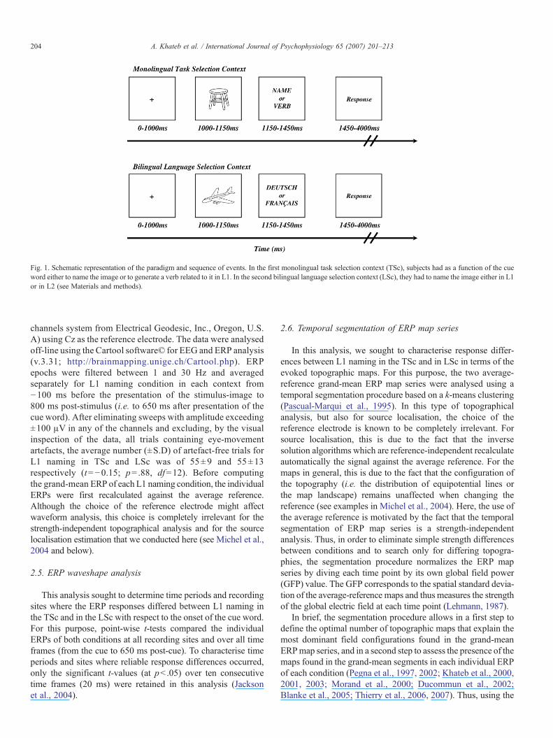
Fig. 1. Schematic representation of the paradigm and sequence of events. In the first monolingual task selection context (TSc), subjects had as a function of the cueword either to name the image or to generate a verb related to it in L1. In the second bilingual language selection context (LSc), they had to name the image either in L1or in L2 (see Materials and methods).
204 A. Khateb et al. / International Journal of Psychophysiology 65 (2007) 201–213
channels system from Electrical Geodesic, Inc., Oregon, U.S.A) using Cz as the reference electrode. The data were analysedoff-line using the Cartool software© for EEG and ERP analysis(v.3.31; http://brainmapping.unige.ch/Cartool.php). ERPepochs were filtered between 1 and 30 Hz and averagedseparately for L1 naming condition in each context from−100 ms before the presentation of the stimulus-image to800 ms post-stimulus (i.e. to 650 ms after presentation of thecue word). After eliminating sweeps with amplitude exceeding±100 μV in any of the channels and excluding, by the visualinspection of the data, all trials containing eye-movementartefacts, the average number (±S.D) of artefact-free trials forL1 naming in TSc and LSc was of 55±9 and 55±13respectively (t=−0.15; p= .88, df=12). Before computingthe grand-mean ERP of each L1 naming condition, the individualERPs were first recalculated against the average reference.Although the choice of the reference electrode might affectwaveform analysis, this choice is completely irrelevant for thestrength-independent topographical analysis and for the sourcelocalisation estimation that we conducted here (see Michel et al.,2004 and below).
2.5. ERP waveshape analysis
This analysis sought to determine time periods and recordingsites where the ERP responses differed between L1 naming inthe TSc and in the LSc with respect to the onset of the cue word.For this purpose, point-wise t-tests compared the individualERPs of both conditions at all recording sites and over all timeframes (from the cue to 650 ms post-cue). To characterise timeperiods and sites where reliable response differences occurred,only the significant t-values (at pb .05) over ten consecutivetime frames (20 ms) were retained in this analysis (Jacksonet al., 2004).
2.6. Temporal segmentation of ERP map series
In this analysis, we sought to characterise response differ-ences between L1 naming in the TSc and in LSc in terms of theevoked topographic maps. For this purpose, the two average-reference grand-mean ERP map series were analysed using atemporal segmentation procedure based on a k-means clustering(Pascual-Marqui et al., 1995). In this type of topographicalanalysis, but also for source localisation, the choice of thereference electrode is known to be completely irrelevant. Forsource localisation, this is due to the fact that the inversesolution algorithms which are reference-independent recalculateautomatically the signal against the average reference. For themaps in general, this is due to the fact that the configuration ofthe topography (i.e. the distribution of equipotential lines orthe map landscape) remains unaffected when changing thereference (see examples in Michel et al., 2004). Here, the use ofthe average reference is motivated by the fact that the temporalsegmentation of ERP map series is a strength-independentanalysis. Thus, in order to eliminate simple strength differencesbetween conditions and to search only for differing topogra-phies, the segmentation procedure normalizes the ERP mapseries by diving each time point by its own global field power(GFP) value. The GFP corresponds to the spatial standard devia-tion of the average-reference maps and thus measures the strengthof the global electric field at each time point (Lehmann, 1987).
In brief, the segmentation procedure allows in a first step todefine the optimal number of topographic maps that explain themost dominant field configurations found in the grand-meanERPmap series, and in a second step to assess the presence of themaps found in the grand-mean segments in each individual ERPof each condition (Pegna et al., 1997, 2002; Khateb et al., 2000,2001, 2003; Morand et al., 2000; Ducommun et al., 2002;Blanke et al., 2005; Thierry et al., 2006, 2007). Thus, using the

205A. Khateb et al. / International Journal of Psychophysiology 65 (2007) 201–213
spatial clustering procedure, we identify periods of quasi-stablemap topographies (i.e. similar field configuration) and computefor each period the mean map that represents the topography of
Fig. 2. ERP waveform analysis. A: Superimposed grand-mean ERP (±SEM) traces frthe whole scalp that highlights the major amplitude differences between contexts oveselected sites for this illustration with nose in the front and left at left. The traces arevertical lines) to 800 ms post-stimulus. The posterior sites (the lowest row) illustratcomponents, respectively at 100, 150 and∼250 ms. Note that the onset of the cue worthe N150, and that the expected P100 exogenous response to the cue coincides withwith the image-related P2 and can't be clearly dissociated. Note that, based on our wcue onset to 650 ms). B: Point-wise t-tests comparing individual ERPs to L1 naming inp values over time and recording sites (the 111 electrodes) and shows that the first macue. As displayed in panel A, these differences were the most prominent over left froappeared also at around 350 and 450 ms. These differences were also widespread andColour scale indicates p values of 0.05 to 0.01.
this period. Once these mean maps (referred to also as “segmentmaps”) are defined, their specificity for a given condition isverified by fitting them to the individual ERP map series in each
om TSc (black lines) and LSc (red lines) showing a subset of 34 electrodes overr time. The electrode schema in the upper right row indicates the position of thedisplayed with a 100 ms baseline before the image-stimulus onset (at 0 ms, rede the exogenous visual responses to the image: the P100, the N150 and the P2d (blue vertical lines), at 150 ms after the image onset, coincides with the peak ofthe ascending phase of the image-related P2. Hence, the cue-related P100 mixesorking hypothesis, the following analyses will focus on the post-cue period (i.e.TSc and LSc from cue onset to 650 ms after. The graph illustrates the significant
jor differences involving a large number of electrodes occurred at ∼220 ms post-ntal sites (see arrowheads on left anterior sites, panel A). Other later differencesaffected both left anterior and right posterior sites (see blue triangles in panel A).

206 A. Khateb et al. / International Journal of Psychophysiology 65 (2007) 201–213
context. Concretely, we calculate a spatial correlation coefficient(Brandeis et al., 1992) between each segment map and each mapin the subject's individual map series (Pegna et al., 1997; Khatebet al., 1999), and then each time point in the individual mapseries is labelled with the segment map it is most highlycorrelated with. Afterwards, different timing and spatialmeasures can be extracted for each segment map, particularlythe segment duration which refers to how many times eachtemplate segment map appeared in each individual ERP mapseries. Here, we compared statistically the segment durationparameter for determining condition-specific segment maps (i.e.those appearing preferentially in one but not in the othercondition) and those that are present in two conditions but mightdiffer in terms of duration.
2.7. Source localisation analysis
This analysis used the LAURA inverse solution (Grave dePeralta Menedez et al., 2001) to estimate brain regions that gaverise to electric field differences between the two contexts.LAURA is a distributed linear inverse solution calculated on arealistic head model that includes 4024 solutions points (i.e.voxels) equally distributed within the cortical and sub-corticalgrey matter of the average brain (Montreal NeurologicalInstitute, Montreal, Canada). Similar to other distributed inversesolutions, LAURA is capable of dealing with a priori unknownnumber and location of simultaneously active sources in thebrain (Ducommun et al., 2002; Khateb et al., 2003; Ortigueet al., 2004; Blanke et al., 2005; Thierry et al., 2006). Here, wefirst applied LAURA to the individual ERP time segments ofinterest. We then contrasted the individual estimated inversesolutions of LSc vs TSc using t-tests in order to determine brainregions that were more significantly activated (at pb .01) duringthe language selection context.
3. Results
3.1. Behavioural results
The results of the translation test using TRANSLOG indi-cated that the quality scores were high and homogenousamong the group (mean score=56±7 out of 80), except in onesubject who showed a relatively poorer score (34/80). Thesubjects' performance in the monolingual TSc showed a meancorrect response rate of 95±3% in verb generation and of 96±2% in L1 naming (p= .6). In the bilingual LSc, the subjectsshowed a mean correct response rate of 95±3% in L1 namingand of 85±10% in L2 naming (t=4.4; pb .001; df=12). Moreparticularly for our purpose, no performance difference wasobserved in L1 naming between TSc and LSc (p= .83).Concerning the response times (RTs), the TSc and the LScyielded globally similar RTs, indicating that difficulty overcontexts was highly comparable (mean RT=1160±140 and1176±138 ms, respectively in TSc and LSc; p= .45). Withinthe TSc, verb generation took longer time than L1 naming(mean=1202±144 and 1117±145 ms respectively; t±4.1;pb .002; df=12). In the LSc, response times for L1 naming did
not differ significantly from those for L2 naming (1156±130and 1196±170 ms respectively; p= .29). In particular for ourpurpose, RTs in L1 naming were slightly longer in LSc than inTSc, but this difference failed to reach significance (1156±130 and 1117±145 ms respectively, t=1.98; pb .073; df=12).
3.2. Analysis of ERP waveforms
The analysis aimed at identifying the earliest responsesdifferentiating L1 naming in the two contexts after cue onset.Fig. 2A shows on a subset of recording sites the superposition ofthe averaged waveforms (±SEM) induced by L1 namingcondition in TSc and LSc from −100 ms before the image(stimulus) onset to 800 ms post-stimulus (i.e. 650 ms post-cue).This illustration shows that some response differences appearedrelatively early after the cue onset (see arrowhead on leftanterior sites), but that other differences were also found later(see triangles on left anterior and right posterior sites). Inparticular and as expected, the most posterior sites (the lowestrow) that depict the primary visual responses in relation withimage presentation (successively the P100, N150 and P2components, at 100, 150 and ∼250 ms respectively) suggestno response difference during these early components. Of notealso is the fact that the cue-expected P100 response coincideswith the ongoing image-related P2, and thus can't be dissociatedhere.
In order to assess statistically the response differences bet-ween L1 naming in TSc and LSc, we compared the individualERP of the two contexts from the cue onset to 650 ms post-cue(i.e. 800 ms post-image) using point-wise t-tests. As shown inFig. 2B, which depicts the significant p values over time andrecording sites, the first major differences appeared at around220 ms post-cue and lasted up to 300 ms. These differencesconcerned various scalp locations but were most prominent atleft anterior recording sites (see examples on Fig. 2A, arrow-heads). Although of lesser significance to our working hypo-thesis, other later differences were also confirmed between∼350–400 ms and slightly after the 450 ms time range. Asexpected from the visual inspection of the grand-mean ERPs,these later differences involved both left anterior and rightposterior scalp regions (see examples in Fig. 2A, triangles).
3.3. Temporal segmentation of ERP map series
This analysis sought to characterise response differences bet-ween the two contexts in terms of the electric field topographies.The strength-independent segmentation of ERP map series of L1naming in TSc and LSc showed that a total of 18 topographictemplate maps (see Fig. 3A) explained the whole dataset.Figure 3B illustrates the time segments of stable topographicalconfiguration where these maps occurred in the grand-meanERPs. It shows that the same sequence of segments was found inthe two contexts up to around 200 ms and then some mapsappeared only in the TSc (e.g. maps #5 and #6) or in the LScgrand-mean (e.g. map #7). This suggested that this same period,where amplitude differences were also found through waveformstatistical comparison, differentiated contexts also in terms of the

Fig. 3. Temporal segmentation of the grand-mean ERP map series and source localisation analysis. A: The 18 topographic maps that explained the dominant fieldconfigurations found in the grand-mean ERP map series of L1 naming in TSc and LSc as revealed by the temporal segmentation procedure. The maps are shown fromtop with left ear left. Blue values indicate negative potentials, red values positive potentials. B: Global field power traces (see Materials and methods) of the grand-mean ERPs of L1 naming in TSc and LSc showing the time segments of stable map configuration (referred to as functional microstates) where these maps occurred ineach context. As in the waveform analysis, the first major differences appeared between 200 and 300 ms as attested by the dominant presence of map 6 in TSc only andof map 7 in LSc only. C: Axial MRI slices illustrating the mean inverse solutions over subjects for the period 220–270 ms (periods of map 6 and 7) in TSc and LSc andshowing similar source distribution with particularly the dominant activation of the bilateral inferior and middle occipital gyri (see text for the other regions). Note thatthe two solutions have the same intensity scale and that left is one left. D: Axial MRI slices and 3D brain images showing the results of the statistical analysis thatcontrasted the individual LSc and TSc inverse solutions during the 220–270 ms time period. The brain regions differentiating LSc and TSc (at pb .01) indicated astronger involvement of left fronto-parietal regions during LSc.
207A. Khateb et al. / International Journal of Psychophysiology 65 (2007) 201–213

208 A. Khateb et al. / International Journal of Psychophysiology 65 (2007) 201–213
global electric field configuration and thus presumably of theunderlying cerebral generators.
In order to verify this hypothesis statistically, we assessed thepresence of the segments' maps (referred to as “segments'duration”, see Materials and methods) in the individual L1naming ERPs of both contexts. For that, we looked for the maps1 to 7 in the time window between 0 and 320 and for maps 8–18in the time window between 310 and 650 ms. In the first timewindow, the 2×7 ANOVA performed on segments' durationusing contexts (2) and maps (7) as within subjects' factorsshowed a highly significant main effect for maps (F(6, 72)=14.4, pb .000001) due to the fact that the successive segmentshad varying durations. More interestingly, the highly significantinteraction, observed between the two analysis factors (F(6, 72)=3.8, pb .0025), indicated that some maps' durations differedbetween conditions. Post-hoc Fisher's LSD tests indeed showedthat map segments #6 and #7 significantly differentiated con-texts. Thus, map #6 was confirmed to occur more frequently inTSc (59±37 ms) than in LSc (34±20 ms, pb .002), while map#7 was more present in LSc (68±31) than in TSc (43±37 ms,pb .0015).
Although of lesser interest to our purpose, the 2×11 ANOVAperformed on the duration of the latter segments' maps (the 11remaining maps) showed again a highly significant main effectfor map (F(10, 120)=5.4, pb .000001), and a significant inter-action between the analysis factors (F(10, 120)=3.4, pb .0007).Post-hoc Fischer's LSD tests indeed showed that: map 13 wasfound more frequently in TSc (103±82 ms) than in LSc (58±47 ms, pb .0003); map 15 was more present in LSc (32±20 ms)than in TSc (4±7 ms, pb .025); and that map 17 was morepresent in TSc (53±60 ms) than in LSc (22±31 ms, pb .01). Ofnote is the fact that the duration in the individual ERPs ofsegments' maps 10, 12 and 18 followed the same tendency as inthe grand-mean segmentation (see Fig. 3B), but that thedurations' differences did not reach the statistical significance.
3.4. Source localisation analysis
This analysis aimed at estimating brain regions accountingfor the first field configuration differences between LSc andTSc and represented by segments' maps #7 and #6. For that, amean map was first calculated from each individual L1 namingERP of each context in the time period between 220 and270 ms. These maps were then individually subjected to theinverse solution algorithm (LAURA) and a t-test contrastingLSc vs TSc was afterwards computed on the current density ofall the LAURA solution points (i.e. voxels). Fig. 3C, whichillustrates the mean inverse solution across subjects for thistime period, shows that the dominant activation in TSc and LScwas globally found in similar brain regions. These includedprincipally the inferior and middle occipital gyri bilaterally(although more dominant in the left), the bilateral temporalgyrus but more dominantly in the left, the superior, middle andinferior frontal gyri, and the superior and inferior parietallobules. Fig. 3D depicts the brain regions where the estimatedindividual activation significantly differentiated LSc from TSc.It shows that this time period in LSc recruited differently left
hemispheric frontal and parietal areas. Antero-posteriorly, thelocalisation of the activated areas according to the Talairachand Tournoux's coordinates (Talairach and Tournoux, 1988)revealed the involvement at the frontal level of the middlefrontal–precentral gyri (BAs 9/6; x,y,z at the centre of gravityof the cluster=−58, 1, 43) and at the parietal level of thesupramarginal gyrus (BA 40/2; −62, −21, 35) and the angulargyrus (BA 39; −51, −62, 41).
4. Discussion
The aim of the present investigation was to determinewhether the cognitive mechanism that allows bilingual speakersto select one language rather than another is different from thatwhich allows the selection between various behavioural patternsincluding the different linguistic registers. To address this ques-tion, we analysed electric responses (ERP waveforms and mapseries) evoked by the same L1 naming condition as a function oftwo selection contexts: a monolingual (or intra-language) taskselection context (TSc) and a bilingual (or between languages)language selection context (LSc). The rationale was that if thebetween-language selection process differs from the intra-language task selection process, then electric brain responsesshould differentiate these two (otherwise exactly similar) L1naming conditions. This design diverges significantly fromother paradigms that manipulate language switching with apredictable task-sequence (Rogers and Monsell, 1995), whichthus allows for the assessment of the switching costs in terms ofresponse time and errors (see Jackson et al., 2001; Swainsonet al., 2003). Here, the presentation of the different conditions ineach selection context was randomised in another way for eachsubject, with a trial-by-trial cuing (Meiran, 1996) and withoutany predictable order. The random naming in L1 and L2 in LScwas a necessary manipulation in order to create the context ofbetween-languages selection, which was contrasted with that ofintra-language task selection.
Behaviourally, the analysis of subjects' correct responses inL1 naming in LSc and TSc showed a highly similar per-formance across contexts, indicating that task difficulty washighly comparable. In terms of response speed, although reac-tion times (RTs) for L1 naming did not differ significantlybetween contexts, our results show a trend towards longer RTs(of ∼40 ms) in LSc than in TSc. This additional time in LSc,presumably due to switching costs, fits within the range ofvalues estimated by previous studies (e.g. 25ms in Jackson et al.,2004 and 102 ms in Jackson et al., 2001). In comparison toHernandez et al.'s study (2001) where image naming conditionswere used, our results show that the RTs measured here for L1naming in both contexts were considerably longer than thosepreviously reported. However, it should be noted that in thestudy of Hernandez et al. (2001) the cue was presented before theimages while in our study it was presented after the images. Thisincrease in RTs corroborates thus previous finding showing thatswitching, which is a time-consuming process, delays responseselection if it occurs after target presentation (Swainson et al.,2006). Presenting a cue before the lexical item is thought toabolish costs related to in-between-language selection processes

209A. Khateb et al. / International Journal of Psychophysiology 65 (2007) 201–213
since selection is limited only to items within a single languagelexicon, a process similar to word production in monolinguals(see Grosjean, 1998). This might explain why in Hernadezet al.'s functional study (2001) no significant activation wasfound in left hemisphere language control areas when comparingswitching between languages to no-switching condition. In ourstudy, the bilingual subjects have first to enter the hypothesisedcommon conceptual representation for the two languages andthen language selection took place to retrieve the correct lexicalitems (supposedly with the concomitant inhibition of the non-target language).
At the electrophysiological level, we predicted that if theselection processes involved in the two contexts were differentin terms of their neural basis, then the electrical responsesevoked by the two L1 naming conditions should diverge rela-tively early after the analysis of the cues. The analysis of ERPwaveforms in terms of response amplitude confirmed our pre-diction by showing that the earliest major differences appearedat around 220 ms after the presentation of the cue and peaked ataround 250 ms. The strength-independent topographicalanalysis of ERP map series confirmed that this same timeperiod differentiated contexts in terms of the electric fieldtopographies. Actually, it revealed that, while L1 naming in LScand in TSc showed the same succession of microstates up to∼200ms, the period between∼200 and 300ms was dominantlycharacterised by the presence of one microstate (segment #6) inTSc and by another one (segment #7) in LSc, each having itsdistinct electric field configuration. Assuming that the two L1naming conditions differed only in terms of the selection processinvolved, it appears reasonable to assume that these early elec-trophysiological differences represent the correlates of thediffering processes involved, and thus that the neural responsesunderlying the between-language selection could differ fromthose involved in the switching from one linguistic register toanother.
In a previous ERP study, Jackson et al. (2001) investigatedthe time course of language switching in bilingual speakersusing a predictable productive switching task. They reportedthat switch trials, compared with non-switch trials, increased thefrontal N2 negativity (at around 320 ms) and this is only for L2trials. Later on, they found a modulation of a late positivecomponent (LPC, between 350 and 700 ms) for switch trials inboth languages. The authors proposed that the modulation of thefrontal N2 might reflect the suppression of the habitual response(L1) during L2 switch trials (Meuter and Allport, 1999) andsuggested that this interpretation was compatible with Green'sInhibitory Control Model of language switching (Green, 1998).Concerning the modulation of the LPC by switch trials in bothlanguages, the authors interpreted it as reflecting the reconfig-uration of the language-specific phonology-to-articulatory setson the basis of the cue. In a subsequent study, the authors in-vestigated a receptive language switching (Jackson et al., 2004)using number words presented in L1 and L2. Subjects wererequired, using motor responses, to judge whether the wordswere odd or even. In this later study, they failed to observe thefrontal and parietal switch-related activity, previously reportedin the productive switching task (Jackson et al., 2001). How-
ever, they reported an early switch-related activity on centralelectrodes that was not language-specific. In a within-sentenceswitching paradigm, Proverbio et al. (2004) used visual presen-tation of unmixed (one language) and mixed (mixing languagesat the final word) sentences and observed that the first effect oflexical and code switching was found between 140 and 200 msat left anterior sites. Due to the differences in the paradigmsused, no direct comparison could be made between our resultsand those of these previous studies. However, it is worth notingthat in these studies, as in our present findings, the first languageswitch-related modulation of the ERP concerned more partic-ularly the left anterior sites, showing thus a certain consistencyacross studies and this is independent of the paradigms used.Nevertheless, we hypothesise that the relatively early LSc-in-duced response observed here is not related to L1 suppression(as suggested by Jackson et al., 2001) since the comparisonsperformed here concerned in both contexts the L1 naming. Inthe language selection dual-schema context (i.e. naming in L1or name in L2, von Studnitz and Green, 1997) that we designedhere, these highly proficient bilingual subjects performed thetask according to a strongly mixed bilingual mode in which theyhad to switch continuously between their L1 and L2 and this isin a completely random fashion. Although, we intentionally didnot compare directly the ERPs to L1 and L2 naming to avoidinterpreting hazardously the ERP differences that can be duemerely to differing proficiency levels between the two lan-guages, we speculate that in such a context, the languageselection process, which is supposed to deactivate the lexicon ofthe non-target language (Green, 1998), is the same whether thesubjects have to name images in L1 or in L2.
Although of a lesser importance to our hypothesis, the laterERP differences, which we observed here between ∼350 and500 ms post-cue, appeared at posterior (central and right) sitesas an increased positivity for LSc as compared to TSc and as anincreased negativity for LSc vs TSc at anterior left sites. Theanalysis of the different segment maps occurring during thissecond time period confirmed that some of the maps were foundmore specifically in TSc (e.g. segment map 13) while otherswere found in LSc (e.g.map 15). Of note here is the fact that themodulation of our late responses resembles that foundpreviously in Jackson et al.'s (2001) study using the productiveswitching task (see above). Actually, in their average-referenceERP results, the authors reported that switch-related modulationof the LPC (350–700 ms) affected principally the parietal sites.However, it is important to note that the augmented LPC for theswitch conditions in their results was accompanied at the frontalsites with an increased negativity exactly as we found here.Given the fact that both studies (Jackson et al. and ours) usedlanguage production tasks, it is thus reasonable to assume thatthe late response difference between LSc and TSc might be dueto the reconfiguration of the language-specific phonology-to-articulatory sets: In our highly mixed LSc a language switchwas roughly required on a trial-by-trial basis, a process that isnot required for L1 naming in the TSc. Finally, one should alsonot exclude the possibility that the small RT difference betweenL1 naming in TSc and LSC might have slightly contributed tothe late ERP differences.
210 A. Khateb et al. / International Journal of Psychophysiology 65 (2007) 201–213
Although to be interpreted with caution due to the relativelylimited spatial resolution of these techniques, the mean inversesolution revealed in both contexts the dominant involvementof the occipital areas, but also of other frontal, temporal andparietal regions. The dominant activation of the occipital areasis consistent with the fact that this period coincide with that ofan increased posterior negativity (the ERP N2 component)where bilateral posterior activation had previously beenreported in relation with pictorial processing (Doniger et al.,2000; Khateb et al., 2002). More particularly, the statisticalcomparison of the individually estimated sources during thistime period showed that brain regions differentiating LSc fromTSc involved a subset of areas known for their participation invarious language and cognitive tasks (see Duncan and Owen,2000; Miller, 2000; Brass et al., 2005; Vigneau et al., 2006). Apoint of interest here is the fact that these areas were restricted tothe left hemisphere, corroborating thus previous clinicalobservations indicating that left lesions may be responsiblefor producing language switching difficulties (Marien et al.,2005). As already pointed out in other switching studies (Priceet al., 1999), the fronto-parietal areas described here were foundoutside of the classical Broca and Wernicke language areas. Thefrontal activation covered partially the posterior prefrontalcortex (BA 9). The prefrontal cortex, which constitutes a largecortical region, had repeatedly been proposed by clinical andfunctional studies to participate in cognitive control and switchingprocesses (Owen et al., 1993; Dove et al., 2000; Duncan andOwen, 2000), and particularly in language selection processesin bilinguals (Fabbro et al., 2000; Hernandez et al., 2001;Rodriguez-Fornells et al., 2002, 2005). For instance Rodri-guez-Fornells et al. (2005) have recently suggested that therecruitment of the left prefrontal cortex (BA 9/46), a typical‘executive function’ brain area, might be crucial in inhibitingthe production of the non-target language when subjects had toname a picture in the target language. The prefrontal cortex,which has also been implicated in working memory anddivided attention (Miller, 2000; Raye et al., 2002; Nebel et al.,2005), has been involved in a variety of language paradigmsincluding word generation, semantic categorisation, semanticfluency and rhyme detection tasks (see Seghier et al., 2004;Vigneau et al., 2006), confirming thus the participation of theprefrontal regions to various task sets (Dosenbach et al., 2006).Similarly, the motor areas and more specifically the precentralgyrus were also found in most language studies (Vigneau et al.,2006) and associated not only with articulatory planning andexecution but also with speech perception (Pulvermuller et al.,2006).
Together with the frontal region, we observed also signi-ficant difference in the anterior part of the left supramarginalgyrus (BA 40) and in the left angular gyrus (BA39), both areaspreviously found in various language studies. In particular, thesupramarginal gyrus has been often involved in phonologicalprocesses (e.g. mapping orthography to phonology, phonolog-ical recoding, rhyme detection etc., see Paulesu et al., 1993;Demonet et al., 1994; Seghier et al., 2004), while the angulargyrus has been involved in semantic processing (Binder et al.,1997; Price, 2000; Binder et al., 2005). The activation here is
also in accordance with previous clinical observations suggest-ing a role for the supramarginal gyrus in language switching(Paradis, 1983; Hernandez et al., 2001). In line with this finding,Price et al. (1999) have previously shown that language switchingis associated with an increased activation in the bilateral supra-marginal gyri. Related to these observations, a recent fMRI study(Venkatraman et al., 2006) showed that language switching ef-fects during exact arithmetic additions were found in the leftinferior frontal gyrus and the left inferior parietal lobule extendingto the angular gyrus. Also, a specific role for the inferior parietallobule in the bilingual brain has recently been supported bystudies using whole-brain mapping techniques. Actually,Mechelli et al. (2004) showed that acquiring the vocabulary of asecond language (L2, English in Italian native speakers) inducedstructural changes in the inferior parietal cortex as attested by greymatter density increases as a function of language proficiency.Interestingly, the same brain region was referred to as the‘language switching talent area’ by the classical German apha-siological literature (Poetzl, 1925, 1930; Leischner, 1948).
Contrary to other studies where a specific activation of theleft dorsolateral prefrontal cortex has been observed (Hernandezet al., 2001), our analysis did not reveal the particular activationof this area in this context. This region, which has been found invarious other contexts requiring increased cognitive controlsuch as task switching and divided/focused attention (D'Espo-sito et al., 1995; Swainson et al., 2003; Brass et al., 2005; Nebelet al., 2005), might have been involved similarly in our mono-lingual TSc and bilingual LSc during this specific time period,and thus did not appear in this statistical comparison. Likewise,differing from clinical (Abutalebi et al., 2000; Marien et al.,2005) and recent functional imaging studies (Crinion et al.,2006) where the left caudate nucleus has been involved inlanguage selection, our analysis did not show the activation ofthis specific area. In Crinion et al.'s study (2006) wheresemantic decision task was used in bilinguals, it has beenindicated that the left caudate responses were highest not onlywhen there was a change in language but also when a change inword meaning occurred. It is worth noting that other clinicalreports in monolingual patients indicated that the left caudatedamage might be associated with naming and word-findingdifficulties, thus suggesting that its lesion might impair thepatients' ability to select the appropriate lexical–semantic res-ponses (see Crinion et al., 2006). In view of such observations,it is likely that the caudate nucleus had also been engagedduring the lexical switching from one linguistic register toanother during TSc and thus did not appear in our analysis. Thepossibility that the caudate nucleus was not involved at allduring this processing step should also not be excluded. Indeed,a recent review by Friederici (2006) suggested that the leftcaudate nucleus, which participates to a variety of languagetasks, thanks to its connections with the frontal, motor andtemporo-parietal cortex, might activate when the languageprocessing system cannot rely entirely on automatic mechan-isms. The late ERP differences between LSc and TSc (but alsothe slight increase of RTs in LSc), which might presumably belinked to phonological re-mapping process, strongly indicatethat L1 naming in the bilingual context was achieved through

211A. Khateb et al. / International Journal of Psychophysiology 65 (2007) 201–213
more controlled than in L1 naming in the monolingual context.Thus, one could not rule out that the left caudate nucleus (butalso other areas involved in cognitive control) was engagedduring other steps of information processing.
To conclude, our results point to the participation of fronto-parietal areas in language selection processes. The involvement ofthese regions in language processing network has recently beensupported by a diffusion tensor imaging study that described aconnection between the posterior frontal cortex (in particular themiddle frontal gyrus) and the inferior parietal cortex (preciselyBA 40 and 39, Catani et al., 2005). This observation, togetherwith the fact that the brain areas found here were also shown toparticipate in the processing of various linguistic processesindicates that language selection might rely not only on regionsinvolved in cognitive control but also on other neuralmodules thatare part of the extended language neural network defined byrecent neuroimaging studies. Further studies comparing languageand non-language switching paradigms are however required tosubstantiate the specific role of such areas in the early steps oflanguage selection processes.
Acknowledgements
This research was supported by the Swiss National ScienceFoundation grant nos. 3151A0-102271/1 and 320000-109928and by the Center for Biomedical Imaging (CIBM) of Genevaand Lausanne. We thank Dr Rolando Grave de Peralta Menedezand Dr Sara Gonzales Andino for providing us with the inversesolutions and Mrs Tanja Heiden for her help in L2 proficiencyassessment and subjects' recruitment.
References
Abutalebi, J., Miozzo, A., Cappa, S., 2000. Do subcortical structures controllanguage selection in bilinguals? Evidence from pathological languagemixing. Neurocase 6, 101–106.
Aglioti, S., Fabbro, F., 1993. Paradoxical selective recovery in a bilingualaphasic following subcortical lesion. NeuroReport 4, 1359–1362.
Binder, J.R., Frost, J.A., Hammeke, T.A., Cox, R.W., Rao, S.M., Prieto, T.,1997. Human brain language areas identified by functional magneticresonance imaging. J. Neurosci. 17, 353–362.
Binder, J.R., Medler, D.A., Desai, R., Conant, L.L., Liebenthal, E., 2005. Someneurophysiological constraints on models of word naming. NeuroImage 27,677–693.
Blanke, O., Mohr, C., Michel, C.M., Pascual-Leone, A., Brugger, P., Seeck, M.,Landis, T., Thut, G., 2005. Linking out-of-body experience and selfprocessing to mental own-body imagery at the temporoparietal junction.J. Neurosci. 25, 550–557.
Brandeis, D., Naylor, H., Halliday, R., Callaway, E., Yano, L., 1992. Scopolamineeffects on visual information processing, attention and event-related potentialmap latencies. Psychophysiology 29, 315–336.
Brass, M., Ullsperger, M., Knoesche, T.R., von Cramon, D.Y., Phillips, N.A.,2005. Who comes first? The role of the prefrontal and parietal cortex incognitive control. J. Cogn. Neurosci. 17, 1367–1375.
Catani, M., Jones, D.K., ffytche, D.H., 2005. Perisylvian language networks ofthe human brain. Ann. Neurol. 57, 8–16.
Chee, M.W., Tan, E.W., Thiel, T., 1999. Mandarin and English single wordprocessing studied with functional magnetic resonance imaging. J. Neurosci.19, 3050–3056.
Colomé, A., 2001. Lexical activation in bilinguals' speech production:language-specific or language independent? J. Mem. Lang. 45, 721–736.
Costa, A., Caramazza, A., 1999. Is lexical selection in bilinguals language-specific? Further evidence from Spanish–English bilinguals and English–Spanish bilinguals. Bilingualism: Lang. Cogn. 2, 231–244.
Crinion, J., Turner, R., Grogan, A., Hanakawa, T., Noppeney, U., Devlin, J.T.,Aso, T., Urayama, S., Fukuyama, H., Stockton, K., Usui, K., Green, D.W.,Price, C.J., 2006. Language control in the bilingual brain. Science 312,1537–1540.
Demonet, J.F., Price, C., Wise, R., Frackowiak, R.S., 1994. Differentialactivation of right and left posterior sylvian regions by semantic andphonological tasks: a positron-emission tomography study in normal humansubjects. Neurosci. Lett. 182, 25–28.
D'Esposito, M., Detre, J.A., Alsop, D.C., Shin, R.K., Atlas, S., Grossman, M.,1995. The neural basis of the central executive system of working memory.Nature 378, 279–281.
Doniger, G.M., Foxe, J.J., Murray, M.M., Higgins, B.A., Snodgrass, J.G.,Schroeder, C.E., 2000. Activation timecourse of ventral visual streamobject-recognition areas: high density electrical mapping of perceptualclosure processes. J. Cogn. Neurosci. 12, 615–621.
Dosenbach, N.U., Visscher, K.M., Palmer, E.D., Miezin, F.M., Wenger, K.K.,Kang, H.C., Burgund, E.D., Grimes, A.L., Schlaggar, B.L., Petersen, S.E.,2006. A core system for the implementation of task sets. Neuron 50,799–812.
Dove, A., Pollmann, S., Schubert, T., Wiggins, C.J., von Cramon, D.Y., 2000.Prefrontal cortex activation in task switching: an event-related fMRI study.Brain Res. Cogn. Brain Res. 9, 103–109.
Ducommun, C.Y., Murray, M.M., Thut, G., Bellmann, A., Viaud-Delmon, I.,Clarke, S., Michel, C.M., 2002. Segregated processing of auditory motionand auditory location: an ERP mapping study. NeuroImage 16, 76–88.
Duncan, J., Owen, A.M., 2000. Common regions of the human frontal loberecruited by diverse cognitive demands. Trends Neurosci. 23, 475–483.
Fabbro, F., Skrap, M., Aglioti, S., 2000. Pathological switching betweenlanguages after frontal lesions in a bilingual patient. J. Neurol. Neurosurg.Psychiatry 68, 650–652.
Francis, W.S., 1999. Cognitive integration of language and memory inbilinguals: semantic representation. Psychol. Bull. 125, 193–222.
Friederici, A.D., 2006. What's in control of language? Nat. Neurosci. 9, 991–992.Gollan, T., Kroll, J., 2001. Lexical access in bilinguals. In: Rapp, B. (Ed.), A
Handbook of Cognitive Neuropsychology: What Deficits Reveal about theHuman Mind. Psychology Press, New York, pp. 321–345.
Grainger, J., 1993. Visual word recognition in bilinguals. In: Schreuder, R.,Weltens, B. (Eds.), The Bilingual Lexicon. John Benjamins, Amsterdam,pp. 11–25.
Grainger, J., Dijkstra, A., 1992. On the representation and use of languageinformation in bilinguals. In: Harris, R.J. (Ed.), Cognitive Processing inBilinguals. Elsevier, Amsterdam, pp. 207–220.
Grave de Peralta Menedez, R., Gonzalez Andino, S., Lantz, G., Michel, C.M.,Landis, T., 2001. Noninvasive localization of electromagnetic epilepticactivity. I. Method descriptions and simulations. Brain Topogr. 14, 131–137.
Green, D., 2003. The neural basis of the lexicon and the grammar in L2acquisition. In: van Hout, R., et al. (Ed.), The Interface between Syntax andthe Lexicon in Second Language Acquisition. John Benjamins, Amsterdam.
Green, D.W., 1998. Mental control of the bilingual lexico-semantic system.Bilingualism: Lang. Cogn. 1, 67–81.
Grosjean, F., 1992. Another view of bilingualism. In: Harris, R. (Ed.), CognitiveProcessing in Bilinguals. Elsevier, Amsterdam, pp. 51–62.
Grosjean, F., 1998. Studying bilinguals: methodological and conceptual issues.Bilingualism: Lang. Cogn. 1, 131–149.
Grosjean, F., 2001. The bilingual's language modes. In: Janet, L. (Ed.), OneMind,Two Languages: Bilingual Sentence Processing. Blackwell, Oxford, pp. 1–22.
Hernandez, A.E., Dapretto, M., Mazziotta, J., Bookheimer, S., 2001. Languageswitching and language representation in Spanish–English bilinguals: anfMRI study. NeuroImage 14, 510–520.
Jackson, G.M., Swainson, R., Cunnington, R., Jackson, S.R., 2001. ERPcorrelates of executive control during repeated language-switching.Bilingualism: Lang. Cogn. 4, 169–178.
Jackson, G.M., Swainson, R., Mullin, A., Cunnington, R., Jackson, S.R., 2004.ERP correlates of a receptive language-switching task. Q. J. Exp. Psychol.A. 57, 223–240.

212 A. Khateb et al. / International Journal of Psychophysiology 65 (2007) 201–213
Jakobsen, A.L.L.S., 1999. Translog documentation. In: Hansen, G. (Ed.),Probing the Process in Translation: Methods and Results. CopenhagenStudies in Language, vol. 24. Samfundslitteratur, Copenhagen.
Khateb, A., Annoni, J.M., Landis, T., Pegna, A.J., Custodi, M.C., Fonteneau, E.,Morand, S.M., Michel, C.M., 1999. Spatio-temporal analysis of electricbrain activity during semantic and phonological word processing. Int.J. Psychophysiol. 32, 215–231.
Khateb, A., Michel, C., Pegna, A., Thut, G., Landis, T., Annoni, J., 2001. Thetime course of semantic category processing in the cerebral hemispheres: anelectrophysiological study. Brain Res. Cogn. Brain Res. 10, 251–264.
Khateb, A., Michel, C.M., Pegna, A.J., Landis, T., Annoni, J.M., 2000. Newinsights into the Stroop effect: a spatio-temporal analysis of electric brainactivity. NeuroReport 11, 1849–1855.
Khateb, A., Michel, C.M., Pegna, A.J., O'Dochartaigh, S.D., Landis, T.,Annoni, J.M., 2003. Processing of semantic categorical and associativerelations: an ERP mapping study. Int. J. Psychophysiol. 49, 41–55.
Khateb, A., Pegna, A.J., Michel, C.M., Landis, T., Annoni, J.M., 2002.Dynamics of brain activation during an explicit word and image recognitiontask: an electrophysiological study. Brain Topogr. 14, 197–213.
Kroll, J.F., Peck, A., 1998. Competing activation across a bilingual's twolanguages: evidence from picture naming. Paper Presented at the 43rd.Annual Meeting of the International Linguistic Association. New YorkUniversity, New York.
Kroll, J.F., Stewart, E., 1994. Category interference in translation and picturenaming: evidence for asymmetric connections between bilingual memoryrepresentations. J. Lang. Mem. 33, 149–174.
Lee, M.W., Williams, J.N., 2001. Lexical access in spoken word recognition bybilinguals: evidence from the semantic competitor priming paradigm.Bilingualism: Lang. Cogn. 4, 233–248.
Lehmann, D., 1987. Principles of spatial analysis. In: Gevins, A.S., Remond, A.(Eds.), Handbook of Electroencephalography and Clinical Neurophysiolo-gy. Vol 1: Methods of Analysis of Brain Electrical and Magnetic Signals.Elsevier, Amsterdam, pp. 309–354.
Leischner, A., 1948. Readings on aphasia in bilinguals and polyglots. In: Paradis,M. (Ed.), On the Aphasia of Multilinguals. Didier, Montreal, pp. 456–502,1983.
Marien, P., Abutalebi, J., Engelborghs, S., De Deyn, P.P., 2005. Pathophysiologyof language switching and mixing in an early bilingual child with subcorticalaphasia. Neurocase 11, 385–398.
Mechelli, A., Crinion, J.T., Noppeney, U., O'Doherty, J., Ashburner, J.,Frackowiak, R.S., Price, C.J., 2004. Neurolinguistics: structural plasticity inthe bilingual brain. Nature 431, 757.
Meiran, N., 1996. Reconfiguration of processing mode prior to taskperformance. J. Exp. Psychol. Learn. Mem. Cogn. 22, 1423–1442.
Meuter, R., Allport, D., 1999. Bilingual language switching in naming: asym-metrical costs of language selection. J. Mem. Lang. 40, 25–40.
Michel, C.M., Murray, M.M., Lantz, G., Gonzalez, S., Spinelli, L., Grave dePeralta, R., 2004. EEG source imaging. Clin. Neurophysiol. 115, 2195–2222.
Miller, E.K., 2000. The prefrontal cortex and cognitive control. Nat. Rev.Neurosci. 1, 59–65.
Morand, S., Thut, G., de Peralta, R.G., Clarke, S., Khateb, A., Landis, T.,Michel, C.M., 2000. Electrophysiological evidence for fast visual processingthrough the human koniocellular pathway when stimuli move. Cereb. Cortex10, 817–825.
Nebel, K., Wiese, H., Stude, P., de Greiff, A., Diener, H.C., Keidel, M., 2005. Onthe neural basis of focused and divided attention. Brain Res. Cogn. BrainRes. 25, 760–776.
Oldfield, R.C., 1971. The assessment and analysis of handedness: the EdinburgInventory. Neuropsychologia 9, 97–113.
Ortigue, S., Michel, C.M., Murray, M.M., Mohr, C., Carbonnel, S., Landis, T.,2004. Electrical neuroimaging reveals early generator modulation toemotional words. NeuroImage 21, 1242–1251.
Owen, A.M., Roberts, A.C., Hodges, J.R., Summers, B.A., Polkey, C.E.,Robbins, T.W., 1993. Contrasting mechanisms of impaired attentional set-shifting in patients with frontal lobe damage or Parkinson's disease. Brain116 (Pt 5), 1159–1175.
Paradis, M., 1983. Readings on Aphasia in Bilinguals and Polyglots. MarcelDidier, Montreal.
Pascual-Marqui, R.D., Michel, C.M., Lehmann, D., 1995. Segmentation of brainelectrical activity into microstates: model estimation and validation. IEEETrans. Biomed. Eng. 7, 658–665.
Paulesu, E., Frith, C.D., Frackowiak, R.S., 1993. The neural correlates of theverbal component of working memory. Nature 362, 342–345.
Pegna, A.J., Khateb, A., Murray, M.M., Landis, T., Michel, C.M., 2002. Neuralprocessing of illusory and real contours revealed by high-density ERPmapping. NeuroReport 13, 965–968.
Pegna, A.J., Khateb, A., Spinelli, L., Seeck, M., Landis, T., Michel, C.M., 1997.Unraveling the cerebral dynamics of mental imagery. Hum. Brain Mapp. 5,410–421.
Perani, D., Abutalebi, J., 2005. The neural basis of first and second languageprocessing. Curr. Opin. Neurobiol. 15, 202–206.
Perani, D., Paulesu, E., Galles, N.S., Dupoux, E., Dehaene, S., Bettinardi, V.,Cappa, S.F., Fazio, F., Mehler, J., 1998. The bilingual brain. Profi-ciency and age of acquisition of the second language. Brain 121 (Pt 10),1841–1852.
Poetzl, O., 1925. Ueber die parietal bedingte Aphasie und ihren Einfluss auf dasSprechen mehrerer Sprachen. eitschrift fuer die gesamte. Neurol. Psychiatr.99, 100–124.
Poetzl, O., 1930. Aphasie und Mehrsprachigkeit. Zeitschrift fuer die gesamte.Neurol. Psychiatr. 124, 145–162.
Price, C.J., 2000. The anatomy of language: contributions from functionalneuroimaging. J Anat. 197 (Pt 3), 335–359.
Price, C.J., Green, D.W., von Studnitz, R., 1999. A functional imaging study oftranslation and language switching. Brain 122, 2221–2235.
Proverbio, A.M., Leoni, G., Zani, A., 2004. Language switching mech-anisms in simultaneous interpreters: an ERP study. Neuropsychologia 42,1636–1656.
Pulvermuller, F., Huss, M., Kherif, F., Moscoso Del Prado Martin, F., Hauk, O.,Shtyrov, Y., 2006. Motor cortex maps articulatory features of speech sounds.Proc. Natl. Acad. Sci. U. S. A. 103, 7865–7870.
Raye, C.L., Johnson, M.K., Mitchell, K.J., Reeder, J.A., Greene, E.J., 2002.Neuroimaging a single thought: dorsolateral PFC activity associated withrefreshing just-activated information. NeuroImage 15, 447–453.
Rodriguez-Fornells, A., Rotte, M., Heinze, H., Noesselt, T., Muente, T., 2002.Brain potential and functional MRI evidence for how to handle twolanguages with one brain. Nature 415, 1026–1029.
Rodriguez-Fornells, A., van der Lugt, A., Rotte, M., Britti, B., Heinze, H.J.,Munte, T.F., 2005. Second language interferes with word production influent bilinguals: brain potential and functional imaging evidence. J. Cogn.Neurosci. 17, 422–433.
Roelofs, A., 2003. Goal-referenced selection of verbal action: modellingattentional control in the Stroop task. Psychol. Rev. 110, 88–125.
Rogers, R., Monsell, S., 1995. Costs of a predictable switch between simplecognitive tasks. J. Exp. Psychol. Gen. 124, 207–231.
Seghier, M.L., Lazeyras, F., Pegna, A.J., Annoni, J.M., Zimine, I., Mayer, E.,Michel, C.M., Khateb, A., 2004. Variability of fMRI activation during aphonological and semantic language task in healthy subjects. Hum. BrainMapp. 23, 140–155.
Shallice, T., 1994.Multiple levels of control processes. In: Umilta, C.,Moscovitch,M. (Eds.), Attention and Performance XV: Conscious and NonconsciousInformation Processing. MIT Press, Cambridge, MA, pp. 395–420.
Snodgrass, J.G., Vanderwart, M., 1980. A standardized set of 260 pictures: normsfor name agreement, image agreement, familiarity and visual complexity.J. Exp. Psychol. 6, 174–215.
Swainson, R., Cunnington, R., Jackson, G.M., Rorden, C., Peters, A.M., Morris,P.G., Jackson, S.R., 2003. Cognitive control mechanisms revealed by ERPand fMRI: evidence from repeated task-switching. J. Cogn. Neurosci. 15,785–799.
Swainson, R., Jackson, S.R., Jackson, G.M., 2006. Using advance informationin dynamic cognitive control: an ERP study of task-switching. Brain Res.1105, 61–72.
Talairach, J., Tournoux, P., 1988. Co-planar Stereotaxic Atlas of the HumanBrain. Thieme, New York.
Thierry, G., Martin, C.D., Downing, P., Pegna, A.J., 2007. Controlling forinterstimulus perceptual variance abolishes N170 face selectivity. Nat.Neurosci. 10, 505–511.

213A. Khateb et al. / International Journal of Psychophysiology 65 (2007) 201–213
Thierry, G., Pegna, A.J., Dodds, C., Roberts, M., Basan, S., Downing, P., 2006.An event-related potential component sensitive to images of the humanbody. NeuroImage 32, 871–879.
Venkatraman, V., Siong, S.C., Chee, M.W., Ansari, D., 2006. Effect of languageswitching on arithmetic: a bilingual FMRI study. J. Cogn. Neurosci. 18,64–74.
Vigneau, M., Beaucousin, V., Herve, P.Y., Duffau, H., Crivello, F., Houde, O.,Mazoyer, B., Tzourio-Mazoyer, N., 2006. Meta-analyzing left hemisphere
language areas: phonology, semantics, and sentence processing. Neuro-Image 30, 1414–1432.
von Studnitz, R., Green, D., 1997. Lexical decision and language switching. Int.J Bilingualism 1, 3–24.
Wartenburger, I., Heekeren, H.R., Abutalebi, J., Cappa, S., Villringer, A., Perani,D., 2003. Early setting of grammatical processing in the bilingual brain.Neuron 37, 159–170.