Lack of Lithium-Like Behavioral and Molecular Effects in IMPA2 Knockout Mice
11
Lack of Lithium-Like Behavioral and Molecular Effects in IMPA2 Knockout Mice Kim Cryns* ,1,3 , Alon Shamir 2,3 , Joseph Shapiro 2 , Gie Daneels 1 , Ilse Goris 1 , Hansfried Van Craenendonck 1 , Roel Straetemans 1 , RH Belmaker 2 , Galila Agam 2 , Dieder Moechars 1 and Thomas Steckler 1 1 Research and Early Development Europe, Johnson & Johnson Pharmaceutical Research and Development, Beerse, Belgium; 2 Stanley Research Center, Faculty of Health Sciences, Ben Gurion University of the Negev and Mental Health Center, Beer-Sheva, Israel Lithium is a potent mood-stabilizing medication in bipolar disorder. Despite 50 years of clinical use, the mechanism of action is unknown. Multiple effects have been attributed to lithium including the uncompetitive inhibition of inositol monophosphatase (IMPase). IMPA2, one of the genes that encode IMPase, is located in a region with linkage to bipolar disorder. Owing to the role of IMPase in cell signaling and the possibility that this enzyme is a target for mood-stabilizing drugs, we generated IMPA2 / mice. Possible involvement of IMPase in complex behaviors related to affective disorders was assessed by monitoring the behavior of the IMPA2 / mice in the forced swim test, the tail suspension test (TST), the elevated zero-maze and open field test. It has been described that chronically lithium-treated mice exhibit reduced immobility time in the forced swim test and decreased exploratory behavior. We found increased rearing of IMPA2 / mice in the open field, suggesting an increased exploratory behavior. Although immobility time of IMPA2 / female but not male mice in the forced swim test was reduced, no difference was found between male and female IMPA2 / and IMPA2 +/+ mice in the TST and overall there was no clear effect of the deletion of IMPA2 on depression-like behavior. Frontal cortex IMPase activity and inositol levels in the IMPA2 / mice did not differ from IMPA2 +/+ mice, but kidney inositol levels were reduced. In conclusion, phenotypic characterization of the IMPA2 / mouse indicates that deleting IMPA2 does not mimic the effects of lithium treatment. Neuropsychopharmacology (2007) 32, 881–891. doi:10.1038/sj.npp.1301154; published online 12 July 2006 Keywords: IMPA2; lithium; bipolar disorder; behavioral analysis; IMPase activity; inositol levels INTRODUCTION Bipolar disorder is a serious psychiatric disorder. It affects at least 1% of the population and is characterized by episodes of mania and depression (Goodwin and Jamison, 1990). Notwithstanding the devastating impact, the under- lying pathophysiology is still unknown. Lithium has been the standard pharmacological treatment for bipolar dis- order over the last 50 years (Goodwin and Jamison, 1990). In vivo and in vitro studies demonstrated that lithium exerts multiple effects on neurotransmitter/receptor-mediated signaling, ion transport, signal transduction cascades, hormonal and circadian regulation, and gene expression (for a review, see Jope (1999) and Quiroz et al (2004)). At therapeutically relevant concentrations, it inhibits a group of at least four related phosphomonoesterases (inositol monophosphate phosphatase, inositol polyphosphate phosphatase, fructose 1,6-bisphosphate 1-phosphatase, and bisphosphate nucleotidase), the metabolic enzyme phosphoglucomutase, and glycogen synthase kinase-3 (GSK3). Despite 50 years of clinical use and despite intensive research, the precise mechanism by which lithium exerts its therapeutic effects is still unknown. A heuristic hypothesis proposed to explain lithium’s mechanism of action is the inositol depletion hypothesis (Berridge et al, 1989), which suggests that uncompetitive inhibition of inositol monophosphatase (IMPase) and inositol poly- phosphate 1-phosphatase (IPPase) by therapeutically rele- vant lithium concentrations leads to an accumulation of inositol phosphates and a corresponding depletion of free myo-inositol. The consequent reduced rate of re-synthesis of phosphoinositide second messenger signal generation attenuates the response to neurotransmission. Valproic acid and carbamazepine, two other mood-stabilizing drugs, also cause depletion of free myo-inositol in rodent brain and in neuronal cells in vitro (Shaltiel et al, 2004; Williams et al, 2002), thereby indicating that alterations in phospha- tidylinositol-cycle activity may indeed play an important Online publication: 7 June 2006 at http://www.acnp.org/citations/ Npp060706050702/default.pdf Received 28 November 2005; revised 20 April 2006; accepted 6 June 2006 *Correspondence: Dr K Cryns, Research and Early Development Europe, Johnson & Johnson Pharmaceutical Research and Develop- ment, Turnhoutseweg 30, B-2340 Beerse, Belgium, Tel: +32 14 603819, Fax: + 32 14 607266, E-mail: [email protected] 3 Both authors contributed equally to this work. Neuropsychopharmacology (2007) 32, 881–891 & 2007 Nature Publishing Group All rights reserved 0893-133X/07 $30.00 www.neuropsychopharmacology.org
Transcript of Lack of Lithium-Like Behavioral and Molecular Effects in IMPA2 Knockout Mice
Lack of Lithium-Like Behavioral and Molecular Effects inIMPA2 Knockout Mice
Kim Cryns*,1,3, Alon Shamir2,3, Joseph Shapiro2, Gie Daneels1, Ilse Goris1, Hansfried Van Craenendonck1,Roel Straetemans1, RH Belmaker2, Galila Agam2, Dieder Moechars1 and Thomas Steckler1
1Research and Early Development Europe, Johnson & Johnson Pharmaceutical Research and Development, Beerse, Belgium; 2Stanley Research
Center, Faculty of Health Sciences, Ben Gurion University of the Negev and Mental Health Center, Beer-Sheva, Israel
Lithium is a potent mood-stabilizing medication in bipolar disorder. Despite 50 years of clinical use, the mechanism of action is unknown.
Multiple effects have been attributed to lithium including the uncompetitive inhibition of inositol monophosphatase (IMPase). IMPA2, one
of the genes that encode IMPase, is located in a region with linkage to bipolar disorder. Owing to the role of IMPase in cell signaling and
the possibility that this enzyme is a target for mood-stabilizing drugs, we generated IMPA2�/� mice. Possible involvement of IMPase in
complex behaviors related to affective disorders was assessed by monitoring the behavior of the IMPA2�/� mice in the forced swim test,
the tail suspension test (TST), the elevated zero-maze and open field test. It has been described that chronically lithium-treated mice
exhibit reduced immobility time in the forced swim test and decreased exploratory behavior. We found increased rearing of IMPA2�/�
mice in the open field, suggesting an increased exploratory behavior. Although immobility time of IMPA2�/� female but not male mice in
the forced swim test was reduced, no difference was found between male and female IMPA2�/� and IMPA2 + / + mice in the TST and
overall there was no clear effect of the deletion of IMPA2 on depression-like behavior. Frontal cortex IMPase activity and inositol levels in
the IMPA2�/� mice did not differ from IMPA2 + / + mice, but kidney inositol levels were reduced. In conclusion, phenotypic
characterization of the IMPA2�/� mouse indicates that deleting IMPA2 does not mimic the effects of lithium treatment.
Neuropsychopharmacology (2007) 32, 881–891. doi:10.1038/sj.npp.1301154; published online 12 July 2006
Keywords: IMPA2; lithium; bipolar disorder; behavioral analysis; IMPase activity; inositol levels
������������������������������������������������
INTRODUCTION
Bipolar disorder is a serious psychiatric disorder. It affectsat least 1% of the population and is characterized byepisodes of mania and depression (Goodwin and Jamison,1990). Notwithstanding the devastating impact, the under-lying pathophysiology is still unknown. Lithium has beenthe standard pharmacological treatment for bipolar dis-order over the last 50 years (Goodwin and Jamison, 1990).In vivo and in vitro studies demonstrated that lithium exertsmultiple effects on neurotransmitter/receptor-mediatedsignaling, ion transport, signal transduction cascades,hormonal and circadian regulation, and gene expression(for a review, see Jope (1999) and Quiroz et al (2004)). At
therapeutically relevant concentrations, it inhibits a groupof at least four related phosphomonoesterases (inositolmonophosphate phosphatase, inositol polyphosphatephosphatase, fructose 1,6-bisphosphate 1-phosphatase,and bisphosphate nucleotidase), the metabolic enzymephosphoglucomutase, and glycogen synthase kinase-3(GSK3). Despite 50 years of clinical use and despiteintensive research, the precise mechanism by which lithiumexerts its therapeutic effects is still unknown. A heuristichypothesis proposed to explain lithium’s mechanism ofaction is the inositol depletion hypothesis (Berridge et al,1989), which suggests that uncompetitive inhibition ofinositol monophosphatase (IMPase) and inositol poly-phosphate 1-phosphatase (IPPase) by therapeutically rele-vant lithium concentrations leads to an accumulation ofinositol phosphates and a corresponding depletion of freemyo-inositol. The consequent reduced rate of re-synthesisof phosphoinositide second messenger signal generationattenuates the response to neurotransmission. Valproicacid and carbamazepine, two other mood-stabilizing drugs,also cause depletion of free myo-inositol in rodent brainand in neuronal cells in vitro (Shaltiel et al, 2004; Williamset al, 2002), thereby indicating that alterations in phospha-tidylinositol-cycle activity may indeed play an important
Online publication: 7 June 2006 at http://www.acnp.org/citations/Npp060706050702/default.pdf
Received 28 November 2005; revised 20 April 2006; accepted 6 June2006
*Correspondence: Dr K Cryns, Research and Early DevelopmentEurope, Johnson & Johnson Pharmaceutical Research and Develop-ment, Turnhoutseweg 30, B-2340 Beerse, Belgium, Tel: + 32 14603819, Fax: + 32 14 607266, E-mail: [email protected] authors contributed equally to this work.
Neuropsychopharmacology (2007) 32, 881–891& 2007 Nature Publishing Group All rights reserved 0893-133X/07 $30.00
www.neuropsychopharmacology.org
role in the pathophysiology and/or treatment of bipolardisorder.
IMPase is a key enzyme in the phosphatidylinositolsignaling system. Two human genes encoding for IMPase(IMPA genes) have been cloned. The IMPA1 gene was foundon chromosome 8q21.13–21.3 (Sjoholt et al, 1997) andIMPA2 was located on chromosome 18p11.2 (Sjoholt et al,2000; Yoshikawa et al, 2000, 1997). Several studies haveindicated the presence of a susceptibility locus for bipolardisorder on chromosome 18p (Berrettini et al, 1994; Rojaset al, 2000). Sjoholt et al (2004) reported an associationbetween bipolar illness and single-nucleotide polymorph-isms in the promoter of IMPA2. Yoon et al (2001) reportedthat B lymphoblast cell lines derived from male bipolarpatients exhibit downregulation of IMPA2 expressionaccompanied by high free intracellular Ca + 2 concentration.
Subconvulsive doses of the cholinergic agonist pilocar-pine induce limbic seizures in rodents pretreated withlithium (Honchar et al, 1983). Lithium by itself does nothave proconvulsant effects in rodents. Induction of lithium–pilocarpine seizures results in a drop in cortical inositol andelevation of inositol monophosphate levels. The reversal oflithium’s effect by inositol provided behavioral support forthe inositol depletion hypothesis (Kofman and Belmaker,1993).
Owing to the important role of IMPase in cell signaling, ingeneral, and the possibility that this enzyme is a target formood-stabilizing drugs, we used a retrovirus-mediated genetrap strategy to generate IMPA2�/� mice. The present studydescribes molecular, biochemical, behavioral, and pharma-cologic findings in IMPA2�/� mice.
MATERIALS AND METHODS
Generation of IMPA2�/� Mouse
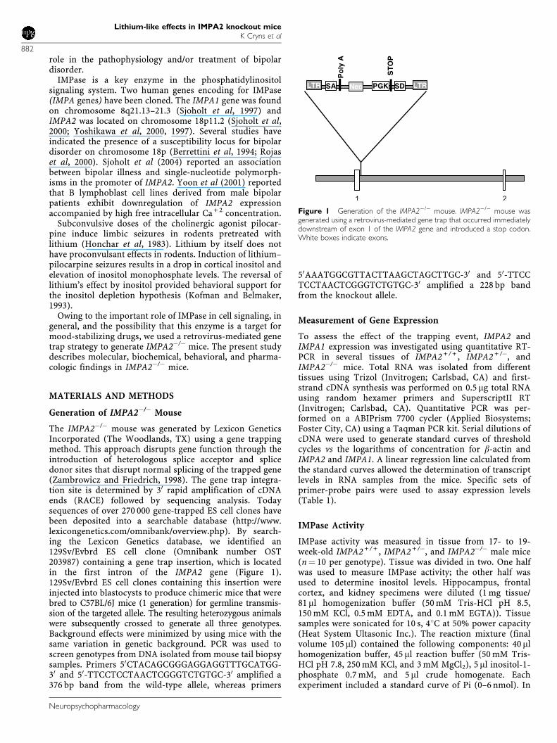
The IMPA2�/� mouse was generated by Lexicon GeneticsIncorporated (The Woodlands, TX) using a gene trappingmethod. This approach disrupts gene function through theintroduction of heterologous splice acceptor and splicedonor sites that disrupt normal splicing of the trapped gene(Zambrowicz and Friedrich, 1998). The gene trap integra-tion site is determined by 30 rapid amplification of cDNAends (RACE) followed by sequencing analysis. Todaysequences of over 270 000 gene-trapped ES cell clones havebeen deposited into a searchable database (http://www.lexicongenetics.com/omnibank/overview.php). By search-ing the Lexicon Genetics database, we identified an129Sv/Evbrd ES cell clone (Omnibank number OST203987) containing a gene trap insertion, which is locatedin the first intron of the IMPA2 gene (Figure 1).129Sv/Evbrd ES cell clones containing this insertion wereinjected into blastocysts to produce chimeric mice that werebred to C57BL/6J mice (1 generation) for germline transmis-sion of the targeted allele. The resulting heterozygous animalswere subsequently crossed to generate all three genotypes.Background effects were minimized by using mice with thesame variation in genetic background. PCR was used toscreen genotypes from DNA isolated from mouse tail biopsysamples. Primers 50CTACAGCGGGAGGAGGTTTGCATGG-30 and 50-TTCCTCCTAACTCGGGTCTGTGC-30 amplified a376 bp band from the wild-type allele, whereas primers
50AAATGGCGTTACTTAAGCTAGCTTGC-30 and 50-TTCCTCCTAACTCGGGTCTGTGC-30 amplified a 228 bp bandfrom the knockout allele.
Measurement of Gene Expression
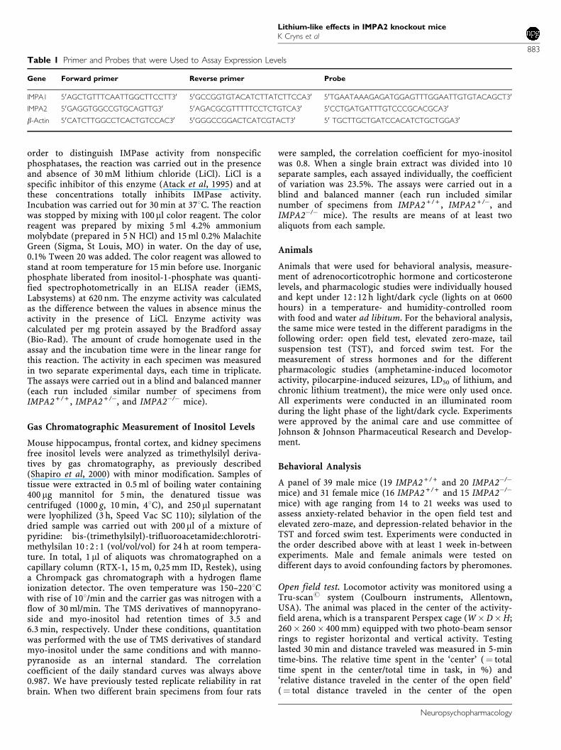
To assess the effect of the trapping event, IMPA2 andIMPA1 expression was investigated using quantitative RT-PCR in several tissues of IMPA2 + / + , IMPA2 + /�, andIMPA2�/� mice. Total RNA was isolated from differenttissues using Trizol (Invitrogen; Carlsbad, CA) and first-strand cDNA synthesis was performed on 0.5 mg total RNAusing random hexamer primers and SuperscriptII RT(Invitrogen; Carlsbad, CA). Quantitative PCR was per-formed on a ABIPrism 7700 cycler (Applied Biosystems;Foster City, CA) using a Taqman PCR kit. Serial dilutions ofcDNA were used to generate standard curves of thresholdcycles vs the logarithms of concentration for b-actin andIMPA2 and IMPA1. A linear regression line calculated fromthe standard curves allowed the determination of transcriptlevels in RNA samples from the mice. Specific sets ofprimer-probe pairs were used to assay expression levels(Table 1).
IMPase Activity
IMPase activity was measured in tissue from 17- to 19-week-old IMPA2 + / + , IMPA2 + /�, and IMPA2�/� male mice(n¼ 10 per genotype). Tissue was divided in two. One halfwas used to measure IMPase activity; the other half wasused to determine inositol levels. Hippocampus, frontalcortex, and kidney specimens were diluted (1 mg tissue/81 ml homogenization buffer (50 mM Tris-HCl pH 8.5,150 mM KCl, 0.5 mM EDTA, and 0.1 mM EGTA)). Tissuesamples were sonicated for 10 s, 41C at 50% power capacity(Heat System Ultasonic Inc.). The reaction mixture (finalvolume 105 ml) contained the following components: 40 mlhomogenization buffer, 45 ml reaction buffer (50 mM Tris-HCl pH 7.8, 250 mM KCl, and 3 mM MgCl2), 5 ml inositol-1-phosphate 0.7 mM, and 5 ml crude homogenate. Eachexperiment included a standard curve of Pi (0–6 nmol). In
LTRLTR Neo PGK
ST
OP
SDSA
Po
ly A
Figure 1 Generation of the IMPA2�/� mouse. IMPA2�/� mouse wasgenerated using a retrovirus-mediated gene trap that occurred immediatelydownstream of exon 1 of the IMPA2 gene and introduced a stop codon.White boxes indicate exons.
Lithium-like effects in IMPA2 knockout miceK Cryns et al
882
Neuropsychopharmacology
order to distinguish IMPase activity from nonspecificphosphatases, the reaction was carried out in the presenceand absence of 30 mM lithium chloride (LiCl). LiCl is aspecific inhibitor of this enzyme (Atack et al, 1995) and atthese concentrations totally inhibits IMPase activity.Incubation was carried out for 30 min at 371C. The reactionwas stopped by mixing with 100 ml color reagent. The colorreagent was prepared by mixing 5 ml 4.2% ammoniummolybdate (prepared in 5 N HCl) and 15 ml 0.2% MalachiteGreen (Sigma, St Louis, MO) in water. On the day of use,0.1% Tween 20 was added. The color reagent was allowed tostand at room temperature for 15 min before use. Inorganicphosphate liberated from inositol-1-phosphate was quanti-fied spectrophotometrically in an ELISA reader (iEMS,Labsystems) at 620 nm. The enzyme activity was calculatedas the difference between the values in absence minus theactivity in the presence of LiCl. Enzyme activity wascalculated per mg protein assayed by the Bradford assay(Bio-Rad). The amount of crude homogenate used in theassay and the incubation time were in the linear range forthis reaction. The activity in each specimen was measuredin two separate experimental days, each time in triplicate.The assays were carried out in a blind and balanced manner(each run included similar number of specimens fromIMPA2 + / + , IMPA2 + /�, and IMPA2�/� mice).
Gas Chromatographic Measurement of Inositol Levels
Mouse hippocampus, frontal cortex, and kidney specimensfree inositol levels were analyzed as trimethylsilyl deriva-tives by gas chromatography, as previously described(Shapiro et al, 2000) with minor modification. Samples oftissue were extracted in 0.5 ml of boiling water containing400 mg mannitol for 5 min, the denatured tissue wascentrifuged (1000 g, 10 min, 41C), and 250 ml supernatantwere lyophilized (3 h, Speed Vac SC 110); silylation of thedried sample was carried out with 200 ml of a mixture ofpyridine: bis-(trimethylsilyl)-trifluoroacetamide:chlorotri-methylsilan 10 : 2 : 1 (vol/vol/vol) for 24 h at room tempera-ture. In total, 1 ml of aliquots was chromatographed on acapillary column (RTX-1, 15 m, 0,25 mm ID, Restek), usinga Chrompack gas chromatograph with a hydrogen flameionization detector. The oven temperature was 150–2201Cwith rise of 101/min and the carrier gas was nitrogen with aflow of 30 ml/min. The TMS derivatives of mannopyrano-side and myo-inositol had retention times of 3.5 and6.3 min, respectively. Under these conditions, quantitationwas performed with the use of TMS derivatives of standardmyo-inositol under the same conditions and with manno-pyranoside as an internal standard. The correlationcoefficient of the daily standard curves was always above0.987. We have previously tested replicate reliability in ratbrain. When two different brain specimens from four rats
were sampled, the correlation coefficient for myo-inositolwas 0.8. When a single brain extract was divided into 10separate samples, each assayed individually, the coefficientof variation was 23.5%. The assays were carried out in ablind and balanced manner (each run included similarnumber of specimens from IMPA2 + / + , IMPA2 + /�, andIMPA2�/� mice). The results are means of at least twoaliquots from each sample.
Animals
Animals that were used for behavioral analysis, measure-ment of adrenocorticotrophic hormone and corticosteronelevels, and pharmacologic studies were individually housedand kept under 12 : 12 h light/dark cycle (lights on at 0600hours) in a temperature- and humidity-controlled roomwith food and water ad libitum. For the behavioral analysis,the same mice were tested in the different paradigms in thefollowing order: open field test, elevated zero-maze, tailsuspension test (TST), and forced swim test. For themeasurement of stress hormones and for the differentpharmacologic studies (amphetamine-induced locomotoractivity, pilocarpine-induced seizures, LD50 of lithium, andchronic lithium treatment), the mice were only used once.All experiments were conducted in an illuminated roomduring the light phase of the light/dark cycle. Experimentswere approved by the animal care and use committee ofJohnson & Johnson Pharmaceutical Research and Develop-ment.
Behavioral Analysis
A panel of 39 male mice (19 IMPA2 + / + and 20 IMPA2�/�
mice) and 31 female mice (16 IMPA2 + / + and 15 IMPA2�/�
mice) with age ranging from 14 to 21 weeks was used toassess anxiety-related behavior in the open field test andelevated zero-maze, and depression-related behavior in theTST and forced swim test. Experiments were conducted inthe order described above with at least 1 week in-betweenexperiments. Male and female animals were tested ondifferent days to avoid confounding factors by pheromones.
Open field test. Locomotor activity was monitored using aTru-scanr system (Coulbourn instruments, Allentown,USA). The animal was placed in the center of the activity-field arena, which is a transparent Perspex cage (W�D�H;260� 260� 400 mm) equipped with two photo-beam sensorrings to register horizontal and vertical activity. Testinglasted 30 min and distance traveled was measured in 5-mintime-bins. The relative time spent in the ‘center’ (¼ totaltime spent in the center/total time in task, in %) and‘relative distance traveled in the center of the open field’(¼ total distance traveled in the center of the open
Table 1 Primer and Probes that were Used to Assay Expression Levels
Gene Forward primer Reverse primer Probe
IMPA1 50AGCTGTTTCAATTGGCTTCCTT30 50GCCGGTGTACATCTTATCTTCCA30 50TGAATAAAGAGATGGAGTTTGGAATTGTGTACAGCT30
IMPA2 50GAGGTGGCCGTGCAGTTG30 50AGACGCGTTTTTCCTCTGTCA30 50CCTGATGATTTGTCCCGCACGCA30
b-Actin 50CATCTTGGCCTCACTGTCCAC30 50GGGCCGGACTCATCGTACT30 50 TGCTTGCTGATCCACATCTGCTGGA30
Lithium-like effects in IMPA2 knockout miceK Cryns et al
883
Neuropsychopharmacology
field/total distance traveled, in %) were analyzed as ameasure for anxiety-like behavior.
Elevated zero-maze. The zero-maze consisted of an annularrunway (diameter: 50 cm, width: 5 cm), elevated 50 cmabove the floor. The maze was divided in four parts, thatis, two opposite open parts and two opposite closed parts.The sidewalls of the opposing closed parts had a height of20 cm; the open parts had borders with a height of 3 mm.The maze was enclosed by a black cabinet (size: 150 cm highby 132 cm wide). Light intensity on the open arms was10 Lux and on the closed arms 2 Lux. All the animals wereplaced in the same start position, just in front of theopening of the closed part, facing the closed part. Theanimals were allowed to freely explore the maze for 5 minand their behavior was recorded and analyzed using theEthovision Pro video tracking system (Noldus, The Nether-lands). The software was capable of measuring the distancetraveled and the time spent in each part of the maze. ThreeIMPA2 + / + and 1 IMPA2�/� male mice were excluded fromthe analysis because they fell off the maze during testing.
TST. Mice were suspended by their tail to a hook in a testchamber using adhesive tape. Total duration of immobilitywas measured over a period of 6 min using the Videotracksystem (Viewpoint, France). The Videotrack system wasvalidated in-house with standard antidepressant com-pounds. For both mouse panels, one IMPA2 + / + and twoIMPA2�/� mice were excluded for analysis because the micecurled up towards their tail or fell off during testing.
Forced swim test. Mice were placed in a cylinder (diameter10 cm), filled with water to a height of 10 cm (such that themouse could not touch the bottom or rim of the cylinder atany point) and a temperature of 25711C. Each mouse waspre-exposed to swim-stress for 6 min on day 1 and a testsession of 6 min was performed 24 h later. Total duration ofimmobility was measured using the Videotrack system(Viewpoint, France). The Videotrack system was validatedin-house with standard antidepressant compounds.
Measurement of ACTH and CORT after Forced SwimTesting
Plasma adrenocorticotrophic hormone (ACTH) and corti-costerone (CORT) levels were measured in 21- to 32-week-old male and female mice (n¼ 6 for both female IMPA2 + / +
and IMPA2�/�mice, n¼ 9 and 7 for male IMPA2 + / + andIMPA2�/�mice, respectively). Animals were brought underlight ether anesthesia and trunk blood was collected inEDTA blood collection tubes (Microtainer EDTA, BectonDickinson, Switserland). All tubes were kept on ice and thencentrifuged at 1090 g for 5 min at 41C. Separated plasma wasfrozen at �701C for subsequent ACTH or CORT determina-tion (both in duplicate) using the RSL 125I ACTH radio-immunoassay (RIA) kit and ImmuChemt double antibodyCORT 125I kit from MP Biomedicals (USA) following themanufacturer’s protocol. ACTH and CORT levels wereexpressed in pg/ml and ng/ml plasma, respectively. In asubsequent experiment, ACTH and CORT levels weremeasured in 10- to 18-week-old IMPA2 + / + and IMPA2�/�
female littermates after forced swim testing (n¼ 12 and 11for IMPA2 + / + and IMPA2�/�, respectively). Mice weresubjected to forced swim testing once only for a totalduration of 6 min. At 10 min after the forced swim test,animals were brought under light ether anesthesia andtrunk blood was collected in EDTA blood collection tubes(Microtainer EDTA, Becton Dickinson, Switserland). ACTHand CORT levels were measured as described above.
Pharmacologic Studies
Amphetamine-induced locomotor activity. The effect of d-amphetamine on locomotor behavior was evaluated inIMPA2 + / + , IMPA2 + /�, and IMPA2�/� male littermateswith age ranging from 15 to 32 weeks. d-Amphetamine wasdissolved in saline. Locomotion was evaluated in the Tru-scanr system (Coulbourn instruments, Allentown, USA).Mice were initially placed into the activity monitor chamberfor 1 h, then injected i.p. with vehicle (10 ml/kg of bodyweight; n¼ 9, 10, 7 for IMPA2 + / + , IMPA2 + /�, and IMPA2�/�,respectively) or d-amphetamine (1 mg/kg, n¼ 8, 9, 10 forIMPA2 + / + , IMPA2 + /�, and IMPA2�/�, respectively, or2 mg/kg, n¼ 10, 10, 9 for IMPA2 + / + , IMPA2 + /�, andIMPA2�/�, respectively). After the injection, the animalswere returned to the activity-field arena and monitored for2 h (in 5-min time-bins).
Pilocarpine-induced seizures. IMPA2 + / + and IMPA2�/�
male littermates (18–22 weeks old) were injected subcuta-neously (s.c) with 100 or 200 mg/kg pilocarpine (n¼ 5 foreach dose) and rated for signs of seizures once every 5 minfor 2 h according to a modified version of the scale used byPatel et al (1988). The scoring was as follows: 0¼ noresponse; 1¼ gustatory movements and/or fictive scratch-ing; 2¼ tremor; 3¼ head bobbing; 4¼ forelimb clonus;5¼ rearing, clonus and falling. In addition, the latency toattain rearing, clonus, and falling (a score of 5) wasrecorded for each mouse (Kofman et al, 1993). A well-trained blind observer scored the mice. To further study thepossibility of lithium augmentation to reveal pilocarpinesensitivity of the IMPA2�/� mice, we pretreated IMPA2 + / +
and IMPA2�/� male littermates with 3 meq/kg (n¼ 5) and6 meq/kg (n¼ 5) lithium.
LD50 of lithium. Isotonic lithium (20 meq/kg) was injected(i.p.) in 5- to 7-month-old IMPA2 + / + (n¼ 19) and IMPA2�/�
(n¼ 17) female littermates. The dose was chosen based onBersudsky et al (1993). Lethality was monitored 24 h and 5days after the injection.
Chronic lithium treatment. Adult IMPA2 + / + , IMPA2 + /�,and IMPA2�/� male littermates (4–10 months old) weretreated with control chow (n¼ 12 for IMPA2 + / + andIMPA2 + /�, n¼ 10 for IMPA2�/�) or chow containing0.2% (n¼ 12 for every genotype) or 0.4% lithium carbonate(Teklad, Madison, WI) (n¼ 20 for IMPA2 + / + and IMPA2 + /�,n¼ 18 for IMPA2�/�) for 14 days. Water was available adlibitum. Three IMPA2 + / + , three IMPA2 + /�, and oneIMPA2�/� in the 0.4% lithium carbonate group died duringthe treatment period. After 14 days, forced swim testing wasperformed. Data obtained from a satellite experimentdemonstrated that chronic treatment of mice with a mixed
Lithium-like effects in IMPA2 knockout miceK Cryns et al
884
Neuropsychopharmacology

C57BL/6J-129Sv/Evbrd genetic background resulted inaverage plasma lithium levels of 0.67 mM70.069(mean7SEM) for the 0.2% group and 2.8 7 0.437 in the0.4% group. Sodium plasma levels were comparablebetween all groups (means in mM7SEM: 105.470.73,105.670.78, 106.672.16 for the 0, 0.2, and 0.4% lithiumgroups, respectively).
Statistical Analysis
The Kolmogorov–Smirnov test was used to evaluate thenormality assumption of expression data, behavioral data,and CORT and ACTH data after forced swim stress.Expression data, behavioral data, and CORT and ACTHdata after forced swim stress were analyzed using aStudent’s t-test or a Mann–Whitney Rank Sum Test in caseof data that were not normally distributed. A one-wayANOVA was used to compare IMPase activity and inositollevels between the different genotypes. Parametric statisticalanalysis was chosen based on our previous data of theseassays (Motulsky, 1995).
To evaluate rearing before amphetamine administration, aone-way ANOVA was used. A two-way ANOVA (genotype�treatment) was used to assess the effect of amphetaminetreatment on locomotor behavior and rearing in the openfield test and of lithium on immobility in the forced swimtest. A two-way ANOVA (gender� genotype) was also usedto analyze CORT and ACTH data in baseline conditions.Residual plots were used to diagnose the appropriateness ofthe final ANOVA models used. This graphical examinationincluded histograms, QQ-plots, and plots of residuals vsfitted values. No departures from the ANOVA assumptionswere detected. In case data were not normally distributed, aloge transformation was performed. A Fisher’s exact test wasused to evaluate the effect of acute lithium treatment.
RESULTS
IMPA2 and IMPA1 Expression
IMPA2 expression was found to be completely abolished inIMPA2�/� mice, indicating that the trapping event resultedin a null allele of IMPA2 (Figure 2).
To look for potential compensatory mechanisms, IMPA1mRNA levels were analyzed in brain and liver tissue from
IMPA2 + / + (n¼ 8) and IMPA2�/� (n¼ 8) mice by quanti-tative RT-PCR. No significant difference could be detectedbetween IMPA2 + / + and IMPA2�/� mice (brain (mean inarbitrary units7SEM): 1.870.2 and 2.470.7 for IMPA2 + / +
and IMPA2�/�, respectively, Mann–Whitney U-test, U¼ 63,p¼ 0.645; liver (mean7SEM): 2.070.2 and 2.370.3 forIMPA2 + / + and IMPA2�/�, respectively, Student’s t-test,t¼�0.764, p¼ 0.458).
IMPase Activity and Inositol Levels
IMPase activity and inositol levels were measured in thefrontal cortex and in the hippocampus of IMPA2 + / + ,IMPA2 + /�, and IMPA2�/� mice. As summarized in Table 2,hippocampal IMPase activity was significantly increased by24% in the IMPA2�/� mice (ANOVA: F(2,27) ¼ 3.39, p¼ 0.05)but no difference was observed in the frontal cortex(ANOVA: F(2,27) ¼ 1.08, p¼ 0.35). No difference wasobserved in inositol levels among IMPA2�/�, IMPA2 + /�,or IMPA2 + / + mice in the hippocampus (ANOVA:F(2,26) ¼ 0.677, p¼ 0.51) or in the frontal cortex (ANOVA:F(2,26) ¼ 2.26, p¼ 0.12).
Inositol levels and IMPase activity were also compared inthe kidneys, which is the tissue with the highest expressionof IMPA2 (Figure 2). Reduced IMPA2 gene-dose resulted ina significant reduction in inositol levels (23% in IMPA2 + /�
and 32% in IMPA2�/� mice compared with IMPA2 + / +
Table 2 Inositol and IMPAse Levels
Kidney Hippocampus Frontal cortex
Inositol levels IMPase activity Inositol levels IMPase activity Inositol levelsa IMPase activityb
IMPA2+/+ 13.671.5 (n¼ 10) 2.370.3 (n¼ 10) 18.271.6 (n¼ 10) 2.770.1 (n¼ 10) 14.172.3 (n¼ 10) 8.571.0 (n¼ 10)
IMPA2+/� 10.371.2 (n¼ 9) 1.770.1 (n¼ 10) 16.470.7 (n¼ 9) 2.570.1 (n¼ 10) 9.770.8 (n¼ 10) 6.570.8 (n¼ 10)
IMPA2�/� 9.170.7* (n¼ 9) 1.670.2 (n¼ 10) 16.771.1 (n¼ 10) 3.170.2* (n¼ 10) 13.971.1 (n¼ 10) 7.971.2 (n¼ 10)
Results are means7SEM.ammol/ kg (W/W).bnmol/mg�min.*ANOVA; po0.05.Hippocampal IMPase activity post hoc LSD: IMPA2+/+ vs IMPA2�/�, p¼ 0.064; IMPA2+/+ vs IMPA2+/�, p¼ 0.58; IMPA2+/� vs IMPA2�/�, p¼ 0.019.Kidney inositol levels post hoc LSD: IMPA2+/+ vs IMPA2�/�, p¼ 0.011; IMPA2+/+ vs IMPA2+/�, p¼ 0.06; IMPA2+/� vs IMPA2�/�, p¼ 0.437
0.00
5.00
10.00
15.00
20.00
25.00
B L K H B L K H B L K H B L K H B L K H B L K H
IMPA2-/-
(M)IMPA2-/-
(F)IMPA2+/-
(M)IMPA2+/-
(F)IMPA2+/+
(M)IMPA2+/+
(F)
Rel
ativ
e ex
pre
ssio
n
Figure 2 IMPA2 expression is completely abolished in IMPA2�/� mice. B:Brain, H: Heart, K: Kidney, L: Liver. Results are means7SEM.
Lithium-like effects in IMPA2 knockout miceK Cryns et al
885
Neuropsychopharmacology
mice), which reached statistical significance (ANOVA:F(2,25) ¼ 2.25, p¼ 0.03) but no difference in IMPase activitywas found among the three genotype groups (ANOVA:F(2,27) ¼ 2.19, p¼ 0.13; Table 2).
Behavioral Analysis
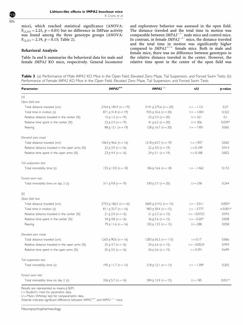
Table 3a and b summarize the behavioral data for male andfemale IMPA2 KO mice, respectively. General locomotor
and exploratory behavior was assessed in the open field.The distance traveled and the total time in motion wascomparable between IMPA2�/� male mice and control mice.In contrast, in female IMPA2�/� mice, the distance traveledand the total time in motion was significantly highercompared to IMPA2 + / + female mice. Both in male andfemale mice, there was no difference between genotypes inthe relative distance traveled in the center. However, therelative time spent in the center of the open field was
Table 3 (a) Performance of Male IMPA2 KO Mice in the Open Field, Elevated Zero Maze, Tail Suspension, and Forced Swim Tests; (b)Performance of Female IMPA2 KO Mice in the Open Field, Elevated Zero Maze, Tail Suspension, and Forced Swim Tests
Parameter IMPA2+/+ IMPA2�/� t/U p-value
(a)
Open field test
Total distance traveled (cm) 27647185.9 (n¼ 19) 31417275.6 (n¼ 20) t¼�1.121 0.27
Total time in motion (s) 871731.8 (n¼ 19) 925742.6 (n¼ 20) t¼�1.004 0.322
Relative distance traveled in the center (%) 1571.2 (n¼ 19) 2373.3 (n¼ 20) U¼ 321 0.1
Relative time spent in the center (%) 2372.4 (n¼ 19) 4176.2 (n¼ 20) U¼ 306 0.039*
Rearing 88712.1 (n¼ 19) 128716.7 (n¼ 20) t¼�1.901 0.065
Elevated zero maze
Total distance traveled (cm) 1063746.6 (n¼ 16) 1218763.7 (n¼ 19) t¼ 1.907 0.065
Relative distance traveled in the open arms (%) 2273.9 (n¼ 16) 2273.0 (n¼ 19) t¼ 0.109 0.914
Relative time spent in the open arms (%) 2374.4 (n¼ 16) 2473.1 (n¼ 19) t¼ 0.188 0.852
Tail suspension test
Total immobility time (s) 155713.0 (n¼ 18) 186716.6 (n¼ 18) t¼�1.462 0.153
Forced swim test
Total immobility time on day 2 (s) 31179.8 (n¼ 19) 33073.7 (n¼ 20) U¼ 338 0.244
(b)
Open field test
Total distance traveled (cm) 27537186.5 (n¼ 16) 36057214.2 (n¼ 15) t¼�3.011 0.005*
Total time in motion (s) 811733.7 (n¼ 16) 983730.4 (n¼ 15) t¼�3.777 o0.001*
Relative distance traveled in the center (%) 2172.0 (n¼ 16) 2172.3 (n¼ 15) t¼�0.0723 0.943
Relative time spent in the center (%) 3474.8 (n¼ 16) 3675.6 (n¼ 15) t¼�0.207 0.838
Rearing 79711.6 (n¼ 16) 102713.5 (n¼ 15) U¼ 288 0.058
Elevated zero maze
Total distance traveled (cm) 1265790.6 (n¼ 16) 1283756.3 (n¼ 115) t¼ 0.17 0.866
Relative distance traveled in the open arms (%) 2573.7 (n¼ 16) 2472.6 (n¼ 15) t¼�0.0524 0.959
Relative time spent in the open arms (%) 2573.5 (n¼ 16) 2672.6 (n¼ 15) t¼ 0.391 0.699
Tail suspension test
Total immobility time (s) 195711.7 (n¼ 15) 218712.1 (n¼ 13) t¼�1.309 0.202
Forced swim test
Total immobility time on day 2 (s) 33675.7 (n¼ 16) 304712.4 (n¼ 15) U¼ 185 0.031*
Results are represented as means7SEM.t¼ Student’s t-test for parametric data.U¼Mann–Whitney test for nonparametric data.Asterisk indicates significant difference between IMPA2+/+ and IMPA2�/� mice.
Lithium-like effects in IMPA2 knockout miceK Cryns et al
886
Neuropsychopharmacology
significantly higher in male IMPA2�/� mice but not infemale IMPA2�/�mice. In addition, a borderline signifi-cantly increased number of rearings was observed in bothmale and female IMPA2�/� mice.
In the elevated zero-maze, anxiety-related behavior wasevaluated by measuring the relative time spent and distancetraveled in the open areas. Measures were not altered inmale and female IMPA2�/� mice. Similarly, analysis oflocomotion on the elevated zero-maze revealed no differ-ences between the two genotypes tested in male and femaleIMPA2�/� mice.
Depression-related behavior was assessed in the tailsuspension and in the forced swim test. As summarized inTable 3a, male IMPA2�/� mice did not exhibit a differencein total immobility time in the tail suspension test or in theforced swimming test. Female IMPA2�/� mice also did notexhibit a difference in total immobility time in the tailsuspension test but in the forced swim test total immobilitytime was significantly lower compared to control mice.
Measurement of Plasma ACTH and CORT after ForcedSwimming
ACTH and CORT levels are presented in Table 4. At baselinecondition, no genotype effect was seen on ACTH(F(1,24) ¼ 2.69, p¼ 0.11) and CORT levels (F(1,24)¼ 0.45,p¼ 0.51). There was a significant difference in ACTH levelsbetween male and female mice (F(1,24) ¼ 29.92, po0.0001)while CORT levels were not different between bothgenders (F(1,24)¼ 2.16, p¼ 0.16). There was no significantgenotype� gender interaction (F(1,24)¼ 0.09, p¼ 0.77 andF(1,24) ¼ 0.01, p¼ 0.93 for, respectively, ACTH and CORT).
In a subsequent experiment, ACTH and CORT levels weremeasured in female IMPA2 + / + and IMPA2�/� mice afterforced swim stress. No difference in the magnitude of ACTH(Student’s t-test, t¼�1.643, p¼ 0.115) and CORT (Mann–Whitney U-test, U¼ 144, p¼ 0.479) response was observedbetween IMPA2 + / + and IMPA2�/� mice.
Pharmacologic Studies
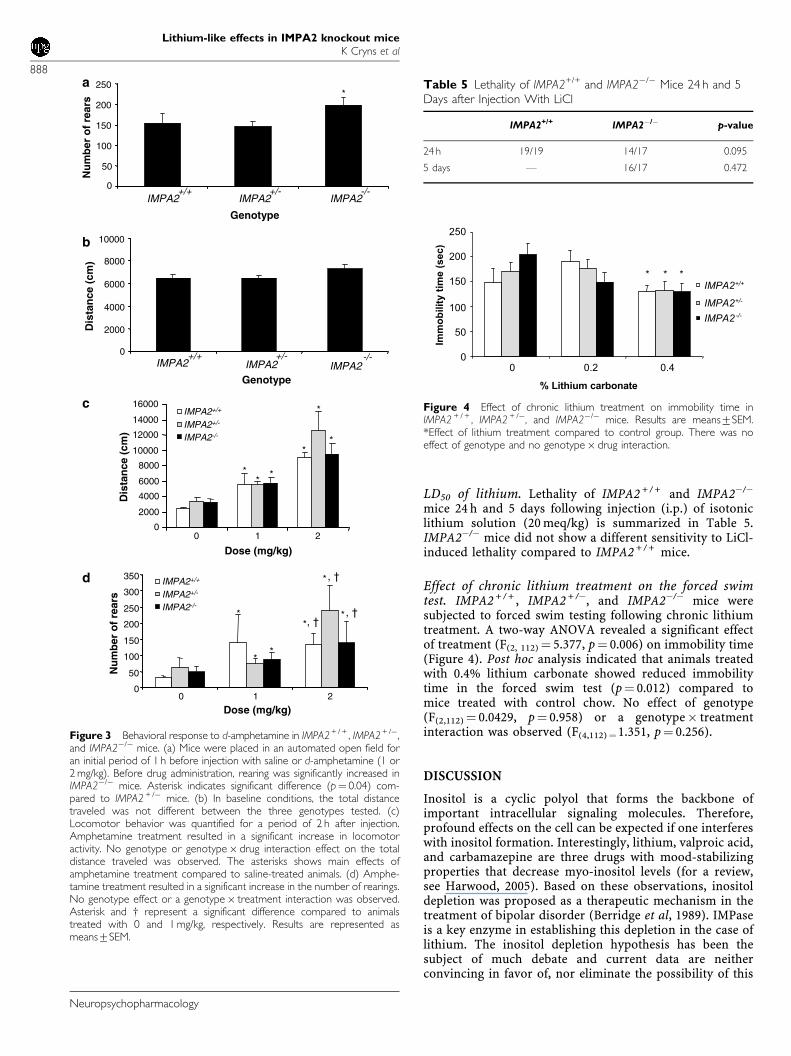
Amphetamine-induced locomotor activity. Locomotorbehavior of IMPA2 + / + , IMPA2 + /�, and IMPA2�/� mice inthe open field test was evaluated after d-amphetamineadministration. Before drug administration, a signifi-cant genotype effect on the number of rears was seen
(F(2, 85)¼ 3.92, p¼ 0.0235, Figure 3a). Post hoc testing indicatedrearing was significantly increased in IMPA2�/� animalscompared to IMPA2 + /�mice (p¼ 0.04). A tendency for anincrease in rearings was seen in IMPA2�/�compared toIMPA2 + / + mice (p¼ 0.062). Baseline locomotor activity asmeasured by total distance traveled before drug adminis-tration was not different between the three genotypes(F(2,84) ¼ 2.209, p¼ 0.116, Figure 3b). Amphetamine admin-istration produced a significant increase in total distancetraveled (drug effect, F(2,73)¼ 49.65, po0.0001, Figure 3c),but no effect of genotype was observed (F(2,73)¼ 1.53,po0.223). Post hoc analysis indicated that animals treatedwith 1 and 2 mg/kg travelled significantly more than controltreated animals (po0.0001 for both doses). There wasno genotype� drug interaction (F(4,73) ¼ 0.28, p¼ 0.889).Detailed analysis of locomotor activity over time confirmedthe absence of a genotype and genotype� drug interactioneffect on the total distance traveled (data not shown).Amphetamine treatment resulted in a significant increase inthe number of rears (F(2,73)¼ 9.79, p¼ 0.0002). Post hocanalysis indicated rearing was increased in animals treatedwith 1 mg/kg (p¼ 0.014) and 2 mg/kg (po0.0001) com-pared to saline-treated animals. Treatment with 2 mg/kgamphetamine also resulted in a significant increasein rearing compared to animals treated with 1 mg/kg(p¼ 0.02). No genotype effect (F(2,73)¼ 0.19, p¼ 0.83) or agenotype� treatment interaction (F(4,73) ¼ 0.38, p¼ 0.819)was observed.
Pilocarpine sensitivity. No limbic seizures were observed inIMPA2 + / + and IMPA2�/� mice following administration of100 or 200 mg/kg pilocarpine. To further study thepossibility of lithium augmentation to reveal pilocarpinesensitivity of the IMPA2�/� mice, we pretreated IMPA2�/�
and IMPA2 + / + mice with 3 or 6 meq/kg lithium. No limbicseizures following administration of 100 mg/kg pilocarpinewere obtained in any group of mice.
Injection of 100 mg/kg pilocarpine has previously beenshown to induce limbic seizures in mice pretreated with10 meq/kg lithium (Bersudsky et al, 1994). As a positivecontrol, 100 mg/kg pilocarpine following 10 meq/kg lithiumwere injected to IMPA2�/� and IMPA2 + / + mice. Asexpected, limbic seizures in both groups were observedwith no difference in latency to clonus and seizure scorebetween the two groups of mice.
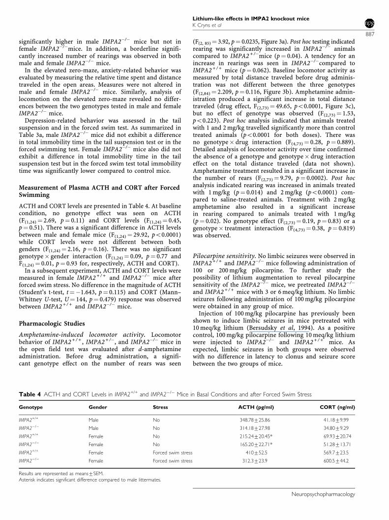
Table 4 ACTH and CORT Levels in IMPA2+/+ and IMPA2�/� Mice in Basal Conditions and after Forced Swim Stress
Genotype Gender Stress ACTH (pg/ml) CORT (ng/ml)
IMPA2+/+ Male No 348.78725.86 41.1879.99
IMPA2�/� Male No 314.18727.98 34.8079.29
IMPA2+/+ Female No 215.24720.45* 69.93720.74
IMPA2�/� Female No 165.20722.71* 51.28713.71
IMPA2+/+ Female Forced swim stress 410752.5 569.7723.5
IMPA2�/� Female Forced swim stress 312.3723.9 600.5744.2
Results are represented as means7SEM.Asterisk indicates significant difference compared to male littermates.
Lithium-like effects in IMPA2 knockout miceK Cryns et al
887
Neuropsychopharmacology
LD50 of lithium. Lethality of IMPA2 + / + and IMPA2�/�
mice 24 h and 5 days following injection (i.p.) of isotoniclithium solution (20 meq/kg) is summarized in Table 5.IMPA2�/� mice did not show a different sensitivity to LiCl-induced lethality compared to IMPA2 + / + mice.
Effect of chronic lithium treatment on the forced swimtest. IMPA2 + / + , IMPA2 + /�, and IMPA2�/� mice weresubjected to forced swim testing following chronic lithiumtreatment. A two-way ANOVA revealed a significant effectof treatment (F(2, 112) ¼ 5.377, p¼ 0.006) on immobility time(Figure 4). Post hoc analysis indicated that animals treatedwith 0.4% lithium carbonate showed reduced immobilitytime in the forced swim test (p¼ 0.012) compared tomice treated with control chow. No effect of genotype(F(2,112) ¼ 0.0429, p¼ 0.958) or a genotype� treatmentinteraction was observed (F(4,112)¼1.351, p¼ 0.256).
DISCUSSION
Inositol is a cyclic polyol that forms the backbone ofimportant intracellular signaling molecules. Therefore,profound effects on the cell can be expected if one interfereswith inositol formation. Interestingly, lithium, valproic acid,and carbamazepine are three drugs with mood-stabilizingproperties that decrease myo-inositol levels (for a review,see Harwood, 2005). Based on these observations, inositoldepletion was proposed as a therapeutic mechanism in thetreatment of bipolar disorder (Berridge et al, 1989). IMPaseis a key enzyme in establishing this depletion in the case oflithium. The inositol depletion hypothesis has been thesubject of much debate and current data are neitherconvincing in favor of, nor eliminate the possibility of this
a
0
50
100
150
200
250N
um
ber
of
rear
s
IMPA2+/+ +/- -/-
Genotype
IMPA2 IMPA2
0
2000
4000
6000
8000
10000
Genotype
Dis
tan
ce (
cm)
b
-/-+/-+/+IMPA2 IMPA2 IMPA2
c
0
2000
4000
6000
8000
10000
12000
14000
16000
0 1 2
Dose (mg/kg)
Dis
tan
ce (
cm)
IMPA2+/+
IMPA2+/-
IMPA2-/-
0
50
100
150
200
250
300
350
0 1 2
Dose (mg/kg)
Nu
mb
er o
f re
ars
IMPA2+/+
IMPA2+/-
IMPA2-/-
d
*
**
**
*
*
*
***
*
, †, †
*, †
Figure 3 Behavioral response to d-amphetamine in IMPA2+ / + , IMPA2+ /�,and IMPA2�/� mice. (a) Mice were placed in an automated open field foran initial period of 1 h before injection with saline or d-amphetamine (1 or2 mg/kg). Before drug administration, rearing was significantly increased inIMPA2�/� mice. Asterisk indicates significant difference (p¼ 0.04) com-pared to IMPA2 + /� mice. (b) In baseline conditions, the total distancetraveled was not different between the three genotypes tested. (c)Locomotor behavior was quantified for a period of 2 h after injection.Amphetamine treatment resulted in a significant increase in locomotoractivity. No genotype or genotype� drug interaction effect on the totaldistance traveled was observed. The asterisks shows main effects ofamphetamine treatment compared to saline-treated animals. (d) Amphe-tamine treatment resulted in a significant increase in the number of rearings.No genotype effect or a genotype� treatment interaction was observed.Asterisk and w represent a significant difference compared to animalstreated with 0 and 1 mg/kg, respectively. Results are represented asmeans7SEM.
Table 5 Lethality of IMPA2+/+ and IMPA2�/� Mice 24 h and 5Days after Injection With LiCl
IMPA2+/+ IMPA2�/� p-value
24 h 19/19 14/17 0.095
5 days F 16/17 0.472
0
50
100
150
200
250
0 0.2 0.4
% Lithium carbonate
Imm
ob
ilit
y t
ime (
sec)
IMPA2+/+
IMPA2+/-
IMPA2-/-
***
Figure 4 Effect of chronic lithium treatment on immobility time inIMPA2 + / + , IMPA2 + /�, and IMPA2�/� mice. Results are means7SEM.*Effect of lithium treatment compared to control group. There was noeffect of genotype and no genotype� drug interaction.
Lithium-like effects in IMPA2 knockout miceK Cryns et al
888
Neuropsychopharmacology
hypothesis. In the present work, we phenotyped IMPA2�/�
mice molecularly and behaviorally to better understand thecontribution of IMPA2 to brain IMPase activity and inositollevels and the possible involvement of this gene in affectivedisorders.
IMPA2�/� mice grow and reproduce normally, suggestingthat their somatic and sexual development is normal. Theabsence of major developmental abnormalities in thesemice also suggests that the IMPA2 gene does not play anessential role during development.
Possible involvement of IMPase in complex behaviorsrelated to affective disorders was assessed by monitoringthe behavior of IMPA2�/� mice in the forced swim test andthe tail suspension test modeling depression and in theelevated zero-maze and open field test modeling anxiety.Male IMPA2�/� mice did not show aberrant behavior in theforced swim test. In contrast, female IMPA2�/� miceshowed a small (9.5%) but significant decrease in immo-bility time in the forced swim test. A significant increase inthe total time in motion and the total distance traveled inthe open field test was also observed in female IMPA2�/�
mice while these parameters were unaltered in maleIMPA2�/� mice. Therefore, the aberrant behavior of femaleIMPA2�/� mice in the forced swim test most likely reflectsan increased locomotor behavior rather than an anti-depressant phenotype. Interestingly, a different depressivephenotype in male vs female mice has also been shownin 5HT knockout mice (Jones and Lucki, 2005). Therewas no difference between male and female IMPA2�/�
and IMPA2 + / + mice in the tail suspension test, overallsuggesting no effect of the deletion of the IMPA2 gene onthe depression-like behavior.
A trend towards an increased number of rears in the openfield was observed in male and female IMPA2�/� micemonitored for 30 min. This trend reached significance in athird mouse panel monitored for 60 min (prior toamphetamine administration), suggesting an increasedexploratory behavior. Kofman et al (1991) reported reducedrearing in lithium-treated mice, an effect reversed byinositol administration, and enhanced rearing followingdietary inositol load (Kofman et al, 1998). Thus, it may havebeen expected that IMPA2�/� mice would show reducedrearing as a lithium-like effect. Counterintuitively, anenhancement was observed. Interestingly, it has recentlybeen shown that mice treated with the antidepressantfluoxetine at an early postnatal age (pd 4–pd 20)demonstrate depressive- and anxiety-like behaviors atadulthood in contrast with the antidepressant and anxioly-tic effects of SSRIs when administered at an adult age(Ansorge et al, 2004). In parallel, it may be that lifelongreduction in IMPA2 levels affects rearing differently thanchronic lithium treatment of adult mice.
In the anxiety-related open field test, the relative timespent in the center was significantly higher in male but notfemale IMPA2�/� mice. This finding, together with the factthat IMPA2�/� mice showed increased rearing in the openfield test, could indicate that IMPA2�/� exhibit a loweranxiety level compared to control mice. In contrast, noanxiolytic phenotype was observed in the elevated zero-maze test in either gender of the KO mice. Interestingly,similar contrasting findings have been described byMontkowski et al (1997) who compared different mouse
strains in the open field test and elevated plus maze. Theysuggested that as both tests measure rodents’ innate fear ofa novel environment, the contrasting findings support thenotion that the open field is a stronger fear-inducingstimulus than the plus maze. However, detailed analysis ofrearing and of relative time spent in the center of the openfield over 5 min time-bins in mouse panel 1 indicatedrearing was only significantly increased in IMPA2�/� mice10 min after introduction to the open field and thisdifference disappeared after 15 min (data not shown).Moreover, although an overall significant difference wasseen between IMPA2�/� and wild-type mice in the relativetime spent in the center, no difference was seen when datawere analyzed in 5 min time-bins (data not shown), that is,for neither parameter a genotype effect was seen during thefirst 10 min of the test session. This would suggest thatIMPA2�/� mice show increased exploratory behavior ratherthan an anxiolytic phenotype.
CORT and ACTH levels were measured in male andfemale IMPA2 + / + and IMPA2�/� mice. No significant effectof genotype or a genotype� gender interaction was seen onCORT and ACTH levels. ACTH levels were significantlydifferent between male and female mice, whereas CORTlevels were not different between both genders. It should benoted that gender differences in HPA-axis activity havebeen well demonstrated by others (eg Timpl et al, 1998). Ina subsequent experiment, it was shown that female IMPA2�/�
mice did not show altered stress-hormone levels comparedto female IMPA2�/� mice after forced swim stress, therebysuggesting that IMPA2 is not involved in modulation of theHPA axis.
Amphetamine is an indirect dopamine-receptor agonistas it elevates extracellular dopamine levels in mousestriatum. Hyperlocomotion in the open field test after d-amphetamine administration is considered to be an animalmodel for mania (Einat and Belmaker, 2001), and lithiumsalts are reported to antagonize this behavior (Einat andBelmaker, 2001). Amphetamine clearly induced hyperloco-motion as measured by a significant increase in the distancetraveled, but no effect of genotype or a genotype� druginteraction was observed, indicating that IMPA2�/� mice donot differently respond to amphetamine-induced manic-likebehavior.
To study the possible involvement of the IMPA2 gene inthe molecular mechanism of lithium’s inhibitory effect onIMPase, we used the behavioral paradigm of lithium–pilocarpine seizures. It has been suggested that braininositol depletion due to IMPase inhibition is critical tothe development of lithium–pilocarpine-induced seizures(Kofman and Belmaker, 1993). We therefore hypothesizedthat IMPA2�/� mice would present a lithium-like effect inthis paradigm. No limbic seizures were observed inIMPA2�/� mice following subconvulsive pilocarpine doses.These data suggest that the IMPA2 gene does notsignificantly contribute to brain IMPase activity and there-fore knocking it out is not enough to mimic this behavioraleffect of lithium. The fact that we did not find inositoldepletion or reduced IMPase activity in the frontal cortexand in the hippocampus of IMPA2�/� compared withIMPA2 + / + mice is consistent with this conclusion. Chroniclithium treatment resulted in a significant reduction inimmobility time but no genotype or genotype� treatment
Lithium-like effects in IMPA2 knockout miceK Cryns et al
889
Neuropsychopharmacology
interaction was seen, indicating lack of augmentation effectof IMPA2 knockout on the antidepressant effect of lithiumin the forced swim test (O’Brien et al, 2004). It should benoted that the reduction in immobility time was onlyobserved in the animals treated with food containing 0.4%lithium. This treatment regime results in lithium plasmalevels that are higher than the therapeutic range in humans(data not shown). Furthermore, mice from the 0.4% groupshowed a reduction in body weight (data not shown), whichmight interfere with their behavior. Therefore, the reductionin immobility time should be interpreted cautiously.
Among tissues studied, IMPA2 expression is highest inkidney. We therefore studied the effect of IMPA2�/� onIMPase activity and inositol levels in this tissue. We did finda gene-dose effect on kidney inositol levels in the IMPA2�/�
mice raising the possibility that the IMPA2 gene is involvedin renal side effects of lithium treatment. Bersudsky et al(1992) have previously reported that lithium-treated ratsthat drank inositol solution developed polydipsia muchslower than rats drinking mannitol solution.
Altogether our results indicate that deletion of the IMPA2gene did not resemble the pharmacological effects oflithium. Gene targeting to create null mutations in mice isa powerful tool to study complex phenotypes and assessinghow genes influence biological functions in vivo. Yet, if aKO mouse does not exhibit a phenotypical change it maybedue to compensatory effects of other genes. However, nodifference in brain or liver IMPA1 expression levels wasfound between IMPA2�/� and IMPA2 + / + mice ruling outthe possibility that the lack of phenotypical changes inIMPA2�/� mice results from a compensatory upregulationof the IMPA1 gene. In this context, it is also not conceivablethat the 24% increase in hippocampal IMPase activity in theIMPA2�/� mice is responsible for the lack of phenotypicalchanges as our assay measures the Vmax of the enzyme,whereas in vivo a much lower enzyme rate is physiologicallysufficient.
It is noteworthy that the effectiveness of inositol depletionwill also depend on influx of inositol from exogenoussources. In the brain, there are two myo-inositol transpor-ters. SMIT (sodium/myo-inositol transporter) is dependenton Na + and inhibited at reduced pH (Hager et al, 1995).The second transporter is HMIT (H+/myo-inositol trans-porter), which is activated as the pH lowers. Afterdepolarization and activation of protein kinase C (PKC),HMIT is translocated to the plasma membrane at growthcones, leading to an increased myo-inositol uptake (Uldryet al, 2004). Interestingly, gene expression profilingindicated that PKCe and PKCi are both upregulated inhippocampus of IMPA2�/� mice (data not shown). Thisupregulation may lead to an increased exogenous inositoluptake via HMIT, thereby compensating for the inositoldepletion. Alternatively, absence of a lithium-like pheno-type in IMPA2�/� mice may point to functional redundancyof IMPA2, potentially provided by IMPA1 (Cryns et al,2005). This is supported by the fact that the expression ofIMPA1 is several fold higher than that of IMPA2 in mice andin human frontal cortex (Agam et al, 2002).
In conclusion, molecular, behavioral, and pharmacologicanalysis of the IMPA2�/� mouse indicates that deletingthe IMPA2 gene does not mimic the effects of lithiumtreatment. Compensatory mechanisms and/or functional
redundancy may be responsible for the absence ofdepression- or mania-related phenotypes. To exhibitlithium-like behavioral and molecular effects, IMPA1 orboth IMPase genes may have to be knocked out. Yet, even inSMIT�/� mice with the most severe inositol deficiency everrecorded in mammals, there is no significant loss ofphosphatidylinositol (Berry et al, 2004).
ACKNOWLEDGEMENTS
We gratefully acknowledge Ms Ilse Leenaerts, Dr JosPrickaerts, Dr Adriaan Bouwknecht, and Ms CarolienJanssen for their experimental/analytical work contribution.
REFERENCES
Agam G, Shamir A, Shaltiel G, Greenberg ML (2002). Myo-inositol-1-phosphate (MIP) synthase: a possible new target for anti-bipolar drugs. Bipolar Disord 4: 15–20.
Ansorge MS, Zhou M, Lira A, Hen R, Gingrich JA (2004). Early-lifeblockade of the 5-HT transporter alters emotional behavior inadult mice. Science 306: 879–881.
Atack JR, Broughton HB, Pollack SJ (1995). Inositol mono-phosphataseFa putative target for Li+ in the treatment ofbipolar disorder. Trends Neurosci 18: 343–349.
Berrettini WH, Ferraro TN, Goldin LR, Weeks DE, Detera-Wadleigh S, Nurnberger JI et al (1994). Chromosome 18 DNAmarkers and manic-depressive illness: evidence for a suscept-ibility gene. Proc Natl Acad Sci USA 91: 5918–5921.
Berridge MJ, Downes CP, Hanley MR (1989). Neural anddevelopmental actions of lithium: a unifying hypothesis. Cell59: 411–419.
Berry GT, Buccafusca R, Greer JJ, Eccleston E (2004). Phospho-inositide deficiency due to inositol depletion is not a mechanismof lithium action in brain. Mol Genet Metab 82: 87–92.
Bersudsky Y, Mahler O, Kofman O, Belmaker RH (1994). Speciesdifferences in susceptibility to Li–pilocarpine seizures. EurNeuropsychopharmacol 4: 429–430.
Bersudsky Y, Vinnitsky I, Ghelber D, Kofman O, Kaplan Z,Belmaker RH (1993). Mechanism of lithium lethality in rats. JPsych Res 27: 415–422.
Bersudsky Y, Vinnitsky I, Grisaru N, Yaroslavsky U, Gheorghiu S,Ivgi D et al (1992). The effect of inositol on litihum-induced-polyuria-polydipsia in rats and human. Hum Psychopharmacol7: 403–407.
Cryns K, Daneels G, Goris I, Peeters PJ, Van Craenendonck H,Steckler T et al (2005). Phenotypic characterization of themyo-inositol monophosphatase 1 (IMPA1)Fdeficient mice.‘Neuroscience Meeting 2005. Washington, DC, pp No. 559.1.
Einat H, Belmaker RH (2001). The effects of inositol treatment inanimal models of psychiatric disorders. J Affect Disord 62: 113–121.
Goodwin FK, Jamison KR (1990). Manic-Depressive Illness. OxfordUniversity Press: New York.
Hager K, Hazama A, Kwon HM, Loo DD, Handler JS, Wright EM(1995). Kinetics and specificity of the renal Na+/myo-inositolcotransporter expressed in Xenopus oocytes. J Membr Biol 143:103–113.
Harwood AJ (2005). Lithium and bipolar mood disorder:the inositol-depletion hypothesis revisited. Mol Psychiatry 10:117–126.
Honchar MP, Olney JW, Sherman WR (1983). Systemic cholinergicagents induce seizures and brain damage in lithium-treated rats.Science 220: 323–325.
Jones MD, Lucki I (2005). Sex differences in the regulation ofserotonergic transmission and behavior in 5-HT receptorknockout mice. Neuropsychopharmacology 30: 1039–1047.
Lithium-like effects in IMPA2 knockout miceK Cryns et al
890
Neuropsychopharmacology
Jope RS (1999). A bimodal model of the mechanism of action oflithium. Mol Psychiatry 4: 21–25.
Kofman O, Agam G, Shapiro J, Spencer A (1998). Chronic dietaryinositol enhances locomotor activity and brain inositol levels inrats. Psychopharmacology (Berl) 139: 239–242.
Kofman O, Belmaker RH (1993). Ziskind-Somerfeld ResearchAward 1993. Biochemical, behavioral, and clinical studies of therole of inositol in lithium treatment and depression. BiolPsychiatry 34: 839–852.
Kofman O, Belmaker RH, Grisaru N, Alpert C, Fuchs I, Katz V et al(1991). Myo-inositol attenuates two specific behavioral effects ofacute lithium in rats. Psychopharmocol Bull 27: 185–190.
Kofman O, Sherman WR, Katz V, Belmaker RH (1993). Restorationof brain myo-inositol levels in rats increases latency to lithium–pilocarpine seizures. Psychopharmacology (Berl) 110: 229–234.
Montkowski A, Poettig M, Mederer A, Holsboer F (1997).Behavioural performance in three substrains of mouse strain129. Brain Res 762: 12–18.
Motulsky H (1995). Intuitive Biostatistics. Oxford University Press:New-York. pp 299–300.
O’Brien WT, Harper AD, Jove F, Woodgett JR, Maretto S, Piccolo Set al (2004). Glycogen synthase kinase-3beta haploinsufficiencymimics the behavioral and molecular effects of lithium. JNeurosci 24: 6791–6798.
Patel S, Meldrum BS, Fine A (1988). Susceptibility to pilocarpine-induced seizures in rats increases with age. Behav Brain Res31: 165–167.
Quiroz JA, Gould TD, Manji HK. (2004). Molecular effects oflithium. Mol Interv 4: 259–272.
Rojas K, Liang L, Johnson EI, Berraettini WH, Overhauser J (2000).Identification of candidate genes for psychiatric disorders on18p11. Mol Psychiatry 5: 389–395.
Shaltiel G, Shamir A, Shapiro J, Ding D, Dalton E, Bialer M et al(2004). Valproate decreases inositol biosynthesis. Biol Psychiatry56: 868–874.
Shapiro J, Belmaker RH, Biegon A, Seker A, Agam G (2000). Scyllo-inositol in post-mortem brain of bipolar, unipolar and schizo-phrenic patients. J Neural Transm 107: 603–607.
Sjoholt G, Ebstein RP, Lie RT, Berle JO, Mallet J, Deleuze JFet al (2004). Examination of IMPA1 and IMPA2 genes inmanic-depressive patients: association between IMPA2promoter polymorphisms and bipolar disorder. Mol Psychiatry9: 621–629.
Sjoholt G, Gulbrandsen AK, Lovlie R, Berle J, Molven A, Steen VM(2000). A human myo-inositol monophosphatase gene (IMPA2)localized in a putative susceptibility region for bipolar disorder onchromosome 18p11.2: genomic structure and polymorphismscreening in manic-depressive patients. Mol Psychiatry 5: 172–180.
Sjoholt G, Molven A, Lovlie R, Wilcox A, Sikela JM, Steen VM(1997). Genomic structure and chromosomal localization of ahuman myo-inositol monophosphatase gene (IMPA). Genomics45: 113–122.
Timpl P, Spanagel R, Sillaber I, Kresse A, Reul JMH, Stalla GK et al(1998). Impaired stress response and reduced anxiety in micelacking a functional corticotropin-releasing hormone receptor 1.Nat Genet 19: 162–166.
Uldry M, Steiner P, Zurich MG, Beguin P, Hirling H, Dolci W et al(2004). Regulated exocytosis of an H+/myo-inositol symporter atsynapses and growth cones. EMBO J 23: 531–540 (Epub 2004 Jan 29).
Williams RS, Cheng L, Mudge AW, Harwood AJ (2002). A commonmechanism of action for three mood-stabilizing drugs. Nature417: 292–295.
Yoon IS, Li PP, Siu KP, Kennedy JL, Cooke RG, Parikh SV et al(2001). Altered IMPA2 gene expression and calcium homeostasisin bipolar disorder. Mol Psychiatry 6: 678–683.
Yoshikawa T, Padigaru M, Karkera JD, Sharma M, Berrettini WH,Esterling LE et al (2000). Genomic structure and novel variantsof myo-inositol monophosphatase 2 (IMPA2). Mol Psychiatry 5:165–171.
Yoshikawa T, Turner G, Esterling LE, Sanders AR, Detera-Wadleigh SD (1997). A novel human myo-inositol mono-phosphatase gene, IMP.18p, maps to a susceptibility region forbipolar disorder. Mol Psychiatry 2: 393–397.
Zambrowicz BP, Friedrich GA (1998). Comprehensive mammaliangenetics: history and future prospects of gene trapping in themouse. Int J Dev Biol 42: 1025–1036.
Lithium-like effects in IMPA2 knockout miceK Cryns et al
891
Neuropsychopharmacology