L-Type Amino Acid Transporters LAT1 and LAT4 in Cancer: Uptake of 3-O-Methyl-6- 18F-Fluoro-L-Dopa in...
10
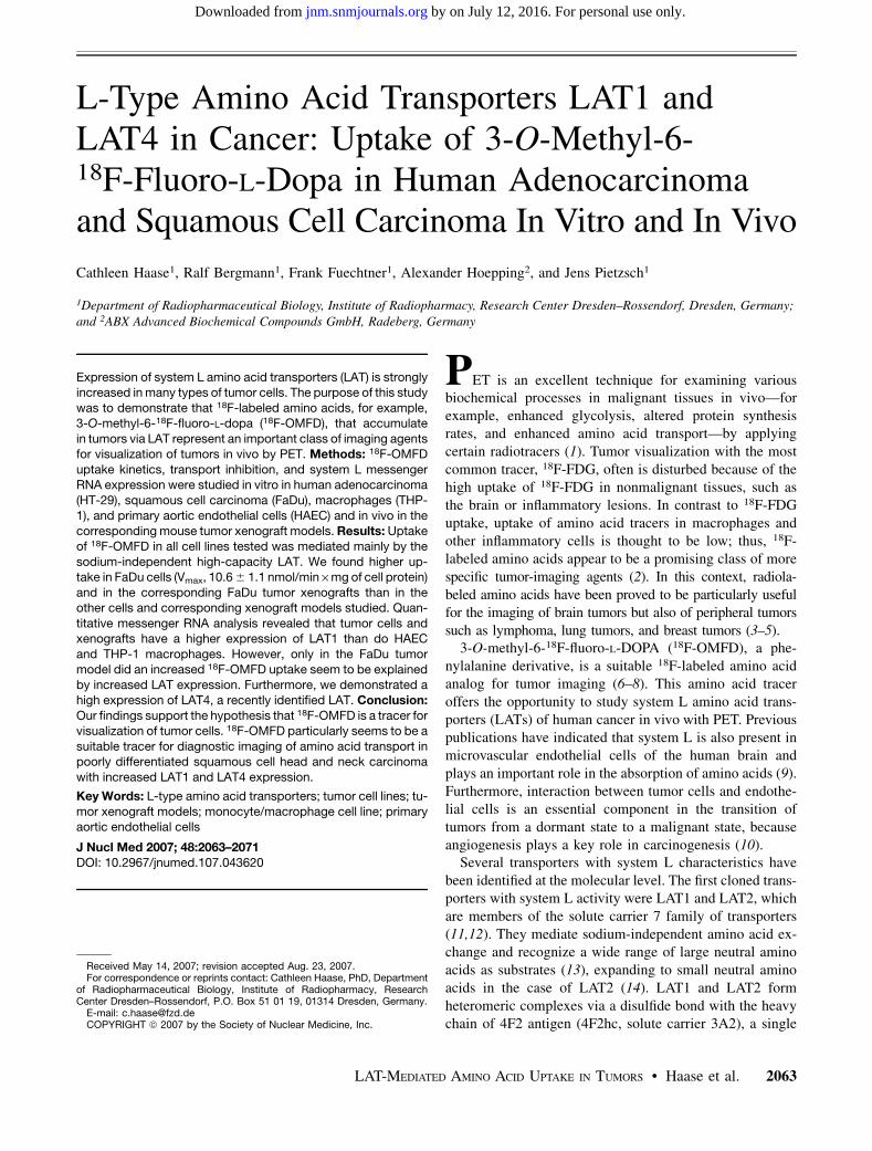
L-Type Amino Acid Transporters LAT1 and LAT4 in Cancer: Uptake of 3-O-Methyl-6- 18 F-Fluoro-L-Dopa in Human Adenocarcinoma and Squamous Cell Carcinoma In Vitro and In Vivo Cathleen Haase 1 , Ralf Bergmann 1 , Frank Fuechtner 1 , Alexander Hoepping 2 , and Jens Pietzsch 1 1 Department of Radiopharmaceutical Biology, Institute of Radiopharmacy, Research Center Dresden–Rossendorf, Dresden, Germany; and 2 ABX Advanced Biochemical Compounds GmbH, Radeberg, Germany Expression of system L amino acid transporters (LAT) is strongly increased in many types of tumor cells. The purpose of this study was to demonstrate that 18 F-labeled amino acids, for example, 3-O-methyl-6- 18 F-fluoro-L-dopa ( 18 F-OMFD), that accumulate in tumors via LAT represent an important class of imaging agents for visualization of tumors in vivo by PET. Methods: 18 F-OMFD uptake kinetics, transport inhibition, and system L messenger RNA expression were studied in vitro in human adenocarcinoma (HT-29), squamous cell carcinoma (FaDu), macrophages (THP- 1), and primary aortic endothelial cells (HAEC) and in vivo in the corresponding mouse tumor xenograft models. Results: Uptake of 18 F-OMFD in all cell lines tested was mediated mainly by the sodium-independent high-capacity LAT. We found higher up- take in FaDu cells (V max , 10.6 6 1.1 nmol/min · mg of cell protein) and in the corresponding FaDu tumor xenografts than in the other cells and corresponding xenograft models studied. Quan- titative messenger RNA analysis revealed that tumor cells and xenografts have a higher expression of LAT1 than do HAEC and THP-1 macrophages. However, only in the FaDu tumor model did an increased 18 F-OMFD uptake seem to be explained by increased LAT expression. Furthermore, we demonstrated a high expression of LAT4, a recently identified LAT. Conclusion: Our findings support the hypothesis that 18 F-OMFD is a tracer for visualization of tumor cells. 18 F-OMFD particularly seems to be a suitable tracer for diagnostic imaging of amino acid transport in poorly differentiated squamous cell head and neck carcinoma with increased LAT1 and LAT4 expression. Key Words: L-type amino acid transporters; tumor cell lines; tu- mor xenograft models; monocyte/macrophage cell line; primary aortic endothelial cells J Nucl Med 2007; 48:2063–2071 DOI: 10.2967/jnumed.107.043620 PET is an excellent technique for examining various biochemical processes in malignant tissues in vivo—for example, enhanced glycolysis, altered protein synthesis rates, and enhanced amino acid transport—by applying certain radiotracers (1). Tumor visualization with the most common tracer, 18 F-FDG, often is disturbed because of the high uptake of 18 F-FDG in nonmalignant tissues, such as the brain or inflammatory lesions. In contrast to 18 F-FDG uptake, uptake of amino acid tracers in macrophages and other inflammatory cells is thought to be low; thus, 18 F- labeled amino acids appear to be a promising class of more specific tumor-imaging agents (2). In this context, radiola- beled amino acids have been proved to be particularly useful for the imaging of brain tumors but also of peripheral tumors such as lymphoma, lung tumors, and breast tumors (3–5). 3-O-methyl-6- 18 F-fluoro-L-DOPA ( 18 F-OMFD), a phe- nylalanine derivative, is a suitable 18 F-labeled amino acid analog for tumor imaging (6–8). This amino acid tracer offers the opportunity to study system L amino acid trans- porters (LATs) of human cancer in vivo with PET. Previous publications have indicated that system L is also present in microvascular endothelial cells of the human brain and plays an important role in the absorption of amino acids (9). Furthermore, interaction between tumor cells and endothe- lial cells is an essential component in the transition of tumors from a dormant state to a malignant state, because angiogenesis plays a key role in carcinogenesis (10). Several transporters with system L characteristics have been identified at the molecular level. The first cloned trans- porters with system L activity were LAT1 and LAT2, which are members of the solute carrier 7 family of transporters (11,12). They mediate sodium-independent amino acid ex- change and recognize a wide range of large neutral amino acids as substrates (13), expanding to small neutral amino acids in the case of LAT2 (14). LAT1 and LAT2 form heteromeric complexes via a disulfide bond with the heavy chain of 4F2 antigen (4F2hc, solute carrier 3A2), a single Received May 14, 2007; revision accepted Aug. 23, 2007. For correspondence or reprints contact: Cathleen Haase, PhD, Department of Radiopharmaceutical Biology, Institute of Radiopharmacy, Research Center Dresden–Rossendorf, P.O. Box 51 01 19, 01314 Dresden, Germany. E-mail: [email protected] COPYRIGHT ª 2007 by the Society of Nuclear Medicine, Inc. LAT-MEDIATED AMINO ACID UPTAKE IN TUMORS • Haase et al. 2063 by on July 12, 2016. For personal use only. jnm.snmjournals.org Downloaded from
-
Upload
independent -
Category
Documents
-
view
2 -
download
0
Transcript of L-Type Amino Acid Transporters LAT1 and LAT4 in Cancer: Uptake of 3-O-Methyl-6- 18F-Fluoro-L-Dopa in...
L-Type Amino Acid Transporters LAT1 andLAT4 in Cancer: Uptake of 3-O-Methyl-6-18F-Fluoro-L-Dopa in Human Adenocarcinomaand Squamous Cell Carcinoma In Vitro and In Vivo
Cathleen Haase1, Ralf Bergmann1, Frank Fuechtner1, Alexander Hoepping2, and Jens Pietzsch1
1Department of Radiopharmaceutical Biology, Institute of Radiopharmacy, Research Center Dresden–Rossendorf, Dresden, Germany;and 2ABX Advanced Biochemical Compounds GmbH, Radeberg, Germany
Expression of system L amino acid transporters (LAT) is stronglyincreased in many types of tumor cells. The purpose of this studywas to demonstrate that 18F-labeled amino acids, for example,3-O-methyl-6-18F-fluoro-L-dopa (18F-OMFD), that accumulatein tumors via LAT represent an important class of imaging agentsfor visualization of tumors in vivo by PET. Methods: 18F-OMFDuptake kinetics, transport inhibition, and system L messengerRNA expression were studied in vitro in human adenocarcinoma(HT-29), squamous cell carcinoma (FaDu), macrophages (THP-1), and primary aortic endothelial cells (HAEC) and in vivo in thecorresponding mouse tumor xenograft models. Results: Uptakeof 18F-OMFD in all cell lines tested was mediated mainly by thesodium-independent high-capacity LAT. We found higher up-take in FaDu cells (Vmax, 10.6 6 1.1 nmol/min · mg of cell protein)and in the corresponding FaDu tumor xenografts than in theother cells and corresponding xenograft models studied. Quan-titative messenger RNA analysis revealed that tumor cells andxenografts have a higher expression of LAT1 than do HAECand THP-1 macrophages. However, only in the FaDu tumormodel did an increased 18F-OMFD uptake seem to be explainedby increased LAT expression. Furthermore, we demonstrated ahigh expression of LAT4, a recently identified LAT. Conclusion:Our findings support the hypothesis that 18F-OMFD is a tracer forvisualization of tumor cells. 18F-OMFD particularly seems to be asuitable tracer for diagnostic imaging of amino acid transport inpoorly differentiated squamous cell head and neck carcinomawith increased LAT1 and LAT4 expression.
Key Words: L-type amino acid transporters; tumor cell lines; tu-mor xenograft models; monocyte/macrophage cell line; primaryaortic endothelial cells
J Nucl Med 2007; 48:2063–2071DOI: 10.2967/jnumed.107.043620
PET is an excellent technique for examining variousbiochemical processes in malignant tissues in vivo—forexample, enhanced glycolysis, altered protein synthesisrates, and enhanced amino acid transport—by applyingcertain radiotracers (1). Tumor visualization with the mostcommon tracer, 18F-FDG, often is disturbed because of thehigh uptake of 18F-FDG in nonmalignant tissues, such asthe brain or inflammatory lesions. In contrast to 18F-FDGuptake, uptake of amino acid tracers in macrophages andother inflammatory cells is thought to be low; thus, 18F-labeled amino acids appear to be a promising class of morespecific tumor-imaging agents (2). In this context, radiola-beled amino acids have been proved to be particularly usefulfor the imaging of brain tumors but also of peripheral tumorssuch as lymphoma, lung tumors, and breast tumors (3–5).
3-O-methyl-6-18F-fluoro-L-DOPA (18F-OMFD), a phe-nylalanine derivative, is a suitable 18F-labeled amino acidanalog for tumor imaging (6–8). This amino acid traceroffers the opportunity to study system L amino acid trans-porters (LATs) of human cancer in vivo with PET. Previouspublications have indicated that system L is also present inmicrovascular endothelial cells of the human brain andplays an important role in the absorption of amino acids (9).Furthermore, interaction between tumor cells and endothe-lial cells is an essential component in the transition oftumors from a dormant state to a malignant state, becauseangiogenesis plays a key role in carcinogenesis (10).
Several transporters with system L characteristics havebeen identified at the molecular level. The first cloned trans-porters with system L activity were LAT1 and LAT2, whichare members of the solute carrier 7 family of transporters(11,12). They mediate sodium-independent amino acid ex-change and recognize a wide range of large neutral aminoacids as substrates (13), expanding to small neutral aminoacids in the case of LAT2 (14). LAT1 and LAT2 formheteromeric complexes via a disulfide bond with the heavychain of 4F2 antigen (4F2hc, solute carrier 3A2), a single
Received May 14, 2007; revision accepted Aug. 23, 2007.For correspondence or reprints contact: Cathleen Haase, PhD, Department
of Radiopharmaceutical Biology, Institute of Radiopharmacy, ResearchCenter Dresden–Rossendorf, P.O. Box 51 01 19, 01314 Dresden, Germany.
E-mail: [email protected] ª 2007 by the Society of Nuclear Medicine, Inc.
LAT-MEDIATED AMINO ACID UPTAKE IN TUMORS • Haase et al. 2063
by on July 12, 2016. For personal use only. jnm.snmjournals.org Downloaded from
transmembrane domain protein essential for the functionalexpression of LAT1 and LAT2 at the plasma membrane(15,16). Recently, LAT3 (solute carrier 43) was identified,and several properties distinguish LAT3 from LAT1 andLAT2. For example, LAT3 does not require coexpressionwith 4F2hc and further exhibits a narrow substrate selec-tivity, preferring phenylalanine to others as substrates (17).Recently, LAT4 (solute carrier 43) was shown to havetransport activity similar to that of LAT3 but a differenttissue distribution (17). LAT1 is supposed to be the onlytransporter that plays an important role in cell proliferationand shows increased transport activity in many cancer cells(18,19).
The aim of the present study was to investigate the maintransport system for uptake of the amino acid tracer 18F-OMFD in different cells: human colorectal adenocarcinomacells (HT-29), human head and neck squamous cell carci-noma cells (FaDu), and primary human aortic endothelialcells (HAEC). For differentiation of concomitant traceruptake in tumor and inflammatory cells, we used a phorbolester–stimulated human monocyte/macrophage cell line(THP-1), which represents a well-characterized model forproinflammatory cells and tissue-associated macrophages,respectively. We further studied 18F-OMFD uptake in thecorresponding tumor (HT-29 and FaDu) xenograft modelsin nude mice in vivo.
Using quantitative reverse transcription (RT) polymerasechain reaction (PCR), we characterized the expression ofmessenger RNA (mRNA) in the LAT1, LAT2, LAT3,LAT4, and 4F2hc subtypes.
MATERIALS AND METHODS
Cell CultureHuman aortic endothelial cells (HAEC) were obtained from
Cascade Biologics and cultivated in EC medium MV2 (Promo-
cell). Human tumor cell lines FaDu (head and neck squamous cellcarcinoma) (American Type Culture Collection number HTB-43)and HT-29 (colorectal adenocarcinoma) (American Type CultureCollection number HTB-38) were obtained from DSMZ andcultivated in McCoy medium (FaDu) and RPMI (HT-29) supple-mented with 10% fetal calf serum. THP-1 cells (European Col-lection of Cell Cultures number 88081201; human monocyte/macrophage cell line, stimulated with phorbol myristate acetate[64 nmol/L] for 72 h) were cultivated in RPMI with 10% fetal calfserum. All media and supplements were obtained from Biochrom.Cells were cultivated with 5% CO2 and 37�C in a CO2 incubator(Heracell; Heraeus).
RNA IsolationFor preparation of total RNA from cells and xenograft tumors,
the Perfect RNA Mini Kit (Eppendorf) was used according to themanufacturer’s recommendations.
Real-Time PCRRT was done with a 2-step cMaster RT Kit (Eppendorf)
according to the manufacturer’s instructions.SYBR GREEN I (1:30, 000) (Sigma) real-time PCR assay was
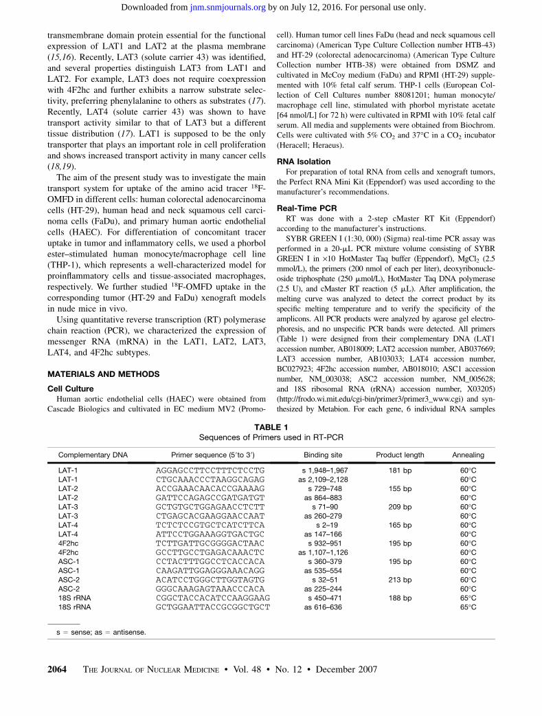
performed in a 20-mL PCR mixture volume consisting of SYBRGREEN I in ·10 HotMaster Taq buffer (Eppendorf), MgCl2 (2.5mmol/L), the primers (200 nmol of each per liter), deoxyribonucle-oside triphosphate (250 mmol/L), HotMaster Taq DNA polymerase(2.5 U), and cMaster RT reaction (5 mL). After amplification, themelting curve was analyzed to detect the correct product by itsspecific melting temperature and to verify the specificity of theamplicons. All PCR products were analyzed by agarose gel electro-phoresis, and no unspecific PCR bands were detected. All primers(Table 1) were designed from their complementary DNA (LAT1accession number, AB018009; LAT2 accession number, AB037669;LAT3 accession number, AB103033; LAT4 accession number,BC027923; 4F2hc accession number, AB018010; ASC1 accessionnumber, NM_003038; ASC2 accession number, NM_005628;and 18S ribosomal RNA (rRNA) accession number, X03205)(http://frodo.wi.mit.edu/cgi-bin/primer3/primer3_www.cgi) and syn-thesized by Metabion. For each gene, 6 individual RNA samples
TABLE 1Sequences of Primers used in RT-PCR
Complementary DNA Primer sequence (59to 39) Binding site Product length Annealing
LAT-1 AGGAGCCTTCCTTTCTCCTG s 1,948–1,967 181 bp 60�CLAT-1 CTGCAAACCCTAAGGCAGAG as 2,109–2,128 60�CLAT-2 ACCGAAACAACACCGAAAAG s 729–748 155 bp 60�CLAT-2 GATTCCAGAGCCGATGATGT as 864–883 60�CLAT-3 GCTGTGCTGGAGAACCTCTT s 71–90 209 bp 60�CLAT-3 CTGAGCACGAAGGAACCAAT as 260–279 60�CLAT-4 TCTCTCCGTGCTCATCTTCA s 2–19 165 bp 60�CLAT-4 ATTCCTGGAAAGGTGACTGC as 147–166 60�C4F2hc TCTTGATTGCGGGGACTAAC s 932–951 195 bp 60�C4F2hc GCCTTGCCTGAGACAAACTC as 1,107–1,126 60�CASC-1 CCTACTTTGGCCTCACCACA s 360–379 195 bp 60�CASC-1 CAAGATTGGAGGGAAACAGG as 535–554 60�CASC-2 ACATCCTGGGCTTGGTAGTG s 32–51 213 bp 60�CASC-2 GGGCAAAGAGTAAACCCACA as 225–244 60�C18S rRNA CGGCTACCACATCCAAGGAAG s 450–471 188 bp 65�C18S rRNA GCTGGAATTACCGCGGCTGCT as 616–636 65�C
s 5 sense; as 5 antisense.
2064 THE JOURNAL OF NUCLEAR MEDICINE • Vol. 48 • No. 12 • December 2007
by on July 12, 2016. For personal use only. jnm.snmjournals.org Downloaded from
from each cell line were analyzed in a dual assay. The thresholdcycle, which records the cycle when sample fluorescence exceeds achosen threshold above background fluorescence, was dividedthrough the threshold cycle of the internal 18S rRNA housekeepinggene, and the quotient was termed the relative expression level.
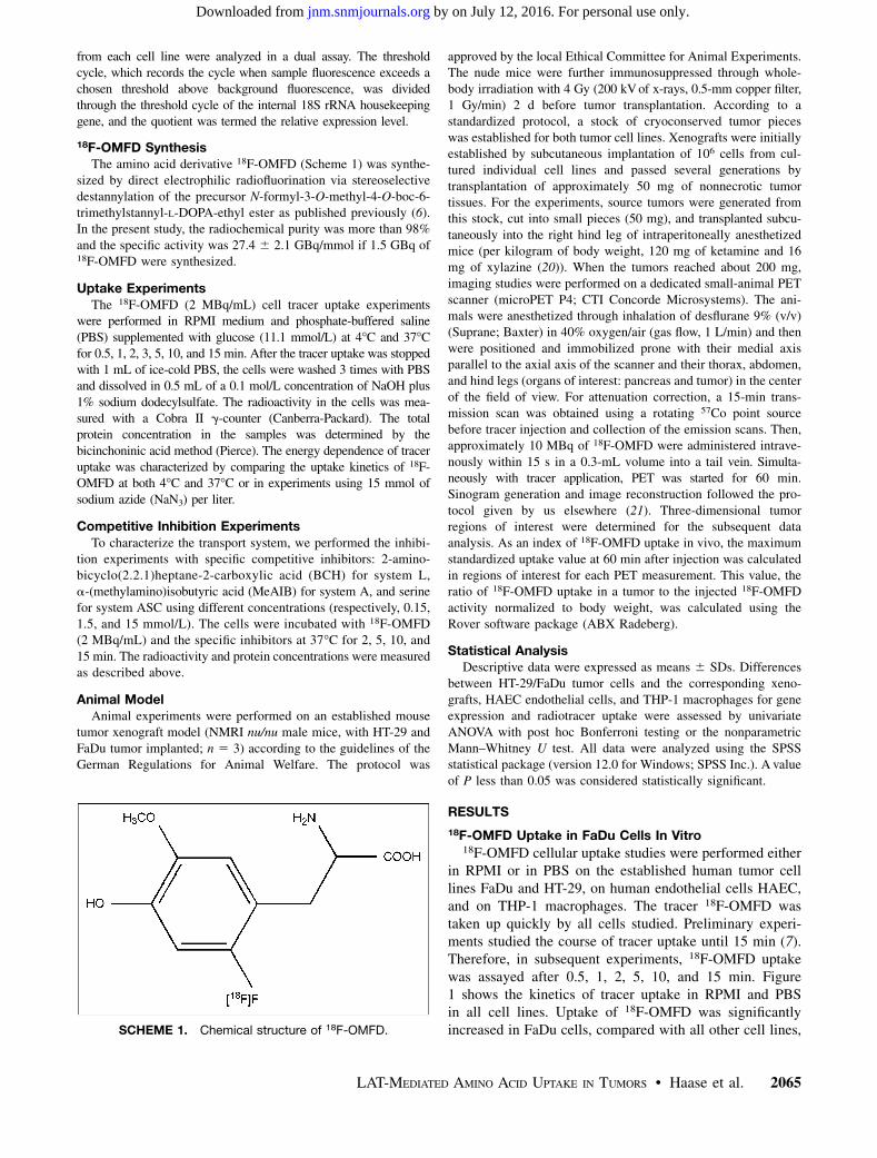
18F-OMFD SynthesisThe amino acid derivative 18F-OMFD (Scheme 1) was synthe-
sized by direct electrophilic radiofluorination via stereoselectivedestannylation of the precursor N-formyl-3-O-methyl-4-O-boc-6-trimethylstannyl-L-DOPA-ethyl ester as published previously (6).In the present study, the radiochemical purity was more than 98%and the specific activity was 27.4 6 2.1 GBq/mmol if 1.5 GBq of18F-OMFD were synthesized.
Uptake ExperimentsThe 18F-OMFD (2 MBq/mL) cell tracer uptake experiments
were performed in RPMI medium and phosphate-buffered saline(PBS) supplemented with glucose (11.1 mmol/L) at 4�C and 37�Cfor 0.5, 1, 2, 3, 5, 10, and 15 min. After the tracer uptake was stoppedwith 1 mL of ice-cold PBS, the cells were washed 3 times with PBSand dissolved in 0.5 mL of a 0.1 mol/L concentration of NaOH plus1% sodium dodecylsulfate. The radioactivity in the cells was mea-sured with a Cobra II g-counter (Canberra-Packard). The totalprotein concentration in the samples was determined by thebicinchoninic acid method (Pierce). The energy dependence of traceruptake was characterized by comparing the uptake kinetics of 18F-OMFD at both 4�C and 37�C or in experiments using 15 mmol ofsodium azide (NaN3) per liter.
Competitive Inhibition ExperimentsTo characterize the transport system, we performed the inhibi-
tion experiments with specific competitive inhibitors: 2-amino-bicyclo(2.2.1)heptane-2-carboxylic acid (BCH) for system L,a-(methylamino)isobutyric acid (MeAIB) for system A, and serinefor system ASC using different concentrations (respectively, 0.15,1.5, and 15 mmol/L). The cells were incubated with 18F-OMFD(2 MBq/mL) and the specific inhibitors at 37�C for 2, 5, 10, and15 min. The radioactivity and protein concentrations were measuredas described above.
Animal ModelAnimal experiments were performed on an established mouse
tumor xenograft model (NMRI nu/nu male mice, with HT-29 andFaDu tumor implanted; n 5 3) according to the guidelines of theGerman Regulations for Animal Welfare. The protocol was
approved by the local Ethical Committee for Animal Experiments.The nude mice were further immunosuppressed through whole-body irradiation with 4 Gy (200 kV of x-rays, 0.5-mm copper filter,1 Gy/min) 2 d before tumor transplantation. According to astandardized protocol, a stock of cryoconserved tumor pieceswas established for both tumor cell lines. Xenografts were initiallyestablished by subcutaneous implantation of 106 cells from cul-tured individual cell lines and passed several generations bytransplantation of approximately 50 mg of nonnecrotic tumortissues. For the experiments, source tumors were generated fromthis stock, cut into small pieces (50 mg), and transplanted subcu-taneously into the right hind leg of intraperitoneally anesthetizedmice (per kilogram of body weight, 120 mg of ketamine and 16mg of xylazine (20)). When the tumors reached about 200 mg,imaging studies were performed on a dedicated small-animal PETscanner (microPET P4; CTI Concorde Microsystems). The ani-mals were anesthetized through inhalation of desflurane 9% (v/v)(Suprane; Baxter) in 40% oxygen/air (gas flow, 1 L/min) and thenwere positioned and immobilized prone with their medial axisparallel to the axial axis of the scanner and their thorax, abdomen,and hind legs (organs of interest: pancreas and tumor) in the centerof the field of view. For attenuation correction, a 15-min trans-mission scan was obtained using a rotating 57Co point sourcebefore tracer injection and collection of the emission scans. Then,approximately 10 MBq of 18F-OMFD were administered intrave-nously within 15 s in a 0.3-mL volume into a tail vein. Simulta-neously with tracer application, PET was started for 60 min.Sinogram generation and image reconstruction followed the pro-tocol given by us elsewhere (21). Three-dimensional tumorregions of interest were determined for the subsequent dataanalysis. As an index of 18F-OMFD uptake in vivo, the maximumstandardized uptake value at 60 min after injection was calculatedin regions of interest for each PET measurement. This value, theratio of 18F-OMFD uptake in a tumor to the injected 18F-OMFDactivity normalized to body weight, was calculated using theRover software package (ABX Radeberg).
Statistical AnalysisDescriptive data were expressed as means 6 SDs. Differences
between HT-29/FaDu tumor cells and the corresponding xeno-grafts, HAEC endothelial cells, and THP-1 macrophages for geneexpression and radiotracer uptake were assessed by univariateANOVA with post hoc Bonferroni testing or the nonparametricMann–Whitney U test. All data were analyzed using the SPSSstatistical package (version 12.0 for Windows; SPSS Inc.). A valueof P less than 0.05 was considered statistically significant.
RESULTS
18F-OMFD Uptake in FaDu Cells In Vitro18F-OMFD cellular uptake studies were performed either
in RPMI or in PBS on the established human tumor celllines FaDu and HT-29, on human endothelial cells HAEC,and on THP-1 macrophages. The tracer 18F-OMFD wastaken up quickly by all cells studied. Preliminary experi-ments studied the course of tracer uptake until 15 min (7).Therefore, in subsequent experiments, 18F-OMFD uptakewas assayed after 0.5, 1, 2, 5, 10, and 15 min. Figure1 shows the kinetics of tracer uptake in RPMI and PBSin all cell lines. Uptake of 18F-OMFD was significantlyincreased in FaDu cells, compared with all other cell lines,SCHEME 1. Chemical structure of 18F-OMFD.
LAT-MEDIATED AMINO ACID UPTAKE IN TUMORS • Haase et al. 2065
by on July 12, 2016. For personal use only. jnm.snmjournals.org Downloaded from
for both PBS and RPMI (Fig. 1). Accumulated activity inFaDu cells was 5- to 10-fold higher for PBS than for RPMI(14% injected dose [%ID]/mg of protein) and reached 135%ID/mg of protein. FaDu cells had already revealed anincreased uptake (5.7-fold) after 5 min, compared with allother cell lines (Fig. 1). No significant differences wereseen between tracer uptake of HT-29, HAEC, and THP-1macrophages in RPMI or PBS. The average accumulatedactivity in these cells was about 3 %ID/mg of protein inRPMI and 35 %ID/mg of protein in PBS after 15 min.
To study the energy dependence of the observed 18F-OMFD transport process, we compared uptake at 37�C withthat at 4�C with and without adding 15 mmol of BCH perliter. No significant differences were found in any cell line.Furthermore, oxidative phosphorylation was inhibited byadding a 15 mmol/L concentration of NaN3, which mimicsthe effects of hypoxia under normoxic conditions. How-ever, NaN3 did not significantly influence the uptake kinet-ics in any cell line.
Kinetics of 18F-OMFD Uptake
To compare the functional expression of the L-aminotransport systems in HT-29 and FaDu, as well as in dif-ferentiated THP-1 macrophages and primary HAEC, westudied the kinetics of 18F-OMFD uptake. Uptake of 18F-OMFD was measured in the presence of various concentra-tions of unlabeled OMFD. Uptake of 18F-OMFD in all celltypes studied was saturable and apparently followed Mi-chaelis–Menton kinetics, with the Km and Vmax values givenin Table 2. The maximum velocity of 18F-OMFD uptake(Vmax) was 10.6 6 1.1 nmol/min · mg of cell protein andapproximately 5-fold higher than in HT-29 cells, THP-1macrophages, and HAEC.
Inhibition of 18F-OMFD Uptake
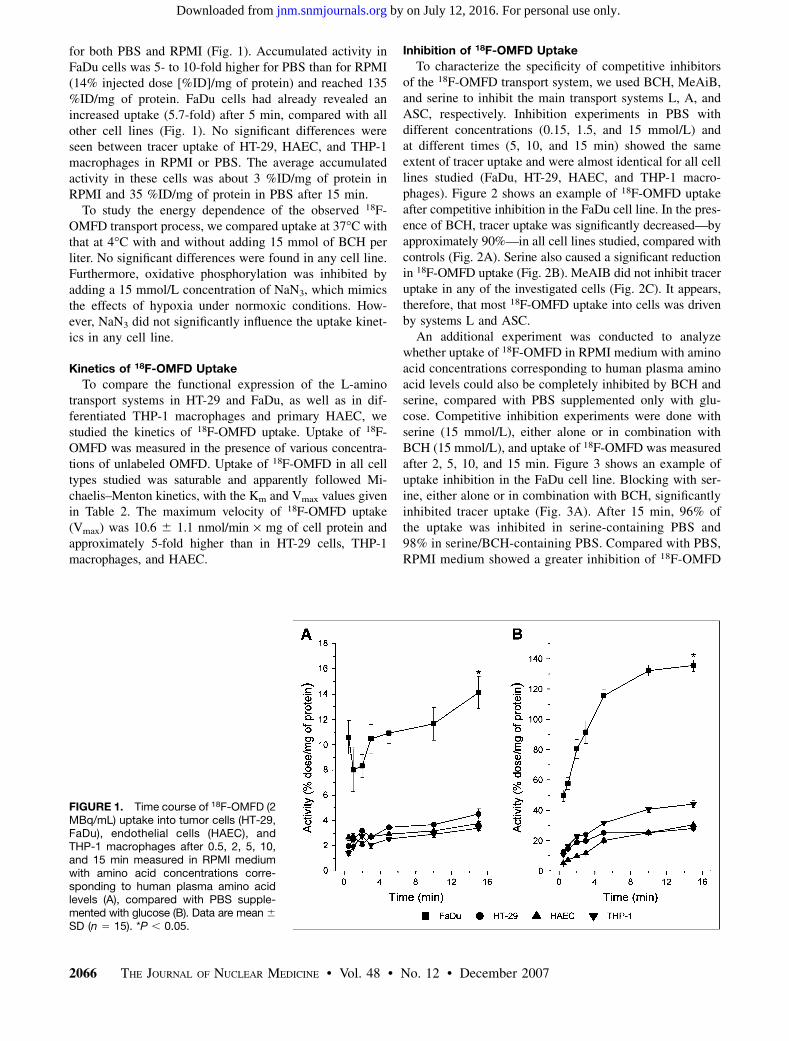
To characterize the specificity of competitive inhibitorsof the 18F-OMFD transport system, we used BCH, MeAiB,and serine to inhibit the main transport systems L, A, andASC, respectively. Inhibition experiments in PBS withdifferent concentrations (0.15, 1.5, and 15 mmol/L) andat different times (5, 10, and 15 min) showed the sameextent of tracer uptake and were almost identical for all celllines studied (FaDu, HT-29, HAEC, and THP-1 macro-phages). Figure 2 shows an example of 18F-OMFD uptakeafter competitive inhibition in the FaDu cell line. In the pres-ence of BCH, tracer uptake was significantly decreased—byapproximately 90%—in all cell lines studied, compared withcontrols (Fig. 2A). Serine also caused a significant reductionin 18F-OMFD uptake (Fig. 2B). MeAIB did not inhibit traceruptake in any of the investigated cells (Fig. 2C). It appears,therefore, that most 18F-OMFD uptake into cells was drivenby systems L and ASC.
An additional experiment was conducted to analyzewhether uptake of 18F-OMFD in RPMI medium with aminoacid concentrations corresponding to human plasma aminoacid levels could also be completely inhibited by BCH andserine, compared with PBS supplemented only with glu-cose. Competitive inhibition experiments were done withserine (15 mmol/L), either alone or in combination withBCH (15 mmol/L), and uptake of 18F-OMFD was measuredafter 2, 5, 10, and 15 min. Figure 3 shows an example ofuptake inhibition in the FaDu cell line. Blocking with ser-ine, either alone or in combination with BCH, significantlyinhibited tracer uptake (Fig. 3A). After 15 min, 96% ofthe uptake was inhibited in serine-containing PBS and98% in serine/BCH-containing PBS. Compared with PBS,RPMI medium showed a greater inhibition of 18F-OMFD
FIGURE 1. Time course of 18F-OMFD (2MBq/mL) uptake into tumor cells (HT-29,FaDu), endothelial cells (HAEC), andTHP-1 macrophages after 0.5, 2, 5, 10,and 15 min measured in RPMI mediumwith amino acid concentrations corre-sponding to human plasma amino acidlevels (A), compared with PBS supple-mented with glucose (B). Data are mean 6
SD (n 5 15). *P , 0.05.
2066 THE JOURNAL OF NUCLEAR MEDICINE • Vol. 48 • No. 12 • December 2007
by on July 12, 2016. For personal use only. jnm.snmjournals.org Downloaded from
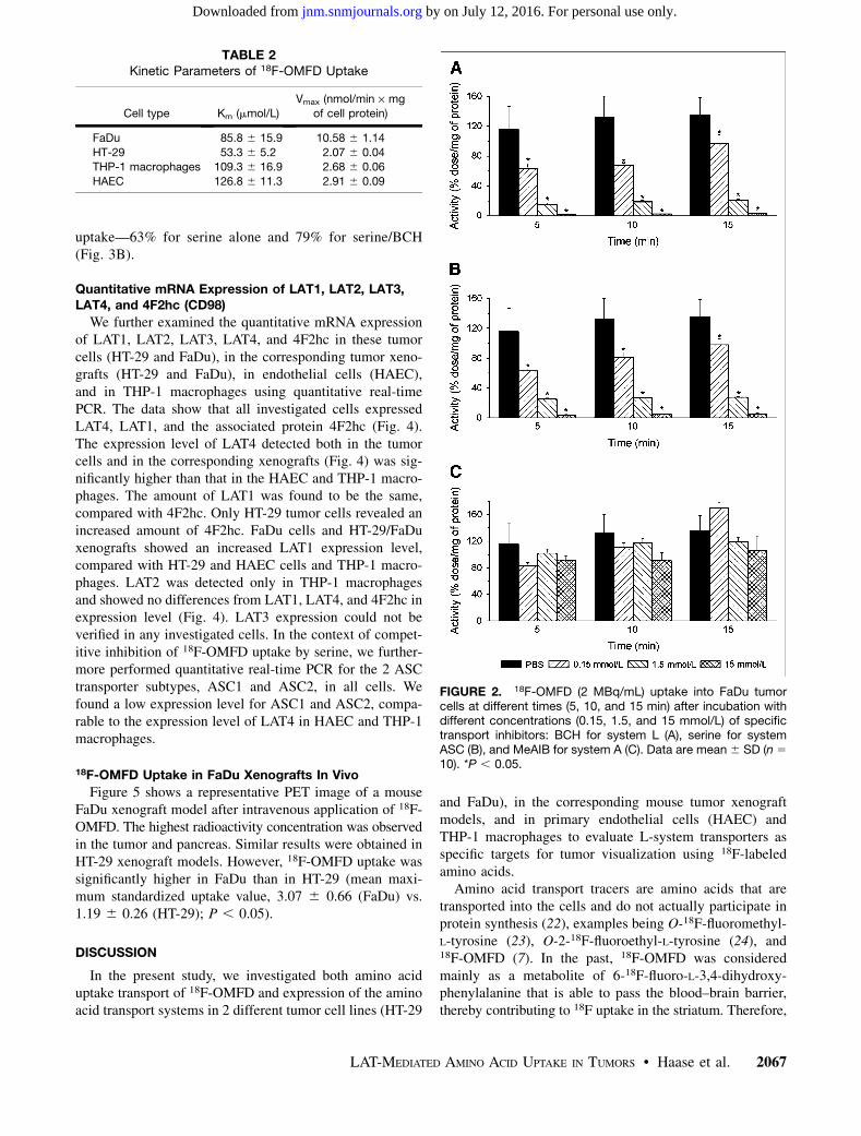
uptake—63% for serine alone and 79% for serine/BCH(Fig. 3B).
Quantitative mRNA Expression of LAT1, LAT2, LAT3,LAT4, and 4F2hc (CD98)
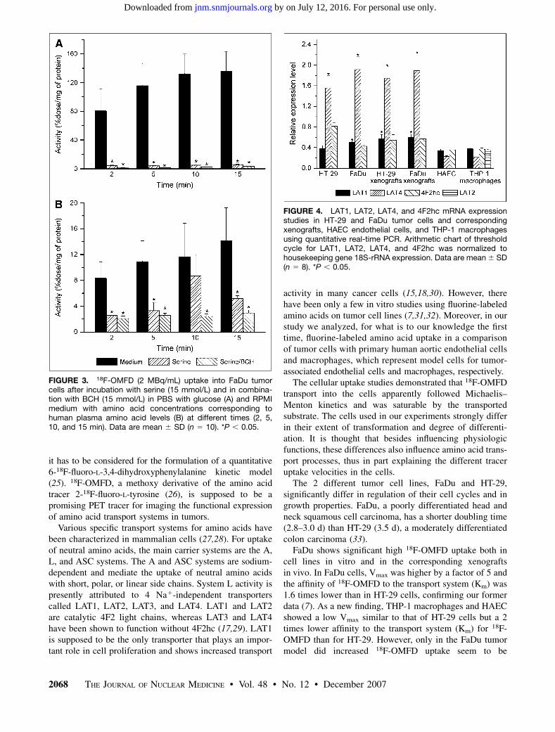
We further examined the quantitative mRNA expressionof LAT1, LAT2, LAT3, LAT4, and 4F2hc in these tumorcells (HT-29 and FaDu), in the corresponding tumor xeno-grafts (HT-29 and FaDu), in endothelial cells (HAEC),and in THP-1 macrophages using quantitative real-timePCR. The data show that all investigated cells expressedLAT4, LAT1, and the associated protein 4F2hc (Fig. 4).The expression level of LAT4 detected both in the tumorcells and in the corresponding xenografts (Fig. 4) was sig-nificantly higher than that in the HAEC and THP-1 macro-phages. The amount of LAT1 was found to be the same,compared with 4F2hc. Only HT-29 tumor cells revealed anincreased amount of 4F2hc. FaDu cells and HT-29/FaDuxenografts showed an increased LAT1 expression level,compared with HT-29 and HAEC cells and THP-1 macro-phages. LAT2 was detected only in THP-1 macrophagesand showed no differences from LAT1, LAT4, and 4F2hc inexpression level (Fig. 4). LAT3 expression could not beverified in any investigated cells. In the context of compet-itive inhibition of 18F-OMFD uptake by serine, we further-more performed quantitative real-time PCR for the 2 ASCtransporter subtypes, ASC1 and ASC2, in all cells. Wefound a low expression level for ASC1 and ASC2, compa-rable to the expression level of LAT4 in HAEC and THP-1macrophages.
18F-OMFD Uptake in FaDu Xenografts In Vivo
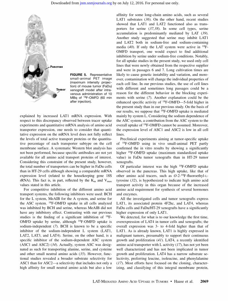
Figure 5 shows a representative PET image of a mouseFaDu xenograft model after intravenous application of 18F-OMFD. The highest radioactivity concentration was observedin the tumor and pancreas. Similar results were obtained inHT-29 xenograft models. However, 18F-OMFD uptake wassignificantly higher in FaDu than in HT-29 (mean maxi-mum standardized uptake value, 3.07 6 0.66 (FaDu) vs.1.19 6 0.26 (HT-29); P , 0.05).
DISCUSSION
In the present study, we investigated both amino aciduptake transport of 18F-OMFD and expression of the aminoacid transport systems in 2 different tumor cell lines (HT-29
and FaDu), in the corresponding mouse tumor xenograftmodels, and in primary endothelial cells (HAEC) andTHP-1 macrophages to evaluate L-system transporters asspecific targets for tumor visualization using 18F-labeledamino acids.
Amino acid transport tracers are amino acids that aretransported into the cells and do not actually participate inprotein synthesis (22), examples being O-18F-fluoromethyl-L-tyrosine (23), O-2-18F-fluoroethyl-L-tyrosine (24), and18F-OMFD (7). In the past, 18F-OMFD was consideredmainly as a metabolite of 6-18F-fluoro-L-3,4-dihydroxy-phenylalanine that is able to pass the blood–brain barrier,thereby contributing to 18F uptake in the striatum. Therefore,
FIGURE 2. 18F-OMFD (2 MBq/mL) uptake into FaDu tumorcells at different times (5, 10, and 15 min) after incubation withdifferent concentrations (0.15, 1.5, and 15 mmol/L) of specifictransport inhibitors: BCH for system L (A), serine for systemASC (B), and MeAIB for system A (C). Data are mean 6 SD (n 5
10). *P , 0.05.
TABLE 2Kinetic Parameters of 18F-OMFD Uptake
Cell type Km (mmol/L)
Vmax (nmol/min · mg
of cell protein)
FaDu 85.8 6 15.9 10.58 6 1.14
HT-29 53.3 6 5.2 2.07 6 0.04THP-1 macrophages 109.3 6 16.9 2.68 6 0.06
HAEC 126.8 6 11.3 2.91 6 0.09
LAT-MEDIATED AMINO ACID UPTAKE IN TUMORS • Haase et al. 2067
by on July 12, 2016. For personal use only. jnm.snmjournals.org Downloaded from
it has to be considered for the formulation of a quantitative6-18F-fluoro-L-3,4-dihydroxyphenylalanine kinetic model(25). 18F-OMFD, a methoxy derivative of the amino acidtracer 2-18F-fluoro-L-tyrosine (26), is supposed to be apromising PET tracer for imaging the functional expressionof amino acid transport systems in tumors.
Various specific transport systems for amino acids havebeen characterized in mammalian cells (27,28). For uptakeof neutral amino acids, the main carrier systems are the A,L, and ASC systems. The A and ASC systems are sodium-dependent and mediate the uptake of neutral amino acidswith short, polar, or linear side chains. System L activity ispresently attributed to 4 Na1-independent transporterscalled LAT1, LAT2, LAT3, and LAT4. LAT1 and LAT2are catalytic 4F2 light chains, whereas LAT3 and LAT4have been shown to function without 4F2hc (17,29). LAT1is supposed to be the only transporter that plays an impor-tant role in cell proliferation and shows increased transport
activity in many cancer cells (15,18,30). However, therehave been only a few in vitro studies using fluorine-labeledamino acids on tumor cell lines (7,31,32). Moreover, in ourstudy we analyzed, for what is to our knowledge the firsttime, fluorine-labeled amino acid uptake in a comparisonof tumor cells with primary human aortic endothelial cellsand macrophages, which represent model cells for tumor-associated endothelial cells and macrophages, respectively.
The cellular uptake studies demonstrated that 18F-OMFDtransport into the cells apparently followed Michaelis–Menton kinetics and was saturable by the transportedsubstrate. The cells used in our experiments strongly differin their extent of transformation and degree of differenti-ation. It is thought that besides influencing physiologicfunctions, these differences also influence amino acid trans-port processes, thus in part explaining the different traceruptake velocities in the cells.
The 2 different tumor cell lines, FaDu and HT-29,significantly differ in regulation of their cell cycles and ingrowth properties. FaDu, a poorly differentiated head andneck squamous cell carcinoma, has a shorter doubling time(2.8–3.0 d) than HT-29 (3.5 d), a moderately differentiatedcolon carcinoma (33).
FaDu shows significant high 18F-OMFD uptake both incell lines in vitro and in the corresponding xenograftsin vivo. In FaDu cells, Vmax was higher by a factor of 5 andthe affinity of 18F-OMFD to the transport system (Km) was1.6 times lower than in HT-29 cells, confirming our formerdata (7). As a new finding, THP-1 macrophages and HAECshowed a low Vmax similar to that of HT-29 cells but a 2times lower affinity to the transport system (Km) for 18F-OMFD than for HT-29. However, only in the FaDu tumormodel did increased 18F-OMFD uptake seem to be
FIGURE 3. 18F-OMFD (2 MBq/mL) uptake into FaDu tumorcells after incubation with serine (15 mmol/L) and in combina-tion with BCH (15 mmol/L) in PBS with glucose (A) and RPMImedium with amino acid concentrations corresponding tohuman plasma amino acid levels (B) at different times (2, 5,10, and 15 min). Data are mean 6 SD (n 5 10). *P , 0.05.
FIGURE 4. LAT1, LAT2, LAT4, and 4F2hc mRNA expressionstudies in HT-29 and FaDu tumor cells and correspondingxenografts, HAEC endothelial cells, and THP-1 macrophagesusing quantitative real-time PCR. Arithmetic chart of thresholdcycle for LAT1, LAT2, LAT4, and 4F2hc was normalized tohousekeeping gene 18S-rRNA expression. Data are mean 6 SD(n 5 8). *P , 0.05.
2068 THE JOURNAL OF NUCLEAR MEDICINE • Vol. 48 • No. 12 • December 2007
by on July 12, 2016. For personal use only. jnm.snmjournals.org Downloaded from
explained by increased LAT1 mRNA expression. Withrespect to this discrepancy observed between tracer uptakeexperiments and quantitative mRNA analysis of amino acidtransporter expression, one needs to consider that quanti-tative expression on the mRNA level does not fully reflectthe levels of total active transport proteins or the quantita-tive percentage of each transporter subtype on the cellmembrane surface. A systematic Western blot analysis hasnot been performed, because specific antibodies are not yetavailable for all amino acid transport proteins of interest.Considering this constraint of the present study, however,the total number of transporters can be higher in FaDu cellsthan in HT-29 cells although showing a comparable mRNAexpression level (related to the housekeeping gene 18SrRNA). This fact is, in part, reflected by the Km and Vmax
values stated in this article.For competitive inhibition of the different amino acid
transport systems, the following inhibitors were used: BCHfor the L system, MeAIB for the A system, and serine forthe ASC system. 18F-OMFD uptake in all cells analyzedwas inhibited by BCH and serine, whereas MeAIB did nothave any inhibitory effect. Contrasting with our previousstudies is the finding of a significant inhibition of 18F-OMFD uptake by serine, although 18F-OMFD uptake issodium-independent (7). BCH is known to be a specificinhibitor of the sodium-independent L system (LAT1,LAT2, LAT3, and LAT4). Serine, on the other hand, is aspecific inhibitor of the sodium-dependent ASC system(ASC1 and ASC2) (34). Actually, system ASC was desig-nated as such for transporting alanine, serine, and cysteineand other small neutral amino acids (35). However, func-tional studies revealed a broader substrate selectivity forASC1 than for ASC2—a selectivity that includes not only ahigh affinity for small neutral amino acids but also a low
affinity for some long-chain amino acids, such as severalLAT1 substrates (36). On the other hand, recent studiesshowed that LAT1 and LAT2 functioned also as trans-porters for serine (37,38). In some cell types, serineaccumulation is predominantly mediated by LAT (39).Another study suggested that serine may inhibit LAT1and LAT2 both in sodium-free and sodium-containingmedia (40). If only the LAT system were active in 18F-OMFD transport, one would expect to find additionalinhibition by serine under sodium-free conditions. Notably,for all uptake studies in the present study, we used only celllines that were newly obtained from the respective supplierand were in passages 6 and 7. Long cultivation times arelikely to cause genetic instability and variation, and more-over, contamination will change the individual properties ofeach cell line. In our previous studies, the use of cell lineswith different and sometimes long passages could be areason for the different behavior in the blocking experi-ments with serine (7). Another explanation could be theenhanced specific activity of 18F-OMFD—5-fold higher inthe present study than in our previous study. On the basis ofour results, we suppose that 18F-OMFD uptake is mediatedmainly by system L. Considering the sodium dependence ofthe ASC system, a contribution from the ASC system to theoverall uptake of 18F-OMFD cannot be assumed. Moreover,the expression level of ASC1 and ASC2 is low in all celllines.
Preclinical experiments aiming at tumor-specific uptakeof 18F-OMFD using in vivo small-animal PET partlyconfirmed the in vitro results by showing a significantlyhigher 18F-OMFD uptake (maximum standardized uptakevalue) in FaDu tumor xenografts than in HT-29 tumorxenografts.
Of particular interest was the high 18F-OMFD uptakeobserved in the pancreas. This high uptake, like that ofother amino acid tracers, such as O-2-18F-fluoroethyl-L-tyrosine (32), is hypothesized to indicate high amino acidtransport activity in this organ because of the increasedamino acid requirement for synthesis of several hormonesand enzymes.
All the investigated cells and tumor xenografts expressLAT1, its associated protein 4F2hc, and LAT4, whereasFaDu cells and FaDu/HT-29 xenografts have a significantlyhigher expression of only LAT1.
We detected, for what is to our knowledge the first time,overexpression of LAT4 in tumor cells and xenografts; theoverall expression was 3- to 4-fold higher than that ofLAT1. As is already known, LAT1 is highly expressed inmalignant tumors, presumably to support their continuousgrowth and proliferation (41). LAT4, a recently identifiedamino acid transporter with L activity (17), has not yet beenwell characterized and has not been implicated in tumorgrowth and proliferation. LAT4 has a narrow substrate se-lectivity, preferring leucine, isoleucine, and phenylalanine(17). Most efforts have focused on the cloning, character-izing, and classifying of this integral membrane protein,
FIGURE 5. Representativesmall-animal PET image(maximum intensity projec-tion) of mouse tumor (FaDu)xenograft model after intra-venous administration of 10MBq of 18F-OMFD (60 minafter injection).
LAT-MEDIATED AMINO ACID UPTAKE IN TUMORS • Haase et al. 2069
by on July 12, 2016. For personal use only. jnm.snmjournals.org Downloaded from
whereas studies of the biologic utility are still ongoing.No evidence has suggested that LAT4 has a role incarcinogenesis.
Furthermore, we found LAT2 mRNA expression only inTHP-1 macrophages, and we found no LAT2 transcripts inany of the tumor cells or corresponding xenografts. LAT2 isbelieved to function predominantly as an efflux pathway(12). Studies using real-time PCR and Western blot analysishave already shown that different tumor cells express LAT1and its associated protein, 4F2hc, at high levels, but notLAT2 (18,42). The similarity in 18F-OMFD uptake betweenTHP-1 cells and the HT-29 tumor model, despite a signif-icantly lower LAT4 expression in the former (expression ofLAT1 is comparable), could, in addition, be explained by acompensatory uptake via LAT2 that is expressed in THP-1cells. For HAEC, a difference from HT-29 cells in thequantitative percentage of transporter subtypes on the cellmembrane surface can be assumed.
Based on our studies, the recently identified transporterLAT4 and the well known LAT1 seem to be an excellentand promising target for further studies on tumor diagnosisand molecular imaging. We showed here, for what is to ourknowledge the first time, high expression of LAT4, andfurther investigations need to focus on this transportersubtype with respect to tumor proliferation and growth.This newly gained knowledge of the molecular mechanisminvolved in the transport of essential amino acids willamplify the potential of radiolabeled amino acids in thediagnosis of, for example, squamous cell head and neckcancer.
CONCLUSION
Because of high accumulation of 18F-OMFD in FaDucells and the corresponding xenografts, PET with 18F-OMFD possibly has the potential to improve staging anddetection of certain types of cancer, particularly of poorlydifferentiated squamous cell head and neck tumors. Inaddition, 18F-OMFD may allow quantification of importantcellular processes related to tumor proliferation in certaintumor entities, such as squamous cell head and neck cancer.However, further study is needed of, on the one hand, thecontribution of endothelial cells and proinflammatory cellssuch as macrophages and, on the other hand, the role ofdifferent LAT subtypes in overall uptake of 18F-OMFD intumors and inflammatory lesions.
ACKNOWLEDGMENTS
This work was supported in part by the Federal Ministryfor Education and Research (BMBF) (Sign 03/4021) and agrant from the European Union (Molecular Imaging forBiologically Optimized Cancer Therapy, BIOCARE, con-tract 505785). We thank Dr. Stefan Hamm (ABX) forproviding the precursor for the 18F-OMFD and the refer-ence standard. The excellent technical help of NicoleEwald, Mareike Barth, and Regina Herrlich is gratefully
acknowledged. We thank Christina Schutze, DorotheePfitzmann, Katja Schumann, and the staff of the Experi-mental Centre of the Medical Faculty of the University ofTechnology Dresden for providing the tumor xenograftmodels. We also thank the staff of GMP laboratory at theInstitute of Radiopharmacy for expert technical assistancein 18F-OMFD synthesis and quality control. We are alsograteful to Bettina Beuthien-Baumann, MD, for her expertadvice and many stimulating discussions.
REFERENCES
1. Gambhir SS, Czernin J, Schwimmer J, Silverman DH, Coleman RE, Phelps ME.
A tabulated summary of the FDG PET literature. J Nucl Med. 2001;42(suppl):
1–93.
2. Stober B, Tanase U, Herz M, Seidl C, Schwaiger M, Senekowitsch-Schmidtke R.
Differentiation of tumour and inflammation: characterisation of [methyl-3H]
methionine (MET) and O-(2-[18F]fluoroethyl)-L-tyrosine (FET) uptake in
human tumour and inflammatory cells. Eur J Nucl Med Mol Imaging. 2006;33:
932–939.
3. Langen KJ, Ziemons K, Kiwit JC, et al. 3-[123I]iodo-alpha-methyltyrosine and
[methyl-11C]-L-methionine uptake in cerebral gliomas: a comparative study
using SPECT and PET. J Nucl Med. 1997;38:517–522.
4. Leskinen-Kallio S, Nagren K, Lehikoinen P, Ruotsalainen U, Joensuu H. Uptake
of 11C-methionine in breast cancer studied by PET: an association with the size
of S-phase fraction. Br J Cancer. 1991;64:1121–1124.
5. Nettelbladt OS, Sundin AE, Valind SO, et al. Combined fluorine-18-FDG and
carbon-11-methionine PET for diagnosis of tumors in lung and mediastinum.
J Nucl Med. 1998;39:640–647.
6. Fuchtner F, Steinbach J. Efficient synthesis of the 18F-labelled 3-O-methyl-6-
[18F]fluoro-L-DOPA. Appl Radiat Isot. 2003;58:575–578.
7. Bergmann R, Pietzsch J, Fuechtner F, et al. 3-O-methyl-6-18F-fluoro-L-dopa, a
new tumor imaging agent: investigation of transport mechanism in vitro. J Nucl
Med. 2004;45:2116–2122.
8. Beuthien-Baumann B, Bredow J, Burchert W, et al. 3-O-methyl-6-[18F]fluoro-L-
DOPA and its evaluation in brain tumour imaging. Eur J Nucl Med Mol Imaging.
2003;30:1004–1008.
9. Umeki N, Fukasawa Y, Ohtsuki S, et al. mRNA expression and amino acid
transport characteristics of cultured human brain microvascular endothelial cells
(hBME). Drug Metab Pharmacokinet. 2002;17:367–373.
10. Naumov GN, Akslen LA, Folkman J. Role of angiogenesis in human tumor
dormancy: animal models of the angiogenic switch. Cell Cycle. 2006;5:1779–1787.
11. Torrents D, Estevez R, Pineda M, et al. Identification and characterization of a
membrane protein (y1L amino acid transporter-1) that associates with 4F2hc to
encode the amino acid transport activity y1L: a candidate gene for lysinuric
protein intolerance. J Biol Chem. 1998;273:32437–32445.
12. Verrey F. System L: heteromeric exchangers of large, neutral amino acids
involved in directional transport. Pflugers Arch. 2003;445:529–533.
13. Uchino H, Kanai Y, Kim DK, et al. Transport of amino acid-related compounds
mediated by L-type amino acid transporter 1 (LAT1): insights into the
mechanisms of substrate recognition. Mol Pharmacol. 2002;61:729–737.
14. Rossier G, Meier C, Bauch C, et al. LAT2, a new basolateral 4F2hc/CD98-
associated amino acid transporter of kidney and intestine. J Biol Chem. 1999;274:
34948–34954.
15. Lahoutte T, Caveliers V, Camargo SM, et al. SPECT and PET amino acid tracer
influx via system L (h4F2hc-hLAT1) and its transstimulation. J Nucl Med. 2004;
45:1591–1596.
16. Campbell WA, Thompson NL. Overexpression of LAT1/CD98 light chain is
sufficient to increase system L-amino acid transport activity in mouse hepato-
cytes but not fibroblasts. J Biol Chem. 2001;276:16877–16884.
17. Bodoy S, Martin L, Zorzano A, Palacin M, Estevez R, Bertran J. Identification of
LAT4, a novel amino acid transporter with system L activity. J Biol Chem. 2005;
280:12002–12011.
18. Kim Do K, Kim IJ, Hwang S, et al. System L-amino acid transporters are dif-
ferently expressed in rat astrocyte and C6 glioma cells. Neurosci Res. 2004;50:
437–446.
19. Kobayashi H, Ishii Y, Takayama T. Expression of L-type amino acid transporter
1 (LAT1) in esophageal carcinoma. J Surg Oncol. 2005;90:233–238.
20. Haase C, Bergmann R, Oswald J, Zips D, Pietzsch J. Neurotensin receptors in
adeno- and squamous cell carcinoma. Anticancer Res. 2006;26:3527–3533.
2070 THE JOURNAL OF NUCLEAR MEDICINE • Vol. 48 • No. 12 • December 2007
by on July 12, 2016. For personal use only. jnm.snmjournals.org Downloaded from
21. Berndt M, Pietzsch J, Wuest F. Labeling of low-density lipoproteins using the18F-labeled thiol-reactive reagent N-[6-(4-[18F]fluorobenzylidene)aminooxyhexyl]
maleimide. Nucl Med Biol. 2007;34:5–15.
22. Jager PL, Vaalburg W, Pruim J, de Vries EG, Langen KJ, Piers DA. Radiolabeled
amino acids: basic aspects and clinical applications in oncology. J Nucl Med.
2001;42:432–445.
23. Ishiwata K, Kawamura K, Wang WF, et al. Evaluation of O-[11C]methyl-L-
tyrosine and O-[18F]fluoromethyl-L-tyrosine as tumor imaging tracers by PET.
Nucl Med Biol. 2004;31:191–198.
24. Wester HJ, Herz M, Weber W, et al. Synthesis and radiopharmacology of O-(2-
[18F]fluoroethyl)-L-tyrosine for tumor imaging. J Nucl Med. 1999;40:205–212.
25. Brust P, Bauer R, Walter B, et al. Simultaneous measurement of [18F]FDOPA
metabolism and cerebral blood flow in newborn piglets. Int J Dev Neurosci.
1998;16:353–364.
26. Coenen HH, Kling P, Stocklin G. Cerebral metabolism of L-[2-18F]fluorotyro-
sine, a new PET tracer of protein synthesis. J Nucl Med. 1989;30:1367–1372.
27. Segel GB, Woodlock TJ, Murant FG, Lichtman MA. Photoinhibition of 2-amino-
2-carboxybicyclo[2,2,1]heptane transport by O-diazoacetyl-L-serine: an initial
step in identifying the L-system amino acid transporter. J Biol Chem. 1989;264:
16399–16402.
28. Saier MH Jr, Daniels GA, Boerner P, Lin J. Neutral amino acid transport systems
in animal cells: potential targets of oncogene action and regulators of cellular
growth. J Membr Biol. 1988;104:1–20.
29. Babu E, Kanai Y, Chairoungdua A, et al. Identification of a novel system L
amino acid transporter structurally distinct from heterodimeric amino acid trans-
porters. J Biol Chem. 2003;278:43838–43845.
30. Yoon JH, Kim IJ, Kim H, et al. Amino acid transport system L is differently
expressed in human normal oral keratinocytes and human oral cancer cells.
Cancer Lett. 2005;222:237–245.
31. Langen KJ, Jarosch M, Muhlensiepen H, et al. Comparison of fluorotyrosines
and methionine uptake in F98 rat gliomas. Nucl Med Biol. 2003;30:501–508.
32. Heiss P, Mayer S, Herz M, Wester HJ, Schwaiger M, Senekowitsch-Schmidtke R.
Investigation of transport mechanism and uptake kinetics of O-(2-[18F]fluoroethyl)-
L-tyrosine in vitro and in vivo. J Nucl Med. 1999;40:1367–1373.
33. Azrak RG, Cao S, Slocum HK, et al. Therapeutic synergy between irinotecan
and 5-fluorouracil against human tumor xenografts. Clin Cancer Res. 2004;10:
1121–1129.
34. Christensen HN, Handlogten ME, Lam I, Tager HS, Zand R. A bicyclic amino
acid to improve discriminations among transport systems. J Biol Chem. 1969;244:
1510–1520.
35. Christensen HN, Liang M, Archer EG. A distinct Na1-requiring transport
system for alanine, serine, cysteine, and similar amino acids. J Biol Chem. 1967;
242:5237–5246.
36. Fuchs BC, Bode BP. Amino acid transporters ASCT2 and LAT1 in cancer:
partners in crime? Semin Cancer Biol. 2005;15:254–266.
37. Kanai Y, Segawa H, Miyamoto K, Uchino H, Takeda E, Endou H. Expression
cloning and characterization of a transporter for large neutral amino acids activated
by the heavy chain of 4F2 antigen (CD98). J Biol Chem. 1998;273:23629–23632.
38. Segawa H, Fukasawa Y, Miyamoto K, Takeda E, Endou H, Kanai Y. Identification
and functional characterization of a Na1-independent neutral amino acid
transporter with broad substrate selectivity. J Biol Chem. 1999;274:19745–19751.
39. Takarada T, Balcar VJ, Baba K, et al. Uptake of [3H]L-serine in rat brain
synaptosomal fractions. Brain Res. 2003;983:36–47.
40. Budy B, O’Neill R, DiBello PM, Sengupta S, Jacobsen DW. Homocysteine trans-
port by human aortic endothelial cells: identification and properties of import
systems. Arch Biochem Biophys. 2006;446:119–130.
41. Yanagida O, Kanai Y, Chairoungdua A, et al. Human L-type amino acid trans-
porter 1 (LAT1): characterization of function and expression in tumor cell lines.
Biochim Biophys Acta. 2001;1514:291–302.
42. Yoon JH, Kim YB, Kim MS, et al. Expression and functional characterization of
the system L amino acid transporter in KB human oral epidermoid carcinoma
cells. Cancer Lett. 2004;205:215–226.
LAT-MEDIATED AMINO ACID UPTAKE IN TUMORS • Haase et al. 2071
by on July 12, 2016. For personal use only. jnm.snmjournals.org Downloaded from
Doi: 10.2967/jnumed.107.0436202007;48:2063-2071.J Nucl Med.
Cathleen Haase, Ralf Bergmann, Frank Fuechtner, Alexander Hoepping and Jens Pietzsch Vitro and In Vivo
F-Fluoro-l-Dopa in Human Adenocarcinoma and Squamous Cell Carcinoma In18-Methyl-6- OL-Type Amino Acid Transporters LAT1 and LAT4 in Cancer: Uptake of 3-
http://jnm.snmjournals.org/content/48/12/2063This article and updated information are available at:
http://jnm.snmjournals.org/site/subscriptions/online.xhtml
Information about subscriptions to JNM can be found at:
http://jnm.snmjournals.org/site/misc/permission.xhtmlInformation about reproducing figures, tables, or other portions of this article can be found online at:
(Print ISSN: 0161-5505, Online ISSN: 2159-662X)1850 Samuel Morse Drive, Reston, VA 20190.SNMMI | Society of Nuclear Medicine and Molecular Imaging
is published monthly.The Journal of Nuclear Medicine
© Copyright 2007 SNMMI; all rights reserved.
by on July 12, 2016. For personal use only. jnm.snmjournals.org Downloaded from


![Usefulness of [18F]-DA and [18F]-DOPA for PET imaging in a mouse model of pheochromocytoma](https://static.fdokumen.com/doc/165x107/6325a7d9852a7313b70e9a7d/usefulness-of-18f-da-and-18f-dopa-for-pet-imaging-in-a-mouse-model-of-pheochromocytoma.jpg)