Total Synthesis of Taxol. 1. Retrosynthesis, Degradation, and Reconstitution
Kupffer Cells are Depleted with HIV Immunodeficiency andPartially Recovered with Antiretroviral Immunereconstitution:HIV, Kupffer Cells and Antiretroviral Therapy
Ashwin Balagopal1, Stuart C. Ray1, Ruben Montes De Oca2, Catherine G. Sutcliffe2,Perumal Vivekanandan3, Yvonne Higgins1, Shruti H. Mehta2, Richard D. Moore1,2, Mark S.Sulkowski1, David L. Thomas1,2, and Michael S. Torbenson3
1 Division of Infectious Diseases, Department of Medicine, The Johns Hopkins MedicalInstitutions, Baltimore, Maryland, U.S.A2 Department of Epidemiology, Johns Hopkins Bloomberg School of Public Health, Baltimore,Maryland, U.S.A3 Department of Pathology, The Johns Hopkins Medical Institutions, Baltimore, Maryland, U.S.A
IntroductionIn the modern era of highly active anti-retroviral therapy (HAART), a leading cause of deathin HIV-infected persons is liver disease, most often due to chronic hepatitis C virus (HCV)infection [1]. Approximately one quarter of HIV infected persons have chronic hepatitis C[2], and HIV-HCV co-infected persons have demonstrated a more rapid progression of liverdisease compared with HCV mono-infected persons. Although the mechanism(s) are notfully known, liver disease is more severe in those with more profound CD4+ lymphocytedepletion compared to those with preserved peripheral blood CD4+ lymphocyte counts [3–8].
HIV induced depletion of intestinal CD4+ lymphocytes [9–12] has recently been linked tomicrobial translocation and HIV progression [13]. Moreover, in a cohort of HCV-infectedpersons at different stages of liver disease we have shown that microbial translocation inHIV co-infected persons was strongly associated with liver disease progression [14]. In thatstudy we also found that HIV seroconversion amongst HCV-infected persons was associatedwith an increase in microbial translocation over time. It is, therefore, compelling to considermicrobial translocation as a common mechanism that contributes to both HIV and HCVprogression. Interestingly, animal models of other forms of liver disease have demonstrateda critical role of intestinal microbial translocation in promoting fibrosis [15–20].
Hepatic macrophages, or Kupffer cells, are responsible for clearing microbial translocationproducts and play a role in liver disease. Kupffer cells, however, can be infected by HIV,and this may result in their impaired ability to clear these potentially fibrogenic microbialtranslocation products [21–27]. In this investigation, we tested the hypotheses that Kupffercell quantities are associated with peripheral CD4+ lymphocyte count in HIV-HCV co-infection, and that changes in CD4+ due to antiretroviral therapy are associated withcorresponding alterations in Kupffer cell quantities. In addition, since in chronic viralhepatitis fibrosis begins in the portal and periportal regions where microbial translocation
Correspondence: David L. Thomas, 1830 Building Room 455-ID, 600 North Wolfe Street, Baltimore, MD 21287, [email protected],fax: (410) 614-7564.The authors have no financial conflicts of interest to report.
NIH Public AccessAuthor ManuscriptAIDS. Author manuscript; available in PMC 2011 May 11.
Published in final edited form as:AIDS. 2009 November 27; 23(18): 2397–2404. doi:10.1097/QAD.0b013e3283324344.
NIH
-PA Author Manuscript
NIH
-PA Author Manuscript
NIH
-PA Author Manuscript

products first enter the liver, we tested the hypothesis that Kupffer cells would be mostabundant in these regions.
MethodsThe study population derives from the HIV-HCV co-infected members of the Johns HopkinsUniversity clinical cohort (Baltimore, MD) [3;28]. Seventy-six individuals were identifiedwho had at least two archived liver tissue samples and correlated clinical data characterizingHIV and HCV stage between January, 1997 and February, 2005. All subjects providedwritten informed consent for testing through a protocol approved by the Committees onHuman Research of the Johns Hopkins School of Medicine or Bloomberg School of PublicHealth.
Data on clinical and lab parameters were abstracted from the clinical and laboratorydatabases. Transcutaneous liver biopsies were obtained using an 18-gauge needle. Livertissue was fixed in 10% formalin and paraffin-embedded. Tissues were stained withhematoxylin and eosin (H&E) as well as trichrome. As previously described, tissues werescored by an experienced liver pathologist (M.T.) for fibrosis according to the Metavirscoring system for HCV infection and were graded for the degree of inflammation by usingthe Ishak modified hepatic activity index (MHAI) [29]. Hepatic fat (steatosis) was assessedas an average percentage of fat (0, 1–30, 31–60, > 60%) on H&E section. The pathologistwas blinded to the subjects’ clinical history and laboratory values. In a select group ofsubjects who were studied longitudinally, slides obtained from the same subject wereseparately encoded and de-identified before handling by the pathologist. Adequacy of tissuesize was determined by the pathologist and subjects with inadequate tissues were excludedfrom the investigation. The median length of tissues was 12 mm. To detect Kupffer cells,tissue sections were immunostained following heat antigen retrieval with mouse monoclonalanti-CD68 antibodies (Dako, Carpinteria, CA), used at a 1:100 dilution. The DAKOEnVision+ Peroxidase kit was used for immunostaining. Kupffer cells were identified bytheir strong cytoplasmic staining. Kupffer cell density (KCD) was determined as thearithmetic mean number of Kupffer cells per 5 high power fields. Portal and periportalKupffer cells were further quantified and compared with centrilobular Kupffer cells. [Todetect CD68+/HLA-DRα+ Kupffer cells, paraffin-embedded tissue sections from 20available liver blocks were selected from a subset of subjects matched for MHAI score whohad liver biopsies obtained at a second time point. Blocks were deparaffinized andsequentially immunostained with the anti-CD68+ antibody followed by a rabbit polyclonalanti-HLA-DRα IgG antibody (Santa Cruz, Santa Cruz, CA), both at 1:100 dilution. Dualsequential immunostaining was performed using the EnVision G|2 Doublestain System(Dako, Carpinteria, CA). CD68+/ HLA-DRα-, and CD68−/ HLA-DRα+, and CD68+/HLA-DRα+ cells were enumerated in 5–10 parenchymal high powered fields and averaged.]
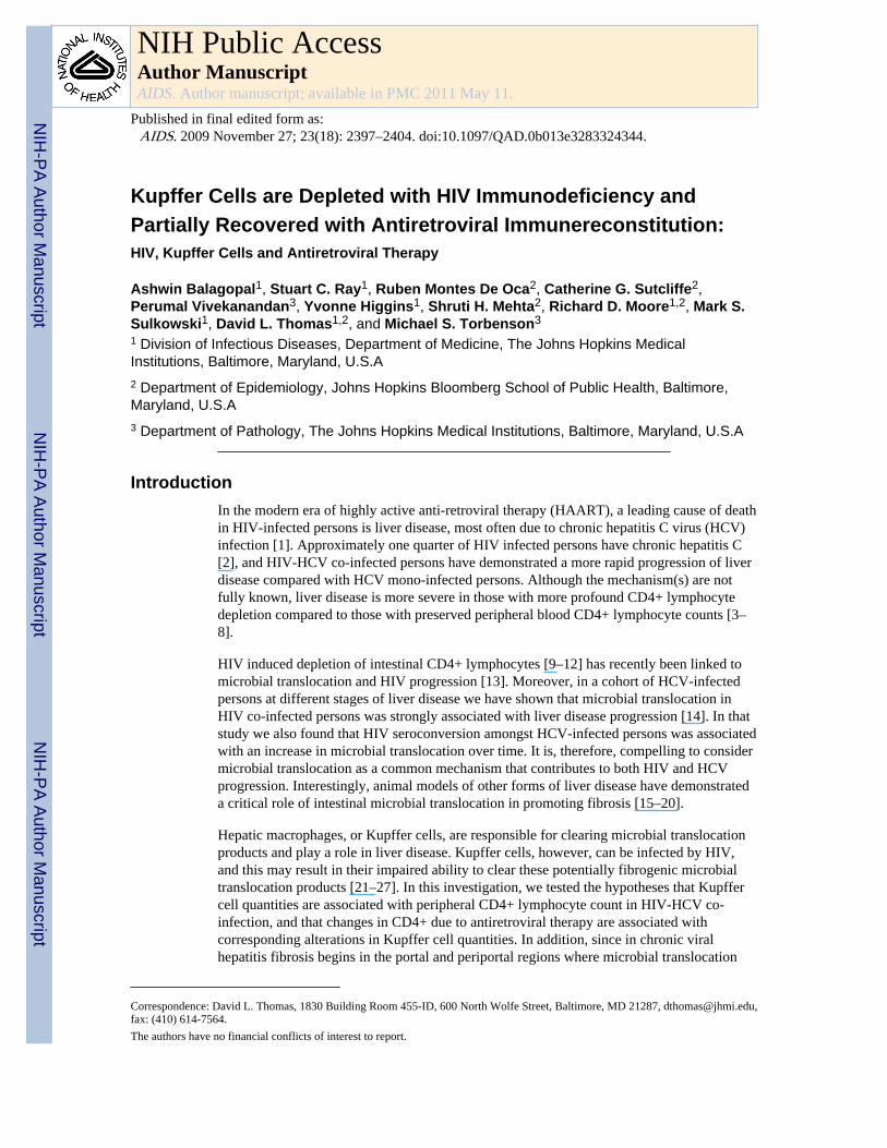
Regional differences in KCD were characterized by quantifying all CD68+ Kupffer cellswithin a 50 μm radius of selected portal and centrilobular veins. A Zeiss PALMR
MicroLaser system was used to accurately determine a 50 μm radius around vessels. Whilethis distance extends from portal to periportal regions depending on the section, it allows forprecise comparisons between portal and centrilobular Kupffer cells. Cases were selected foranalysis if they had at least one suitable complete cross section of both a well oriented portaltract and central vein. To avoid skewed results on the basis of oversampling regions ofabundant inflammation, only 1 representative portal or centrilobular vein was used from acluster of branching vessels. Multiple sequential sections were only used to confirmfindings, and not used for counting to avoid oversampling the same cells. Kupffer cellnumber was normalized per μm2 to account for differences in vessel size using the adaptedarea of an elliptical ring, π *(½ a + 50)*(½ b + 50) − π*(½ a)* (½ b), where a and b were
Balagopal et al. Page 2
AIDS. Author manuscript; available in PMC 2011 May 11.
NIH
-PA Author Manuscript
NIH
-PA Author Manuscript
NIH
-PA Author Manuscript

the measured long and short axes of the vessel in μm (Fig. 1). Kupffer cell counts of zerowere conservatively assigned values of 0.5 per given area as the lower limit of detection.Results are reported in mm2.
Liver biopsies were obtained a median (range) of 1.25 (0.1 – 139.6) months from theassessment of CD4+ lymphocyte count and clinical data, which were also ascertained usingstructured instruments before and after the initial liver biopsy, as described elsewhere [28].KCD in all subjects was approximately normally distributed. Wilcoxon rank-sum andKruskal-Wallis tests were used to compare median KCD values across different covariatesof interest. Linear regression models were used to model the association between KCD andboth current and nadir CD4+ lymphocyte count. Kupffer cell quantities per vessel were notnormally distributed and were compared using Wilcoxon rank-sum tests. When log-transformed, Kupffer cell quantities per vessel yielded normally distributed data that wassimilarly found to be statistically significant using paired t tests.
ResultsSeventy-six HIV-HCV co-infected persons with archived liver biopsies were identified. Themean age of the participants was 44.8 years at the time of the first biopsy, 59 (77.6%) weremale, and 67 (88.2%) were African-American (Table 1). Peripheral blood CD4+ lymphocytecounts ranged from 9 to 1277/mm3 and serum HIV RNA ranged from undetectable to >400,000 copies per mL. Antiretroviral therapy use was reported in 57 (75%) persons. Allpersons had chronic HCV infection with a median RNA level of 596,000 IU/mL. HCVinfection was predominantly genotype 1a (66.2%) or 1b (25.4%).
Baseline liver fibrosis stage ranged from zero to cirrhosis (Metavir 0–4). Liver inflammationranged from MHAI 0 to 9, and steatosis from 0 to 60%. KCD was normally distributed overthe cohort, with a median (IQR) of 23 cells/HPF (17.7–27.8). No differences were detectedin the KCD distribution according to age, gender, or HCV genotype (Table 1). Importantly,peripheral blood monocyte (Kupffer cell precursors) quantity was not associated with KCD(p=0.25). KCD was also not associated with liver disease fibrosis stage on the concurrent orsubsequent biopsy, and was not associated with grade of hepatic inflammation or steatosis.
Mean KCD was significantly lower in subjects with lower peripheral blood CD4+lymphocyte counts compared to those with higher levels (p<0.05, by Wilcoxon rank-sumtest). KCD was associated with both contemporaneous CD4+ lymphocyte count and thelowest result recorded before histologic sampling (nadir CD4+ count, Table 2).
Since microbial translocation products enter the liver through the portal vein, we examinedwhether Kupffer cells were distributed more heavily in portal and periportal regions thancentrilobular regions, as has been seen in rats [30;31]. From the 76 archived liver biopsies,51 cases had sufficient tissue for additional analysis. There were no significant differencesin the clinical characteristics between subjects with adequate liver tissue for the regionalanalysis and those with insufficient tissue. In total, Kupffer cells from 122 portal and 95centrilobular regions were counted. The median (range) number of portal and periportalKupffer cells, 328.6 (41.4 – 3252.8) /mm2 , was markedly higher than for centrilobularKupffer cells, 90.5 (18.2 – 731.9) /mm2 (Fig. 2A, B).
[Quiescent Kupffer cells are normally immunotolerant to physiologic amounts of microbialtranslocation. In order to determine if progression of AIDS was associated with Kupffer cellimmune activation, 20 available liver blocks from equal numbers of subjects with high andlow MHAI scores were dually immunostained for CD68 and the activation marker HLA-DRα. Single- and double-positive cells/HPF were enumerated in the hepatic parenchyma.Median KCD in this subgroup was lower than in the larger cohort (7.25 cells/HPF, range 1.8
Balagopal et al. Page 3
AIDS. Author manuscript; available in PMC 2011 May 11.
NIH
-PA Author Manuscript
NIH
-PA Author Manuscript
NIH
-PA Author Manuscript

– 20 cells/HPF), and the median quantity of CD68+/HLA-DRα+ was 4.15 cells/HPF(range 0– 11.2 cells/HPF). HLA-DRα staining was found almost entirely on CD68+ cells; only amedian of 0.1 (range 0 – 2.3) cells/HPF were CD68−/HLA-DR+, and this staining wasconfined to sinusoidal endothelial cells. There was a reciprocal trend between the number ofdually-stained Kupffer cells per HPF and peripheral CD4+ lymphocyte count, but this wasnot statistically significant. Interestingly, the percentage of total Kupffer cells that wereHLA-DR+ followed the same trend.]
The relationship between CD4+ lymphocyte count and KCD was examined further in aselect subgroup of individuals who had immunosuppression (CD4+ lymphocyte count <350/mm3) at the time of the initial biopsy, began HAART and achieved immune restoration(≥ 2-fold increase in CD4+ lymphocyte count), then had a second biopsy with sufficientachieved tissue. In the eight evaluable subjects, biopsies were obtained at a median intervalof 36.8 months (range 28.1–58.4 months). All 8 subjects had exposure to nucleoside reversetranscriptase inhibitors and protease inhibitors, and 3 subjects also had exposure to non-nucleoside reverse transcriptase inhibitors. The median (IQR) increase in CD4+ lymphocytecount was 246.5 (203–317) cells/mm3 and all subjects had virologic suppression on HAARTto < 400 copies/mL. (Fig. 3) KCD went up in all subjects, from a median (IQR) of 16.6 (11– 23.1) before HAART to a median (IQR) of 32 (24.6 – 39.2) during HAART exposure(p=0.007).
DiscussionKupffer cells are the first line of defense against microbial translocation products that havebeen associated with liver fibrosis. In this investigation of HIV-HCV co-infected persons wehave demonstrated that KCD is lowest in persons with the most AIDS-relatedimmunosuppression compared to those with preserved CD4+ lymphocytes. In a subset ofsubjects who were given HAART and had immune reconstitution, KCD increased alongwith absolute CD4+ lymphocyte counts. These data suggest that Kupffer cell loss maycontribute to liver fibrosis progression in persons with HIV-HCV co-infection and supportobservational studies linking HAART with slowed liver disease progression.
Immunosuppression due to HIV and other causes has been associated with HCV progressionin numerous studies [3;6–8;32–35], and liver disease progression is slowed with theinitiation of HAART [36;37]. There have been several explanations suggested for how HIVworsens HCV progression, and we propose that Kupffer cells may also play a role. Our datashowing lower Kupffer cells in persons with the lowest CD4+ lymphocyte count, and thepartial recovery of Kupffer cells with HAART and immune reconstitution suggest that theassociation between liver disease progression and HIV-related immunosuppression may befacilitated by Kupffer cells.
Activated Kupffer cells produce pro-inflammatory and pro-fibrogenic cytokines, such asTNFα and TGFβ that in turn activate hepatic stellate cells, the proven precursors of liverfibrosis [38]. Animal models of liver injury of different causes have revealed frommacrophage-depletion experiments that liver fibrosis is Kupffer cell-dependent [16;19;39–41]. Interestingly, these experiments have shown a critical role for microbial translocation inKupffer cell-dependent fibrosis: portal vein-derived lipopolysaccharide (LPS) and itssensing apparatus on Kupffer cells (CD14, LPS-binding protein, and TLR4) are required forthe development of liver disease [16;20]. While [activated] Kupffer cells can promotefibrosis in the setting of liver injury, in the quiescent host Kupffer cells are responsible forclearing microbial translocation products [42]. [Activation of Kupffer cells, as measured byincreased HLA-DR expression, has been found in HCV mono-infection [43]. In this studywe noted a trend suggesting that Kupffer cells are more frequently activated in persons with
Balagopal et al. Page 4
AIDS. Author manuscript; available in PMC 2011 May 11.
NIH
-PA Author Manuscript
NIH
-PA Author Manuscript
NIH
-PA Author Manuscript

the lowest CD4+ lymphocyte counts, though further work is necessary to evaluatedifferences between HIV-HCV co-infected and HCV mono-infected persons.] Our findingsprovide insights into how HIV might worsen chronic viral hepatitis.
Though it is not understood how Kupffer cell function is affected by HIV infection, earlierwork has shown that HIV preferentially infects Kupffer cells over other hepatic cells [21–27]. Indeed, biopsies from HIV-infected humans and SIV-infected macaques have shownthat Kupffer cells are enriched for HIV and SIV proteins, respectively [21–23;25].Moreover, human and macaque Kupffer cells support productive HIV infection in vitro[24;26;27]. As a consequence, Kupffer cell loss may be due to the direct cytotoxic effects ofHIV on the Kupffer cell, or as the result of soluble viral or host factors that induceprogrammed cell death. HIV infection may also alter the trafficking and migration ofKupffer cells and their precursors to target sites in the liver. Although we found noassociation between KCD and circulating monocyte quantities, recent data from SIV-infected macaques demonstrate that while circulating monocyte levels are unchanged frombaseline in animals with AIDS, high monocyte turnover correlated tightly with AIDSprogression and mortality [44].
This study also provides an explanation for the early development of periportal fibrosis inHCV infection despite clusters of HCV replication being diffusely found in the hepaticlobules. While injury and inflammation in chronic hepatitis C infection are found throughoutthe lobules [45], patterns of fibrosis in chronic viral hepatitis have long been identified asbeginning in portal and periportal regions, supporting a role for intestinally-derivedsubstances in enhancing fibrosis. In this study we found that Kupffer cells clustered next toportal veins where microbial translocation products first engage the liver, furtherstrengthening an association between Kupffer cells, microbial translocation, and fibrosis andproviding clues to the discordant locations of HCV replication and fibrosis in chronic HCVinfection. Similar portal and periportal predominance of Kupffer cells have been found innormal and diseased rats, but the implications of an intervessel Kupffer cell gradient arepoorly understood [30;31]. In our study, since the majority of study subjects had lowMetavir and MHAI scores, it is unlikely that portal and periportal Kupffer cell clustering issimply a reflection of non-specific infiltrating inflammatory cells.
In this study we did not include HCV mono-infected persons, a group that will be ofimportance in future studies of Kupffer cell dynamics in various mono- and co-infectedpopulations. In addition, further studies are needed to show that HIV infection of Kupffercells is directly responsible for Kupffer cell loss. Though we did not find an associationbetween KCD and fibrosis, our study was enriched for persons at earlier stages of liverdisease, with only 9 persons who had Metavir scores ≥2 at baseline and only 10 personswho had significantly progressed fibrosis between the two biopsies. Of note, this cohortderived from a larger cohort of HIV-HCV co-infected persons who had limited amounts offibrosis at early time points [3]. Also, because of the cross-sectional nature of our study, theimpact of past Kupffer cell activity on contemporaneous liver pathology may not becaptured at the instant of biopsy. As a further limitation, we did not have more than oneperson independently assess KCD on all slides, although ten percent of slides were scoredby a second investigator with high correlation between results.
Too little is known about the relationship of KCD and their physiologic function to interpretthe magnitude of differences that were observed in this study. Although the dynamic rangeof Kupffer cell density was limited compared to CD4+ lymphocyte counts, the LPS-clearance capacity of Kupffer cells is thought to be quite large. Indeed, after intravenousinjection of LPS into rats, most was substantially cleared by the liver and nearly all of it was
Balagopal et al. Page 5
AIDS. Author manuscript; available in PMC 2011 May 11.
NIH
-PA Author Manuscript
NIH
-PA Author Manuscript
NIH
-PA Author Manuscript

found in Kupffer cells [46]. Therefore, small differences in Kupffer cell density maytranslate to significant changes in net clearance of microbial translocation products.
Our results suggest that Kupffer cells form a dynamic cellular population that is closely tiedto peripheral CD4+ lymphocyte counts and localizes to portals of microbial translocation.Hepatic macrophages are a filter of intestinally-derived products, and dysregulatedmacrophage survival and/or trafficking in the HIV-HCV-coinfected host may be onemechanism by which liver fibrosis rapidly progresses. Further studies into the mechanismsand consequences of HIV infection of Kupffer cells as well as the physiologic effect ofKupffer cell depletion on microbial translocation will need to be explored in in vitro andanimal models.
AcknowledgmentsThe authors would like to thank the research subjects for the use of clinical and laboratory data, and the use ofspecimens. We also thank Dr. Christine Zink, Dr. Joseph Mankowski, and Chris Bartizal in the Retrovirologylaboratory and Dr. Carlos Pardo and Dr. Arun Azhagiri in Neurology, both at Johns Hopkins Medical Institutions.This study is supported by R01 DA 013806, and R01 DK078686, and subjects were seen through the ClinicalResearch Unit at the Johns Hopkins Medical Institutions, M01RR-02719. The Johns Hopkins University clinicalcohort database is supported by R01 DA11602, R01 AA16893, and K24 DA00432.
Abbreviations
BD bile duct
CLV centrilobular vein
HA hepatic artery
HAART highly active anti-retroviral therapy
HCV hepatitis C virus
HIV human immunodeficiency virus
IQR interquartile range
KCD Kupffer cell density
LPS lipopolysaccharide
NNRTI non-nucleoside reverse transcriptase inhibitor
NRTI nucleoside reverse transcriptase inhibitors
PI protease inhibitor
PV portal vein
SIV simian immunodeficiency virus
TGFβ transforming growth factor beta
TLR4 toll-like receptor-4
References1. Weber R, Sabin CA, Friis-Moller N, et al. Liver-related deaths in persons infected with the human
immunodeficiency virus: the D:A:D study. Arch Intern Med. 2006; 166:1632–41. [PubMed:16908797]
2. Sherman KE, Rouster SD, Chung RT, Rajicic N. Hepatitis C Virus prevalence among patientsinfected with Human Immunodeficiency Virus: a cross-sectional analysis of the US adult AIDSClinical Trials Group. Clin Infect Dis. 2002; 34:831–7. [PubMed: 11833007]
Balagopal et al. Page 6
AIDS. Author manuscript; available in PMC 2011 May 11.
NIH
-PA Author Manuscript
NIH
-PA Author Manuscript
NIH
-PA Author Manuscript

3. Sulkowski MS, Mehta SH, Torbenson MS, et al. Rapid fibrosis progression among HIV/hepatitis Cvirus-co-infected adults. AIDS. 2007; 21:2209–16. [PubMed: 18090048]
4. Thein HH, Yi Q, Dore GJ, Krahn MD. Natural history of hepatitis C virus infection in HIV-infectedindividuals and the impact of HIV in the era of highly active antiretroviral therapy: a meta-analysis.AIDS. 2008; 22:1979–91. [PubMed: 18784461]
5. Goedert JJ, Eyster ME, Lederman MM, et al. End-stage liver disease in persons with hemophiliaand transfusion- associated infections. Blood. 2002; 100:1584–9. [PubMed: 12176875]
6. Benhamou Y, Bochet M, Di MV, et al. Liver fibrosis progression in human immunodeficiency virusand hepatitis C virus coinfected patients. The Multivirc Group. Hepatology. 1999; 30:1054–8.[PubMed: 10498659]
7. Martin-Carbonero L, Benhamou Y, Puoti M, et al. Incidence and predictors of severe liver fibrosisin human immunodeficiency virus-infected patients with chronic hepatitis C: a Europeancollaborative study. Clin Infect Dis. 2004; 38:128–33. [PubMed: 14679458]
8. Graham CS, Baden LR, Yu E, et al. Influence of human immunodeficiency virus infection on thecourse of hepatitis c virus infection: a meta-analysis. Clin Infect Dis. 2001; 33:562–9. [PubMed:11462196]
9. Smit-McBride Z, Mattapallil JJ, McChesney M, Ferrick D, Dandekar S. Gastrointestinal Tlymphocytes retain high potential for cytokine responses but have severe CD4(+) T-cell depletion atall stages of simian immunodeficiency virus infection compared to peripheral lymphocytes. J Virol.1998; 72:6646–56. [PubMed: 9658111]
10. Veazey RS, DeMaria M, Chalifoux LV, et al. Gastrointestinal tract as a major site of CD4+ T celldepletion and viral replication in SIV infection. Science. 1998; 280:427–31. [PubMed: 9545219]
11. Mehandru S, Poles MA, Tenner-Racz K, et al. Primary HIV-1 infection is associated withpreferential depletion of CD4(+) T lymphocytes from effector sites in the gastrointestinal tract. JExp Med. 2004; 200:761–70. [PubMed: 15365095]
12. Guadalupe M, Reay E, Sankaran S, et al. Severe CD4(+) T-cell depletion in gut lymphoid tissueduring primary human immunodeficiency virus type 1 infection and substantial delay inrestoration following highly active antiretroviral therapy. J Virol. 2003; 77:11708–17. [PubMed:14557656]
13. Brenchley JM, Price DA, Schacker TW, et al. Microbial translocation is a cause of systemicimmune activation in chronic HIV infection. Nat Medicine. 2006; 12:1365–71.
14. Balagopal A, Philp FH, Astemborski J, et al. Human immunodeficiency virus-related microbialtranslocation and progression of hepatitis C. Gastroenterology. 2008; 135:226–33. [PubMed:18457674]
15. Paik YH, Schwabe RF, Bataller R, Russo MP, Jobin C, Brenner DA. Toll-like receptor 4 mediatesinflammatory signaling by bacterial lipopolysaccharide in human hepatic stellate cells.Hepatology. 2003; 37:1043–55. [PubMed: 12717385]
16. Seki E, De Minicis S, Osterreicher CH, et al. TLR4 enhances TGF-beta signaling and hepaticfibrosis. Nat Medicine. 2007; 13:1324–32.
17. Kono H, Rusyn I, Yin M, et al. NADPH oxidase-derived free radicals are key oxidants in alcohol-induced liver disease. J Clin Invest. 2000; 106:867–72. [PubMed: 11018074]
18. Rivera CA, Adegboyega P, van Rooijen N, Tagalicud A, Allman M, Wallace M. Toll-likereceptor-4 signaling and Kupffer cells play pivotal roles in the pathogenesis of non-alcoholicsteatohepatitis. J Hepatol. 2007; 47:571–9. [PubMed: 17644211]
19. Thurman RG. Mechanisms of Hepatic Toxicity II. Alcoholic liver injury involves activation ofKupffer cells by endotoxin. Am Journal Phys Gastrointest Liver Physiol. 1998; 38:G605–G611.
20. Su GL. Lipopolysaccharides in liver injury: molecular mechanisms of Kupffer cell activation. AmJournal Phys Gastrointest Liver Physiol. 2002; 283:G256–G265.
21. Housset C, Boucher O, Girard PM, et al. Immunohistochemical evidence for humanimmunodeficiency virus-1 infection of liver Kupffer cells. Hum Pathol. 1990; 21:404–8. [PubMed:2108080]
22. Cao YZ, Dieterich D, Thomas PA, Huang YX, Mirabile M, Ho DD. Identification and quantitationof HIV-1 in the liver of patients with AIDS. AIDS. 1992; 6:65–70. [PubMed: 1543567]
Balagopal et al. Page 7
AIDS. Author manuscript; available in PMC 2011 May 11.
NIH
-PA Author Manuscript
NIH
-PA Author Manuscript
NIH
-PA Author Manuscript

23. Hufert FT, Schmitz J, Schreiber M, Schmitz H, Racz P, von Laer DD. Human Kupffer cellsinfected with HIV-1 in vivo. J Acquir Immune Defic Syndr. 1993; 6:772–7. [PubMed: 8099611]
24. Persidsky Y, Steffan AM, Gendrault JL, et al. Permissiveness of Kupffer cells for simianimmunodeficiency virus (SIV) and morphological changes in the liver of rhesus monkeys atdifferent periods of SIV infection. Hepatology. 1995; 21:1215–25. [PubMed: 7737626]
25. Persidsky Y, Berger S, Gendrault JL, et al. Signs of Kupffer cell involvement in productive simianimmunodeficiency virus infection in monkey liver. Res Virol. 1994; 145:229–37. [PubMed:7800950]
26. Schmitt MP, Gendrault JL, Schweitzer C, et al. Permissivity of primary cultures of human Kupffercells for HIV-1. AIDS Res Hum Retroviruses. 1990; 6:987–91. [PubMed: 2121193]
27. Schmitt MP, Steffan AM, Gendrault JL, et al. Multiplication of human immunodeficiency virus inprimary cultures of human Kupffer cells--possible role of liver macrophage infection in thephysiopathology of AIDS. Res Virol. 1990; 141:143–52. [PubMed: 1693219]
28. Moore RD. Understanding the clinical and economic outcomes of HIV therapy: the Johns HopkinsHIV clinical practice cohort. J Acquir Immune Defic Syndr. 1998; 17 (Suppl 1):S38–S41.
29. Wilson LE, Torbenson M, Astemborski J, et al. Progression of liver fibrosis among injection drugusers with chronic hepatitis C. Hepatology. 2006; 43:788–95. [PubMed: 16557548]
30. Sleyster EC, Knook DL. Relation between localization and function of rat liver Kupffer cells. LabInvest. 1982; 47:484–90. [PubMed: 6182391]
31. Bouwens L, De Bleser P, Vanderkerken K, Geerts B, Wisse E. Liver cell heterogeneity: functionsof non-parenchymal cells. Enzyme. 1992; 46:155–68. [PubMed: 1289080]
32. Gane EJ, Portmann BC, Naoumov NV, et al. Long-term outcome of hepatitis C infection after livertransplantation. N Engl J Med. 1996; 334:815–20. [PubMed: 8596547]
33. Gane EJ, Naoumov NV, Qian KP, et al. A longitudinal analysis of hepatitis C virus replicationfollowing liver transplantation. Gastroenterology. 1996; 110:167–77. [PubMed: 8536853]
34. Prieto M, Berenguer M, Rayon JM, et al. High incidence of allograft cirrhosis in hepatitis C virusgenotype 1b infection following transplantation: relationship with rejection episodes. Hepatology.1999; 29:250–6. [PubMed: 9862874]
35. Berenguer M, Prieto M, Rayon JM, et al. Natural history of clinically compensated hepatitis Cvirus-related graft cirrhosis after liver transplantation. Hepatology. 2000; 32:852–8. [PubMed:11003634]
36. Thein HH, Yi Q, Dore GJ, Krahn MD. Natural history of hepatitis C virus infection in HIV-infected individuals and the impact of HIV in the era of highly active antiretroviral therapy: ameta-analysis. AIDS. 2008; 22:1979–91. [PubMed: 18784461]
37. Brau N, Salvatore M, Rios-Bedoya CF, et al. Slower fibrosis progression in HIV/HCV- coinfectedpatients with successful HIV suppression using antiretroviral therapy. J Hepatol. 2006; 44:47–55.[PubMed: 16182404]
38. Friedman SL. Mechanisms of hepatic fibrogenesis. Gastroenterology. 2008; 134:1655–69.[PubMed: 18471545]
39. Enomoto N, Yamashina S, Kono H, et al. Development of a new, simple rat model of earlyalcohol-induced liver injury based on sensitization of Kupffer cells. Hepatology. 1999; 29:1680–9.[PubMed: 10347108]
40. Bhagwandeen BS, Apte M, Manwarring L, Dickeson J. Endotoxin Induced Hepatic- Necrosis inRats on An Alcohol Diet. J Pathol. 1987; 152:47–53. [PubMed: 3305847]
41. Adachi Y, Bradford BU, Gao WS, Bojes HK, Thurman RG. Inactivation of Kupffer Cells PreventsEarly Alcohol-Induced Liver-Injury. Hepatology. 1994; 20:453–60. [PubMed: 8045507]
42. Kirn A, Bingen A, Steffan AM, Wild MT, Keller F, Cinqualbre J. Endocytic capacities of Kupffercells isolated from the human adult liver. Hepatology. 1982; 2:216–22. [PubMed: 7068115]
43. Burgio VL, Ballardini G, Artini M, Caratozzolo M, Bianchi FB, Levrero M. Expression of co-stimulatory molecules by Kupffer cells in chronic hepatitis of hepatitis C virus etiology.Hepatology. 1998; 27:1600–6. [PubMed: 9620333]
44. Hasegawa A, Liu H, Ling B, et al. The level of monocyte turnover predicts disease progression inthe macaque model of AIDS. Blood (in press). 2009
Balagopal et al. Page 8
AIDS. Author manuscript; available in PMC 2011 May 11.
NIH
-PA Author Manuscript
NIH
-PA Author Manuscript
NIH
-PA Author Manuscript

45. Pal S, Shuhart MC, Thomassen L, et al. Intrahepatic hepatitis C virus replication correlates withchronic hepatitis C disease severity in vivo. J Virol. 2006; 80:2280–90. [PubMed: 16474135]
46. Praaningvandalen DP, Brouwer A, Knook DL. Clearance Capacity of Rat-Liver Kupffer,Endothelial, and Parenchymal-Cells. Gastroenterology. 1981; 81:1036–44. [PubMed: 7286581]
Balagopal et al. Page 9
AIDS. Author manuscript; available in PMC 2011 May 11.
NIH
-PA Author Manuscript
NIH
-PA Author Manuscript
NIH
-PA Author Manuscript
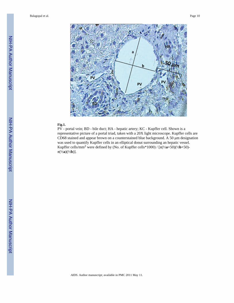
Fig.1.PV - portal vein; BD - bile duct; HA - hepatic artery; KC - Kupffer cell. Shown is arepresentative picture of a portal triad, taken with a 20X light microscope. Kupffer cells areCD68 stained and appear brown on a counterstained blue background. A 50 μm designationwas used to quantify Kupffer cells in an elliptical donut surrounding an hepatic vessel.Kupffer cells/mm2 were defined by (No. of Kupffer cells*1000) / [π(½a+50)(½b+50)-π(½a)(½b)].
Balagopal et al. Page 10
AIDS. Author manuscript; available in PMC 2011 May 11.
NIH
-PA Author Manuscript
NIH
-PA Author Manuscript
NIH
-PA Author Manuscript
Fig. 2.A. PV - portal vein; BD - bile duct; HA - hepatic artery; CLV - centrilobular vein; KC -Kupffer cell. Shown are representative pictures of a portal triad (upper panel) and acentrilobular vein (lower panel) from the same individual, taken with a 20X lightmicroscope. Kupffer cells are CD68 stained and appear brown on a counterstained bluebackground. B. Biopsies from 51/76 subjects with ≥ 1 postal vein and ≥1 centrilobular veinwere evaluated for a total of 122 portal and 95 centrilobular veins. Kupffer cells werequantified as described. Groups were compared using Wilcoxon rank-sum tests.
Balagopal et al. Page 11
AIDS. Author manuscript; available in PMC 2011 May 11.
NIH
-PA Author Manuscript
NIH
-PA Author Manuscript
NIH
-PA Author Manuscript

Fig. 3.Eight HIV-HCV co-infected persons with at least 2 liver biopsies were selected for theirinitiation and response to HAART between biopsies. Biopsies were obtained at a medianinterval of 36.8 months apart (range 28.1 – 58.4 months). Response to HAART was definedas having a CD4+ lymphocyte count at the time of the second biopsy that was ≥ 2× thevalue at the time of the first biopsy (left upper panel). All subjects had virologic suppressionof serum HIV RNA (right upper panel hashed reference line indicates 400 copies viral RNA/mL). KCD was measured as described on the first and second biopsies (lower panel).Symbols denote the same individuals over time and are consistent between the upper andlower panels. Because persons were preselected for increases in CD4+ lymphocyte count onHAART, a p-value was not generated for the top panel.*comparison of mean KCD on HAART with mean KCD pre-HAART using Student’s t test.† This individual, distinct in having the smallest increase in KCD during HAART, also hadthe highest pre-HAART CD4+ lymphocyte count and HIV RNA.
Balagopal et al. Page 12
AIDS. Author manuscript; available in PMC 2011 May 11.
NIH
-PA Author Manuscript
NIH
-PA Author Manuscript
NIH
-PA Author Manuscript

NIH
-PA Author Manuscript
NIH
-PA Author Manuscript
NIH
-PA Author Manuscript
Balagopal et al. Page 13
Table 1
Baseline characteristics of 76 HIV-HCV co-infected subjects.
Characteristics N (%) Median KCD IQR p valuef
Agea
<40 years 17 (22.4) 24.4 17.4 – 30.2 0.48
40 – 50 years 40 (52.6) 23 18 – 26.8 -
≥ 50 years 19 (25) 21.2 16. – 26.2 -
Gender
Male 59 (77.6) 22.8 17.4 – 27.6 0.48
Female 17 (22.4) 24.4 18.4 – 29.6 -
Race
Black 67 (88.2) 23.4 18 – 28.6 0.25
All others 9 (11.8) 20.8 14.2 – 23 -
HCV Genotype
1a 47 (66.2) 23 17.4 – 29.4 0.76
1b 18 (25.2) 23.2 19 – 24.8 -
CD4+ T cell countb
< 350/mm3 39 (51.3) 20.8 14.2 – 24.7 0.03
≥ 350/mm3 37 (48.7) 24.4 21.2 – 28.6 -
HIV Viral RNAc
< 400 cp/mL 42 (55.3) 23 18 – 26.2 0.51
≥ 400 cp/mL 34 (44.7) 23.1 14.4 – 30.4
HAARTd
Yes 53 (69.7) 22 14.4 – 27.6 0.10
No 23 (30.3) 24.4 19 – 31 -
Metavire
< 2 67 (88.2) 23.2 18 – 28 0.63
≥ 2 9 (11.8) 20.8 16.2 – 27.4 -
MHAIe
≤ 5 61 (80.3) 22.4 18 – 25.2 0.28
> 5 9 (11.8) 25.7 18.4 – 32.2 -
HAART - highly active anti-retroviral therapy.
aAge is given at the time of the first biopsy.
bCD4+ lymphocyte determinations were made within a median (range) of 1.25 (0.1 – 139.6) months of the first biopsy.
cHIV viral RNA was determined within a median (range) of 1.50 (0.1 – 68.9) months of the first biopsy.
dHAART was defined as use of at least 2 nucleoside reverse transcriptase inhibitors (NRTI) + 1 protease inhibitor (PI), or 2 NRTIs +1 non-
nucleoside reverse transcriptase inhibitor (NNRTI), or 2 NRTIs + 1 NNRTI.
eMetavir and MHAI staging were determined by liver biopsy by a trained pathologist (M.T.)
AIDS. Author manuscript; available in PMC 2011 May 11.

NIH
-PA Author Manuscript
NIH
-PA Author Manuscript
NIH
-PA Author Manuscript
Balagopal et al. Page 14
fp values were generated from Wilcoxon rank-sum and Kruskal-Wallis tests.
AIDS. Author manuscript; available in PMC 2011 May 11.

NIH
-PA Author Manuscript
NIH
-PA Author Manuscript
NIH
-PA Author Manuscript
Balagopal et al. Page 15
Table 2
Independent association of Kupffer cell density with CD4+ lymphocyte count.a
Parameter Estimate p Value
CD4+ T cell countb .0114 0.027
Nadir CD4+ T cell countc .0164 0.006
aShown are univariate models of Kupffer cell density.
bCD4+ lymphocyte determinations were made within a median (range) of 1.3 (0.1 – 139.6) months of the first biopsy.
cNadir CD4+ lymphocyte count was determined within a median (range) of 17.3 (0.1 – 139.6) months before the first biopsy, and was defined as
the lowest recorded CD4+ lymphocyte count available.
AIDS. Author manuscript; available in PMC 2011 May 11.
Copyright © 2022 FDOKUMEN


![Polyclonal hematopoietic reconstitution in leukemia patients at remission after suppression of specific gene rearrangements [see comments]](https://static.fdokumen.com/doc/165x107/633576362532592417008ca6/polyclonal-hematopoietic-reconstitution-in-leukemia-patients-at-remission-after.jpg)