Knee Joint Laxity and Its Cyclic Variation Influence Tibiofemoral Motion during Weight Acceptance
16
Knee Joint Laxity and Its Cyclic Variation Influence Tibiofemoral Motion during Weight Acceptance Sandra J. Shultz, PhD, ATC, CSCS, FACSM 1 , Randy J. Schmitz, PhD, ATC 1 , Anh-Dung Nguyen, PhD, ATC 2 , Beverly Levine, PhD 1 , Hyunsoo Kim, MS, ATC 1 , Melissa M. Montgomery, MA, ATC 1 , Yohei Shimokochi, PhD, ATC 3 , Bruce D. Beynnon, PhD 4 , and David H. Perrin, PhD, ATC, FACSM 1 1 School of Health and Human Performance, University of North Carolina at Greensboro; Greensboro, NC 2 Department of Health and Human Performance; College of Charleston, Charleston, SC 3 Osaka University of Health and Sport Sciences; Osaka, Japan 4 Department of Orthopaedics, University of Vermont; Burlington, VT Abstract Purpose—To better understand how sex differences in anterior knee joint laxity (AKL) impact knee joint biomechanics, we examined the consequence of greater absolute baseline (males and females) and cyclic increases in AKL during the menstrual cycle (females) on anterior tibial translation (ATT) as the knee transitioned from non-weight bearing (NWB) to weight bearing (WB) conditions, while also controlling for genu recurvatum (GR). Methods—Males and females (71F,48M;18-30 years) were measured for AKL and GR, and underwent measurement of ATT. Females were tested on the days of their cycle when AKL was at its minimum (T1) and maximum (T2); males were matched in time to a female with similar AKL. Linear regressions examined relationships between absolute baseline (AKL T1 , GR T1 ) and cyclic changes (Δ=T2-T1; AKL Δ , GR Δ )(females only) in knee laxity with ATT as measured at T1 and T2, and Δ (T2-T1) (females only). Results—AKL and GR increased in females, but not males, from T1 to T2. Greater AKL T1 and GR T1 predicted greater ATT T1 and ATT T2 in males (R 2 =21.0, P<.007). The combination of greater AKL T1 , AKL Δ and less GR Δ predicted greater ATT T1 and ATT T2 in females (R2=12.5-13.1, P<.05), with AKL Δ being a stronger predictor (coefficient, P-value) of ATT T2 (0.864, P=.027) compared to ATT T1 (0.333, P=.370). AKL Δ was the sole predictor of ATT Δ (R 2 =. 104; 0.740, P=.042). Conclusions—Greater absolute baseline and cyclic increases in AKL were consistently associated with greater ATT produced by transition of the knee from NWB to WB. As the ACL is the primary restraint to ATT, these findings provide insight into possible mechanisms by which Copyright © 2010 American College of Sports Medicine Send Correspondence to: Sandra J Shultz PhD, ATC, CSCS, FACSM, Associate Professor and Co-Director Applied Neuromechanics Research Laboratory, Department of Kinesiology 1408 Walker Ave Greensboro, NC 27402 336-334-3027 (Phone); 336-334-3238 (Fax); [email protected]. Publisher's Disclaimer: This is a PDF file of an unedited manuscript that has been accepted for publication. As a service to our customers we are providing this early version of the manuscript. The manuscript will undergo copyediting, typesetting, and review of the resulting proof before it is published in its final citable form. Please note that during the production process errors may be discovered which could affect the content, and all legal disclaimers that apply to the journal pertain. NIH Public Access Author Manuscript Med Sci Sports Exerc. Author manuscript; available in PMC 2012 February 1. Published in final edited form as: Med Sci Sports Exerc. 2011 February ; 43(2): 287–295. doi:10.1249/MSS.0b013e3181ed118d. NIH-PA Author Manuscript NIH-PA Author Manuscript NIH-PA Author Manuscript
Transcript of Knee Joint Laxity and Its Cyclic Variation Influence Tibiofemoral Motion during Weight Acceptance
Knee Joint Laxity and Its Cyclic Variation Influence TibiofemoralMotion during Weight Acceptance
Sandra J. Shultz, PhD, ATC, CSCS, FACSM1, Randy J. Schmitz, PhD, ATC1, Anh-DungNguyen, PhD, ATC2, Beverly Levine, PhD1, Hyunsoo Kim, MS, ATC1, Melissa M.Montgomery, MA, ATC1, Yohei Shimokochi, PhD, ATC3, Bruce D. Beynnon, PhD4, andDavid H. Perrin, PhD, ATC, FACSM11 School of Health and Human Performance, University of North Carolina at Greensboro;Greensboro, NC2Department of Health and Human Performance; College of Charleston, Charleston, SC3Osaka University of Health and Sport Sciences; Osaka, Japan4Department of Orthopaedics, University of Vermont; Burlington, VT
AbstractPurpose—To better understand how sex differences in anterior knee joint laxity (AKL) impactknee joint biomechanics, we examined the consequence of greater absolute baseline (males andfemales) and cyclic increases in AKL during the menstrual cycle (females) on anterior tibialtranslation (ATT) as the knee transitioned from non-weight bearing (NWB) to weight bearing(WB) conditions, while also controlling for genu recurvatum (GR).
Methods—Males and females (71F,48M;18-30 years) were measured for AKL and GR, andunderwent measurement of ATT. Females were tested on the days of their cycle when AKL was atits minimum (T1) and maximum (T2); males were matched in time to a female with similar AKL.Linear regressions examined relationships between absolute baseline (AKLT1, GRT1) and cyclicchanges (Δ=T2-T1; AKLΔ, GRΔ)(females only) in knee laxity with ATT as measured at T1 andT2, and Δ (T2-T1) (females only).
Results—AKL and GR increased in females, but not males, from T1 to T2. Greater AKLT1 andGRT1 predicted greater ATTT1 and ATTT2 in males (R2=21.0, P<.007). The combination ofgreater AKLT1, AKLΔ and less GRΔ predicted greater ATTT1 and ATTT2 in females(R2=12.5-13.1, P<.05), with AKLΔ being a stronger predictor (coefficient, P-value) of ATTT2(0.864, P=.027) compared to ATTT1 (0.333, P=.370). AKLΔ was the sole predictor of ATTΔ (R2=.104; 0.740, P=.042).
Conclusions—Greater absolute baseline and cyclic increases in AKL were consistentlyassociated with greater ATT produced by transition of the knee from NWB to WB. As the ACL isthe primary restraint to ATT, these findings provide insight into possible mechanisms by which
Copyright © 2010 American College of Sports MedicineSend Correspondence to: Sandra J Shultz PhD, ATC, CSCS, FACSM, Associate Professor and Co-Director AppliedNeuromechanics Research Laboratory, Department of Kinesiology 1408 Walker Ave Greensboro, NC 27402 336-334-3027 (Phone);336-334-3238 (Fax); [email protected]'s Disclaimer: This is a PDF file of an unedited manuscript that has been accepted for publication. As a service to ourcustomers we are providing this early version of the manuscript. The manuscript will undergo copyediting, typesetting, and review ofthe resulting proof before it is published in its final citable form. Please note that during the production process errors may bediscovered which could affect the content, and all legal disclaimers that apply to the journal pertain.
NIH Public AccessAuthor ManuscriptMed Sci Sports Exerc. Author manuscript; available in PMC 2012 February 1.
Published in final edited form as:Med Sci Sports Exerc. 2011 February ; 43(2): 287–295. doi:10.1249/MSS.0b013e3181ed118d.
NIH
-PA Author Manuscript
NIH
-PA Author Manuscript
NIH
-PA Author Manuscript

greater AKL may be associated with at risk knee biomechanics during the weight acceptancephase of dynamic tasks.
KeywordsInjury Mechanism; anterior knee laxity; genu recurvatum; knee biomechanics; hormone response;menstrual cycle
INTRODUCTIONSerious knee trauma, such as anterior cruciate ligament (ACL) injury, most often occurswhen the knee transitions from non-weight bearing (NWB) to weight bearing (WB) duringsudden deceleration or change-of-direction with the knee relatively extended (12,20).Studies have shown that as the knee transitions to WB at low knee flexion angles (e.g.15-30°), there is anterior translation of the tibia relative to the femur (ATT) (8,37) that isrestrained by the ACL in the normal knee (8). The ACL's role in maintaining normal kneebiomechanics during weight acceptance is further supported by evidence of substantialincreases in ATT with ACL-sectioning in the cadaveric knee (37), and increased ATT insubjects with ACL-deficient knees (1). Yet despite the role of the ACL in controlling ATT,the impact of anterior knee laxity (AKL) on knee biomechanics during the transition fromNWB to WB is not well understood. Understanding this relationship is important as greatermagnitudes of AKL have been associated with an increased risk of suffering ACL injury infemales (39), and females are reported to have greater AKL values than males (24,28,39),with these differences being more pronounced at certain times of the menstrual cyclecompared to others (28).
AKL is a measure of the displacement of the tibia relative to the femur in response to ananterior directed load applied to the tibia while the knee is non-weight bearing with themuscles relaxed, and is typically considered a biomechanical assessment of the ACL whichis the primary ligament restraint to this motion (2). As the ACL is the primary ligamentrestraint to both AKL and ATT, the greater AKL observed in females suggest they mayexperience greater ATT in response to weight bearing loads that act at the knee. However, itis well accepted that additional factors may contribute to control of anterior tibial motionduring weight acceptance, such as knee flexion angle, joint geometry and congruency (e.g.,condylar depth, the slope of the tibial plateau), the extensor moment arm, and the sequenceand magnitude of muscle co-activation. This is supported by Shultz et al (34) who examinedthe relationship between AKL, produced by a 133N anterior directed load applied to thetibia, and the magnitude of ATT produced during the transition from non-weight bearing toa weight bearing load (40% of body weight) applied to the limb. While a strong relationshipbetween AKL and ATT (R2=35.5%) was reported, 65% of the variance remainedunexplained by AKL alone. Moreover, males and females were examined collectively, andgiven known sex differences in knee joint geometry (10) and muscle activation patterns(31), it is possible the relationship between AKL and ATT is not the same for males andfemales. Further, cyclic variations in AKL across the female's cycle were not controlled.Individual variations in hormone profiles among females makes this later assessmentdifficult, as not all women experience cyclic changes in AKL, and the time during themenstrual cycle when these cyclic changes occur is quite variable (21,32). This inter-subjectvariability also raises the question whether some women may have greater difficulty withneuromuscular control of the tibiofemoral joint during dynamic activities such as weightacceptance if their cyclic variations in AKL are of sufficient magnitude to disrupt normalknee joint biomechanics.
Shultz et al. Page 2
Med Sci Sports Exerc. Author manuscript; available in PMC 2012 February 1.
NIH
-PA Author Manuscript
NIH
-PA Author Manuscript
NIH
-PA Author Manuscript
In an effort to better understand how sex differences in AKL may impact knee jointbiomechanics when transitioning from non-weight bearing to weight bearing (for example,during the initial application and transmission of the joint compressive loads that areproduced during activities that involve foot strike with the ground), our purpose was toexamine the consequence of both greater absolute baseline (males and females) and cyclicincreases in AKL during the menstrual cycle (females only) on ATT as measured at twotime points; the days of minimum and maximum AKL in females. ATT was measured as theknee transitioned from NWB to WB while also accounting for thigh muscle strength, themagnitude of thigh muscle activation during the measurement of ATT, and the magnitude ofgenu recurvatum. Thigh muscle strength and activation levels are often observed to bedifferent between males and females (31), and both the position of the tibia relative to thefemur and the absolute magnitude of anterior tibial displacement can be altered by muscleforces near full knee extension (37). Examining the contributions of genu recurvatum toATT is also important, given its association with ACL injury risk (11,18,23), the greatervalues reported in females compared to males (19), its potential to also vary across themenstrual cycle (29) and its potential to alter the neutral position of the tibiofemoral jointand the posterior tibial slope (5,16,17) (the later of which can in turn influence ATT (37)).Our expectations were that once controlling for these factors, strong associations would benoted between AKL and ATT in both males and females, and that greater cyclic changes inAKL would predict greater cyclic changes in ATT in females.
METHODSSubjects initially included 74 females and 50 males between 18 and 30 years of age, whowere recreationally active (2.5- 10 hrs/week) for the past 3 months and non-smokers, andhad a body mass index (weight/height2) ≤ 30 and no history of ligament or cartilage injuryto the knee. We chose to exclude individuals categorized as obese (those with a body massindex > 30) to limit the potential for abnormal hormone levels and menstrual cycleirregularities (40), and to control for excessive spatial filtering of the EMG signals whichcan occur with excess tissue between the surface electrode and muscle (6). Additionalcriteria for females were self-reported menstrual cycles lasting 26-32 days (varying no morethan ±1day month to month), no use of exogenous hormones for the past 6 months, and nohistory of or plans to become pregnant. Potential subjects were prescreened to obtain a widedistribution of baseline AKL values so that we could 1) include more subjects with a widerange of AKL values found to be predictive of ACL injury in females (39), and 2) determineif the effects of cyclic changes in AKL vary according to the subject's baseline AKL values.We then used a prospective study design to evaluate the relationship between absolutebaseline and cyclic changes in AKL with ATT produced by the transition of the knee fromNWB to WB. All subjects were informed of study procedures and risk, and signed aninformed consent approved by the University's institutional review board for the protectionof human subjects.
Because there is no uniform day in the cycle when AKL is known to be at its minimum ormaximum, females were measured each morning (7:00-9:00a.m., within ± 30 minutesbetween test days) for AKL (and genu recurvatum) during the first 6 days following theonset of menses (per self report) and the first 8 days of the early luteal phase followingevidence of ovulation [CVS One Step Ovulation Predictor (sensitivity 20 mIU/ml LH,accuracy 99%); CVS Corporation, Woonsocket, RI] for two consecutive cycles. In thefollowing month, males and females were tested on all variables at two time points (T1 andT2; within ± 1 hour of the same time of day). For females, T1 and T2 corresponded to theestimated days of minimum AKL (AKLT1) during menses and maximum AKL (AKLT2)during the early luteal phase, respectively, based on the patterns of AKL observed during thetwo prior months. For males, the time between tests was matched to a female with a similar
Shultz et al. Page 3
Med Sci Sports Exerc. Author manuscript; available in PMC 2012 February 1.
NIH
-PA Author Manuscript
NIH
-PA Author Manuscript
NIH
-PA Author Manuscript
baseline AKL value (within ± 0.5mm). Subjects were familiarized to all proceduresapproximately two weeks prior to testing, and asked to refrain from any physical activityprior to testing. All measures were obtained on the dominant stance limb (preferred limbwhen kicking a ball).
AKL was measured as the anterior displacement of the tibia relative to the femur when a133N anterior directed load was applied with the KT2000™ Knee Arthrometer (MEDmetricCorp; San Diego, CA) while the knee was positioned in 25° of flexion. Genu recurvatum(GR) was measured in supine (with a 10.2cm bolster under the distal tibia) as the amount ofknee hyperextension while the subject performed a maximal active knee extension (30). Asingle tester with establish reliability [ICC(2,3)(SEM) = 0.96(0.3mm) for AKL, 0.97(0.5°)for GR] (26) obtained all laxity measures.
To obtain thigh muscle torques and to normalize muscle responses recorded with surfaceelectromyography (sEMG) during ATT testing, participants were positioned in adynamometer (Biodex Medical Systems Inc., Shirley, NY) at 20° of knee flexion (thisduplicated positioning of the knee during measurement of ATT) and asked to complete three5s maximal effort isometric knee extension and knee flexion contractions (MVIC). This wasdone after first warming up on a bicycle ergometer and completing practice trials at 25%,50%, 75%, and 100% of maximal effort. Peak knee flexor and knee extensor torques (Nm)were recorded for each trial. Maximum sEMG signals were recorded with a 16 channelMyopac telemetric system (Run Technologies, Mission Viejo, CA: amplification 1 mV / V,frequency bandwidth 10-1000 Hz, CMRR 90 dB min at 60 Hz, input resistance 1 MΩ, andinternal sampling rate 8 KHz) via 10mm bipolar Ag–AgCl surface electrodes (MedicotestBlue Sensor Model #N-00-S; Ambu Products, Germany) applied over prepared skin areas ofthe vastus medialis and lateralis, the medial hamstring and biceps femoris, with a 2.5cmcenter-to-center distance. The reference electrode was placed on the anterior tibial shaft.Manual muscle testing confirmed signal fidelity and absence of cross talk.
With the sEMG electrodes still attached, subjects were positioned supine in the VermontKnee Laxity Device (VKLD, University of Vermont, Burlington, VT) to measure ATT asthe knee transitioned from NWB to a WB load equal to 40% body weight as previouslydescribed (Figure 1) (26,34). It is appreciated that muscular strategies used to control kneejoint motion upon weight acceptance using this supine model may differ from what occursduring upright weight bearing postures. We chose this model to accurately control themagnitude, direction and rate of application of the external loads applied to the limb, and tominimize the extraneous factors that may limit measurement precision of ATT duringupright weight bearing postures in order to test our hypothesis. These factors include theability to control the knee position upon weight acceptance, trunk position relative to theknee, the tibiofemoral shear loads acting at the knee prior to application of the weightbearing loads, and the magnitude and direction of the weight bearing load acting in areproducible manner through the ankle and hip joint centers of rotation. Specifically, thisapproach allowed us to: (1) established a repeatable, initial zero-shear load referenceposition of the tibia relative to the femur through counterbalanced weighting, and (2) apply aconsistent axial compression load that acts through the axes of rotation of the ankle and hipjoints (34). The 40% weight bearing load utilized in this study is consistent with what wouldbe experienced during double leg stance (assuming 50% of bodyweight applied to each leg,and 10% of bodyweight distributed below the knee) (34), and is intended to represent theearly loading period as one transitions to full weight bearing.
Six-degree of freedom position sensors (Mini Birds Ascension Technologies, Burlington,VT, USA) were secured to the lateral thigh, patella and the anteromedial aspect of theproximal tibia. Hip, knee, and ankle joint centers were determined using the centroid method
Shultz et al. Page 4
Med Sci Sports Exerc. Author manuscript; available in PMC 2012 February 1.
NIH
-PA Author Manuscript
NIH
-PA Author Manuscript
NIH
-PA Author Manuscript
(34). Initial anatomical position was established with the knee fully extended and the 2nd
metatarsal aligned with the vertical axis of the VKLD. A counter weight system was appliedto the thigh and shank to offset gravitational loads acting on the lower extremity, creating aninitial zero anterior-posterior directed shear load across the tibiofemoral joint while it wasunweighted (38). The ankle and knee were flexed to 90° and 20° respectively, and thesubjects were instructed to relax their leg muscles. Prior to each loading trial, 3 anterior-to-posterior directed forces were applied to the proximal tibia to obtain a reproducible initialposition of the tibia relative to the femur, and knee flexion angle was confirmed within ±5°.Data collection began with the subjects’ knee in the initial NWB position and continued as acompressive force equal to 40% of the subject's bodyweight was applied under the control ofgravity through the ankle and hip joint axes of rotation to load the tibiofemoral joint (34). Asix degree-of-freedom load transducer (Model MC3A, Advanced Medical Technology, Inc;Watertown, MA) measured compressive loads at the foot (500Hz), and initiation of weightacceptance was defined as when the compressive force exceeded 10 N. Subjects were nottold when the weight would be released, but were asked to try and maintain the 20° kneeposition during load acceptance. Three practice trials were followed by 6 test trials.Kinematic data were collected (100Hz) during trials 1-3 and analyzed using the MotionMonitor (Innovative Sports Training, Chicago, IL, USA) electromagnetic tracking systemand software. SEMG data were collected during trials 4-6 due to noise interference from theelectromagnetic system. Pilot testing prior to commencement of the study confirmed thatmuscle activation amplitudes obtained in trials 4-6 were quite consistent with those obtainedin trials 1-3 (All ICC(2,3) >.98) (26).
Data ReductionFor each time point (T1, T2), 3 trials of AKL and GR were measured and averaged.Quadriceps and hamstring torques were recorded as the mean of the peak torques obtainedover the 3 MVIC trials, and normalized to the subject's body mass (Nm/kg). ATT wascalculated as the change in displacement of the proximal shank sensor relative to the patellasensor in the A-P plane between the onset of weight acceptance (NWB) and peak axialcompressive load (WB). During this same time frame, knee flexion excursion in degrees wasalso recorded. SEMG data (1000 Hz) were synchronized with the kinematic data using thesame foot contact force threshold of 10N. SEMG signals were band pass filtered from 10 Hzto 350 Hz, using a fourth-order, zero-lag Butterworth filter(31), and MVIC (100ms) andweight acceptance (5ms) trials were processed using a centered root mean square algorithm,and ensemble averaged over their respective trials. Muscle reflex activation amplitude wasdefined as the mean normalized RMS amplitude (%MVIC) over the duration from muscleonset to peak WB load. The medial and lateral aspects of each muscle were averaged torepresent a single quadriceps and hamstring normalized reflex amplitude, respectively.
Statistical AnalysesSeparate 2 × 2 repeated measures ANOVAs examined mean differences in AKL, GR andATT by sex and time (T1, T2). To further explore potential relationships between absolutebaseline AKL as measured at T1 (herein called AKLT1) and cyclic changes in AKL asmeasured from T1 to T2 (herein called AKLΔ) with ATT in females, separate multiple linearregressions examined these relationships when ATT was measured at T1 (ATTT1), at T2(ATTT2), and the cyclic change from T1 to T2 (ATTΔ). This was done while also accountingfor GR as measured at T1 and the change from T1 to T2 (herein called GRT1 and GRΔ), aswell as 5 supressor variables [quadriceps and hamstring strength (QTrq, HTrq) andactivation (Q%MIVC, H%MVIC), and changes in knee flexion angle (KFLEX) during loadacceptance] as measured at each time point, given their potential to confound themeasurement of ATT (34,37). The three full models examined for females were:
Shultz et al. Page 5
Med Sci Sports Exerc. Author manuscript; available in PMC 2012 February 1.
NIH
-PA Author Manuscript
NIH
-PA Author Manuscript
NIH
-PA Author Manuscript
1. ATTT1=α0+β1AKLT1+β2GRT1+β3AKLΔ+β4GRΔ+β5QTrqT1+β6HTrqT1+β7Q%MVICT1+β8H%MVICT1+β9KFLEX T1+ε
2. ATTT2=α0+β1AKLT1+β2GRT1+β3AKLΔ+β4GRΔ+β5QTrqT2+β6HTrqT2+β7Q%MVICT2+β8H%MVICT2 + β9KFLEX T2+ε
3. ATTΔ=α0+β1AKLT1+β2GRT1+β3AKLΔ+βGRΔ+β5QTrqΔ+β6HTrqΔ+β7Q%MVICΔ+β8H%MVICΔ+β9KFLEXΔ+ε
Because none of these variables vary cyclically in males, T1 and T2 values for all variableswould be expected to differ by random error only, and, consequently, all delta variableswould be expected to have a mean value of zero. For this reason, and to better control forvariables highly correlated with sex, males were examined separately. We present modelresults for males for both ATTT1 and ATTT2 in order to demonstrate whether the expectedsimilarity of findings across the two time points is indeed observed. Model results for ATTΔare not presented, because this variable in males represents essentially random error. Thetwo full models for males were:
1. ATTT1=α0+β1AKL T1+β2GRT1+β3QTrqT1+β4HTrqT1+β5Q%MVICT1+β6H%MVICT1+β7KFLEX T1+ξ
2. ATTT2=α0+β1AKLT1+β2GRT1+β3QTrqT2+β4HTrqT2+β5Q%MVICT2+β6H%MVICT2+β7KFLEX T2+ε
For each analysis, we used a two step modeling procedure. On the first step, we examinedhow much variance in the ATT variables was accounted for by the five suppressor variables.On the second step, we added AKL and GR to the model (AKLT1, GRT1, AKLΔ, GRΔ forfemales; AKLT1, GRT1 for males) to examine their contribution and parameter estimatesrelative to ATT while accounting for the suppressor variables. All analyses were performedwith PASW Statistics 17.0 (release 17.0.2; SPSS Inc., Chicago, IL). Alpha-level was set apriori at p<.05.
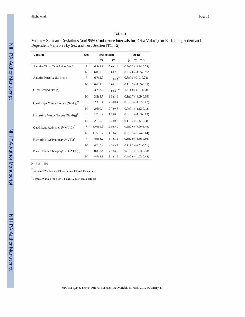
RESULTSThree females did not complete the study and were therefore excluded from the dataanalysis. One male subject was excluded due to mechanical errors in the acquisition ofATTT1. A second male was excluded due to noncompliance with refraining from exerciseprior to the 2nd test session that resulted in a 2mm increase in AKL at T2 compared to T1.Table 1 list the means ± standard deviations for each dependent and independent variable bysex for the remaining 71 females and 48 males at T1, T2 and Δ change from T1 to T2 (95%confidence intervals are also presented for Δ values). Significant sex by time interactionswere observed for both AKL and GR (P<.001). As expected, AKL and GR increased infemales but not males from T1 to T2, and this resulted in greater values in females comparedto males at T2 (Table 1). Sex differences in AKLΔ and GRΔ are further described in figure2. These frequency distributions confirm that the majority of males changed less than±0.5mm for AKLΔ (73% of cases) and ±0.5° for GRΔ (79% of the cases) between T1 andT2, while only 34% and 15% of the females changed less than ±0.5mm(or ±0.5°) for AKLΔand GRΔ, respectively. In 34% and 54% of the female cases, respectively, AKL changedmore than 1.5mm, and GR changed more than 1.5° (values that exceed 3-5 times whatwould be expected simply due to measurement error). Despite these sex differences inAKLΔ and GRΔ from T1 to T2, no mean differences were observed in ATT between sex(P=.841), time (P=.713) or the sex by time interaction (P=.550). However, there wasconsiderable dispersion in the absolute (T1 and T2) and delta values for ATT (figure 2) andmany of the suppressor variables (see delta means and standard deviations reported in Table1).
Shultz et al. Page 6
Med Sci Sports Exerc. Author manuscript; available in PMC 2012 February 1.
NIH
-PA Author Manuscript
NIH
-PA Author Manuscript
NIH
-PA Author Manuscript
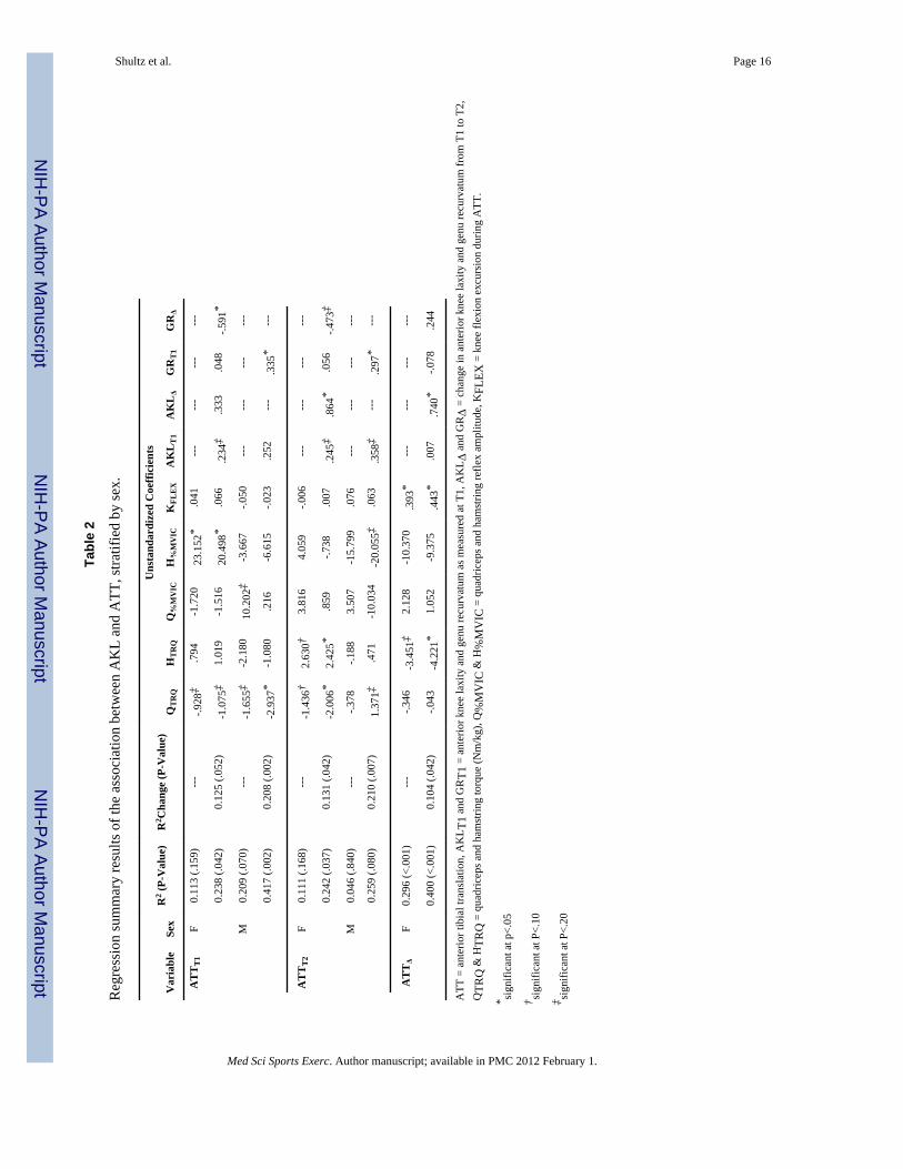
Table 2 presents the regression model summary results. When examining the results forfemales, the 5 suppressor variables explained 11.3% and 11.1% of the variance in ATTT1and ATTT2, respectively (P>.159), and 29.6% of the variance for ATTΔ (P<.001). WhenAKL(T1 and Δ) and GR(T1 and Δ) were added to the model, R2 values increased by comparableamounts for ATTT1 (0.125, P=.052), ATTT2 (0.131, P=.042) and ATTΔ (0.104, P=.042).While greater AKLT1 and less GRΔ were consistent predictors of ATTT1 and ATTT2 (bothin terms of the magnitude of their coefficients and p-values), AKLΔ was a strong predictorof ATTT2 than ATTT1, and the sole predictor of ATTΔ. Thus, the major difference betweenthe model results for ATTT1, ATTT2, and ATTΔ in females appears to be the substantiallylarger coefficients for AKLΔ when predicting ATTT2 (0.864, P=.027) and ATTΔ (0.740, P=.028), than when predicting ATTT1 (0.333, P=.135).
When examining the results for males, the 5 suppressor variables explained 20.9% (P=.070)and 4.9% (P=.826) of the variance in ATTT1 and ATTT2, respectively. When AKL and GRwere entered on the next step, they explained an additional 21% of the variance in ATTT1and ATTT2 (P<.018). In both models, greater AKL and GR were associated with greaterATT, with GR being the stronger predictor. As expected, there did not appear to be anynoteworthy or consistent differences for any of the modeling steps given for ATTT1 andATTT2 in males. (When using AKLT2 and GRT2 in the model for predicting ATTT2, theresults were essentially the same, supporting the consistency in these values across time inmales).
DISCUSSIONOur primary findings were that AKL and GR were significant predictors of ATT in bothmales and females, accounting for a larger percentage of the variance in ATT in men (R2
change ~21%) than women (R2 change ~10-13%). This was also apparent when comparingthe strength of the coefficients for AKLT1 and GRT1 between males and females, wheretheir associations with ATT were generally smaller in magnitude for women than for men.While the strength of the association of AKLT1 with ATT did not differ markedly betweenmales and females for either T1 or T2, GRT1 was only a significant positive predictor ofATT in males, and GRΔ was a significant negative predictor of ATT in females. Asexpected, the model results for ATT as measured at T1 (when AKL was at its minimum infemales) vs. T2 (when AKL was at its maximum in females) were essentially the same formales, but were substantially different in females relative to the larger coefficients andstronger associations between AKLΔ with ATTT2 compared to ATTT1. When examining thechange in ATT from T1 to T2 in females (ATTΔ), AKLΔ was the sole significant predictoronce differences in thigh strength and activation and knee flexion angles between T1 and T2were accounted for. Therefore, our expectations that greater baseline AKL would positivelypredict greater ATT in both males and females, and that greater AKLΔ would independentlypredict greater ATTΔ in females was in large part supported. However, the difference inmodel results between males and females suggests that ATT may be further increased infemales who experience large cyclic changes in AKL across their cycle, and that therelationship between AKL and ATT may be somewhat dependent on the magnitude of GR.
AKL represents the anterior displacement of the tibia relative to the femur once the neutralor initial reference position of the tibia relative to the femur is obtained, where the ACL isthe primary restraint to this motion (2). Conversely, posterior ligament structures (i.e.oblique popliteal, posterior cruciate and the posterior lateral corner) are the primaryrestraints to GR (16). As such, the combined measurements of AKL and GR may be areflection of how the overall anterior-posterior alignment of the tibiofemoral joint surfaces iscontrolled when the knee transitions from NWB to WB, thus the overall magnitude of ATTthat occurs. The positive relationships between both AKL and GR with ATT in males
Shultz et al. Page 7
Med Sci Sports Exerc. Author manuscript; available in PMC 2012 February 1.
NIH
-PA Author Manuscript
NIH
-PA Author Manuscript
NIH
-PA Author Manuscript
suggest that the tibia may initially be positioned more posterior in individuals with greaterGR (owing to the greater posterior laxity), allowing greater anterior displacement of the tibiafrom the initial NWB condition until the motion is restrained by the ACL during WB.However, these results are based on a controlled load applied through the centers of the hipand ankle joints, and may in part be attributed to the testing protocol. Specifically, westandardized initial tibiofemoral joint position prior to each ATT measurement by placinganterior-posterior directed loads on the tibia relative to the femur and then allowing thecounter weight system to create a zero shear load across the knee prior to application of thecompressive, weight bearing load to the foot. The implication of the balance in magnitudebetween AKL and GR and their influence on tibiofemoral joint position in upright weightbearing deserves further study.
But while GRT1 (measured when AKL was at its minimum in females) along with AKL wasoften an important predictor in males, GRT1 had little impact on the prediction of ATT infemales. We considered three possible explanations for these findings. First, AKL and GRwere more highly correlated in females (.528) compared to males (.361), thus potentiallyexplaining less overall variance in ATT in females due to a greater shared variance.Secondly, sex differences in lower extremity and knee joint anatomy may also modify therelationships between AKL, GR and ATT. For example, sex differences have been noted inthe magnitude and variability of the lateral and medial posterior inferior tibial slopes as wellas the coronal tibial slope (10,36). These slope differences along with the depth of theconcavity of the medial compartment of the tibiofemoral joint (tibiofemoral jointcongruence) may impact articular contact and joint biomechanics, and contribute more orless resistance to ATT upon joint loading (9). Known sex differences in other aspects oflower extremity anatomy, such as lower extremity alignment, the length of the knee extensormoment arm, or the size and material properties of the ACL, may also play a role (3,4,7,30).Work is ongoing to examine these potential anatomical contributions, and how they mayinteract with AKL and GR to impact joint biomechanics as the knee transitions from NWBto WB.
A third consideration is that cyclic variation in GR across the female menstrual cycle mayhave impacted the relationship between GRT1 with ATT. Although GRT1 did not addsubstantially to the model in females, greater ATT at both T1 and T2 was generallyobserved in females who had greater AKLT1 and AKLΔ values but who experienced lesscyclic changes in GRΔ. The reciprocal relationship between AKLΔ and GRΔ whenpredicting ATT is not entirely clear. Although significant low to moderate correlations werenoted between GRΔ and AKLΔ in females (r=.379, P=.001), there were considerablevariations across women as to how much of an increase in GR they experienced relative tothe change in AKL from T1 to T2. Because GR was only measured on the days when AKLwas at minimum and maximum values, and the timing of changes in GR and AKL are notalways consistent across the cycle (29) the true magnitude of baseline and cyclic changes inGR within an individual may not always have been reflected in GRT1 and GRΔ. As such, weconsidered whether GRΔ may represent somewhat of an adjustment for GRT1, togetherproviding a better estimate of the magnitude of GR within that individual. However, whenwe removed GRΔ from the model, this resulted in little to no change in the coefficients forAKLT1 (.264) or GRT1 (.044) and the amount of variance explained in ATT decreased by5%. Perhaps a more likely explanation for this reciprocal relationship is that the balancebetween cyclic variations in AKL and GR (thus the extent to which they are correlated) maybe influenced by various anatomical contributions to GR. While the ACL is the primaryrestraint to AKL, the magnitude of GR can be influenced by multiple factors, including thebony alignment of the proximal tibia (e.g. a reduced / reversed posterior slope of the tibialplateau has been associated with greater GR), excessive capsuloligamentous laxity, or acombination of both (5,13,16,22). If the cause is more structural in nature (e.g. associated
Shultz et al. Page 8
Med Sci Sports Exerc. Author manuscript; available in PMC 2012 February 1.
NIH
-PA Author Manuscript
NIH
-PA Author Manuscript
NIH
-PA Author Manuscript
with a reduced posterior slope of the tibial plateau rather than posterior ligament laxity), thisform of GR may be less likely to produce cyclic variations across the cycle, and in turn, beless likely to produce ATT with axial compression (37) as compared to someone with anormal or increased posterior directed slope of the tibial plateau, and greater total anterior-posterior laxity. This is further supported by the model results when predicting the change inATT from T1 to T2; while greater increases in both AKL and GR predicted greater increasesATT from T1 to T2, the change in GR was a much weaker predictor than the change inAKL.
AKLΔ (the cyclic increases in AKL from T1 to T2) was the strongest predictor for ATTT2and the sole significant predictor of ATTΔ. Although AKLΔ explained only 10% additionalvariance in ATTΔ once all suppressor variables were accounted for, the regressioncoefficient indicates that for every 1.00 mm increase in AKL from T1 to T2, there is apredicted 0.74 mm increase in ATT (see Table 2). These findings suggests that if a female'sAKL changes as much as 3mm across her cycle, she may experience an increase in ATTover 2 mm, a change that represents approximately 30% of the mean magnitude in ATT weobserved at T1. Of interest, the magnitude of change in ATT in response to changes in AKLwas not dependent on the individual's minimum AKL, suggesting that biomechanical effectsassociated with these cyclic variations may be additive to those associated with one'sbaseline laxity values.
Clinical ImplicationsWhen considering the biomechanical implications of these findings and how they are relatedto the risk of suffering ACL trauma, there are a number of considerations that require furtherstudy. First, it is well appreciated that the ACL is the primary restraint controlling ATT(1,37). As such, it is possible that during the initial weight acceptance of the limb duringdynamic tasks (e.g. landing from a jump) that greater magnitudes of AKL, thus greatermagnitudes of ATT, may be associated with greater acceleration and movement of the tibiabefore the ACL restrains the motion. This may be of particular concern when combined withunopposed quadriceps contractions (14,15,37) and higher axial directed compressive loads(25,37) at lower knee flexion angles, which have also been associated with greater anteriordirected shear forces and tibial translation. Second, greater anterior tibial displacement mayalso impact tibiofemoral joint biomechanics upon full weight acceptance. While there isminimal tibiofemoral translation once full axial compression is applied to the joint, greaterATT during the transition to full weight bearing may result in starting subsequent fullweight bearing motions with the tibia in a more anterior position relative to the femur, whichmay potentially lead to abnormal joint kinematics and muscle function (37,38). While thiscontention is supported by studies noting altered lateral hamstring activation in females whohave greater magnitudes of AKL (24,27), the actual impact this altered tibiofemoral jointposition may have on ACL strain biomechanics has yet to be determined.
Finally, greater ATT may be coupled with frontal and transverse plane rotationalbiomechanics of the knee, and this may also have an important impact on ACL strainbiomechanics. Previous research indicates that females who had greater AKL also hadgreater varus-valgus and internal-external rotational laxity of the knee (35), and these laxityvalues have been associated with altered transverse and frontal plane hip and knee jointkinematics and kinetics during landing (33). Other work (21) suggests that small cyclicincreases in AKL as little as 1.3-mm may increase knee adduction moment during a stopjump task as much as 20%. Still other work has noted an association between increasedAKL with greater peak lateral tibial acceleration immediately after foot strike during gait inACL deficient knees compared to normal knees and ACL reconstructed knees (42). Theyattributed this finding to greater adduction moment during walking, potentially causing an
Shultz et al. Page 9
Med Sci Sports Exerc. Author manuscript; available in PMC 2012 February 1.
NIH
-PA Author Manuscript
NIH
-PA Author Manuscript
NIH
-PA Author Manuscript
increase in the shear and compression loading of the medial compartment, and acceleratingthe development of osteoarthritis. Based on these collective findings, and the observedassociation between increased AKL and increased ATT, further research is needed toexamine the implications of ATT (both absolute and cyclic increases) on coupled frontal andtransverse plane knee biomechanics, and their combined influence on ACL strain.
In summary, greater magnitudes of AKL are associated with greater magnitudes of ATT andthese relationships were often influenced by the amount of baseline (males) or cyclicchanges (females) in GR. Moreover, females who experience greater AKLΔ across the cyclealso experienced greater ATTΔ, further supporting a link between AKL and ATT. Althoughthis study accounted for more factors than previously examined (34), approximately 60-75%of the variance in ATT remained unexplained by these factors, indicating that otherimportant factors that regulate ATT remain unaccounted for. As previously noted, factorssuch as condylar geometry, extensor moment arm, and lower extremity structural alignmentshould be examined in future studies as they relate to the association between knee jointlaxity and ATT. Further, it is important to note that for the weight bearing conditions used inthis study, the muscles were relaxed prior to the application of the weight bearing load, andthe weight bearing load was substantially lower than what one would experience during highdemand activities associated with sport. While we believe this research model represents animportant first step in understanding the relationship between knee laxity (a risk factor forinjury to the ACL) and tibiofemoral joint biomechanics during the transition from NWB toWB (a very important event which includes application and transmission of largecompression and shear loads across the knee and is associated with a majority of non-contact ACL injuries), it is well accepted that muscle pre-activation occurs in anticipation ofground contact during common athletic tasks such as landing from a jump. Research hasshown that muscle pre-activation may in part be modulated by the amount of AKL anindividual possesses (24,27) and consequently, it is possible that an individual mayeffectively compensate for the increased AKL through appropriate timing and amplitude ofcontraction of their dynamic restraints when the action is anticipated.(41) However, should amismatch occur between what is anticipated and what actually occurs (i.e. so that the thighmuscles are not able to respond in an appropriate manner to an unanticipated external load),the current findings suggest that greater joint laxity may lead to greater joint displacementprior to any restraint provided by reactive muscle action. This may be particularly true withthe higher magnitudes of axial compressive loads and loading rates associated with physicalactivity, as ATT has been shown to increase with increasing magnitudes of axial loadsdespite faster and stronger reactive muscle response times and activation amplitudes (25).
AcknowledgmentsThe project described was supported by Grant Number R01- AR53172 NIH-NIAMS, and through a U54 Centergrant as part of the Specialized Cooperative Centers Program in Reproductive Research [NICHD (SCCPRR) GrantU54 HD28934, University of Virginia, Center for Research in Reproduction, Ligand Assay and Analysis Core].The results of the present study do not constitute endorsement by the American College of Sports Medicine.
Funding Source: Supported by Grant Number R01- AR53172 NIH-NIAMS, and through a U54 Center grant as partof the Specialized Cooperative Centers Program in Reproductive Research [NICHD (SCCPRR) Grant U54HD28934]
REFERENCES1. Beynnon BD, Fleming BC, Labovitch R, et al. Chronic anterior cruciate ligament deficiency is
associated with increased anterior translation of the tibia during the transition from non-weightbearing to weightbearing. J Orthop Res 2002;20:332–337. [PubMed: 11918313]
2. Butler DL, Noyes FR, Grood ES. Ligamentous restraints to anterior-posterior drawer in the humanknee. J Bone Joint Surg 1980;62-A(2):259–270. [PubMed: 7358757]
Shultz et al. Page 10
Med Sci Sports Exerc. Author manuscript; available in PMC 2012 February 1.
NIH
-PA Author Manuscript
NIH
-PA Author Manuscript
NIH
-PA Author Manuscript
3. Chandrashekar N, Mansour JM, Slauterbeck J, et al. Sex-based differences in the tensile proerties ofthe human anterior cruciate ligament. J Biomech 2006;39:2943–2950. [PubMed: 16387307]
4. Chandrashekar N, Slauterbeck J, Hashemi J. Sex-Based Differences in the AnthropometricCharacteristics of the Anterior Cruciate Ligament and Its Relation to Intercondylar NotchGeometry. Am J Sports Med 2005;33(10):1492–1498. [PubMed: 16009992]
5. Dejour D, Bonin N, Locatelli E. Tibial antirecurvatum osteotomies. Oper Tech Sports Med2000;8(1):67–70.
6. DeLuca CJ. The use of surface electromyography in biomechanics. J Appl Biomech 1997;13:135–163.
7. Fayad LM, Rosenthal EH, Morrison WB, et al. Anterior cruciate ligament volume: Analysis ofgender differences. J Magnetic Res Imag 2008;27:218–223.
8. Fleming BC, Renstrom PA, Beynnon BD, et al. The effect of weightbearing and external loading onanterior cruciate ligament strain. J Biomech 2001;34(3):163–170. [PubMed: 11165279]
9. Giffin JR, Vogrin TM, Zantop T, et al. Effects of increasing tibial slope on the biomechanics of theknee. Am J Sports Med 2004;32(2):376–382. [PubMed: 14977661]
10. Hashemi J, Chandrashekar N, Gill B, et al. The geometry of the tibial plateau and its influence onthe biomechanics of the tibiofemoral joint. J Bone Joint Surg Am 2008;90(12):2724–2734.[PubMed: 19047719]
11. Kramer LC, Denegar CR, Buckley WE, et al. Factors associated with anterior cruciate ligamentinjury: history in female athletes. J Sports Med Phys Fit 2007;47:446–454.
12. Krosshaug T, Slauterbeck JR, Engebretsen L, et al. Biomechanical analysis of anterior cruciateligament injury mechanisms: three-dimensional motion reconstruction from video sequences.Scand J Med Sci Sports 2007;17(5):508–519. [PubMed: 17181770]
13. Laura G, Berruto M, Bianchi M. Genu recurvatum following distal epiphysiodesis of the femur: X-ray evaluation and therapeutical approach. Ital J Orthop Traumatol 1992;18(4):505–514. [PubMed:1345644]
14. Li G, Rudy TW, Sakane M, et al. The importance of quadriceps and hamstring muscle loading onknee kinematics and in-situ forces in the ACL. J Biomech 1999;32:395–400. [PubMed: 10213029]
15. Markolf KL, O'Neill G, Jackson SR, et al. Effects of applied quadriceps and hamstrings muscleloads on forces in the anterior and posterior cruciate ligaments. Am J Sports Med 2004;32(5):1144–1149. [PubMed: 15262635]
16. Morgan PM, LaPrade RF, Wentorf FA, et al. The role of the oblique popliteal ligament and otherstructures in preventing knee hyperextension. Am J Sports Med 2010;38(3):550–557. [PubMed:20097929]
17. Moroni A, Pezzuto V, Pompili M, et al. Proximal osteotomy of the tibia for treatment of genurecurvatum in adults. J Bone Jt Surg [Am] 1992;74A(4):577–586.
18. Myer GD, Ford KR, Paterno MV, et al. The effects of generalized joint laxity on risk of anteriorcruciate ligament injury in young female athletes. Am J Sports Med 2008;36(6):1073–1080.[PubMed: 18326833]
19. Nguyen AD, Shultz SJ. Sex Differences in Lower Extremity Posture. J Orthop Sports Phys Ther2007;37(7):389–398. [PubMed: 17710908]
20. Olsen O, Myklebust G, Engebretsen L, et al. Injury mechanisms for anterior cruciate ligamentinjuries in team handball. Am J Sports Med 2004;32(4):1002–1012. [PubMed: 15150050]
21. Park SK, Stefanyshyn DJ, Ramage B, et al. Alterations in Knee Joint Laxity During the MenstrualCycle in Healthy Women Leads to Increases in Joint Loads During Selected Athletic Movements.Am J Sports Med 2009;37(6):1169–1177. [PubMed: 19289541]
22. Piriou P, Garreau C, Combelles F, et al. Original technique for the treatment of ligament-relatedgenu recurvatum: preliminary results. Knee Surg Sports Traumatol Arthrosc 2002;10(4):260–264.[PubMed: 12211187]
23. Ramesh R, VonArx O, Azzopardi T, et al. The risk of anterior cruciate ligament rupture withgeneralised joint laxity. J Bone Jt Surg 2005;87-B:800–803.
24. Rozzi SL, Lephart SM, Gear WS, et al. Knee joint laxity and neuromuscular characteristics of maleand female soccer and basketball players. Am J Sports Med 1999;27(3):312–319. [PubMed:10352766]
Shultz et al. Page 11
Med Sci Sports Exerc. Author manuscript; available in PMC 2012 February 1.
NIH
-PA Author Manuscript
NIH
-PA Author Manuscript
NIH
-PA Author Manuscript
25. Schmitz RJ, Kim HS, Shultz SJ. Effect of Axial Load on Anterior Tibial Translation whenTransitioning from Non-Weight Bearing to Weight Bearing. Clin Biomech 2010;25(1):77–82.
26. Shultz SJ, Beynnon BD, Schmitz RJ. Sex Differences in Coupled Knee Motions During theTransition from Non-Weight Bearing to Weight Bearing. J Orthop Res 2009;27(6):717–723.[PubMed: 19030172]
27. Shultz SJ, Carcia CR, Perrin DH. Knee joint laxity affects muscle activation patterns in the healthyknee. J Electromyogr Kinesiol 2004;14:475–483. [PubMed: 15165597]
28. Shultz SJ, Kirk SE, Sander TC, et al. Sex differences in knee laxity change across the femalemenstrual cycle. J Sports Med Phys Fit 2005;45(4):594–603.
29. Shultz SJ, Levine BJ, Nguyen AD, et al. A Comparison of Cyclic Variations in Anterior KneeLaxity, Genu Recurvatum and General Joint Laxity Across the Menstrual Cycle. J Orth Res. InPress.
30. Shultz SJ, Nguyen A, Levine BJ. The Relationship Between Lower Extremity AlignmentCharacteristics and Anterior Knee Joint Laxity. J Sports Health 2009;1(1):54–60.
31. Shultz SJ, Nguyen AD, Leonard MD, et al. Thigh Strength and Activation as Predictors of KneeBiomechanics During a Drop Jump Task. Med Sci Sports Exer 2009;41:857–66.
32. Shultz SJ, Sander TC, Kirk SE, et al. Relationship between sex hormones and anterior knee laxityacross the menstrual cycle. Med Sci Sports Exerc 2004;36(7):1165–1174. [PubMed: 15235320]
33. Shultz SJ, Schmitz RJ. Effects of Transverse and Frontal Plane Knee Laxity on Hip and KneeNeuromechanics During Drop Landings. Am J Sports Med 2009;37(9):1821–1830. [PubMed:19483075]
34. Shultz SJ, Shimokochi Y, Nguyen A, et al. Non-Weight Bearing Anterior Knee Laxity is Relatedto Anterior Tibial Translation During Transition from Non-Weight Bearing to Weight Bearing. JOrthop Res 2006;24(3):516–523. [PubMed: 16456828]
35. Shultz SJ, Shimokochi Y, Nguyen A, et al. Measurement of Varus-Valgus and Internal-ExternalRotational Knee Laxities In-Vivo Part II: Relationship with Anterior-Posterior and General JointLaxity in Males and Females. J Orthop Res 2007;25(8):989–996. [PubMed: 17469179]
36. Todd MS, Lalliss S, Garcia S, et al. The Relationship Between Posterior Tibial Slope and AnteriorCruciate Ligament Injuries. Am J Sports Med 2010;38(1):63–67. [PubMed: 19737987]
37. Torzilli PA, Deng X, Warren RF. The effect of joint-compressive load and quadriceps muscleforce on knee motion in the intact and anterior cruciate ligament-sectioned knee. Am J Sports Med1994;22(1):105–112. [PubMed: 8129092]
38. Uh BS, Beynnon BD, Churchill DL, et al. A new device to measure knee laxity duringweightbearing and non-weight bearing conditions. J Orthop Res 2001;19:1185–1191. [PubMed:11781022]
39. Uhorchak JM, Scoville CR, Williams GN, et al. Risk factors associated with non-contact injury ofthe anterior cruciate ligament. Am J Sports Med 2003;31(6):831–842. [PubMed: 14623646]
40. Wei S, Schmidt MD, Dwyer T, et al. Obesity and menstrual irregularity: associations with SHBG,testosterone, and insulin. Obesity 2009;17(5):1070–1076. [PubMed: 19180069]
41. Yack HJ, Riley LM, Whieldon TR. Anterior tibial translation during progressive loading of theACL-deficient knee during weight-bearing and nonweight-bearing isometric exercise. J OrthopSports Phys 1994;20:247–253.
42. Yoshimura I, Naito M, Hara M, et al. Analysis of the significance of the measurement ofacceleration with respect to lateral laxity of the anterior cruciate ligament insufficient knee. IntOrthop 2000;24(5):276–278. [PubMed: 11153458]
Shultz et al. Page 12
Med Sci Sports Exerc. Author manuscript; available in PMC 2012 February 1.
NIH
-PA Author Manuscript
NIH
-PA Author Manuscript
NIH
-PA Author Manuscript
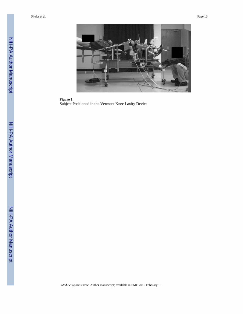
Figure 1.Subject Positioned in the Vermont Knee Laxity Device
Shultz et al. Page 13
Med Sci Sports Exerc. Author manuscript; available in PMC 2012 February 1.
NIH
-PA Author Manuscript
NIH
-PA Author Manuscript
NIH
-PA Author Manuscript
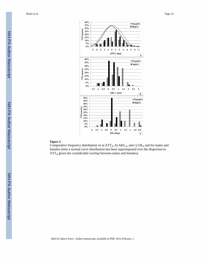
Figure 2.Comparative frequency distribution of a) ATTΔ, b) AKLΔ, and c) GRΔ and for males andfemales (note a normal curve distribution has been superimposed over the dispersion inATTΔ given the considerable overlap between males and females).
Shultz et al. Page 14
Med Sci Sports Exerc. Author manuscript; available in PMC 2012 February 1.
NIH
-PA Author Manuscript
NIH
-PA Author Manuscript
NIH
-PA Author Manuscript
NIH
-PA Author Manuscript
NIH
-PA Author Manuscript
NIH
-PA Author Manuscript
Shultz et al. Page 15
Table 1
Means ± Standard Deviations (and 95% Confidence Intervals for Delta Values) for Each Independent andDependent Variables by Sex and Test Session (T1, T2)
Variable Sex Test Session Delta
T1 T2 (Δ = T2 - T1)
Anterior Tibial Translation (mm) F 6.8±2.3 7.0±2.4 0.2±2.3 (-0.34-0.74)
M 6.8±2.9 6.8±2.9 -0.0±2.0 (-0.55-0.55)
Anterior Knee Laxity (mm) F 6.7±2.0 7.4±2.1* 0.6±0.8 (0.42-0.78)
M 6.6±1.8 6.6±1.8 0.1±0.5 (-0.05-0.25)
Genu Recurvatum (°) F 3.7±3.6 5.0±3.6* 1.3±1.0 (1.07-1.53)
M 3.5±3.7 3.5±3.6 -0.1±0.7 (-0.29-0.09)
Quadriceps Muscle Torque (Nm/kg)† F 2.3±0.4 2.3±0.4 -0.0±0.3 (-0.07-0.07)
M 2.6±0.4 2.7±0.6 0.0±0.4 (-0.12-0.12)
Hamstring Muscle Torque (Nm/Kg)† F 1.7±0.2 1.7±0.3 -0.0±0.1 (-0.03-0.03)
M 2.1±0.3 2.2±0.3 0.1±0.2 (0.06-0.14)
Quadriceps Activation (%MVIC)† F 13.6±5.8 13.9±5.0 0.3±5.0 (-0.88-1.48)
M 11.5±5.7 11.2±4.5 -0.3±3.3 (-1.24-0.64)
Hamstrings Activation (%MVIC)† F 4.9±3.2 5.1±3.2 0.3±2.9 (-0.36-0.96)
M 4.2±3.4 4.3±3.2 0.1±2.2 (-0.51-0.71)
Knee Flexion Change @ Peak ATT (°) F 8.3±3.4 7.7±3.3 -0.6±3.1 (-1.33-0.13)
M 8.5±3.3 8.1±3.2 -0.4±2.9 (-1.22-0.42)
N= 71F, 48M
*Female T2 > female T1 and male T1 and T2 values
†Female ≠ male for both T1 and T2 (sex main effect)
Med Sci Sports Exerc. Author manuscript; available in PMC 2012 February 1.
NIH
-PA Author Manuscript
NIH
-PA Author Manuscript
NIH
-PA Author Manuscript
Shultz et al. Page 16
Tabl
e 2
Reg
ress
ion
sum
mar
y re
sults
of t
he a
ssoc
iatio
n be
twee
n A
KL
and
ATT
, stra
tifie
d by
sex.
R2 (
P-V
alue
)R
2 Cha
nge
(P-V
alue
)U
nsta
ndar
dize
d C
oeffi
cien
ts
Var
iabl
eSe
xQ
TR
QH
TR
%M
VIC
H%
MV
ICK
FLE
XA
KL
T1
AK
LΔ
GR
T1
GRΔ
AT
TT
1F
0.11
3 (.1
59)
---
-.928
‡.7
94-1
.720
23.1
52*
.041
---
---
---
---
0.23
8 (.0
42)
0.12
5 (.0
52)
-1.0
75‡
1.01
9-1
.516
20.4
98*
.066
.234
‡.3
33.0
48-.5
91*
M0.
209
(.070
)--
--1
.655
‡-2
.180
10.2
02‡
-3.6
67-.0
50--
---
---
---
-
0.41
7 (.0
02)
0.20
8 (.0
02)
-2.9
37*
-1.0
80.2
16-6
.615
-.023
.252
---
.335
*--
-
AT
TT
2F
0.11
1 (.1
68)
---
-1.4
36†
2.63
0†3.
816
4.05
9-.0
06--
---
---
---
-
0.24
2 (.0
37)
0.13
1 (.0
42)
-2.0
06*
2.42
5*.8
59-.7
38.0
07.2
45‡
.864
*.0
56-.4
73‡
M0.
046
(.840
)--
--.3
78-.1
883.
507
-15.
799
.076
---
---
---
---
0.25
9 (.0
80)
0.21
0 (.0
07)
1.37
1‡.4
71-1
0.03
4-2
0.05
5‡.0
63.3
58‡
---
.297
*--
-
AT
TΔ
F0.
296
(<.0
01)
---
-.346
-3.4
51‡
2.12
8-1
0.37
0.3
93*
---
---
---
---
0.40
0 (<
.001
)0.
104
(.042
)-.0
43-4
.221
*1.
052
-9.3
75.4
43*
.007
.740
*-.0
78.2
44
ATT
= a
nter
ior t
ibia
l tra
nsla
tion,
AK
L T1
and
GR
T1 =
ant
erio
r kne
e la
xity
and
gen
u re
curv
atum
as m
easu
red
at T
1, A
KL Δ
and
GRΔ
= ch
ange
in a
nter
ior k
nee
laxi
ty a
nd g
enu
recu
rvat
um fr
om T
1 to
T2,
QTR
Q &
HTR
Q =
qua
dric
eps a
nd h
amst
ring
torq
ue (N
m/k
g), Q
%M
VIC
& H
%M
VIC
= q
uadr
icep
s and
ham
strin
g re
flex
ampl
itude
, KFL
EX =
kne
e fle
xion
exc
ursi
on d
urin
g A
TT.
* sign
ifica
nt a
t p<.
05
† sign
ifica
nt a
t P<.
10
‡ sign
ifica
nt a
t P<.
20
Med Sci Sports Exerc. Author manuscript; available in PMC 2012 February 1.