Modify Self-attention via Skeleton Decomposition for Effective ...
Upload
khangminh22Category
view
1download
0
�����������������
Citation: Leggio, A.; Gallieni, M.;
Puzo, P.; Introna, F.; Sablone, S.
Klippel–Feil Syndrome: The Curious
Case of the Skeleton of a Young Slavic
Soldier Who Died in 1946. Forensic
Sci. 2022, 2, 155–162. https://
doi.org/10.3390/forensicsci2010012
Academic Editors: Ricardo
Dinis-Oliveira, Pier Matteo Barone
and Francisca Alves Cardoso
Received: 9 November 2021
Accepted: 11 February 2022
Published: 14 February 2022
Publisher’s Note: MDPI stays neutral
with regard to jurisdictional claims in
published maps and institutional affil-
iations.
Copyright: © 2022 by the authors.
Licensee MDPI, Basel, Switzerland.
This article is an open access article
distributed under the terms and
conditions of the Creative Commons
Attribution (CC BY) license (https://
creativecommons.org/licenses/by/
4.0/).
Case Report
Klippel–Feil Syndrome: The Curious Case of the Skeleton of aYoung Slavic Soldier Who Died in 1946Alessia Leggio 1,* , Massimo Gallieni 2, Pasquale Puzo 3, Francesco Introna 1 and Sara Sablone 1
1 Department of Interdisciplinary Medicine, Section of Legal Medicine, Policlinico di Bari Hospital,University of Bari, Piazza Giulio Cesare 11, 70124 Bari, Italy; [email protected] (F.I.);[email protected] (S.S.)
2 Department of Neurosurgery, International Neuroscience Institute, Rudolf Pichlmayr Str. 4,30625 Hannover, Germany; [email protected]
3 Department of Medical Science, Neuroscience and Sense Organs, Section of Ophthalmology,Bari Policlinico Hospital, University of Bari, 70124 Bari, Italy; [email protected]
* Correspondence: [email protected]
Abstract: This paper describes the curious case history of the famous and rare Klippel Feil syndrometype II, identified in the skeleton of a young Slavic soldier who died in 1946. It is a very interestingcase given the fusion of the C1 and C2 cervical vertebrae, which prevented the young soldier fromrotating his skull while alive. Klippel–Feil syndrome is a rare osteopathology and involves fusion ofthe vertebrae of the spine and is linked to other pathologies that indicate the presence of this patho-logical condition. In the present study, several basic investigations were carried out: a macroscopicobservation to document the abnormalities throughout the rest of the skeleton, a morphological oneto determine the identifying anthropological analysis, a pathological one to determine the pathologiespresent and a radiographic one to diagnose and confirm the pathology. Studying the pathologies ofthe past is fundamental in order to know the evolution and behaviour of the disease today, and theinvestigations carried out in this case study determined what the limitations of the young soldierwere, how this disease may have influenced his activities as a soldier during World War II andthrough which therapies the syndrome may have been treated in such an important historical period.
Keywords: paleopathology; Klippel–Feil syndrome; forensic anthropology; Slavic soldier; World War II
1. Introduction
In the summer of 2019, the Institute of Forensic Medicine of Bari and the ForensicAnthropology Laboratory of the same department undertook research work within theMonumental Cemetery of Bari that led to the discovery of 93 skeletal remains of Yugoslavianorigin, most belonging to soldiers missing for a very long time who died during the SecondWorld War in the famous Prison Camp of Torre Tresca (Bari, Puglia). Among these remainswas the skeleton of a young soldier who died in 1946, probably from complications of arare disease. This disease had been sporadically documented in anthropological literatureas Klippel–Feil syndrome, in this case type II with fusion of the first and second cervicalvertebrae. The detection of skeletal pathology and trauma provides important informationthat can help identify an unknown subject and may provide possible evidence of thecircumstances or cause of death. Skeletal pathology can be very important in forensiccontexts in cases where other aspects of the osteological profile are very similar [1].
Klippel–Feil syndrome was first described by Klippel and Feil (1912) in a clinical casein which massive fusion of the cervical vertebrae was observed. The classic clinical triad ofsymptoms is short neck, low back hairline and restriction of movement in the neck [2]. Thedifferent manifestations due to the different genetic pathways made it possible to definethree degrees of cervical fusion (I–III):
Forensic Sci. 2022, 2, 155–162. https://doi.org/10.3390/forensicsci2010012 https://www.mdpi.com/journal/forensicsci

Forensic Sci. 2022, 2 156
Type I: the classic Klippel–Feil syndrome with several cervical and upper thoracicvertebrae embedded in a bony block showing a complex abnormal appearance and oftenassociated with other major defects [3].
Type II: only two or three vertebral segments are involved, with the second and thirdcervical segments most commonly affected, and there is evidence of autosomal dominantinheritance; the fifth and sixth cervical segments are the next most commonly involved andare usually expressed as an autosomal recessive trait. If thoracic vertebrae are involved,the defect most commonly occurs between T2 and T5. In some cases, hemivertebral oratlanto–occipital fusions are present [2,4].
Type III: fusion of cervical vertebrae coexisting with segmental vertebral defects in thethoracic and lumbar regions [2].
Only a few old cases of Klippel–Feil syndrome have been reported so far, particularlyin Europe [5]. It is very important to study and document rare osteopathies in individualcases to gather information that can be used in population studies [6]. The bones of theyoung soldier’s skeletal remains were subjected to a preliminary investigation, which atfirst only revealed Klippel–Feil syndrome. A second, more specific analysis allowed allthe anomalies associated with this pathology to be examined: bilateral and symmetricaloccipital condylar hypoplasia, cranial asymmetry, deviation of the nasal septum, spinabifida occulta type III and mild cervical dextroscoliosis [7–9].
2. Case Report
In 2019, the Institute of Forensic Medicine of Bari (Apulia, Italy) undertook researchthat led to the discovery of 93 skeletal remains of Slavic origin, abandoned in the ossuaryof the Monumental Cemetery of Bari. The skeletal remains were placed inside small steelboxes, and during the first general inspection, war badges were found (Figure 1) thatallowed the identification and historical contextualizing of these skeletal remains. Aftera thorough historical analysis of the badges, it was determined that the skeletal remainsbelonged to young Slavic soldiers of the Royal Yugoslav Army, Cetnico of the Ravna Goranational movement in exile in 1941, under the command of Serbian General Dragoljub“Draža” Mihailovic [10]. These soldiers were most likely, given the historical evidence,deported, and then, died in the Torre Tresca prison camp (Bari, Apulia) during WorldWar II [11].
Forensic Sci. 2022, 2, FOR PEER REVIEW 2
of symptoms is short neck, low back hairline and restriction of movement in the neck [2]. The different manifestations due to the different genetic pathways made it possible to define three degrees of cervical fusion (I–III):
Type I: the classic Klippel–Feil syndrome with several cervical and upper thoracic vertebrae embedded in a bony block showing a complex abnormal appearance and often associated with other major defects [3].
Type II: only two or three vertebral segments are involved, with the second and third cervical segments most commonly affected, and there is evidence of autosomal dominant inheritance; the fifth and sixth cervical segments are the next most commonly involved and are usually expressed as an autosomal recessive trait. If thoracic vertebrae are involved, the defect most commonly occurs between T2 and T5. In some cases, hemivertebral or atlanto–occipital fusions are present [2,4].
Type III: fusion of cervical vertebrae coexisting with segmental vertebral defects in the thoracic and lumbar regions [2].
Only a few old cases of Klippel–Feil syndrome have been reported so far, particularly in Europe [5]. It is very important to study and document rare osteopathies in individual cases to gather information that can be used in population studies [6]. The bones of the young soldier’s skeletal remains were subjected to a preliminary investigation, which at first only revealed Klippel–Feil syndrome. A second, more specific analysis allowed all the anomalies associated with this pathology to be examined: bilateral and symmetrical occipital condylar hypoplasia, cranial asymmetry, deviation of the nasal septum, spina bifida occulta type III and mild cervical dextroscoliosis [7–9].
2. Case Report In 2019, the Institute of Forensic Medicine of Bari (Apulia, Italy) undertook research
that led to the discovery of 93 skeletal remains of Slavic origin, abandoned in the ossuary of the Monumental Cemetery of Bari. The skeletal remains were placed inside small steel boxes, and during the first general inspection, war badges were found (Figure 1) that allowed the identification and historical contextualizing of these skeletal remains. After a thorough historical analysis of the badges, it was determined that the skeletal remains belonged to young Slavic soldiers of the Royal Yugoslav Army, Cetnico of the Ravna Gora national movement in exile in 1941, under the command of Serbian General Dragoljub “Draža” Mihailović [10]. These soldiers were most likely, given the historical evidence, deported, and then, died in the Torre Tresca prison camp (Bari, Apulia) during World War II [11].
Figure 1. Objects placed in coffin No. 33. Frieze of the Royal Yugoslav Army, bearing the monogramof Peter II Karadordevic (a), and military badge of Ravnogorksi, representing the movement’s struggleof Ravna Gora under the leadership of General Dragoljub Draža Mihailovi’c (b).

Forensic Sci. 2022, 2 157
3. Materials and Methods
The remains of Private Chetnic were not subjected to AMS radiocarbon dating [12]because there is historical documentation on paper that certifies the subject’s personal datasuch as date of birth and death: 1921–1946; there is also an identification tag engravedinside the iron box in which the skeleton is stored, with personal data such as first andlast name and military rank, which also confirms the origin of these remains. The skeletonwas recovered almost 90% intact [13], completely skeletonised without soft tissue andwith green pigmentation on the front of the skull due to excessive humidity in the areawhere the box was stored. Pathologies can influence the results of the anthropologicalinvestigation during the reconstruction of the biological profile and for these reasons, in thepresent case, different methods were applied during the investigation. The skeletal remainswere subjected to several stages of investigation: macroscopic, morphological, dental andpathological analysis. Given the known subject, age at death was confirmed biologically,using epiphyseal fusion, age-related tooth wear and the degree of obliteration of cranialsutures [14,15].
Dental analysis for age determination was applied to the entire dental system throughmorphological and macroscopic observation of dental wear [16]. Finally, suture oblitera-tion is at level 1 [17]. Congenital anomalies could influence sex determination based onmorphological data; therefore, even in the case of a known subject, it was necessary todetermine sex by applying different methods. Sex determination was mainly based onthe characteristics of the skull and pelvis, the skeletal districts with the greatest sexualdimorphism [18–22]. After reconstructing and confirming the subject’s biological profile, apathological analysis revealed multiple abnormalities in the skeletal system under inves-tigation. The first and most obvious is the fusion of the C1 and C2 vertebrae. The skullshows symmetrical and bilateral condylar hypoplasia and deviation of the nasal septum.The cervical vertebrae are affected by mild dextroscoliosis and the sacrum shows spinabifida occulta type III. However, the morphology of these bones is apparently normal.A radiographic examination confirmed the actual C1–C2 fusion, allowing the diagnosis ofKlippel–Feil syndrome type II and affirming the correlation of the additional pathologiesfound (Figure 2c).
Forensic Sci. 2022, 2, FOR PEER REVIEW 4
atlanto–occipital fusion; in the case at hand, there is total fusion of the C1 and C2 verte-brae, causing severe limitation in flexion and extension of neck rotation (Figure 2a, 2b). The entire skeletal system presents several anomalies associated with this syndrome as bilateral and symmetric occipital condylar hypoplasia, cranial asymmetry, nasal septum deviation, spina bifida occulta type III and mild cervical dextroscoliosis.
Figure 2. Klippel–Feil Syndrome, fused vertebral block of C1 and C2. (1a) Front projection, (1b) rear projection. X-ray front projection (1c), evident fusion between the C1 vertebra and the C2 vertebrae.
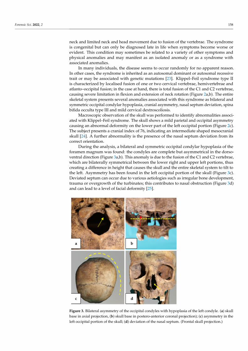
Macroscopic observation of the skull was performed to identify abnormalities asso-ciated with Klippel–Feil syndrome. The skull shows a mild parietal and occipital asym-metry causing an abnormal deformity on the lower part of the left occipital portion (Figure 2c). The subject presents a cranial index of 76, indicating an intermediate shaped mesocra-nial skull [24]. A further abnormality is the presence of the nasal septum deviation from its correct orientation.
During the analysis, a bilateral and symmetric occipital condylar hypoplasia of the foramen magnum was found: the condyles are complete but asymmetrical in the dorso-ventral direction (Figure 3a, 3b). This anomaly is due to the fusion of the C1 and C2 ver-tebrae, which are bilaterally symmetrical between the lower right and upper left portions, thus creating a difference in height that causes the skull and the entire skeletal system to tilt to the left. Asymmetry has been found in the left occipital portion of the skull (Figure 3c). Deviated septum can occur due to various aetiologies such as irregular bone develop-ment, trauma or overgrowth of the turbinates; this contributes to nasal obstruction (Figure 3d) and can lead to a level of facial deformity [25].
Figure 2. Klippel–Feil Syndrome, fused vertebral block of C1 and C2. (a) Front projection, (b) rearprojection. X-ray front projection (c), evident fusion between the C1 vertebra and the C2 vertebrae.
4. Results
The epiphyseal fusion process suggested an age at death between 18 and 25 years [10,11].Dental analysis for age determination based on dental wear indicates an age at deathbetween 20 and 24 years [14]. Finally, the degree of obliteration of the suture is stage 1 [13].Overall, the different methods applied indicate an age at death between 23 and 25 years,confirming the age data indicated on the plaque found inside the box containing the youngsoldier’s remains.
Klippel–Feil syndrome is a rare skeletal disease mainly characterised by an abnormalunion or fusion of two or more vertebrae that concentrates specifically on the cervicalvertebrae, as in the present case. Some affected individuals have an abnormally short
Forensic Sci. 2022, 2 158
neck and limited neck and head movement due to fusion of the vertebrae. The syndromeis congenital but can only be diagnosed late in life when symptoms become worse orevident. This condition may sometimes be related to a variety of other symptoms andphysical anomalies and may manifest as an isolated anomaly or as a syndrome withassociated anomalies.
In many individuals, the disease seems to occur randomly for no apparent reason.In other cases, the syndrome is inherited as an autosomal dominant or autosomal recessivetrait or may be associated with genetic mutations [23]. Klippel–Feil syndrome type IIis characterized by localised fusion of one or two cervical vertebrae, hemivertebrae andatlanto–occipital fusion; in the case at hand, there is total fusion of the C1 and C2 vertebrae,causing severe limitation in flexion and extension of neck rotation (Figure 2a,b). The entireskeletal system presents several anomalies associated with this syndrome as bilateral andsymmetric occipital condylar hypoplasia, cranial asymmetry, nasal septum deviation, spinabifida occulta type III and mild cervical dextroscoliosis.
Macroscopic observation of the skull was performed to identify abnormalities associ-ated with Klippel–Feil syndrome. The skull shows a mild parietal and occipital asymmetrycausing an abnormal deformity on the lower part of the left occipital portion (Figure 2c).The subject presents a cranial index of 76, indicating an intermediate shaped mesocranialskull [24]. A further abnormality is the presence of the nasal septum deviation from itscorrect orientation.
During the analysis, a bilateral and symmetric occipital condylar hypoplasia of theforamen magnum was found: the condyles are complete but asymmetrical in the dorso-ventral direction (Figure 3a,b). This anomaly is due to the fusion of the C1 and C2 vertebrae,which are bilaterally symmetrical between the lower right and upper left portions, thuscreating a difference in height that causes the skull and the entire skeletal system to tilt tothe left. Asymmetry has been found in the left occipital portion of the skull (Figure 3c).Deviated septum can occur due to various aetiologies such as irregular bone development,trauma or overgrowth of the turbinates; this contributes to nasal obstruction (Figure 3d)and can lead to a level of facial deformity [25].
Forensic Sci. 2022, 2, FOR PEER REVIEW 5
Figure 3. Bilateral asymmetry of the occipital condyles with hypoplasia of the left condyle. (3a skull base in axial projection, 3b skull base in postero-anterior coronal projection); 3c, asymmetry in the left occipital portion of the skull; 3d, deviation of the nasal septum. (Frontal skull projection.)
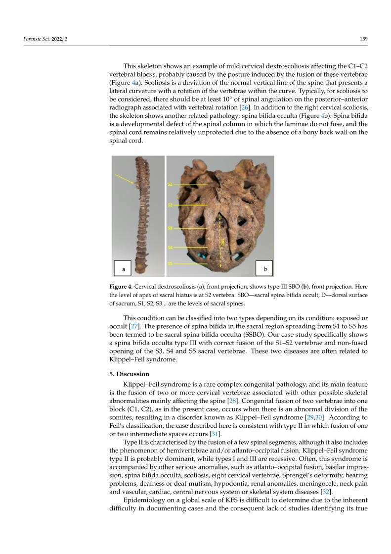
This skeleton shows an example of mild cervical dextroscoliosis affecting the C1–C2 vertebral blocks, probably caused by the posture induced by the fusion of these vertebrae (Figure 4a). Scoliosis is a deviation of the normal vertical line of the spine that presents a lateral curvature with a rotation of the vertebrae within the curve. Typically, for scoliosis to be considered, there should be at least 10° of spinal angulation on the posterior–anterior radiograph associated with vertebral rotation [26]. In addition to the right cervical scolio-sis, the skeleton shows another related pathology: spina bifida occulta (Figure 4b). Spina bifida is a developmental defect of the spinal column in which the laminae do not fuse, and the spinal cord remains relatively unprotected due to the absence of a bony back wall on the spinal cord.
Figure 4. Cervical dextroscoliosis 4a, front projection; shows type-III SBO 4b, front projection. Here the level of apex of sacral hiatus is at S2 vertebra. SBO—sacral spina bifida occult, D—dorsal surface of sacrum, S1, S2, S3... are the levels of sacral spines.
Figure 3. Bilateral asymmetry of the occipital condyles with hypoplasia of the left condyle. (a) skullbase in axial projection, (b) skull base in postero-anterior coronal projection); (c) asymmetry in theleft occipital portion of the skull; (d) deviation of the nasal septum. (Frontal skull projection.)
Forensic Sci. 2022, 2 159
This skeleton shows an example of mild cervical dextroscoliosis affecting the C1–C2vertebral blocks, probably caused by the posture induced by the fusion of these vertebrae(Figure 4a). Scoliosis is a deviation of the normal vertical line of the spine that presents alateral curvature with a rotation of the vertebrae within the curve. Typically, for scoliosis tobe considered, there should be at least 10◦ of spinal angulation on the posterior–anteriorradiograph associated with vertebral rotation [26]. In addition to the right cervical scoliosis,the skeleton shows another related pathology: spina bifida occulta (Figure 4b). Spina bifidais a developmental defect of the spinal column in which the laminae do not fuse, and thespinal cord remains relatively unprotected due to the absence of a bony back wall on thespinal cord.
Forensic Sci. 2022, 2, FOR PEER REVIEW 5
Figure 3. Bilateral asymmetry of the occipital condyles with hypoplasia of the left condyle. (3a skull base in axial projection, 3b skull base in postero-anterior coronal projection); 3c, asymmetry in the left occipital portion of the skull; 3d, deviation of the nasal septum. (Frontal skull projection.)
This skeleton shows an example of mild cervical dextroscoliosis affecting the C1–C2 vertebral blocks, probably caused by the posture induced by the fusion of these vertebrae (Figure 4a). Scoliosis is a deviation of the normal vertical line of the spine that presents a lateral curvature with a rotation of the vertebrae within the curve. Typically, for scoliosis to be considered, there should be at least 10° of spinal angulation on the posterior–anterior radiograph associated with vertebral rotation [26]. In addition to the right cervical scolio-sis, the skeleton shows another related pathology: spina bifida occulta (Figure 4b). Spina bifida is a developmental defect of the spinal column in which the laminae do not fuse, and the spinal cord remains relatively unprotected due to the absence of a bony back wall on the spinal cord.
Figure 4. Cervical dextroscoliosis 4a, front projection; shows type-III SBO 4b, front projection. Here the level of apex of sacral hiatus is at S2 vertebra. SBO—sacral spina bifida occult, D—dorsal surface of sacrum, S1, S2, S3... are the levels of sacral spines.
Figure 4. Cervical dextroscoliosis (a), front projection; shows type-III SBO (b), front projection. Herethe level of apex of sacral hiatus is at S2 vertebra. SBO—sacral spina bifida occult, D—dorsal surfaceof sacrum, S1, S2, S3... are the levels of sacral spines.
This condition can be classified into two types depending on its condition: exposed oroccult [27]. The presence of spina bifida in the sacral region spreading from S1 to S5 hasbeen termed to be sacral spina bifida occulta (SSBO). Our case study specifically showsa spina bifida occulta type III with correct fusion of the S1–S2 vertebrae and non-fusedopening of the S3, S4 and S5 sacral vertebrae. These two diseases are often related toKlippel–Feil syndrome.
5. Discussion
Klippel–Feil syndrome is a rare complex congenital pathology, and its main featureis the fusion of two or more cervical vertebrae associated with other possible skeletalabnormalities mainly affecting the spine [28]. Congenital fusion of two vertebrae into oneblock (C1, C2), as in the present case, occurs when there is an abnormal division of thesomites, resulting in a disorder known as Klippel–Feil syndrome [29,30]. According toFeil’s classification, the case described here is consistent with type II in which fusion of oneor two intermediate spaces occurs [31].
Type II is characterised by the fusion of a few spinal segments, although it also includesthe phenomenon of hemivertebrae and/or atlanto–occipital fusion. Klippel–Feil syndrometype II is probably dominant, while types I and III are recessive. Often, this syndrome isaccompanied by other serious anomalies, such as atlanto–occipital fusion, basilar impres-sion, spina bifida occulta, scoliosis, eight cervical vertebrae, Sprengel’s deformity, hearingproblems, deafness or deaf-mutism, hypodontia, renal anomalies, meningocele, neck painand vascular, cardiac, central nervous system or skeletal system diseases [32].
Epidemiology on a global scale of KFS is difficult to determine due to the inherentdifficulty in documenting cases and the consequent lack of studies identifying its true

Forensic Sci. 2022, 2 160
prevalence. According to some authors, its incidence in the modern population is estimatedat between 0.0025% and 0.5%, this means approximately 1:42,000 births [33].
The present case presents Klippel–Feil syndrome type II, and five anomalies werenoted: deviated septum, cranial occipital symmetry, bilateral occipital condylar hypoplasia,spina bifida occulta type III and cervical dextroscoliosis. Type II occurs more frequentlywith a prevalence of 7.3/1000 [34,35]. Both sexes are equally affected [2,4].
Regarding treatment, most patients receive non-surgical management unless there is aneurological deficit, chronic neurological problems, cervical instability, in which case therecommendation is for surgical management. Overall, treatment is conservative and symp-tom driven. For patients with 1 or 2 level fusions below C3, monitoring and conservativemanagement are sufficient. Those with a fusion above C3, particularly at the occiput, aremore likely to be symptomatic and prone to the risk of spinal injury [36–40].
The syndrome is generally caused by mutations in the GDF6 or GDF3 genes, be-longing to the family of bone morphogenetic proteins involved in regulating the growthand maturation of bone and cartilage; this condition is inherited as an autosomal dom-inant character, i.e., only one copy of the altered gene in each cell is sufficient to causethe disorder [41–44]. The robustness index and pilastric index of the femur were calcu-lated in addition to macroscopic observation, detection of abnormalities and radiographicinvestigation that diagnosed the pathology under investigation [44].
The findings indicate muscle weakness in the thighs and confirm a reduced biome-chanical activity of the individual during his life (Table 1).
Table 1. Anthropometric indices applied to the bones of the young Slavic soldier for the evaluationof aspects of the individual’s biomechanical activity during his lifetime.
Femur Strength Index Result
WEAK (X-12.5)
STRONG (12.6-X) INDEX 8
PILASTRIC FEMUR INDEX
NULL (X-99)WEAK (100–109)
MEDIUM (110–119)STRONG (120-X) INDEX 102
TOTAL ANALYSIS WEAK
These results play an important role in determining how much the syndrome mayhave affected the entire skeletal system, especially the activity of the lower limbs. In thepresent case, it is possible to affirm the presence of the symptoms in the living subjectbecause the fusion of the C1 and C2 vertebrae did not allow a lateral rotation of the skull,with consequent cervical dextroscoliosis that inevitably caused an imbalance betweenskeletal and muscular development, particularly in puberty when bone growth is high.
6. Conclusions
The palaeopathological study of Klippel–Feil syndrome in anthropological archaeologyis well known but underdeveloped, as there is no rich documentation to describe the originof this pathology in past populations. This study investigated type II pathology andobserved a number of related anomalies including deviated septum, cranial occipitalsymmetry, hypoplasia of the bilateral occipital condyles, spina bifida occulta type III andcervical dextroscoliosis. Considering the clinical presentation of the living subject, it is amystery how he could have been part of a platoon of Slavic soldiers during the SecondWorld War. Given the clinical condition of the living subject, it is speculated that he mayhave been a young, disabled volunteer who enlisted during the war.
Forensic Sci. 2022, 2 161
7. Impact Statement
The authors would like to thank the collaboration with the forensic physicians of theDepartment of Forensic Medicine of Bari (Apulia, Italy) and the Forensic Anthropologylaboratory of the same department for the anthropological analysis performed on thecollection of historical skeletal remains. In addition, the authors thank the anonymousreviewers for their comments.
Author Contributions: Conceptualisation, A.L. and F.I.; methodology, A.L. and F.I.; software, P.P. andM.G.; validation, F.I. and S.S.; formal analysis, A.L., P.P. and M.G.; survey, S.S. and M.G.; resources,F.I.; data curation, A.L.; writing—preparation of original draft, A.L.; writing—revision and editing,A.L. and S.S.; visualisation, A.L.; supervision, F.I. and A.L.; project administration, F.I. All authorshave read and agreed to the published version of the manuscript.
Funding: This research received no external funding.
Institutional Review Board Statement: Not applicable.
Informed Consent Statement: Informed consent was obtained from all subjects involved in the study.
Data Availability Statement: The data presented in this study are available on request from thecorresponding author.
Conflicts of Interest: The authors declare no conflict of interest.
References1. Cunha, E. Pathology as a Factor of Personal Identity in Forensic Anthropology. In Forensic Anthropology and Medicine; Humana
Press Inc.: Totowa, NJ, USA, 2006. [CrossRef]2. Gunderson, C.H.; Greenspan, R.H.; Glaser, G.H.; Lubs, H.A. The Klippel-Feil syndrome. Medicine 1967, 46, 491–512. [CrossRef]
[PubMed]3. Barnes, E. Developmental Defects of the Axial Skeleton in Paleopathology; University Press Colorado: Boulder, CO, USA, 1994.4. Dulic, T. Sentenced “for Ideological and Political Reasons”: The Rehabilitation of Dragoljub “Draža” Mihailovic and Social
Memory in Serbia. Sociologija 2012, 49, 625–648. [CrossRef]5. Resnick, D. Diagnosis of Bone and Joint Disorders; W.B. Saunders: Philadelphia, PA, USA, 2002.6. Gladykowska-Rzeczycka, J. A serious defect of two cervical vertebrae from a medieval cemetery in Poland; Klippel-Feil syndrome?
Acta Biologica Szegediensis 1997, 42, 49–53.7. Samartzis, D.; Herman, J.; Lubicky, J.P.; Shen, F.H. Sprengel’s deformity in Klippel-Feil syndrome. Spine (Phila Pa 1976) 2007, 32,
E512–E516. [CrossRef]8. Thomsen, M.N.; Schneider, U.; Weber, M.; Johannisson, R.; Niethard, F.U. Scoliosis and congenital anomalies associated with
Klippel-Feil syndrome types I-III. Spine (Phila Pa 1976) 1997, 22, 396–401. [CrossRef]9. David, K.M.; Copp, A.J.; Stevens, J.M.; Hayward, R.D.; Crockard, H.A. Split cervical spinal cord with Klippel-Feil syndrome:
Seven cases. Brain 1996, 119 Pt 6, 1859–1872. [CrossRef]10. Roberts, C.A.; Knüsel, C.J.; Race, L. A foot deformity from a Romano-British cemetery at Gloucester, England, and the current
evidence for Talipes in paleopathology. Int. J. Osteoarchaeol. 2004, 14, 389–403. [CrossRef]11. Scotti, G. Il Bosco Dopo il Mare. Partigiani Italiani in Jugoslavia, 1943–1945; Infinito Edizioni: Castel Gandolfo, Italy, 2009.12. Sidell, J.; Thomas, C.; Bayliss, A. Validating and improving archaeological phasing at St. Mary Spital, London. Radiocarbon 2007,
49, 593–610. [CrossRef]13. Powers, N. Human Osteology Method Statement. Museum of London. 2012. Available online: https://www.museumoflondon.
org.uk/application/files/4814/5633/5269/osteologymethod-statement-revised-2012.pdf (accessed on 14 January 2022).14. Wolff-Heidegger, G. Atlas der systematischen Anatomie des Menschen; Band 1; Karger Publishers: Basel, Switzerland; New York, NY,
USA, 1954.15. Black, S.; Scheuer, L. Developmental Juvenile Osteology; Academic Press: San Diego, CA, USA, 2000.16. Ubelaker, D.H. Human Skeletal Remains: Excavation, Analysis, Interpretation; Smithsonian Institute Press: Washington, DC, USA,
1989.17. Acsadi, G.; Nemeskèri, J. History of Human Life Span and Mortality; Akadémiai Kiado: Budapest, Hungary, 1970.18. Ferembach, D.; Schwidetzky, I.; Stloukal, M. Empfehlungen für die Alters- und Geschlechtsdiagnose am Skelett. Homo 1979, 30,
1–32.19. Krogman, W.M.; Iscan, M.Y. The Human Skeleton in Forensic Medicine; Charles, C., Ed.; Thomas: Springfield, MO, USA, 1986.20. Novotny, V. Sex Determination of the Pelvic Bone: A System Approach. Anthropologie 1986, 24, 197–206.21. Bruzek, J. A Method for Visual Determination of Sex, Using the Human Hip Bone. Am. J. Phys. Anthropol. 2002, 117, 157–168.
[CrossRef] [PubMed]

Forensic Sci. 2022, 2 162
22. Tassabehj, M.; Fang, Z.M.; Hilton, E.N.; McGaughran, J.; Zhao, Z.; de Bock, C.E.; Howard, E.; Malass, M.; Donnai, D.; Diwan,A.; et al. Mutations in GDF6 are associated with vertebral segmentation defects in Klippel-Feil syndrome. Hum. Mutat. 2008, 29,1017–1027. [CrossRef] [PubMed]
23. Capecchi, V.; Messeri, P. Antropologia, Il Cranio nel suo Insieme dal Punto di vista Antropologico; Capitolo III: Roma, Italy, 1979;pp. 275–287.
24. Kucybała, I.; Janik, K.A.; Ciuk, S.; Storman, D.; Urbanik, A. Nasal septal deviation and concha bullosa—Do they have an impacton maxillary sinus volumes and prevalence of maxillary sinusitis? Pol. J. Radiol. 2017, 82, 126–133. [CrossRef] [PubMed]
25. Lovell, W.W.; Winter, R.B.; Morrissy, R.T.; Weinstein, S.L. Lovell and Winter’s Pediatric Orthopaedics; Lippincott Williams & Wilkins:Philadelphia, PA, USA; The British Editorial Society of Bone & Joint Surgery: London, UK, 2006; pp. 693–762.
26. Sadler, T.W. Langman’s Medical Embryology, 11th ed.; Lippincot Williams and Wilkins: Philadelphia, PA, USA, 2009; pp. 302–303.27. Andrews, J.W.; Berger, T.; Elston, D. Andrews’ Diseases of the Skin: Clinical Dermatology, 10th ed.; Saunders: London, UK, 2005;
ISBN 0-7216-2921-0.28. Aufderheide, A.C.; Rodríguez-Martín, C. The Cambridge Encyclopedia of Human Paleopathology; Cambridge University Press:
Cambridge, UK, 1998.29. Netter, F.H. The CIBA Collection of Medical Illustrations—Muscoloskeletal System: Developmental Disorders, Tumors, Rheumatic Diseases
and Joint Replacement; Part II A; Ciba-Geigy: Toms River, NJ, USA, 2002; Volume 8.30. Larson, A.R.U.; Josephson, K.D.; Pauli, R.M.; Opitz, J.M.; Williams, M.S. Klippel-Feil anomaly, omovertebral bone, thumb
abnormalities, and flexion-crease changes: Novel association or syndrome? Am. J. Med. Genet. 2001, 101, 158–162. [CrossRef]31. Hensinger, R.N.; Lang, J.E.; MacEwen, G.D. Klippel-Feil Syndrome: A constellation of associated anomalies. J. Bone Jt. Surg. 1974,
56, 1246–1253. [CrossRef]32. Yuksel, M.; Karabiber, H.; Yuksel, K.Z.; Parmaksiz, G. Diagnostic Importance of 3D CT Images in Klippel- Feil Syndrome with
Multiple Skeletal Anomalies: A Case Report. Korean J. Radiol. 2005, 6, 278–281. [CrossRef]33. Papagrigorakis, M.J.; Synodinos, P.N.; Daliouris, C.P.; Metaxotou, C. De novo inv (2)(p12q34) associated with Klippel-Feil
anomaly and hypodontia. Eur. J. Pediatrics 2003, 162, 594–597. [CrossRef]34. Tracy, M.R.; Dormans, J.P.; Kusumi, K. Klippel-Feil Syndrome. Clin. Orthop. Relat. Res. 2004, 424, 183–190. [CrossRef]35. Brown, M.W.; Templeton, A.W.; Hodges, F.J. The incidence of acquired and congenital fusions in the cervical spine. Am. J.
Roentgenol. Radium. Ther. Nucl. Med. 1964, 92, 1255–1259.36. Menger, R.P.; Rayi, A.; Notarianni, C. Klippel Feil Syndrome. In StatPearls [Internet]; StatPearls Publishing: Treasure Island, FL,
USA, 2022.37. Nagib, M.G.; Maxwell, R.E.; Chou, S.N. Identificazione e gestione dei pazienti ad alto rischio con sindrome di Klippel-Feil.
J. Neurochir. 1984, 61, 523–530.38. Brokinkel, B.; Wiebe, K.; Hesselmann, V.; Filler, T.J.; Ewelt, C.; Müller-Hofstede, C.; Stummer, W.; Klingenhöfer, M. Trattamento
chirurgico in un paziente con sindrome di Klippel-Feil e meningomielocele cervicale anteriore: Un caso clinico e revisione dellaletteratura. Eur. Spine J. 2013, 22 (Suppl. S3), S517–S520. [CrossRef] [PubMed]
39. Bayrakli, F.; Guclu, B.; Yakicier, C.; Balaban, H.; Kartal, U.; Erguner, B.; Sagiroglu, M.S.; Yuksel, S.; Ozturk, A.R.; Kazanci, B.; et al.Mutation in MEOX1 gene causes a recessive Klippel-Feil syndrome subtype. BMC Genet. 2013, 14, 95. [CrossRef] [PubMed]
40. Mohamed, J.Y.; Faqeih, E.; Alsiddiky, A.; Alshammari, M.J.; Ibrahim, N.A.; Alkuraya, F.S. Mutations in MEOX1, encodingmesenchyme homeobox 1, cause Klippel-Feil anomaly. Am. J. Hum. Genet. 2013, 92, 157–161. [CrossRef] [PubMed]
41. Ye, M.; Berry-Wynne, K.M.; Asai-Coakwell, M.; Sundaresan, P.; Footz, T.; French, C.R.; Abitbol, M.; Fleisch, V.C.; Corbett, N.;Allison, W.T.; et al. Mutation of the bone morphogenetic protein GDF3 causes ocular and skeletal anomalies. Hum. Mol. Genet.2010, 19, 287–298. [CrossRef]
42. Alazami, A.M.; Kentab, A.Y.; Faqeih, E.; Mohamed, J.Y.; Alkhalidi, H.; Hijazi, H.; Alkuraya, F.S.; Abitbol, M.; Fleisch, V.C.; Corbett,N.; et al. A novel syndrome of Klippel-Feil anomaly, myopathy, and characteristic facies is linked to a null mutation in MYO18B.J. Med. Genet. 2015, 52, 400–404. [CrossRef]
43. Karaca, E.; Yuregir, O.O.; Bozdogan, S.T.; Aslan, H.; Pehlivan, D.; Jhangiani, S.N.; Akdemir, Z.C.; Gambin, T.; Bayram, Y.; Atik,M.M.; et al. rare variants in the notch signaling pathway describe a novel type of autosomal recessive Klippel-Feil syndrome. Am.J. Med. Genet. Part A 2015, 167, 2795–2799. [CrossRef]
44. Martin, R.; Saller, K. Lehrbuch der Anthropologie; G. Fisher: Stuttgart, Germany, 1957; Volume 62.
Copyright © 2022 FDOKUMEN


![Curious cases of flagellation in France [electronic resource]](https://static.fdokumen.com/doc/165x107/63227405887d24588e043de9/curious-cases-of-flagellation-in-france-electronic-resource.jpg)