Postcranial Skeleton of Glironia venusta (Didelphimorphia, Didelphidae, Caluromyinae): Description...
30
Postcranial Skeleton of Glironia venusta (Didelphimorphia, Didelphidae, Caluromyinae): Description and Functional Morphology David A. Flores * , 1, 2 and M. Mȓnica DȄaz 2, 3 1 Museo Argentino de Ciencias Naturales “Bernardino Rivadavia”, Divisiȓn Mastozoologia, Av. Angel Gallardo 470, CP 1405, Ciudad de Buenos Aires, Argentina 2 CONICET. Consejo Nacional de Investigaciones CientȄficas y Tŗcnicas, Argentina 3 PIDBA. Programa de Investigaciones de Biodiversidad Argentina, Facultad de Ciencias Naturales e Instituto Miguel Lillo, Universidad Nacional de Tucumőn, Argentina Introduction The genus Glironia includes a single species, G. venusta, distributed in the amazonian rainforests of Bolivia, Ecua- dor, Peru, Brazil, and Colombia (Barkley 2007; DȄaz & Willig 2004). This species represents one of the most enigmatic and least known living marsupials of the New World (Marshall 1978). Simpson (1945) included the genus in the subfamily Didelphinae, but later Reig (1955) considered this, together with Caluromys, Caluro- mysiops, and Dromiciops, as Microbiotheriinae, based on dental and basicranial morphology. Nonetheless, the ba- sicranial resemblance among microbiotherids with Gliro- nia, Caluromys, and Caluromysiops were argued by Segall (1969). Kirsch (1977) created the subfamily Caluromyinae to include these genera. At present, the si- milarities of the dentary between Caluromyinae and Dro- miciops are recognized as convergencies, and thus are not the result of common ancestry (e.g. Kirsch 1977; Reig et al. 1987). In the morphological pattern exhibited by caluro- myines, the cranio-dental morphology and some tegu- # 2009 WILEY-VCH Verlag GmbH & Co. KGaA, Weinheim Received 16 September 2008 Accepted 14 May 2009 Published 24 September 2009 Key Words marsupials comparative anatomy locomotor system Abstract The postcranial skeleton of the neotropical living marsupial Glironia venusta is de- scribed and compared in a functional framework. Osteological and myological charac- ters of 19 species of living didelphids and some additional placentals were consulted as models to explain functional implications from the morphology. The skeleton of G. ve- nusta provides evidence about locomotory behavior and specific capacities of move- ments, and reveals patterns comparable to arboreal didelphids and placentals with high capacity to climb. In general terms, G. venusta has few diagnostic characters in the context of the didelphid sample analyzed, which includes representatives of all recog- nized clades in the family (second sacral not fused to the ilium, humeral greater tro- chanter well developed, tibia shorter than femur). Most of the postcranial pattern in G. venusta is consistent with arboreal locomotion, but unlike Caluromys and Caluromy- siops, it seems to have faster locomotion. The morphology of the vertebral column, at the thoracic and lumbar portions, shows features that allow powerful lateral and sagittal movements during different phases of locomotion. The patterns evidenced in the fore- limbs, pelvic girdle, and hindlimbs point to arboreal habits as well, except for some features on the humerus, illium and fibula. Even if most didelphids have been cata- loged as generalized with respect to their mode of gait, the skeletal morphology of G. venusta and the high variation existent in further neotropical marsupials with a vari- ety of body sizes, reveal a diverse combination of features associated to specialized capacities of movements. This indicates a diversity of locomotory modes and postures in didelphids. Zoosyst. Evol. 85 (2) 2009, 311 – 339 / DOI 10.1002/zoos.200900009 * Corresponding author, e-mail: [email protected]
-
Upload
independent -
Category
Documents
-
view
0 -
download
0
Transcript of Postcranial Skeleton of Glironia venusta (Didelphimorphia, Didelphidae, Caluromyinae): Description...

Postcranial Skeleton of Glironia venusta(Didelphimorphia, Didelphidae, Caluromyinae):Description and Functional Morphology
David A. Flores*, 1, 2 and M. M�nica D�az 2, 3
1 Museo Argentino de Ciencias Naturales “Bernardino Rivadavia”, Divisi�n Mastozoologia, Av. Angel Gallardo 470, CP 1405,Ciudad de Buenos Aires, Argentina
2 CONICET. Consejo Nacional de Investigaciones Cient�ficas y T�cnicas, Argentina3 PIDBA. Programa de Investigaciones de Biodiversidad Argentina, Facultad de Ciencias Naturales e Instituto Miguel Lillo,
Universidad Nacional de Tucum�n, Argentina
Introduction
The genus Glironia includes a single species, G. venusta,distributed in the amazonian rainforests of Bolivia, Ecua-dor, Peru, Brazil, and Colombia (Barkley 2007; D�az &Willig 2004). This species represents one of the mostenigmatic and least known living marsupials of the NewWorld (Marshall 1978). Simpson (1945) included thegenus in the subfamily Didelphinae, but later Reig(1955) considered this, together with Caluromys, Caluro-mysiops, and Dromiciops, as Microbiotheriinae, based on
dental and basicranial morphology. Nonetheless, the ba-sicranial resemblance among microbiotherids with Gliro-nia, Caluromys, and Caluromysiops were argued bySegall (1969). Kirsch (1977) created the subfamilyCaluromyinae to include these genera. At present, the si-milarities of the dentary between Caluromyinae and Dro-miciops are recognized as convergencies, and thus arenot the result of common ancestry (e.g. Kirsch 1977;Reig et al. 1987).
In the morphological pattern exhibited by caluro-myines, the cranio-dental morphology and some tegu-
# 2009 WILEY-VCH Verlag GmbH & Co. KGaA, Weinheim
Received 16 September 2008Accepted 14 May 2009Published 24 September 2009
Key Words
marsupialscomparative anatomylocomotor system
Abstract
The postcranial skeleton of the neotropical living marsupial Glironia venusta is de-scribed and compared in a functional framework. Osteological and myological charac-ters of 19 species of living didelphids and some additional placentals were consulted asmodels to explain functional implications from the morphology. The skeleton of G. ve-
nusta provides evidence about locomotory behavior and specific capacities of move-ments, and reveals patterns comparable to arboreal didelphids and placentals with highcapacity to climb. In general terms, G. venusta has few diagnostic characters in thecontext of the didelphid sample analyzed, which includes representatives of all recog-nized clades in the family (second sacral not fused to the ilium, humeral greater tro-chanter well developed, tibia shorter than femur). Most of the postcranial pattern inG. venusta is consistent with arboreal locomotion, but unlike Caluromys and Caluromy-
siops, it seems to have faster locomotion. The morphology of the vertebral column, atthe thoracic and lumbar portions, shows features that allow powerful lateral and sagittalmovements during different phases of locomotion. The patterns evidenced in the fore-limbs, pelvic girdle, and hindlimbs point to arboreal habits as well, except for somefeatures on the humerus, illium and fibula. Even if most didelphids have been cata-loged as generalized with respect to their mode of gait, the skeletal morphology ofG. venusta and the high variation existent in further neotropical marsupials with a vari-ety of body sizes, reveal a diverse combination of features associated to specializedcapacities of movements. This indicates a diversity of locomotory modes and posturesin didelphids.
Zoosyst. Evol. 85 (2) 2009, 311–339 / DOI 10.1002/zoos.200900009
* Corresponding author, e-mail: [email protected]
mentary characters are mostly shared between Calu-romys and Caluromysiops (see Marshall 1978; Voss &Jansa 2003). Hershkovitz (1992) created the familyGlironiidae (including only Glironia venusta), definedby cranio-dental morphology and some external and te-gumentary features, but the name is unavailable. Thephylogenetic analysis performed by Voss & Jansa(2003), includes several diagnostic conditions for G. ve-nusta in the context of caluromyine group: manual dig-it III and IV subequal and longer than other manual di-git, absence of pouch, tail morphology, premaxillaeextended to upper canine alveoli, transverse canal pre-sent, anterior limb of ectotympanic directly attachedvia malleus, fenestra cochleae of petrosal exposed inlateral view, angular process acute and strongly in-flected, first premolar large, second and third premolarssubequal in size, M4 wider than M1, and upper molarswith a distinct ectoflexus. Although those cranio-dentaland external features have been properly defined anddescribed in previous papers, its postcranial morphol-ogy is scarcely known, except for some details of tarsalmorphology (see Szalay 1994; but see Flores 2009). Todate, the skeletal material of G. venusta deposited insystematic collections is quite scarce, and just one spe-cimen with complete cleaned postcranial skeleton isknown, on which our description and comparisons arebased. Detailed information of the postcranial morphol-ogy of G. venusta is significant in order to add newevidence on a functional background of this scarcelyknown marsupial. We focused our study on two mainpurposes: 1) to provide a comprehensive description ofthe skeleton of G. venusta, and 2) to draw conclusionsabout its functional morphology through comparisonwith models developed from better known didelphids,and thereby understand its locomotory mode and sub-strate use in the rainforest.
Material and methods
Our description of the postcranial morphology of Glironia venusta isbased on one cleaned skeleton of an adult lactating female collectedby M. M�nica D�az (collections catalogue MMD 607, deposited in theMuseo de Historia Natural de San Marcos, Peru; not cataloged). Thespecimen was captured at Puerto Almendra, km 6 de la carretera Iqui-tos-Nauta, 6.5 km al W del camino a Zungarococha, Maynas Pro-vince, Loreto Department, Per� (3�50.0390 S, 73�22.6330 W). Evenwhen few specimens of this taxon are deposited in several museums(see D�az & Willig 2004), most of them are preserved in alcohol orskin-skull preparations. Although at present it is not possible to ana-lyze the individual or sexual variations, the collection of more speci-mens with complete skeletons will facilitate descriptions and compar-isons. We compared the skeletal morphology of G. venusta with19 other species of arboreal, generalized, and terrestrial didelphid spe-cies, essential in order to deduce about its ecological habits and loco-motory abilities. Several inferences on locomotion are based on spe-cific models linked with precise functional capacities in thepostcranium of living and fossil marsupials and some placental mam-mals (e.g. Slijper 1946; Oxnard 1963; Yalden 1972; Taylor 1974,1976; Jenkins & Camazine 1977; Jenkins & Weijs 1979; Gebo 1989;Larson 1993; Curtis 1995; Johnson & Shapiro 1998; Szalay & Sargis2001; Argot 2001, 2002, 2003; Sargis 2001a, 2001b, 2002a, 2002b;
Muizon & Argot 2003; Weisbecker & Warton 2006; Candela & Picas-so 2008; Weisbecker & Archer 2008; Flores 2009). In agreement withthe variety of locomotory habits exhibited by didelphids, the postcra-nial skeleton shows important anatomical variability whose phyloge-netic interpretations were recently incorporated (Flores 2009). Thus,although the descriptions and comparisons include the morphology ofeach skeletal element, only the major, functionally important postcra-nial features are discussed and illustrated, to understand the functionalcompromise of the skeleton of G. venusta.
Study specimens (Appendix). For comparisons, we analyzed a sampleof the arboreal Caluromys and Caluromysiops skeletons among calu-romyines, and postcranial material of some didelphines with diversehabits of posture and locomotion (Didelphis, Chironectes, Cryptona-nus, Metachirus, Micoureus, Philander, Lestodelphys, Monodelphis,Marmosa, Marmosops, and Thylamys), including representatives of allrecognized clades in Didelphidae (Voss & Jansa 2003; Flores 2009).
For terminology of bones, foramina, processes, and muscles we fol-low complete descriptions of marsupial and placental mammals (e.g.Evans 1993; Szalay 1994; Marshall & Sigogneau Rusell 1995; Szalay& Sargis 2001; Bezuidenhout & Evans 2005; Flores 2009).
Results
The postcranial morphology of G. venusta differs insome characters from the morphology described inother didelphids. Important differences with the remain-ing caluromyines were also detected in most of thepostcranial elements. However, some characters arehighly conservative across our caluromyine sample, andin some cases, G. venusta is autapomorphic in the con-text of living didelphids. We offer a meticulous descrip-tion of the morphology of each element of the postcra-nial skeleton of G. venusta, and comparisons with otherdidelphids.
Vertebral Morphology
In Glironia venusta, as in the remaining didelphids, thecolumn is formed by 7 cervical, 13 thoracic, 6 lumbar,2 sacral, and 24 caudal elements. However, the generalvertebral morphology shows some remarkable differ-ences compared with other analyzed didelphids.
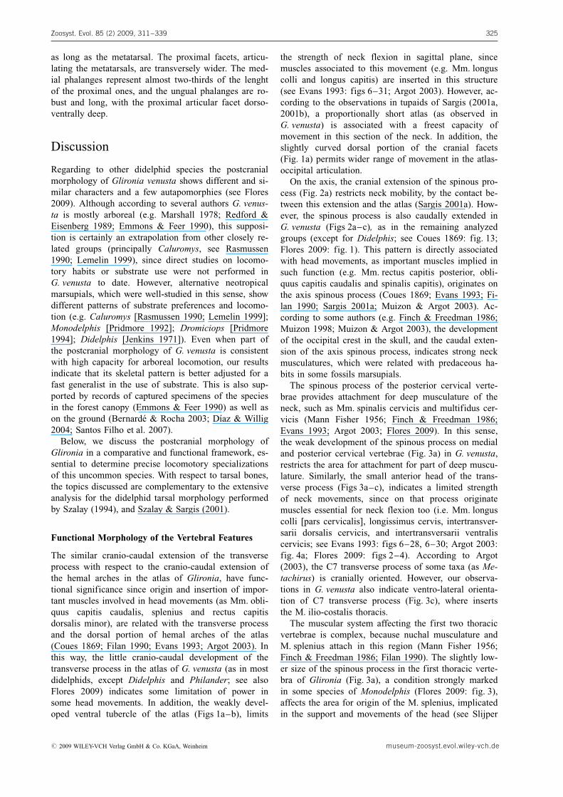
Atlas. The general morphology of the atlas of Glironiavenusta (Figs 1a–b) is essentially similar to other didel-phids. It is a small element, with neural arches dorsallyconvergent and cranio-caudally extended in relation tothe intercentrum I. Behind the cranial articular fovea asmall atlantal foramen for the cranial nerve I is present(Fig. 1b). In dorsal view, the neural arches are similarto the cranio-caudal extension of the transverse pro-cesses, being longer in Caluromys and Caluromysiops.The transverse processes are not caudally extended, butreaching the same level than the caudal articulation. Inventral plane, the processes are oblique with respect tothe vertebral body as in other caluromyines, but thecontact to this (Fig. 1b) is comparatively thinner than inCaluromys (Fig. 1d) and Caluromysiops. The transverseforamina are incompletely closed, forming a canal (Figs1a–b), whereas in adult specimens of Caluromys
Flores, D. A. & Dıaz, M. M.: Postcranial morphology in Glironia venusta312
museum-zoosyst.evol.wiley-vch.de # 2009 WILEY-VCH Verlag GmbH & Co. KGaA, Weinheim

(Figs 1c–d) and Caluromysiops the foramina are com-pletely closed. However, ontogenetic variations wereevidenced in some taxa, as in Caluromys philander,where the foramina are incompletely closed in youngspecimens, showing asymmety in some cases.
As in Caluromys, Caluromisiops, and most of didel-phines analyzed, the cranial facets of the atlas have thedorsal borders curved, although in lesser degree thanother caluromyines (Figs 1a, c). The ventral arches arein contact and the sutures with the intercentrum I areslightly evident. The ventral tubercle is scarcely devel-oped (Figs 1a–b), whereas in Caluromys (Figs c–d) andCaluromysiops it is evident. In caudal view, the caudalarticular foveae are rounded (Fig. 1b), and the trans-
verse canal is well developed. Compared to Caluromysand Caluromysiops, the articular foveae of G. venustaare more caudally oriented.
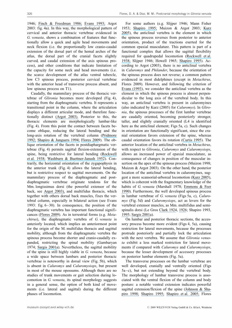
Axis. The axis (Figs 2a–c) of G. venusta is structurallysimilar to those of Caluromys and Caluromysiops. Thesutures among its different components are absent,although an almost imperceptible suture is present be-tween the centrum I and II (Fig. 2c). The sutures amongits remaining components are clearly evident in youngor subadult specimens of other didelphines analyzed(e.g. Didelphis, Philander). The transverse foramen iscompletely closed, and the spinous process is well de-veloped cranially and caudally extended beyond the
Zoosyst. Evol. 85 (2) 2009, 311–339 313
Figure 1. Atlas of Glironia venustaMMD 607 (a–b) and Caluromys philan-der AMNH 267337 (c–d). a, c. Cranialview; b, d. Caudal view. af – atlantalforamen; caf – caudal articular fovea;crf – cranial articular fovea; I – inter-centrum; na – neural arch; tf – trans-verse foramen; tp – transverse process;vt – ventral tubercle. Scale bars: 5 mm.
Figure 2. Axis of Glironia venusta MMD 607 (a–c) and Philander frenatus MVZ 182067 (d–f). a, d. Lateral view; b, e. Dorsalview; c, f. Ventral view. caf – caudal articular fovea; crf – cranial articular fovea; de – dens; na – neural arch; sp – spinousprocess; tf – transverse foramen; tp – transverse process; vt – ventral tubercle. Scale bars: 5 mm.
# 2009 WILEY-VCH Verlag GmbH & Co. KGaA, Weinheim museum-zoosyst.evol.wiley-vch.de
neural arches (Fig. 2a) forming a sagittal crest, as in allanalyzed groups, except Didelphis, which lacks of suchcaudal extension (see Coues 1869). The crest is tallerand more robust in Caluromys and Caluromysiops,being almost parallel to the ventral plane, whereas it isanteroventrally inclined in G. venusta (Fig. 2a). Thedens is anterodorsally oriented, cranially extended withrespect to the anterior tip of the spinous process(Figs 2a–b), a condition observed also in the remainingcaluromyines and some didelphines (e.g. Didelphis,Marmosops, Thylamys). The transverse process is caud-ally oriented (Figs 2b–c) but not enlarged, reaching justthe half of the C3 body (as in Caluromysiops), whereasin some specimens of Caluromys it is more caudallyextended, reaching the posterior border of C3 body.
The cranial articular foveae are almost rounded andcranio-laterally oriented (Fig. 2a). Similarly to Caluromysand Caluromysiops, the caudal articular foveae of G. ve-nusta are oval shape and caudo-ventrally oriented. In
other taxa, as Didelphis, Metachirus, and Philander, thecaudal articular foveae are rounded and almost ventrallyoriented. In lateral view, the inferior part of the neuralarch is thin (Fig. 2a), as evidenced in some mouse opos-sums (e.g Thylamys, Marmosa, Marmosops, Lestodel-phys). On the ventral surface, 3 pairs of small foraminaare present, one posterior to the dens, one medial (whichis also observed on the interior floor), and one posterior(Fig. 2c). The ventral tubercle is formed by two weak se-parated lobes (Fig. 2c), which is also shown in some di-delphines, as Lestodelphys, Marmosops, Metachirus,some species of Thylamys, and Cryptonanus unduavien-sis. The anterior notch of the neural arches is wide(Fig. 2a), a condition widely spread among mouse opos-sums, and present also in Chironectes and Lutreolina.
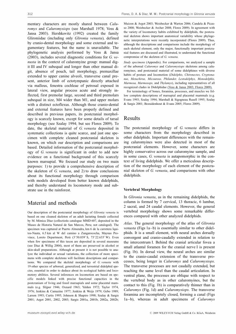
Posterior Cervical Vertebrae (C3–C7). In G. venustathe spinous process is tiny in C3, and gradually becomemore developed from C4 to C7 (Fig. 3a). This condi-
Flores, D. A. & Dıaz, M. M.: Postcranial morphology in Glironia venusta314
Figure 3. Posterior cervical and anterior thoracic vertebrae of Glironia venusta MMD 607 (a–c) and cervical vertebrae of Calu-romys philander AMNH 267337 (d–f). a, d. Lateral view; b, e. Dorsal view; c, f. Ventral view. Ax – axis; C3 – third cervicalvertebra; C6 – sixth cervical vertebra; crf – cranial articular fovea; de – dens; la – lamellae; poz – postzygapophysis; pz –prezygapophysis; ri – rib; sp – spinous process; T1 – first thoracic vertebra; T6 – sixth thoracic vertebra; tp – transverseprocess. Arrow indicates the different orientation of prezygapophysis. Not in scale.
museum-zoosyst.evol.wiley-vch.de # 2009 WILEY-VCH Verlag GmbH & Co. KGaA, Weinheim
tion is a significant difference with Caluromys and Ca-luromysiops in which the spinous process is well devel-oped and laminar shape in all post-axis cervical verte-brae. The transverse process on C3 has one head(Fig. 3c), as in most of mouse opossums, whereas inCaluromys, Caluromysiops, Metachirus, and the2n = 22 large opossums, it has two heads. The prezyga-pophysis is dorso-mesially oriented, and the postzyga-pophysis is ventro-laterally oriented from (Fig. 3a). InC6 a well developed lamella is observed as in all taxaanalyzed (Figs 3a, c). The transverse foramen is presentin C7, and the transverse processes are caudally or-iented from C3 to C5, almost perpendicular to the ver-tebral body in C6, and slightly anterior and ventro-lat-erally oriented in C7 (Figs 3a–c), as in Caluromys,Caluromysiops, Didelphis, and some mouse opossums.
Thoracic Vertebrae (T1–13). The thoracic vertebrae ofG. venusta show some remarkable differences with re-spect to Caluromys and Caluromysiops. The spinousprocess of T1 is scarcely lower than in T2 and T3(although not as in Monodelphis, see Flores 2009:fig. 3) and slightly wider cranio-caudally. The spinousprocesses become more caudally oriented starting fromT3 (Fig. 3a). In Caluromys and Caluromysiops the pro-cesses from T1 to T3 are similar in size and orienta-tion, being in T3 slightly caudal. In didelphines, theprocess of T1 is taller or equal than in T2, except inMonodelphis, where it is notably shorter.
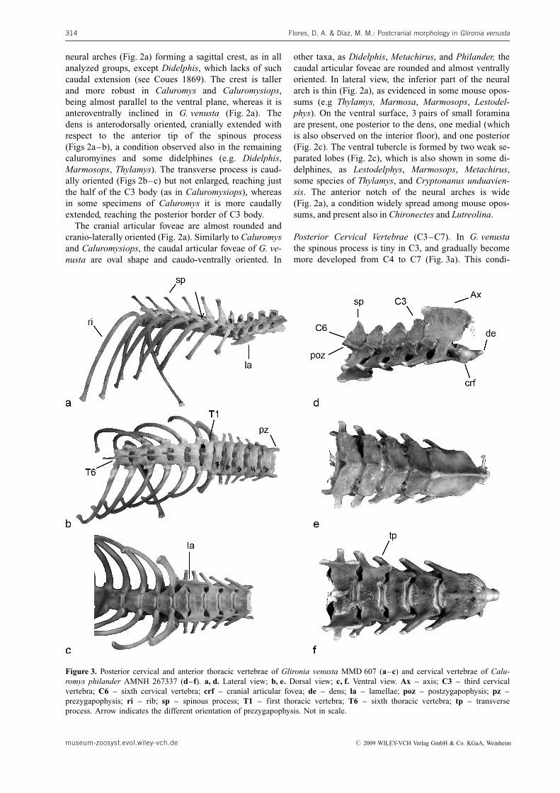
In G. venusta, as in all studied didelphines, the firstthoracic vertebra with the prezygapophysis facing later-ally is placed on the third element (Fig. 3a), whereas inCaluromys and Caluromysiops it is placed on the sec-ond position. From the eighth vertebra, the accessoryprocess is completely separated from the transverseprocess (Fig. 4), whereas in Caluromys this conditionoccurs in the ninth thoracic vertebra; and in the seventhin Caluromysiops. The accessory processes are small(Fig. 4) if compared with those of Caluromys, Caluro-mysiops or Metachirus.
From T10 to T13, the spinous processes become no-tably shorter and cranio-caudally enlarged, and the gen-eral vertebral morphology resembles to anterior lumbar
elements (Fig. 4). Until T9 or T10 the facets for verte-bral articulation are parallel with respect to the ventralplane, but from T11 a change in orientation occurs, be-coming it more oblique (Fig. 4). In this sense, T11 isidentified as diaphragmatic vertebra in G. venusta(Flores 2009). In the same way, T11 is the first elementthat shows the mammilary process notably developed(Fig. 4), as in Caluromysiops and Caluromys philander.Both G. venusta and Caluromys display the T11 spinousprocess almost vertical respect to the long axis of ver-tebral body, whereas in T12–13 they are caudally ori-ented.
Lumbar Vertebrae (L1–6). The lumbar vertebrae ofG. venusta show both the spinous and mammilary pro-cesses well developed (Fig. 5a). Starting from the thirdlumbar element, the mammilary process surpassesslightly the level of articulation with the anterior verte-bra (Fig. 5a). This condition is shared with Caluromys,Caluromysiops, and several didelphines, such as Meta-chirus, Chironectes, Micoureus, Marmosa robinsoni,and Cryptonanus unduaviensis. Small accessory pro-cesses are present in L1–4 (Fig. 5a), contrasting withCaluromys and Caluromysiops, where those are well de-veloped and present in all lumbar vertebrae. In dorsalview, an evident intervertebral space is observed(Fig. 5b), a state shared with mouse opossums, but ab-sent in the remaining caluromyines and large didel-phines. A small foramen on the base of the neuralarches in the last lumbar element is present, as in allother taxa analyzed.
Caudally, the spinous process becomes taller and cra-nio-caudally expanded (Figs 5a–c). The process at L4 ismore expanded and vertically oriented, whereas in pos-terior lumbar vertebrae they are cranially oriented, beingtaller and thinner in L6 (Figs 5a, c). In this sense, the an-ticlinal vertebra in G. venusta is located at L4, whereasin other taxa its position shows notable variation. For in-stance, in Caluromys the process is vertical in L4 and L5but in L6 it is cranially oriented (Fig. 5d), whereas in Ca-luromysiops and Micoureus the vertical process is placedon L6. On the other hand, in Metachirus this element isplaced at L3, and at L4 in Philander.
Zoosyst. Evol. 85 (2) 2009, 311–339 315
Figure 4. Lateral view of posterior thoracic ver-tebrae of Glironia venusta MMD 607. ap – ac-cessory process; dv – diaphragmatic vertebra;mp – mammilary process; poz – postzygapo-physis; pz – prezygapophysis; ri – rib; sp –spinous process; T7 – seventh thoracic vertebra.Scale bar: 10 mm.
# 2009 WILEY-VCH Verlag GmbH & Co. KGaA, Weinheim museum-zoosyst.evol.wiley-vch.de
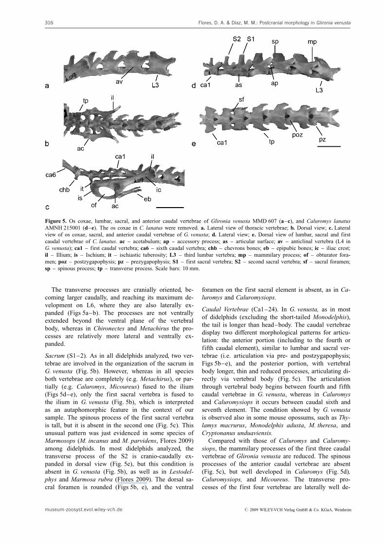
The transverse processes are cranially oriented, be-coming larger caudally, and reaching its maximum de-velopment on L6, where they are also laterally ex-panded (Figs 5a–b). The processes are not ventrallyextended beyond the ventral plane of the vertebralbody, whereas in Chironectes and Metachirus the pro-cesses are relatively more lateral and ventrally ex-panded.
Sacrum (S1–2). As in all didelphids analyzed, two ver-tebrae are involved in the organization of the sacrum inG. venusta (Fig. 5b). However, whereas in all speciesboth vertebrae are completely (e.g. Metachirus), or par-tially (e.g. Caluromys, Micoureus) fused to the ilium(Figs 5d–e), only the first sacral vertebra is fused tothe ilium in G. venusta (Fig. 5b), which is interpretedas an autaphomorphic feature in the context of oursample. The spinous process of the first sacral vertebrais tall, but it is absent in the second one (Fig. 5c). Thisunusual pattern was just evidenced in some species ofMarmosops (M. incanus and M. parvidens, Flores 2009)among didelphids. In most didelphids analyzed, thetransverse process of the S2 is cranio-caudally ex-panded in dorsal view (Fig. 5e), but this condition isabsent in G. venusta (Fig. 5b), as well as in Lestodel-phys and Marmosa rubra (Flores 2009). The dorsal sa-cral foramen is rounded (Figs 5b, e), and the ventral
foramen on the first sacral element is absent, as in Ca-luromys and Caluromysiops.
Caudal Vertebrae (Ca1–24). In G. venusta, as in mostof didelphids (excluding the short-tailed Monodelphis),the tail is longer than head–body. The caudal vertebraedisplay two different morphological patterns for articu-lation: the anterior portion (including to the fourth orfifth caudal element), similar to lumbar and sacral ver-tebrae (i.e. articulation via pre- and postzygapophysis;Figs 5b–e), and the posterior portion, with vertebralbody longer, thin and reduced processes, articulating di-rectly via vertebral body (Fig. 5c). The articulationthrough vertebral body begins between fourth and fifthcaudal vertebrae in G. venusta, whereas in Caluromysand Caluromysiops it occurs between caudal sixth andseventh element. The condition showed by G. venustais observed also in some mouse opossums, such as Thy-lamys macrurus, Monodelphis adusta, M. theresa, andCryptonanus unduaviensis.
Compared with those of Caluromys and Caluromy-siops, the mammilary processes of the first three caudalvertebrae of Glironia venusta are reduced. The spinousprocesses of the anterior caudal vertebrae are absent(Fig. 5c), but well developed in Caluromys (Fig. 5d),Caluromysiops, and Micoureus. The transverse pro-cesses of the first four vertebrae are laterally well de-
Flores, D. A. & Dıaz, M. M.: Postcranial morphology in Glironia venusta316
Figure 5. Os coxae, lumbar, sacral, and anterior caudal vertebrae of Glironia venusta MMD 607 (a–c), and Caluromys lanatusAMNH 215001 (d–e). The os coxae in C. lanatus were removed. a. Lateral view of thoracic vertebrae; b. Dorsal view; c. Lateralview of os coxae, sacral, and anterior caudal vertebrae of G. venusta; d. Lateral view; e. Dorsal view of lumbar, sacral and firstcaudal vertebrae of C. lanatus. ac – acetabulum; ap – accessory process; as – articular surface; av – anticlinal vertebra (L4 inG. venusta); ca1 – first caudal vertebra; ca6 – sixth caudal vertebra; chb – chevrons bones; eb – epipubic bones; ic – iliac crest;il – Illium; is – Ischium; it – ischiastic tuberosity; L3 – third lumbar vertebra; mp – mammilary process; of – obturator fora-men; poz – postzygapophysis; pz – prezygapophysis; S1 – first sacral vertebra; S2 – second sacral vertebra; sf – sacral foramen;sp – spinous process; tp – transverse process. Scale bars: 10 mm.
museum-zoosyst.evol.wiley-vch.de # 2009 WILEY-VCH Verlag GmbH & Co. KGaA, Weinheim
veloped and cranio-caudally extended (Fig. 5b), beingreduced to just one anterior and one posterior promi-nences on the fifth element. The same reduction is ob-served at the seventh element in Caluromys and Calur-omysiops. In most didelphids, there is a caudal elementwhere the transverse process is craniocaudally length-ened as the vertebral body (see Argot 2003: fig. 10a),which is absent in G. venusta. The chevrons bones arewell developed on the first caudal vertebrae (Fig. 5c),becoming smaller and weaker between the fourth andfifth elements.
Pectoral Girdle and Forelimb Morphology
Ribs and Sternum. In Glironia venusta the ribs are cy-lindrical, as in all didelphines (Figs 3–4), being cranio-caudally enlarged in Caluromys (see Argot 2003:fig. 8a) and Caluromysiops. The first rib is thin and no-tably less curved in (Fig. 3c), even when compared withthat of the arboreal Micoureus, whereas in Caluromysand Caluromysiops they are notably robust and curved.In old specimens of Caluromys and Caluromysiops, thecostal cartilages are almost completely ossified, withonly small portions without ossification. Such a condi-tion is not observed in G. venusta.
The sternum is formed by 6 sternebrae (includingmanubrium and xiphoides), although some occasional
individual variation is evidenced in Caluromys philan-der, where 5 or 7 elements were occasionally detected(e.g. AMNH 267002, RMNH 19646). The manubriumof Glironia venusta is less robust compared with Calu-romys, slightly keeled, and the cranial section lessenlarged anteriorly. The second sternebrae is com-pressed, and the remainings are flattened. From third tofifth element, the morphology is basically similar. Thexiphoid is enlarged with a small medial compression.Alternatively, in Caluromysiops and Caluromys all ster-nebrae are laterally compressed and caudally expanded,becoming posteriorly smaller.
Clavicle. The clavicles of Glironia venusta are thin andslightly concave, with the sternal tip notably expandedcompared with the scapular tip, and proportionally thincompared with that of Metachirus, Didelphis, Micou-reus, and Philander. In Caluromys and Caluromysiops,the clavicles are also curved but with both tips of simi-lar size.
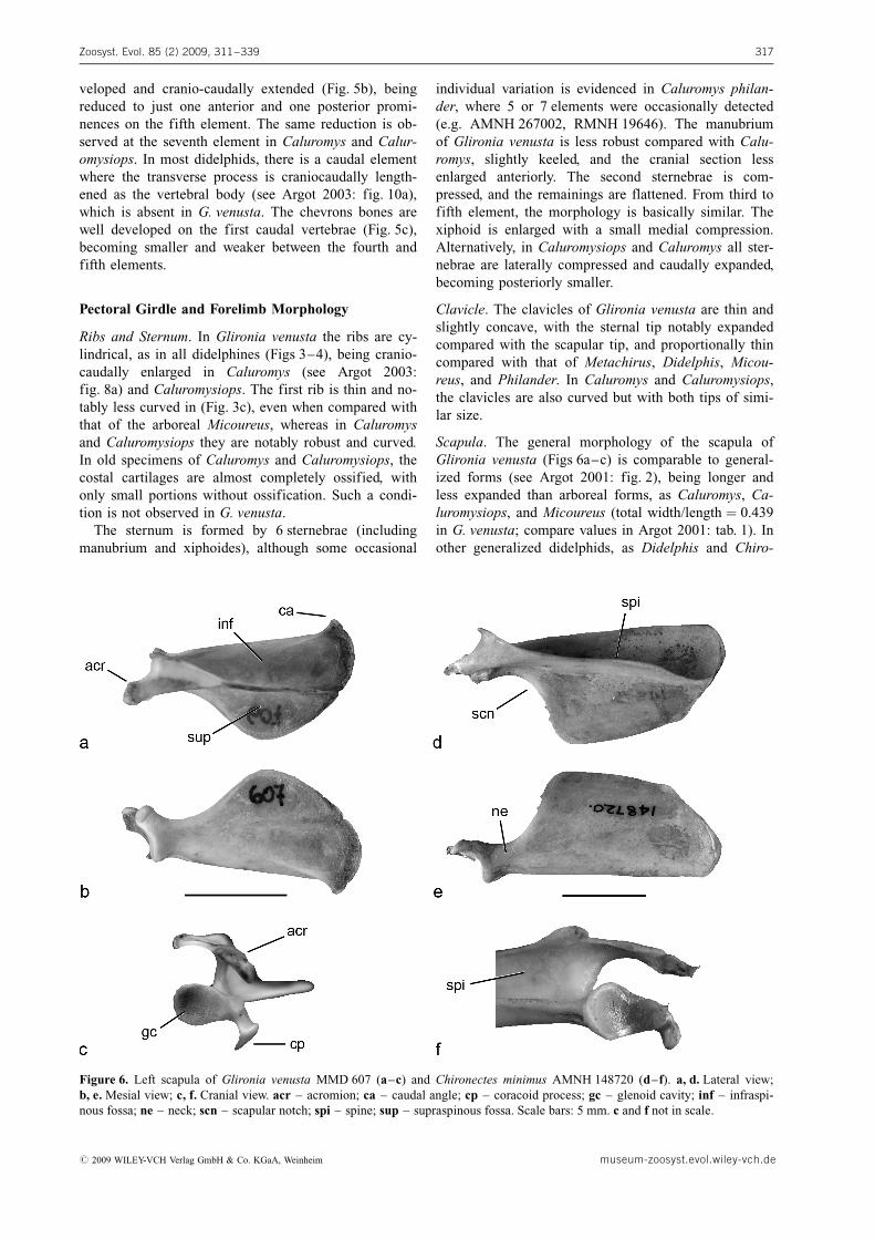
Scapula. The general morphology of the scapula ofGlironia venusta (Figs 6a–c) is comparable to general-ized forms (see Argot 2001: fig. 2), being longer andless expanded than arboreal forms, as Caluromys, Ca-luromysiops, and Micoureus (total width/length ¼ 0.439in G. venusta; compare values in Argot 2001: tab. 1). Inother generalized didelphids, as Didelphis and Chiro-
Zoosyst. Evol. 85 (2) 2009, 311–339 317
Figure 6. Left scapula of Glironia venusta MMD 607 (a–c) and Chironectes minimus AMNH 148720 (d–f). a, d. Lateral view;b, e. Mesial view; c, f. Cranial view. acr – acromion; ca – caudal angle; cp – coracoid process; gc – glenoid cavity; inf – infraspi-nous fossa; ne – neck; scn – scapular notch; spi – spine; sup – supraspinous fossa. Scale bars: 5 mm. c and f not in scale.
# 2009 WILEY-VCH Verlag GmbH & Co. KGaA, Weinheim museum-zoosyst.evol.wiley-vch.de

nectes, the scapula is rectangular (Fig. 6d), being widerin terrestrial taxa as Metachirus or Monodelphis. Thesupraspinous fossa is wider and larger than the infraspi-nous one, as in Metachirus, Philander, and Marmosa.Similarly to arboreal forms, the caudal angle is acuteand the scapular notch is extended to the half of thescapula (Figs 6a–b). The coracoid process is well de-veloped (Fig. 6c), being higly variable across the didel-phines. For instance, in Philander and Chironectes it issmall (Fig. 6f), whereas in Metachirus and Didelphis itis notably wide. The acromion is strong and extendsbeyond the glenoid fossa of the scapula (Fig. 6b); ingeneral, it is more developed in caluromyines than di-delphines. The glenoid fossa is piriform shape (Fig. 6c)and anteroventrally oriented, as in all groups analyzed.The scapular spine is well developed (Figs 6a, c) inG. venusta, as wide as the infraspinous fossa at theneck level, a condition shared with Caluromysiops andCaluromys. In contrast, the spine is proportionally low-er in Metachirus, Philander, Didelphis, and Chironectes(Fig. 6f), descending more abruptly compared withstrongly arboreal forms.
Humerus. The body is robust, with the diaphysis almoststraight (Figs 7a–c). The humeral head is slightly oval(Fig. 7b), being proportionally lesser in G. venusta com-pared with those of Caluromys and Caluromysiops,where it is more rounded. As in Caluromys and Calu-romysiops, the neck is not strongly marked in G. venus-ta, whereas it is more evident in some didelphines, asMetachirus, Micoureus and Philander (Fig. 7f). In theproximal section, the humeral greater and lesser tuber-
osities are well developed (Fig. 7b) and the greater oneslightly surpasses the level of the head. Among didel-phids, this condition was detected only in Glironia(compare with Philander; Fig. 7e), and additionally insome Australasian groups, as Dasyurus and Vombatus(Horovitz & S�nchez-Villagra 2003). The deltopectoralcrest is well developed, extending to the proximal halfof the diaphysis and determining a deep bicipitalgroove (Figs 7a, c), as in Caluromys, Caluromysiops,and some mouse opossums. Instead, in Philander(Fig. 7f) and Metachirus the crest is low, but extendingbeyond the proximal half of the humerus, and the bici-pital groove is comparatively shallow.
In mesial view, a medial relief for the insertion ofthe M. teres major is observed, which is also present inCaluromys and Caluromysiops. The relief is less evi-dent in Didelphis, Micoureus, Metachirus, and Philan-der, and well developed in the terrestrial Monodelphis.In lateral view, the tuberosity for the insertion ofM. teres minor is evident (Fig. 7c), as in Monodelphis,Metachirus, and Philander. On the distal portion of thehumerus, the ectepicondylar crest (or supinator ridge)is well developed and laterally expanded in G. venusta,showing a small proximal process (Figs 7a–c). The tro-chlea is evident, and clearly separated from the capitu-lum (Fig. 7c), although in Caluromys, Caluromysiops,and Micoureus both structures are comparatively moreseparated by a wider zone for the humero-ulnar articu-lation. The capitulum is spherical, more proximallyextended than the trochlea, showing a clear lateral pro-jection (Fig. 7a) as in the large opossums. Theentepicondyle is well developed protruding mesially
Flores, D. A. & Dıaz, M. M.: Postcranial morphology in Glironia venusta318
Figure 7. Left humerus of Glironia venusta MMD 607 (a–c) and Philander opossum AMNH 190446 (d–f). a, d. Cranial view;b, e. Caudal view; c, f. Lateral view. bg – bicipital groove; cap – capitulum; dpc – deltopectoral crest; ef – entepicondylar fora-men; en – entepicondyle; gt – greater tuberosity; hcf – humeral coronoid fossa; hh – humeral head; hmt – humeral lateraltuberosity; lex – lateral extension of the capitulum; lt – lesser tuberosity; ne – neck; of – olecraneon fossa; prp – proximalprocess of the supinator ridge; sur – supinator ridge; tr – trochlea. Scale bars: 10 mm.
museum-zoosyst.evol.wiley-vch.de # 2009 WILEY-VCH Verlag GmbH & Co. KGaA, Weinheim
and separated from the trochlea by a small groove (Figs7a–b). The separation of both structures is variable indifferent groups. For instance, in Micoureus the grooveis wide, whereas in Didelphis, Philander (Fig. 7e), andMetachirus it is almost absent. The entepicondylar orhumeral supracondyloid foramen is well developed andoval shape (Fig. 7a), as in most the groups analyzed,although it is rounded and relatively smaller in Meta-chirus and Micoureus. As in Caluromys, Micoureus andPhilander (Fig. 7e), the olecraneon fossa is visible butshallow in G. venusta (Fig. 7b), and the humeral coro-noid fossa is also shallow (Fig. 7a).
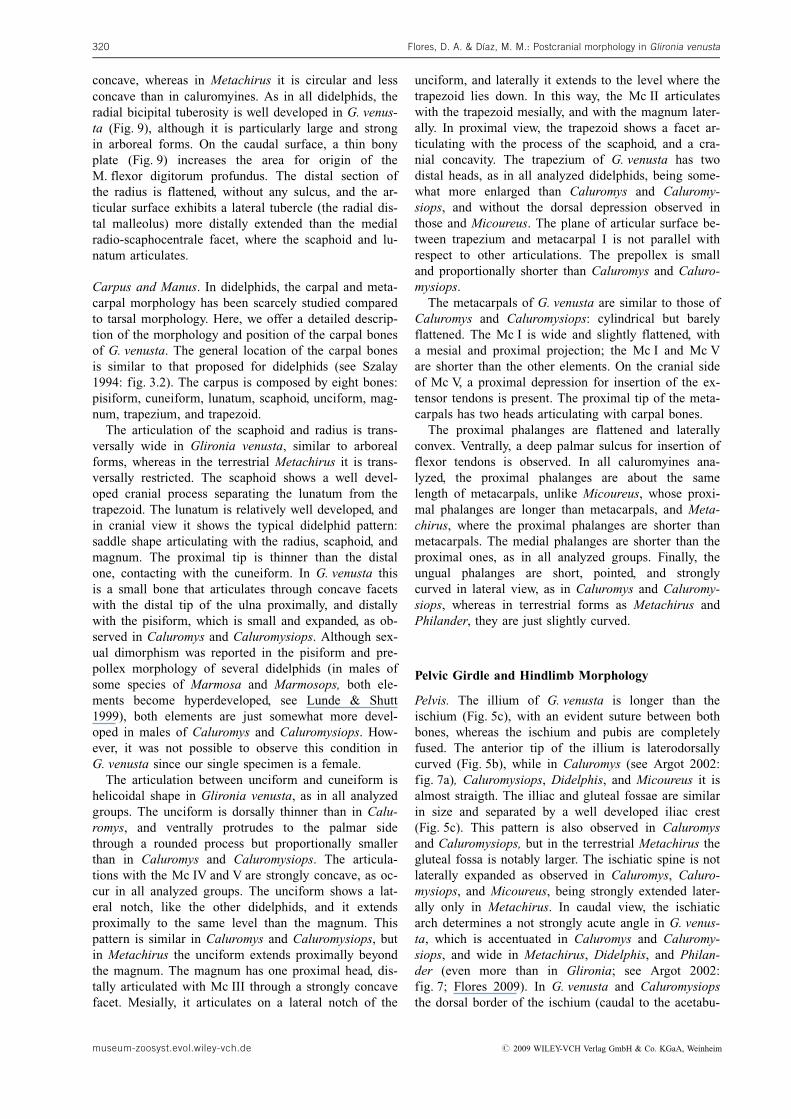
Ulna and Radius. In Glironia the ulna and radius arestrong bones (Figs 8a, 9). The diaphysis of the ulna islaterally compressed as in arboreal forms, but in largerdidelphines (as Didelphis, Metachirus, and Philander),it is rather cylindrical. The olecranon is short and ro-bust (Fig. 8a), with its proximal tip slightly curved cra-nially. Such pattern was also seen in didelphids as Ca-luromys, Caluromysiops, Micoureus, and Marmosa(Flores 2009). In Monodelphis (Fig. 8b) and Didelphis,the olecranon is longer, whereas in Metachirus, Hyla-delphys, and Marmosops parvidens it is shorter and ro-bust (see Flores 2009: fig. 19). The anconeal process ispoorly developed in G. venusta, being defined by twosmall crests (Fig. 8a): the ulnar lateral proximal tro-chlear (ulptcl) and the ulnar medial proximal trochlear(ulptcm). Both crests are about the same size, and theulptcm is less extended proximally than the ulptcl, afeature also observed in Didelphis, Caluromys, Caluro-
mysiops, and Microures. Alternatively, the anconealprocess is more developed in Metachirus than in Phi-lander, although in both genera the ulptcm is longerand more proximally extended than ulptcl. The angleformed by the small radial notch and the border of thecoronoid process is obtuse in G. venusta (Fig. 8a), Ca-luromys, and Caluromysiops. The coronoid process andthe greater sigmoid cavity are well developed, orientedperpendicular respect to the ulnar diaphysis (Fig. 8a),as in arboreal forms. On the diaphysis, the ulnar fossafor M. flexor digitorum profundus is deep, extendingbeyond the trochlear notch (Flores 2009) and a weakgroove for the M. abductor pollicis longus is present.The fossa for the insertion of M. anconeus is evidenton the lateral side (Fig. 8a), although in lesser degreethan observed in Caluromysiops (Flores 2009: fig. 19).On the cranial side, there is a marked crest for the ori-gin of M. pronator cuadratus and M. flexor digitorumprofundus. Distally, the ulna is cylindrical in transverseplane, with an anterior sulcus. The ulnar styloid processis well developed, and articulates with the cuneiformand pisiform through the ulnar-cuneiform facet.
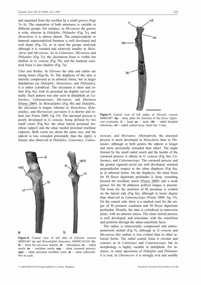
The radius is transversally compressed and antero-posteriorly arched (Fig. 9), although in G. venusta andMicoureus this outline is less evident than in other ar-boreal forms. The radial central fossa is circular andconcave as in Caluromys and Caluromysiops, but itsmorphology is highly variable in didelphids. For in-stance, in some specimens of Didelphis and Philanderit is oval, in Chironectes it is strongly oval and notably
Zoosyst. Evol. 85 (2) 2009, 311–339 319
Figure 8. Cranial view of left ulna of Glironia venustaMMD 607 (a) and Monodelphis domestica AMNH 261241 (b).fa – fossa for anconeus muscle; ol – olecraneon; rn – radialnotch; tn – trochlear notch; ucp – ulnar coronoid process;uptc – ulnar proximal trochlear crest; ut – ulnar tuberosity.Not in scale.
Figure 9. Lateral view of left radius of Glironia venustaMMD 607. bp – bony plate for insertion of the flexor digito-rum profundus; h – head, ne – neck; rbt – radial bicipitaltuberosity; rcf – radial central fossa. Scale bar: 5 mm.
# 2009 WILEY-VCH Verlag GmbH & Co. KGaA, Weinheim museum-zoosyst.evol.wiley-vch.de
concave, whereas in Metachirus it is circular and lessconcave than in caluromyines. As in all didelphids, theradial bicipital tuberosity is well developed in G. venus-ta (Fig. 9), although it is particularly large and strongin arboreal forms. On the caudal surface, a thin bonyplate (Fig. 9) increases the area for origin of theM. flexor digitorum profundus. The distal section ofthe radius is flattened, without any sulcus, and the ar-ticular surface exhibits a lateral tubercle (the radial dis-tal malleolus) more distally extended than the medialradio-scaphocentrale facet, where the scaphoid and lu-natum articulates.
Carpus and Manus. In didelphids, the carpal and meta-carpal morphology has been scarcely studied comparedto tarsal morphology. Here, we offer a detailed descrip-tion of the morphology and position of the carpal bonesof G. venusta. The general location of the carpal bonesis similar to that proposed for didelphids (see Szalay1994: fig. 3.2). The carpus is composed by eight bones:pisiform, cuneiform, lunatum, scaphoid, unciform, mag-num, trapezium, and trapezoid.
The articulation of the scaphoid and radius is trans-versally wide in Glironia venusta, similar to arborealforms, whereas in the terrestrial Metachirus it is trans-versally restricted. The scaphoid shows a well devel-oped cranial process separating the lunatum from thetrapezoid. The lunatum is relatively well developed, andin cranial view it shows the typical didelphid pattern:saddle shape articulating with the radius, scaphoid, andmagnum. The proximal tip is thinner than the distalone, contacting with the cuneiform. In G. venusta thisis a small bone that articulates through concave facetswith the distal tip of the ulna proximally, and distallywith the pisiform, which is small and expanded, as ob-served in Caluromys and Caluromysiops. Although sex-ual dimorphism was reported in the pisiform and pre-pollex morphology of several didelphids (in males ofsome species of Marmosa and Marmosops, both ele-ments become hyperdeveloped, see Lunde & Shutt1999), both elements are just somewhat more devel-oped in males of Caluromys and Caluromysiops. How-ever, it was not possible to observe this condition inG. venusta since our single specimen is a female.
The articulation between unciform and cuneiform ishelicoidal shape in Glironia venusta, as in all analyzedgroups. The unciform is dorsally thinner than in Calu-romys, and ventrally protrudes to the palmar sidethrough a rounded process but proportionally smallerthan in Caluromys and Caluromysiops. The articula-tions with the Mc IV and V are strongly concave, as oc-cur in all analyzed groups. The unciform shows a lat-eral notch, like the other didelphids, and it extendsproximally to the same level than the magnum. Thispattern is similar in Caluromys and Caluromysiops, butin Metachirus the unciform extends proximally beyondthe magnum. The magnum has one proximal head, dis-tally articulated with Mc III through a strongly concavefacet. Mesially, it articulates on a lateral notch of the
unciform, and laterally it extends to the level where thetrapezoid lies down. In this way, the Mc II articulateswith the trapezoid mesially, and with the magnum later-ally. In proximal view, the trapezoid shows a facet ar-ticulating with the process of the scaphoid, and a cra-nial concavity. The trapezium of G. venusta has twodistal heads, as in all analyzed didelphids, being some-what more enlarged than Caluromys and Caluromy-siops, and without the dorsal depression observed inthose and Micoureus. The plane of articular surface be-tween trapezium and metacarpal I is not parallel withrespect to other articulations. The prepollex is smalland proportionally shorter than Caluromys and Caluro-mysiops.
The metacarpals of G. venusta are similar to those ofCaluromys and Caluromysiops: cylindrical but barelyflattened. The Mc I is wide and slightly flattened, witha mesial and proximal projection; the Mc I and Mc Vare shorter than the other elements. On the cranial sideof Mc V, a proximal depression for insertion of the ex-tensor tendons is present. The proximal tip of the meta-carpals has two heads articulating with carpal bones.
The proximal phalanges are flattened and laterallyconvex. Ventrally, a deep palmar sulcus for insertion offlexor tendons is observed. In all caluromyines ana-lyzed, the proximal phalanges are about the samelength of metacarpals, unlike Micoureus, whose proxi-mal phalanges are longer than metacarpals, and Meta-chirus, where the proximal phalanges are shorter thanmetacarpals. The medial phalanges are shorter than theproximal ones, as in all analyzed groups. Finally, theungual phalanges are short, pointed, and stronglycurved in lateral view, as in Caluromys and Caluromy-siops, whereas in terrestrial forms as Metachirus andPhilander, they are just slightly curved.
Pelvic Girdle and Hindlimb Morphology
Pelvis. The illium of G. venusta is longer than theischium (Fig. 5c), with an evident suture between bothbones, whereas the ischium and pubis are completelyfused. The anterior tip of the illium is laterodorsallycurved (Fig. 5b), while in Caluromys (see Argot 2002:fig. 7a), Caluromysiops, Didelphis, and Micoureus it isalmost straigth. The illiac and gluteal fossae are similarin size and separated by a well developed iliac crest(Fig. 5c). This pattern is also observed in Caluromysand Caluromysiops, but in the terrestrial Metachirus thegluteal fossa is notably larger. The ischiatic spine is notlaterally expanded as observed in Caluromys, Caluro-mysiops, and Micoureus, being strongly extended later-ally only in Metachirus. In caudal view, the ischiaticarch determines a not strongly acute angle in G. venus-ta, which is accentuated in Caluromys and Caluromy-siops, and wide in Metachirus, Didelphis, and Philan-der (even more than in Glironia; see Argot 2002:fig. 7; Flores 2009). In G. venusta and Caluromysiopsthe dorsal border of the ischium (caudal to the acetabu-
Flores, D. A. & Dıaz, M. M.: Postcranial morphology in Glironia venusta320
museum-zoosyst.evol.wiley-vch.de # 2009 WILEY-VCH Verlag GmbH & Co. KGaA, Weinheim
lum) is straight, but laterally oriented in Caluromys, Di-delphis, and Metachirus. The ischiatic tuberosity is en-larged in Glironia (Fig. 5c) and arboreal forms as Ca-luromys, Caluromysiops, and Micoureus, but less thanDidelphis, Metachirus, or Philander.
The iliopubic process is scarcely developed in Gliro-nia, although in some specimens of Caluromys and Ca-luromysiops it is well developed. In caluromyines andMicoureus, the symphysis pubis is as large as the cra-nio-caudal length of the obturator foramen (Fig. 5c),being shorter in Didelphis and Philander. The obturatorforamen is rounded (Fig. 5c) and proportionally widerthan in Micoureus, Caluromys, or Caluromysiops. Theacetabulum is opened, oval shape, and with a wellmarked and concave dorsal border (Figs 5b–c). Thismorphology is common in all analyzed taxa, except inMetachirus where the acetabulum is notably deep androunded.
Epipubic bones. The epipubic bones of Glironia venus-ta are robust, wider on its proximal section, becomingthinner and slightly curved laterally at the distal tip,and extending approximately to half of the illium(Fig. 5c). This pattern is also observed in our entiresample, although some variation is found in Caluromys,Caluromysiops, and Metachirus. Flores (2009: fig. 24)report epipubic bones notably elongated and stronglycurved ventrally in Caluromysiops, Caluromys, Marmo-sops parvidens, Chironectes, and Marmosa rubra.
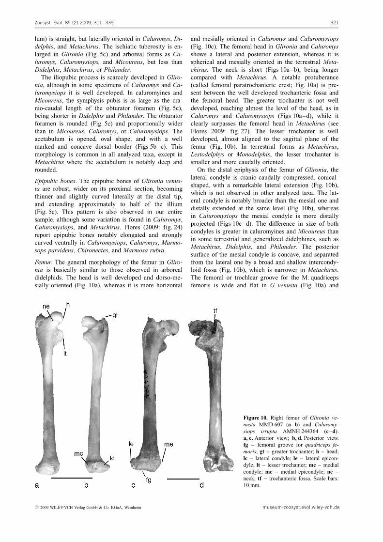
Femur. The general morphology of the femur in Gliro-nia is basically similar to those observed in arborealdidelphids. The head is well developed and dorso-me-sially oriented (Fig. 10a), whereas it is more horizontal
and mesially oriented in Caluromys and Caluromysiops(Fig. 10c). The femoral head in Glironia and Caluromysshows a lateral and posterior extension, whereas it isspherical and mesially oriented in the terrestrial Meta-chirus. The neck is short (Figs 10a–b), being longercompared with Metachirus. A notable protuberance(called femoral paratrochanteric crest; Fig. 10a) is pre-sent between the well developed trochanteric fossa andthe femoral head. The greater trochanter is not welldeveloped, reaching almost the level of the head, as inCaluromys and Caluromysiops (Figs 10a–d), while itclearly surpasses the femoral head in Metachirus (seeFlores 2009: fig. 27). The lesser trochanter is welldeveloped, almost aligned to the sagittal plane of thefemur (Fig. 10b). In terrestrial forms as Metachirus,Lestodelphys or Monodelphis, the lesser trochanter issmaller and more caudally oriented.
On the distal epiphysis of the femur of Glironia, thelateral condyle is cranio-caudally compressed, conical-shaped, with a remarkable lateral extension (Fig. 10b),which is not observed in other analyzed taxa. The lat-eral condyle is notably broader than the mesial one anddistally extended at the same level (Fig. 10b), whereasin Caluromysiops the mesial condyle is more distallyprojected (Figs 10c–d). The difference in size of bothcondyles is greater in caluromyines and Micoureus thanin some terrestrial and generalized didelphines, such asMetachirus, Didelphis, and Philander. The posteriorsurface of the mesial condyle is concave, and separatedfrom the lateral one by a broad and shallow intercondy-loid fossa (Fig. 10b), which is narrower in Metachirus.The femoral or trochlear groove for the M. quadricepsfemoris is wide and flat in G. venusta (Fig. 10a) and
Zoosyst. Evol. 85 (2) 2009, 311–339 321
Figure 10. Right femur of Glironia ve-nusta MMD 607 (a–b) and Caluromy-siops irrupta AMNH 244364 (c–d).a, c. Anterior view; b, d. Posterior view.fg – femoral groove for quadriceps fe-moris; gt – greater trochanter; h – head;lc – lateral condyle; le – lateral epicon-dyle; lt – lesser trochanter; mc – medialcondyle; me – medial epicondyle; ne –neck; tf – trochanteric fossa. Scale bars:10 mm.
# 2009 WILEY-VCH Verlag GmbH & Co. KGaA, Weinheim museum-zoosyst.evol.wiley-vch.de
Micoureus, whereas it is slightly concave in Caluromysand Caluromysiops (Fig. 10c), and narrow and concavein the terrestrials Metachirus and Monodelphis.
Fibular fabella. The fabella or parafibula (Szalay &Sargis 2001) is an ossification fixed on the proximo-lateral surface of the fibula. In Glironia, it is basicallysimilar to all analyzed taxa: the distal tip, which articu-lates to the fibula, is wider and flattened, and the prox-imal tip is thinner and curved. However, the fabella ofG. venusta shows the proximal tip rounded with astrong ventral curvature, compared to the oval shapeand less curved structure observed in Caluromys andCaluromysiops.
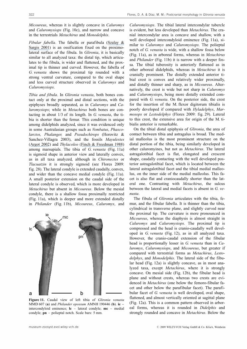
Tibia and fibula. In Glironia venusta, both bones con-tact only at the proximal and distal sections, with theepiphyses broadly separated, as in Caluromys and Ca-luromysiops; while in Metachirus both bones are con-tacting in about 1/3 of its length. In G. venusta, the ti-bia is shorter than the femur. This condition is uniqueamong didelphids analyzed, since it was evidenced onlyin some Australasian groups such as Vombatus, Phasco-larctos, Phalanger, and Pseudochirops (Horovitz &Sanchez-Villagra 2003), and the fossils Mayulestes(Argot 2002) and Thylacoleo (Finch & Freedman 1988)among marsupials. The tibia of G. venusta (Fig. 11a)is sigmoid shape in anterior view and laterally convex,as in all taxa analyzed, although in Chironectes orTlacuatzin it is strongly sigmoid (see Flores 2009:fig. 28). The lateral condyle is extended caudally, convex,and wider than the concave medial condyle (Fig. 11a).A small posterior extension on the caudal side of thelateral condyle is observed, which is more developed inMetachirus but absent in Micoureus. Below the mesialcondyle, there is a shallow fossa proximally restricted(Fig. 11a), which is deeper and more extended distallyin Philander (Fig. 11b), Micoureus, Caluromys, and
Caluromysiops. The tibial lateral intercondylar tubercleis evident, but less developed than Metachirus. The cra-nial intercondylar area is concave and shallow, with awell developed intercondyloid eminence (Fig. 11a), si-milar to Caluromys and Caluromysiops. The polieptalnotch of G. venusta is wide, with a shallow fossa below(Fig. 11a), as in arboreal forms, whereas in Metachirusand Philander (Fig. 11b) it is narrow with a deeper fos-sa. The tibial tuberosity is anteriorly flattened as inother arboreal didelphids, whereas in Metachirus it iscranially prominent. The distally extended anterior ti-bial crest is convex and relatively wider proximally,and distally thinner and sharp, as in Micoureus. Alter-natively, the crest is wide but not sharp in Caluromysand Caluromysiops, being more distally extended com-pared with G. venusta. On the posterior side, the crestfor the insertion of the M. flexor digitorum tibialis ispoorly developed if compared with Hyladelphys, Mar-mosops or Lestodelphys (Flores 2009: fig. 29). Lateralto this crest, the extensive area for origin of the M. ti-bialis anterior is remarkable.
On the tibial distal epiphysis of Glironia, the area ofcontact between tibia and astragalus is broad. The med-ial malleolus is the most prominent structure on thedistal portion of the tibia, being similarly developed inother caluromyines, but not as Metachirus. The lateralastragalotibial facet is flat, elongated and crescentshape, caudally contacting with the well developed pos-terior astragalotibial facet, which is located between thelateral astragalotibial facet and the tibial medial malleo-lus, on the inner side of the medial malleolus. This fa-cet is also flat and craniocaudally shorter than the lat-eral one. Contrasting with Metachirus, the sulcusbetween the lateral and medial facets is absent in G. ve-nusta.
The fibula of Glironia articulates with the tibia, fe-mur, and the fibular fabella. It is thinner than the tibia,cylindrical in transverse plane, and slightly curved nearthe proximal tip. The curvature is more pronounced inMicoureus, whereas the diaphysis is almost straight inCaluromys and Caluromysiops. The proximal tip iscompressed and the head is cranio-caudally well devel-oped in G. venusta (Fig. 12), as in all analyzed taxa.However, the cranio-caudal extension of the fibularhead is proportionally lesser in G. venusta than in Ca-luromys, Caluromysiops, and Micoureus, but greater ifcompared with terrestrial forms as Metachirus, Lesto-delphys, and Monodelphis. The lateral side of the fibu-lar head (Fig. 12a) is slightly concave, as in most ana-lyzed taxa, except Metachirus, where it is stronglyconcave. On mesial side (Fig. 12b), the fibular head isplane and without crests, whereas two crests are evi-denced in Metachirus (one below the femoro-fibular fa-cet and other below the parafibular facet). The parafi-bular facet of G. venusta is well developed, oval shape,flattened, and almost vertically oriented at sagittal plane(Fig. 12a). This is a common pattern observed in arbor-eal forms, whereas it is rounded in Didelphis andstrongly rounded and concave in Metachirus. Below the
Flores, D. A. & Dıaz, M. M.: Postcranial morphology in Glironia venusta322
Figure 11. Caudal view of left tibia of Glironia venustaMMD 607 (a) and Philander opossum AMNH 190446 (b). ie –intercondyloid eminence; lc – lateral condyle; mc – medialcondyle; pn – polieptal notch. Scale bars: 5 mm.
museum-zoosyst.evol.wiley-vch.de # 2009 WILEY-VCH Verlag GmbH & Co. KGaA, Weinheim
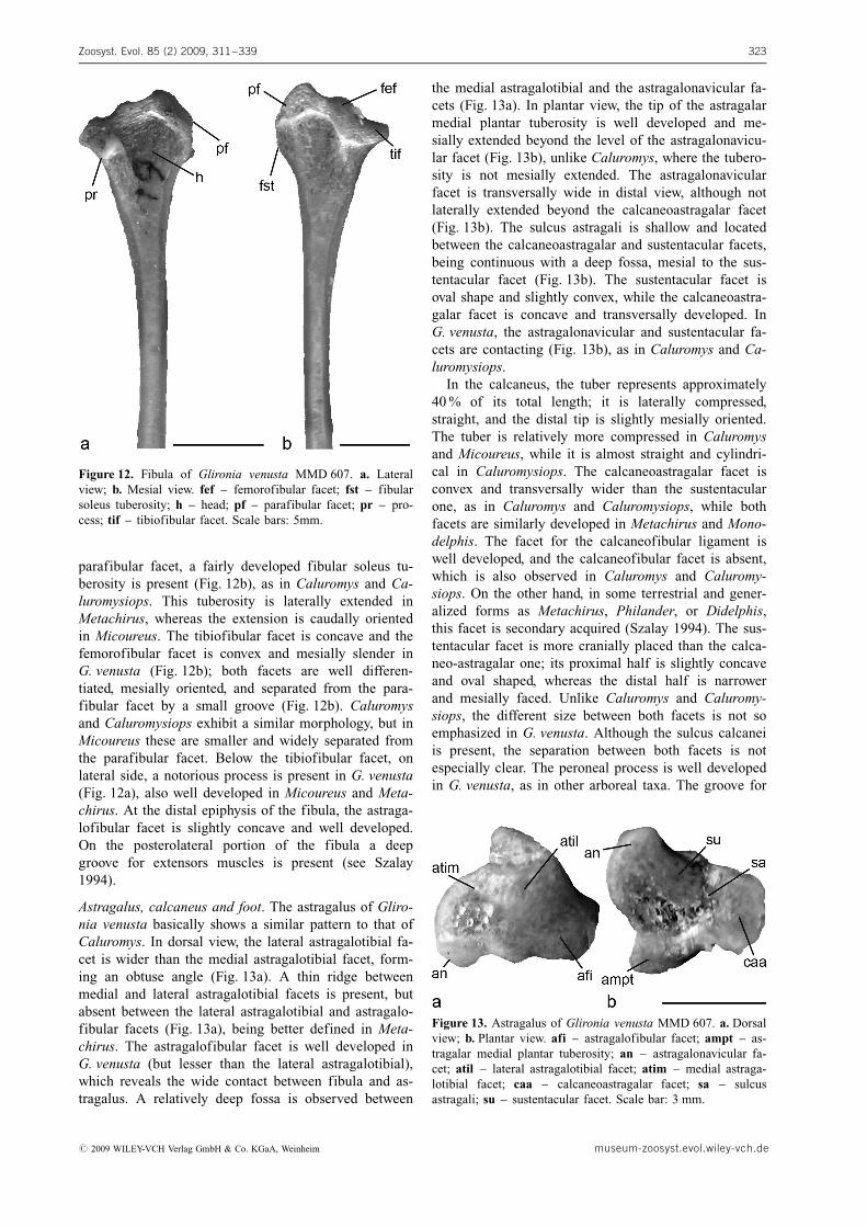
parafibular facet, a fairly developed fibular soleus tu-berosity is present (Fig. 12b), as in Caluromys and Ca-luromysiops. This tuberosity is laterally extended inMetachirus, whereas the extension is caudally orientedin Micoureus. The tibiofibular facet is concave and thefemorofibular facet is convex and mesially slender inG. venusta (Fig. 12b); both facets are well differen-tiated, mesially oriented, and separated from the para-fibular facet by a small groove (Fig. 12b). Caluromysand Caluromysiops exhibit a similar morphology, but inMicoureus these are smaller and widely separated fromthe parafibular facet. Below the tibiofibular facet, onlateral side, a notorious process is present in G. venusta(Fig. 12a), also well developed in Micoureus and Meta-chirus. At the distal epiphysis of the fibula, the astraga-lofibular facet is slightly concave and well developed.On the posterolateral portion of the fibula a deepgroove for extensors muscles is present (see Szalay1994).
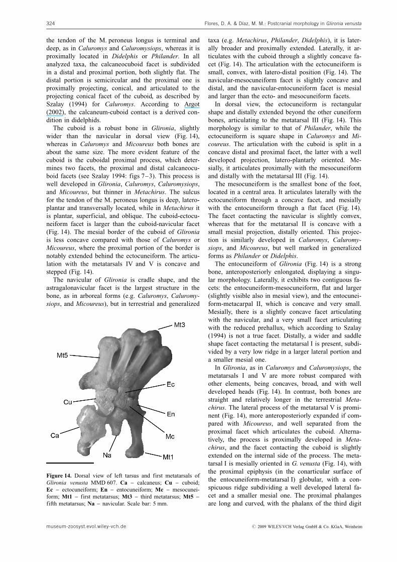
Astragalus, calcaneus and foot. The astragalus of Gliro-nia venusta basically shows a similar pattern to that ofCaluromys. In dorsal view, the lateral astragalotibial fa-cet is wider than the medial astragalotibial facet, form-ing an obtuse angle (Fig. 13a). A thin ridge betweenmedial and lateral astragalotibial facets is present, butabsent between the lateral astragalotibial and astragalo-fibular facets (Fig. 13a), being better defined in Meta-chirus. The astragalofibular facet is well developed inG. venusta (but lesser than the lateral astragalotibial),which reveals the wide contact between fibula and as-tragalus. A relatively deep fossa is observed between
the medial astragalotibial and the astragalonavicular fa-cets (Fig. 13a). In plantar view, the tip of the astragalarmedial plantar tuberosity is well developed and me-sially extended beyond the level of the astragalonavicu-lar facet (Fig. 13b), unlike Caluromys, where the tubero-sity is not mesially extended. The astragalonavicularfacet is transversally wide in distal view, although notlaterally extended beyond the calcaneoastragalar facet(Fig. 13b). The sulcus astragali is shallow and locatedbetween the calcaneoastragalar and sustentacular facets,being continuous with a deep fossa, mesial to the sus-tentacular facet (Fig. 13b). The sustentacular facet isoval shape and slightly convex, while the calcaneoastra-galar facet is concave and transversally developed. InG. venusta, the astragalonavicular and sustentacular fa-cets are contacting (Fig. 13b), as in Caluromys and Ca-luromysiops.
In the calcaneus, the tuber represents approximately40 % of its total length; it is laterally compressed,straight, and the distal tip is slightly mesially oriented.The tuber is relatively more compressed in Caluromysand Micoureus, while it is almost straight and cylindri-cal in Caluromysiops. The calcaneoastragalar facet isconvex and transversally wider than the sustentacularone, as in Caluromys and Caluromysiops, while bothfacets are similarly developed in Metachirus and Mono-delphis. The facet for the calcaneofibular ligament iswell developed, and the calcaneofibular facet is absent,which is also observed in Caluromys and Caluromy-siops. On the other hand, in some terrestrial and gener-alized forms as Metachirus, Philander, or Didelphis,this facet is secondary acquired (Szalay 1994). The sus-tentacular facet is more cranially placed than the calca-neo-astragalar one; its proximal half is slightly concaveand oval shaped, whereas the distal half is narrowerand mesially faced. Unlike Caluromys and Caluromy-siops, the different size between both facets is not soemphasized in G. venusta. Although the sulcus calcaneiis present, the separation between both facets is notespecially clear. The peroneal process is well developedin G. venusta, as in other arboreal taxa. The groove for
Zoosyst. Evol. 85 (2) 2009, 311–339 323
Figure 12. Fibula of Glironia venusta MMD 607. a. Lateralview; b. Mesial view. fef – femorofibular facet; fst – fibularsoleus tuberosity; h – head; pf – parafibular facet; pr – pro-cess; tif – tibiofibular facet. Scale bars: 5mm.
Figure 13. Astragalus of Glironia venusta MMD 607. a. Dorsalview; b. Plantar view. afi – astragalofibular facet; ampt – as-tragalar medial plantar tuberosity; an – astragalonavicular fa-cet; atil – lateral astragalotibial facet; atim – medial astraga-lotibial facet; caa – calcaneoastragalar facet; sa – sulcusastragali; su – sustentacular facet. Scale bar: 3 mm.
# 2009 WILEY-VCH Verlag GmbH & Co. KGaA, Weinheim museum-zoosyst.evol.wiley-vch.de
the tendon of the M. peroneus longus is terminal anddeep, as in Caluromys and Caluromysiops, whereas it isproximally located in Didelphis or Philander. In allanalyzed taxa, the calcaneocuboid facet is subdividedin a distal and proximal portion, both slightly flat. Thedistal portion is semicircular and the proximal one isproximally projecting, conical, and articulated to theprojecting conical facet of the cuboid, as described bySzalay (1994) for Caluromys. According to Argot(2002), the calcaneum-cuboid contact is a derived con-dition in didelphids.
The cuboid is a robust bone in Glironia, slightlywider than the navicular in dorsal view (Fig. 14),whereas in Caluromys and Micoureus both bones areabout the same size. The more evident feature of thecuboid is the cuboidal proximal process, which deter-mines two facets, the proximal and distal calcaneocu-boid facets (see Szalay 1994: figs 7–3). This process iswell developed in Glironia, Caluromys, Caluromysiops,and Micoureus, but thinner in Metachirus. The sulcusfor the tendon of the M. peroneus longus is deep, latero-plantar and transversally located, while in Metachirus itis plantar, superficial, and oblique. The cuboid-ectocu-neiform facet is larger than the cuboid-navicular facet(Fig. 14). The mesial border of the cuboid of Glironiais less concave compared with those of Caluromys orMicoureus, where the proximal portion of the border isnotably extended behind the ectocuneiform. The articu-lation with the metatarsals IV and V is concave andstepped (Fig. 14).
The navicular of Glironia is cradle shape, and theastragalonavicular facet is the largest structure in thebone, as in arboreal forms (e.g. Caluromys, Caluromy-siops, and Micoureus), but in terrestrial and generalized
taxa (e.g. Metachirus, Philander, Didelphis), it is later-ally broader and proximally extended. Laterally, it ar-ticulates with the cuboid through a slightly concave fa-cet (Fig. 14). The articulation with the ectocuneiform issmall, convex, with latero-distal position (Fig. 14). Thenavicular-mesocuneiform facet is slightly concave anddistal, and the navicular-entocuneiform facet is mesialand larger than the ecto- and mesocuneiform facets.
In dorsal view, the ectocuneiform is rectangularshape and distally extended beyond the other cuneiformbones, articulating to the metatarsal III (Fig. 14). Thismorphology is similar to that of Philander, while theectocuneiform is square shape in Caluromys and Mi-coureus. The articulation with the cuboid is split in aconcave distal and proximal facet, the latter with a welldeveloped projection, latero-plantarly oriented. Me-sially, it articulates proximally with the mesocuneiformand distally with the metatarsal III (Fig. 14).
The mesocuneiform is the smallest bone of the foot,located in a central area. It articulates laterally with theectocuneiform through a concave facet, and mesiallywith the entocuneiform through a flat facet (Fig. 14).The facet contacting the navicular is slightly convex,whereas that for the metatarsal II is concave with asmall mesial projection, distally oriented. This projec-tion is similarly developed in Caluromys, Caluromy-siops, and Micoureus, but well marked in generalizedforms as Philander or Didelphis.
The entocuneiform of Glironia (Fig. 14) is a strongbone, anteroposteriorly enlongated, displaying a singu-lar morphology. Laterally, it exhibits two contiguous fa-cets: the entocuneiform-mesocuneiform, flat and larger(slightly visible also in mesial view), and the entocunei-form-metacarpal II, which is concave and very small.Mesially, there is a slightly concave facet articulatingwith the navicular, and a very small facet articulatingwith the reduced prehallux, which according to Szalay(1994) is not a true facet. Distally, a wider and saddleshape facet contacting the metatarsal I is present, subdi-vided by a very low ridge in a larger lateral portion anda smaller mesial one.
In Glironia, as in Caluromys and Caluromysiops, themetatarsals I and V are more robust compared withother elements, being concaves, broad, and with welldeveloped heads (Fig. 14). In contrast, both bones arestraight and relatively longer in the terrestrial Meta-chirus. The lateral process of the metatarsal V is promi-nent (Fig. 14), more anteroposteriorly expanded if com-pared with Micoureus, and well separated from theproximal facet which articulates the cuboid. Alterna-tively, the process is proximally developed in Meta-chirus, and the facet contacting the cuboid is slightlyextended on the internal side of the process. The meta-tarsal I is mesially oriented in G. venusta (Fig. 14), withthe proximal epiphysis (in the conarticular surface ofthe entocuneiform-metatarsal I) globular, with a con-spicuous ridge subdividing a well developed lateral fa-cet and a smaller mesial one. The proximal phalangesare long and curved, with the phalanx of the third digit
Flores, D. A. & Dıaz, M. M.: Postcranial morphology in Glironia venusta324
Figure 14. Dorsal view of left tarsus and first metatarsals ofGlironia venusta MMD 607. Ca – calcaneus; Cu – cuboid;Ec – ectocuneiform; En – entocuneiform; Mc – mesocunei-form; Mt1 – first metatarsus; Mt3 – third metatarsus; Mt5 –fifth metatarsus; Na – navicular. Scale bar: 5 mm.
museum-zoosyst.evol.wiley-vch.de # 2009 WILEY-VCH Verlag GmbH & Co. KGaA, Weinheim
as long as the metatarsal. The proximal facets, articu-lating the metatarsals, are transversely wider. The med-ial phalanges represent almost two-thirds of the lenghtof the proximal ones, and the ungual phalanges are ro-bust and long, with the proximal articular facet dorso-ventrally deep.
Discussion
Regarding to other didelphid species the postcranialmorphology of Glironia venusta shows different and si-milar characters and a few autapomorphies (see Flores2009). Although according to several authors G. venus-ta is mostly arboreal (e.g. Marshall 1978; Redford &Eisenberg 1989; Emmons & Feer 1990), this supposi-tion is certainly an extrapolation from other closely re-lated groups (principally Caluromys, see Rasmussen1990; Lemelin 1999), since direct studies on locomo-tory habits or substrate use were not performed inG. venusta to date. However, alternative neotropicalmarsupials, which were well-studied in this sense, showdifferent patterns of substrate preferences and locomo-tion (e.g. Caluromys [Rasmussen 1990; Lemelin 1999];Monodelphis [Pridmore 1992]; Dromiciops [Pridmore1994]; Didelphis [Jenkins 1971]). Even when part ofthe postcranial morphology of G. venusta is consistentwith high capacity for arboreal locomotion, our resultsindicate that its skeletal pattern is better adjusted for afast generalist in the use of substrate. This is also sup-ported by records of captured specimens of the speciesin the forest canopy (Emmons & Feer 1990) as well ason the ground (Bernard� & Rocha 2003; D�az & Willig2004; Santos Filho et al. 2007).
Below, we discuss the postcranial morphology ofGlironia in a comparative and functional framework, es-sential to determine precise locomotory specializationsof this uncommon species. With respect to tarsal bones,the topics discussed are complementary to the extensiveanalysis for the didelphid tarsal morphology performedby Szalay (1994), and Szalay & Sargis (2001).
Functional Morphology of the Vertebral Features
The similar cranio-caudal extension of the transverseprocess with respect to the cranio-caudal extension ofthe hemal arches in the atlas of Glironia, have func-tional significance since origin and insertion of impor-tant muscles involved in head movements (as Mm. obli-quus capitis caudalis, splenius and rectus capitisdorsalis minor), are related with the transverse processand the dorsal portion of hemal arches of the atlas(Coues 1869; Filan 1990; Evans 1993; Argot 2003). Inthis way, the little cranio-caudal development of thetransverse process in the atlas of G. venusta (as in mostdidelphids, except Didelphis and Philander; see alsoFlores 2009) indicates some limitation of power insome head movements. In addition, the weakly devel-oped ventral tubercle of the atlas (Figs 1a–b), limits
the strength of neck flexion in sagittal plane, sincemuscles associated to this movement (e.g. Mm. longuscolli and longus capitis) are inserted in this structure(see Evans 1993: figs 6–31; Argot 2003). However, ac-cording to the observations in tupaids of Sargis (2001a,2001b), a proportionally short atlas (as observed inG. venusta) is associated with a freest capacity ofmovement in this section of the neck. In addition, theslightly curved dorsal portion of the cranial facets(Fig. 1a) permits wider range of movement in the atlas-occipital articulation.
On the axis, the cranial extension of the spinous pro-cess (Fig. 2a) restricts neck mobility, by the contact be-tween this extension and the atlas (Sargis 2001a). How-ever, the spinous process is also caudally extended inG. venusta (Figs 2a–c), as in the remaining analyzedgroups (except for Didelphis; see Coues 1869: fig. 13;Flores 2009: fig. 1). This pattern is directly associatedwith head movements, as important muscles implied insuch function (e.g. Mm. rectus capitis posterior, obli-quus capitis caudalis and spinalis capitis), originates onthe axis spinous process (Coues 1869; Evans 1993; Fi-lan 1990; Sargis 2001a; Muizon & Argot 2003). Ac-cording to some authors (e.g. Finch & Freedman 1986;Muizon 1998; Muizon & Argot 2003), the developmentof the occipital crest in the skull, and the caudal exten-sion of the axis spinous process, indicates strong neckmusculatures, which were related with predaceous ha-bits in some fossils marsupials.
The spinous process of the posterior cervical verte-brae provides attachment for deep musculature of theneck, such as Mm. spinalis cervicis and multifidus cer-vicis (Mann Fisher 1956; Finch & Freedman 1986;Evans 1993; Argot 2003; Flores 2009). In this sense,the weak development of the spinous process on medialand posterior cervical vertebrae (Fig. 3a) in G. venusta,restricts the area for attachment for part of deep muscu-lature. Similarly, the small anterior head of the trans-verse process (Figs 3a–c), indicates a limited strengthof neck movements, since on that process originatemuscles essential for neck flexion too (i.e. Mm. longuscolli [pars cervicalis], longissimus cervis, intertransver-sarii dorsalis cervicis, and intertransversarii ventraliscervicis; see Evans 1993: figs 6–28, 6–30; Argot 2003:fig. 4a; Flores 2009: figs 2–4). According to Argot(2003), the C7 transverse process of some taxa (as Me-tachirus) is cranially oriented. However, our observa-tions in G. venusta also indicate ventro-lateral orienta-tion of C7 transverse process (Fig. 3c), where insertsthe M. ilio-costalis thoracis.
The muscular system affecting the first two thoracicvertebrae is complex, because nuchal musculature andM. splenius attach in this region (Mann Fisher 1956;Finch & Freedman 1986; Filan 1990). The slightly low-er size of the spinous process in the first thoracic verte-bra of Glironia (Fig. 3a), a condition strongly markedin some species of Monodelphis (Flores 2009: fig. 3),affects the area for origin of the M. splenius, implicatedin the support and movements of the head (see Slijper
Zoosyst. Evol. 85 (2) 2009, 311–339 325
# 2009 WILEY-VCH Verlag GmbH & Co. KGaA, Weinheim museum-zoosyst.evol.wiley-vch.de
1946; Finch & Freedman 1986; Evans 1993; Argot2003: fig. 4a). In this way, the morphological pattern ofcervical and anterior thoracic vertebrae evidenced inG. venusta, shows a combination of features that func-tionally allow a quick and agile head movements andneck flexion (i.e. the proportionally low cranio-caudalextension of the dorsal part of the hemal arches of theatlas, the dorsal part of the cranial facets slightlycurved, and caudal extension of the axis spinous pro-cess), and other conditions that indicate limitation ofthe capacity for some neck and head movements (i.e.the scarce development of the atlas ventral tubercle,low C3 spinous process, posterior cervical vertebraewith the anterior head of transverse process absent, andlow spinous process on T1).
Caudally, the mammilary process of the thoracic ver-tebrae of Glironia becomes more developed (Fig. 4),starting from the diaphragmatic vertebra. It represents atransitional point in the column, where the articulationdisplays a different articular mode, and therefore func-tionally distinct (Argot 2003). Posterior to this, thethoracic elements are morphologically lumbar-like(Fig. 4). From this point the contact between facets be-come oblique, reducing the lateral bending and thelong-axis rotation of the vertebral column (Pridmore1992; Shapiro & Junguers 1994; Flores 2009). The ob-lique orientation of the facets in postdiaphargmatic ver-tebrae (Fig. 4) permits sagittal flexion-extension of thespine, being restrictive for lateral bending (Rockwellet al. 1938; Washburn & Buettner-Janush 1952). Con-trarily, the horizontal orientation of the zygapophysis inthe anterior trunk (Fig. 4) allows the lateral bending,but is restrictive respect to sagittal movements. On themammilary process of the diaphragmatic and post-diaphragmatic vertebrae originate tendons for theMm. longissimus dorsi (the powerful extensor of theback, see Argot 2003), and multifidus thoracis, whichtogether with others dorsal back muscles, fixes the ver-tebral column, especially in bilateral action (see Evans1993: fig. 6–30). In consequence, the position of thediaphragmatic vertebra has important functional signifi-cances (Flores 2009). As in terrestrial forms (e.g. Meta-chirus), the diaphragmatic vertebra of G. venusta isanteriorly located, which implies an anteriormost pointfor the origin of the M. multifidus thoracis and sagittalmobility, although from the diaphragmatic vertebra thespinous process become shorter and cranio-caudally ex-panded, restricting the spinal mobility (Gambaryan1974; Sargis 2001a). Nevertheless, the sagittal mobilityof the spine is still highly viable in G. venusta, becausea wide space between lumbars and posterior thoracicvertebrae is noteworthy in dorsal view (Fig. 5b), whichis absent in Caluromys and Caluromysiops, but presentin most of the mouse opossums. Although there are nostudies of trunk movements or gait selection during lo-comotion in G. venusta, its spinal morphology suggestsin a general sense, the option of both kind of move-ments (i.e. lateral and sagittal) during the differentphases of locomotion.
For some authors (e.g. Slijper 1946; Mann Fisher1953; Shapiro 1995; Muizon & Argot 2003; Kurz2005), the anticlinal vertebra is the element in whichthe spinous process reverses from posterior to anteriororientation, product of the tractions exerted for thecommon epaxial musculature. This pattern is part of afunctional complex that allows the sagittal flexibilityrequired for quadrupedal locomotion (Rockwell et al.1938; Slijper 1946; Howell 1965; Shapiro 1995). Ac-cording to Argot (2003), there is no anticlinal vertebrain Caluromys and Philander, because the orientation ofthe spinous process does not reverse; a common patternevidenced in most didelphines (except in Metachirus,Flores 2009). However, and following the criterion ofEvans (1993), we consider the anticlinal vertebra as theelement in which the spinous process is almost perpen-dicular to the long axis of the vertebral body. In thisway, an anticlinal vertebra is present in caluromyines(also indicated by Kurz (2005) for Caluromys). In Gliro-nia, the spinous processes of the first lumbar vertebraeare caudally oriented, becoming posteriorly stronger,taller, and slightly cranially oriented (L4 is identifiedhere as the anticlinal element; Figs 5a, c). Such changesin orientation are functionally significant, since the cra-nial orientation favors extension of the spine, whereascaudal orientation favors its rotation (Curtis 1995). Theanterior location of the anticlinal vertebra in Metachirus,with respect to Glironia, Caluromys and Caluromysiops,allows an increased power of epaxial musculature, asconsequence of changes in position of the muscular in-sertion on the apex of the spinous process (Muizon 1998;Muizon & Argot 2003). On the other hand, the posteriorlocation of the anticlinal vertebra in caluromyines, sug-gest a more scansorial-arboreal locomotion (Kurz 2005),which is coherent with the fragmentary knowledge of thehabits of G. venusta (Marshall 1978; Emmons & Feer1990). Furthermore, the well developed spinous processin lumbar vertebrae of G. venusta (Figs 5a, c), Caluro-mys (Fig. 5d) and Caluromysiops, act as levers for thevertebral extensor muscles, as Mm. multifidus and semi-spinalis dorsi (Le Gros Clark 1924, 1926; Shapiro 1993,1995; Sargis 2001a).
On lumbar and posterior thoracic sections, the acces-sory process become more evident (Figs 4, 5a), causingrestriction for lateral movements, because the processesprotrude posteriorly and partially lock the articulationwith the next vertebra. We assume that Glironia venus-ta exhibit a less marked restriction for lateral move-ments if compared with Caluromys and Caluromysiops,because the lesser development of accessory processeson posterior lumbar elements (Fig. 5a).
The transverse processes on the lumbar vertebrae arewell developed, cranially and ventrally oriented (Figs5a–c), but not extending beyond the vertebral body.The morphology of lumbar transverse process is asso-ciated with the ventral flexion of the column and bodyposture: a notable ventral extension indicates powerfulsagittal extension/flexion of the spine (Johnson & Sha-piro 1998; Shapiro 1995; Shapiro et al. 2005; Flores
Flores, D. A. & Dıaz, M. M.: Postcranial morphology in Glironia venusta326
museum-zoosyst.evol.wiley-vch.de # 2009 WILEY-VCH Verlag GmbH & Co. KGaA, Weinheim

2009), since it improves the mechanical advantage ofspinal flexor muscles (i.e. Mm. psoas major and quad-ratus lumborum), which attach in the ventral surface ofthe transverse processes (Gambrayan 1974; Stein 1981;Currey 1984; Shapiro 1995; Sargis 2001a). Further-more, the ventral extension of the transverse process inlumbar vertebrae, increase the space for the attachmentof erector spinae muscles, which extend the vertebralcolumn (Sanders & Bodenbender 1994; Shapiro 1995;Sargis 2001b). On the other hand, the transverse pro-cess cranially oriented in Glironia (Figs 5b, e), improvethe leverage for lateral flexion (Gambaryan 1974), andthe leverage for spinal extensor muscles, which attachon its dorsal surfaces (Sanders & Bodenbender 1994;Sanders 1995). Among living didelphids, Metachirus isthe only taxon with extremely developed transverseprocess, because its saltatorial locomotion habits(Grand 1983; Muizon & Argot 2003; Flores 2009:fig. 7), although other taxa as Lutreolina and Chiro-nectes show large transverse processes as well. How-ever, the morphology of the lumbar vertebrae of G. ve-nusta is consistent with high capacity for flexion andextension of the column. Additionally, the flexion ofthe spine in this species is facilitated by the low rigidityof the rib cage, since the ribs does not show the cranio-caudal extension observed in Caluromys and Caluromy-siops (Figs 3a–c; see discussion below). According toV�zquez Molinero et al. (2001), the characteristic flex-ion of the vertebral column, as observed in small ther-ian taxa of Didelphidae and Tupaiidae (e.g. Jenkins1974; Sargis 2001a) may have been retained from anancestral therian mode of locomotion, since it was de-tected in the jurassic Henkelotherium.
The structure of caudal vertebrae in small mammalsreflects types of dynamic use; prehensile tails are char-acterized by short and wide caudal vertebrae (V�zquezMolinero et al. 2001). A tail formed by short vertebrae(as the anterior caudal elements in Glironia) is able tofix the tail efficiently around a substrate, and the widthand robusticity of the caudal vertebrae provide a largerarea for the insertions of the Mm. sacrococcygei asflexors and extensors of the tail. On the anterior caudalvertebrae, the transverse process form a strong structurethat protrudes laterally and expand cranio-caudally(Fig. 5b), where insert the Mm. ischio-caudalis and ab-ductor caudae dorsalis (Argot 2003), which move thetail in transverse plane. Furthermore, some muscles re-lated with hindlimb movements (i.e. Mm. caudofemora-lis, femoro-coccygus, and semitendinosus caput dorsa-lis) are originated on the transverse processes of theanterior caudal elements. The absence of spinous pro-cess on the anterior caudal vertebrae (Fig. 5c) indicatesa restricted capacity for vertical movements of the tail.In this sense, the morphology of the anterior caudalvertebrae of Glironia is associated to high mobility ofthe tail in lateral plane and limited movements in verti-cal plane.
The two types of caudal articulation found in Gliro-nia (see Fig. 5c), are product of morphological modifi-
cations in the vertebral body (i.e. the vertebrae becomelong, slender, with reduced apophysis). The articulationthrough intervertebral discs increases the flexibility ofthe middle and posterior portion of the tail (Argot2003). In G. venusta, the presence of this articulationamong caudal vertebrae 4–5 (Fig. 5c) increases theflexibility in an anterior position, compared with Calur-omys and Caluromysiops. In this way, the tail of G. ve-nusta shows high capacity for lateral movements, andhigh faculty for flexibility, which allows a significantcompetence for tail prehensility.
Functional Morphology of the Pectoral Girdleand Forelimb Features
The function of the vertebral column is indirectly influ-enced by the rib morphology (Jenkins 1970). The cra-nio-caudal extension of the ribs of Caluromys and Ca-luromysiops, reduces the space filled up by intercostalmuscles, increasing the rigidity of the rib cage and re-ducing the flexibility provided by muscles and liga-ments (Jenkins 1970, Sargis 2001b; Argot 2003). Thismorphology was also evidenced in bats, some primates,and arboreal myrmecophagids (Jenkins 1970; MacPhee& Jacobs 1986; Gebo 1989). Jenkins (1970) concludesthat the cranio-caudally expanded ribs increase the sta-bility in the rib cage, and the vertebral column aswhole, connecting forequarters and hindquarters. Insome medium sized taxa as Caluromys and Caluromy-siops, this stability is necessary during arboreal loco-motion (see Flores 2009: fig. 5). However, in Glironiaand other arboreal opossums (e.g. Micoureus) this pat-tern is absent (Figs 3a–c, 4). Although G. venusta pre-sents several arboreal attributes in its skeletal morphol-ogy, the different configuration of the ribs comparedwith Caluromys and Caluromysiops, likely is a conse-quence of their lesser body size and weight. The shortand robust first pair of ribs of G. venusta (Figs 3a–c),as in the remaining taxa analyzed, develops a particu-larly strong connection with the sternal complex (Klima1987). Some important muscles (e.g. Mm. pectoralis,scalenus, subclavius, and serratus ventralis thorcis) im-plied in the support of the head and forequarters, andtransmission of forces between forelimb and trunk (Ar-got 2003), are inserted on the anterior section of thefirst pair of ribs (Bezuidenhout & Evans 2005), whichsupports high mechanical demands.
The morphology of the breast-shoulder apparatus evi-denced in Glironia is similarly arranged as the generalpattern described by Klima (1987) for adult marsupials(i.e. scapula and sternum connected via clavicle). Onthe sternum, the well keeled manubrium and the later-ally compressed sternebrae post-manubrium of Caluro-mys and Caluromysiops, suggests important function ofthe M. pectoralis (Cheng 1955; Jenkins & Weijs 1979;Argot 2003), which move the trunk cranially over theadvanced limb, and extend the shoulder joint during lo-comotion (Evans 1993; Bezuidenhout & Evans 2005).The weakly keeled manubrium and the flattened ster-
Zoosyst. Evol. 85 (2) 2009, 311–339 327
# 2009 WILEY-VCH Verlag GmbH & Co. KGaA, Weinheim museum-zoosyst.evol.wiley-vch.de
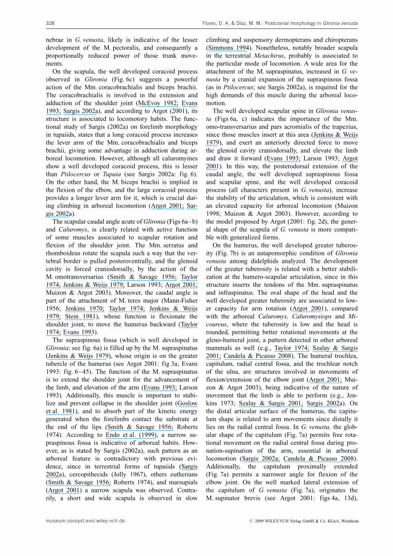
nebrae in G. venusta, likely is indicative of the lesserdevelopment of the M. pectoralis, and consequently aproportionally reduced power of those trunk move-ments.
On the scapula, the well developed coracoid processobserved in Glironia (Fig. 6c) suggests a powerfulaction of the Mm. coracobrachialis and biceps brachii.The coracobrachialis is involved in the extension andadduction of the shoulder joint (McEvoy 1982; Evans1993; Sargis 2002a), and according to Argot (2001), itsstructure is associated to locomotory habits. The func-tional study of Sargis (2002a) on forelimb morphologyin tupaiids, states that a long coracoid process increasesthe lever arm of the Mm. coracobrachialis and bicepsbrachii, giving some advantage in adduction during ar-boreal locomotion. However, although all caluromyinesshow a well developed coracoid process, this is lesserthan Ptilocercus or Tupaia (see Sargis 2002a: fig. 6).On the other hand, the M. biceps brachii is implied inthe flexion of the elbow, and the large coracoid processprovides a longer lever arm for it, which is crucial dur-ing climbing in arboreal locomotion (Argot 2001; Sar-gis 2002a).
The scapular caudal angle acute of Glironia (Figs 6a–b)and Caluromys, is clearly related with active functionof some muscles associated to scapular rotation andflexion of the shoulder joint. The Mm. serratus andrhomboideus rotate the scapula such a way that the ver-tebral border is pulled posteroventrally, and the glenoidcavity is forced craniodorsally, by the action of theM. omotransversarius (Smith & Savage 1956; Taylor1974; Jenkins & Weijs 1979; Larson 1993; Argot 2001;Muizon & Argot 2003). Moreover, the caudal angle ispart of the attachment of M. teres major (Mann-Fisher1956; Jenkins 1970; Taylor 1974; Jenkins & Weijs1979; Stein 1981), whose function is flexionate theshoulder joint, to move the humerus backward (Taylor1974; Evans 1993).
The supraspinous fossa (which is well developed inGlironia; see fig. 6a) is filled up by the M. supraspinatus(Jenkins & Weijs 1979), whose origin is on the greatertubercle of the humerus (see Argot 2001: fig 3a; Evans1993: fig. 6–45). The function of the M. supraspinatusis to extend the shoulder joint for the advancement ofthe limb, and elevation of the arm (Evans 1993; Larson1993). Additionally, this muscle is important to stabi-lize and prevent collapse in the shoulder joint (Goslowet al. 1981), and to absorb part of the kinetic energygenerated when the forelimbs contact the substrate atthe end of the lips (Smith & Savage 1956; Roberts1974). According to Endo et al. (1999), a narrow su-praspinous fossa is indicative of arboreal habits. How-ever, as is stated by Sargis (2002a), such pattern as anarboreal feature is contradictory with previous evi-dence, since in terrestrial forms of tupaiids (Sargis2002a), cercopithecids (Jolly 1967), others eutherians(Smith & Savage 1956; Roberts 1974), and marsupials(Argot 2001) a narrow scapula was observed. Contra-rily, a short and wide scapula is observed in slow
climbing and suspensory dermopterans and chiropterans(Simmons 1994). Nonetheless, notably broader scapulain the terrestrial Metachirus, probably is associated tothe particular mode of locomotion. A wide area for theattachment of the M. supraspinatus, increased in G. ve-nusta by a cranial expansion of the supraspinous fossa(as in Ptilocercus; see Sargis 2002a), is required for thehigh demands of this muscle during the arboreal loco-motion.
The well developed scapular spine in Glironia venus-ta (Figs 6a, c) indicates the importance of the Mm.omo-transversarius and pars acromialis of the trapezius,since those muscles insert at this area (Jenkins & Weijs1979), and exert an anteriorly directed force to movethe glenoid cavity craniodorsally, and elevate the limband draw it forward (Evans 1993; Larson 1993; Argot2001). In this way, the posterodorsal extension of thecaudal angle, the well developed supraspinous fossaand scapular spine, and the well developed coracoidprocess (all characters present in G. venusta), increasethe stability of the articulation, which is consistent withan elevated capacity for arboreal locomotion (Muizon1998; Muizon & Argot 2003). However, according tothe model proposed by Argot (2001: fig. 2d), the gener-al shape of the scapula of G. venusta is more compati-ble with generalized forms.
On the humerus, the well developed greater tuberos-ity (Fig. 7b) is an autapomorphic condition of Glironiavenusta among didelphids analyzed. The developmentof the greater tuberosity is related with a better stabili-zation at the humero-scapular articulation, since in thisstructure inserts the tendons of the Mm. supraspinatusand infraspinatus. The oval shape of the head and thewell developed greater tuberosity are associated to low-er capacity for arm rotation (Argot 2001), comparedwith the arboreal Caluromys, Caluromysiops and Mi-coureus, where the tuberosity is low and the head isrounded, permitting better rotational movements at thegleno-humeral joint, a pattern detected in other arborealmammals as well (e.g., Taylor 1974; Szalay & Sargis2001; Candela & Picasso 2008). The humeral trochlea,capitulum, radial central fossa, and the trochlear notchof the ulna, are structures involved in movements offlexion/extension of the elbow joint (Argot 2001; Mui-zon & Argot 2003), being indicative of the nature ofmovement that the limb is able to perform (e.g., Jen-kins 1973; Szalay & Sargis 2001; Sargis 2002a). Onthe distal articular surface of the humerus, the capitu-lum shape is related to arm movements since distally itlies on the radial central fossa. In G. venusta, the glob-ular shape of the capitulum (Fig. 7a) permits free rota-tional movement on the radial central fossa during pro-nation-supination of the arm, essential in arboreallocomotion (Sargis 2002a; Candela & Picasso 2008).Additionally, the capitulum proximally extended(Fig. 7a) permits a narrower angle for flexion of theelbow joint. On the well marked lateral extension ofthe capitulum of G. venusta (Fig. 7a), originates theM. supinator brevis (see Argot 2001: figs 4a, 13d),
Flores, D. A. & Dıaz, M. M.: Postcranial morphology in Glironia venusta328
museum-zoosyst.evol.wiley-vch.de # 2009 WILEY-VCH Verlag GmbH & Co. KGaA, Weinheim
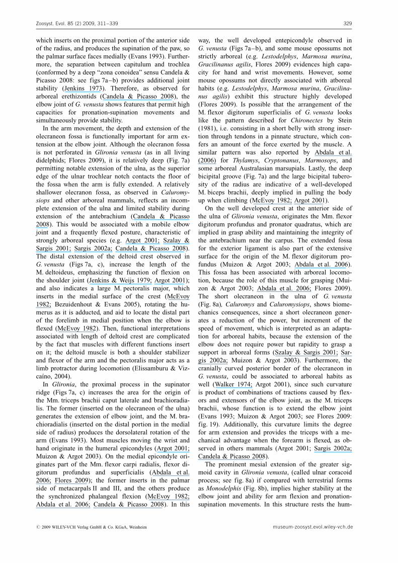
which inserts on the proximal portion of the anterior sideof the radius, and produces the supination of the paw, sothe palmar surface faces medially (Evans 1993). Further-more, the separation between capitulum and trochlea(conformed by a deep “zona conoidea” sensu Candela &Picasso 2008: see figs 7a–b) provides additional jointstability (Jenkins 1973). Therefore, as observed forarboreal erethizontids (Candela & Picasso 2008), theelbow joint of G. venusta shows features that permit highcapacities for pronation-supination movements andsimultaneously provide stability.
In the arm movement, the depth and extension of theolecraneon fossa is functionally important for arm ex-tension at the elbow joint. Although the olecranon fossais not perforated in Glironia venusta (as in all livingdidelphids; Flores 2009), it is relatively deep (Fig. 7a)permitting notable extension of the ulna, as the superioredge of the ulnar trochlear notch contacts the floor ofthe fossa when the arm is fully extended. A relativelyshallower olecranon fossa, as observed in Caluromy-siops and other arboreal mammals, reflects an incom-plete extension of the ulna and limited stability duringextension of the antebrachium (Candela & Picasso2008). This would be associated with a mobile elbowjoint and a frequently flexed posture, characteristic ofstrongly arboreal species (e.g. Argot 2001; Szalay &Sargis 2001; Sargis 2002a; Candela & Picasso 2008).The distal extension of the deltoid crest observed inG. venusta (Figs 7a, c), increase the length of theM. deltoideus, emphasizing the function of flexion onthe shoulder joint (Jenkins & Weijs 1979; Argot 2001);and also indicates a large M. pectoralis major, whichinserts in the medial surface of the crest (McEvoy1982; Bezuidenhout & Evans 2005), rotating the hu-merus as it is adducted, and aid to locate the distal partof the forelimb in medial position when the elbow isflexed (McEvoy 1982). Then, functional interpretationsassociated with length of deltoid crest are complicatedby the fact that muscles with different functions inserton it; the deltoid muscle is both a shoulder stabilizerand flexor of the arm and the pectoralis major acts as alimb protractor during locomotion (Elissamburu & Viz-ca�no, 2004).
In Glironia, the proximal process in the supinatorridge (Figs 7a, c) increases the area for the origin ofthe Mm. triceps brachii caput laterale and brachioradia-lis. The former (inserted on the olecraneon of the ulna)generates the extension of elbow joint, and the M. bra-chioradialis (inserted on the distal portion in the medialside of radius) produces the dorsolateral rotation of thearm (Evans 1993). Most muscles moving the wrist andhand originate in the humeral epicondyles (Argot 2001;Muizon & Argot 2003). On the medial epicondyle ori-ginates part of the Mm. flexor carpi radialis, flexor di-gitorum profundus and superficialis (Abdala et al.2006; Flores 2009); the former inserts in the palmarside of metacarpals II and III, and the others producethe synchronized phalangeal flexion (McEvoy 1982;Abdala et al. 2006; Candela & Picasso 2008). In this
way, the well developed entepicondyle observed inG. venusta (Figs 7a–b), and some mouse opossums notstrictly arboreal (e.g. Lestodelphys, Marmosa murina,Gracilinanus agilis, Flores 2009) evidences high capa-city for hand and wrist movements. However, somemouse opossums not directly associated with atborealhabits (e.g. Lestodelphys, Marmosa murina, Gracilina-nus agilis) exhibit this structure highly developed(Flores 2009). Is possible that the arrangement of theM. flexor digitorum superficialis of G. venusta lookslike the pattern described for Chironectes by Stein(1981), i.e. consisting in a short belly with strong inser-tion through tendons in a pinnate structure, which con-fers an amount of the force exerted by the muscle. Asimilar pattern was also reported by Abdala et al.(2006) for Thylamys, Cryptonanus, Marmosops, andsome arboreal Australasian marsupials. Lastly, the deepbicipital groove (Fig. 7a) and the large bicipital tubero-sity of the radius are indicative of a well-developedM. biceps brachii, deeply implied in pulling the bodyup when climbing (McEvoy 1982; Argot 2001).
On the well developed crest at the anterior side ofthe ulna of Glironia venusta, originates the Mm. flexordigitorum profundus and pronator quadratus, which areimplied in grasp ability and maintaining the integrity ofthe antebrachium near the carpus. The extended fossafor the exterior ligament is also part of the extensivesurface for the origin of the M. flexor digitorum pro-fundus (Muizon & Argot 2003; Abdala et al. 2006).This fossa has been associated with arboreal locomo-tion, because the role of this muscle for grasping (Mui-zon & Argot 2003; Abdala et al. 2006; Flores 2009).The short olecraneon in the ulna of G. venusta(Fig. 8a), Caluromys and Caluromysiops, shows biome-chanics consequences, since a short olecraneon gener-ates a reduction of the power, but increment of thespeed of movement, which is interpreted as an adapta-tion for arboreal habits, because the extension of theelbow does not require power but rapidity to grasp asupport in arboreal forms (Szalay & Sargis 2001; Sar-gis 2002a; Muizon & Argot 2003). Furthermore, thecranially curved posterior border of the olecraneon inG. venusta, could be associated to arboreal habits aswell (Walker 1974; Argot 2001), since such curvatureis product of combinations of tractions caused by flex-ors and extensors of the elbow joint, as the M. tricepsbrachii, whose function is to extend the elbow joint(Evans 1993; Muizon & Argot 2003; see Flores 2009:fig. 19). Additionally, this curvature limits the degreefor arm extension and provides the triceps with a me-chanical advantage when the forearm is flexed, as ob-served in others mammals (Argot 2001; Sargis 2002a;Candela & Picasso 2008).
The prominent mesial extension of the greater sig-moid cavity in Glironia venusta, (called ulnar coracoidprocess; see fig. 8a) if compared with terrestrial formsas Monodelphis (Fig. 8b), implies higher stability at theelbow joint and ability for arm flexion and pronation-supination movements. In this structure rests the hum-
Zoosyst. Evol. 85 (2) 2009, 311–339 329
# 2009 WILEY-VCH Verlag GmbH & Co. KGaA, Weinheim museum-zoosyst.evol.wiley-vch.de
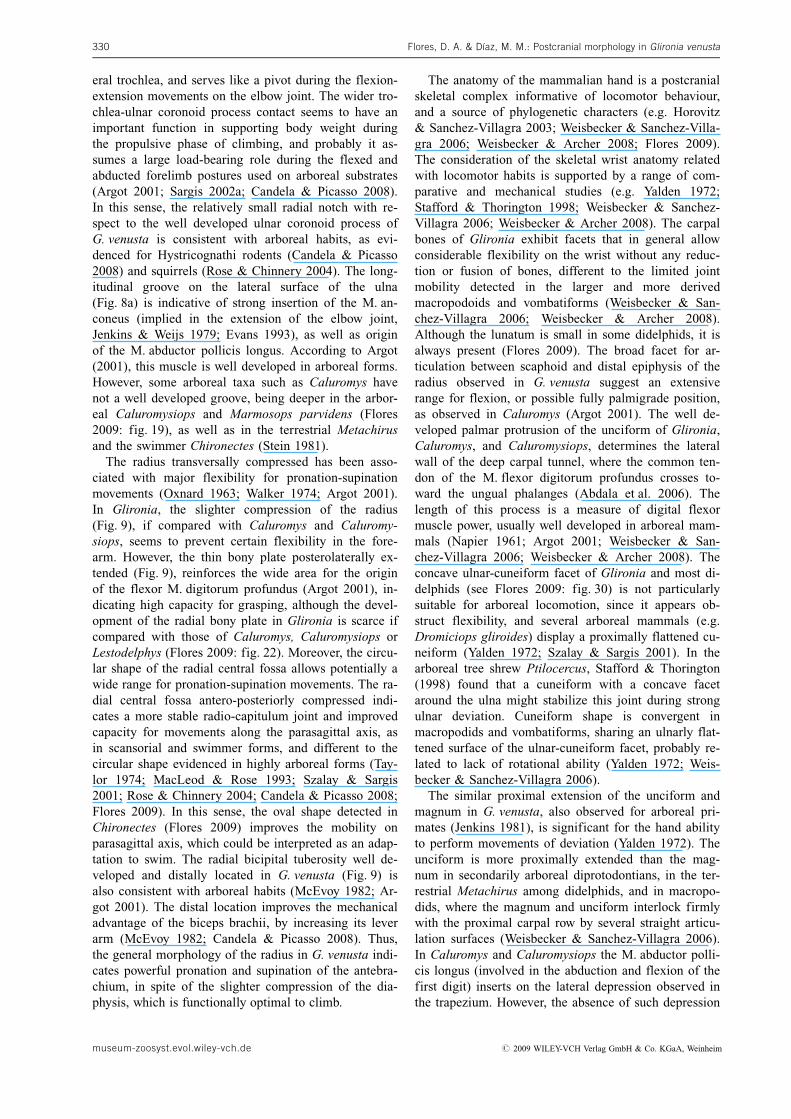
eral trochlea, and serves like a pivot during the flexion-extension movements on the elbow joint. The wider tro-chlea-ulnar coronoid process contact seems to have animportant function in supporting body weight duringthe propulsive phase of climbing, and probably it as-sumes a large load-bearing role during the flexed andabducted forelimb postures used on arboreal substrates(Argot 2001; Sargis 2002a; Candela & Picasso 2008).In this sense, the relatively small radial notch with re-spect to the well developed ulnar coronoid process ofG. venusta is consistent with arboreal habits, as evi-denced for Hystricognathi rodents (Candela & Picasso2008) and squirrels (Rose & Chinnery 2004). The long-itudinal groove on the lateral surface of the ulna(Fig. 8a) is indicative of strong insertion of the M. an-coneus (implied in the extension of the elbow joint,Jenkins & Weijs 1979; Evans 1993), as well as originof the M. abductor pollicis longus. According to Argot(2001), this muscle is well developed in arboreal forms.However, some arboreal taxa such as Caluromys havenot a well developed groove, being deeper in the arbor-eal Caluromysiops and Marmosops parvidens (Flores2009: fig. 19), as well as in the terrestrial Metachirusand the swimmer Chironectes (Stein 1981).
The radius transversally compressed has been asso-ciated with major flexibility for pronation-supinationmovements (Oxnard 1963; Walker 1974; Argot 2001).In Glironia, the slighter compression of the radius(Fig. 9), if compared with Caluromys and Caluromy-siops, seems to prevent certain flexibility in the fore-arm. However, the thin bony plate posterolaterally ex-tended (Fig. 9), reinforces the wide area for the originof the flexor M. digitorum profundus (Argot 2001), in-dicating high capacity for grasping, although the devel-opment of the radial bony plate in Glironia is scarce ifcompared with those of Caluromys, Caluromysiops orLestodelphys (Flores 2009: fig. 22). Moreover, the circu-lar shape of the radial central fossa allows potentially awide range for pronation-supination movements. The ra-dial central fossa antero-posteriorly compressed indi-cates a more stable radio-capitulum joint and improvedcapacity for movements along the parasagittal axis, asin scansorial and swimmer forms, and different to thecircular shape evidenced in highly arboreal forms (Tay-lor 1974; MacLeod & Rose 1993; Szalay & Sargis2001; Rose & Chinnery 2004; Candela & Picasso 2008;Flores 2009). In this sense, the oval shape detected inChironectes (Flores 2009) improves the mobility onparasagittal axis, which could be interpreted as an adap-tation to swim. The radial bicipital tuberosity well de-veloped and distally located in G. venusta (Fig. 9) isalso consistent with arboreal habits (McEvoy 1982; Ar-got 2001). The distal location improves the mechanicaladvantage of the biceps brachii, by increasing its leverarm (McEvoy 1982; Candela & Picasso 2008). Thus,the general morphology of the radius in G. venusta indi-cates powerful pronation and supination of the antebra-chium, in spite of the slighter compression of the dia-physis, which is functionally optimal to climb.
The anatomy of the mammalian hand is a postcranialskeletal complex informative of locomotor behaviour,and a source of phylogenetic characters (e.g. Horovitz& Sanchez-Villagra 2003; Weisbecker & Sanchez-Villa-gra 2006; Weisbecker & Archer 2008; Flores 2009).The consideration of the skeletal wrist anatomy relatedwith locomotor habits is supported by a range of com-parative and mechanical studies (e.g. Yalden 1972;Stafford & Thorington 1998; Weisbecker & Sanchez-Villagra 2006; Weisbecker & Archer 2008). The carpalbones of Glironia exhibit facets that in general allowconsiderable flexibility on the wrist without any reduc-tion or fusion of bones, different to the limited jointmobility detected in the larger and more derivedmacropodoids and vombatiforms (Weisbecker & San-chez-Villagra 2006; Weisbecker & Archer 2008).Although the lunatum is small in some didelphids, it isalways present (Flores 2009). The broad facet for ar-ticulation between scaphoid and distal epiphysis of theradius observed in G. venusta suggest an extensiverange for flexion, or possible fully palmigrade position,as observed in Caluromys (Argot 2001). The well de-veloped palmar protrusion of the unciform of Glironia,Caluromys, and Caluromysiops, determines the lateralwall of the deep carpal tunnel, where the common ten-don of the M. flexor digitorum profundus crosses to-ward the ungual phalanges (Abdala et al. 2006). Thelength of this process is a measure of digital flexormuscle power, usually well developed in arboreal mam-mals (Napier 1961; Argot 2001; Weisbecker & San-chez-Villagra 2006; Weisbecker & Archer 2008). Theconcave ulnar-cuneiform facet of Glironia and most di-delphids (see Flores 2009: fig. 30) is not particularlysuitable for arboreal locomotion, since it appears ob-struct flexibility, and several arboreal mammals (e.g.Dromiciops gliroides) display a proximally flattened cu-neiform (Yalden 1972; Szalay & Sargis 2001). In thearboreal tree shrew Ptilocercus, Stafford & Thorington(1998) found that a cuneiform with a concave facetaround the ulna might stabilize this joint during strongulnar deviation. Cuneiform shape is convergent inmacropodids and vombatiforms, sharing an ulnarly flat-tened surface of the ulnar-cuneiform facet, probably re-lated to lack of rotational ability (Yalden 1972; Weis-becker & Sanchez-Villagra 2006).
The similar proximal extension of the unciform andmagnum in G. venusta, also observed for arboreal pri-mates (Jenkins 1981), is significant for the hand abilityto perform movements of deviation (Yalden 1972). Theunciform is more proximally extended than the mag-num in secondarily arboreal diprotodontians, in the ter-restrial Metachirus among didelphids, and in macropo-dids, where the magnum and unciform interlock firmlywith the proximal carpal row by several straight articu-lation surfaces (Weisbecker & Sanchez-Villagra 2006).In Caluromys and Caluromysiops the M. abductor polli-cis longus (involved in the abduction and flexion of thefirst digit) inserts on the lateral depression observed inthe trapezium. However, the absence of such depression
Flores, D. A. & Dıaz, M. M.: Postcranial morphology in Glironia venusta330
museum-zoosyst.evol.wiley-vch.de # 2009 WILEY-VCH Verlag GmbH & Co. KGaA, Weinheim
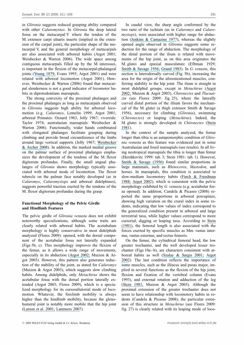
in Glironia suggests reduced grasping ability comparedwith other Caluromyines. In Glironia the deep lateralfossa on the metacarpal V where the tendon of theM. extensor carpi ulnaris inserts (implied in the exten-sion of the carpal joint), the particular shape of the me-tacarpal V, and the general morphology of metacarpalsare also associated with arboreal habits (Argot 2001;Weisbecker & Warton 2006). The wide space amongcontiguous metacarpals filled up by the M. interossei,is important in the flexion of the metacarpal-phalangealjoints (Young 1879; Evans 1993; Argot 2001) and wererelated with arboreal locomotion (Argot 2001). How-ever, Weisbecker & Warton (2006) found that metacar-pal slenderness is not a good indicator of locomotor ha-bits in diprotodontians marsupials.
The strong convexity of the proximal phalanges andthe proximal phalanges as long as metacarpals observedin Glironia suggests high ability for arboreal loco-motion (e.g. Caluromys, Lemelin 1999; Argot 2001;arboreal Primates: Oxnard 1963, Jolly 1967; viverrids:Taylor 1974; australasian marsupials: Weisbecker &Warton 2006). Functionally, wider hands combinatedwith elongated phalanges facilitate grasping duringclimbing and provide broad circumference of the limbsaround large vertical supports (Jolly 1967; Weisbecker& Archer 2008). In addition, the marked medial grooveon the palmar surface of proximal phalanges empha-sizes the development of the tendons of the M. flexordigitorum profundus. Finally, the small ungual pha-langes of Glironia shows morphology typically asso-ciated with arboreal mode of locomotion. The flexortubercle on the palmar face notably developed (as inCaluromys, Caluromysiops and arboreal didelphines),suggests powerful traction exerted by the tendons of theM. flexor digitorum profundus during the grasp.
Functional Morphology of the Pelvic Girdleand Hindlimb Features
The pelvic girdle of Glironia venusta does not exhibitnoteworthy specializations, although some traits areclearly related with arboreal habits. The acetabulummorphology is highly conservative in most didelphidsanalyzed (Flores 2009): opened, with the dorsal compo-nent of the acetabular fossa not laterally expanded(Figs 5b, c). This morphology improves the flexion ofthe femur, as it allows a wide range of movements,especially in its abduction (Argot 2002; Muizon & Ar-got 2003). However, this pattern also generates reduc-tion of the stability of the joint, as stated for Caluromys(Muizon & Argot 2003), which suggests slow climbinghabits. Among didelphids, only Metachirus shows theacetabular fossa with the dorsal portion laterally ex-tended (Argot 2003; Flores 2009), which is a specia-lized morphology for its cursosaltatorial mode of loco-motion. Whatever, the forelimb mobility is alwayshigher than the hindlimb mobility, because the gleno-humeral joint is notably more mobile that the hip joint(Larson et al. 2001; Lammers 2007).
In caudal view, the sharp angle conformed by thetwo rami of the ischium (as in Caluromys and Caluro-mysiops), were associated with higher range for abduc-tion (Jenkins & Camazine 1977), whereas the slightlyopened angle observed in Glironia suggests some re-duction for the range of abduction. The morphology ofthe distal portion of the ilium is related with move-ments of the hip joint, as on this area originates theM. glutei and epaxial musculature (Efltman 1929;Smith & Savage 1956; Grand 1983). In G. venusta, thissection is laterodorsally curved (Fig. 5b), increasing thearea for the origin of the aforementioned muscles, con-ferring stability to the hip joint. The ilium is straight inmost didelphid groups, except in Metachirus (Argot2002; Muizon & Argot 2003), Chironectes and Tlacuat-zin (see Flores 2009: fig. 25), where the laterallycurved distal portion of the illium favors the mechani-cal of the M. glutei (a thigh extensor Smith & Savage1956), necessary for climbing (Glironia), swimming(Chironectes) or leaping (Metachirus). Indeed, theM. glutei is strongly developed in Chironectes (Stein1981).
In the context of the sample analyzed, the femurlonger than tibia is an autapomorphic condition of Gliro-nia venusta as this feature was evidenced just in someAustralasian and fossil marsupials (see results). In all liv-ing neotropical marsupials the tibia is longer than femur(Hershkovitz 1999: tab. 3; Stein 1981: tab. 1). However,Smith & Savage (1956) found similar proportions inlarge mammals, such as Rhinoceros, Mastodon, andhorses. In marsupials, this condition is associated toslow-medium locomotory habits (Finch & Freedman1988; Argot 2002), which is consistent with the pelvicmorphology exhibited by G. venusta (e.g. acetabular fos-sa opened). In addition, Candela & Picasso (2008) re-ported the same proportions in arboreal porcupines,showing high variation on the crural index in some ro-dents, indicating that low values of index correspond tothe generalized condition present in arboreal and largeterrestrial taxa, while higher values correspond to morecursorial, digging or leaping taxa. According to Stein(1981), the femoral length is also associated with theforces exerted by specific muscles as Mm. vastus inter-nus, vastus externus, and rectus femoris.
On the femur, the cylindrical femoral head, the lowgreater trochanter, and the well developed lesser tro-chanter (Figs 10a–b), are characters consistent with ar-boreal habits as well (Szalay & Sargis 2001; Argot2002). The last condition reflects the importance ofsome muscles, such as the illiacus and psoas major, im-plied in several functions as the flexion of the hip joint,flexion and fixation of the vertebral column (Evans1993), and external rotation and adduction of the leg(Stein 1981; Muizon & Argot 2003). Although theproximal extension of the greater trochanter does notseem to have relationship with locomotory habits in ro-dents (Candela & Picasso 2008); the particular exten-sion of this structure in Metachirus (see Flores 2009:fig. 27) is clearly related with its leaping mode of loco-
Zoosyst. Evol. 85 (2) 2009, 311–339 331
# 2009 WILEY-VCH Verlag GmbH & Co. KGaA, Weinheim museum-zoosyst.evol.wiley-vch.de
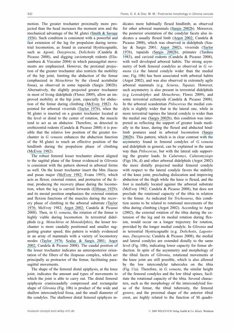
motion. The greater trochanter proximally more pro-jected than the head increases the moment arm and themechanical advantage of the M. glutei (Smith & Savage1956). Such condition is consistent with a powerful andfast extension of the leg for propulsion during terres-trial locomotion, as found in cursorial Hystricognathi,such as Agouti, Dasyprocta, Dolichotis (Candela &Picasso 2008), and digging caviomorph rodents (Elis-samburu & Vizcaino 2004) in which parasagittal move-ments are emphasized. However, the proximal projec-tion of the greater trochanter also restricts the mobilityof the hip joint, limiting the abduction of the femur(emphasized in Metachirus by the closed acetabularfossa), as observed in some tupaiids (Sargis 2002b).Alternatively, the slightly projected greater trochanterin most of living didelphids (Flores 2009), allow an im-proved mobility at the hip joint, especially for abduc-tion of the femur during climbing (McEvoy 1982). Aspointed for arboreal viverrids (Taylor 1976), when theM. glutei is inserted on a greater trochanter located atthe level or distal to the center of rotation, the muscletend to act as an abductor. Therefore, as detected inerethizontid rodents (Candela & Picasso 2008) it is pos-sible that the relative low position of the greater tro-chanter in G. venusta enhances the abduction functionof the M. glutei to reach an effective position of thehindlimb during the propulsion phase of climbing(McEvoy 1982).
The robust femoral lesser trochanter almost alignedto the sagittal plane of the femur evidenced in Glironiais consistent with the pattern showed in arboreal formsas well. On the lesser trochanter insert the Mm. iliacusand psoas major (McEvoy 1982; Evans 1993), whichacts as flexor, external rotator, and protractor of the fe-mur, producing the recovery phase during the locomo-tion, when the leg is carried forwards (Elftman 1929);and its mesial position emphasizes the external rotationand flexion functions of the muscles during the recov-ery phase of climbing in the arboreal substrate (Taylor1976; McEvoy 1982; Argot 2002; Candela & Picasso2008). Then, in G. venusta, the rotation of the femur ishighly viable during locomotion. In terrestrial didel-phids (e.g. Metachirus or Monodelphis), the lesser tro-chanter is more caudally positioned and smaller sug-gesting greater speed; this pattern is widely evidencedin an array of mammals with a variety of locomotorymodes (Taylor 1976; Szalay & Sargis 2001; Argot2002; Candela & Picasso 2008). The caudal position ofthe lesser trochanter indicates an anteroposterior orien-tation of the fibers of the iliopsoas complex, which actprincipally as protractor of the femur, facilitating para-sagittal movements.
The shape of the femoral distal epiphysis, at the kneejoint, indicates the amount and types of movements towhich the joint is able to carry out. The distal femoralepiphysis craniocaudally compressed and rectangularshape of Glironia (Fig. 10b) is product of the wide andshallow intercondyloid fossa, and the small diameter ofthe condyles. The shallower distal femoral epiphysis in-
dicates more habitually flexed hindlimb, as observedfor other arboreal mammals (Sargis 2002b). Moreover,the posterior orientation of the condylar facets also in-dicates a usually flexed limb (Argot 2002; Candela &Picasso 2008), which was observed in didelphids (Sza-lay & Sargis 2001; Argot 2002), viverrids (Taylor1976), tupaiids (Sargis 2002b), primates (Tardieu1983), and cavioid rodents (Candela & Picasso 2008),with well developed arboreal habits. The strong asym-metry of both femoral condyles as observed in G. ve-nusta (i.e the lateral condyle wider than the medialone; Fig. 10b) has been associated with arboreal habits(Argot 2002), and was also observed in extremely agilearboreal mammals (e.g. Sciurus, Tupaia). However,such asymmetry is also present in terrestrial didelphids(e.g Lestodelphys and Metachirus; Flores 2009), andsome terrestrial echimyids (Candela & Picasso 2008).In the arboreal scandentian Ptilocercus the medial con-dyle is slightly wider that in the lateral one, while inmore terrestrial tupines the lateral condyle is wider thanthe medial one (Sargis 2002b), this condition was inter-preted as reflecting the support of greater loads medi-ally in the knee, during the flexed and abducted hind-limb postures used in arboreal locomotion (Sargis2002b). This pattern, which is in clear contrast with theasymmetry found in femoral condyles of G. venustaand didelphids in general, can be explained in the sameway than Ptilocercus, but with the lateral side support-ing the greater loads. In Caluromys, Caluromysiops(Figs 10c, d) and other arboreal didelphids (Argot 2002)the more distally projected medial femoral condylewith respect to the lateral condyle favors the stabilityof the knee joint, precluding dislocation and improvingabduction of the thigh while the knee is flexed, and thefoot is medially located against the arboreal substrate(McEvoy 1982; Candela & Picasso 2008), but does notpreclude the rotational capacity of the tibia in relationto the femur. As indicated for Trichosurus, this condi-tion seems to be related to rotational movements of thetibia during climbing (Argot 2002). According to Argot(2002), the external rotation of the tibia during the ex-tension of the leg and its medial rotation during flex-ion, would occur on a longer trajectory for rotation,provided by the longer medial condyle. In Glironia andin terrestrial Hystricognathi (e.g. Dolichotis, Lagosto-mus, Dasyprocta; Candela & Picasso 2008), the medialand lateral condyles are extended distally to the samelevel (Fig. 10b), indicating lower capacity for femur ab-duction. In spite of the asymmetry and morphology ofthe tibial facets of Glironia, rotational movements atthe knee joint are still possible, which is also allowedby the low intercondylar tubercules on the tibia(Fig. 11a). Therefore, in G. venusta, the similar heightof the femoral condyles and the low tibial spines, facil-itate the rotational capacity of the tibia. Several charac-ters, such as the morphology of the intercondyloid fos-sa of the femur, the tibial tuberosity, the femoralgroove, and the proximal shape of the anterior tibialcrest, are highly related to the function of M. quadri-
Flores, D. A. & Dıaz, M. M.: Postcranial morphology in Glironia venusta332
museum-zoosyst.evol.wiley-vch.de # 2009 WILEY-VCH Verlag GmbH & Co. KGaA, Weinheim

ceps femoris during the locomotion (Muizon 1998;Szalay & Sargis 2001; Argot 2002). The femoralgroove for the M. quadriceps femoris (a powerful ex-tensor of the knee) is notably flat in G. venusta(Fig. 10a), which is typical of fast running forms, beingmore concave in slower arboreal species (e.g. Caluro-mys and Caluromysiops; see Fig. 10c). This demon-strates that, as stated by Muizon & Argot (2003), themorphology of the femoral distal epiphysis is more re-lated to the activity (slow vs. fast) than behavioral loco-motion (terrestrial vs arboreal; Tardieu 1983).
The sigmoid shape of the tibia is a state sharedamong most didelphids, and according to Szalay & Sar-gis (2001) it is a plesiomorphic condition in marsu-pials. Even when the curvature observed in G. venustais less conspicuous than the aquatic Chironectes (seeFlores 2009: fig. 28), the contact between tibia and fi-bula is restricted to the proximal and distal portion ofthe diaphyses, which confers some capacity for rotationbetween both bones, necessary during arboreal locomo-tion. However, both bones are provided by strong liga-ments that reinforce their interconnection (Stein 1981;Argot 2002). The wide space between tibia and fibularelates to the development of the M. flexor digitorumfibularis (Stein 1981), important in arboreal forms,since the digital flexion on the hindlimb is steadilyused to force the claws into irregularities in the sub-strate during climbing and to grasp branches (McEvoy1982). The weak tibial tuberosity observed in G. venus-ta seems to indicate arboreal habits, since other arbor-eal caluromyines and didelphines (e.g. Caluromys, Ca-luromysiops, Micoureus), exhibit this structure flat andnot cranially expanded and in terrestrial forms the tu-berosity is prominent (Muizon & Argot 2003; Flores2009). The tendon for the M. quadriceps femoris insertsdirectly on the tibial tuberosity, as the patella is absentin didelphids (Szalay & Sargis 2001; Argot 2002).Equally to the asymmetry exhibited by femoral con-dyles, the tibial condyles of G. venusta show strongasymmetry in shape and size (Fig. 11a). The most pro-nounced difference between both tibial condyles is ob-served in Metachirus implying increment of the stabi-lity on the articulation in the terrestrial form. In thissense, the lesser stabilized joint between femur andtibia of G. venusta, permits independent rotation of thetibia in relation to the femur, which is necessary forchanges of position, whereas the foot is fixed (seeabove). Additionally, an evident triangular area on themedial side of the tibia is present in Caluromys andCaluromysiops, where inserts strong ligaments comingfrom the medial femoral condyle, fixing the tibia to fe-mur during the rotation. The development and exten-sion of this structure in G. venusta is less than in Ca-luromys and Caluromysiops, suggesting reducedmechanical stress likely because the lesser body size ofG. venusta. Such ligaments are also present in other ar-boreal mammals, as primates (Tardieu 1983), andSciuridae (Haines 1942). According to Argot (2002),the rotation between tibia and femur is emphasized by
the slight lateral torsion of the distal epiphysis of thefemur in Caluromys. However, we did not observe suchtorsion in Caluromyines.
The M. gracilis, one of the main adductors of the hipand a powerful flexor of the knee, is inserted on thetibial crest (McEvoy 1982; Evans 1993). In arborealmammals, as extant Erehizontidae, the well developedgracilis is associated with a powerful adduction of thehindlimb during climbing (McEvoy 1982; Candela &Picasso 2008). The long moment arm of the gracilis, asconsequence of its distal insertion on the tibial crest,increases the mechanical advantage of this muscle(Candela & Picasso 2008). The well developed anteriortibial crest in Glironia is indicative of faster mode oflocomotion (Muizon 1998; Szalay & Sargis 2001). InCaluromys and Caluromysiops, the area for insertion ofM. gracilis seems to be more distally located than inG. venusta, and this aparent well developed muscle isconsistent with the larger body size of those speciescompared with G. venusta. In addition, the distal exten-sion of the tibial crest indicates distal insertion of ham-string muscles, which implies long moment arms, gen-erating powerful movement of the leg at the beginningof propulsion (Elftman 1929; Smith & Savage 1956;Candela & Picasso 2008). The more proximal insertionof the M. semitendinosus on the tibia is associated togreater speed of the initial phase of step in caviomorphrodents (Elissamburu & Vizcaino 2004) and marsupials(Elftman 1929). In agreement with the relatively welldeveloped and distally projected tibial crest of G. ve-nusta, the extensive lateral area for the origin of thetibial portion of the M. tibialis anterior is evident, sug-gesting high importance of this muscle inverting thefoot to locate its plantar side against the arboreal sub-strate (McEvoy 1982; Evans 1993). However, the graspcapacity of the foot seems to be partially limited inG. venusta, which is inferred by the poorly developedarea for the origin of the M. flexor digitorum tibialis,although this function could be substituted by the welldeveloped M. flexor digitorum fibularis (see above). Inagreement with the broad contact between tibia and as-tragalus, the cruro-astragalar joint of G. venusta ishighly mobile, as in most of arboreal marsupials (Sza-lay 1994). The elongated and crescent shaped lateralastragalotibial facet of G. venusta facilitates the rota-tional movement of the foot during dorsiflexion-plantar-flexion movements, a condition also evidenced in ar-boreal rodents (e.g. Coendou, Candela & Picasso2008). The well developed and flattened posterior andmedial astragalotibial facets of the tibia, permits a widerange of movements at the cruro-astragalar joint. More-over, intercondylar ridges between the facets are notevidenced, which add mobility to the joint, as observedin arboreal marsupials (Szalay 1994; Szalay & Sargis2001; Argot 2002) and rodents (Candela & Picasso2008). Alternatively, the slightly concave shape of thecruro-astragalar facets of some terrestrial didelphids(e.g. Metachirus, Lestodelphys) limits the lateral move-ments, because the articular surface of the tibia shows
Zoosyst. Evol. 85 (2) 2009, 311–339 333
# 2009 WILEY-VCH Verlag GmbH & Co. KGaA, Weinheim museum-zoosyst.evol.wiley-vch.de
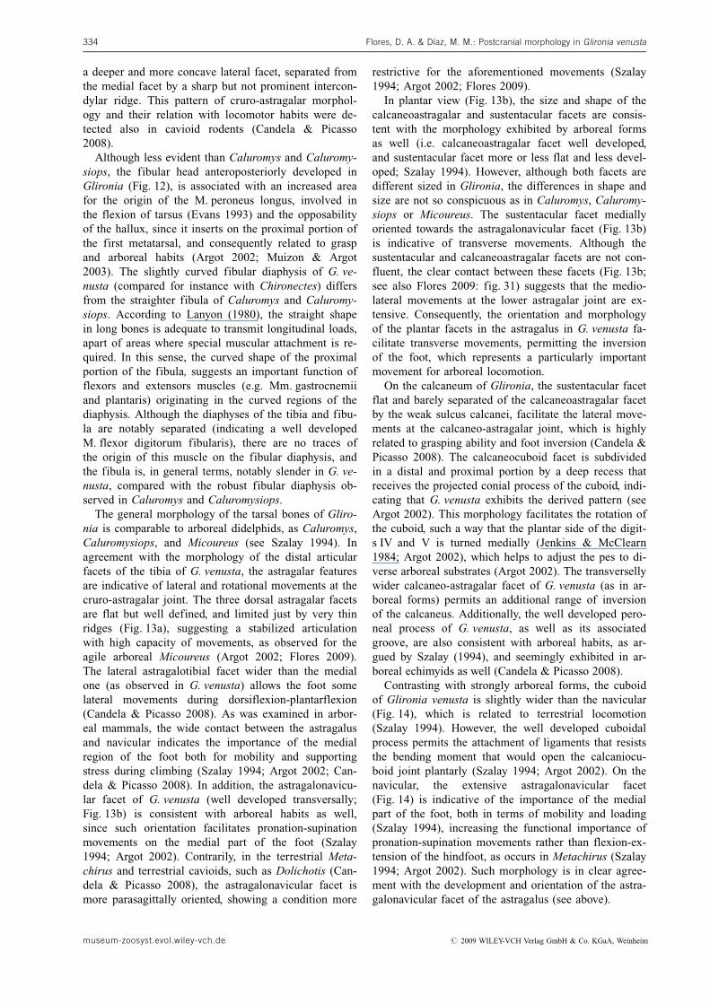
a deeper and more concave lateral facet, separated fromthe medial facet by a sharp but not prominent intercon-dylar ridge. This pattern of cruro-astragalar morphol-ogy and their relation with locomotor habits were de-tected also in cavioid rodents (Candela & Picasso2008).
Although less evident than Caluromys and Caluromy-siops, the fibular head anteroposteriorly developed inGlironia (Fig. 12), is associated with an increased areafor the origin of the M. peroneus longus, involved inthe flexion of tarsus (Evans 1993) and the opposabilityof the hallux, since it inserts on the proximal portion ofthe first metatarsal, and consequently related to graspand arboreal habits (Argot 2002; Muizon & Argot2003). The slightly curved fibular diaphysis of G. ve-nusta (compared for instance with Chironectes) differsfrom the straighter fibula of Caluromys and Caluromy-siops. According to Lanyon (1980), the straight shapein long bones is adequate to transmit longitudinal loads,apart of areas where special muscular attachment is re-quired. In this sense, the curved shape of the proximalportion of the fibula, suggests an important function offlexors and extensors muscles (e.g. Mm. gastrocnemiiand plantaris) originating in the curved regions of thediaphysis. Although the diaphyses of the tibia and fibu-la are notably separated (indicating a well developedM. flexor digitorum fibularis), there are no traces ofthe origin of this muscle on the fibular diaphysis, andthe fibula is, in general terms, notably slender in G. ve-nusta, compared with the robust fibular diaphysis ob-served in Caluromys and Caluromysiops.
The general morphology of the tarsal bones of Gliro-nia is comparable to arboreal didelphids, as Caluromys,Caluromysiops, and Micoureus (see Szalay 1994). Inagreement with the morphology of the distal articularfacets of the tibia of G. venusta, the astragalar featuresare indicative of lateral and rotational movements at thecruro-astragalar joint. The three dorsal astragalar facetsare flat but well defined, and limited just by very thinridges (Fig. 13a), suggesting a stabilized articulationwith high capacity of movements, as observed for theagile arboreal Micoureus (Argot 2002; Flores 2009).The lateral astragalotibial facet wider than the medialone (as observed in G. venusta) allows the foot somelateral movements during dorsiflexion-plantarflexion(Candela & Picasso 2008). As was examined in arbor-eal mammals, the wide contact between the astragalusand navicular indicates the importance of the medialregion of the foot both for mobility and supportingstress during climbing (Szalay 1994; Argot 2002; Can-dela & Picasso 2008). In addition, the astragalonavicu-lar facet of G. venusta (well developed transversally;Fig. 13b) is consistent with arboreal habits as well,since such orientation facilitates pronation-supinationmovements on the medial part of the foot (Szalay1994; Argot 2002). Contrarily, in the terrestrial Meta-chirus and terrestrial cavioids, such as Dolichotis (Can-dela & Picasso 2008), the astragalonavicular facet ismore parasagittally oriented, showing a condition more
restrictive for the aforementioned movements (Szalay1994; Argot 2002; Flores 2009).
In plantar view (Fig. 13b), the size and shape of thecalcaneoastragalar and sustentacular facets are consis-tent with the morphology exhibited by arboreal formsas well (i.e. calcaneoastragalar facet well developed,and sustentacular facet more or less flat and less devel-oped; Szalay 1994). However, although both facets aredifferent sized in Glironia, the differences in shape andsize are not so conspicuous as in Caluromys, Caluromy-siops or Micoureus. The sustentacular facet mediallyoriented towards the astragalonavicular facet (Fig. 13b)is indicative of transverse movements. Although thesustentacular and calcaneoastragalar facets are not con-fluent, the clear contact between these facets (Fig. 13b;see also Flores 2009: fig. 31) suggests that the medio-lateral movements at the lower astragalar joint are ex-tensive. Consequently, the orientation and morphologyof the plantar facets in the astragalus in G. venusta fa-cilitate transverse movements, permitting the inversionof the foot, which represents a particularly importantmovement for arboreal locomotion.
On the calcaneum of Glironia, the sustentacular facetflat and barely separated of the calcaneoastragalar facetby the weak sulcus calcanei, facilitate the lateral move-ments at the calcaneo-astragalar joint, which is highlyrelated to grasping ability and foot inversion (Candela &Picasso 2008). The calcaneocuboid facet is subdividedin a distal and proximal portion by a deep recess thatreceives the projected conial process of the cuboid, indi-cating that G. venusta exhibits the derived pattern (seeArgot 2002). This morphology facilitates the rotation ofthe cuboid, such a way that the plantar side of the digit-s IV and V is turned medially (Jenkins & McClearn1984; Argot 2002), which helps to adjust the pes to di-verse arboreal substrates (Argot 2002). The transversellywider calcaneo-astragalar facet of G. venusta (as in ar-boreal forms) permits an additional range of inversionof the calcaneus. Additionally, the well developed pero-neal process of G. venusta, as well as its associatedgroove, are also consistent with arboreal habits, as ar-gued by Szalay (1994), and seemingly exhibited in ar-boreal echimyids as well (Candela & Picasso 2008).
Contrasting with strongly arboreal forms, the cuboidof Glironia venusta is slightly wider than the navicular(Fig. 14), which is related to terrestrial locomotion(Szalay 1994). However, the well developed cuboidalprocess permits the attachment of ligaments that resiststhe bending moment that would open the calcaniocu-boid joint plantarly (Szalay 1994; Argot 2002). On thenavicular, the extensive astragalonavicular facet(Fig. 14) is indicative of the importance of the medialpart of the foot, both in terms of mobility and loading(Szalay 1994), increasing the functional importance ofpronation-supination movements rather than flexion-ex-tension of the hindfoot, as occurs in Metachirus (Szalay1994; Argot 2002). Such morphology is in clear agree-ment with the development and orientation of the astra-galonavicular facet of the astragalus (see above).
Flores, D. A. & Dıaz, M. M.: Postcranial morphology in Glironia venusta334
museum-zoosyst.evol.wiley-vch.de # 2009 WILEY-VCH Verlag GmbH & Co. KGaA, Weinheim
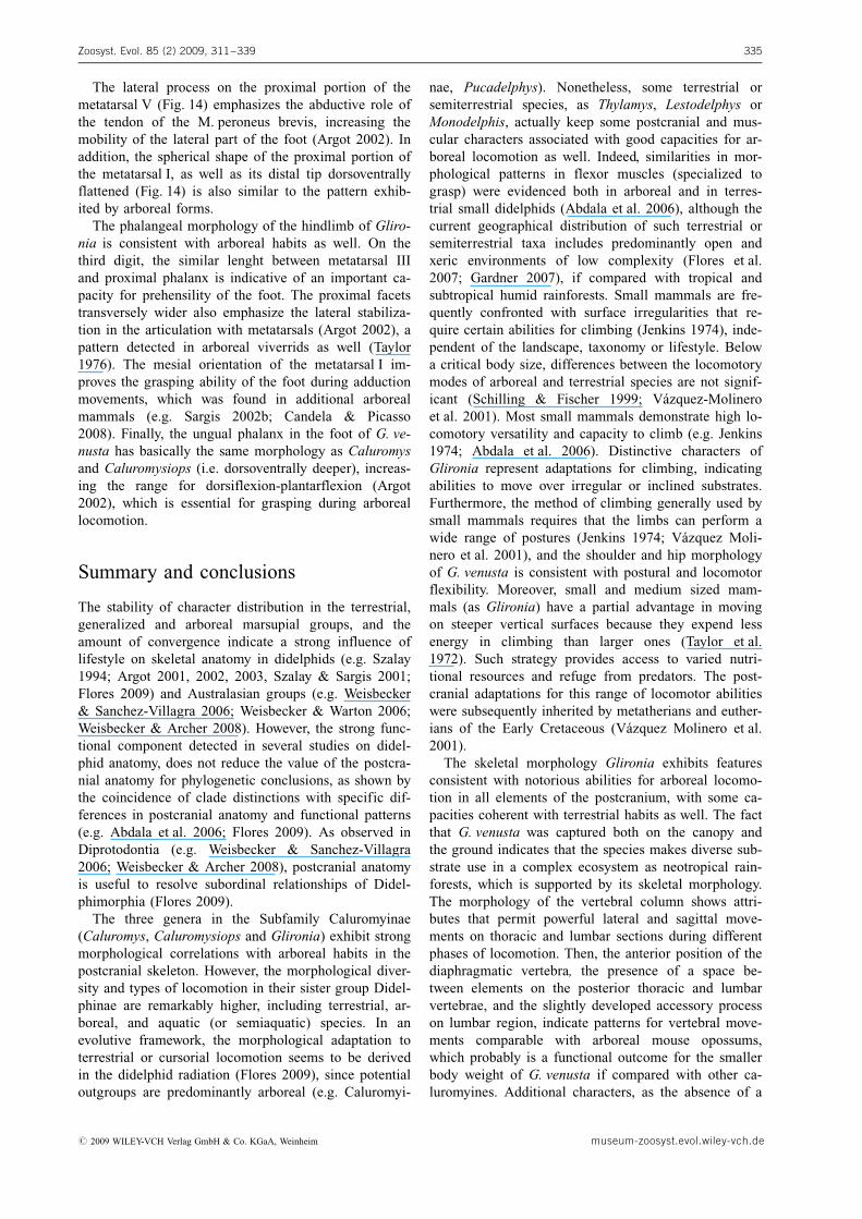
The lateral process on the proximal portion of themetatarsal V (Fig. 14) emphasizes the abductive role ofthe tendon of the M. peroneus brevis, increasing themobility of the lateral part of the foot (Argot 2002). Inaddition, the spherical shape of the proximal portion ofthe metatarsal I, as well as its distal tip dorsoventrallyflattened (Fig. 14) is also similar to the pattern exhib-ited by arboreal forms.
The phalangeal morphology of the hindlimb of Gliro-nia is consistent with arboreal habits as well. On thethird digit, the similar lenght between metatarsal IIIand proximal phalanx is indicative of an important ca-pacity for prehensility of the foot. The proximal facetstransversely wider also emphasize the lateral stabiliza-tion in the articulation with metatarsals (Argot 2002), apattern detected in arboreal viverrids as well (Taylor1976). The mesial orientation of the metatarsal I im-proves the grasping ability of the foot during adductionmovements, which was found in additional arborealmammals (e.g. Sargis 2002b; Candela & Picasso2008). Finally, the ungual phalanx in the foot of G. ve-nusta has basically the same morphology as Caluromysand Caluromysiops (i.e. dorsoventrally deeper), increas-ing the range for dorsiflexion-plantarflexion (Argot2002), which is essential for grasping during arboreallocomotion.
Summary and conclusions
The stability of character distribution in the terrestrial,generalized and arboreal marsupial groups, and theamount of convergence indicate a strong influence oflifestyle on skeletal anatomy in didelphids (e.g. Szalay1994; Argot 2001, 2002, 2003, Szalay & Sargis 2001;Flores 2009) and Australasian groups (e.g. Weisbecker& Sanchez-Villagra 2006; Weisbecker & Warton 2006;Weisbecker & Archer 2008). However, the strong func-tional component detected in several studies on didel-phid anatomy, does not reduce the value of the postcra-nial anatomy for phylogenetic conclusions, as shown bythe coincidence of clade distinctions with specific dif-ferences in postcranial anatomy and functional patterns(e.g. Abdala et al. 2006; Flores 2009). As observed inDiprotodontia (e.g. Weisbecker & Sanchez-Villagra2006; Weisbecker & Archer 2008), postcranial anatomyis useful to resolve subordinal relationships of Didel-phimorphia (Flores 2009).
The three genera in the Subfamily Caluromyinae(Caluromys, Caluromysiops and Glironia) exhibit strongmorphological correlations with arboreal habits in thepostcranial skeleton. However, the morphological diver-sity and types of locomotion in their sister group Didel-phinae are remarkably higher, including terrestrial, ar-boreal, and aquatic (or semiaquatic) species. In anevolutive framework, the morphological adaptation toterrestrial or cursorial locomotion seems to be derivedin the didelphid radiation (Flores 2009), since potentialoutgroups are predominantly arboreal (e.g. Caluromyi-
nae, Pucadelphys). Nonetheless, some terrestrial orsemiterrestrial species, as Thylamys, Lestodelphys orMonodelphis, actually keep some postcranial and mus-cular characters associated with good capacities for ar-boreal locomotion as well. Indeed, similarities in mor-phological patterns in flexor muscles (specialized tograsp) were evidenced both in arboreal and in terres-trial small didelphids (Abdala et al. 2006), although thecurrent geographical distribution of such terrestrial orsemiterrestrial taxa includes predominantly open andxeric environments of low complexity (Flores et al.2007; Gardner 2007), if compared with tropical andsubtropical humid rainforests. Small mammals are fre-quently confronted with surface irregularities that re-quire certain abilities for climbing (Jenkins 1974), inde-pendent of the landscape, taxonomy or lifestyle. Belowa critical body size, differences between the locomotorymodes of arboreal and terrestrial species are not signif-icant (Schilling & Fischer 1999; V�zquez-Molineroet al. 2001). Most small mammals demonstrate high lo-comotory versatility and capacity to climb (e.g. Jenkins1974; Abdala et al. 2006). Distinctive characters ofGlironia represent adaptations for climbing, indicatingabilities to move over irregular or inclined substrates.Furthermore, the method of climbing generally used bysmall mammals requires that the limbs can perform awide range of postures (Jenkins 1974; V�zquez Moli-nero et al. 2001), and the shoulder and hip morphologyof G. venusta is consistent with postural and locomotorflexibility. Moreover, small and medium sized mam-mals (as Glironia) have a partial advantage in movingon steeper vertical surfaces because they expend lessenergy in climbing than larger ones (Taylor et al.1972). Such strategy provides access to varied nutri-tional resources and refuge from predators. The post-cranial adaptations for this range of locomotor abilitieswere subsequently inherited by metatherians and euther-ians of the Early Cretaceous (V�zquez Molinero et al.2001).
The skeletal morphology Glironia exhibits featuresconsistent with notorious abilities for arboreal locomo-tion in all elements of the postcranium, with some ca-pacities coherent with terrestrial habits as well. The factthat G. venusta was captured both on the canopy andthe ground indicates that the species makes diverse sub-strate use in a complex ecosystem as neotropical rain-forests, which is supported by its skeletal morphology.The morphology of the vertebral column shows attri-butes that permit powerful lateral and sagittal move-ments on thoracic and lumbar sections during differentphases of locomotion. Then, the anterior position of thediaphragmatic vertebra, the presence of a space be-tween elements on the posterior thoracic and lumbarvertebrae, and the slightly developed accessory processon lumbar region, indicate patterns for vertebral move-ments comparable with arboreal mouse opossums,which probably is a functional outcome for the smallerbody weight of G. venusta if compared with other ca-luromyines. Additional characters, as the absence of a
Zoosyst. Evol. 85 (2) 2009, 311–339 335
# 2009 WILEY-VCH Verlag GmbH & Co. KGaA, Weinheim museum-zoosyst.evol.wiley-vch.de
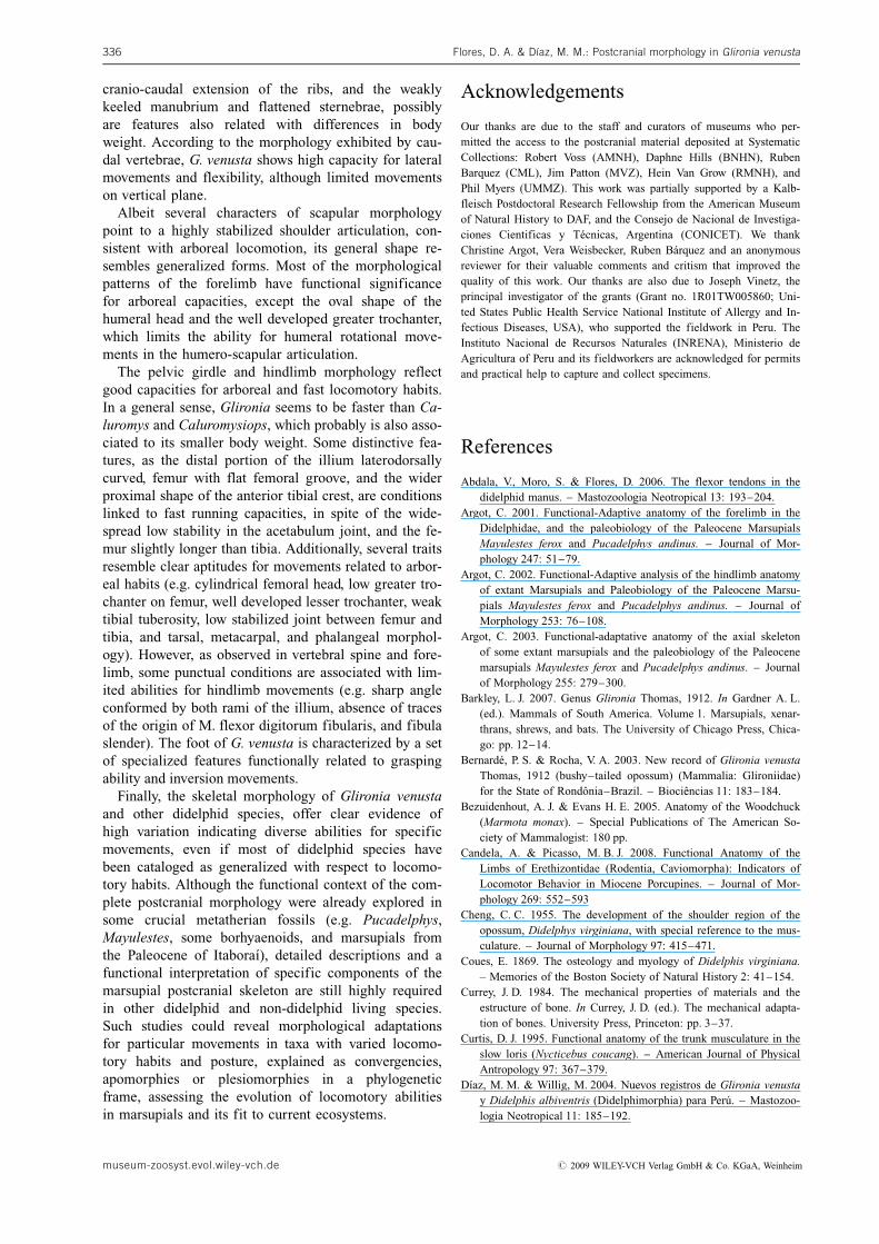
cranio-caudal extension of the ribs, and the weaklykeeled manubrium and flattened sternebrae, possiblyare features also related with differences in bodyweight. According to the morphology exhibited by cau-dal vertebrae, G. venusta shows high capacity for lateralmovements and flexibility, although limited movementson vertical plane.
Albeit several characters of scapular morphologypoint to a highly stabilized shoulder articulation, con-sistent with arboreal locomotion, its general shape re-sembles generalized forms. Most of the morphologicalpatterns of the forelimb have functional significancefor arboreal capacities, except the oval shape of thehumeral head and the well developed greater trochanter,which limits the ability for humeral rotational move-ments in the humero-scapular articulation.
The pelvic girdle and hindlimb morphology reflectgood capacities for arboreal and fast locomotory habits.In a general sense, Glironia seems to be faster than Ca-luromys and Caluromysiops, which probably is also asso-ciated to its smaller body weight. Some distinctive fea-tures, as the distal portion of the illium laterodorsallycurved, femur with flat femoral groove, and the widerproximal shape of the anterior tibial crest, are conditionslinked to fast running capacities, in spite of the wide-spread low stability in the acetabulum joint, and the fe-mur slightly longer than tibia. Additionally, several traitsresemble clear aptitudes for movements related to arbor-eal habits (e.g. cylindrical femoral head, low greater tro-chanter on femur, well developed lesser trochanter, weaktibial tuberosity, low stabilized joint between femur andtibia, and tarsal, metacarpal, and phalangeal morphol-ogy). However, as observed in vertebral spine and fore-limb, some punctual conditions are associated with lim-ited abilities for hindlimb movements (e.g. sharp angleconformed by both rami of the illium, absence of tracesof the origin of M. flexor digitorum fibularis, and fibulaslender). The foot of G. venusta is characterized by a setof specialized features functionally related to graspingability and inversion movements.
Finally, the skeletal morphology of Glironia venustaand other didelphid species, offer clear evidence ofhigh variation indicating diverse abilities for specificmovements, even if most of didelphid species havebeen cataloged as generalized with respect to locomo-tory habits. Although the functional context of the com-plete postcranial morphology were already explored insome crucial metatherian fossils (e.g. Pucadelphys,Mayulestes, some borhyaenoids, and marsupials fromthe Paleocene of Itabora�), detailed descriptions and afunctional interpretation of specific components of themarsupial postcranial skeleton are still highly requiredin other didelphid and non-didelphid living species.Such studies could reveal morphological adaptationsfor particular movements in taxa with varied locomo-tory habits and posture, explained as convergencies,apomorphies or plesiomorphies in a phylogeneticframe, assessing the evolution of locomotory abilitiesin marsupials and its fit to current ecosystems.
Acknowledgements
Our thanks are due to the staff and curators of museums who per-mitted the access to the postcranial material deposited at SystematicCollections: Robert Voss (AMNH), Daphne Hills (BNHN), RubenBarquez (CML), Jim Patton (MVZ), Hein Van Grow (RMNH), andPhil Myers (UMMZ). This work was partially supported by a Kalb-fleisch Postdoctoral Research Fellowship from the American Museumof Natural History to DAF, and the Consejo de Nacional de Investiga-ciones Cientificas y T�cnicas, Argentina (CONICET). We thankChristine Argot, Vera Weisbecker, Ruben B�rquez and an anonymousreviewer for their valuable comments and critism that improved thequality of this work. Our thanks are also due to Joseph Vinetz, theprincipal investigator of the grants (Grant no. 1R01TW005860; Uni-ted States Public Health Service National Institute of Allergy and In-fectious Diseases, USA), who supported the fieldwork in Peru. TheInstituto Nacional de Recursos Naturales (INRENA), Ministerio deAgricultura of Peru and its fieldworkers are acknowledged for permitsand practical help to capture and collect specimens.
References
Abdala, V., Moro, S. & Flores, D. 2006. The flexor tendons in thedidelphid manus. – Mastozoologia Neotropical 13: 193–204.
Argot, C. 2001. Functional-Adaptive anatomy of the forelimb in theDidelphidae, and the paleobiology of the Paleocene MarsupialsMayulestes ferox and Pucadelphys andinus. – Journal of Mor-phology 247: 51–79.
Argot, C. 2002. Functional-Adaptive analysis of the hindlimb anatomyof extant Marsupials and Paleobiology of the Paleocene Marsu-pials Mayulestes ferox and Pucadelphys andinus. – Journal ofMorphology 253: 76–108.
Argot, C. 2003. Functional-adaptative anatomy of the axial skeletonof some extant marsupials and the paleobiology of the Paleocenemarsupials Mayulestes ferox and Pucadelphys andinus. – Journalof Morphology 255: 279–300.
Barkley, L. J. 2007. Genus Glironia Thomas, 1912. In Gardner A. L.(ed.). Mammals of South America. Volume 1. Marsupials, xenar-thrans, shrews, and bats. The University of Chicago Press, Chica-go: pp. 12–14.
Bernard�, P. S. & Rocha, V. A. 2003. New record of Glironia venustaThomas, 1912 (bushy–tailed opossum) (Mammalia: Glironiidae)for the State of Rond�nia–Brazil. – Bioci�ncias 11: 183–184.
Bezuidenhout, A. J. & Evans H. E. 2005. Anatomy of the Woodchuck(Marmota monax). – Special Publications of The American So-ciety of Mammalogist: 180 pp.
Candela, A. & Picasso, M. B. J. 2008. Functional Anatomy of theLimbs of Erethizontidae (Rodentia, Caviomorpha): Indicators ofLocomotor Behavior in Miocene Porcupines. – Journal of Mor-phology 269: 552–593
Cheng, C. C. 1955. The development of the shoulder region of theopossum, Didelphys virginiana, with special reference to the mus-culature. – Journal of Morphology 97: 415–471.
Coues, E. 1869. The osteology and myology of Didelphis virginiana.– Memories of the Boston Society of Natural History 2: 41–154.
Currey, J. D. 1984. The mechanical properties of materials and theestructure of bone. In Currey, J. D. (ed.). The mechanical adapta-tion of bones. University Press, Princeton: pp. 3–37.
Curtis, D. J. 1995. Functional anatomy of the trunk musculature in theslow loris (Nycticebus coucang). – American Journal of PhysicalAntropology 97: 367–379.
D�az, M. M. & Willig, M. 2004. Nuevos registros de Glironia venustay Didelphis albiventris (Didelphimorphia) para Per�. – Mastozoo-logia Neotropical 11: 185–192.
Flores, D. A. & Dıaz, M. M.: Postcranial morphology in Glironia venusta336
museum-zoosyst.evol.wiley-vch.de # 2009 WILEY-VCH Verlag GmbH & Co. KGaA, Weinheim
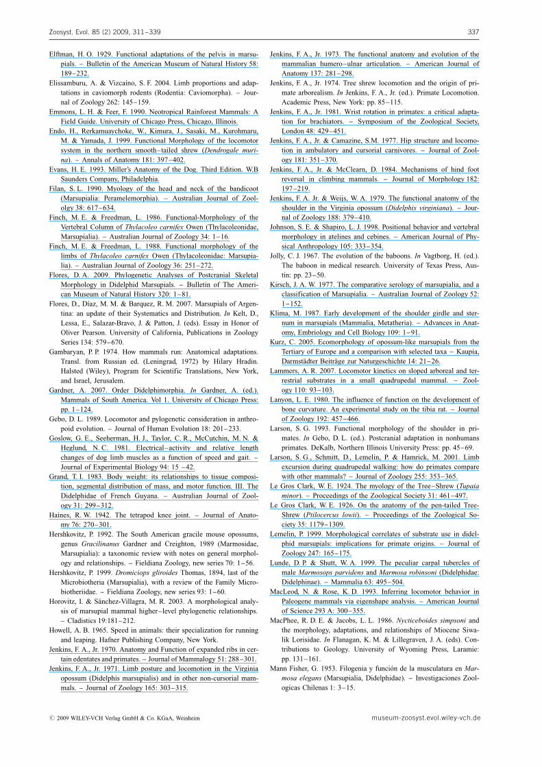
Elftman, H. O. 1929. Functional adaptations of the pelvis in marsu-pials. – Bulletin of the American Museum of Natural History 58:189–232.
Elissamburu, A. & Vizca�no, S. F. 2004. Limb proportions and adap-tations in caviomorph rodents (Rodentia: Caviomorpha). – Jour-nal of Zoology 262: 145–159.
Emmons, L. H. & Feer, F. 1990. Neotropical Rainforest Mammals: AField Guide. University of Chicago Press, Chicago, Illinois.
Endo, H., Rerkamuaychoke, W., Kimura, J., Sasaki, M., Kurohmaru,M. & Yamada, J. 1999. Functional Morphology of the locomotorsystem in the northern smooth–tailed shrew (Dendrogale muri-na). – Annals of Anatomy 181: 397–402.
Evans, H. E. 1993. Miller’s Anatomy of the Dog. Third Edition. W.BSaunders Company, Philadelphia.
Filan, S. L. 1990. Myology of the head and neck of the bandicoot(Marsupialia: Peramelemorphia). – Australian Journal of Zool-olgy 38: 617–634.
Finch, M. E. & Freedman, L. 1986. Functional-Morphology of theVertebral Column of Thylacoleo carnifex Owen (Thylacoleonidae,Marsupialia). – Australian Journal of Zoology 34: 1–16.
Finch, M. E. & Freedman, L. 1988. Functional morphology of thelimbs of Thylacoleo carnifex Owen (Thylacoleonidae: Marsupia-lia). – Australian Journal of Zoology 36: 251–272.
Flores, D. A. 2009. Phylogenetic Analyses of Postcranial SkeletalMorphology in Didelphid Marsupials. – Bulletin of The Ameri-can Museum of Natural History 320: 1–81.
Flores, D., D�az, M. M. & Barquez, R. M. 2007. Marsupials of Argen-tina: an update of their Systematics and Distribution. In Kelt, D.,Lessa, E., Salazar-Bravo, J. & Patton, J. (eds). Essay in Honor ofOliver Pearson. University of California, Publications in ZoologySeries 134: 579–670.
Gambaryan, P. P. 1974. How mammals run: Anatomical adaptations.Transl. from Russian ed. (Leningrad, 1972) by Hilary Hradin.Halsted (Wiley), Program for Scientific Translations, New York,and Israel, Jerusalem.
Gardner, A. 2007. Order Didelphimorphia. In Gardner, A. (ed.).Mammals of South America. Vol 1. University of Chicago Press:pp. 1–124.
Gebo, D. L. 1989. Locomotor and pylogenetic consideration in anthro-poid evolution. – Journal of Human Evolution 18: 201–233.
Goslow, G. E., Seeherman, H. J., Taylor, C. R., McCutchin, M. N. &Heglund, N. C. 1981. Electrical–activity and relative lengthchanges of dog limb muscles as a function of speed and gait. –Journal of Experimental Biology 94: 15 –42.
Grand, T. I. 1983. Body weight: its relationships to tissue composi-tion, segmental distribution of mass, and motor function. III. TheDidelphidae of French Guyana. – Australian Journal of Zool-ogy 31: 299–312.
Haines, R. W. 1942. The tetrapod knee joint. – Journal of Anato-my 76: 270–301.
Hershkovitz, P. 1992. The South American gracile mouse opossums,genus Gracilinanus Gardner and Creighton, 1989 (Marmosidae,Marsupialia): a taxonomic review with notes on general morphol-ogy and relationships. – Fieldiana Zoology, new series 70: 1–56.
Hershkovitz, P. 1999. Dromiciops gliroides Thomas, 1894, last of theMicrobiotheria (Marsupialia), with a review of the Family Micro-biotheriidae. – Fieldiana Zoology, new series 93: 1–60.
Horovitz, I. & S�nchez-Villagra, M. R. 2003. A morphological analy-sis of marsupial mammal higher–level phylogenetic relationships.– Cladistics 19:181–212.
Howell, A. B. 1965. Speed in animals: their specialization for runningand leaping. Hafner Publishing Company, New York.
Jenkins, F. A., Jr. 1970. Anatomy and Function of expanded ribs in cer-tain edentates and primates. – Journal of Mammalogy 51: 288–301.
Jenkins, F. A., Jr. 1971. Limb posture and locomotion in the Virginiaopossum (Didelphis marsupialis) and in other non-cursorial mam-mals. – Journal of Zoology 165: 303–315.
Jenkins, F. A., Jr. 1973. The functional anatomy and evolution of themammalian humero–ulnar articulation. – American Journal ofAnatomy 137: 281–298.
Jenkins, F. A., Jr. 1974. Tree shrew locomotion and the origin of pri-mate arborealism. In Jenkins, F. A., Jr. (ed.). Primate Locomotion.Academic Press, New York: pp. 85–115.
Jenkins, F. A., Jr. 1981. Wrist rotation in primates: a critical adapta-tion for brachiators. – Symposium of the Zoological Society,London 48: 429–451.
Jenkins, F. A., Jr. & Camazine, S.M. 1977. Hip structure and locomo-tion in ambulatory and cursorial carnivores. – Journal of Zool-ogy 181: 351–370.
Jenkins, F. A., Jr. & McClearn, D. 1984. Mechanisms of hind footreversal in climbing mammals. – Journal of Morphology 182:197–219.
Jenkins, F. A. Jr. & Weijs, W. A. 1979. The functional anatomy of theshoulder in the Virginia opossum (Didelphis virginiana). – Jour-nal of Zoology 188: 379–410.
Johnson, S. E. & Shapiro, L. J. 1998. Positional behavior and vertebralmorphology in atelines and cebines. – American Journal of Phy-sical Anthropology 105: 333–354.
Jolly, C. J. 1967. The evolution of the baboons. In Vagtborg, H. (ed.).The baboon in medical research. University of Texas Press, Aus-tin: pp. 23–50.
Kirsch, J. A. W. 1977. The comparative serology of marsupialia, and aclassification of Marsupialia. – Australian Journal of Zoology 52:1–152.
Klima, M. 1987. Early development of the shoulder girdle and ster-num in marsupials (Mammalia, Metatheria). – Advances in Anat-omy, Embriology and Cell Biology 109: 1–91.
Kurz, C. 2005. Ecomorphology of opossum-like marsupials from theTertiary of Europe and a comparison with selected taxa – Kaupia,Darmstdter Beitrge zur Naturgeschichte 14: 21–26.
Lammers, A. R. 2007. Locomotor kinetics on sloped arboreal and ter-restrial substrates in a small quadrupedal mammal. – Zool-ogy 110: 93–103.
Lanyon, L. E. 1980. The influence of function on the development ofbone curvature. An experimental study on the tibia rat. – Journalof Zoology 192: 457–466.
Larson, S. G. 1993. Functional morphology of the shoulder in pri-mates. In Gebo, D. L. (ed.). Postcranial adaptation in nonhumansprimates. DeKalb, Northern Illinois University Press: pp. 45–69.
Larson, S. G., Schmitt, D., Lemelin, P. & Hamrick, M. 2001. Limbexcursion during quadrupedal walking: how do primates comparewith other mammals? – Journal of Zoology 255: 353–365.
Le Gros Clark, W. E. 1924. The myology of the Tree–Shrew (Tupaiaminor). – Proceedings of the Zoological Society 31: 461–497.
Le Gros Clark, W. E. 1926. On the anatomy of the pen-tailed Tree-Shrew (Ptilocercus lowii). – Proceedings of the Zoological So-ciety 35: 1179–1309.
Lemelin, P. 1999. Morphological correlates of substrate use in didel-phid marsupials: implications for primate origins. – Journal ofZoology 247: 165–175.
Lunde, D. P. & Shutt, W. A. 1999. The peculiar carpal tubercles ofmale Marmosops parvidens and Marmosa robinsoni (Didelphidae:Didelphinae). – Mammalia 63: 495–504.
MacLeod, N. & Rose, K. D. 1993. Inferring locomotor behavior inPaleogene mammals via eigenshape analysis. – American Journalof Science 293 A: 300–355.
MacPhee, R. D. E. & Jacobs, L. L. 1986. Nycticeboides simpsoni andthe morphology, adaptations, and relationships of Miocene Siwa-lik Lorisidae. In Flanagan, K. M. & Lillegraven, J. A. (eds). Con-tributions to Geology. University of Wyoming Press, Laramie:pp. 131–161.
Mann Fisher, G. 1953. Filogenia y funci�n de la musculatura en Mar-mosa elegans (Marsupialia, Didelphidae). – Investigaciones Zool-ogicas Chilenas 1: 3–15.
Zoosyst. Evol. 85 (2) 2009, 311–339 337
# 2009 WILEY-VCH Verlag GmbH & Co. KGaA, Weinheim museum-zoosyst.evol.wiley-vch.de
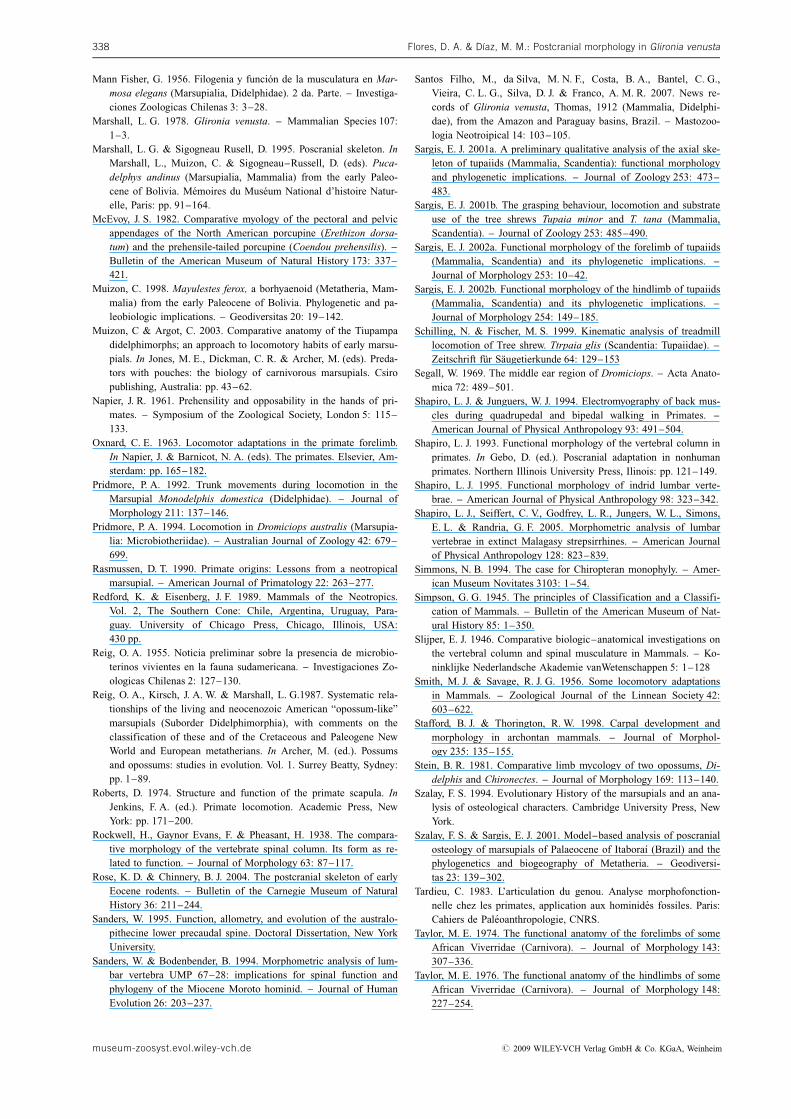
Mann Fisher, G. 1956. Filogenia y funci�n de la musculatura en Mar-mosa elegans (Marsupialia, Didelphidae). 2 da. Parte. – Investiga-ciones Zoologicas Chilenas 3: 3–28.
Marshall, L. G. 1978. Glironia venusta. – Mammalian Species 107:1–3.
Marshall, L. G. & Sigogneau Rusell, D. 1995. Poscranial skeleton. InMarshall, L., Muizon, C. & Sigogneau–Russell, D. (eds). Puca-delphys andinus (Marsupialia, Mammalia) from the early Paleo-cene of Bolivia. M�moires du Mus�um National d’histoire Natur-elle, Paris: pp. 91–164.
McEvoy, J. S. 1982. Comparative myology of the pectoral and pelvicappendages of the North American porcupine (Erethizon dorsa-tum) and the prehensile-tailed porcupine (Coendou prehensilis). –Bulletin of the American Museum of Natural History 173: 337–421.
Muizon, C. 1998. Mayulestes ferox, a borhyaenoid (Metatheria, Mam-malia) from the early Paleocene of Bolivia. Phylogenetic and pa-leobiologic implications. – Geodiversitas 20: 19–142.
Muizon, C & Argot, C. 2003. Comparative anatomy of the Tiupampadidelphimorphs; an approach to locomotory habits of early marsu-pials. In Jones, M. E., Dickman, C. R. & Archer, M. (eds). Preda-tors with pouches: the biology of carnivorous marsupials. Csiropublishing, Australia: pp. 43–62.
Napier, J. R. 1961. Prehensility and opposability in the hands of pri-mates. – Symposium of the Zoological Society, London 5: 115–133.
Oxnard, C. E. 1963. Locomotor adaptations in the primate forelimb.In Napier, J. & Barnicot, N. A. (eds). The primates. Elsevier, Am-sterdam: pp. 165–182.
Pridmore, P. A. 1992. Trunk movements during locomotion in theMarsupial Monodelphis domestica (Didelphidae). – Journal ofMorphology 211: 137–146.
Pridmore, P. A. 1994. Locomotion in Dromiciops australis (Marsupia-lia: Microbiotheriidae). – Australian Journal of Zoology 42: 679–699.
Rasmussen, D. T. 1990. Primate origins: Lessons from a neotropicalmarsupial. – American Journal of Primatology 22: 263–277.
Redford, K. & Eisenberg, J. F. 1989. Mammals of the Neotropics.Vol. 2, The Southern Cone: Chile, Argentina, Uruguay, Para-guay. University of Chicago Press, Chicago, Illinois, USA:430 pp.
Reig, O. A. 1955. Noticia preliminar sobre la presencia de microbio-terinos vivientes en la fauna sudamericana. – Investigaciones Zo-ologicas Chilenas 2: 127–130.
Reig, O. A., Kirsch, J. A. W. & Marshall, L. G.1987. Systematic rela-tionships of the living and neocenozoic American “opossum-like”marsupials (Suborder Didelphimorphia), with comments on theclassification of these and of the Cretaceous and Paleogene NewWorld and European metatherians. In Archer, M. (ed.). Possumsand opossums: studies in evolution. Vol. 1. Surrey Beatty, Sydney:pp. 1–89.
Roberts, D. 1974. Structure and function of the primate scapula. InJenkins, F. A. (ed.). Primate locomotion. Academic Press, NewYork: pp. 171–200.
Rockwell, H., Gaynor Evans, F. & Pheasant, H. 1938. The compara-tive morphology of the vertebrate spinal column. Its form as re-lated to function. – Journal of Morphology 63: 87–117.
Rose, K. D. & Chinnery, B. J. 2004. The postcranial skeleton of earlyEocene rodents. – Bulletin of the Carnegie Museum of NaturalHistory 36: 211–244.
Sanders, W. 1995. Function, allometry, and evolution of the australo-pithecine lower precaudal spine. Doctoral Dissertation, New YorkUniversity.
Sanders, W. & Bodenbender, B. 1994. Morphometric analysis of lum-bar vertebra UMP 67–28: implications for spinal function andphylogeny of the Miocene Moroto hominid. – Journal of HumanEvolution 26: 203–237.
Santos Filho, M., da Silva, M. N. F., Costa, B. A., Bantel, C. G.,Vieira, C. L. G., Silva, D. J. & Franco, A. M. R. 2007. News re-cords of Glironia venusta, Thomas, 1912 (Mammalia, Didelphi-dae), from the Amazon and Paraguay basins, Brazil. – Mastozoo-logia Neotroipical 14: 103–105.
Sargis, E. J. 2001a. A preliminary qualitative analysis of the axial ske-leton of tupaiids (Mammalia, Scandentia): functional morphologyand phylogenetic implications. – Journal of Zoology 253: 473–483.
Sargis, E. J. 2001b. The grasping behaviour, locomotion and substrateuse of the tree shrews Tupaia minor and T. tana (Mammalia,Scandentia). – Journal of Zoology 253: 485–490.
Sargis, E. J. 2002a. Functional morphology of the forelimb of tupaiids(Mammalia, Scandentia) and its phylogenetic implications. –Journal of Morphology 253: 10–42.
Sargis, E. J. 2002b. Functional morphology of the hindlimb of tupaiids(Mammalia, Scandentia) and its phylogenetic implications. –Journal of Morphology 254: 149–185.
Schilling, N. & Fischer, M. S. 1999. Kinematic analysis of treadmilllocomotion of Tree shrew. Ttrpaia glis (Scandentia: Tupaiidae). –Zeitschrift fr Sugetierkunde 64: 129–153
Segall, W. 1969. The middle ear region of Dromiciops. – Acta Anato-mica 72: 489–501.
Shapiro, L. J. & Junguers, W. J. 1994. Electromyography of back mus-cles during quadrupedal and bipedal walking in Primates. –American Journal of Physical Anthropology 93: 491–504.
Shapiro, L. J. 1993. Functional morphology of the vertebral column inprimates. In Gebo, D. (ed.). Poscranial adaptation in nonhumanprimates. Northern Illinois University Press, Ilinois: pp. 121–149.
Shapiro, L. J. 1995. Functional morphology of indrid lumbar verte-brae. – American Journal of Physical Anthropology 98: 323–342.
Shapiro, L. J., Seiffert, C. V., Godfrey, L. R., Jungers, W. L., Simons,E. L. & Randria, G. F. 2005. Morphometric analysis of lumbarvertebrae in extinct Malagasy strepsirrhines. – American Journalof Physical Anthropology 128: 823–839.
Simmons, N. B. 1994. The case for Chiropteran monophyly. – Amer-ican Museum Novitates 3103: 1–54.
Simpson, G. G. 1945. The principles of Classification and a Classifi-cation of Mammals. – Bulletin of the American Museum of Nat-ural History 85: 1–350.
Slijper, E. J. 1946. Comparative biologic–anatomical investigations onthe vertebral column and spinal musculature in Mammals. – Ko-ninklijke Nederlandsche Akademie vanWetenschappen 5: 1–128
Smith, M. J. & Savage, R. J. G. 1956. Some locomotory adaptationsin Mammals. – Zoological Journal of the Linnean Society 42:603–622.
Stafford, B. J. & Thorington, R. W. 1998. Carpal development andmorphology in archontan mammals. – Journal of Morphol-ogy 235: 135–155.
Stein, B. R. 1981. Comparative limb mycology of two opossums, Di-delphis and Chironectes. – Journal of Morphology 169: 113–140.
Szalay, F. S. 1994. Evolutionary History of the marsupials and an ana-lysis of osteological characters. Cambridge University Press, NewYork.
Szalay, F. S. & Sargis, E. J. 2001. Model–based analysis of poscranialosteology of marsupials of Palaeocene of Itabora� (Brazil) and thephylogenetics and biogeography of Metatheria. – Geodiversi-tas 23: 139–302.
Tardieu, C. 1983. L’articulation du genou. Analyse morphofonction-nelle chez les primates, application aux hominides fossiles. Paris:Cahiers de Paleoanthropologie, CNRS.
Taylor, M. E. 1974. The functional anatomy of the forelimbs of someAfrican Viverridae (Carnivora). – Journal of Morphology 143:307–336.
Taylor, M. E. 1976. The functional anatomy of the hindlimbs of someAfrican Viverridae (Carnivora). – Journal of Morphology 148:227–254.
Flores, D. A. & Dıaz, M. M.: Postcranial morphology in Glironia venusta338
museum-zoosyst.evol.wiley-vch.de # 2009 WILEY-VCH Verlag GmbH & Co. KGaA, Weinheim
Taylor, C. R., Caldwell, S. L. & Rowntree, V. J. 1972. Running up anddown hills: some consequences of size. – Science 178: 1096–1097.
V�zquez-Molinero, R., Martin, T., Fischer, M. S. & Frey, R. 2001.Comparative anatomical investigations of the postcranial skeletonof Henkelotherium guimarotae Krebs, 1991 (Eupantotheria, Mam-malia) and their implications for its locomotion. – Mitteilungenaus dem Museum fr Naturkunde in Berlin. ZoologischeReihe 77: 207–216.
Voss, R. S. & Jansa, S. A. 2003. Phylogenetic studies on Didelphidmarsupials II. Nonmolecular data and new IRBP sequences: sepa-rate and combined analyses of Didelphine relationships with den-ser taxon sampling. – Bulletin of the American Museum of Nat-ural History 276: 1–82.
Walker, A. 1974. Locomotor adaptations in past and present prosi-mian primates. In Jenkins, F. A. (ed.). Primate Locomotion. Aca-demic Press, New York: pp. 349–381.
Washburn, S. & Buettner-Janush, J. 1952. The definition of thoracicand lumbar vertebrae. – American Journal of Physical Anthropol-ogy 10: 251–252.
Weisbecker V. & Archer, M. 2008. Parallel evolution of hand anatomyin kangaroos and vombatiform marsupials: functional and evolu-tionary implications. – Palaeontology 51: 321–338.
Weisbecker, V. & Sanchez-Villagra, M. R. 2006. Carpal evolution indiprotodontian marsupials. – Zoological Journal of the LinneanSociety 146: 369–384.
Weisbecker, V. & Warton, D. I. 2006. Evidence at hand: diversity,functional implications, and locomotory prediction in intrinsichand proportions of diprotodontian marsupials. – Journal of Mor-phology 267: 1469–1485.
Yalden, D. W. 1972. The form and function of the carpal bones in somearboreally adapted mammals. – Acta Anatomica 82: 383–406.
Young, A. H. 1879. The intrinsic muscles of the marsupial hand. –Journal of Anatomy and Physiology 14: 149–165.
Appendix
Specimens examined
The skeletons analyzed for this study are deposited in the followingsystematics collections, listed in alphabetical order by their acronyms:
AMNH American Museum of Natural History, New YorkBMNH British Museum (Natural History), LondonCML Colecci�n Mam�feros Lillo, Universidad Nacional de Tucu-
m�n, Tucum�n, ArgentinaMVZ Museum of Vertebrate Zoology, University of California,
Berkeley
RMNH Rijksmuseum van Natuurlijke Historie, LeidenUWZM University of Wisconsin Zoological Museum (Madison)MMD Personal catalog of M. M�nica D�az (deposited at CML and
Museo de Historia Natural de San Marcos, Peru uncata-loged)
Caluromys lanatus: MMD 2973, 4508; CML 7573, 7574; AMNH133199, 133200, 215001.
Caluromys philander: AMNH 95761, 267001, 267002, 267337,95974; RMNH uncataloged, 12866, 10790, 19646, 20664.
Caluromysiops irrupta: AMNH 208101, 244364.Chironectes minimus: AMNH 97319, 148720, 212909, 264571;
RMNH uncataloged.Cryptonanus unduaviensis: AMNH 210369, 262401.Didelphis albiventris: AMNH 13102, 148320, 170653, 170654,
170664, 204406, 205301, 205382, 238006.Didelphis marsupialis: MMD 3313; CML 7575; AMNH 13448,
97318, 132784, 209164, 210427, 210428, 210439, 210447,255854
Glironia venusta: MMD 607 (deposited at the Museo de Historia Nat-ural de San Marcos, Peru uncataloged).
Lestodelphys halli: UWZM 224223; BMNH 21.6.7.19.Marmosa robinsoni: AMNH 206596, 206597, 206766, 207766,
257209, 257210.Marmosops bishopi: CML 7564, 7565.Marmosops noctivagus: AMNH 136157, 231952; MMD 2201, 3503,
3520, 4663, 4776, 4789.Metachirus nudicaudatus: MMD 785, 801; CML 7579, 7580, 7342;
AMNH 97320, 136151, 136155, 244617, 267009.Micoureus regina: AMNH 61391, 148757.Micoureus demerarae: AMNH 257211, 257212; RMNH 12871, 998,
18228.Micoureus sp.: MMD 556, 613, 1252, 1984, 2203, 2206, 2586, 2590,
3458, 4617.Monodelphis adusta: MMD 4305; CML 7591; AMNH 136158,
139227.Monodelphis brevicaudata: AMNH 48133, 257203; RMNH 18079,
12851, 17907.Monodelphis theresa: MVZ 182775.P. opossum: MMD 581, 1033, 1046, 1255, 1973, 2214, 2972, 3314,
3379, 3735, 3398, 3737, 4294; CML 7583, 7585; AMNH61396, 61864, 97332, 133074, 190446, 210406, 210410,248703, 254509, 261273, 261276, 262415; RMNH 12835,12838, 12834.
Philander andersoni: MMD 4621, 4652.Thylamys pallidior: AMNH 262405, 262406, 262408; CML 3189,
3192, 3574, 3575.
Zoosyst. Evol. 85 (2) 2009, 311–339 339
# 2009 WILEY-VCH Verlag GmbH & Co. KGaA, Weinheim museum-zoosyst.evol.wiley-vch.de