Ankylosis patterns in the postcranial skeleton and hyoid bones of the harbour porpoise ( Phocoena...
11
Ankylosis patterns in the postcranial skeleton and hyoid bones of the harbour porpoise (Phocoena phocoena) in the Baltic and North Sea Anders Galatius and Carl Christian Kinze Abstract: The onset and timing of epiphyseal ankylosis in the vertebral column and flippers and ankylosis of the hyoid and sternal bones were studied in 350 skeletons of the harbour porpoise (Phocoena phocoena) originating from the Baltic and North Sea and held in the collections of the Zoological Museum (University of Copenhagen), the Museum of Natural History (Gothenburg), the National Museum of Natural History (Stockholm), and the German Oceanographic Museum (Stralsund). Epiphyseal ankylosis in the vertebral column started in the anterior cervical region and then initi- ated around the 23rd to 26th caudal vertebrae from where it proceeded in both directions. The progression of vertebral epiphyseal ankylosis eventually terminated in the thoracic and lumbar regions. Epiphyseal ankylosis in the flippers be- gan at the distal end of the humerus and the proximal ends of the radius and ulna. The timing of ankylosis in the flip- pers was more consistent across the specimens than the timing of vertebral ankylosis. Males and females had similar timing of ankylosis in the vertebral column and the flippers. Complete fusion of the hyoid and sternal bones occurred within the first year of life in most specimens. The early development of the hyoid apparatus may be linked to use of suction in feeding. Résumé : L’étude de 350 squelettes de marsouins communs (Phocoena phocoena) de la Baltique et de la mer du Nord provenant des collections du Musée zoologique de l’Université de Copenhague, du Musée d’histoire naturelle de Go- thenburg, du Musée national d’histoire naturelle de Stockholm et du Musée Allemand d’océanographique de Stralsund nous à permis d’étudier le début et la progression dans le temps de l’ankylose des épiphyses de la colonne vertébrale et des nageoires, ainsi que l’ankylose des os hyoïde et sternal. L’ankylose des épiphyses de la colonne vertébrale dé- bute dans la région cervicale antérieure, puis elle s’installe dans la région des vertèbres caudales 23–26, pour ensuite s’étendre dans les deux directions. La progression de l’ankylose des épiphyses de la colonne vertébrale se termine éventuellement dans les régions thoraciques et lombaires. L’ankylose des épiphyses des nageoires commence dans la région distale de l’humérus et les extrémités proximales du radius et du cubitus. Le développement dans le temps de l’ankylose dans les nageoires est plus semblable d’un spécimen à un autre que celle des vertèbres. Les mâles et les fe- melles subissent l’ankylose de la colonne vertébrale et des nageoires à des rythmes semblables. La fusion complète des os hyoïde et sternal est complétée au cours de la première année chez la plupart des spécimens. Le développement pré- coce de l’appareil hyoïde peut peut-être s’expliquer par l’utilisation de la succion durant l’alimentation. [Traduit par la Rédaction] Galatius and Kinze 1861 Introduction The ankylosis of epiphyses and the fusion of other postcranial elements as a gross measure of age are well known, but very few papers have addressed this phenome- non in cetaceans, and in those cases, mainly in mysticete species (Wheeler 1930; Ohsumi et al. 1958; Aguilar and Lockyer 1987; Kemper and Leppard 1999). The only com- parable studies on ankylosis of vertebral epiphyses in odon- tocete species that we could find were carried out by Ito and Miyazaki (1990) and Calzada et al. (1997) on the striped dolphin (Stenella coeruleoalba) and Yoshida et al. (1994) on the finless porpoise (Neophocaena phocaenoides). Ferrero and Walker (1999) investigated epiphyseal ankylosis of three thoracic vertebrae as a measure of physical maturity in Dall’s porpoise (Phocoenoides dalli). The general character- istics of the pattern of epiphyseal ankylosis in the vertebral column of the investigated cetaceans can be described as a rapid and early ankylosis of the cervical vertebrae immedi- ately followed by the ankylosis of posterior caudal verte- brae; ankylosis then progresses anteriorly from the caudal vertebrae, and at a slower pace posteriorly from the cervical vertebrae. Can. J. Zool. 81: 1851–1861 (2003) doi: 10.1139/Z03-181 © 2003 NRC Canada 1851 Received 23 April 2003. Accepted 30 September 2003. Published on the NRC Research Press Web site at http://cjz.nrc.ca on 23 December 2003. A. Galatius. 1,2 Department of Zoomorphology, Zoological Institute, University of Copenhagen, Universitetsparken 15, 2100 Copenhagen Ø, Denmark. C.C. Kinze. Zoological Museum, University of Copenhagen, Universitetsparken 15, 2100 Copenhagen Ø, Denmark. 1 Corresponding author (e-mail: [email protected]). 2 Present address: Skovvangen 3, DK-2920 Charlottenlund, Denmark.
-
Upload
independent -
Category
Documents
-
view
0 -
download
0
Transcript of Ankylosis patterns in the postcranial skeleton and hyoid bones of the harbour porpoise ( Phocoena...

Ankylosis patterns in the postcranial skeleton andhyoid bones of the harbour porpoise (Phocoenaphocoena) in the Baltic and North Sea
Anders Galatius and Carl Christian Kinze
Abstract: The onset and timing of epiphyseal ankylosis in the vertebral column and flippers and ankylosis of the hyoidand sternal bones were studied in 350 skeletons of the harbour porpoise (Phocoena phocoena) originating from theBaltic and North Sea and held in the collections of the Zoological Museum (University of Copenhagen), the Museumof Natural History (Gothenburg), the National Museum of Natural History (Stockholm), and the German OceanographicMuseum (Stralsund). Epiphyseal ankylosis in the vertebral column started in the anterior cervical region and then initi-ated around the 23rd to 26th caudal vertebrae from where it proceeded in both directions. The progression of vertebralepiphyseal ankylosis eventually terminated in the thoracic and lumbar regions. Epiphyseal ankylosis in the flippers be-gan at the distal end of the humerus and the proximal ends of the radius and ulna. The timing of ankylosis in the flip-pers was more consistent across the specimens than the timing of vertebral ankylosis. Males and females had similartiming of ankylosis in the vertebral column and the flippers. Complete fusion of the hyoid and sternal bones occurredwithin the first year of life in most specimens. The early development of the hyoid apparatus may be linked to use ofsuction in feeding.
Résumé : L’étude de 350 squelettes de marsouins communs (Phocoena phocoena) de la Baltique et de la mer du Nordprovenant des collections du Musée zoologique de l’Université de Copenhague, du Musée d’histoire naturelle de Go-thenburg, du Musée national d’histoire naturelle de Stockholm et du Musée Allemand d’océanographique de Stralsundnous à permis d’étudier le début et la progression dans le temps de l’ankylose des épiphyses de la colonne vertébraleet des nageoires, ainsi que l’ankylose des os hyoïde et sternal. L’ankylose des épiphyses de la colonne vertébrale dé-bute dans la région cervicale antérieure, puis elle s’installe dans la région des vertèbres caudales 23–26, pour ensuites’étendre dans les deux directions. La progression de l’ankylose des épiphyses de la colonne vertébrale se termineéventuellement dans les régions thoraciques et lombaires. L’ankylose des épiphyses des nageoires commence dans larégion distale de l’humérus et les extrémités proximales du radius et du cubitus. Le développement dans le temps del’ankylose dans les nageoires est plus semblable d’un spécimen à un autre que celle des vertèbres. Les mâles et les fe-melles subissent l’ankylose de la colonne vertébrale et des nageoires à des rythmes semblables. La fusion complète desos hyoïde et sternal est complétée au cours de la première année chez la plupart des spécimens. Le développement pré-coce de l’appareil hyoïde peut peut-être s’expliquer par l’utilisation de la succion durant l’alimentation.
[Traduit par la Rédaction] Galatius and Kinze 1861
Introduction
The ankylosis of epiphyses and the fusion of otherpostcranial elements as a gross measure of age are wellknown, but very few papers have addressed this phenome-non in cetaceans, and in those cases, mainly in mysticetespecies (Wheeler 1930; Ohsumi et al. 1958; Aguilar andLockyer 1987; Kemper and Leppard 1999). The only com-parable studies on ankylosis of vertebral epiphyses in odon-tocete species that we could find were carried out by Ito andMiyazaki (1990) and Calzada et al. (1997) on the striped
dolphin (Stenella coeruleoalba) and Yoshida et al. (1994) onthe finless porpoise (Neophocaena phocaenoides). Ferreroand Walker (1999) investigated epiphyseal ankylosis of threethoracic vertebrae as a measure of physical maturity inDall’s porpoise (Phocoenoides dalli). The general character-istics of the pattern of epiphyseal ankylosis in the vertebralcolumn of the investigated cetaceans can be described as arapid and early ankylosis of the cervical vertebrae immedi-ately followed by the ankylosis of posterior caudal verte-brae; ankylosis then progresses anteriorly from the caudalvertebrae, and at a slower pace posteriorly from the cervicalvertebrae.
Can. J. Zool. 81: 1851–1861 (2003) doi: 10.1139/Z03-181 © 2003 NRC Canada
1851
Received 23 April 2003. Accepted 30 September 2003. Published on the NRC Research Press Web site at http://cjz.nrc.ca on23 December 2003.
A. Galatius.1,2 Department of Zoomorphology, Zoological Institute, University of Copenhagen, Universitetsparken 15, 2100Copenhagen Ø, Denmark.C.C. Kinze. Zoological Museum, University of Copenhagen, Universitetsparken 15, 2100 Copenhagen Ø, Denmark.
1Corresponding author (e-mail: [email protected]).2Present address: Skovvangen 3, DK-2920 Charlottenlund, Denmark.
J:\cjz\cjz8111\Z03-181.vpDecember 17, 2003 2:07:10 PM
Color profile: DisabledComposite Default screen

© 2003 NRC Canada
1852 Can. J. Zool. Vol. 81, 2003
Epiphyseal ankylosis in the flipper bones has been investi-gated by radiological techniques in the striped dolphin byCalzada and Aguilar (1996) and DiGiancamillo et al. (1998),the characteristics being that the distal epiphyses of the ra-dius and ulna fuse later than the proximal epiphyses of thesebones and the humeral epiphyses. Calzada et al. (1997)noted that in this species, maturity was reached much earlierin the flippers than in the vertebral column. Hui (1979) em-ployed the development of the flipper bones of the commondolphin (Delphinus delphis) as one correlate among othersfor sexual maturity.
Here, we describe the process of epiphyseal ankylosis in
the vertebral column and flipper bones as well as ankylosisof the hyoid and sternal bones in the harbour porpoise (Pho-coena phocoena) using the extensive collections of skeletonsat the Zoological Museum of Copenhagen, the Museum ofNatural History in Gothenburg, the National Museum ofNatural History in Stockholm, and the German Oceano-graphic Museum of Stralsund.3
The harbour porpoise exhibits a pattern of sexual dimor-phy unusual among odontocetes and phocoenids, the femalebeing the larger sex and having an extended period ofgrowth compared with the male (Gaskin and Blair 1977;Gaskin et al. 1984; Kull and Berggren 1995; Lockyer 1995;Miyazaki et al. 1987; Stuart and Morejohn 1980; Van
State Definition
A No ankylosis. Both epiphyseal plates free or not yetossified
B Initial ankylosis. At least one epiphyseal plate looselyfused to centra
C Progressing ankylosis. Both epiphyseal plates fused tocentra, showing clear sutures
D Complete ankylosis. Both epiphyseal plates fused tocentra, showing no sutures
Table 1. States of epiphyseal ankylosis of the vertebral columnof harbour porpoise (Phocoena phocoena).
Fig. 1. States of epiphyseal ankylosis of the vertebral column ofharbour porpoise (Phocoena phocoena). A, B, C, and D are thestates described in Table 1.
State Definition
A No ankylosis. Epiphysis free or not yet ossifiedB Initial ankylosis. Epiphysis loosely attached to the boneC Progressing ankylosis. Epiphysis fused to the bone,
showing clear sutureD Complete ankylosis. Epiphysis fused to the bone, show-
ing no suture
Table 2. States of epiphyseal ankylosis of the flippers of harbourporpoise.
Fig. 2. States of epiphyseal ankylosis of the flippers (distal ra-dius depicted) of harbour porpoise. A, B, C, and D are the statesdescribed in Table 2.
3 Supplementary data for this article are available on the Web site or may be purchased from the Depository of Unpublished Data, DocumentDelivery, CISTI, National Research Council of Canada, Ottawa, ON K1A 0S2, Canada. DUD 3539. For more information on obtaining ma-terial refer to http://cisti-icist.nrc-cnrc.gc.ca/irm/unpub_e.shtml.
J:\cjz\cjz8111\Z03-181.vpDecember 17, 2003 2:07:14 PM
Color profile: DisabledComposite Default screen

Utrecht 1978; Read and Tolley 1997). We test if this is mir-rored in the osteologic development of the species.
Materials and methods
Observations were made on 350 harbour porpoise skele-tons (195 females and 155 males). Three hundred and tenspecimens originated from bycatches, 17 were of uncertainorigin, and 25 originated from strandings. All of these ani-mals originated from the Baltic or North Sea. Age estima-tions by dentinal growth layer groups were available formost of the specimens from Copenhagen and Stralsund, pro-vided by Christina Lockyer and Hartwig Kremer, respec-tively. Age estimations for the remaining specimens fromthese collections and the animals from Stockholm andGothenburg were performed using the protocol of Hohn andLockyer (1995).
Specimens were selected to represent 17 age classes fromfoetuses, yearlings, 1-year-olds, etc., up to 15 years andolder. Where possible, each age class consisted of at least 15specimens of each sex.
It was impossible to achieve full sample sizes for all ageclasses, since older animals are rare in a sample originatingfrom incidental catches. Because of the scarcity of speci-mens, 10- and 11-year-old specimens and 12- to 14-year-old
specimens were lumped for both sexes resulting in a total of14 age classes.
Vertebral columnThe degree of epiphyseal ankylosis in the vertebral col-
umn was recorded for each vertebra of each specimen anddescribed as state A, B, C, or D (see Table 1 for definitionsand Fig. 1). The vertebrae were divided into cervical, tho-racic, lumbar, and caudal series as defined by de Smet(1977). The average vertebral formula presented in Fig. 5 iscalculated on the basis of all specimens except the foetuses(in foetuses, it was impossible to distinguish between thedifferent series of vertebrae, since transverse processes werenot fused to the centra and facets for hemal arches were notdeveloped, but the sequence of the individual vertebraecould be deduced) and is as follows: 7 cervical vertebrae(fixed), 12 thoracic, 15 lumbar, and the first 30 caudal verte-brae. Up to about 35 caudal vertebrae were present in speci-mens with all vertebrae intact, but vertebrae beyond the 30thcaudal were often missing.
FlippersThe degree of epiphyseal ankylosis in the flipper bones
was recorded at the proximal and distal ends of the humerus,radius, and ulna for each specimen and described as state A,
© 2003 NRC Canada
Galatius and Kinze 1853
Fig. 3. States of ankylosis of the hyoid bones of harbour porpoise. A, B, and C are the states described in Table 3.
Fig. 4. States of ankylosis of the sternal bones of harbour porpoise. A, B, and C are the states described in Table 4.
State Definition
A No ankylosis. Both thyroids freeB Initial ankylosis. At least one thyroid free or loosely
attached to the basihyoid, showing clear suturesC Complete ankylosis. Both thyroids fused to the
basihyoid, showing no sutures
Table 3. States of ankylosis of the hyoid bones of harbour por-poise.
State Definition
A No ankylosis. None of the three sternal bones fusedB Initial ankylosis. At least two sternal bones loosely
attached, showing clear suturesC Complete ankylosis. All three sternal bones fused,
showing no sutures
Table 4. States of ankylosis of the sternal bones in harbour por-poise.
J:\cjz\cjz8111\Z03-181.vpDecember 17, 2003 2:07:18 PM
Color profile: DisabledComposite Default screen

B, C, or D (Table 2, Fig. 2). The mode of state of ankylosiswas determined for both ends of each bone in each ageclass.
Logistic regressionsTo investigate differences between the sexes and to obtain
a firmer assessment of the average timing of the onset ofphysical maturity in the vertebral column and flippers, logis-tic regressions were performed on the attainment of physicalmaturity. For this purpose, specimens were considered phys-ically mature when the development of all vertebral and flip-per epiphyses had reached state C, comparable with thedegree of fusion used by Ferrero and Walker (1999) to de-fine physical maturity in the vertebral columns of Dall’s por-poise. Specimens having attained physical maturity werescored as 1 for the regression, whereas immature specimenswere scored as 0.
The logistic regression feature of SYSTAT (Systat Soft-ware Inc. 1998) was used to estimate the parameters of thelogistic equation
[1] P(t) = 1/[1 + e–(a+bt)]
where t is the age (class), P(t) is the proportion of the popu-lation expected to show physical maturity in a given ageclass, and a and b are the parameters of the model.
Hyoid and sternal bonesThe degree of ankylosis of the hyoid and sternal bones was
described as state A, B, or C (Tables 3 and 4, Figs. 3 and 4).
Results
Epiphyseal ankylosis in the vertebral columnComplete ankylosis of all vertebral epiphyses occurs in
the adult harbour porpoise. Sexual maturity is attained sev-eral years before physical maturity.
In younger age classes, the modal states of ankylosis indi-cate a slightly delayed progression in females comparedwith males, up to and including age class 6. From age class7 and up, there is a slight tendency for the females to showmore progressed states of ankylosis than the males.
The modal progression of ankylosis in the vertebral col-umn (Fig. 5) can be described as follows.
Foetuses exhibit no ankylosis except for the cervical ver-tebrae. Age class 0 is characterized by ankylosis progessingin all cervical vertebrae and initiation of ankylosis aroundthe 23rd to 26th caudal vertebrae in some specimens.Ankylosis then progresses in both directions from these untilin age class 6, ankylosis is initiated or completed in the en-tire vertebral column in a majority of the males, whereasthere are still some areas in the lumbar region in femaleswhere ankylosis typically is not initiated. From age class 7to age classes 12–14, the general picture is one of completedankylosis in the posterior caudals advancing in an anteriordirection, whereas the cervical region also shows completeankylosis, which is progressing posteriorly into the first fewthoracic vertebrae. In these age classes, ankylosis is gener-ally slightly more advanced in females. Among the speci-mens aged 15 years and older, a majority of the femalesshow complete ankylosis of the entire vertebral column,whereas in males, the majority of the specimens do not fea-ture complete closure of the sutures of the posterior thor-acics, the lumbar region, and the first few caudal vertebrae.
There is considerable individual variation with respect toage around the typical progression depicted here but no de-viations from the general pattern.
The youngest specimens in the sample to show completeankylosis in the entire vertebral column are a 6-year-oldmale and a 7-year-old female, whereas a male of 22 and afemale of 17 do not show complete ankylosis in all thoracicand lumbar vertebrae.
If physical maturity is defined by epiphyseal ankylosis ofthe entire vertebral column resulting in termination ofgrowth of the individual, then it appears from the data thatthe harbour porpoise shows a tendency to attain sexual matu-rity several years prior to physical maturity, and many maynever achieve full physical maturity. Sexual maturity is gen-erally attained around an age of 2–3 years for males and 3–4for females (Kinze 1994). The average life span of a harbourporpoise is 8–10 years as determined from mainly by-caughtanimals, whereas only about 5% exceed 12 years of age(Lockyer and Kinze 2003), so it seems that most harbourporpoises do not live long enough to attain full physical ma-turity.
The logistic regressions of physical maturity in the verte-bral column (Fig. 7, Table 5) revealed no intersexual differ-ence, the estimates of age where 50% of the specimens werephysically mature being 8.1 years for females (95% confi-dence interval (CI) = 7.3–9.2 years) and 8.4 years for males(95% CI = 7.4–9.9 years).
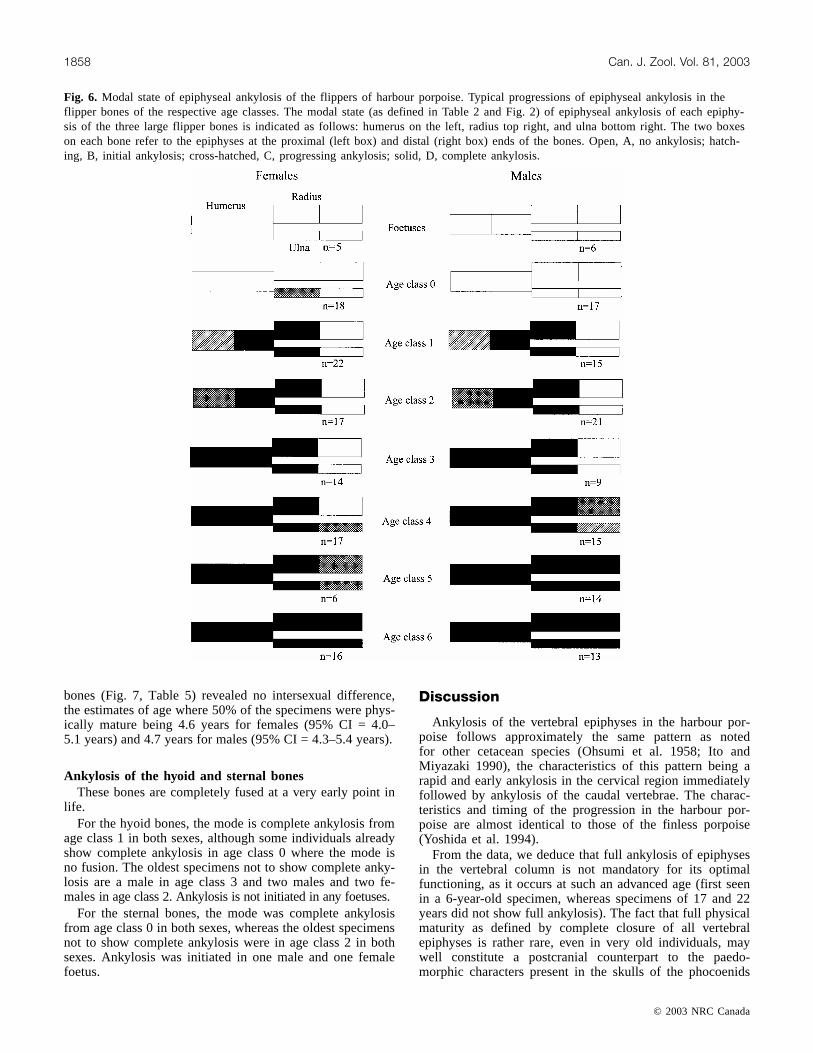
Epiphyseal ankylosis in the flippersEpiphyseal ankylosis of the humerus, radius, and ulna be-
gins at the joint of the humerus to the radius and ulna, pro-ceeds at the proximal end of the humerus, and ends with theankylosis of the epiphyses at the distal ends of the radiusand ulna (Fig. 6).
© 2003 NRC Canada
1854 Can. J. Zool. Vol. 81, 2003
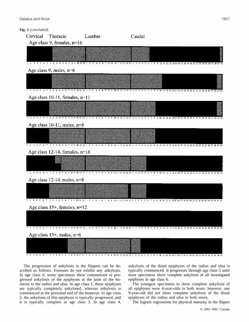
Fig. 5. Modal state of epiphyseal ankylosis of the vertebral column of harbour porpoise. Typical progression of epiphyseal ankylosis inthe vertebral columns of the respective age classes. The modal state (as defined in Table 1 and Fig. 1) of epiphyseal ankylosis of eachvertebra in the average vertebral formula is indicated as follows: open, A, no ankylosis; hatched, B, initial ankylosis; cross-hatched, C,progressing ankylosis; solid, D, complete ankylosis. Squares indicate that the concerned vertebra(e) was present in less than three spec-imens. Numbers of the vertebrae in the cervical, thoracic, lumbar, and caudal series are given below the columns progressing from an-terior to posterior.
Regression a SE b SE
Vertebral column, females –391 53 48 7Vertebral column, males –4.14 0.62 0.49 0.09Flippers, females –5.78 1.01 1.27 0.22Flippers, males –5.45 0.97 1.16 0.20
Table 5. Parameters and standard errors (SE) of the logisticequations (eq. 1) for the regressions on physical maturity of har-bour porpoise.
J:\cjz\cjz8111\Z03-181.vpDecember 17, 2003 2:07:18 PM
Color profile: DisabledComposite Default screen

© 2003 NRC Canada
Galatius and Kinze 1855
J:\cjz\cjz8111\Z03-181.vpDecember 17, 2003 2:07:22 PM
Color profile: DisabledComposite Default screen

© 2003 NRC Canada
1856 Can. J. Zool. Vol. 81, 2003
Fig. 5 (continued).
J:\cjz\cjz8111\Z03-181.vpDecember 17, 2003 2:07:25 PM
Color profile: DisabledComposite Default screen
© 2003 NRC Canada
Galatius and Kinze 1857
The progression of ankylosis in the flippers can be de-scribed as follows. Foetuses do not exhibit any ankylosis.In age class 0, some specimens show commenced or pro-gressed ankylosis of the epiphyses at the joint of the hu-merus to the radius and ulna. In age class 1, these epiphysesare typically completely ankylosed, whereas ankylosis iscommenced at the proximal end of the humerus. In age class2, the ankylosis of this epiphysis is typically progressed, andit is typically complete in age class 3. In age class 4,
ankylosis of the distal epiphyses of the radius and ulna istypically commenced. It progresses through age class 5 untilmost specimens show complete ankylosis of all investigatedepiphyses in age class 6.
The youngest specimens to show complete ankylosis ofall epiphyses were 4-year-olds in both sexes; however, one9-year-old did not show complete ankylosis of the distalepiphyses of the radius and ulna in both sexes.
The logistic regressions for physical maturity in the flipper
Fig. 5 (concluded).
J:\cjz\cjz8111\Z03-181.vpDecember 17, 2003 2:07:28 PM
Color profile: DisabledComposite Default screen
bones (Fig. 7, Table 5) revealed no intersexual difference,the estimates of age where 50% of the specimens were phys-ically mature being 4.6 years for females (95% CI = 4.0–5.1 years) and 4.7 years for males (95% CI = 4.3–5.4 years).
Ankylosis of the hyoid and sternal bonesThese bones are completely fused at a very early point in
life.For the hyoid bones, the mode is complete ankylosis from
age class 1 in both sexes, although some individuals alreadyshow complete ankylosis in age class 0 where the mode isno fusion. The oldest specimens not to show complete anky-losis are a male in age class 3 and two males and two fe-males in age class 2. Ankylosis is not initiated in any foetuses.
For the sternal bones, the mode was complete ankylosisfrom age class 0 in both sexes, whereas the oldest specimensnot to show complete ankylosis were in age class 2 in bothsexes. Ankylosis was initiated in one male and one femalefoetus.
Discussion
Ankylosis of the vertebral epiphyses in the harbour por-poise follows approximately the same pattern as notedfor other cetacean species (Ohsumi et al. 1958; Ito andMiyazaki 1990), the characteristics of this pattern being arapid and early ankylosis in the cervical region immediatelyfollowed by ankylosis of the caudal vertebrae. The charac-teristics and timing of the progression in the harbour por-poise are almost identical to those of the finless porpoise(Yoshida et al. 1994).
From the data, we deduce that full ankylosis of epiphysesin the vertebral column is not mandatory for its optimalfunctioning, as it occurs at such an advanced age (first seenin a 6-year-old specimen, whereas specimens of 17 and 22years did not show full ankylosis). The fact that full physicalmaturity as defined by complete closure of all vertebralepiphyses is rather rare, even in very old individuals, maywell constitute a postcranial counterpart to the paedo-morphic characters present in the skulls of the phocoenids
© 2003 NRC Canada
1858 Can. J. Zool. Vol. 81, 2003
Fig. 6. Modal state of epiphyseal ankylosis of the flippers of harbour porpoise. Typical progressions of epiphyseal ankylosis in theflipper bones of the respective age classes. The modal state (as defined in Table 2 and Fig. 2) of epiphyseal ankylosis of each epiphy-sis of the three large flipper bones is indicated as follows: humerus on the left, radius top right, and ulna bottom right. The two boxeson each bone refer to the epiphyses at the proximal (left box) and distal (right box) ends of the bones. Open, A, no ankylosis; hatch-ing, B, initial ankylosis; cross-hatched, C, progressing ankylosis; solid, D, complete ankylosis.
J:\cjz\cjz8111\Z03-181.vpDecember 17, 2003 2:07:30 PM
Color profile: DisabledComposite Default screen

described by Barnes (1985). This would yield further sup-port to the theory that porpoises have developed their smallsize through paedomorphosis.
There were no substantial intersexual differences in thetiming of epiphyseal ankylosis in the vertebral column, al-though some difference might have been expected, consider-ing the prolonged period of growth for females comparedwith males that has been suggested by several investigators(Gaskin and Blair 1977; Gaskin et al. 1984; Kull andBerggren 1995; Lockyer 1995; Miyazaki et al. 1987; Stuartand Morejohn 1980; Van Utrecht 1978; Read and Tolley1997). A two-phase Gompertz growth model applied tooverall body lengths of the present sample predicted attain-ment of 95% of predicted asymptotic length for females at6.7 years and for males at 4.8 years (A. Galatius, unpub-lished data). In their analysis of physical maturity in Dall’sporpoise, Ferrero and Walker (1999) also found almost iden-tical timing of physical maturity in females and males usinglogistic regressions, although the males in their studyshowed a prolonged period of growth compared with the fe-males, reaching 95% of the estimated asymptotic length at5.4 years of age, whereas the females did so at 4.6 years ofage (calculations on the basis of their estimated models).
The onset and the specific timing of ankylosis show aconsiderable variation within each age class. The ratherlarge variation in the timing of the ankylosis process may be
the result of lesser importance of ankylosis for the properfunctioning of the vertebral column.
The corresponding results for the flippers are more con-sistent, and ankylosis occurs at a younger age. This maymirror an earlier development of the flippers, supported byanalysis of flipper growth relative to overall growth by Readand Tolley (1997), Van Utrecht (1978), and A. Galatius (un-published data) on the present sample. Earlier ankylosis ofthe epiphyses of the flipper bones as compared with verte-bral epiphyses was also found by Calzada et al. (1997) in thestriped dolphin.
As with the vertebral column, there was no substantialintersexual difference in the progression of ankylosis in theflipper bones. In the striped dolphin, however, there is con-siderable intersexual difference in flipper development; thefemales reach maturity at 5–6 years of age, whereas themales, which in this species have a prolonged period ofgrowth, do so at 8–9 years of age (Calzada and Aguilar1996).
The progression of epiphyseal ankylosis in the flippersfollows a pattern similar to that found in the striped dolphin,except for the earlier fusion noted by Calzada and Aguilar(1996) for the epiphysis at the proximal end of the humerus.The progression of ankylosis of this epiphysis may be hardto assess by the radiologic approach used by these authors,however, since it wraps around the diaphysis.
© 2003 NRC Canada
Galatius and Kinze 1859
Fig. 7. Logistic regressions of attainment of physical maturity in the vertebral column and flippers of harbour porpoise. Thick lines,predicted models; thin lines, 95% confidence interval; dotted lines, predictions of the ages at which 50% of the population has reachedphysical maturity; circles, occurrence of specimen(s) not showing physical maturity (y = 0) and specimens showing physical maturity(y = 1) in the respective age classes.
J:\cjz\cjz8111\Z03-181.vpDecember 17, 2003 2:07:31 PM
Color profile: DisabledComposite Default screen

Ankylosis of the hyoid bones occurs at a much youngerage (complete ankylosis is predominant in age class 1) thanin the white-beaked dolphin (Lagenorhynchus albirostris),where it occurs very late in development (A. Galatius andC.C. Kinze, unpublished data), and in the striped dolphin,where complete ankylosis is the norm at age 17 in Japanesewaters (Ito and Miyazaki 1990) and occurs somewhat earlierin the Mediterranean Sea (DiGiancamillo et al. 1998). Fullfusion of this bone is also the norm at age 1 in the finlessporpoise (Yoshida et al. 1994). This may be indicative of adifferent function of the hyoid bones in the phocoenids thanin the striped dolphin and delphinids. Since the timing offull development of the hyoid apparatus in the two investi-gated porpoise species is similar to the time of weaning, it islikely that the development is related to foraging. Kasteleinet al. (1997) described pressure changes in the mouth of afeeding harbour porpoise and proposed that suction played apart in ingestion of prey in odontocetes. The early develop-ment of the hyoid bones in the harbour and finless porpoisesmay mirror a difference in the specifics of the use of suctionin feeding between phocoenids and delphinids.
Acknowledgements
The Zoological Museum of Copenhagen, the Museum ofNatural History in Gothenburg, the National Museum ofNatural History in Stockholm, and the German Oceano-graphic Museum of Stralsund are thanked for access to theircollections. Mogens Andersen of the Zoological Museum ofCopenhagen; Petra Rudd of the Museum of Natural Historyin Gothenburg; Bo Fernholm, Olavi Grönwall, and PeterMortensen of the National Museum of Natural History inStockholm; and Dr. Harald Benke, Klaus Harder, andGerhard Schulze of the German Oceanographic Museum ofStralsund are thanked for support at their respective institu-tions. The age determinations of Christina Lockyer andHartwig Kremer are acknowledged. Christina Lockyer isthanked for advice in the process and comments and sugges-tions on the manuscript. Louise Botfeldt, Lars H. Hansen,and Pia Ø. Nielsen are thanked for initial discussions.
References
Aguilar, A., and Lockyer, C. 1987. Growth, physical maturity andmortality of fin whales inhabiting the temperate waters of theNorth–East Atlantic. Can. J. Zool. 65: 253–264.
Barnes, L.G. 1985. Evolution, taxonomy and antitropical distribu-tion of the porpoises (Phocoenidae, Mammalia). Mar. MammalSci. 1: 149–165.
Calzada, N., and Aguilar, A. 1996. Flipper development in theMediterranean striped dolphin (Stenella coeruleoalba). Anat.Rec. 245: 708–714.
Calzada, N., Aguilar, A., Lockyer, C., and Grau, E. 1997. Patternsof growth and physical maturity in the western Mediterraneanstriped dolphin (Stenella coeruleoalba) (Cetacea: Odontoceti).Can. J. Zool. 75: 632–637.
de Smet, W.M.A. 1977. The regions of the cetacean vertebral col-umn. In Functional anatomy of marine mammals. Vol. III.Edited by R.J. Harrison. Academic Press, London. pp. 59–80.
DiGiancamillo, M., Rattegni, G., Podestà, M., Cagnolaro, L.,Cozzi, B., and Leonardi, L. 1998. Postnatal ossification of the
thoracic limb in striped dolphins (Stenella coeruleoalba) (Meyen,1833) from the Mediterranean Sea. Can. J. Zool. 76: 1286–1293.
Ferrero, R.C., and Walker, W.A. 1999. Age, growth and reproduc-tive patterns of Dall’s porpoise (Phocoenoides dalli) in the cen-tral North Pacific Ocean. Mar. Mammal Sci. 15: 273–313.
Gaskin, D.E., and Blair, B.A. 1977. Age determination of harbourporpoise (Phocoena phocoena) (L.) in the western North Atlan-tic. Can. J. Zool. 55: 18–30.
Gaskin, D.E., Smith, G.J.D., Watson, A.P., Yasui, W.Y., andYurick, D.B. 1984. Reproduction in the porpoises (Phoco-enidae): implications for management. Rep. Int. Whaling Comm.Spec. Issue, 6: 135–148.
Hohn, A.A., and Lockyer, C. 1995. Protocol for obtaining age esti-mates from harbour porpoise teeth. Rep. Int. Whaling Comm.Spec. Issue, 16: 494–496.
Hui, C.A. 1979. Correlates of maturity in the common dolphin,Delphinus delphis. Fish. Bull. U.S. 77: 295–300.
Ito, H., and Miyazaki, N. 1990. Skeletal development of the stripeddolphin Stenella coeruleoalba in Japanese waters. J. Mammal.Soc. Jpn. 14: 79–96.
Kastelein, R.A., Staal, C., Terlouw, A., and Muller, M. 1997. Pres-sure changes in the mouth of a feeding harbour porpoise(Phocoena phocoena). In The biology of the harbour porpoise.Edited by A.J. Read, P.R. Wiepkema, and P.E. Nachtigall. DeSpil Publishers, Woerden, the Netherlands. pp. 279–291.
Kemper, C.M., and Leppard, P. 1999. Estimating body length ofpygmy right whales (Caperea marginata) from measurements ofthe skeleton and baleen. Mar. Mammal Sci. 15: 683–700.
Kinze, C.C. 1994. Phocoena phocoena — Schweinswal oderKleintümmler. In Handbuch der Säugetiere Europas, Band 6:Meeresäuger, Teil I: Wale und Delphine. Edited by D. Robineau,R. Duguy, and M. Klima. AULA-Verlag, Wiesbaden, Germany.pp. 241–264.
Kull, M., and Berggren, P. 1995. Growth of harbour porpoises(Phocoena phocoena) in Swedish waters. In Abstracts from the11th Biennial Conference on the Biology of Marine Mammals,Orlando, Florida, 14–18 December 1995. Society for MarineMammalogy, San Francisco, Calif. p. 64. [Abstract]
Lockyer, C. 1995. Aspects of the biology of the harbour porpoisePhocoena phocoena, from British waters. In Whales, seals, fishand Man. Edited by A.S. Blix, L. Walloe, and Ø. Ulltang.Elsevier Science Publishers, Amsterdam, the Netherlands.pp. 443–457.
Lockyer, C., and Kinze, C.C. 2003. Status and life history ofharbour porpoise, Phocoena phocoena, in Danish waters.NAMMCO Sci. Publ. In press.
Miyazaki, N., Amano, M., and Fujise, Y. 1987. Growth and skullmorphology of the harbour porpoise, Phocoena phocoena, inJapanese waters. Mem. Natl. Sci. Mus. (Tokyo), 20: 137–146.
Ohsumi, S., Nishiwaki, M., and Hibiya, T. 1958. Growth of finwhale in the North Pacific. Sci. Rep. Whales Res. Inst. Tokyo,13: 97–133.
Read, A.J., and Tolley, K.A. 1997. Postnatal growth and allometryof harbour porpoises from the Bay of Fundy. Can. J. Zool. 75:122–130.
Stuart, L.J., and Morejohn, G.V. 1980. Developmental patterns inosteology and external morphology in Phocoena phocoena. Rep.Int. Whaling Comm. Spec. Issue, 3: 133–142.
Systat Software Inc. 1998. SYSTAT: statistics. Version 8.0 [com-puter program]. Systat Software Inc., Richmond, Calif.
Van Utrecht, W.L. 1978. Age and growth in Phocoena phocoenaLinnaeus, 1758 (Cetacea, Odontoceti) from the North Sea. Bijdr.Dierkd. 48: 16–28.
© 2003 NRC Canada
1860 Can. J. Zool. Vol. 81, 2003
J:\cjz\cjz8111\Z03-181.vpDecember 17, 2003 2:07:31 PM
Color profile: DisabledComposite Default screen

© 2003 NRC Canada
Galatius and Kinze 1861
Wheeler, J.F.G. 1930. The age of fin whales at physical maturitywith a note on multiple ovulations. Discovery Rep. (Camb.), 2:403–434.
Yoshida, H., Shirakihara, M., Takemura, A., and Shirakihara, K.
1994. Development, sexual dimorphism, and individual varia-tion in the finless porpoise, Neophocaena phocaenoides, in thecoastal waters of Western Kyushu, Japan. Mar. Mammal Sci.10: 266–282.
J:\cjz\cjz8111\Z03-181.vpDecember 17, 2003 2:07:31 PM
Color profile: DisabledComposite Default screen