Journal 2016 A.pdf - Persatuan Dermatologi Malaysia
72
Volume 36 | Jul 2016 | ISSN: 1511-5356 www.dermatology.org.my Indexed in: Western Pacific Research Index Medicus Dermatology Malaysian Journal of JURNAL DERMATOLOGI MALAYSIA PERSATUAN DERMATOLOGI MALAYSIA DERMATOLOGICAL SOCIETY OF MALAYSIA
-
Upload
khangminh22 -
Category
Documents
-
view
2 -
download
0
Transcript of Journal 2016 A.pdf - Persatuan Dermatologi Malaysia
Volume 36 | Jul 2016 | ISSN: 1511-5356
www.dermatology.org.myIndexed in: Western Pacific Research Index Medicus
DermatologyM a l a y s i a n J o u r n a l o f
J U R N A L D E R M A T O L O G I M A L A Y S I A
PERSATUAN DERMATOLOGI MALAYSIA DERMATOLOGICAL SOCIETY OF MALAYSIA
MA
LAY
SIA
N J
OU
RN
AL O
F DE
RM
AT
OLO
GY
Volume 36 | Jul 2016 | ISSN
: 1511-5356


Malaysian Journal of Dermatology
MJD 2016 Jul Vol 36
The Malaysian Journal of Dermatology welcomes manuscripts on all aspects of cutaneous medicine and surgery in the form of original articles, research papers, case reports and correspondence. Contributions are accepted for publication on condition that they are submitted exclusively to the Malaysian Journal of Dermatology. The Publisher and Editors cannot be held responsible for errors or any consequences arising from the use of information contained in this journal; the views and opinions expressed do not necessarily reflect those of the publisher and Editors, neither does the publication of advertisements constitute any endorsement by the publisher.
Manuscripts should be submitted via email:[email protected]
Questions regarding the Malaysian Journal of Dermatology can be sent to me at:[email protected]
Contributions should be written for one of the following categories:
Case Report*A report of 400-600 words, illustrated by no more than three illustrations. This category offers a means for rapid communication about a single subject.
Clinical TrialAn article of 700-1200 words concerning a drug evaluation. This category provides rapid publications and is meant to be a succinct presentation with a minimum of graphs and tables.
Commentary*An editorial 700-1200 words in length with approximately five references. The author may express his or her opinion without complete documentation.
Clinicopathological ChallengeA photographic essay that includes both clinical and pathological photographs in color. The diagnosis and legends for the photographs should be listed after the references in the article. The article should be no more than 2-3 pages in length.
Correspondence*Letters to the editor and short notes. Contributions should not exceed 600 words, two figures, and 10 references.
Dermatological Surgery An article relating to the surgical aspects of treatment. Article types may include Review, Report or Case Report Format.
Original ArticleAn original article including, whenever possible, an Introduction, Materials and Methods , Results, Comment and References. A Structured Abstract of not more than 240 words must be included. It should consist of four paragraphs, labelled Background, Methods, Results, and Conclusions. It should describe the problem studies, how the study was performed, the main results, and what the author(s) concluded from the results.
ReviewBy invitation only. A major didactic article that clarifies and summarizes the existing knowledge in a particular field. It should not be an exhaustive review of the literature, and references should not exceed 100 in number. Tables, diagrams, and selected figures are often helpful. The length is left to the judgment of the author, although it generally should not exceed 5000 words. Topics may include updates in clinically relevant basic science and cutaneous biology.
*No abstract required
Manuscripts should include a title page bearing the title of the paper, the author(s)’ name(s), degrees, and affiliation(s), the category of the article, the number of figures and tables, and three key words for indexing purposes. The name and full postal address (including a street address), phone and fax numbers and an email address of the corresponding author who will be responsible for reading the proofs must also be given on the title page. The author(s) must also declare any affiliation or significant financial involvement in any organizations or entity with a direct financial interest in the subject matter or materials discussed in the manuscript on this page.
All measurements should be according to the metric system. If confusion could result, please include other measurement systems in parentheses.
Refer to patients by number or letters; names or initials should not be used.
References must be listed in the order in which they appear in the manuscript. References from journals should include: (1) name(s) followed by the initials of the author(s), up to four authors: if more than four authors, include the first three authors followed by et al.; (2) title of paper; (3) title of the journal as abbreviated in the Index Medicus; (4) year of publication; (5) volume number; (6) first and final page numbers of the article.
For example:Ambrose D, Gangaram HB, Hussein SH. Sporotrichosis: A Hospital Kuala Lumpur experience. M J Dermatol 2006;19:52-55.
References to books should include: (1) author(s) or editor(s); (2) chapter (if any) book titles; (3) edition, volume, etc.; (4) place of publication; (5) publisher; (6) year; (7) page(s) referred to.
For example: Foong HBB. Transcontinental Dermatology: Virtual Grand Rounds. In: Wootton R and Oakley A, editors. Teledermatology. London. Royal Society of Medicine 2002. p.127-134.
The author is responsible for the accuracy and completeness of all references; incomplete references may result in a delay to publication.
Tables should be typed, double-spaced with a heading, each on a separate sheet, and should only include essential information. Drawings, graphs, and formulas should be submitted on separate pages.
Send illustrations as tiff or jpeg files. In the case of photomicrographs, the stain type and original magnification should be stated. Each figure should bear a reference number corresponding to a similar number in the text.
To minimise the publication time of your manuscript it is important that all electronic artwork is supplied to the Editorial Office in the correct format and resolution.
DisclaimerThe Publisher and Editors cannot be held responsible for errors or any consequences arising from the use of information contained in this journal; the views and opinions expressed do not necessarily reflect those of the publisher and Editors, neither does the publication of advertisements constitute any endorsement by the publisher and Editors of the products advertised.
Notice to Authors

Malaysian Journal of Dermatology
MJD 2016 Jul Vol 36
LETTER TO EDITOR
1 A Rare Case of Cutaneous Actinomycosis of the Upper Limb XT Wu, JY Pan
3 Bell’s Palsy Secondary to Probable Herpes Zoster Infection in a Psoriasis Patient on Infliximab and High-Dose Methotrexate Su P, Pan JY
ORIGINAL ARTICLE
5 Staphylococcus Aureus Antibiotic Resistance in Atopic Eczema Lee CK, Yusof MY, Lee YY, Tan ESS, Wong SM, Ch’ng CC, Koh CK
11 Clinical Presentation and Outcome of Herpes Zoster Infection in a Tertiary Dermatology Outpatient Referral Clinic in Malaysia Yeoh CA, Chan LC, Tan WC, Wee HC
18 Knowledge, Attitude And Practices of Adults in Relation to Sun Exposure and Photodamage Yang SSY, Lim JSJ, Liau MMQ, Toh MHS, Aw DCW
28 Significant Lightening Effect of a Whitening Formula (Aebritening Complex-01) Compared to 4% Hydroxyquinone Shazlina ZA, Saadiah S, Sharifah I, Maryam AJ, Elaine TS L
CASE REPORT
33 A Case of Primary Colonic Melanoma Tee SH, Mohd Affandi A, Lee BR
36 Genotyping for SNP RS17822931 in ABCC11 Gene can Help in the Diagnosis of Body Malodour Ho WT, Wang HY, Pan JY
39 A Challenging Case of Medium Vessel Vasculitis Long V, Yang S, Aw DCW, Teo LSS, Aisha L, Teng GG
43 Renal Cell Carcinoma with Cutaneous Metastases Resembling Pyogenic Granuloma Anisha B, Norashikin S
47 Lupus Erythematosus Panniculitis: A Case Series Liew HM, Long V, Koh MJA
51 Atypical Granular Cell Tumor Long V
55 A Painful Erythematous Plaque on the Neck Shamharini N, Anisha B, Zuliatul FB, Azura MA
COMMENTARY
58 Dermatology in Vietnam War Long V
60 Dermatology and Love Long V
62 Turmeric - An Indian Golden Curry Spice, A Golden Cure? Long V
ContentsM A L A Y S I A N J O U R N A L O F D E R M A T O L O G Y

Malaysian Journal of Dermatology
iMJD 2016 Jul Vol 36
Editor-in-Chief Associate Professor Dr Felix Yap Boon Bin MRCP Adv MDermUniversiti Tunku Abdul Rahman
Founding EditorDr Steven Chow Kim WengFRCPIKuala Lumpur
Editorial OfficeMalaysian Dermatological Society Rumah DermatolgyG1, Medical Academics of Malaysia210, Jalan Tun Razak50400 Kuala Lumpur, Malaysia
Editorial BoardDr Henry Foong Boon Bee FRCPIpoh, Perak
Dr Chan Lee Chin MMed, Penang
Dr Ng Ting Guan MRCP AdvMDermKlang, Selangor
Dr Adawiyah Jamil MMed AdvMDermKuala Lumpur
Dr Tang Jyh Jong MRCP AdvMDermIpoh, Perak
Dr Tarita Taib AdvMDermSelayang, Selangor
Dr Tan Wooi Chiang AvdMDermAlor Setar, Malaysia
Dr Chang Choong Chor AdvMDermKuala Lumpur, Malaysia
Dermatological Society of Malaysia | Persatuan Dermatologi Malaysia
Executive StaffHenry Foong Boon Bee, FRCP - PresidentNajeed Ahmad Safdar, MRCP - Past PresidentAgnes Heng Yoke Hui, MRCP - Vice PresidentRohna Ridzwan, MRCP - SecretaryNoor Zalmy Azizan, AdvMDerm - TreasurerChan Lee Chin, MMedKhor Guat Ee, MRCPSabeera Begum, MMedTan Wooi Chiang, AdvMDerm
Dermatological Society of MalaysiaRumah DermatolgyG1, Medical Academics of Malaysia210, Jalan Tun Razak50400 Kuala Lumpur, Malaysia
Published by Dermatological Society of Malaysia twice a year from year 2009 (July and December issues)
Printed by Percetakan Sri Jaya, No.27, Jalan Emas SD 5/1A, Bandar Sri Damansara, 52200 Kuala LumpurTel : 03-6276 4082 Fax : 03-6275 9514
®2014 Persatuan Dermatologi Malaysia. All rights reserved.No part of this journal can be reproduced without the written permission from editorial board.

Malaysian Journal of Dermatology
1 MJD 2016 Jul Vol 36
TROPICAL DERMATOLOGY - Letter to Editor
A RARE CASE OF CUTANEOUS ACTINOMYCOSISOF THE UPPER LIMBXT Wu1, JY Pan2
Corresponding Author and Reprint Request Dr Xiaotian Wu1E Kent Ridge Road, Singapore 119228 NUHS Tower Block, Level 11Email: [email protected]
1 Yong Loo Lin School of Medicine, National University of Singapore2 National Skin Centre, Singapore
3 clinical types: cervicofacial - the most common form described in literature, abdominal-pelvic, and pulmonary-thoracic. Actinomycosis of the extremities is extremely rare, with less than 50 cases described in the literature worldwide2, and is usually postulated to be due to hematogenous spread of the micro-organism, although inoculation of the wound with saliva has also been less commonly reported2-3.
The clinical presentation of primary cutaneous actinomycosis of the extremity is heterogeneous and can be similar to other dermatological conditions, which makes the condition a diagnostic challenge. Nodular lesions, subcutaneous abscesses and even a mass lesion mimicking a neoplasm have been reported in the literature3. Actinomycosis presenting as a cystic lesion, as in our patient, is uncommon, with only 2 other reported cases in English literature4-5. There might be a history of intermittent and recurring symptoms which improves with antibiotic treatment. Local spread to surrounding structures such as subcutaneous tissue, muscle and bone has been described2.
Sir,
A 66-year-old Chinese healthy female presented to the National Skin Centre in Singapore with a tender and erythematous lump on the medial aspect of her right arm of 2 days duration. The lump has been present for many years with no change in nature previously. She was unable to recall any trauma. On examination, a 1.5 x 2cm tender, cystic lesion with a central punctum and surrounding erythema was noted (Fig. 1). She was otherwise well. A diagnosis of an inflamed epidermal cyst was made clinically and the patient underwent incision and drainage of cyst contents, where a ruptured capsule was seen and copious amount of pus was extruded. The pus was sent for pyogenic culture. The patient was also started on oral amoxicillin-clavunalate. Actinomyces species was cultured from the pus specimen. The patient underwent surgical excision of the lesion and was treated with 6 weeks of phenoxymethylpenicllin for actinomycosis.
Actinomycosis is a rare infection caused by bacteria belonging to the Actinomyces genus, which are commensals of the oropharynx, aerodigestive and female genital tract. The usual pathogen involved is Actinomyces israelii, an anaerobic, non-spore-forming Gram-positive bacillus.
Due to its low virulence, actinomyces cannot penetrate intact mucosa, and traumatic injury is usually required to cause a break in the mucosa and to create the anaerobic conditions for the organism to proliferate1. Actinomycosis is usually classified into
Figure 1. A 66-year-old Chinese female presented with a tender and erythematous cystic lesion of 2 days duration.

Malaysian Journal of Dermatology
2MJD 2016 Jul Vol 36
Imaging modalities such as CT and MRI, while helpful in determining the extent of involvement, are usually non-contributory to establishing the diagnosis6. The diagnosis of actinomycosis is usually made only after histopathological examination or microbiological culture. Due to the fastidious nature of the organism, it is often difficult to culture2 and diagnosis is often made on histological examination of the characteristic “sulphur granules” extruded in pus. In this reported case, we managed to culture Actinomyces spp. from the pus specimen after 1 week of incubation. Contamination of the pus from normal flora was unlikely because of sterile surgical technique. A subsequent Actinomyces-specific culture was attempted, but yielded negative results. This is likely because the patient had already been started on antibiotic treatment for actinomycosis.
The treatment of actinomycosis usually consists of surgical excision and a prolonged course of suitable antimicrobial therapy to ensure eradication. Actinomyces spp. are generally susceptible to penicillin1, and it remains the drug of choice. Tetracyclines can be given to patients allergic to penicillin. Our patient responded well to surgical excision and a 6-week course of antibiotics.
In summary, this is a case of cutaneous actinomycosis of the upper extremity presenting as an inflamed epidermal cyst. Actinomycosis is difficult to diagnose clinically and requires a high degree of suspicion. Accurate diagnosis and prompt treatment is important to prevent the recurrence of symptoms.
References 1. Bowden GHW. Actinomyces, Propionibacterium
propionicus, and Streptomyces. Baron S (ed). Medical Microbiology, 4th ed. Galveston (TX): University of Texas Medical Branch at Galveston; 1996. Chapter 34.
2. Reiner SL, Harrelson JM, Miller SE, Hill GB, Gallis HA. Primary Actinomycosis of an Extremity: a Case Report and Review. Rev Infect Dis. 1987; 9(3): 581-9.
3. Yang CH. Primary Cutaneous Actinomycosis of an Extremity: a Case Report. J Intern Med Taiwan 2010; 21: 290-3.
4. Min KW, Park SY, Paik SS. Penile Actinomycosis clinically diagnosed as an epidermal Cyst: a Case Report. Ann R Coll Surg Engl. 2012; 94(1): e22-23. http://dx.doi.org/1.1308/003588412X13171221499388 (accessed 2 March 2016).
5. Jain A, Narula V, Alam K, Shukla I. Cervicofacial Actinomycosis mimicking sebaceous Cyst.. BMJ Case Rep. 2013. doi:10.1136/bcr-2012-008429 (accessed 2 March 2016).
6. Hossain MI, Khan AKMS. Primary cutaneous Actinomycosis of lower Extremity: a rare Case Report. Anwer Khan Modern Medical College Journal 2015; 6(1): 55-57.

Malaysian Journal of Dermatology
3 MJD 2016 Jul Vol 36
INFECTIOUS DISEASE DERMATOLOGY - Letter to Editor
BELL’S PALSY SECONDARY TO PROBABLE HERPES ZOSTER INFECTION IN A PSORIASIS PATIENT ON INFLIXIMAB AND HIGH-DOSE METHOTREXATESu P1, Pan JY2
Corresponding Author and Reprint Request Dr Xiaotian Wu1E Kent Ridge Road, Singapore 119228 NUHS Tower Block, Level 11Email: [email protected]
1 Yong Loo Lin School of Medicine, National University of Singapore2 National Skin Centre, Singapore
palsy and with ectropion of the lower eyelid. There was no vesiculation but he had a prodrome of persistent pain and tingling of the left side of the face and ear. He was treated for possible Ramsay-Hunt syndrome with acyclovir, and switched to secukinumab with improvement of his psoriasis and arthritis.
The risk of herpes zoster has been reported to be as high as 61% in patients with rheumatoid arthritis receiving anti-tumour necrosis factor (anti-TNF) blockers4. Disseminated zoster infection has also been reported in patients with inflammatory arthritis on methotrexate5,6. These patients were successfully treated with parenteral acyclovir.
Although Bell’s palsy has been reported in children following immunization with inactivated trivalent influenza vaccine (TIV) and hepatitis B virus (HBV) vaccine, it has not been reported in association with methotrexate or biologics7. To our knowledge, the current case is the first report of Ramsay-Hunt syndrome with Bell’s palsy associated with biologics. In such patients, other than treatment with acyclovir, lowering the dose of immunosuppressive agents should be considered.
Secukinumab is relatively new US FDA approved human interleukin-17A (IL-17A) antagonist, for the treatment of plaque psoriasis. Trials evaluating the efficacy and safety of secukinumab have shown it to have good tolerability although the risk of infections, particularly respiratory infections, appear to be a common side effect8.
Secukinumab, however, may be less immunosuppressive than the other biologics and thus could have a lower risk of infections. Long-term safety data for secukinumab are still lacking. With the increasing use of biologic therapy in the treatment of psoriasis, physicians should be cognisant of the potential risks of adverse effects.
Sir,
In recent years, the use of biologics has revolutionised the treatment of psoriasis. However, the efficacy of these treatments must be balanced against potential adverse events. A recent multicentre, longitudinal study found a higher risk of serious infections, particularly pneumonia and cellulitis, with adalimumab and infliximab compared with non-methotrexate and non-biologic therapies1. Herpes zoster infections may also be increased in patients receiving methotrexate and biologics2. In addition, combination treatment with methotrexate and biologics may increase the risk of Herpes zoster infection further3.
In the present study, we describe a patient who developed Ramsay-Hunt syndrome complicated by Bell’s palsy whilst on infliximab.
A 49 year old Malay man had psoriasis with psoriatic arthritis. His previous treatments included phototherapy, methotrexate, ciclosporin, acitretin and biologic therapy.
His first biologic was ustekinumab but he had a paradoxical flare of her disease with pustular psoriasis. He was switched to adalimumab but developed secondary failure. Infliximab was commenced and methotrexate up to 20mg/week was added for better control of his arthritis. However, after 3 months of therapy, his response remained suboptimal and he developed a profound left Bell’s

Malaysian Journal of Dermatology
4MJD 2016 Jul Vol 36
References 1. Kalb RE, Fiorentino DF, Lebwohl MG at al. Risk of
Serious Infection With Biologic and Systemic Treatment of Psoriasis: Results From the PsoriasisLongitudinal Assessment and Registry (PSOLAR). JAMA Dermatol. 2015; 151(9): 961-9.
2. Dreiher J, Kresch FS, Comaneshter D et al. Risk of Herpes zoster in patients with psoriasis treated with biologic drugs. J Eur Acad Dermatol Venereol. 2012; 26(9): 1127-32.
3. Shalom G, Zisman D, Bitterman H et al. Systemic Therapy for Psoriasis and the Risk of Herpes Zoster: A 500,000 Person-year Study. JAMA Dermatol. 2015; 151(5): 533-8.
4. Che H, Likas C, Morel J et al. Risk of herpes/herpes zoster during anti-tumor necrosis factor therapy in patients with rheumatoid arthritis. Systematic review and meta-analysis. Joint Bone Spine. 2014; 81(3): 215-21.
5. Patel N, Singh D, Patel K et al. Atypical Presentation of Disseminated Zoster in a Patient with Rheumatoid Arthritis. Case Rep Med. 2015; 2015: 124840.
6. Agarwal V, Singh R, Chauhan S. Remission of rheumatoid arthritis after acute disseminated varicella-zoster infection. Clin Rheumatol. 2007; 26(5): 779-80.
7. Rowhani-Rahbar A, Klein NP, Lewis N at al. Immunization and Bell’s palsy in children: a case-centered analysis. Am J Epidemiol. 2012; 175(9): 878-85
8. Ryoo JY, Yang HJ, Ji E et al. Meta-analysis of the Efficacy and Safety of Secukinumab for the Treatment of Plaque Psoriasis. Ann Pharmacother. 2016; 50(5): 341-51.

Malaysian Journal of Dermatology
5 MJD 2016 Jul Vol 36
INFECTIOUS DISEASE DERMATOLOGY - Original Article
STAPHYLOCOCCUS AUREUS ANTIBIOTIC RESISTANCEIN ATOPIC ECZEMALee CK1,2, Yusof MY3, Lee YY2,4, Tan ESS1, PhD, Wong SM2, Ch’ng CC2, Koh CK2
Abstract
Background: Atopic Dermatitis (AD) is a chronic relapsing, pruritic inflammation of the skin which is often colonized by Staphylococcus aureus. Antibiotic resistance of S. aureus is a constant challenge for clinicians who manages atopic dermatitis.
Aim: To determine S. aureus antibiotic resistance pattern among patients with non-infected atopic dermatitis and its association with disease severity.
Methods: One hundred and seventy eight participants (89 AD patients and 89 controls) were recruited from Universiti Malaya Medical Centre (UMMC). Participants were subjected to a questionnaire on demographics, personal and family medical conditions as well as antibiotic administration. AD severity were determined using Scoring Atopic Dermatitis (SCORAD). Skin swab was taken from eczematous lesion in patients and from left forearm in controls. Antibiotic susceptibility towards methicillin, vancomycin, rifampicin, fusidic acid, erythromycin, gentamicin, clindamycin, sulphamethoxazole, cefuroxime and penicillin were determined using disk diffusion method. Results for antibiotic resistance were categorized as none, sensitive and resistant.
Results: Colonization of S. aureus in AD were significantly higher than control (p<0.001). Highest antibiotic resistance was reported for Penicillin (32/39, 82.1%), followed by Fusidic Acid (7/39, 17.9%) as well as Clindamycin and Erythromycin (3/39, 7.7% respectively). Two AD patient (5.1%) were resistant to Gentamicin. In addition, 1 AD patient (2.6%) was resistant towards Methicillin, Sulfamethoxazole and Cefuroxime respectively. No antibiotic resistance was reported for Vancomycin and Rifampicin among the AD patients.
Conclusion: High resistance were found for Penicillin and Fusidic acid. Their usage and prescription should be reduced to preserve its sensitivity.
Keywords: antibiotic resistance, atopic dermatitis, Staphylococcus aureus, SCORAD
Corresponding Author and Reprint Request Dr Irene Lee Chew Kek, MRCPSchool of Anti-aging,Aesthetic and Regenerative Medicine,Faculty of Medicine and Health Sciences,UCSI UniversityEmail: [email protected]
1 School of Anti-aging, Aesthetic and Regenerative Medicine, Faculty of Medicine, UCSI University, Kuala Lumpur, Malaysia2 Department of Medicine, Universiti Malaya, Kuala Lumpur, Malaysia3 Department of Microbiologi, Universiti Malaya, Kuala Lumpur, Malaysia 4 Sunway Medical Centre, Selangor, Malaysia
IntroductionAtopic dermatitis (AD) is a chronic relapsing, pruritic inflammation of the skin, affecting 10-20% of children and 1-3% of adults worldwide1. Staphylococcus aureus can colonised both the lesional and non-lesional skin2. In fact, S. aureus is the main colonizer in more than 90% of AD without causing apparent skin infections2-4.
Due to the compromised physical skin barrier, patients with AD are more susceptible to recurrent pyogenic infections due to S. aureus such as impetigo, folliculitis and furunculosis3. Infections

Malaysian Journal of Dermatology
6MJD 2016 Jul Vol 36
due to S. aureus tend to be more common during severe exacerbation of the disease6. These recurrent infections results in the recurrent usage of topical and systemic antibiotics, especially among patients with severe AD6.
Antibiotics resistance is on the rise. The recurrent usage of antibiotics may encourage the development of antibiotics resistance among this group of patient. Hence, it is important to recognise the resistance pattern of S. aureus and its association with AD severity to guide clinicians on the antibiotic of choice for the treatment of AD associated pyogenic infections.
Material and methodsEighty-nine AD patients as well as 89 ethnicity-, age- and gender-matched control were recruited in University Malaya Medical Centre (UMMC).
Questionnaires were administered to gather information on demographics and clinical characteristics.
AD severity was determined by a dermatologist using Scoring Atopic Dermatitis (SCORAD). SCORAD results were catergorised as mild (<25), moderate (25-50) and severe (>50). Skin swab of was taken from 1cm2 exzematous lesion in patients and from 1cm2 left forearm in controls for cultures as well as antibiotic sensitivity towards methicillin, vancomycin, rampicin, fusidic acid, erythromycin, gentamicin, clindamycin, sulphamethoxazole, cefuroxime and penicillin. Antibiotic resistances
were determined using agar disk diffussion method in accordance to the Clinical and Laboratory Standards Institute (CLSI). Antibiotic resistance were classified based on zone diameter interpretive standards.
Data obtained were analyzed using Predictive Analytics Software (PASW) Version 18.0. Association between categorical variables were analyzed using the chi-square test. Statistical signifance was determined as p<0.05.
ResultsPatient and control group were statistically homogeneous (Table 1). Positive cultures for S. aureus in AD group were significantly higher than control (39 patients, 43.8% vs 2 controls, 2.2%, p<0.001). Meanwhile, 22 out of 48 (45.8%) patients below 16 years grew S. aureus compared to 17 out of 41 (41.5%) patients above 16 years (p=0.679).
Highest antibiotic resistance was reported for Penicillin (32/39, 82.1%), followed by Fusidic Acid (7/39, 17.9%) as well as Clindamycin and Erythromycin (3/39, 7.7% respectively). Two AD patient (5.1%) were resistant to Gentamicin. In addition, 1 resistant AD patient (2.6%) was reported for Methicillin, Sulfamethoxazole and Cefuroxime respectively. Only 1 AD patient showed no resistance to the tested antibiotics. No antibiotic resistance was reported for Vancomycin and Rifampicin among AD patients. Out of the two cultures in the control arm, only one culture was resistant to penicillin. No other antibiotic resistance was reported. (Table 2).
Demographics
Gender, no (%)MaleFemale
Ethnicity, no (%)MalayChineseIndian
Age (median, IQR)
Age Group, no (%)<16 years≥ 16 years
Participants
Patient(n=89)
34 (38.2) 55(6.8)
50 (56.2) 26 (29.2) 13 (14.6)
15 (13.5)
48 (53.9) 41 (46.1)
Control(n=89)
35 (39.3) 54 (60.7)
49 (55.1) 27 (30.3) 13 (14.6)
15 (14.5)
47 (52.8) 42 (47.2)
p-value
p=0.878
p=0.986
p=0.974
p=0.881
Table 1. Demographics of Patients and Controls.
Malaysian Journal of Dermatology
7 MJD 2016 Jul Vol 36
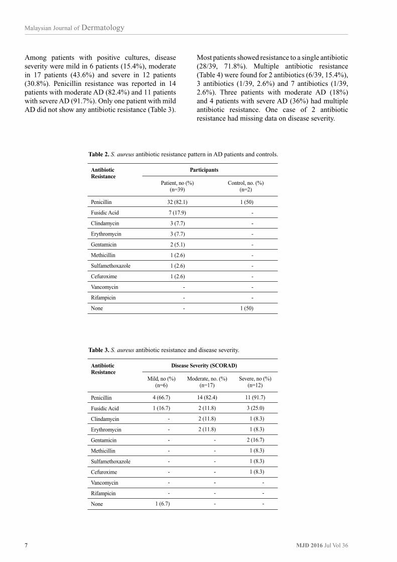
Among patients with positive cultures, disease severity were mild in 6 patients (15.4%), moderate in 17 patients (43.6%) and severe in 12 patients (30.8%). Penicillin resistance was reported in 14 patients with moderate AD (82.4%) and 11 patients with severe AD (91.7%). Only one patient with mild AD did not show any antibiotic resistance (Table 3).
Most patients showed resistance to a single antibiotic (28/39, 71.8%). Multiple antibiotic resistance (Table 4) were found for 2 antibiotics (6/39, 15.4%), 3 antibiotics (1/39, 2.6%) and 7 antibiotics (1/39, 2.6%). Three patients with moderate AD (18%) and 4 patients with severe AD (36%) had multiple antibiotic resistance. One case of 2 antibiotic resistance had missing data on disease severity.
AntibioticResistance
Penicillin
Fusidic Acid
Clindamycin
Erythromycin
Gentamicin
Methicillin
Sulfamethoxazole
Cefuroxime
Vancomycin
Rifampicin
None
Participants
Patient, no (%)(n=39)
32 (82.1)
7 (17.9)
3 (7.7)
3 (7.7)
2 (5.1)
1 (2.6)
1 (2.6)
1 (2.6)
-
-
-
Control, no. (%)(n=2)
1 (50)
-
-
-
-
-
-
-
-
-
1 (50)
Table 2. S. aureus antibiotic resistance pattern in AD patients and controls.
AntibioticResistance
Penicillin
Fusidic Acid
Clindamycin
Erythromycin
Gentamicin
Methicillin
Sulfamethoxazole
Cefuroxime
Vancomycin
Rifampicin
None
Disease Severity (SCORAD)
Mild, no (%)(n=6)
4 (66.7)
1 (16.7)
-
-
-
-
-
-
-
-
1 (6.7)
Moderate, no. (%)(n=17)
14 (82.4)
2 (11.8)
2 (11.8)
2 (11.8)
-
-
-
-
-
-
-
Severe, no (%)(n=12)
11 (91.7)
3 (25.0)
1 (8.3)
1 (8.3)
2 (16.7)
1 (8.3)
1 (8.3)
1 (8.3)
-
-
-
Table 3. S. aureus antibiotic resistance and disease severity.

Malaysian Journal of Dermatology
8MJD 2016 Jul Vol 36
Concurrent antibiotic resistance
2 antibioticsFusidic Acid & Penicillin*Erythromycin & ClindamycinGentamicin & Penicillin
3 antibioticsErythromycin, Clindamycin & Penicillin
7 antibioticsMethicillin, Erythromycin, Sulfamethoxazole, Gentamicin, Clindamycin, Penicillin & Cefuroxime
Mild, no (%)(n=6)
---
-
-
Moderate, no. (%)(n=17)
1 (5.9)1 (5.9)
-
1 (5.9)
-
Severe, no (%)(n=12)
2 (16.7)-
1 (8.3)
-
1 (8.3)
Table 4. Multiple S. aureus resistance and disease severity.
Disease Severity (SCORAD)
* One case had missing data for disease severity
AntibioticResistance
Penicillin
Fusidic Acid
Clindamycin
Erythromycin
Gentamicin
Methicillin
Sulfamethoxazole
Cefuroxime
Vancomycin
Rifampicin
Percentage of Resistance
This Study (%)
82.1
17.9
7.7
7.7
5.1
2.6
2.6
2.6
-
-
NSAR (%)
82.4
13.0
11.2
18.3
14.0
17.3
12.2
13.4
-
2.2
Table 5. S. aureus antibiotic resistance pattern in AD patients versusMalaysian National Surveillance of Antibiotic Resistance (NSAR).
DiscussionFrequent prescription as well as prolonged usage of topical and oral antibiotics promotes its resistance. Owing to increased prevalence of antibiotic resistances, some authors discouraged to use antibiotics for purpose of decolonization without clinical signs of infections7.
S. aureus antibiotic resistance in this study is compared to Malaysian National Surveillance of Antibiotic Resistance (NSAR) report (Table 5).
Antibiotic resistances reported in NSAR are based on S. aureus all isolates that were analyzed during routine laboratory test results done in hospitals.
Penicillin resistance rates in this study (82.1%) are surprisingly comparable to hospital antibiotic resistance rates (82.4%). Several studies also reported similar penicillin resistance rates among AD patients; Poland (82%), and Singapore (91.7% in adults and 93.3% in children). According to Kedzierska et al., resistance towards penicillin did not increased in recolonized lesion8.
Malaysian Journal of Dermatology
9 MJD 2016 Jul Vol 36
Alarmingly, fusidic acid resistance (17.9%) in this study is higher by 4.9% compared to NSAR. Prolonged use of fusidic acid had been associated with increased antibiotic resistances. Kedzierska et al. found increased resistance from 0 to 18% in cases of subsequent recolonization within 75 days of treatment8. In another separate incidence, 78% of atopic eczema patient who had applied topical fusidic acid over the past 6 months were found to be resistant to fusidic acid9. In addition, its resistance tripled during prolonged application from infancy to adolescents10.
Fusidic acid is a commonly used anti-staphylococcal drug. Recently, emergence of resistance to fusidic acid is escalating particularly in impetigo where it is recommended as the first line medication11. Fusidic acid resistance is also found among methicillin resistant S. aureus (MRSA)12. In line with this, ANVISA (Brazilian National Health Surveillance Agency) now requires prescription for fusidic acid effective 2011. Mason et al. reported significant correlation between prescription of fusidic acid and its resistance (p=0.001) with average resistance of 2.8%13.
Results had shown low rate of resistance for erythromycin, clindamycin and gentamicin among AD patients. Their resistances were lower than NSAR data; erythromycin (18%), clindamycin (11%) and gentamicin (14%). Erythromycin is often prescribed in cases of penicillin allergy.
In this study, low resistances were found for methicillin, sulfamethoxazole and cefuroxime with nearly full susceptibility. Their resistances were lower than NSAR data of antibiotic resistances; methicillin
(17%), sulfamethoxazole (12%) and cefuroxime (13%). Hoeger reported full susceptibility among 115 pediatric in Germany with moderate to severe AD towards cefuroxime and methicillin as well14. Methicillin’s low resistances rate in AD were also comparable to other study in Germany, United States, New Zealand and Singapore15-18.
Full susceptibilities were found for vancomycin and rifampicin in this study. Similarly, NSAR reported no vancomycin resistant S. aureus; however 2% of resistance was reported for Rifampicin.
Concurrent antibiotic resistances are largely unexplored. Giliani et al. reported concurrent antibiotic resistance between penicillin with intermediate fusidic acid resistance (24%) as well as penicillin with complete fusidic acid (10%) (19). Similar trend were observed for 8% of our AD patient; 1 with moderate and 2 with severe disease severity. Incidences of concurrent antibiotic resistances increase with disease severity.
Clinicians are recommended to conduct susceptibility tests of clinical S. aureus towards antibiotic resistance before each therapy. In addition, it is also worthwhile to monitor rates of resistance in cases of prolong application. Lastly, strong consideration is posed to reduce prescription of penicillin and fusidic acid to preserve their sensitivity.
AcknowledgementThis study was supported by research grant from Persatuan Dermatologi Malaysia. Authors would like to extend their appreciation for their financial support.
References 1. Roll A, Cozzio A, Fischer B, Schmid-Grendelmeier P.
Microbial colonization and atopic dermatitis. Curr Opin Allergy Clin Immunol. 2004;4(5):373-8.
2. Matsui K, Nishikawa A, Suto H, Tsuboi R, Ogawa H. Comparative study of Staphylococcus aureus isolated from lesional and non-lesional skin of atopic dermatitis patients. Microbiol Immunol. 2000;44(11):945-7.
3. Hoare C, Li Wan Po A, Williams H. Systematic review of treatments for atopic eczema. Health Technol Assess. 2000;4(37):1-191.
4. Guzik TJ, Bzowska M, Kasprowicz A, Czerniawska-Mysik G, Wojcik K, Szmyd D, et al. Persistent skin colonization with Staphylococcus aureus in atopic dermatitis: relationship to clinical and immunological parameters. Clin Exp Allergy. 2005;35(4):448-55.
5. Hanifin JM, Rogge JL. Staphylococcal infections in patients with atopic dermatitis. Arch Dermatol. 1977;113(10):1383-6.
6. DiNubile MJ, Lipsky BA. Complicated infections of skin and skin structures: when the infection is more than skin deep. J Antimicrob Chemoth. 2004;53(Supp. S2):ii37 - ii50.
7. Buys LM, D. P, B.C.P.S. Treatment Options for Atopic Dermatitis. Am Fam Physician. 2007;75(4):523 - 8.
8. Kedzierska A, Kapinska-Mrowiecka M, Czubak-Macugowska M, Wojcik K, Kedzierska J. Susceptibility testing and resistance phenotype detection in Staphylococcus aureus strains isolated from patients with atopic dermatitis, with apparent and recurrent skin colonization. Brit J Dermatol. 2008;159(6):1290-9.

Malaysian Journal of Dermatology
10MJD 2016 Jul Vol 36
9. Shah M, Mohanraj M. High levels of fusidic acid-resistant Staphylococcus aureus in dermatology patients. Brit J Dermatol. 2003;148(5):1018-20.
10. Arkwright PD, Daniel TO, Sanyal D, David TJ, Patel L. Age-related prevalence and antibiotic resistance of pathogenic staphylococci and streptococci in children with infected atopic dermatitis at a single-specialty center. Arch Dermatol. 2002;138(7):939-41.
11. Koning S, van Suijlekom-Smit LW, Nouwen JL, Verduin CM, Bernsen RM, Oranje AP, et al. Fusidic acid cream in the treatment of impetigo in general practice: double blind randomised placebo controlled trial. BMJ. 2002;324(7331):203-6.
12. Howden BP, Grayson ML. Dumb and dumber - the potential waste of a useful antistaphylococcal agent: emerging fusidic acid resistance in Staphylococcus aureus. Clin Infec Dis. 2006;42(3):394-400.
13. Mason BW, Howard AJ, Magee JT. Fusidic acid resistance in community isolates of methicillin-susceptible Staphylococcus aureus and fusidic acid prescribing. J Antimicrob Chemother. 2003;51(4):1033-6.
14. Hoeger PH. Antimicrobial susceptibility of skin-colonizing S. aureus strains in children with atopic dermatitis. Pediatr Allergy Immunology. 2004;15(5):474-7.
15. Goh C-L, Wong JS, Giam YC. Skin colonization of Staphylococcus aureus in atopic dermatitis patients seen at the National Skin Centre, Singapore. Int J Dermatol. 1997;36(9):653-7.
16. Niebuhr M, Mai U, Kapp A, Werfel T. Antibiotic treatment of cutaneous infections with Staphylococcus aureus in patients with atopic dermatitis: current antimicrobial resistances and susceptibilities. Exp Dermatol. 2008;17(11):953-7.
17. Matiz C, Tom WL, Eichenfield LF, Pong A, Friedlander SF. Children with atopic dermatitis appear less likely to be infected with community acquired methicillin-resistant Staphylococcus aureus: the San Diego experience. Pediatr Dermatol. 2011;28(1):6-11.
18. Hill SE, Yung A, Rademaker M. Prevalence of Staphylococcus aureus and antibiotic resistance in children with atopic dermatitis: A New Zealand experience. Australas J Dermatol. 2011;52(1):27-31.
19. Gilani SJK, Gonzalez M, Hussain I, Finlay AY, Patel GK. Staphylococcus aureus re-colonization in atopic dermatitis: beyond the skin. Clin Exp Dermatol. 2005;30(1):10-3.
LEARNING POINTS FROM THIS STUDY
1. Staphylococcus aureus antibiotics resistance is high in patients with atopic eczema, with penicillin resistance highest followed by fucidic acid. This is not surprising as penicillin based antibiotics are frequently prescribed and in most times inappropriately for multiple reasons. Resistance to fucidic acid is also not surprising as this is the most prescribed topical antibiotics for patients with eczema. Combination of fucidic acid and steroid cream are also frequently used in patients with eczema, usually prolonged over many months. Thus, it is important for health care settings to limit use of such topical antibiotics alone or in combination to 2 weeks treatment to prevent resistance.
2. Resistance to clindamycin and erythromycin was also a problem in this study. These antibiotics are highly utilized both in oral and topical forms in the treatment of acne and in most times inappropriately prolonged. Again, time limit to the use of such medication is vital.
3. This is the experience of Universiti Malaya Medical Centre and it is imperative that all hospitals and medical centres have their own antibiotic resistance pattern and a committee looking into its use including the topical antibiotics to reduce antibiotic resistance. It is also encouraged in the GP setting where antibiotics are freely prescribed.
Yap FBB MD MRCP AdvMDermEditor-in-Chief, MJD

Malaysian Journal of Dermatology
11 MJD 2016 Jul Vol 36
INFECTIOUS DISEASE DERMATOLOGY - Original Article
CLINICAL PRESENTATION AND OUTCOME OF HERPES ZOSTER INFECTION IN A TERTIARY DERMATOLOGY OUTPATIENT REFERRAL CLINIC IN MALAYSIAYeoh CA, MRCP1, Chan LC, M Med2; Tan WC, MRCP1, Wee HC, MD3
Abstract
Introduction: Herpes zoster (HZ) is a common acute, cutaneous viral infection caused by reactivation of latent varicella zoster virus with devastating effects on quality of life. This study aims to describe the demographic and clinical characteristic and complications of HZ.
Methodology: This was a retrospective study of 179 HZ patients from the Dermatology department of Penang Hospital between January 2010 and June 2013.
Results: The 179 patients had a median age of 53 years. Chinese ethnicity was more affected. Majority of the patients came late to seek treatment with the median of disease duration of 4 days. The commonest presenting complaint was pain (98.9%), followed by itching (25.7%) and fever (9.5%). Single dermatome involvement was seen in 90.5% of the patients, of which the thoracic dermatome (54.9%) being the commonest. The incidence of complications such as secondary bacterial infection, post-herpertic neuralgia, eye complication(s) and scar were 36.3%, 4.5%, 5.6% and 2.8% respectively. The complications were not statistically different between the younger and the older patient. However, it was more common among male patients.
Conclusion: Patients with HZ in Penang presented late and tend to have complications. Hence, public education and vaccination should be recommended.
Keywords: Herpes zoster, postherpetic neuralgia, Malaysia
Corresponding Author and Reprint Request Dr Yeoh Chin Aun,Department of Dermatology,Hospital Sultanah Bahiyah, Alor Setar, KedahEmail: [email protected]
1 Dermatology Department, Sultanah Bahiyah Hospital2 Dermatology Department, Penang Hospital3 Clinical Research Centre, Penang Hospital
America, Europe and Asia-Pacific ranged between 3 and 5/1000 person-years1. The age-specific incidence rate of HZ rises significantly after 50 years of age1.
Characteristically, HZ presents with a self-limiting localized dermatomal painful blistering rash. The most common complication of HZ is post-herpetic neuralgia, which can cause devastating effects on patients’ quality of life and significant global health burden1. Other complications are neurological sequelae, secondary bacterial infection, HZ ophthalmicus with eye involvement, disseminated disease, and scar formation.
IntroductionHerpes zoster (shingles) is a common acute, cutaneous viral infection caused by reactivation of latent varicella zoster virus (VZV) that has remained dormant within the dorsal root ganglia. The incidence rate of Herpes zoster (HZ) in North

Malaysian Journal of Dermatology
12MJD 2016 Jul Vol 36
the Dermatology department of Penang Hospital were included. Random sampling methods were used. All patients aged more than 18 years old diagnosed as HZ from January 2010 to June 2013 were included into this study. The diagnosis of HZ was established by historical and clinical presentations of the patient. Demographic data that were available for this study included age, gender, race, referral centres and onset of the disease. Other disease-related data such as medical history, presentation, Herpes Zoster related complication, immune status, treatment responses and disease outcome were also retrieved for statistical analysis.
We characterized the patients as having complications if they developed either one of the following signs: post HZ scars, post-herpetic neuralgia, visceral involvement, Ramsay hunt, eyes complications and secondary infections. Post herpetic neuralgia is was defined as pain that persists more than 30 days after cutaneous healing2. Conjunctivitis, keratitis, uveitis or ocular cranial-nerve palsies was considered as eye complications. The patients were categorized as immunocompromised if they were having malignancy, diabetes mellitus, chronic kidney disease, end stage kidney failure, HIV or taking immunosuppressants.
To our knowledge, there is no published study on HZ especially on clinical presentation in Malaysia. Additionally in the latest progress in HZ, vaccination for HZ is getting more important in the prevention of this disease. Vaccination can help to reduce the severity of complications due to HZ. Thus, it is important to look into our local population on clinical presentation of HZ to ensure that our patients present or develop complication(s) same like other country or study population. This can help us to plan for a good cost effective strategy to combat HZ in our country.
The primary objective of our study was to describe the demographic and clinical characteristic of HZ patients in our dermatology clinic. We also describe the involvement of dermatome among two age groups and sex, and to explore factors associated with HZ complications. This descriptive clinical information can provide the local health care provider with a better understanding of the disease and to further define the disease characteristics unique to our local multi-ethnic population.
Materials and methodsThis was a single-centre, hospital-based, retrospective-descriptive study. A total of 179 clinically diagnosed Herpes zoster infections from
Variables
Age ( years)
Age Group ( years)
Young [ < 60]
Old [ ≥ 60]
Gender
Male
Female
Ethnicity
Malay
Chinese
Indian
Others
Referral sources
Primary care
Other than Primary Care
Self referral
Median (IQR)
53 (34)a
Frequency, n (%)
106 (59.2)
73 (40.8)
103 (57.5)
76 (42.5)
75 (41.9)
85 (47.5)
11(6.1)
8 (4.5)
133 (74.3)
43 (24.0)
3 (1.7)
Table 1. Demographic of the HZ (n=179)
a Skewed Data IQR = Interquartile Range

Malaysian Journal of Dermatology
13 MJD 2016 Jul Vol 36
were male (n=103; 57.4%) and 47.5% (n=85) were of Chinese ethnic origin. About 27.9% (n=50) of the patients were immunocompromised as they were having malignancy, diabetes mellitus, chronic kidney disease, end stage kidney failure, HIV or taking immunosuppressant. None of these diseases had a significant association with the age group of the patients (< 60 or ≥ 60 years old) despite younger patients co-existed with immunocompromised state.
More than half of the patients (n=98; 54.8%) came late to seek treatment with the median of disease duration of 4 days. All the referral for HZ cases were seen within the same day.
Clinical Presentation and treatmentThe commonest presenting complaint of our cohort was pain (n=164; 98.9%), followed by itching (n=42; 25.7%), fever (n=16; 9.5%), skin swelling (n=2; 1.7%) and acute abdomen (n=1; 0.6%).
Statistical AnalysisStatistical analysis was performed using Statistical Package for the Social Sciences software (Version 15.0). Continuous variables were expressed as median ± with interquartile range (IQR); categorical variables were expressed as a frequency and percentage (%). Simple logistic regression was used to investigate the factors influencing HZ complications. The odds ratios (ORs) and their 95% confidence intervals (CIs) for HZ complications were shown in the final results, with p < 0.05 considered to be statistically significant.
ResultsA total of 179 patients’ data with diagnosis of HZ were retrieved from the clinic cards.
Demographics of the patientsThe median age of this cohort was 53 ± 34 years old with 59.2% (n=106) of the patients less than 60 years old (young patients). Majority of the patients
Dermatome
Thoracic
Cervical
V1 (Trigeminal)
Lumbar
V2, V3 (Trigeminal)
Sacral
Total[n, %]
89 (54.9)
22 (13.6)
21 (13.0)
19 (11.7)
8 (4.9)
3 (1.9)
Young (< 60 years)
n (%)
54 (55.7)
10 (10.3)
12 (12.4)
12 (12.4)
7 (7.2)
2 (2.0)
Older ( ≥ 60 years)
n (%)
35 (53.8)
12 (18.5)
9 (13.8)
7 (10.8)
1 (1.5)
1(1.5)
P valuea
p= 0.464
Table 2. Comparison of the distribution of dermatome involved between two age groups (n= 162).
a Fisher Exact test
Dermatome
Thoracic
Cervical
V1 (Trigeminal)
Lumbar
V2,V3 (Trigeminal)
Sacral
Total[n, %]
89 (54.9)
22 (15.5)
21 (12.9)
16 (9.9)
8 (4.9)
3 (1.9)
Male[ n,%]
51 (56.0%)
11 (12.1%)
15 (16.5%)
6 (6.6%)
6 (6.6%)
2 (2.2%)
Female( ≥ 60 years old)
[n,%]
38 (53.5%)
11 (15.5%)
6 (8.5%)
13 (18.3%)
2 (2.8%)
1 (1.4%)
Table 3. Comparison of the distribution of unilateral dermatome involved between genders (n=162).
Malaysian Journal of Dermatology
14MJD 2016 Jul Vol 36
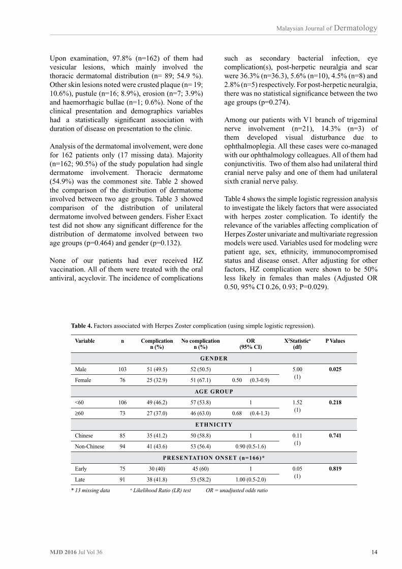
such as secondary bacterial infection, eye complication(s), post-herpetic neuralgia and scar were 36.3% (n=36.3), 5.6% (n=10), 4.5% (n=8) and 2.8% (n=5) respectively. For post-herpetic neuralgia, there was no statistical significance between the two age groups (p=0.274).
Among our patients with V1 branch of trigeminal nerve involvement (n=21), 14.3% (n=3) of them developed visual disturbance due to ophthalmoplegia. All these cases were co-managed with our ophthalmology colleagues. All of them had conjunctivitis. Two of them also had unilateral third cranial nerve palsy and one of them had unilateral sixth cranial nerve palsy.
Table 4 shows the simple logistic regression analysis to investigate the likely factors that were associated with herpes zoster complication. To identify the relevance of the variables affecting complication of Herpes Zoster univariate and multivariate regression models were used. Variables used for modeling were patient age, sex, ethnicity, immunocompromised status and disease onset. After adjusting for other factors, HZ complication were shown to be 50% less likely in females than males (Adjusted OR 0.50, 95% CI 0.26, 0.93; P=0.029).
Upon examination, 97.8% (n=162) of them had vesicular lesions, which mainly involved the thoracic dermatomal distribution (n= 89; 54.9 %). Other skin lesions noted were crusted plaque (n= 19; 10.6%), pustule (n=16; 8.9%), erosion (n=7; 3.9%) and haemorrhagic bullae (n=1; 0.6%). None of the clinical presentation and demographics variables had a statistically significant association with duration of disease on presentation to the clinic.
Analysis of the dermatomal involvement, were done for 162 patients only (17 missing data). Majority (n=162; 90.5%) of the study population had single dermatome involvement. Thoracic dermatome (54.9%) was the commonest site. Table 2 showed the comparison of the distribution of dermatome involved between two age groups. Table 3 showed comparison of the distribution of unilateral dermatome involved between genders. Fisher Exact test did not show any significant difference for the distribution of dermatome involved between two age groups (p=0.464) and gender (p=0.132).
None of our patients had ever received HZ vaccination. All of them were treated with the oral antiviral, acyclovir. The incidence of complications
Variable
Male
Female
<60
≥60
Chinese
Non-Chinese
Early
Late
n
103
76
106
73
85
94
75
91
Complicationn (%)
51 (49.5)
25 (32.9)
49 (46.2)
27 (37.0)
35 (41.2)
41 (43.6)
30 (40)
38 (41.8)
No complicationn (%)
52 (50.5)
51 (67.1)
57 (53.8)
46 (63.0)
50 (58.8)
53 (56.4)
45 (60)
53 (58.2)
X2Statistica
(df)
5.00(1)
1.52(1)
0.11(1)
0.05(1)
P Values
0.025
0.218
0.741
0.819
OR(95% CI)
1
0.50 (0.3-0.9)
1
0.68 (0.4-1.3)
1
0.90 (0.5-1.6)
1
1.00 (0.5-2.0)
Table 4. Factors associated with Herpes Zoster complication (using simple logistic regression).
* 13 missing data a Likelihood Ratio (LR) test OR = unadjusted odds ratio
GENDER
AGE GROUP
ETHNICITY
PRESENTATION ONSET (n=166)*

Malaysian Journal of Dermatology
15 MJD 2016 Jul Vol 36
the Singapore study where only 3% of their study population had ophthalmic dermatome involvement despite both of the studies were done in the tertiary dermatology referral center.
This dermatome involvement can cause serious irreversible complication to the eye. Among our HZ ophthalmicus group, all of them had conjunctivitis and 14.3% had cranial nerve involvement. This was higher than a reported incidence of extraocular muscle palsies in HZ ophthalmicus in North Africa, which was 5.8% 9. This problem can give rise to eye motility disorders with diplopia. This finding added our worry to our local population on complication of HZ other than the post-herpetic neuralgia complication.
More than 50% of our patients presented or were diagnosed late. This had caused a delay in treatment. Early diagnosis followed by appropriate treatment is essential to improve the disease outcome. Acute pain management and prevention of secondary infection can be offered early to reduce the severity of the disease. Antiviral treatment, preferably started within 72 hour of rash onset, can help to resolve the acute disease and inhibit late inflammatory recurrences10. A study done in Singapore, has concluded that there is a need to educate patients at risk to identify the prodrome and skin eruptions of herpes zoster so that early antiviral therapy can be considered8. Unfortunately, even with early treatment, the incidence of post-herpetic neuralgia is not significantly reduced with antiviral treatment11,12. Thus, an alternative solution should be offered to our patients in reducing the severity of HZ and post-herpetic neuralgia. Proactive strategy with vaccination to the elderly population may be is a good solution to prevent the disease from our population group who tend to seek treatment late.
Herpes zoster vaccine was licensed in 2006 and recommended by the Advisory Committee on Immunization Practices in 2008 for prevention of herpes zoster and its complications among adults aged ≥60 years13. It is a live attenuated vaccine. Thus, it should not be given to a patient who is receiving immunosuppressive therapy, including high-dose corticosteroids, has primary or acquired immunodeficiency state, including leukemia, lymphoma, or other malignant neoplasm affecting the bone marrow or lymphatic system, or with acquired immunodeficiency syndrome or other clinical manifestation of infection with human immunodeficiency viruses13.
DiscussionHerpes Zoster is a common disease which is caused by reactivation of latent varicella zoster virus. Most of the reactivation can be either prevented or quickly aborted with the presence of adequate T cell-mediated immune response. However, in immunocompromised or age-related immunosenescence patients, immune response might be inadequate to contain the reactivation of VZV. This explains the more severe form of the disease in these groups of patients. The incidence rate of HZ was about 6-8/1000 person-years at 60 years of age and 8–12/1000 person years at 80 years of age1. The age-specific incidence rates of HZ were similar across countries, with a steep rise after 50 years of age1.
In our study, 40.8% of the study population was from age group more than 60 years old, and the median age was 53 years old. Patients younger than 60 years old had more complications of HZ when compared with older group. This is possible due to the fact that the study was done in a hospital-based dermatology clinic with more in-patient referrals. The younger population had more immunocompromised state as defined by presence of malignancy, diabetes mellitus, chronic kidney disease, end stage kidney failure, HIV or taking immunosuppressant.
The prevalence of HZ in our study for both genders was almost the same. This finding is similar to other researchers’ result which found no difference by sex in HZ3,4. Majority of our study population was Chinese despite our clinic attendees mainly is Malay ethnicity. The racial distribution in the study dermatology clinic were 63.5%, 24.9%, 10.1% and 1.5% respectively for Malay, Chinese, Indian and others. A study done in United Kingdom indicated that zoster risk in patients was 54% lower among blacks5. However, the reasons for these racial differences are unknown.
In HZ, the stages of eruption elements are macula, papules, blisters, crusting then followed by scar or post inflammatory hypo/hyperpigmentation formation. Majority of HZ patients presented with a self-limiting localized dermatomal painful blistering rash. In general, thoracic, cervical, and ophthalmic involvement are most common6,7. We noted a similar presentation among our patients. Goh and Khoo from Singapore also reported the same findings8. In our study, both genders and age groups presented with similar dermatomal distribution. V1 division of Trigeminal nerve was the third most common (13%) dermatome in our study. This is much higher than

Malaysian Journal of Dermatology
16MJD 2016 Jul Vol 36
global health burden, especially post herpetic neuralgia1. Although all of our patients received systemic antiviral treatment, up to 42.5% of our patients developed complications of HZ. 36.3% of our study cohort had secondary bacterial infection. These might be due to late presentation of the patients.
To our knowledge, this is the first published observational study that gave the overview picture of the HZ in Malaysia. The main limitation of our study was that this is a retrospective single center study. Some useful data like education level, family income, transportation accessibility and economic impact of HZ to the patients were not available. There was also presence of incomplete documentation that limit our further analysis.
ConclusionDespite majority of the HZ patients in our study presented with typical presentation of HZ majority of them were still late for treatment (> 3 days). Complication of HZ is common in our study population. Hence, we suggest proactive strategies with education of the disease and HZ vaccination should be recommended to our community.
With vaccination, the risk of having HZ, the burden of disease, and the incidence of post-herpetic neuralgia reduced by 51%, 61%, and 66% respectively, over 3 years period13,14. However, none of our patients has ever received HZ vaccination. There was no statistical significance in terms of complication of HZ between the two age groups, ethnicity, immune status or late/early presentation in this study. The non-significance among the two age groups might be due to higher proportion of immunocompromised patient among the younger age group, which was 46.2% (49/106). Whereas, there was only 37.0% (27/73) of older age group who was immunocompromised. Post multivariate analysis, male gender was found to be the only risk factor to get the HZ complications.
This finding is contradicts other studies which found women with zoster might also be at increased age-specific risk for developing post herpetic neuralgia compared with men17,18. However the studies did not look into other complication of HZ except post herpetic neuralgia.
All the complication of HZ can cause devastating effects on patients’ quality of life and significant
References 1. Kawai K, Gebremeskel BG, Acosta CJ. Systematic review
of incidence and complications of herpes zoster: towards a global perspective. BMJ open 2014; 4(6): e004833.
2. Gnann JW, Whitley RJ. Clinical practice for Herpes zoster. The New England journal of medicine 2002; 347:340-6
3. Di Luzio PU, Arpinelli F, Visona G. Herpes zoster and its complications in Italy: an observational survey; J Infect 1999; 38(2): 116-120.
4. Donahue JG, Choo PW, Manson JE et al. The incidence of herpes zoster; Arch Intern Med 1995; 155(15): 1605-1609.
5. Thomas SL, Hall AJ. What does epidemiology tell us about risk factors for herpes zoster?; Lancet Infect Dis 2004; 4(1): 26-33.
6. Schmader KE, Oxman MN. Varicella and Herpes Zoster. In: Fitzpatrick’s Dermatology in General Medicine (8th eds) 2012; The McGraw-Hill Companies, Inc.
7. Kost RG, Straus SE. Postherpetic neuralgia---pathogenesis, treatment, and prevention. N Eng J Med 1996; 335:32--42.
8. Goh CL, Khoo L. A retrospective study of the clinical presentation and outcome of herpes zoster in a tertiary dermatology outpatient referral clinic. International journal of dermatology 1997; 36(9):667-672.
9. Kahloun R, Attia S, Jelliti B et al. Ocular involvement and visual outcome of herpes zoster ophthalmicus: review of 45 patients from Tunisia, North Africa; Journal of ophthalmic inflammation and infection 2014; 4:25.
10. Dworkin RH, Johnson RW, Breuer J et al: Recommendations for the management of herpes zoster. Clinical infectious diseases 2007; 44 (Suppl 1): S1-26.
11. Schmader K. Herpes zoster in older adults. Clinical infectious diseases 2001; 32:1481-6.
12. Volpi A, Gross G, Hercogova J, et al. Current management of herpes zoster: the European view. American journal of clinical dermatology 2005; 6: 317-25.
13. Schmader K. Herpes zoster in older adults. Clinical infectious diseases 2001; 32:1481-6.
14. Volpi A, Gross G, Hercogova J et al. Current management of herpes zoster: the European view. American journal of clinical dermatology 2005; 6: 317-25.
15. Harpaz R, Ortega-Sanchez IR, Seward JF et al. Prevention of herpes zoster: recommendations of the Advisory Committee on Immunization Practices (ACIP). MMWR. Recommendations and reports: Morbidity and mortality weekly report. Recommendations and reports / Centers for Disease Control. 2008; 57:1-30.
16. Oxman MN, Levin MJ, Johnson GR et al. A vaccine to prevent herpes zoster and postherpetic neuralgia in older adults. The New England journal of medicine 2005; 352: 2271-84.
17. Schmader K. Postherpetic neuralgia in immunocompetent elderly people. Vaccine 1998; 16:1768-70.
18. Yawn BP, Saddier S, Wollan P et al. A population-based study of the incidence and complications of herpes zoster before zoster vaccine introduction. Mayo Clin Proc 2007; 82:1341--9.

Malaysian Journal of Dermatology
17 MJD 2016 Jul Vol 36
LEARNING POINTS FROM THIS STUDY
1. The study showed that the median age of patients was 53 years with 59.2% less than 60 years old. There is a recent observation that the incidence of herpes zoster is increasing in immunocompetent young adults and also in children. Thus, it is imperative that clinicians do not miss the diagnosis in the younger individuals to prevent morbidities.
2. In this study, majority of patients presented late with a median of 4 days from initial symptoms/ signs. This is not uncommon as the symptom of pain usually precedes the sign by a few days, delaying the diagnosis.
3. This study found a higher incidence of herpes zoster in the Chinese population. However, this might be due to the demographics of the population in Penang.
4. Complications and morbidities of herpes zoster is high and thus it is vital that clinicians make an early and accurate diagnosis. This will allow early treatment to prevent such complications. Education of health care workers and patients are also vital as has been alluded by the authors. Another point is that patients with herpes zoster affecting the ophthalmic branch of the trigeminal nerve should be screened by opthlamologist, more so in those with the nasociliary branch involvement. All health care workers and junior doctors need to be aware of this as to prevent devastating eye complications of herpes zoster.
5. This study highlighted males to have higher complications rate than females. This might be related to the delayed diagnosis and treatment among males. Nevertheless, aggressive treatment is necessary for herpes zoster irrespective of gender. Clinicians should be more wary of complications of the disease among male patients.
6. Vaccination of herpes zoster to prevent the disease and its complications is effective and recommended in those more than 60 years. Education of health care workers and general public about availability of such vaccination is important as mentioned by the authors and will lead to reduction of herpes zoster and its complications in the elderly.
Yap FBB MD MRCP AdvMDermEditor-in-Chief, MJD

Malaysian Journal of Dermatology
18MJD 2016 Jul Vol 36
GENERAL DERMATOLOGY - Original Article
KNOWLEDGE, ATTITUDE AND PRACTICES OF ADULTSIN RELATION TO SUN EXPOSURE AND PHOTODAMAGESam SYY1, Lim JSJ1, Liau MMQ1, Toh MHS2, Aw DCW1
Abstract
Background: Protection from sun exposure is key in the prevention of photodamage and skin cancer, and is particularly important in countries that experience high ultraviolet exposure. We compare the knowledge, attitude and behaviour towards sun exposure in Singapore between adults with and without photodamage. We also describe the clinical features of patients with photodamage in Singapore.
Methods: 532 subjects were recruited from the dermatology specialist outpatient of a tertiary hospital in Singapore. Each subject was assessed clinically by a dermatologist for evidence of photodamage, and answered a questionnaire assessing his knowledge, attitude and behaviour towards sun exposure and protection.
Results: Subjects with photodamage were older, and had lower education and employment rates compared to subjects without photodamage. There was no significant difference in knowledge on the harmful effects of sun exposure and on sun protection or in sun avoidance behaviour (other than use of protective sunglasses) between the two groups, though more patients with photodamage felt that they take adequate sun protection measures. Of note, only a low percentage of subjects in both groups (24.5% of subjects with photodamage and 23.1% of subjects without photodamage) practise regular use of sunscreen.
Conclusion: There was no significant difference between the knowledge, attitudes and behaviours of subjects with and without photodamage, though demographic differences between the two groups exist. Regular sunscreen usage is low in Singapore, a country with high exposure to ultraviolet light, and measures to educate and modify the behaviour of the public need to be developed.
Keywords: UVA protection, UVB protection, photodamage, sunscreen, sunprotection
Corresponding Author and Reprint Request Dr Sam Shiyao Yang, MBBS, MRCP(UK)National University Health System, SingaporeDepartment of Dermatology, 1E Kent Ridge RoadTower Block, Level 10, Singapore 119228Email: [email protected]
1 Division of Dermatology, National University Health System, Singapore2 Saw Swee Hock School of Public Health, National Health Group, Singapore
(1968 – 1972) to 8.9/100,000 person-years (1993-1997), affecting mainly older adults2. Singapore is located near the equator, and experiences one of the highest ultraviolet (UV) exposures in the world throughout the year, with UV index scores ranging from 10 to 13 based on the World Health Organisation UV Index values3.
UV radiation is recognized as a group 1 carcinogen to humans by the International Agency for Research on Cancer4-8. It is responsible for photodamage4-6, photoaging, photocarcinogenesis, and eventually skin cancer7,8. Skin cancer and photodamage are highly preventable by adequate sun protection
IntroductionSkin cancer is the 6th most common cancer in both men and women in Singapore1. Its incidence has increased from a rate of 6.0/100,000 person-years
Malaysian Journal of Dermatology
19 MJD 2016 Jul Vol 36
MethodologyWe performed a cross-sectional study over a 6-month period on patients aged 21 years and above, at the dermatology specialist outpatient clinic at a tertiary centre in National University Hospital, Singapore. Patients with photodermatitis and photoaggravated dermatoses were excluded.
measures9,10, such as avoiding sun exposure between 10am and 2pm, seeking shade, using sunscreen, and wearing protective clothing10-12.
We intend to study the knowledge, attitudes, and behaviour of adults in Singapore relating to sun exposure, and whether this has any impact on photodamage.
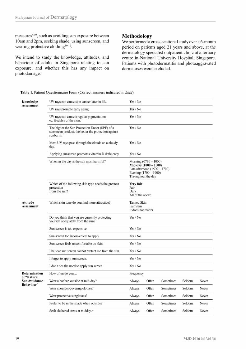
KnowledgeAssessment
Attitude Assessment
Determination of “Natural Sun Avoidance Behaviour”
UV rays can cause skin cancer later in life.
UV rays promote early aging.
UV rays can cause irregular pigmentationeg. freckles of the skin.
The higher the Sun Protection Factor (SPF) of a sunscreen product, the better the protection against sunburns.
Most UV rays pass through the clouds on a cloudy day.
Applying sunscreen promotes vitamin D deficiency.
When in the day is the sun most harmful?
Which of the following skin type needs the greatest protectionfrom the sun?
Which skin tone do you find more attractive?
Do you think that you are currently protecting yourself adequately from the sun?
Sun screen is too expensive.
Sun screen too inconvenient to apply.
Sun screen feels uncomfortable on skin.
I believe sun screen cannot protect me from the sun.
I forget to apply sun screen.
I don’t see the need to apply sun screen.
How often do you…
Wear a hat/cap outside at mid-day?
Wear shoulder-covering clothes?
Wear protective sunglasses?
Prefer to be in the shade when outside?
Seek sheltered areas at midday>
Yes / No
Yes / No
Yes / No
Yes / No
Yes / No
Yes / No
Morning (0730 – 1000)Mid-day (1000 – 1500)Late afternoon (1500 – 1700) Evening (1700 – 1900) Throughout the day
Very fairFairDarkAll of the above
Tanned SkinFair SkinIt does not matter
Yes / No
Yes / No
Yes / No
Yes / No
Yes / No
Yes / No
Yes / No
Frequency
Always Often Sometimes Seldom Never
Always Often Sometimes Seldom Never
Always Often Sometimes Seldom Never
Always Often Sometimes Seldom Never
Always Often Sometimes Seldom Never
Table 1. Patient Questionnaire Form (Correct answers indicated in bold).

Malaysian Journal of Dermatology
20MJD 2016 Jul Vol 36
The first two questions surveyed attitudes towards skin colour attractiveness and adequacy of personal sun protection. The next five questions studied perceptions on the use of sunscreen. Those who answered ‘no’ in at least 5 of the 6 statements on sunscreen were taken to have a positive attitude towards the use of sunscreen. (See Table 2).
Behavioural AssessmentQuestions were asked on the frequency and extent of application, and the amount of sunscreen applied. Natural sun avoidance behavioral activities were also assessed (Table 3). Responses indicating ‘sometimes, often, or always’ were classified as having practiced the stated sun avoidance behaviour. A response of “never” or “seldom” would be deemed as a negative response. A patient would be classified as having ‘natural sun avoidance behaviour’ if they practiced 3 out of the 5 behavioural activities.
QuestionnaireThe questionnaire assessed participants’ knowledge, attitude and behaviour with respect to sun exposure and protection, and was designed based on the American Skin Association recommendations13, WHO Fact Sheet (2010)14 and Reinau15. Certain questions with established reliability and validity pertaining to sunscreen use, frequency of acquiring tans or sunburns, shade-seeking behaviour, and the wearing of protective clothes were included16.
Eight questions were asked about UV exposure and its cutaneous effects, and protection from using sunscreen. Each correct answer scored 1 point. Knowledge scores were classified into ‘high’ (7-8 points), ‘medium’ (5-6 points), or ‘low’ (0-4 points).
Category
Gender
Ethnicity
Age (years)
Education level
Employment
Exposure to sunlight at work
YesN=220 (41.4%)
n (%)
115 (52.3)
105 (47.7)
168 (76.4)
11 (5.0)
21 (9.5)
20 (9.1)
55.3 ±13.3
12 (5.5)
15 (6.8)
38 (17.3)
63 (28.6)
65 (29.5)
27 (12.3)
31 (14.1)
74 (33.6)
54 (24.5)
61 (27.7)
133 (60.5)
87 (39.5)
43 (19.5)
177 (80.5)
0.6 ±1.5
NoN=312 (58.6%)
n (%)
161 (51.6)
151 (48.4)
234 (75.0)
23 (7.4)
27 (8.7)
28 (9.0)
33.2 ±12.0
155 (49.7)
80 (25.6)
44 (14.1)
20 (6.4)
7 (2.2)
6 (1.9)
8 (2.6)
43 (13.8)
98 (31.4)
163 (52.2)
240 (76.9)
72 (23.1)
63 (20.2)
249 (79.8)
0.6 ±1.8
P value
0.930
0.733
<0.001
<0.001
<0.001
0.854
0.788
Photodamage
Table 2. Comparison of demographic data between patients with photodamage and patients without photodamage.
Male
Female
Chinese
Malay
Indian
Others
Mean
21-29
30-39
40-49
50-59
60-69
70+
No formal education/ primary
Secondary
Junior college/ Polytechnic/Diploma
University/post-graduate
Employed
Unemployed
Yes
No
Mean duration of sun exposure (hrs)

Malaysian Journal of Dermatology
21 MJD 2016 Jul Vol 36
Level of Knowledge (classified according to the scores obtained from the Knowledge Assessment Questionnaire - see Table 1)
High (7-8 points)
Medium (5-6 points)
Low (0-4 points)
Was previously given advice on sun protection by health care provider
YesN=220 (41.4%)
n (%)
81 (36.8)
126 (57.3)
13 (5.9)
64 (29.1)
156 (70.9)
NoN=312 (58.6%)
n (%)
120 (38.5)
175 (56.1)
17 (5.4)
53 (17.0)
259 (83.0)
P value
0.917
0.001
Photodamage
Table 3. Knowledge of the harmful effects of excessive sun exposure and on sun protection - comparison between patients with photodamage and patients without photodamage.
Yes
No
Category
Skin colour preference
Tanned skin
Fair skin
No preference for either tanned or fair skin
Self-assessment of own current protection measures
Perception that current protection measures are adequate
Attitude toward sunscreen
Sun screen too expensive (answered ‘Yes’)
Sun screen too inconvenient to apply (answered ‘Yes’)
Sun screen feels uncomfortable on skin (answered ‘Yes’)
I believe sun screen cannot protect me from the sun (anwered ‘Yes’)
I forget to apply sun screen (answered ‘Yes’)
I don’t see the need to apply sun screen (answered ‘Yes’)
Overall positive attitude towards use of sun screen(answered ‘No’ to at least 5 of the above 6 questions on sun screen)
YesN=220 (41.4%)
n (%)
32 (14.5)
89 (40.5)
99 (45.0)
116 (52.7)
93 (42.3%)
107 (48.6%)
107 (48.6%)
51 (23.2%)
130 (59.1%)
108 (49.1%)
103 (46.8%)
NoN=312 (58.6%)
n (%)
57 (18.3)
109 (34.9)
146 (46.8)
130 (41.7)
109 (34.9%)
165 (52.9%)
187 (59.9%)
54 (17.3%)
190 (60.9%)
130 (41.7%)
147 (47.1%)
P value
0.330
0.012
0.086
0.334
0.010
0.094
0.675
0.090
0.946
Photodamage
Table 4. Comparison of the attitude towards skin preference and sunscreen use between patients with photodamage and patients without photodamage.
Statistical analysisData analysis was performed using the IBM Statistical Package for the Social Science (SPSS) version 22.0. The unpaired t-test was used to compare means, chi-square or Fisher’s exact test for comparing proportional data. A probability (p) of <0.05 was considered statistically significant.
Clinical assessmentAll patients were assessed by their attending dermatologist for evaluation on their Fitzpatrick Skin Type, severity of photodamage using the Glogau Photoaging classification, and also the areas of involvement such as the face, neck or limbs.
In addition, we screened for presence of risk factors for malignant melanoma17,18. This included a history of sunburns, and sun-tanning habits.
Malaysian Journal of Dermatology
22MJD 2016 Jul Vol 36
Education and EmploymentMore patients with photodamage lacked tertiary education (72.3% vs 47.8% of patients without photodamage; p < 0.001). More patients with photodamage also lacked employment (76.9% vs 60.5% of patients without photodamage,; p < 0.001).
There was no significant difference in gender, ethnicity and exposure to sunlight at work between patients with and without photodamage.
Knowledge on sun exposure and protection (Table 5)Both groups of patients scored almost equally in the knowledge domain. However, those with photodamage were significantly more likely to have received advice on sun damage than those without. (29.1% vs 17.0%, p=0.001); the temporal sequence between development of photodamage and reception of advice could not be established.
ResultsOf a total of 609 administered questionnaires, 532 were completed with a response rate of 87.3%. Of the 532 participants, 220 (41.4%) were assessed to have photodamaged skin, and 312 (58.6%) did not have photodamaged skin. We then go on to compare the demographic differences between these two groups of patients, and also assess for differences in their knowledge, attitude and practices pertaining to sun-exposure and sun-avoidance behaviours. Finally, we also characterize this group of 220 patients with sun-damaged skin as seen in Tables 8 and 9 respectively.
Demographic differences between patients with photodamage and patients without photodamage (Table 4)Age & EthnicityThe mean age of our participants was 42.3 ±16.6 years. There were approximately equal numbers of males and females (ratio 1.08:1). 75.6% of participants were Chinese, 9% were Indians, 6.4% were Malays, and 9% were of ‘other’ ethnicities. Patients with photodamage were more likely to be older than patients without photodamage (mean age 55.3 ± 13.3 vs 33.2 ± 12.0, p < 0.001).
Category
Users of sun screen (once or several times/day)
Non-users (never/ only when necessary)
Area(s) of use for sunscreen in sun screen users (percentages expressed are that of sunscreen users in each category only)
Face+
Neck+
Arms+
Legs+
Amount of sunscreen applied each time (percentages expressed are that of sunscreen users in each category only)
Rice-grain
Pea-size
Half tea-spoon
Sun avoidance behavior
Wear hat/cap†
Wear shoulder-covering clothes†
Protective sunglasses†
Prefer to be shade when outside†
Seek sheltered areas at midday†
Natural sun avoidance behaviour*
YesN=220n (%)
54 (24.5)
166 (75.5)
53 (98.1)
22 (40.7)
18 (33.3)
6 (11.1)
21 (16.9)
58 (46.8)
45 (36.3)
56 (25.5)
120 (54.5)
106 (48.2)
204 (92.7)
204 (92.7)
154 (70.0)
NoN=312 n (%)
72 (23.1)
240 (76.9)
70 (97.2)
32 (44.4)
17 (23.6)
5 (6.9)
15 (8.8)
98 (57.6)
57 (33.5)
63 (20.2)
176 (56.4)
113 (36.2)
273 (87.5)
293 (93.9)
217 (69.6)
P value
0.695
0.736
0.678
0.228
0.412
0.061
0.151
0.670
0.006
0.051
0.588
0.912
Photodamage
Table 5. Comparison of behaviour relating to Sunscreen use and Natural Sun Avoidance between patients with photodamage and patients without photodamage.

Malaysian Journal of Dermatology
23 MJD 2016 Jul Vol 36
Category
Skin type (Fitzpatrick)
1
2
3
4
5
6
Photoaging grade (Glogau)
Mild
Moderate
Advanced
Severe
Skin condition
Dyspigmentation
Freckles
Irregular pigmentation
Solar lentigines
Collagen degeneration
Solar elastosis
Static wrinkles
Pre-malignant and malignancies
Actinic keratoses
Basal cell carcinoma
Squamous cell carcinoma in-situ
Areas of sun damage
Face
Neck and ‘V’
Limbs
Patients with clinical features of photodamage (%)
N = 220
1 (0.4)
18 (8.2)
54 (24.7)
116 (52.7)
24 (10.9)
7 (3.2)
62 (28.2)
98 (44.5)
59 (26.8)
1 (0.5)
64 (29.1)
155 (70.5)
102 (46.4)
76 (34.5)
114 (51.8)
7 (3.2)
3 (0.6)
1 (0.2)
201 (91.4)
83 (37.7)
96 (43.6)
Table 6. Clinical features of patients with photodamage.
Behaviour Relating to Sunscreen Use and Natural Sun-avoidance (Table 7)There was no significant difference in frequency of application of sunscreen in patients with and without photodamage, the areas of application, and the amount of sunscreen applied.
Only 70.0% of those with photodamage had natural sun avoidance behaviour, the commonest of which were preference for shade when outside (92.7%) and seeking sheltered areas at midday (92.7%). Patients with photodamage were more likely to wear protective sunglasses when outside (48.2% vs 36.2%, p = 0.006).
Attitudes toward skin colour and sun screen (Table 6)In general, there was no significant difference in attitude towards using sunscreen between patients with and without photodamage. More patients with photodamage tended to think that they had taken adequate protection measures (52.7% with photodamage vs 41.7% without photodamage, p = 0.012).The commonest reason for not using sunscreen was forgetfulness (59.1%), followed failing to see a need for it (49.1%). Interestingly, more patients without photodamage felt that sunscreen was uncomfortable on the skin (59.9% vs 48.6% of patients with photodamage, p=0.010).

Malaysian Journal of Dermatology
24MJD 2016 Jul Vol 36
Age is indeed a significant factor in photoaging and is also an established factor in wrinkling5. In a study looking at skin cancer trends in Singapore, Sng et al2 also found that preponderance of skin cancer (BCC, SCC, and malignant melanoma) cases in Singapore occurred among older persons.
Given the significant association between photodamaged patients and unemployment, one explanation could be a reduction of overall sun-exposure due to being indoors during work-hours in employed people. It is heartening to note that having a lower education level did not translate to lower knowledge scores.
Both photodamaged and those without photodamage have mostly high and medium knowledge scores. More photodamaged patients received information about sun-protection by healthcare providers compared to those without. Advice given by physicians has been shown to be associated with higher sun-safety compliance20 compared to media campaigns21, and in the study by Nyiri, schools and parents were shown to comply if a doctor advised sun protection measures19. The lack of difference in knowledge scores may suggest that a greater emphasis needs to be placed on the translation of knowledge to actual practice.
Clinical Features and Types of Photodamage (Table 8) Of the 220 (41.4%) patients with photodamage, most were of skin type 4 (52.7%) and had a moderate degree of (44.5%) photoaging by Glogau grading. The most common signs observed were irregular pigmentation (70.5%) and static wrinkles (51.8%), with the face being the predominant site (91.4%).
Risk factors for skin cancer/malignant melanoma (Table 9)A surprising finding was that most patients with photodamage were less frequently sunburnt than those without photodamage. More patients with photodamage actually do not sun-tan, 93.4% vs 87.1%, (p = 0.041).
DiscussionFew studies have been done to assess the knowledge, attitude, and sun-protection behaviour in Singapore and of the photodamaged-skin cancer population. A previous study by Nyiri19 assessed sun-protection attitude, measures and incidence of sunburn of Singapore’s school children and found that over 50% of school children had sunburns during their first 10 years, supporting the importance of sun-safety campaigns in schools.
Risk Factors
History of sunburn over the past year
Never
1-2x
3-5x
5-10x
>10x
Frequency of sun-tanning
Daily
Once a week
Once every 2 week
Once every 4 weeks
Less than once a month
Never
Time of day when sun-tanning under natural sunlight
Morning
Mid-day
Late afternoon
YesN=219 (41.2%)
n (%)
105 (63.3)
36 (21.7)
14 (8.4)
6 (3.6)
5 (3.0)
3 (1.8)
3 (1.8)
0 (0.0)
1 (0.6)
4 (2.4)
155 (93.4)
8 (3.6)
2 (0.9)
4 (1.8)
NoN=211 (58.8%)
n (%)
101 (42.1)
103 (42.9)
20 (8.3)
11 (4.6)
5 (2.1)
2 (0.8)
4 (1.7)
2 (0.8)
4 (1.7)
19 (7.9)
209 (87.1)
18 (5.8)
14 (4.5)
7 (2.2)
P value
<0.001
0.120
0.041
0.062
Photodamage
Table 7. Comparison of risk factors for skin cancer between patients with photodamage and patients without photodamage.

Malaysian Journal of Dermatology
25 MJD 2016 Jul Vol 36
With respect to the frequency of sunburns and sunbed use as a risk factor for melanoma17,28, those with photodamage were significantly less frequently sun burnt and tended to refrain from sun tanning practices compared to those without photodamage. A likely reason for this unusual finding is that a previous experience of having been sunburnt before may have caused a behavioural change to avoid excessive sun exposure. Additionally, sun-tanning practices are not common in Singapore because of year-round high temperature and strong sunlight.
Study LimitationsOur study population, taken from a tertiary dermatology clinic, may have a different profile from that of the general population and its findings may not be fully generalisable to the general Singaporean population. Limitations of a self-report questionnaire include recall bias and underreporting of actual harmful practices (e.g. frequency of sunburn and suntanning) because of the need to provide socially desirable answers. Causal relationships cannot be established.
ConclusionIn conclusion, our study showed our patients had similar knowledge and attitude towards sunscreen use regardless of presence of photodamage. However, photodamaged patients were more informed by healthcare providers on sun-safety, placed less value on tanned skin, and had less frequency of sunburns and sun-tanning. Subsequent sun-safety studies on the general public, and on skin-cancer patients could be undertaken in the future. We would advise undertaking public education campaigns on sun protection, especially regarding sunscreen use, and targeting the ones at risk of photodamage such as the elderly and the lower educated. Emphasis may need to be placed on behavioural modification instead of merely imparting knowledge. Prospective studies can be considered to evaluate if changes to knowledge, attitude and behaviours of patients from a young age can have an impact on the development of photodamage.
The general study population either preferred fair skin or had no preference for skin colour. Similarly, a sun-safety study done on a Chinese population showed more than half found a suntan unattractive22. This is significantly different from Caucasian populations, where a tan is deemed more visually appealing23.
Of note, in our study population, less than half of the entire study population had a positive attitude towards sunscreen. Only about a quarter of each group applied sunscreen daily despite having high-medium knowledge scores. The most common barrier to sunscreen use was discomfort on the skin, which we believe to be compatible with a hot and humid climate in Singapore. In contrast, forgetfulness and inconvenience were most commonly cited in a Canadian outpatient population20. Additionally, our photodamaged responders also applied a lesser amount of sunscreen than non-photodamaged. This difference approached statistical significance with a p value of 0.061. It is conceivable that the photodamaged skin be a reflection of lower protection. The recommended amount of sunscreen for an entire face is half a tea-spoon24, and may show deficiency in knowledge of sunscreen users. In general, the perception and use of sunscreen in our population is low – and more is required to promote its use nation-wide.
In contrast, a Chinese study by Cheng et al22 and a Western study by Halpern et al25, showed that sunscreen use was the most popular preventive behaviour with 58.8% and 85% of responders using sunscreen respectively. In a study on skin cancer patients, there was a marked increase in the application of sunscreen after diagnosis26. Sunscreen is the gold standard against photodamage27 and skin cancer, and its use should be emphasized more to the public.
When characterizing the features of photoaging amongst our photodamaged patients, we noted that most were of skin type 4 (52.7%) and had moderate (44.5%) photoaging by Glogau grading. Consistent with excessive and unprotected sun exposure, our commonest observation was irregular pigmentation (70.5%) and static wrinkles (51.8%) on the face.

Malaysian Journal of Dermatology
26MJD 2016 Jul Vol 36
References 1. Teo MC, Soo KC. Cancer trends and incidences in
Singapore. Jpn J ClinOncol2013;43:219-224.2. Sng J, Koh D, Siong WC, Choo TB. Skin cancer trends
among Asians living in Singapore from 1968 to 2006. J Am AcadDermatol2009;61:426-432.
3. World Health Organization 2014. Ultraviolet radiation and the INTERSUN Programme. [WWW document]. URL http://www.who.int/uv/intersunprogramme/activities/uv_index/en/index3.html
4. Eun HC. Cutaneous photodamage in Asians. J Dermatol 2001;28:614-616.
5. Chung JH, Lee SH, Youn CSet al. Cutaneous photodamage in Koreans: influence of sex, sun exposure, smoking, and skin color. Arch Dermatol 2001; 137:1043-1051.
6. Chung JH. Photoaging in Asians. Photodermatol PhotoimmunolPhotomed 2003; 19:109-121.
7. Gonzaga ER. Role of UV light in photodamage, skin aging, and skin cancer: importance of photoprotection. Am J ClinDermatol 2009; 10Suppl 1:S19-24.
8. Grether-Beck S, Wlaschek M, Krutmann J, Scharffetter-Kochanek K. Photodamage and photoaging--prevention and treatment. J DtschDermatolGes 2005; 3Suppl 2:S19-25.
9. Armstrong BK, Kricker A. The epidemiology of UV induced skin cancer. J PhotochemPhotobiol B 2001; 63:8-18.
10. Phillips TJ, Bhawan J, Yaar M et al. Effect of daily versus intermittent sunscreen application on solar simulated UV radiation-induced skin response in humans. J Am AcadDermatol2000; 43:610-618.
11. O’Riordan DL, Nehl E, Gies Pet al. Validity of covering-up sun-protection habits: Association of observations and self-report. J Am AcadDermatol2009; 60:739-744.
12. Berret J, Liardet S, Scaletta C et al. Use of sunscreens in families living in Switzerland. Dermatology 2002; 204:202-208.
13. American Skin Association 2012. Sun Safety. [WWW document]. URL www.americanskin.org/resource/safety.php#
14. World Health Organization 2014. Ultraviolet radiation and the INTERSUN Programme – Sun protection. [WWW document]. URL www.who.int/uv/sun_protection/en/
15. Reinau D, Meier C, Gerber N et al. Sun protective behavior of primary and secondary school students in North-Western Switzerland. Swiss Med Wkly2012; 142:w13520. doi: 10.4414/smw.2012.13520
16. Falk M, Anderson CD. Measuring sun exposure habits and sun protection behavior using a comprehensive scoring instrument--an illustration of a possible model based on Likert scale scorings and on estimation of readiness to increase sun protection. Cancer Epidemiol2012; 36:e265-269. doi: 10.1016/j.canep.2012.03.004.
17. Diffey BL, Norridge Z. Reported sun exposure, attitudes to sun protection and perceptions of skin cancer risk: a survey of visitors to Cancer Research UK’s SunSmart campaign website. Br J Dermatol2009; 160:1292-1298.
18. Thomson CS, Woolnough S, Wickenden M, Hiom S, Twelves CJ. Sunbed use in children 11-17 in England: face to face quota sampling surveys in the National Prevalence Study and Six Cities Study. BMJ2010; 340:c877. doi: 10.1136/bmj.c877.
19. Nyiri P. Sun protection in Singapore’s schools. Singapore Med J2005; 46:471-475.
20. Boggild AK, From L. Barriers to sun safety in a Canadian outpatient population. J Cutan Med Surg 2003; 7:292-299.
21. Smith BJ, Ferguson C, McKenzie J et al. Impacts from repeated mass media campaigns to promote sun protection in Australia. Health PromotInt2002; 17:51-60.
22. Cheng S, Lian S, Hao Y et al. Sun-exposure knowledge and protection behavior in a North Chinese population: a questionnaire-based study. Photodermatol Photoimmunol Photomed2010; 26:177-181.
23. Jones B, Oh C, Corkery E et al. Attitudes and perceptions regarding skin cancer and sun protection behavior in an Irish population. J Eur Acad Dermatol Venereol 2007; 21:1097-1101.
24. Schneider J. The teaspoon rule of applying sunscreen. Arch Dermatol2002; 138:838-839.
25. Halpern AC, Kopp LJ. Awareness, knowledge and attitudes to non-melanoma skin cancer and actinic keratosis among the general public. Int J Dermatol2005; 44:107-111.
26. Soto E, Lee H, Saladi RN et al. Behavioral factors of patients before and after diagnosis with melanoma: a cohort study – are sun-protection measures being implemented? Melanoma Res 2010; 20:147-152.
27. DeBuys HV, Levy SB, Murray JC et al. Modern approaches to photoprotection. DermatolClin2000; 18:577-590.
28. Whiteman D, Green A. Melanoma and sunburn. Cancer Causes Control1994; 5:564-572.

Malaysian Journal of Dermatology
27 MJD 2016 Jul Vol 36
LEARNING POINTS FROM THIS STUDY
1. This study highlight the fact that knowledge among individuals, even with photodamage, will not necessarily translate into better sun protection practice. This phenomenon is not only seen in Singapore but also in Malaysia where most are educated via various sources about the harm of sun exposure and the importance of sun avoidance but still have poor sun avoidance habits. Usage of sunscreen is generally poor among Southeast Asians.
2. The two main reasons for not using regular sunscreen in this study were forgetfulness and not seeing the need for regular use. This also occurs in those with photodamaged skin. Thus, more campaigns need to be done to encourage regular use of sunblock. Medical practitioners not only in the field of dermatology and aesthetics but all across the board should encourage their patients to use sunscreen to prevent the complications of sun exposure especially skin cancers. Advise and reinforcements by medical practitioners proved to have the highest rate of compliance.
Yap FBB MD MRCP AdvMDermEditor-in-Chief, MJD

Malaysian Journal of Dermatology
28MJD 2016 Jul Vol 36
COSMETIC DERMATOLOGY - Original Article
SIGNIFICANT LIGHTENING EFFECT OF A WHITENING FORMULA (AEBRITENING COMPLEX-01) COMPAREDTO 4% HYDROXYQUINONEShazlina ZA1, Saadiah S1, Sharifah I2, Maryam AJ2, Elaine TS L3
Abstract
Introduction: Fair skin complexion is much preferred by the Asian population. Four percent hydroquinone has been known to be effective as a whitening agent albeit unwanted effects such as worsening pigmentation, onchronosis and irritation have been well documented. This study aims to compare the lightening effects and the safety profile of a novel topical formulation derived from Vitamin C in combination with plant’s extract ,known as AEBritening Complex- 01 with a standard formulation containing four percent Hydroquinone.
Material & Method: A case control study was conducted to evaluate the efficacy of AEBritening Complex-01 versus 4% Hydroquinone cream in lightening normal skin colour. AEBritening Complex-01* contains refined stabilized vitamin C Complex and plant’s extract as active ingredient . All 20 subjects were applied with AEBritening Complex-01 on their right arm , 4% hydroquinone on their left arm twice a day for 28 days. Their left forearm were left untreated. Visual and colorimeter assessment of the right arm, left arm and left forearm were done on day 0, Day7, Day 14, Day 21 and Day 28.
Results: Skin areas treated with AEBritening Complex-01 showed significant degree of lightening effect (+1.69) after 21 days of treatment compared to areas treated with 4% hydroquinone (+0.47) and untreated area (+0.13). This was tested using using Skin Colorimeter Konica Minolta CR 10. There was further improvement at day 28 of the treated area with AEBritening Complex-01 (+1.96), 4% hydroquinone (+0.66) and untreated area (-0.09). The AEBritening Complex-01 formulation showed significant skin lightening effect compared to standard 4 % Hydroxyquinone with p< 0.05.
Conclusion: The AEBrightening Complex-01 formulation is significantly effective to lighten normal skin colour compared to 4% Hydroquinone.
Keywords: lightening cream, brightening cream, whitening cream, sunprotection, photoprotection.
Corresponding Author and Reprint Request Dr Saadiah Sulaiman Klinik Pakar Kulit dan Alahan Annur,Hospital Pakar Annur, Seksyen 9, Bangi,43650 SelangorEmail: [email protected]
1 Klinik Pakar Kulit dan Alahan Annur, Hospital Pakar Annur, Bangi ,Selangor, Malaysia2 Healthmedic Research, Bandar Permaisuri, Cheras Kuala Lumpur Malaysia3 Ample Effect Sdn Bhd,Taman Industri Mega, Semenyih, Selangor Malaysia
IntroductionAs opposed to the tanned skin sought by Caucasians, fair complexion is preferred by Asians who are exposed to countless hours of highly intense sun exposure that contribute to skin darkening. In order to achieve a fair skin complexion, many methods have been practiced. The most convenient of which is the use of whitening cream which has gained considerable interest among women. Four percent hydroquinone has been known to be effective as whitening agent, however its unwanted effects such as worsening pigmentation, onchronosis and irritation is well documented. For this reason, we conduct this study to compare the lightening effect

Malaysian Journal of Dermatology
29 MJD 2016 Jul Vol 36
The colorimetric measurements for skin lightening effects were taken on both treated and untreated area at the beginning of the study (T0) and after day 7 (T7), Day 14(T14), Day 21 (T21) and Day 28 (T28) of treatment procedure. The data collected were analysed using Excel. A paired samples Student t-test was performed where the differences between the two groups of data were considered significant if the probability p value ≤ 0.05.
ResultA total of 20 female subjects completed the study, age range was between 23-52 years old, mean age obtained 36 ± 9.46.The study was done between 27th May 2015 and 30th June 2015. The study involved 14 Malays, 4 Indians and 2 Chinese subjects.
Using the Student T test, the area treated with test material A (AEBritening Complex-01) has significant lightening effect compared to 4% Hydroquinone at day 21 and 28. P = 0.0001 and 0.0005 respectively, as shown in Table 2a.
Using the Student T test, the area treated with test material A has significant lightening effect compared to untreated area at day 21 and 28. P = 0.001 and 0.0007 respectively, as shown in table 2b.
and the safety profile of a novel topical formulation derived from Vitamin C in combination with plant extracts, namely AEBritening Complex-01 with a standard formulation containing 4% hydroquinone.
Materials And MethodsA case control study was conducted at Makmal Bioserasi dan Klinikal Cheras. Criteria for inclusion into the study are healthy, non pregnant, non lactating adult female, age range between 18 to 65 years free of skin diseases and those with no history of intolerance to drugs,cosmetic products and allergy to nickle or any other allergens.
The study was to evaluate the efficacy of AEBritening Complex-01 with that of 4% Hydroquinone cream in skin lightening. Each subject was treated with AEBritening Complex-01 on their right arm, while their left arm was treated with 4% hydroquinone. Their left forearm were left untreated. The application of test material was made twice a day, morning and night on the test sites (see table 1) for 28 days under close supervision and close monitoring throughout the study.
All test materials was applied on the treatment area of the skin evenly twice a day (morning and night). This procedure of application was repeated everyday for 28 days.
Treatment area
Dorsal aspect of right arm(AEBritening Complex-01)
Dorsal aspect of left arm (4% Hydroxyquinone)
Untreated area (control)
Untreated area (control)
Dorsal aspect of left forearm (Control)
Table 1. Area of topical application.
Mean different A
Mean different B
Student T testp value
Day 7 (T7-T0)
0.87 ± 1.63
0.46± 1.66
0.27
Day 14 (T14-T0)
1.07 ± 1.31
0.56 ± 1.66
0.07
Day 21 (T21-T0)
1.69 ± 1.45
0.47 ± 1.1
0.0001
Day 28 (T28-T0)
1.96 ± 1.81
0.66 ± 1.28
0.0005
Table 2a. The improvement of lightening of area treated with test material A (AEBritening Complex-01) and test material B (4% Hydroxyquinone) compared to baseline at day 7, day 14, day 21 and day 28.

Malaysian Journal of Dermatology
30MJD 2016 Jul Vol 36
to upper class income due to the associated financial cost. The use of sunscreen is also only highlighted for the last 10 years and many are still are not aware of its pigmentation protection benefit. However, whitening cream has gained significant interest among women of the East due to the perception of association of fairer skin and beauty. This has led to competitive production of whitening cream due to increasing demand. Bernstein’s research in China beauty market in 2010, showed whitening skincare products accounted approximately 70% of the total market value2.
DiscussionIntense UV light exposure can stimulate melanocytes to produce more melanosome, causing patchy hyperpigmentation on sun exposed area such as the face, neck and arms1.The hyperpigmentation cause much dismay with social and psychological effect. This condition is a social inconvenience especially to the professionals and the skin health conscious individuals. In Malaysia, majority are not aware of the need to protect the skin against the sun from early age. The use of sunscreen only lingered in the middle
Mean different A
Mean different C
Student T testp value
Day 7(T7-T0)
0.87 ± 1.63
0.22 ±0.95
0.094
Day 14 (T14-T0)
1.07 ± 1.31
0.44 ± 0.94
0.123
Day 21 (T21-T0)
1.69 ± 1.45
0.13 ± 0.95
0.001
Day 28 (T28-T0)
1.96 ± 1.81
-0.09 ± 1.09
0.0007
Table 2b. Student T test for the degree in changes following treatment (compared to the initial value) between treated area with test material A (AEBritening Complex-01) and untreated area at day 7, day 14, day 21 and day 28.
0.87
1.07
1.69
1.96
Deg
ree
of L
ight
enin
g “L
”
Time (Days)
DAY 0 DAY 7 DAY 14 DAY 21 DAY 28
0.44
0.220.13
0.090
0.460.56
0.47
0.66
AEBritening Complex-01 (A)
4% Hydroquinone (B)
Untreated Area (C)
Figure 1. is a line chart that demonstrate the degree lightening effect of the AEBritening Complex-01 (A) and 4% Hyroxyquinone (B) and the untreated area (C) compared to baseline at different time interval.

Malaysian Journal of Dermatology
31 MJD 2016 Jul Vol 36
The treated area with the AEBritening Complex-01 showed significant lightening effect compared to non treated area after 21 days of treatment. The measurement of degree of lightening was done objectively using skin colorimeter by the same trained observer. This is done to alleviate interobserver bias and subjective colour assessment.As the test was done on normal skin, the lightening effect compared to the baseline colour can be due to the photoprotective properties that prevent interaction of UV rays and the skin and also the availability of antioxidant under the skin that can indirectly inhibit activation of melanosome production.
Hydroquinone which has the ability to inhibit the enzyme tyrosine kinase thereby inhibit melanin synthesis was not use in this study. Hydroquinone elicits reversible depigmentation of the skin by inhibiting enzymatic oxidation of tyrosine to 3, 4 - dihydroxyphenylalanine (DOPA) and also suppresses other melanocyte metabolic processes11,12.
This study showed the ability of AEBritening Complex-01 to significantly lightening the treated skin compared to 4% hydroquinone. The area treated with AEBritening Complex-01 have +1.96 L ‘Degree of Lightening compared to 4% hydroxyquinone and untreated skin. No skin irritation was observed in the AEBritening Complex-01 cream compared to irritation caused by 4 % hydroquinone.
It is expected that this formulation may contribute to safe and effective treatment for melasma in the future. The ingredient for lightening effect may have treatment effect towards melasma as both condition need photoprotective properties and inhibition of melanosome production. Based on study by Grimes et al, hyperpigmentation in melasma showed an increased in deposition of melanin in the epidermis and dermis whereas in hyperpigmented tone, there is a quantitative increase in melanocytes which are larger and intensely stained with very prominent dendrites. Electron microscopy also revealed more melanosomes in keratinocytes, melanocytes, and dendrites in the hyperpigmented skin13. Now that AEBritening Complex-01 formulation is proven to be able to lighten the normal colour skin far better than 4% Hydroxyquinone, it is anticipated it could also lighten the pigmentation of melasma.
The ideal lightening cream functions to prevent interaction of the ultraviolet rays with the skin, thus facilitating scavenging of free radicals. This inhibits the activity of melanosome production. The combination effect prevents hyperpigmentation and maintains pigmentat free skin4,5.
AEBritening Complex-01 formulation is developed free of hydroxyquinone. It is formulated with a refined, stabilised vitamin C and plant extract, Chamomile Recutitita extract. The ingredients provide antioxidant properties and photoprotection for the skin contributing to the significant lightening effect.
Vitamin C is a water soluble vitamin and is known as potent naturally occurring antioxidant which is acquired from natural sources such as citrus fruits, green leafy vegetables, papaya and strawberries4. L Ascorbic acid (LAA) is the biologically active form of Vitamin C used in medicinal or cosmetic products. Vitamin C forms a part of complex enzymatic and non-enzymatic antioxidants that coexist to protect skin from reactive species (ROS) generated by the ultraviolet rays. It also protects the skin from oxidative stress by donating electrons to neutralize free radicals. Vitamin C also interacts with copper ions at the tyrosinase – active site and inhibits the action of tyrosinase, thereby decreasing the melanin formation. Additionally, it acts on perifollicular pigment. Hence, this helps in lightening the skin tone5,6. Apart from acting as antioxidant, Vitamin C also function as anti-inflammatory agent. It inhibits NFkB which is responsible for activation of inflammatory cytokines such as TNF-alfa, IL1, IL6 and IL8, therefore promoting wound healing and post inflammatory hyperpigmentation which occurs after chronic or intense exposure to ultraviolet light7.
Other main ingredient in this novel complex is Chamomile vecutita extract which is also known to have wound healing property and regulate immunodulatory effect in the skin8.
Titanium dioxide is also use as ingredient for photoprotective properties in this novel complex. It helps hasten the lightening effect by reflecting and blocking the UV rays, thereby reduce activity of melanosome9. Low dose of UVA radiation can induce lipid peroxidation of both keratinocytes and fibroblasts via pathway involving singlet oxygen and iron promoting hyperpigmentation and blocking this ray is essential in the process of skin lightening10.

Malaysian Journal of Dermatology
32MJD 2016 Jul Vol 36
ConclusionThe AEBritening Complex-01 formulation is effective in lightening the skin. The effect could be more significant if the study is conducted longer duration. We recommend for the study to be conducted on subjects with melasma.
Our suggestion for future would be to test the AEBritening Complex-01 formulation in a double blind randomized trial on melasma using MASI (Melasma Assesment Score Index and Patient /Physician Asessment of Improvement) in combination with skin colorimetric measurement for objective assessment.
References 1. Brenner M and Hearing VJ. The protective role of melanin
against UV damage in human skin. Photochem Photobiol 2008; 84(3): 539-549.
2. Elysia Pan. Beautiful White: An illumination of Asian skin – whitening culture. Degree Thesis. Duke University Durham, North Carolina USA.
3. Afaq F. Natural Agents : Cellular and molecular mechanisms of photoprotection. Arch Biochem Biophys 2011; 508 (2);144-151.
4. Pumori ST. Vitamin C in dermatology. Indian Dermatol Online J. 2013; 4 (2): 143-146.
5. Ebanks JP, Wickett RR and Boissy RE. Mechanisms regulating skin pigmentation: The rise and fall of complexion coloration. Int J Mol Sci. 2009; 10: 4066-4087.
6. Matsuda S et al. Inhibitory effects of Novel Ascorbic derivative VCP – IS-2Na on melanogenesis. Chem Pharm Bull.2008; 56:292-7.
7. Traikovich SS. Use of topical ascorbic acid and its’ effect on photodamaged skin topography. Arch.Othorhinol Head and Neck Surg.1999; 125: 1091-8.
8. Safety assessment of Chamomilla Recutita-Derived ingredients as used in cosmetics. Cosmetic Ingredient Review Expert Panel report 2013.
9. Jansen R, Wang SQ, Burnett M, et al. Photoprotection: part 1. Photoprotection by naturally occurring, physical and systemic agents. J Am Acad Dermatol 2013; 69(6): 853. e1-12.
10. Gaiba S et al. Biological Effects Induced by Ultraviolet Radiation in Fibroblast. Flow Cytometry-Recent Perspectives. M.Sc.Ingrid Schmid (Ed) 2012: 439-455.
11. Smit N , Vicanova J and Pavel S. The hunt for natural skin whitening agents.Int.J.Mol.Sci. 2009; 10: 5326-5349.
12. Solano, F, Bringati, S, Picardo, M, Ghanem, G., Hypopigmenting agents: an updated review on biological, chemical and clinical aspects. Pigment Cell Res. 2007; 19: 550–571.
13. Grimes PE, Yamada N, Bhawan J. Light microscopic, immunohistochemical and ultrastructural alterations in patients with melasma. Am J Dermatopathol. 2005.27 (2): 96-101.
LEARNING POINTS FROM THIS STUDY
1. This new lightening agent seems to reduce the tone of the skin compared to placebo and 4% hydroquinone. However, the number of patients in this study is small and the effect is somewhat comparable to 4% hydroquinone. More studies with higher number of patients are needed to confirm this effect.
2. The authors suggest that the cream might be beneficial for melasma. However, large scale case control or split face study is needed. A more comprehensive methodology needs to be undertaken including photographs, quality of life tool, calorimeter and preferably skin biopsy to determine the effectiveness of this cream.
Yap FBB MD MRCP AdvMDermEditor-in-Chief, MJD

Malaysian Journal of Dermatology
33 MJD 2016 Jul Vol 36
ONCOLOGIC DERMATOLOGY - Case Report
A CASE OF PRIMARY COLONIC MELANOMA Tee SH, Mohd Affandi A, Lee BR
Corresponding Author and Reprint Request Dr Tee Shwu HoonDepartment of Dermatology, Hospital Kuala Lumpur, Jalan Pahang, 50586 Kuala Lumpur, MalaysiaEmail: [email protected]
1 Department of Dermatology, Kuala Lumpur Hospital2 Department of Pathology, Faculty of Medicine, Universiti Putra Malaysia
Case ReportWe report a 53 year old lady who was referred by the surgical team to rule out primary cutaneous melanoma. She presented with altered bowel habit and constitutional symptoms. Colonoscopy revealed a fungating mass arising from the ascending colon. Tissue biopsy from the ascending colon showed ulcerated colonic mucosa with solid sheets of tumour cells and adjacent necrosis. The malignant cells exhibited hyperchromatic and pleomorphic nuclei with prominent macronucleoli and scanty cytoplasm. The immunohistochemistry was positive for S100 and HMB-45 confirming the diagnosis of malignant melanoma (Figure 1 a-b).
On examination, we observed multiple depigmented patches, resembling vitiligo on the face and chest (Figure 2), multiple, irregular, hyperpigmented macules and patches over both axillae (Figure 3) and a hyperpigmented patch, surrounded by a rim of hypopigmentation on the left calf (Figure 4).
She denied prior history of skin cancer, excised melanotic nevi, ocular lesions, or family history of melanoma. Skin biopsies taken from both areas, right axilla and left calf excluded primary cutaneous melanoma. Ophthalmology assessment was normal.
IntroductionGastrointestinal involvement by malignant melanoma is predominantly a metastatic phenomenon from a primary cutaneous or ocular melanoma. Only 20% of all malignant melanoma of GIT presented with GI tract involvement as the first site of the disease1. Primary colonic melanoma differs from the cutaneous form of melanoma in its biology, clinical manifestations and management. Diagnosis of primary colonic melanoma is usually late due to a lack of early or specific signs and the location of lesions in areas that are difficult to access during physical examination. We herein describe a case of primary colonic melanoma associated with vitiligo-like depigmentation.
Figure 1. Histopathologic and immunohistochemistry findings of ascending colon.
A: Ascending colon mass biopsy showed ulcerated colonic mucosa with solid sheets of tumour cells which exhibited hyperchromatic and pleomorphic nuclei with prominent macronucleoli and scanty cytoplasm. (H & E, x 400)
B: Immunohistochemistry of the tissue showing S-100 positivity. (x400)

Malaysian Journal of Dermatology
34MJD 2016 Jul Vol 36
that were either excised or spontaneously regressed. Skin examination however revealed atypical lesions on her axillas and left calf, but biopsies from these two sites excluded primary cutaneous melanoma. It is also important to rule out ocular melanoma as the primary source, which was excluded in our patient. This patient presented with vitiligo on her face concomitant with the onset of altered bowel habit. The prevalence of vitiligo among melanoma patients is estimated to be between 3% and 6%3. The association between vitiligo and melanoma is probably the result of a dual immune response against antigens present in both melanocytes and melanoma cells3,4, where the primary immunogenic effect would be tumor rejection, but with a simultaneous secondary autoimmune effect characterized by hypopigmented macules5. Longer survival has been observed in patients who develop leukoderma associated with melanoma3.
CT scan of thorax, abdomen and pelvis demonstrated an ascending colonic mass with regional lymph nodes enlargement with no metastases elsewhere. She was diagnosed of stage II primary malignant melanoma of the ascending colon and subsequently underwent right hemicolectomy. During her follow-up, she recovered well post-operatively. Her skin lesions remained stable with no progression of depigmented lesions.
DiscussionPrimary colonic melanoma is rare with 12 cases being reported to date2. The rarity of colonic melanoma rightfully raises suspicion for a regressed primary cutaneous melanoma. An extensive dermatologic workup is warranted to identify any potential metastatic sources for the disease. Our patient had no history of previous cutaneous lesions
Figure 2. Depigmented patches on the chest of a 53 year-old woman.
Figure 3. Irregular, hyperpigmented macules and patches on the right axilla.
Figure 4. A hyperpigmented patch, surrounded by a rim of hypopigmentation on the left calf (with the biopsy site marked with ink).

Malaysian Journal of Dermatology
35 MJD 2016 Jul Vol 36
For all GIT melanoma, it is fundamental to investigate for a primary source of malignant melanoma, possibly from tumor regression or occult malignant melanoma. Oculocutaneous melanomas being the most common primary melanoma must be excluded. Every effort must be made to obtain a thorough history (prior excised lesions, prior skin and ocular lesions, personal and family history of melanoma) and perform a comprehensive physical examination.
There is no universal staging system for mucosal melanoma. Ballantyne et al proposed a simplified staging system that can be applied to all types of mucosal melanoma: stage 1: Localized disease; stage II: Regional lymph node involvement and stage III: Distant metastasis6. Our patient had stage II primary colonic melanoma as evidenced by lymph nodes metastases. The mainstay of treatment for locoregional mucosal melanoma is surgical excision with or without radiation therapy to achieve local control. Mucosal melanomas are more aggressive compared to cutaneous melanomas7. However, the prognosis of primary colonic melanoma of colon appears to be better than metastatic colonic melanoma with a reported mortality rate of 22.2%2 and 47.1%8 respectively.
References 1. K Patel, S.T. Ward, T. Packer et al. Malignant melanoma of
the gastrointestinal tract:A case series. Int J Surg 2014; 12: 523-7.
2. Khalid U, Saleem T, Imam AM, Khan MR. Pathogenesis, diagnosis and management of primary melanoma of the colon. World J Surg Oncol. 2011;9:14
3. Gül Ü, Kilic¸ A, Tulunay Ö, Kaygusuz G. Vitiligo associated with malignant melanoma and lupus erythematosus. J Dermatol.2007;34:142---5.
4. Arpaia N, Cassano N, Vena GA. Regressing cutaneous malignant melanoma and vitiligo-like depigmentation. Int J Dermatol. 2006;45:952---6.
5. Ramírez-Montagut T, Turk MJ, Wolchok JD et al. Immunity to melanoma: unraveling the relation of tumor immunity and autoimmunity. Oncogene. 2003;22:3180---7.
6. Ballantyne AJ. Malignant melanoma of the skin of the head and neck. An analysis of 405 cases. Am J Surg. 1970;120:425
7. R. Ballester Sanchez, B. de Unamuno Bustos, M. Navarro Mira et al. Mucosal melanoma:An update. Actas Dermo-Sifiliograficas. 2015; 106:96-103
8. Barth A, Wanek LA, Morton DL. Prognostic factors in 1,521 melanoma patients with distant metastases. J Am Coll Surg. 1995;181:193–201.

Malaysian Journal of Dermatology
36MJD 2016 Jul Vol 36
GENERAL DERMATOLOGY - Case Report
GENOTYPING FOR SNP RS17822931 IN ABCC11 GENECAN HELP IN THE DIAGNOSIS OF BODY MALODOUR Ho WT1, Wang HY2, Pan JY3
Corresponding Author and Reprint Request Dr Wen-Tsao HoDepartment of DermatologyHo Wen Tsao Skin Clinic, Taipei City, TaiwanEmail: [email protected]
1 Department of Dermatology, Ho Wen Tsao Skin Clinic, Taipei City, Taiwan2 Department of Plastic and Reconstructive Surgery, Taipei Ars Plastic Clinic, Taipei City, Taiwan; 3 Department of Dermatology, National Skin Centre, 1 Mandalay Road, 308205 Singapore
Case ReportA 37-year old healthy, white-collar, Taiwanese male without any underlying diseases has a chief complaint of excessive perspiration and body odor since he was in his twenties, and has been bothered by it for a period of time. This is especially so during the summer or after strenuous exercise, causing embarrassing wet marks on his shirts and severe malodour. These problems have seriously affected his personal relationships with others especially the opposite sex. He had consulted many doctors with many different diagnoses. Use of antiperspirant gave him some relief. His body mass index was 24.5 and Wood’s lamp examination was negative. Wet earwax was not found in a physical examination and he described an ambiguous type of earwax when taking his medical history. In the family, only his father had body odor but not severe. Recently there were many articles reporting about wet ear wax and the strong genotype relationship and familial association3,4,5. We decided to do a genetic study which was positive for SNP rs1782291 with G genotype. Polymerase chain reaction (PCR) amplification with around 300bp product including rs17822931 was performed. Restriction fragment length polymorphism analysis (RFLP) was done with non-template control. The existence of allele A generates a 200bp and 100bp product following BseMII digestion. The AG and AA genotypes of rs17822931 gave three bands and two bands respectively in 2% agarose gel electrophoresis (Fig.1). Sequencing chromatogram of the sample indicated AG heterozygous (Fig.2). Both methods had identical results as AG heterozygous. With this evidence, we managed to make a strong case for body malodor, and submitted him for axillary surgery which resulted in a very good outcome.
IntroductionBromhidrosis is an unpleasant axillary odour due to the interaction of apocrine glands with microorganisms, especially in postpubertal individuals. On physical examination, it is often found to be associated with wet earwax. Earwax is a dimorphic inhabitant trait of wet and dry types1. An experienced doctor can usually give the correct diagnosis and treatment to patients with bromhidrosis, based on a physical examination for malodor, wet ear wax, or family history2. The treatments included nonsurgical intervention such as botox or 1440nm laser and surgical intervention such as liposuction-assistant curettage or minimal invasive surgery. Complete surgical removal of apocrine glands is thought to be eradicated for body malodour. In many cases, we still encounter situations where diagnosis is difficult or results are ambiguous, thereby making it a problem for physicians to decide on the appropriate treatment. According to the literature, the single nucleotide polymorphism (SNP) rs17822931 in the ATP-binding cassette transporter sub-family C member 11 (ABCC11) genes determines the earwax type; the AA genotype corresponds to dry earwax and GA or GG to the wet type. Here, we have report a case who had excessive sweating but dry earwax whom we performed a genetic analysis of the ABCC11 gene.

Malaysian Journal of Dermatology
37 MJD 2016 Jul Vol 36
The mechanism was believed that the ABCC11 gene encodes an apical efflux pump and apolipoprotein D (ApoD), an odor precursor carrier, which is crucial for the formation of the characteristic axillary odor. The mRNA expression levels of ApoD are significantly higher in the apocrine glands of GG/GA genotypes, which enhance the transition of odor precursors via the ApoD pathway6.
The majority of body malodor can easily be diagnosed clinically and proper treatment be administered. However, some cases can be a challenge. Such is our patient whom the wet earwax is not obvious, thus delaying the diagnosis. We cannot explain why the inconsistency between earwax character and bromhidrosis. Hence, further studies investigating the genotype and phenotype correlation are essential. Although the mutation found in this report is not novel, we would like to emphasize that genetic studies can help in the diagnosis of of suspicious patients with bromhidrosis.
Discussion According to literature, Yoshiura et al. published that a SNP, 538G > A (rs17822931), in the ABCC11 gene, can determine a dry or wet type of earwax3. The AA genotype corresponds to dry earwax, and the GA and GG to the wet type. Also, because of strong clinical correlations between earwax and axillary osmodrosis, Motoi Nakano further reported an important finding of a total of 78 (98.7%) of 79 axillary bromhidrosis patients who had either the GG or GA genotype, while these genotypes were found only in 35.4% (57/161) of the subjects from the general population. The strong association between the wet earwax-related genotypes (GG and GA) and bromhidrosis suggest the SNP genotype can be a good marker for the diagnosis of bromhidrosis5.
Figure 1. RFLP analysis for patient’s sample (labeled 1) and non-template control (labeled 2) showed AG and AA genotype with three bands and two bands respectively in 2% agarose gel electrophoresis
Figure 2. Sequencing chromatogram of the sample indicated AG heterozygous.

Malaysian Journal of Dermatology
38MJD 2016 Jul Vol 36
References 1. Matsunaga E. The dimorphism in human normal cerumen.
Ann Hum Genet. 1962;25:273-86. 2. Miura K, Yoshiura K, Miura S, et al. A strong association
between human earwaxtype and apocrine colostrum secretion from the mammary gland. Hum. Genet. 2007;121: 631–3.
3. Yoshiura K, Kinoshita A, Ishida T, et al. A SNP in the ABCC11 gene is the determinant of human earwax type. Nat. Genet. 2006; 38: 324–30.
4. Shang D, Zhang X, Sun M, et al.. Strong association of the SNP rs17822931 with wet earwax and bromhidrosis in a Chinese family. J Genet. 2013;92: 289-91.
5. Motoi N, Nobutomo M, Akiyoshi H, et al. A strong association of axillary osmidrosis with the wet earwax type determined by genotyping of the ABCC11 gene. BMC Genetics 2009; 10: 42.
6. Zhu Z, Zhang H, Luo G, et al. Association between the ABCC11 gene polymorphism and the expression of apolipoprotein D by the apocrine glands in axillary osmidrosis. Mol Med Rep. 2015;11:4463-7.

Malaysian Journal of Dermatology
39 MJD 2016 Jul Vol 36
GENERAL DERMATOLOGY - Case Report
A CHALLENGING CASE OF MEDIUM VESSEL VASCULITIS Long V1, Yang S2, Aw DCW1,2, Teo LLS4, Aisha L1,3, Teng GG1,3
Corresponding Author and Reprint Request Dr Aisha LateefDivision of Rheumatology, University Medicine Cluster, National University Health System,NUHS Tower Block, 1E Kent Ridge Road,Singapore 119228, Singapore. Email: [email protected]
1 Department of Medicine, Yong Loo Lin School of Medicine, National University of Singapore and National University Health System, Singapore2 Division of Dermatology, University Medicine Cluster, National University Health system, Singapore 3 Division of Rheumatology, University Medicine Cluster, National University Health System, Singapore4 Department of Diagnostic Imaging, National University Health system, Singapore
His chest pain resolved, but his upper abdominal discomfort persisted.
A Computer Tomography (CT) scan with contrast revealed bilateral scattered areas of renal parenchymal perfusion defects. This was deemed to be related to infection and he was treated presumptively for pyelonephritis with intravenous ceftriaxone followed by oral ciprofloxacin. Yet, his urinalysis was bland and no cultures were obtaine.
He was discharged with oral antibiotics and antiplatelet agents but returned to the hospital 5 days later with worsening digital pain. He denied any constitutional symptoms, joint pain or swelling, rashes, oral or genital ulcers, loss of appetite or weight, sicca symptoms or hair loss. Examination now revealed dusky discoloration and tenderness at tips of several fingers and toes. Neurological examination showed motor strength of 3/5 of the left wrist flexion and extension. Sensation was intact.
In view of recent coronary angiography, atheroembolic phenomenon with resultant acral ischemia was considered the most likely diagnosis. Transoesophageal echocardiogram (TEE) did not show any intracardiac thrombosis or valvular vegetations. CT aortogram showed normal and patent thoracic and abdominal aorta, upper and lower limb arteries were without focal stenosis. Routine blood investigations such as full blood count, renal panel and creatinine were unremarkable. He had raised liver transaminases and alkaline phosphatase levels.
Magnetic Resonance Imaging (MRI) of the brain demonstrated multiple small foci of acute infarction primarily in the cerebellum, occipital lobes, and also in both left and right medial cerebral arterial territories. In view of widespread infarctions involving central nervous system, progressive digital pain, relative sparing of renal vasculature and absence of significant atheromatous load on angiography, the diagnosis of athero-embolic phenomena was questioned and an underlying vasculitis was considered. Of note, he had elevated
IntroductionSystemic vasculitis, a great mimicker, can present with a wide range of clinical manifestations which are often nonspecific and diagnostically challenging. We report a difficult case of medium vessel vasculitis, which presented with ischemic features of the cutaneous, cardiac, renal and neurological systems over a three month period, requiring multiple hospital admissions and extensive investigations before the definitive diagnosis was made.
Case ReportA 47 year-old Indian man with hypertension, hyperlipidemia and 30 pack-years of cigarette smoking presented with worsening central non-radiating chest pain for one week associated with mild dyspnea.
He also reported pain at his fingertips but denied any discoloration. He also had a non-specific upper abdominal pain that was non-radiating and was unrelated to meals lasting for 3 weeks prior to admission.
Physical examination was unremarkable. Initial investigations were suggestive of a non-ST elevation myocardial infarction (NSTEMI) with raised creatine kinase and tropinin I levels. He was treated appropriately with aspirin, clopidogrel and low molecular weight heparin. Subsequently, a coronary angiogram revealed minor coronary artery disease.

Malaysian Journal of Dermatology
40MJD 2016 Jul Vol 36
He was treated with nifedipine, intravenous hydrocortisone followed by tapering oral prednisolone course. He improved symptomatically. However, the patient was admitted to our institution three weeks later, with increasing pain of his digits. On examination, ulcerations over his fingertips had developed; with increasing duskiness and frank gangrene over the fingers and toes (Figure 1a and 1b).
C-reactive protein of 46.3 mg/L and erythrocyte sedimentation rate of 108 mm/hour. Complement levels, cryoglobulin, homocysteine, Protein C and S, anti-thrombin III, anti-cardiolipin immunoglobulin (Ig) M and IgG as well as anti-b2 glycoprotein IgM and IgG were within normal ranges. There was no hypereosinophilia. Lupus anticoagulant and Factor V Leiden mutation were negative. Antinuclear antibody and both anti-proteinase 3 and anti-myeloperoxidase anti-neutrophil cytoplasmic antibody were negative. There was no evidence of gammaglobulinopathies. Hepatitis B, C and Human Immnodeficiency Virus serology were also negative. Nerve conduction velocities were normal.
Figure 1. Frank gangrene of the fingers (1a) and toes (1b).
Figure 1a Figure 1b
Figure 2. Cardiac MRI revealing infarction, highly suggestive of vasculitis.
Figure 3. Improvement of the fingers after treatment.
2a 2b
2c 2d

Malaysian Journal of Dermatology
41 MJD 2016 Jul Vol 36
artery. In addition to mechanical occlusion of small arteries, CES mimics a systemic vasculitis because cholesterol crystals also incite a foreign-body inflammatory response, intravascular thrombus formation followed by endothelial proliferation and eventually fibrosis. Although cholesterol emboli can lodge in the microvasculature of any organ, feet involvement is much more common than the upper limb due to the presence of atheromatous plaques residing in the abdominal aorta4,5. This patient also had CT evidence of multiple renal infarcts and MRI evidence of multiple intracranial infarcts after the coronary angiogram. Whilst renal involvement may be in keeping with cholesterol embolism, retrograde showers of cholesterol emboli to the brain are less likely to occur in the absence of carotid plaques6. Furthermore, his entire CT aortogram was fairly clean of atherosclerotic plague. Hence, the diagnosis of CES was in doubt.
Due to the variability of its clinical manifestations, and the lack of a discriminatory laboratory feature, the diagnosis of CES normally relies on histologic confirmation on biopsy specimens of the involved organ where crescenteric intraluminal arteriolar clefts or birefringent cholesterol crystals are seen within the vessels of the skin7. Results from a skin biopsy specimen had high specificity and favorable sensitivity (positive in up to 92% of cases)4. If histology does not show the typical birefringent cholesterol crystals, serial sectioning of the biopsy specimen is suggested.
A negative skin biopsy result in our patient was helpful to rule out CES. Anticoagulants and steroids have no influence on the course of the disease process. No definitive treatment has been identified and the outcome is poor5 with renal failure, extensive gangrene and mortality8 as common sequelae.
Buerger’s disease (thromboangiitis obliterans)as differential diagnosis Buerger’s disease (thromboangiitis obliterans) was suspected given the heavy smoking history in a middle aged male9. It is a segmental inflammatory occlusive vasculopathy of small to medium sized arteries and veins in both hands and feet. However, it is unusual for it to affect multiple organs and ESR and CRP levels are normal or slightly elevated. Angiography remains the diagnostic gold standard. Our patient’s CT angiography of the limbs was normal, lacking the distal arterial occlusion and corkscrew collaterals that may otherwise suggest Buerger’s disease. This entity is a diagnosis of
Skin biopsy from the unaffected left middle finger showed minimal perivascular lymphocytic infiltrates with patent blood vessels and a negative direct immunofluorescence. A cardiac MRI revealed infarction features highly suggestive of vasculitis (Figure 2). Although histology was non-conclusive, the clinical and radiological findings were consistent with medium-vessel systemic vasculitis involving the cerebral, cardiac, renal and digital arteries. High dose prednisolone at 1mg/kg/day was re-instituted and slowly tapered. He was also given intravenous cyclophosphamide 700mg/m2 with marked improvement of the digital ulcerations and complete pain resolution after the second dose (Figure 3). Motor functions of the left upper limb also improved.
DiscussionVasculitis is defined by the presence of leukocytes in the vessel wall with reactive inflammatory damage to mural structures, leading to end organ ischemia and infarction. Vasculitides may present in different ways and this is dependent on the size of the affected blood vessel and the organ involved. Hence, it may range from purely cutaneous manifestations such as palpable purpura, to multi-organ systemic involvement such as mononeuritis multiplex, glomerulonephritis, and pulmonary hemorrhage
Defining a primary systemic vasculitis in our patient was a convoluted analytical process. His initial presentation of the pain in his fingertips suggestive of digital ischemia was overlooked due a more overwhelming presentation of acute coronary syndrome, particularly in an Indian man with multiple cardiovascular risk factors. Later, the cardiac catheterization became a red herring to subsequent evaluation of the involvement of other major organs, namely the kidney and brain. The successive manner of multiple organ manifestations in no specific chronological order has been well described at the onset of vasculitis1-3. Further, there was lack of histological evidence, other cardinal signs of autoimmune disorders and specific immunological tests to point towards a specific vasculitic entity.
Cholesterol embolism syndromeas differential diagnosis Cholesterol embolism syndrome (CES) was repeatedly considered as his acral ischemia and digital ulcerations seemed to have developed or evolved after the intravascular instrumentation. He had cardiac catherisation done via the right radial

Malaysian Journal of Dermatology
42MJD 2016 Jul Vol 36
with cyclophosphamide11. Despite such dilemma, treatment had to be instituted aggressively based on the presumptive clinical diagnosis of medium vessel vasculitis, of the idiopathic type, with the goal of preventing morbidity and mortality of multiple organ infarction and failure.
In conclusion, medium vessel vasculitis can truly be a diagnostic challenge owing to its protean manifestations and the lack of a true set of diagnostic criteria. In face of evolution of patient’s clinical presentations over time, clinicians need to rethink a diagnosis, maintain their clinical judgment and be pragmatic in instituting appropriate and timely treatment, despite limitations of angiography, immunological markers and non-specific histopathology.
exclusion and the presence of visceral arterial lesions, especially in the setting of markedly elevated acute-phase reactants, mandated exclusion of another vasculitis.
This case illustrates a challenge of ascribing a diagnostic label to a patient where the manifestations did not fit any defined entity according to the most recent 2012 International Chapel Hill Consensus Conference on the Nomenclature of Vasculitides10
(CHCC2012). Inferring from the CHCC2012 nomenclature (which is neither a classification nor diagnostic criteria), this patient most closely resembles a polyarteritis nodosa phenotype but incompletely so. A case series of isolated acral ischemia have been described as forme-fruste of an unclassified vasculitis; all cases were treated
References 1. Suresh E. Diagnostic approach to patients with suspected
vasculitis. Postgrad Med J. Aug 2006; 82(970): 483–488. 2. Roane D, Griger D. An approach to diagnosis and initial
management of systemic vasculitis. Am Fam Physician 1999;60:1421–30.
3. Luqmani R, Pathare S, Kwok-Fai T. How to diagnose and treat secondary forms of vasculitis? Best Pract Res Clin Rheumatol 2005;19:321–36.
4. Vincent F, Michael J, Wishwa N. The cutaneous manifestations of cholesterol crystal embolization. Arch Dermatol. 1986; 122 (10): 1194-8
5. Schipper H, Gordon M, Berris B. Atheromatous embolic disease. Can Med Assoc J 1975;113:640-644
6. Winter W. Atheromatous emboli: A cause of cerebral infarction. Arch Pathol Lab Med 1957;64:137-142.
7. Anna J, Francesc M, Cristina M. Cholesterol embolism: still an unrecognized entity with a high mortality rate. J Am Acad Dermatol 2006; 55: 786-93
8. Henk D, Beert B, Dick H. Cholesterol embolism as a complication of left heart catheterisation. A report of seven cases. Br Heart J 1984; 52: 339-42
9. Małecki R, Kluz J, Przezdziecka-Dołyk J et al. The pathogenesis and diagnosis of thromboangiitis obliterans: is it still a mystery? Adv Clin Exp Med. 2015 Nov-Dec;24(6):1085-97.
10. Jennette J, Falk R, Bacon P et al. 2012 Revised International Chapel Hill Consensus Conference Nomenclature of Vasculitides. 2013 Arthritis & Rheumatism, 65: 1–11.
11. Pipitone N, Holl-Ulrich K, Gross WL. Unclassified vasculitis with acral ischemic lesions: “forme fruste” or idiopathic vasculitis? Clin Exp Rheumatol. 2008 May-Jun;26(3 Suppl 49):S41-6.

Malaysian Journal of Dermatology
43 MJD 2016 Jul Vol 36
ONCOLOGIC DERMATOLOGY - Case Report
RENAL CELL CARCINOMA WITH CUTANEOUS METASTASES RESEMBLING PYOGENIC GRANULOMA Anisha B, Norashikin S
Corresponding Author and Reprint Request Dr Anisha Bhullar Department of Medicine,Faculty of Medicine and Health SciencesUniversiti Putra Malaysia, 43300 Serdang, MalaysiaEmail: [email protected]
He was subsequently referred 16 months later to the dermatology unit by the nephrologists for a growth on the nose of one month duration. The lesion initially developed as a papule rapidly progressing into a nodule in the span of one month. There was no history of trauma or insect bite to the area prior to the development of the skin lesion. This nodule was not tender to touch but had the tendency to easily bleed. Clinical examination revealed a firm erythematous nodule measuring 1cm by 1cm on the tip of the nose (Figure 1 and 2). It was irregular in shape and had erosive as well as crusted surface, partly haemorrhagic. The examination of the scalp, head and neck region was unremarkable. There was no regional lymph nodes palpable or evidence of systemic involvement. The differential diagnoses include pyogenic granuloma, vascular tumours, appendageal tumours and cutaneous metastasis. An excisional biopsy of the nodule was performed.
Histology of the skin biopsy showed a well circumscribed lesion within the dermis composing of tumour cells infiltrates in a tubular and pseudopapillary pattern forming multiple lobules. The lobules are separated by thin fibrous septae and thin vascular channels. The enlarged tumour cells contain prominent eosinophilic nucleoli with mitosis (Figure 3, 4). The immunochemistry staining were diffusely positive for EMA (Epithelial membrane antigen) and CD10 (Figure 5 and 6). This result was consistent with renal cell carcinoma.
In this case, the nodular lesion on the nose was the first manifestation of metastatic disease.
Post excision, this patient was referred to oncology for further management and was followed-up by a multidisciplinary medical team. The tumour on the nose had rapidly regrown to its previous size shortly after the excision. He managed to complete 3 sessions of palliative radiotherapy to the nose. Thereafter, he developed a right intertrochanteric fracture of femur and was found to have lung metastases on his chest X-ray. Shortly after, he succumbed to his illness.
IntroductionRenal cell carcinoma (RCC) is a malignant tumour that accounts for approximately 2-3% of adult malignancies with the incidence increasing annually1. The incidence of cutaneous metastasis of RCC is reported to be 3.4% and most reported cases having occurred in males2,3. The pattern of metastasis from RCC is poorly defined and as such there have been several case reports of RCC presenting with atypical presentations and rare metastatic sites. This is due the complex lymphatohaematogenous supply of the kidneys. The most common metastatic extension foci of RCC are in the lymph nodes, lungs, liver, and bone2.
Cutaneous lesions that appear in post cancer patients may be an indication of malignancy recurrence. It may be misleading in its appearance and morphology. We report a case of cutaneous metastases arising from RCC presenting as a pyogenic granuloma like lesion on the tip of the nose.
Case ReportA 64 year old Malay gentleman with a background of diabetes and hypertension was diagnosed with renal cell carcinoma in 2012 following an episode of hematuria and flank pain. A right sided renal mass measuring 6.4 x 5.6cm was detected after a CT scan for which a curative intent right nephrectomy was performed. Further adjuvant chemotherapy and radiotherapy was not deemed required. The patient continued to be monitored in renal outpatient clinic with regular computer tomography (CT) abdomen and blood tests.

Malaysian Journal of Dermatology
44MJD 2016 Jul Vol 36
Figure 1. An erythematous nodule with a weepy surface measuring 1cm by 1cm on the tip of the nose.
Figure 2. Haemorrhagic crusts seen on the surface of the nodule.
Figure 3. Islands of well circumscribed tumour cells forming an extensive network of lobules (H&E, magnification X 10).
Figure 5. Positive immunohistochemistry with Epithelial membrane antigen (EMA, magnification X 100).
Figure 4. Enlarged tumour cells contain prominent eosinophilic nucleoli with mitosis is seen (H&E, magnification X 600).
Figure 6. Positive immunohistochemistry staining with CD10 (CD10, magnification X 100).

Malaysian Journal of Dermatology
45 MJD 2016 Jul Vol 36
In our patient the purplish and non-pulsatile nodule was suggestive of a pyogenic granuloma. In the literature there have been a few cases of both visceral and cutaneous malignancies masquerading as pyogenic granulomas9-11. These nodules are rapidly growing with a propensity to bleed. Other clinical description of metastatic RCC lesions are varied from abscesses, pulsating to non- pulsating nodules, cutaneous horn, pyogenic granulomas and zosteriform pattern12-15. Common sites are the scalp, forehead, upper lip and chin, submandibular neck and gland and parotid gland.
This case highlights the importance of comprehensive clinical history as it heightens the clinical suspicion of examination findings. When a patient presents with a background history of internal malignancy, the degree of suspicion for these cutaneous lesions are high and excisions with further histopathology correlation is needed. In this case, the patient presented with an atypical manifestation of metastatic renal cell carcinoma with the only presentation being a solitary skin nodule. Cutaneous metastasis often reflects poor prognosis due to late presentations with high metastatic burden, and this helps to guide clinical decision making with respect to therapeutic and management options. Often, in late disease, the treatment is usually palliative or symptom directed. Hence we would suggest comprehensive and yearly interval skin checks in patients with renal cell carcinomas. This could add value to the current clinical paradigms in managing patients with solid organ malignancies that are deemed to be cured or in remission.
DiscussionThe skin is a rare site of metastases in visceral malignancies. A large case series done in Taiwan involving 12,146 cases of internal malignancy demonstrated a low percentage of cutaneous involvement 1.02%3. In this series, breast carcinoma was found to have the highest rate of cutaneous metastasis followed by lung carcinoma3.
Koga et al, found cutaneous metastasis of RCC to be a late manifestation of the disease, carrying a poor prognosis with most cases having synchronous visceral metastasis and an expected survival of 23.4 months. These lesions can occur years after surgical resection of an organ-confined tumour. One case (1/6) in that series had evidence of cutaneous metastases at the time of diagnosis of RCC5. The lesions have a tendency to mimic common skin lesions in appearance and patients are not subjected to routine screening skin examinations hence their detection rates are low and often late. There have been reported, albeit rare, of skin metastasis prior to the detection of the primary renal cell malignancy6.When suspecting the possibility of cutaneous metastasis in these cases, and extensive workup to detect the primary malignancy is warranted.
RCC is the commonest histological subtype of renal malignancies and is known to be aggressive in nature. In RCC, haematogenous and lymphatic systems are considered the common mode of spread of the tumour cells. The anatomical site of metastases is unpredictable as it has been shown to spread to rare sites such as the orbit, paranasal and nasal cavities, thyroid and parotid glands, heart and muscle and joints4,7. The lungs followed by the bones have been reported as the commonest site of metastatic spread8.
References 1. Ljungberg B, Bensalah K, Bex A, et al Canfield Guidelines
on renal cell carcinoma. European Association of Urology, 2013.
2. Mueller TJ, Wu H, Greenberg RE, et al. Cutaneous metastases from genitourinary malignancies. Urology 2004 Jun 30;63(6):1021-6.
3. Hu SC, Chen GS, Wu CS, et al. Rates of cutaneous metastases from different internal malignancies: experience from a Taiwanese medical center. J Am Acad Dermatol 2009 60(3):379-87.
4. Lookingbill DP, Spangler N, Helm KF, et al. Cutaneous metastases in patients with metastatic carcinoma: a retrospective study of 4020 patients. J Am Acad Dermatol 1993 ; 29(2):228-36.
5. Koga S, Tsuda S, Nishikido M, et al. Renal cell carcinoma metastatic to the skin. Anticancer Res 2000; 20: 1939-1940.
6. Amador A, Monforte NG, Bejarano N, et al. Cutaneous metastasis from hepatocellular carcinoma as the first clinical sign. J Hepato-biliary-pancreatic Surg. 2007; 14(3):328-30.
7. Sountoulides P, Metaxa L, Cindolo L. et al. Atypical presentations and rare metastatic sites of renal cell carcinoma: a review of case reports. J Med Case Rep. 2011; 5(7):429.8.

Malaysian Journal of Dermatology
46MJD 2016 Jul Vol 36
8. Porter NA,Anderson H,Al-Dujaily,et al. Renal cell carcinoma presenting as a solitary cutaneous facial metastases: case report and review of the literature.Int Semiin Surg Oncol.2006;3(1):27-30
9. Tashiro J, Perlyn CA, Melnick SJ, et al. Non-pigmented melanoma with nodal metastases masquerading as pyogenic granuloma in a 1-year old. J Pediatr Surg 2014; 49(4):653-5.
10. Mitamura Y, Nakahara T, Furue M. Clear cell hidradenocarcinoma mimicking pyogenic granuloma after repeated surgical excision. Dermatol Surg 201 ; 40(2):211-2.
11. Lee MC, Huang YL, Yang CH et al. Cutaneous seeding of hepatocellular carcinoma due to percutaneous ethanol injection and masquerading as a pyogenic granuloma. Dermatol Surg 2004; 30(3):438-40.
12. Snow S, Madjar D, Reizner G, et al. Renal cell carcinoma metastatic to the scalp: case report and review of the literature. Dermatol Surg 2001; 27(2):192-4.
13. Peterson JL, McMarlin L. Metastatic Renal‐Cell Carcinoma Presenting as a Cutaneous Horn. J Dermatol Surg Oncol 1983; 9(10):815-8.
14. Guadalupe EC, Maria EV, Rosa ML, et al. Scalp metastases of a renal cell carcinoma. Dermatol for the Clinician 2006;5(3):148-50.
15. Dincer D, Tunca M, Akar A, et al. Zosteriform cutaneous metastasis of renal cell carcinoma. Indian J Dermatol, Venereology, and Leprology. 2011; 77(3):340.

Malaysian Journal of Dermatology
47 MJD 2016 Jul Vol 36
GENERAL DERMATOLOGY - Case Report
LUPUS ERYTHEMATOSUS PANNICULITIS: A CASE SERIES Liew HM1, Long V2, Koh MJA1
Corresponding Author and Reprint Request Dr H.M. LiewDermatology Service,KK Women’s and Children’s Hospital,Singapore, 100 Bukit Timah Road, 229899 Email: [email protected]
Histological examination revealed septo-lobular panniculitis associated with a mid-to-deep dermal perivascular and peri-adnexal chronic lympho-histiocytic infiltrate (Fig. 2a). Focal rimming of the adipocytes by lymphocytes was seen with mild hyaline necrosis.
Alcian blue stain showed focal deposition of mucin in the dermis and subcutis (Fig. 2b). Most of the lymphocytes stained positively for CD3 and CD4, with smaller numbers of CD8+ lymphocytes and CD20+ B cells.
IntroductionLupus erythematosus panniculitis (LEP) is a rare variant of cutaneous lupus erythematosus. Its presentation is variable and slowly progressive, leading to many patients not seeking early medical intervention. LEP is very rare in children, with approximately 20 reported cases1. We report 3 pediatric cases of LEP.
Case SeriesCase 1Patient 1 is a 6 year-old Chinese girl, referred for recurring left cheek swelling, first noticed at 3 years of age. Physical examination revealed lower facial asymmetry caused by a pronounced fullness of her left lower cheek with faint overlying erythema and telangiectasias, extending into the submental area (Fig. 1).
Figure 1. Patient 1 demonstrates linear telangiectatic atrophic skin on the left lower cheek with facial asymmetry.
Figure 2a. A lobular panniculitis consisting mostly of small lymphocytes and histiocytes, with mild fat necrosis and fat rimming. The inflammatory cells do not exhibit atypia. (H&E, magnification X200).
Figure 2b. Alcian blue stain shows mucin deposition in the deep dermis (Magnification X40).

Malaysian Journal of Dermatology
48MJD 2016 Jul Vol 36
Case 3Patient 3 is a 4 year-old Chinese boy, referred for an enlarging, annular rash on the right lower quadrant of the abdomen for 6 months. Examination revealed an annular, erythematous indurated plaque, at the right lower quadrant of the abdomen, extending onto the right thigh (Fig. 5).
Histological examination revealed a lobular panniculitis associated with mild fat necrosis. There was an associated superficial and deep perivascular and perieccrine lymphohistiocytic dermal infiltrate (Fig. 6a). Alcian blue stain was positive for mucin in the dermis and subcutis (Fig. 6b). The histological findings were suggestive of LEP.
After clinical-histopathological correlation, a diagnosis of LEP was made. The patient was first treated with potent topical corticosteroids and topical tacrolimus. Hydroxychloroquine 50mg once daily (2.5mg/kg/day) was started subsequently, with significant improvement seen after 5 months of treatment. There was no recurrence or evidence of systemic lupus after 22 months of follow-up.
Case 2Patient 2 is an 8 year-old Chinese boy, referred for an enlarging, painless erythematous plaque on the forehead for 6 months. On examination, there was an annular, erythematous, indurated plaque over his right forehead, affecting the right eyebrow (Fig. 3). Histology showed lympho-histiocytic lobular panniculitis with scattered plasma cells. There was focal fat rimming by the lymphocytes but fat necrosis was not prominent. There was an associated chronic mononuclear peri-vascular, peri-adnexal and peri-neural infiltrate in the dermis (Fig. 4a). The alcian blue stain highlighted focal mucin deposition (Fig. 4b). The histological findings were suggestive of LEP.
The patient was started on hydroxychloroquine 50mg once daily (2mg/kg/day). After 4 months, induration and erythema had completely resolved with mild residual atrophy. Hydroxychloroquine was stopped after 5 months, with no evidence of recurrence or systemic involvement at 15 months follow-up.
Figure 3. Patient 2 exhibits annular erythematous indurated plaque on the right forehead.
Figure 4a. A lympho-histiocytic lobular panniculitis with scattered plasma cells. There was focal fat rimming by the lymphocytes but fat necrosis was not prominent. There was an associated chronic mononuclear peri-vascular, peri-adnexal and peri-neural infiltrate in the dermis. (H&E, magnification X200).
Figure 4b. Alcian blue stain highlighted focal mucin deposition. (Magnification X200).

Malaysian Journal of Dermatology
49 MJD 2016 Jul Vol 36
differentiating them confidently. Although not entirely diagnostic, the use of immunophenotyping and TCR gene rearrangement studies can assist in distinguishing both entities. Clonality of T-cell receptor genes can usually be demonstrated on molecular analysis in SPTCL.
The histopathologic features of morphea can also overlap with LEP, especially in early cases. Common histologic features include lymphoplasmacytic inflammation around superficial and deep vessels, adnexae and nerves; thickened septa, lymphocytic vasculitis, mucinous change, and the presence of occasional eosinophils4. Morphea however lacks some typical changes of lupus, for example interface activity and epidermal atrophy4.
Hydroxychloroquine 50mg daily (2.5mg/kg/day) was commenced. The plaque completely resolved within 6 months. Treatment was stopped after 8 months, with no recurrence or progression after a further 10 months follow-up.
The full blood count and erythrocyte sedimentation rate were within normal ranges. Anti-nuclear antibodies (ANA), anti-double-stranded DNA antibodies and extractable nuclear antibodies were negative in all patients.
Discussion LEP commonly presents as an erythematous, depressed patch or subcutaneous nodules, on the face and upper arms2. Disease duration is variable, but can be progressive for months to years2. Mean age of onset is 8 years of age1.
Clinical-histopathological correlation is important in the diagnosis of LEP. In view of its variable clinical manifestations, LEP can be confused clinically and histopathologically with other diagnoses, in particular, subcutaneous panniculitis-like T-cell lymphoma (SPTCL) and morphea.
LEP and SPTCL may exhibit overlapping several similar histological features, including lobular lymphocytic infiltrate, eosinophilic fat necrosis, lymphocytic angioinvasion, histiocytic phagocytosis of debris, and eccrinotropic lymphoid infiltrates3. Histologic features that favor a diagnosis of LEP include numerous plasma cells, areas of hyalinization, reactive germinal centers, and epidermal and dermal changes2. Despite these histological criteria, there may still be difficulty
Figure 5. Patient 3 exhibits annular erythematous indurated plaque on the right lower abdomen.
Figure 6a. A lobular panniculitis associated with mild fat necrosis. There was an associated superficial and deep perivascular and perieccrine lymphohistiocytic dermal infiltrate. (H&E magnification X200).
Figure 6b. Alcian blue stain was positive for mucin in the dermis and subcutis. (Magnification X 200).

Malaysian Journal of Dermatology
50MJD 2016 Jul Vol 36
Two of our cases showed features of lipodystrophy, despite treatment with anti-malarials. It is known that as the inflammation subsides, significant lipodystrophy can occur, possibly due to hyaline fat necrosis.
Although LEP can present with a recurrent clinical courses2, pediatric cases seem to be more responsive to treatment with hydroxycholoroquine, with rare recurrences1. However, as long term follow-up data is lacking, regular work-up to exclude SLE needs to be considered, and in atypical cases, repeat biopsies to exclude SPTCL.
In conclusion, clinical-histopathological correlation is important in the making diagnosis, in particular to distinguish it from morphea and SPTCL. We presented 3 pediatric cases of LEP, responding to hydroxychloroquine, which should be considered as first line treatment.
The occurrence of systemic lupus erythematosus (SLE) in the background of LEP is rare. Whilst it has been shown that adults with LEP may have ANA titres greater than 1:120, only 4 out of 40 patients with LEP fulfills the criteria for SLE5. In a case review of 16 pediatric patients, only 4 had ANA titres greater than 1:120 but none of them demonstrated any evidence of SLE1. None of our patients demonstrated clinical evidence of SLE throughout follow-up. However, there has been 4 reported pediatric cases of LEP occurring in the background SLE6,7. A recent report described a case of pediatric linear LEP progressing to SLE within 6 months of diagnosis8.
LEP can be successfully treated with antimalarials, e.g. hydroxychoroquine. All our patients responded to oral hydroxychloroquine. Ophthalmological assessment is recommended as retinopathy is a serious potential side-effect. Other systemic treatment options that can be considered in refractory cases include systemic corticosteroids, thalidomide, mycophenolate mofetil, cyclosporine and cyclophosphamide7.
References 1. J Weigngartner, D Zedek, D Morrell, et al. Lupus
Erythematosus Panniculitis in Children: Report of Three Cases and Review of Previously Reported Cases. Pediatr Dermatol 2012; 29:169-76.
2. HS Park, JW Choi, BK Kim, et al. Lupus Erythematosus Panniculitis: Clinicopathological, Immunophenotypic, and Molecular Studies. Am J Dermatopathol 2010; 32: 24-30.
3. L Pincus, P LeBoit , TH McCalmont, et al. Subcutaneous panniculitis-like T-cell lymphoma with overlapping clinicopathologic features of lupus erythematosus: coexistence of 2 entities? Am J Dermatopathol 2009; 31: 520-6.
4. D Arps, R Patel. Lupus Profundus (Panniculitis): A Potential Mimic of Subcutaneous Panniculitis-like T-Cell Lymphoma. Arch Pathol Lab Med 2013; 137: 1211–15.
5. Martens PB, Moder KG, Ahmed I. Lupus panniculitis: clinical perspectives from a case series. J Rheumatol 1999; 26: 68–72.
6. Bacanli A, Uzun S, Alpsoy, et al. A case of lupus erythematosus profundus with unusual manifestations. Lupus 2005; 14: 403-5.
7. Guissa VR, Trudes G, Jesus AA et al. Lupus erythematosus panniculitis in children and adolescents. Acta Reumatol Port 2012; 37:82-5.
8. Fernandes S, Santos S, Freitas I, et al. Linear lupus erythematosus profundus as an initial manifestation of systemic lupus erythematosus in a child. Pediatr Dermatol 2014; 31: 378-80.

Malaysian Journal of Dermatology
51 MJD 2016 Jul Vol 36
ONCOLOGIC DERMATOLOGY - Case Report
ATYPICAL GRANULAR CELL TUMORLong V
Corresponding Author and Reprint Request Dr Valencia LongTan Tock Seng Hospital, 11 Jalan Tan Tock Seng, 308433 SingaporeEmail: [email protected]
A excisional biopsy of the nodule on the left abdomen showed a diffuse dermal interstitial proliferation of epithelioid cells with abundant finely granular, eosinophilic cytoplasm (Figure 3). These cells were positive for S100 (Figure 4a) and CD68 (Figure 4b) but negative for high and low molecular weight keratins (Figure 4c) and Mart-1/Melan-A (Figure 4d). There was focal spindling of the granular cells (Figure 5). The granular cells show nuclei that are variably pleomorphic, some were intermittently vesicular with large nucleoli (Figure 6). These findings were consistent with atypical granular cell tumor.
The two nodules in the right axilla were diagnosed as hidradenitis suppurativa. One was excised for pathological diagnosis and showed an inflammatory reaction consistent with hidradentis suppurativa.
IntroductionGranular cell tumors (GCTs) are neoplasms that arise from Schwann cells, and may affect patients of all ages. Although most GCTs are benign, their clinical evolution is often unclear and there may be difficulty distinguishing between atypical and malignant GCTs. We report a case of GCT arising in a middle-aged woman showing atypical features, in which follow-up and treatment depended on risk assessment as well as physician-patient shared decision-making to guide subsequent management of her GCT.
Case historyA 55 year-old woman presented with three painful, erythematous nodules. A solitary nodule on her left abdomen measured 8mm in diameter (Figure 1) and had persisted for two months while two firm nodules in her left axilla measuring 7mm in diameter (Figure 2) had been present for one week. She has no significant comorbidities and does not take any medications.
Figure 1. Solitary, erythematous nodule in the left abdomen measuring 8mm in diameter.
Figure 2. Firm nodules in the axilla measuring 7mm in diameter.

Malaysian Journal of Dermatology
52MJD 2016 Jul Vol 36
Figure 3. Low magnification x2 showing a diffuse dermal interstitial proliferation of epithelioid and spindle cells.
Figure 4a. The epithelioid and spindle cells were positive for S100.
Figure 4c. The ephithelioid and spindle cells were negative for high molecular weight cytokeratin (HMCK).
Figure 4b. The ephithelioid and spindle cells were positive for CD68.
Figure 4d. The ephithelioid and spindle cells were negative for Mart-1.

Malaysian Journal of Dermatology
53 MJD 2016 Jul Vol 36
Figure 5. Focal spindling of tumor cells (H&E, magnification X400).
Figure 6. Granular cells showing nuclei that are variably pleomorphic with vesicular and large nucleoli and occasional mitotic figures (H&E, magnification X400).
predict behavior2. Although histological criteria may indicate an increased risk of malignant evolution, some GCTs with apparent benign appearance may metastasize2.
It is understood that a small percentage (2%) of these lesions may demonstrate malignant behaviour. In 1998, Fanburg-Smith et al. established that six histological criteria3 could predict malignant behaviour: sarcomatoid spindling of the tumor cells, presence of vesicular nuclei with large nucleoli, increased mitotic rate (2 mitoses per 10 high-power fields at 200x magnification), high nuclear to cytoplasmic (N:C) ratio, pleomorphism, and necrosis.
If a GCT demonstrates three or more of these criteria, it is classified as “malignant” and those that show one or two are classified as “atypical,” and if it exhibits none of the criteria or only focal pleomorphism, it is classified as “benign.” Most malignant GC tumors have at least 5 or 6 of the criteria with necrosis or increased mitotic activity3. Ki-67 and p53 were significantly higher in atypical and malignant tumors than in benign ones3.
Based on these histological criteria, the likelihood of malignancy for this patient is low. A review by Machado et al demonstrates that GCT with atypical/uncertain features almost never metastasize, and many of these tumors could behave in a benign fashion or recur locally similar to the behaviour of incompletely excised benign tumors2.
DiscussionGranular cell tumor (GCT) was first described in 1926 by Abrikossoff1 on the tongue. It is thought that GCTs arise from Schwann cells1 as these tumor cells are positive for S100 protein. There are also similarities between the ultrastructural features of these tumor cells and those of Schwann cells1.
GCTs may occur in patients of all ages, and most commonly occur in the fourth and sixth decades of life. Women tend to be affected more commonly than men. GCTs are rare in childhood. They can arise anywhere on the body, and commonly presents as a solitary painless nodule affecting the skin, tongue, oral cavity and less frequently, the breast, gastrointestinal tracts.
Differential diagnoses for skin colored painful nodules include rhabdomyosarcoma, paraganglioma, oncocytic tumor, neuroma and leiomyoma. While rhabdomyomas may bear histological resemblance to GCTs, the former usually are desmin and myoglobin positive.
The diagnosis of atypical granular cell tumor is problematic. To date, the clinical evolution of GCTs is poorly understood. Although most GCTs are benign, some display malignant features. The distinction between benign, atypical, and malignant GCT is controversial due to morphological and immunohistochemical overlap and the lack of consistent histological and phenotypic criteria that

Malaysian Journal of Dermatology
54MJD 2016 Jul Vol 36
no associations established between the occurrence of both granular cell tumor and hidradenitis suppurativa in the medical literature, and their co-existence in this patient is presumed to be sporadic. The clinician treating a patient with atypical GCT faces a few challenges - in particular the extent to which the patient should be investigated. A sensible approach would be to discuss the present state of knowledge with the patient and come to a shared decision about how to proceed. The diagnosis and management of GCTs provide a stimulating learning opportunity for the clinician and represent a meaningful opportunity for continuing medical education.
With regard to treatment, wide local excision is an appropriate treatment for benign and atypical tumors to ensure negative margins4. The role of adjuvant chemotherapy and radiotherapy is uncertain, but should be considered in patients with recurrent malignant GCTs or metastatic disease. All patients should be followed up for recurrence and distant metastasis regardless of the initial nature of the disease4 as the appearance of metastasis remains to be the most unequivocal measure of malignancy.
This patient was also diagnosed with hidradenitis suppurativa affecting her left axilla. It appears that her axillary lesions are not related to her diagnosis of an atypical GCT on her abdomen. There have been
References 1. Spencer D. Granular cell tumour of tongue: A case report.
Saudi Dent J. 2009;21(2):75–8. 2. Machado I, Cruz J, Lavernia J et al. Solitary, multiple,
benign, atypical, or malignant: the “Granular Cell Tumor” puzzle. Virchows Arch. 2016;468(5):527-38.
3. Fanburg-Smith JC, Meis-Kindblom JM, Fante R, Kindblom LG. Malignant granular cell tumor of soft tissue: diagnostic criteria and clinicopathologic correlation. Am J Surg Pathol. 1998;22:779–94.
4. Singh VA, Gunasagaran J, Pailoor J. Granular cell tumour: malignant or benign? Singapore Medical Journal. 2015;56(9):513-517.
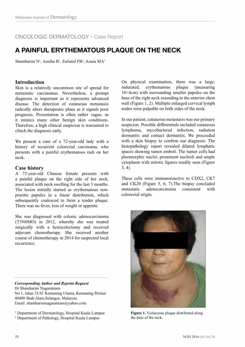
Malaysian Journal of Dermatology
55 MJD 2016 Jul Vol 36
ONCOLOGIC DERMATOLOGY - Case Report
A PAINFUL ERYTHEMATOUS PLAQUE ON THE NECKShamharini N1, Anisha B1, Zuliatul FB2, Azura MA1
Corresponding Author and Reprint Request Dr Shamharini NagaratnamNo 1, Jalan 33/41 Kemuning Utama, Kemuning Permai 40400 Shah Alam,Selangor, Malaysia.Email: [email protected]
1 Department of Dermatology, Hospital Kuala Lumpur2 Department of Pathology, Hospital Kuala Lumpur
On physical examination, there was a large, indurated, erythematous plaque (measuring 10×4cm) with surrounding smaller papules on the base of the right neck extending to the anterior chest wall (Figure 1, 2). Multiple enlarged cervical lymph nodes were palpable on both sides of the neck.
In our patient, cutaneous metastasis was our primary suspicion. Possible differentials included cutaneous lymphoma, mycobacterial infection, radiation dermatitis and contact dermatitis. We proceeded with a skin biopsy to confirm our diagnosis. The histopathology report revealed dilated lymphatic spaces showing tumor emboli. The tumor cells had pleomorphic nuclei, prominent nucleoli and ample cytoplasm with mitotic figures readily seen (Figure 3, 4).
These cells were immunoreactive to CDX2, CK7 and CK20 (Figure 5, 6, 7).The biopsy concluded metastatic adenocarcinoma consistent with colorectal origin.
IntroductionSkin is a relatively uncommon site of spread for metastatic carcinomas. Nevertheless, a prompt diagnosis is important as it represents advanced disease. The detection of cutaneous metastasis radically alters therapeutic plans as it signals poor prognosis. Presentation is often rather vague, as it mimics many other benign skin conditions. Therefore, a high clinical suspicion is warranted to clinch the diagnosis early.
We present a case of a 72-year-old lady with a history of recurrent colorectal carcinoma, who presents with a painful erythematous rash on her neck.
Case historyA 72-year-old Chinese female presents with a painful plaque on the right side of her neck, associated with neck swelling for the last 3 months. The lesion initially started as erythematous non-pruritic papules in a linear distribution, which subsequently coalesced to form a tender plaque. There was no fever, loss of weight or appetite.
She was diagnosed with colonic adenocarcinoma (T3N0MO) in 2012, whereby she was treated surgically with a hemicolectomy and received adjuvant chemotherapy. She received another course of chemotherapy in 2014 for suspected local recurrence.
Figure 1. Violaceous plaque distributed along the base of the neck.

Malaysian Journal of Dermatology
56MJD 2016 Jul Vol 36
Figure 2. On closer inspection, multiple excoriations with serous discharge is visible on the plaque.
Figure 4. On higher magnification, tumor emboli is seen within the lymphovascular space (H&E, magnification X400).
Figure 6. Tumor cells are immunoreactive to CK7.
Figure 3. The image above shows two fragments of skin tissue containing scattered tumor infiltrates within the dermis with areas of subcutaneous fat with haemorrhage (H&E, magnification X100).
Figure 5. Immunohistochemistry shows positive staining for CDX2.
Figure 7. Immunohistochemistry shows positive staining for CK20.
Subcutaneous fat with haemorrhage
Scattered tumor infiltrates
Tumor cells

Malaysian Journal of Dermatology
57 MJD 2016 Jul Vol 36
is considered to be in remission. Histopathology examination is essential to clinch the diagnosis. Demonstration of tumour cells resembling the primary tumour, with positive immunohistochemical markers will favour the diagnosis of cutaneous metastasis.
In this lady, the skin lesion was the first sign of distant spread which prompted her to seek treatment. It is therefore essential for the treating physician to have a high index of suspicion of recurrence in this cohort of patients who present with new onset skin lesions. Cutaneous infiltration is a sign of advanced metastatic disease and treatment options may be limited. There is a role for wide local excision with clear margins in those with isolated skin lesions. However, majority of these patients have widespread disease and will often require multidisciplinary care and further management with an oncologist. The treatment is usually centered on palliative care aiming to prolong survival. Similarly, in the case of our patient, she was referred to the oncology team for further assessment and staging. Contrast enhanced computed tomography (CECT) showed local recurrence of colorectal carcinoma with widespread metastasis to the lungs, liver, spleen and lymph nodes. She is currently undergoing palliative chemotherapy.
DiscussionColorectal carcinoma is a common malignancy seen in both men and women. The incidence of cutaneous metastasis has been reported to be within 2.3-6% of all colorectal carcinomas1,2. It usually occurs within the first 2 years of resection of the primary tumour3. Metastatic dissemination to the skin occurs via the lymphatics, hematogenous spread, direct extension and surgical implantation. The most common site of metastasis is the abdominal wall including surgical incision scars. Other cutaneous sites commonly involved include the pelvis, back, chest, upper extremities, head and neck. Identification of skin metastasis is a poor prognostic sign and reflects widespread disease with simultaneous metastasis to other sites such as lung, liver and peritoneum4.
Metastatic carcinoma can present in various morphological appearances. The most common presentation is as single or multiple painless skin colored to violaceous nodules. Occasionally it can mimic benign dermatoses such as epidermal cysts, neurofibromas, lipomas and cicatricial morphea-like plaques2. Rarely, it can resemble inflammatory lesions known as carcinoma erysipelatoides with well-defined indurated erythema. Cutaneous metastasis may be present de novo, when the patient
References 1. Pereira WA, Humaire CR, Silva CS, Fernandes LH. Sister
Mary Joseph’s nodule: a sign of internal malignancy. An Bras Dermatol. 2011; 86: S118–S120.
2. Lookingbill DP, Spangler N, Helm KF. Cutaneous metastases in patients with metastatic carcinoma: a retrospective study of 4020 patients. J Am Acad Dermatol. 1993; 29: 228–236.
3. Verardino GC, Silva RS, Obadia DL, et al. Rare cutaneous metastasis from a probable basaloid carcinoma of the colon mimicking pyogenic granuloma. An Bras Dermatol. 2011; 86: 537–540.
4. Lookingbill DP, Spangler N, Sexton FM. Skin involvement as the presenting sign of internal carcinoma. A retrospective study of 7316 cancer patients. J Am Acad Dermatol. 1990; 22: 19–26.

Malaysian Journal of Dermatology
58MJD 2016 Jul Vol 36
GENERAL DERMATOLOGY - Commentary
DERMATOLOGY IN VIETNAM WARLong V
Corresponding Author and Reprint Request Dr Valencia LongNational University Hospital, 5 Lower Kent Ridge Rd, Singapore 119074Email: [email protected]
Porphyria Cutanea Tarda(PCT)PCT is more rarely associated with TCDD exposure. PCT is characterised by blistering in sun exposed areas, hypertrichosis, milia formation and development of sclerodermoid plaques in affected areas. Despite studies of Vietnam veterans not finding a clear increased risk of PCT development after exposure4, PCT is regarded as an eligible condition for veterans to seek evaluation under the U.S Department of Veterans Affairs.
Melanoma and non-melanoma cancerAlthough melanoma and non-melanoma skin cancer have been associated with TCDD exposure, most of the human dioxin exposure studies till date have not shown an increased incidence of skin cancers.
Non-Hodgkin LymphomaNon-Hodgkin lymphomas, in particular cutaneous T/B-cell lymphomas are associated with Agent Orange exposure. Studies5 have shown that patients with mycosis fungoides and previous Agent Orange exposure have a higher prevalence of the palmaris et plantaris presentation subtype suggesting that higher disease concentration on affected areas could arise from direct herbicide exposure.
Soft-tissue sarcomaMost studies6 have failed to demonstrate an increased risk of soft-tissue sarcoma in Vietnam veterans. However, 1 study has cited veterans developing leiomyosarcomas, dermatofibrosarcoma protuberans and other soft-tissue sarcomas.
Other skin conditions At least 1 epidemiologic study7 has cited veterans being at increased risk for eczema, benign fatty tumors, milia, epidermoid cysts, dyschromias, increased skin sensitivity and non specific rashes.
IntroductionThe Vietnam War, though over for nearly four decades, had left veterans suffering from a multitude of ailments and dermatological conditions from perhaps the most memorable organochlorine exposure - Agent Orange. This note serves to remind us about the dermatoses arising from the Vietnam War and is a tribute to those who have fought valiantly in it.
During the war, herbicides such as Agent Orange were used to defoliate enemy crops in the Operation Ranch Hand (ORH) campaign. Throughout the decade long campaign, almost 20 million gal of Agent Orange (estimated 366kg of TCDD) was applied to over 3.6 million acres of South Vietnam1. The exposure to Agent Orange containing 2, 3, 7, 8 - tetrachlorodibenzo-p-dioxin (TCDD) has led to various skin disorders.
ChloracneChloracne is the most recognized dermatosis arising from the Vietnam war. It presents with multiple non-inflammatory comedone-like lesions interspersed with straw-colored cysts commonly affecting the malar crescent around the eyes and periauricular areas. There is sometimes genital and truncal involvement. Hypertrichosis, grey discoloration and folliculitis are associated2,3. Perhaps the most notable figure to have been affected by chloracne was former President Viktor Yushchenko in the 2004 poisoning case. Chloracne is known to regress between 6 months to 3 years3.

Malaysian Journal of Dermatology
59 MJD 2016 Jul Vol 36
References 1. Dunagin WG. Cutaneous signs of systemic toxicity due to
dioxins and related chemicals. J Am Acad Dermatol. 1984; 10(4):688-700
2. Burton JE, Michalek JE, Rahe AJ. Serum dioxin, chloracne, and acne in veterans of Operation Ranch Hand. Arch Environ Health. 1998;53(3):199-204.
3. Tindall JP. Chloracne and chloracnegens. J Am Acad Dermatol. 1985;13:539-558.
4. Kim JS, Lim HS, Cho SI et al. Impact of Agent Orange exposure among Korean Vietnam veterans. Ind Health. 2003;41:149-157.
5. Jang MS, Jang JG, Han SH, et al. Clinicopathological features of mycosis fungoides in patients exposed to Agent Orange during the Vietnam War. J Dermatol. 2013;40:606-612.
6. The Selected Cancers Cooperative Study Group. The association of selected cancers with service in the US military in Vietnam. II. Soft-tissue and other sarcomas. Arch Intern Med. 1990;150(12):2485-2492
7. Patterson A, Kaffenberger B, Keller R, et al. Skin diseases associated with Agent Orange and other organochlorine exposures. J Am Acad Dermatol. 2016; 74(1):143-70.

Malaysian Journal of Dermatology
60MJD 2016 Jul Vol 36
GENERAL DERMATOLOGY - Commentary
DERMATOLOGY AND LOVELong V
Corresponding Author and Reprint Request Dr Valencia LongNational University Hospital, 5 Lower Kent Ridge Rd, Singapore 119074Email: [email protected]
early in 19083, kissing nevi have been described to be located on the upper and lower eyelids, recalling perhaps, a most delicate and tender kiss between two lovers holding each other’s hearts.
Kissing ulcers have been commonly used to portray ulcers occurring on opposing surfaces of the vulva due to Herpes simpex infection, or other non herpetic infections such as salmonellosis4 and infectious mononucleosis5.
The honey-crusted appearance of impetigo inspires a sweet feeling of love.
Tenderness is a term sprinkled liberally through the annals of dermatology, and a paradigm example is the exact localisation of tenderness with the help of a pin head in a glomus tumor - also referred to as Love’s sign6.
Heart shaped lesions have been observed as unusual presentations of many common conditions7 - including cafe au lait macules, solar lentigines, nevus spilus, congenital melanocytic nevi, hemangiomas, and even ulcers. Love perhaps, is found in the least expected of places, in the common every-day life.
Gifts and flowers often escort love, with all its strangeness. The singular plant that has captured the imagination of lovers and dermatologists alike is the rose.
The rosette is a popular sign used to describe histological appearances of interstitial granulomatous dermatitis and peripheral neuroblastomas, clinically in linear IgA disease and dermatoscopically in squamous cell carcinoma8.
Pigmented fungiform papillae of the tongue appear dermatoscopically as rose petals9.
Varicella zoster infection manifests as “dew-drops on a rose petal”.
The term Pityriasis rosea reminds us of the greatest symbol of love – the rose.
IntroductionAmong the many passionate addresses1 Vladimir Nabokov, one of Russia’s greatest literary giants to his great love and wife was, “I love you, my sun, my life, I love your eyes - closed - all the little tails of your thoughts, your stretchy vowels, your whole soul from head to heels.”
Dermatology, like love, is infused with metaphor and blessed by the intricacies of language. Terms of endearment, both historical and modern, have been woven intimately with dermatological descriptions. In fact, to many dermatologists, how can dermatology not itself be a form of love?
Cherubism reminds us of the rosy-cheeked child-angel when we gaze into the eyes of our beloved ones. Then, as we plant a gentle angel kiss squarely in the middle of their forehead, we might recollect the nevus simplex upon a newborn’s face.
The term “Venereal” was derived from Latin venerus, from Venus, Roman goddess of love, beauty, sex.
Her son, Cupid, went on to inspire “Cupid’s disease” as a description of syphilis.
Rosacea brings to mind the blush of a lover’s countenance, as do the red, almost himself or herself fiery, papules of Sweet’s syndrome.
Kissing lesions in flexural areas affected by Paederus dermatitis2 are also recognised.
Kissing nevi are rare congenital melanocytic nevi that are located on adjacent places at which division had previously occurred during embryogenesis. Since the first report of such nevi by Von Micheal
Malaysian Journal of Dermatology
61 MJD 2016 Jul Vol 36
More Than Skin DeepDermatology has passed through time, collecting morphological descriptions inspired by the grandness of nature and the exuberance of love. For dermatology, as lovers would quite agree, is more than skin-deep, and certainly more than what meets the eye.
Roseola infantum is caused by the human herpes virus.
Typhoid fever produces rose spots. Is love itself not a feverish dream?
References 1. Nabokov V. (2014). Letters to Véra. London: Penguin
Books Ltd2. Haddad Junior V. “Sign of the kiss” in dermatitis caused by
vesicant beetles (“potós” or Paederus sp.) Anais Brasileiros de Dermatologia. 2014; 89(6): 996-997.
3. Hamming N. Anatomy and embryology of the eyelids: a review with special reference to the development of divided nevi. Pediatr Dermatol. 1983; 1: 51–58
4. Pelletier F, Aubin F, Puzenat E. Lipschutz genital ulceration: a rare manifestation of paratyphoid fever. Eur J Dermatol 2003; 13 (3): 297- 298
5. Portnoy J, Ahronheim GA, Ghibu F, et al. Recovery of Epstein-Barr virus from genital ulcers. N Engl J Med 1984; 311 (15): 966- 968
6. Love JG. Glomus tumors: diagnosis and treatment. Proc Staff Meet Mayo Clin. 1944; 19: 113–6.
7. Bree AF, Cole P, Siegfried EC. A Love of Dermatology. JAMA Dermatol. 2014; 150(2): 121-122.
8. Rubegni P, Tataranno DR, Nami N, Fimiani M. Rosettes: optical effects and not dermoscopic patterns related to skin neoplasms. Australas J Dermatol. 2013; 54(4): 271-272.
9. V. Pinos-León. Dermoscopic Features of Pigmented Fungiform Papillae of the Tongue. Actas Dermo-Sifiliográficas 2015; 106(7): 593.

Malaysian Journal of Dermatology
62MJD 2016 Jul Vol 36
GENERAL DERMATOLOGY - Commentary
TURMERIC- AN INDIAN GOLDEN CURRY SPICE,A GOLDEN CURE?Long V
Corresponding Author and Reprint Request Dr Valencia LongNational University Hospital, 5 Lower Kent Ridge Rd, Singapore 119074Email: [email protected]
Turmeric may also inhibit the expression of TNF-a induced interleukins thought to be contributory in inflammatory psoriasis3. Recently, a Chinese study4 supports turmeric efficacy in mouse models showing significant inhibition of interleukin (IL) - 17, IL-22, IFN-g, IL-2, IL-8 and TNF-a in T cells.
Turmeric and wound healing Turmeric may be efficacious in various forms of wound healing including thermal burns, open wounds, and wounds sustained by immunocompromised patients. Studies performed on diabetic rats demonstrated that oral administration of curcumin reduced the oxidative stress and levels of serum lipid peroxidation, decreased glycation and collagen cross-linking in the rat’s tail tendon and skin5. Curcumin’s role in downregulating both inflammatory and fibrogenic cytokines in mice exposed to ionizing radiation also makes it effective in ameliorating radiation-induced delays in wound repair6.
Turmeric against skin cancersSeminal work by Azuine and Bhide7 demonstrated that dietary turmeric (2%) could prevent DMBA-induced skin tumorigenesis. Since then, other studies8 have shown that turmeric could modulate several cytochromes (cytochrome P-450, hepatic cytochrome b5) conferring protective effects. To date, many in-vitro studies have shown that curcuminoids protect normal keratinocytes from free oxygen radical stress9, and induces apoptosis in human basal carcinoma cells, and mouse melanoma cells. A phase I clinical study10 on humans with Bowen’s disease has demonstrated that administration of large doses of curcumin (up to 8g/day for 3 months) led to histologic improvement of precancerous lesions in around 33% of patients without toxicity.
Turmeric, or Curcuma longa L. is an important spice used in Indian folk medicine. Apart from its use in treating hepatobiliary disorders, turmeric is regarded as an anti-septic, analgesic and wound-healing agent. Preclinical studies have demonstrated that turmeric and curcumin, its principle compound, are effective in treating psoriasis, aids wound healing and preventing UV-induced skin damage. This note recaps the history of turmeric and explores some of its uses that are applicable and validated by modern medicine.
Turmeric is a tropical plant native to India. Its name originated from the medieval Latin term terramerita - meaning “meritorious earth”1. In India, turmeric is closely linked with the socio-cultural life of the populace, and is used extensively in various religious and auspicious ceremonies.
Ancient Indian medicinal texts cite turmeric’s role in treating various skin conditions. In Ayurveda, turmeric is known as Varna datri and was found to be useful for inflammations, abscesses, eczema, leukoderma, urticaria, psoriasis, acne, and bruises.
Turmeric and psoriasis Turmeric is an experimental drug for treating psoriasis. Human studies have shown that topical application of a gel preparation of 1% curcumin induced quicker resolution than calcipotriol ointment (Dovonex).
It is thought that curcumin may inhibit the proliferation of keratinocytes via decreasing the levels of keratinocyte transferring receptor (TRR) expression, the severity of parakeratosis and the density of epidermal CD8+ T cells2.

Malaysian Journal of Dermatology
63 MJD 2016 Jul Vol 36
alternative for patients who are not amenable to surgery or have multiple precancerous lesions such as transplant patients. Despite growing evidence of its efficacy, it is important to note that turmeric cannot be applied directly on skin lesions because of potential allergic contact dermatitis11,12,13.
Turmeric’s efficacy against progression and prevention of basal cell carcinoma, squamous cell carcinoma and melanoma - globally important skin cancers- makes it a valuable compound for further research. It potentially represents a low risk oral
References 1. Ishita C, Kaushik B, Uday B, et al. Turmeric and curcumin:
biological actions and medicinal applications. Curr Sci. 2004;87:44–53.
2. Heng M, Song M, Harker J et al. Drug-induced suppression of phosphorylase kinase activity correlates with resolution of psoriasis as assessed by clinical, histological and immunohistochemical parameters. Br J Dermatol. 2000;143:937–49.
3. Cho JW, Lee KS, Kim CW. Curcumin attenuates the expression of IL-1beta, IL-6, and TNF-alpha as well as cyclin E in TNF-alpha-treated HaCaT cells; NF-kappaB and MAPKs as potential upstream targets. Int J Mol Med. 2007;19:469–74.
4. Kang D, Bowen L, Lei L et al. Cucurmin shows excellent therapeutic effect on psoriasis in mouse model. Biochimie 2016. Article in press.
5. Sajithlal GB, Chithra P, Chandrakasan G. Effect of curcumin on the advanced glycation and cross-linking of collagen in diabetic rats. Biochem Pharmacol. 1998;56:1607–14.
6. Okunieff P, Xu J, Hu D, et al. Curcumin protects against radiation-induced acute and chronic cutaneous toxicity in mice and decreases mRNA expression of in fl ammatory and fi brogenic cytokines. Int J Radiat Oncol Biol Phys. 2006;65:890–8.
7. Azuine MA, Bhide SV. Chemopreventive effect of turmeric against stomach and skin tumors induced by chemical carcinogens in Swiss mice. Nutr Cancer. 1992;17:77–83.
8. Villaseñor IM, Simon MK, Villanueva AM. Comparative potencies of nutraceuticals in chemically induced skin tumor prevention. Nutr Cancer. 2002;44:66–70.
9. Bonté F, Noel-Hudson MS, Wepierre J, et al. Protective effect of curcuminoids on epidermal skin cells under free oxygen radical stress. Planta Med. 1997;63:265–6.
10. Cheng AL, Hsu CH, Lin JK, et al. Phase I clinical trial of curcumin, a chemopreventive agent, in patients with high-risk or pre-malignant lesions. Anticancer Res. 2001;21(4B):2895–900.
11. Fischer L, Agner T. Cucurmin allergy in relation to yellow chlorhexidine solution used for skin disinfection prior to surgery. Contact Dermatitis 2004; 51: 39-40
12. Chaudari S et al. Cucurmin: A contact allergen. J Clin Aesth Dermatol 2015; 8: 43-8
13. Goh C et al. Allergic contact dermatitis to Curcuma longa (turmeric). Contact Dermatitis 1987; 17: 186

NOTES

NOTES