Jaafar Jotheri - “Baguette levee” features in the Mesopotamian floodplain: a new term in river...
7
BioMed Central Page 1 of 7 (page number not for citation purposes) Molecular Cancer Open Access Short communication Bone marrow ectopic expression of a non-coding RNA in childhood T-cell acute lymphoblastic leukemia with a novel t(2;11)(q11.2;p15.1) translocation Maria Corsignano Guastadisegni †1 , Angelo Lonoce †1 , Luciana Impera 1 , Francesco Albano 2 , Pietro D'Addabbo 1 , Sebastiano Caruso 3 , Isabella Vasta 4 , Ioannis Panagopoulos 5 , Anna Leszl 6 , Giuseppe Basso 6 , Mariano Rocchi 1 and Clelia Tiziana Storlazzi* 1 Address: 1 Department of Genetics and Microbiology, University of Bari, Bari, Italy, 2 Department of Hematology, University of Bari, Bari, Italy, 3 Laboratorio di Genetica Medica, AUSL LE, P.O. "Vito Fazzi", Lecce, Italy, 4 Oncoematologia Pediatrica, AUSL LE, P.O. "Vito Fazzi", Lecce, Italy, 5 Department of Clinical Genetics, University Hospital, Lund, Sweden and 6 Department of Pediatrics, Laboratory of Pediatric Onco-Hematology, University of Padova, Padova, Italy Email: Maria Corsignano Guastadisegni - [email protected]; Angelo Lonoce - [email protected]; Luciana Impera - [email protected]; Francesco Albano - [email protected]; Pietro D'Addabbo - [email protected]; Sebastiano Caruso - [email protected]; Isabella Vasta - [email protected]; Ioannis Panagopoulos - [email protected]; Anna Leszl - [email protected]; Giuseppe Basso - [email protected]; Mariano Rocchi - [email protected]; Clelia Tiziana Storlazzi* - [email protected] * Corresponding author †Equal contributors Abstract Chromosomal translocations play a crucial role in tumorigenesis, often resulting in the formation of chimeric genes or in gene deregulation through position effects. T-cell acute lymphoblastic leukemia (T-ALL) is associated with a large number of such rearrangements. We report the ectopic expression of the 3' portion of EST DA926692 in the bone marrow of a childhood T-ALL case showing a t(2;11)(q11.2;p15.1) translocation as the sole chromosome abnormality. The breakpoints, defined at the sequence level, mapped within HPS5 (Hermansky Pudlak syndrome 5) intron 1 at 11p15.1, and DA926692 exon 2 at 2q11.2. The translocation was accompanied by a submicroscopic inversion that brought the two genes into the same transcriptional orientation. No chimeric trancript was detected. Interestingly, Real-Time Quantitative (RQ)-PCR detected, in the patient's bone marrow, expression of a 173 bp product corresponding to the 3' portion of DA926692. Samples from four T-ALL cases with a normal karyotype and normal bone marrow used as controls were negative. It might be speculated that the juxtaposition of this genomic segment to the CpG island located upstream HPS5 activated DA92669 expression. RQ-PCR analysis showed expression positivity in 6 of 23 human tissues examined. Bioinformatic analysis excluded that this small non-coding RNA is a precursor of micro-RNA, although it is conceivable that it has a different, yet unknown, functional role. To the best of our knowledge, this is the first report, in cancer, of the activation of a small non-coding RNA as a result of a chromosomal translocation. Published: 23 October 2008 Molecular Cancer 2008, 7:80 doi:10.1186/1476-4598-7-80 Received: 19 September 2008 Accepted: 23 October 2008 This article is available from: http://www.molecular-cancer.com/content/7/1/80 © 2008 Corsignano et al; licensee BioMed Central Ltd. This is an Open Access article distributed under the terms of the Creative Commons Attribution License (http://creativecommons.org/licenses/by/2.0 ), which permits unrestricted use, distribution, and reproduction in any medium, provided the original work is properly cited.
-
Upload
csm-qadiss -
Category
Documents
-
view
1 -
download
0
Transcript of Jaafar Jotheri - “Baguette levee” features in the Mesopotamian floodplain: a new term in river...

BioMed CentralMolecular Cancer
ss
Open AcceShort communicationBone marrow ectopic expression of a non-coding RNA in childhood T-cell acute lymphoblastic leukemia with a novel t(2;11)(q11.2;p15.1) translocationMaria Corsignano Guastadisegni†1, Angelo Lonoce†1, Luciana Impera1, Francesco Albano2, Pietro D'Addabbo1, Sebastiano Caruso3, Isabella Vasta4, Ioannis Panagopoulos5, Anna Leszl6, Giuseppe Basso6, Mariano Rocchi1 and Clelia Tiziana Storlazzi*1Address: 1Department of Genetics and Microbiology, University of Bari, Bari, Italy, 2Department of Hematology, University of Bari, Bari, Italy, 3Laboratorio di Genetica Medica, AUSL LE, P.O. "Vito Fazzi", Lecce, Italy, 4Oncoematologia Pediatrica, AUSL LE, P.O. "Vito Fazzi", Lecce, Italy, 5Department of Clinical Genetics, University Hospital, Lund, Sweden and 6Department of Pediatrics, Laboratory of Pediatric Onco-Hematology, University of Padova, Padova, Italy
Email: Maria Corsignano Guastadisegni - [email protected]; Angelo Lonoce - [email protected]; Luciana Impera - [email protected]; Francesco Albano - [email protected]; Pietro D'Addabbo - [email protected]; Sebastiano Caruso - [email protected]; Isabella Vasta - [email protected]; Ioannis Panagopoulos - [email protected]; Anna Leszl - [email protected]; Giuseppe Basso - [email protected]; Mariano Rocchi - [email protected]; Clelia Tiziana Storlazzi* - [email protected]
* Corresponding author †Equal contributors
AbstractChromosomal translocations play a crucial role in tumorigenesis, often resulting in the formationof chimeric genes or in gene deregulation through position effects. T-cell acute lymphoblasticleukemia (T-ALL) is associated with a large number of such rearrangements. We report the ectopicexpression of the 3' portion of EST DA926692 in the bone marrow of a childhood T-ALL caseshowing a t(2;11)(q11.2;p15.1) translocation as the sole chromosome abnormality. Thebreakpoints, defined at the sequence level, mapped within HPS5 (Hermansky Pudlak syndrome 5)intron 1 at 11p15.1, and DA926692 exon 2 at 2q11.2. The translocation was accompanied by asubmicroscopic inversion that brought the two genes into the same transcriptional orientation. Nochimeric trancript was detected. Interestingly, Real-Time Quantitative (RQ)-PCR detected, in thepatient's bone marrow, expression of a 173 bp product corresponding to the 3' portion ofDA926692. Samples from four T-ALL cases with a normal karyotype and normal bone marrowused as controls were negative. It might be speculated that the juxtaposition of this genomicsegment to the CpG island located upstream HPS5 activated DA92669 expression. RQ-PCRanalysis showed expression positivity in 6 of 23 human tissues examined. Bioinformatic analysisexcluded that this small non-coding RNA is a precursor of micro-RNA, although it is conceivablethat it has a different, yet unknown, functional role. To the best of our knowledge, this is the firstreport, in cancer, of the activation of a small non-coding RNA as a result of a chromosomaltranslocation.
Published: 23 October 2008
Molecular Cancer 2008, 7:80 doi:10.1186/1476-4598-7-80
Received: 19 September 2008Accepted: 23 October 2008
This article is available from: http://www.molecular-cancer.com/content/7/1/80
© 2008 Corsignano et al; licensee BioMed Central Ltd. This is an Open Access article distributed under the terms of the Creative Commons Attribution License (http://creativecommons.org/licenses/by/2.0), which permits unrestricted use, distribution, and reproduction in any medium, provided the original work is properly cited.
Page 1 of 7(page number not for citation purposes)
Molecular Cancer 2008, 7:80 http://www.molecular-cancer.com/content/7/1/80
FindingsAcquired balanced chromosomal translocations providecrucial diagnostic and prognostic data in cancer patients.They are probably pathogenetically significant as initiat-ing events if present as the only cytogenetic changes [1].These rearrangements often result in the formation of afusion gene, encoding a chimeric oncogenic protein, or inthe deregulation of the expression pattern of genes flank-ing the breakpoint regions by promoter swapping [1-3].Overall, gene deregulation can be accomplished throughposition effects, such as a gene juxtaposition close to anactive chromatin domain. More rarely, translocations mayresult in gene downregulation because of hypermethyla-tion of CpG islands at the breakpoint [4], or in the forma-tion of chimeric transcripts that do not encode for aprotein with oncogenic potential [5].
T-ALL, as other hematological malignancies, is associatedwith a number of chromosomal abnormalities, resultingeither in a position effect or in a gene fusion [6].
Here we report the cloning of a novel balancedt(2;11)(q11.2;p15.1) translocation, found as the solecytogenetic abnormality in the BM of a childhood T-ALLcase, accompanied by the ectopic expression of the 3' por-tion of EST DA926692.
A 14-year-old male patient was referred to our hospital forrhinolalia. Total body CT disclosed a diffuse enlargementof the rhinopharynx vault, lymphoid hyperplasia of theWaldeyer's tonsillar ring, and a mediastinal enlargement.BM aspiration showed 90% of blast cells. Flow cytometryrevealed that blast cell population was positive for CD 45,2, 5, 7, and cytoplasmic CD3, while negative for CD1aand surface CD3. These findings were consistent with adiagnosis of early T-ALL, thus the patient started treatmentbased on AIEOP-BFM ALL 2000 protocol [7].
At the end of induction therapy, the patient was catego-rized as "high risk" due to incomplete remission, as perprotocol stratification criteria. After consolidation ther-apy, the BM aspirate showed no haematological remis-sion, so treatment continued following the AIEOP LLAREC 2003 protocol and complete hematological remis-sion was obtained.
One year from diagnosis, the patient underwent HLA-identical hematopoietic stem cell transplantation from anunrelated donor. He is now doing well and is still inhematologic remission.
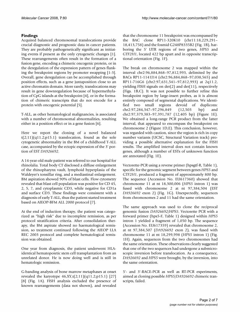
G-banding analysis of bone marrow metaphases at onsetrevealed the karyotype 46,XY,t(2;11)(q11.2;p15.1) [27][8] (Fig. 1A). FISH analysis excluded the presence ofknown rearrangements (data not shown), and revealed
that the chromosome 11 breakpoint was encompassed bythe BAC clone RP11-320K10 (chr11:18,229,291–18,413,758) and the fosmid G248P8335B2 (Fig. 1B), har-boring the 5' UTR regions of two genes, HPS5 andGTF2H1, located 422 bp apart and in opposite transcrip-tional orientation (Fig. 1F).
The break on chromosome 2 was mapped within theinterval chr2:96,884,868–97,812,993, delimited by theBACs RP11-1141J14 (chr2:96,884,868–97,038,565) andRP11-716G6 (chr2:97,631,541–97,812,993) at 2q11.2,yielding FISH signals on der(2) and der(11), respectively(Figs. 1B,C). It was not possible to further refine thisbreakpoint region by large-insert probes, as it is almostentirely composed of segmental duplications. We identi-fied two small regions devoid of duplicons:chr2:97,284,347–97,296,849 (12,503 bp) andchr2:97,379,383–97,391,787 (12,405 bp) (Figure 1E).We obtained a long-range PCR product from the latterinterval, that appeared to encompass the breakpoint onchromosome 2 (Figure 1D,E). This conclusion, however,was regarded with caution, since the region is rich in copynumber variants (UCSC, Structural Variation track) pro-viding a possible alternative explanation for the FISHresults. The amplified interval does not contain knowngenes, although a number of ESTs of unknown functionare annotated (Fig. 1E).
Vectorette-PCR using a reverse primer (hpsgtf-R, Table 1),specific for the genomic segment between genes HPS5 andGTF2H1, produced a fragment of approximately 800 bp.The sequence (Accession No. EU617360) showed thatchromosome 11 at nt 18,300,006 (HPS5 intron 1) wasfused with chromosome 2 at nt 97,384,504 (ESTDA926692 exon 2) (Fig. 1G). Unexpectedly, sequencesfrom chromosomes 2 and 11 had the same orientation.
The same approach was used to clone the reciprocalgenomic fusion DA926692/HPS5. Vectorette PCR with aforward primer (hps5-F, Table 1) designed within HPS5intron 1 yielded a fragment of 1,050 bp. The sequence(Accession No. EU617359) revealed that chromosome 2,at nt 97,384,507 (DA926692 exon 2), was fused withchromosome 11 at nt 18,299,998 (HPS5 intron 1) (Fig.1H). Again, sequences from the two chromosomes hadthe same orientation. These observations clearly suggestedthat one of the two sequences had undergone a submicro-scopic inversion before translocation. As a consequence,DA926692 and HPS5 were brought, by the inversion, intothe same orientation.
5'- and 3'-RACE-PCR as well as RT-PCR experiments,aimed at cloning possible HPS5/DA926692 chimeric tran-scripts, failed.
Page 2 of 7(page number not for citation purposes)

Molecular Cancer 2008, 7:80 http://www.molecular-cancer.com/content/7/1/80
Figure 1 (see legend on next page)
Page 3 of 7(page number not for citation purposes)

Molecular Cancer 2008, 7:80 http://www.molecular-cancer.com/content/7/1/80
Further RQ-PCR analyses aiming at quantifying theexpression levels of genes flanking the breakpoints, i.e.HPS5 (Fig. 2A) and GTF2H1 (data not shown) on chro-mosome 11, and ANKRD36, FAHD2B, KIAA1641, ZAP70on chromosome 2, revealed no misexpression in the
present case compared to other T-ALL cases without thet(2;11) translocation (data not shown).
The same approach was used for DA926692. A primer setspecific for exon 1, upstream to the breakpoint, revealedno statistically significant difference in expression level
Results of the cytogenetic and genomic characterization of the t(2;11) traslocation breakpointsFigure 1 (see previous page)Results of the cytogenetic and genomic characterization of the t(2;11) traslocation breakpoints: A) Karyotype of the case described in the present study. The black arrows point on derivative chromosomes 2 and 11. B) FISH results obtained with fosmids and BAC clones delimiting the breakpoint regions on der(2) and der(11). C) Map of chromosome 2 breakpoint region, according to the latest release of the UCSC Genome Browser (March 2006), delimited by BAC clones RP11-1141J14 (left) and RP11-716G6 (right). RefSeq Genes and CpG islands are represented in blue and green, respectively. Intra-chromo-somal duplications are reported at the bottom of the figure. Gray, yellow and orange colours refer to the percentage of sequence similarity of the duplication (respectively 90–98%, 98–99%, and >99%). D) FISH results obtained using the Chr2-97-1 Long-PCR product on the bone marrow of the patient. E) Detailed magnified map of the chromosome 2 breakpoint region, showing the location of the Chr2-97-1 probe. F) Map of the breakpoint region on chromosome 11. G) and H) Partial chroma-tograms of the junctions sequences on both derivative chromosomes 11 (G) and 2 (H). Inserted nucleotides [three (CAT) and sixteen (GGCGGTATGTCCGTAC) at der(11) and der(2), respectively] at the junctions are underlined in purple.
Table 1: Primers for PCR and sequencing.
Designationa Sequence (5'->3') Positionb Gene
chr2-97-1F AGCCTGTGGTGTGTGTATGAAC chr2:97,380,441–97,380,462 -chr2-97-1R TGAGTGTAGGTGACTGGTGAGG chr2: 97,391,465–97,391,486 -hpsgtf-R TGACACCTTCCGCTAGTTCC chr11:18,300,606–18,300,625 -Hps5-F CCTCCCTGCTTCTTTTTCCA chr11:18,299,339–18,299,358 HPS5HPS5ex20-3216Fc TGCAGGTCTTGTGGTTTCTG chr11:18,263,475 HPS5HPS5ex21-3255Rc TCTTCTCTCCAGCTCCAAACA chr11:18,261,979–18,261,999 HPSHPS5ex1-19Fc TTCACGTTCCGCTCTTAGTG chr11:18,300,260–18,300,279 HPS5HPS5ex1-96Rc CACATCCAGGGCAGTACCTC chr11:18,300,183–18,300,202 HPS5HPS5ex1-181F TGGTTACTGGGTCTCCTCTCA chr11:18,300,107–18,300,127 HPS5HPS5-intr1-F TGTGTTTTGTTCATCCCTGTG chr11:18,300,006–18,300,026 HPS5DA926692 ex1Fc TCAGTCCCAGTCAGGACACA chr2:97,384,972–97,384,991 DA926692DA926692 ex1Rc AGAGCCAGAGCAGCAGGAG chr2:97,384,918–97,384,936 DA926692DA926692 ex2aF CCATCAAGGGAAGCAGATGT chr2:97,384,457–97,384,476 DA926692DA926692 ex2aR GAGGCACCAGGAGAAGCAT chr2:97,384,418–97,384,436 DA926692DA926692ex2anewFc CCCACAGTGTTACAAGTCATAACATA chr2:97,384,479–97,384,504 DA926692DA926692ex2anewRc AGGAAGCTTCATGGCTCCTT chr2:97,384,334–97,384,353 DA926692Beta-act Fc CTGGAACGGTGAAGGTGACA chr7:5,533,739–5,533,758 ACTBBeta-act Rc AAGGGACTTCCTGTAACAACGCA chr7:5,533,619–5,533,641 ACTBCD110063-R CACCAGGAGGCAGACGAG chr2:97,391,951–97,391,968 CD110063ACTB-F GGCATCGTGATGGACTCCG chr7:5,534,773–5,534,789 ACTBACTB-R GCTGGAAGGTGGACAGCGA chr7:5,533,972–5,533,990 ACTBGTF2H1ex9-1201F AGCAGTCAAAAGGGCGAAAT chr11:18,326,030–18,326,043/18,330,004–18,330,010 GTF2H1GTF2H1ex10-1272R TTCTTGAGGTTTAGTGCAATCG chr11:18,330,062–18,330,083 GTF2H1ANKex2-351F CCAACCGGAAATGGTACATC chr2:97,147,558–97,147,577 ANKRD36ANKex2-451R AGAGTTGCACAAGCCTCCTG chr2:97,147,823–97,147,842 ANKRD36FAHD2Bex5-829F TGTAGCAGATCCACACAACTTAAA chr2:97,113,730–97,113,742/97,115,163–97,115,171 FAHD2BFAHD2Bex6-864R GACGACTTCCCCATTCACTC chr2:97,113,695–97,113,714 FAHD2BKIAA1641ex2-214F AGAAAATGGGATGCAGGATT chr2:97,492,508–97,492,527 KIAA1641KIAA1641ex2-245R GAAATTGACTTCTCATCTGGTCTAA chr2:97,490,960–97,490,957/chr2:97,492,476–97,492,496 KIAA1641ZAP70ex11-1678F AGCTACTACACTGCCCGCTCA chr2:97,720,549–97,720,560/97,720,651–97,720,660 ZAP70ZAP70ex11-1749R CTGCGGCTGGAGAACTTG chr2:97,720,709–97,720,726 ZAP70
a F denotes forward; R denotes reverse;b Position, at nucleotide level, according to the UCSC database, using the BLAT tool http://genome.ucsc.edu/cgi-bin/hgBlat;cPrimer used in Real-Time PCR analysis.
Page 4 of 7(page number not for citation purposes)

Molecular Cancer 2008, 7:80 http://www.molecular-cancer.com/content/7/1/80
compared to controls (Fig, 2B). Interestingly, a primerpair designed in exon 2 (Table 1), downstream to thebreakpoint, detected expression only in the t(2;11) case.
RT-PCR with the same primer pair revealed a band of 173pb only in the patient's BM and in the genomic DNA sam-ple, but neither in the T-ALL controls, nor in normal BMor PB (Fig. 2C). These results were confirmed by nestedRT-PCR with the primer pair DA926692 ex2a, further val-idated by the sequencing of PCR products (data notshown). Purity of all RNA samples was tested using ACTBprimers in PCR experiments (Fig. 2D).
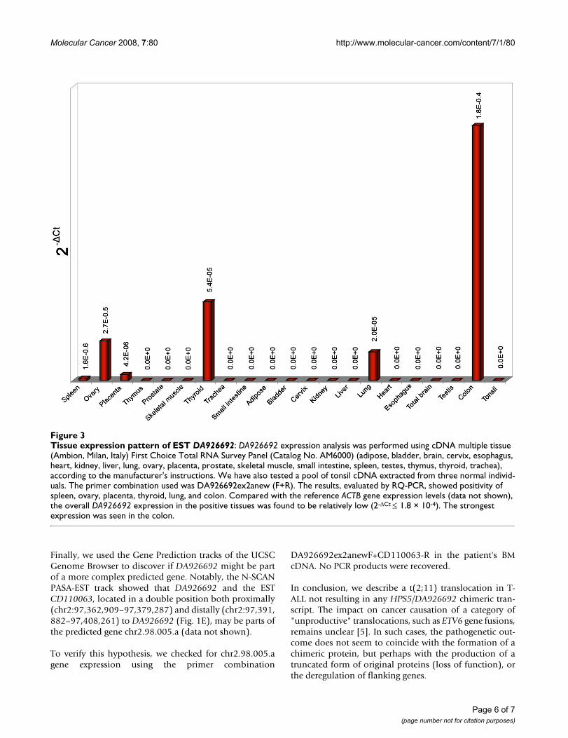
Next, we investigated the expression pattern of3'DA926692 by RQ-PCR in 21 adult tissues. The resultsshowed the occurrence of the transcript in 6 of 23 exam-ined tissues (Fig. 3) [normal BM and PB were alreadyshown to be negative (Fig. 2C)].
Notably, in silico translation of the sequence correspond-ing to the 173 bp PCR product showed that it contained ahigh density of stop codons resulting in very short ORFs.
We hypothesized that this ncRNA could represent a pre-cursor of an miRNA, but the miRNA prediction table pro-duced by miRRim http://mirrim.ncrna.org/TableS1.htmland a sequence similarity search at miRBase http://microrna.sanger.ac.uk/sequences/search.shtml showed no sig-nificant matches (data not shown). Similarly, miRNA pre-dictions by ProMiR II http://cbit.snu.ac.kr/~ProMiR2/and miR-abela http://www.mirz.unibas.ch/ did notretrieve any positive results (data not shown).
Figure 2
Results of the HPS5 and DA926692 gene expression analysisFigure 2Results of the HPS5 and DA926692 gene expression analysis: A) and B) RQ-PCR analyses of HPS5, and DA926692 exon1, respectively, in the present case [t(2;11), four control childhood T-ALL samples [immunophenotype: early (#1,2), thymic (#3), and mature (#4) T-ALL], the mean Ct value of the controls (mean), normal BM and PB. A) The results showed no statistically significant change in HPS5 expression level with primers for exon1 (HPS5ex1-19F+HPS5ex1-96R) and exons 20-21 (HPS5ex20-3216F and HPS5ex21-3255R), if compared with the mean Ct value of the childhood T-ALL controls. B) The results showed com-parable DA926692 transcriptional levels, when evaluated with exon 1 primers (DA926692ex2F and DA926692ex2R), between the patient with t(2;11) and the mean Ct value of the controls. C) RT-PCR results obtained with DA926692ex2anewF and DA926692ex2anewR (Table 1), showing a band of 173 bp only in the patient's bone marrow (lane 1) and normal genomic DNA (lane 8). Lanes 2-5 corre-spond to four control childhood T-ALL bone marrow. Lanes 6, 7 and 9 are normal bone marrow, normal peripheral blood, and blank, respectively. M: 2-Log DNA Ladder (New England Biolabs, Milan, Italy). D) Control RT-PCR with ACTB primers to check the RNA quality, excluding contamination of genomic DNA in the patient's bone marrow RNA.
Page 5 of 7(page number not for citation purposes)
Molecular Cancer 2008, 7:80 http://www.molecular-cancer.com/content/7/1/80
Finally, we used the Gene Prediction tracks of the UCSCGenome Browser to discover if DA926692 might be partof a more complex predicted gene. Notably, the N-SCANPASA-EST track showed that DA926692 and the ESTCD110063, located in a double position both proximally(chr2:97,362,909–97,379,287) and distally (chr2:97,391,882–97,408,261) to DA926692 (Fig. 1E), may be parts ofthe predicted gene chr2.98.005.a (data not shown).
To verify this hypothesis, we checked for chr2.98.005.agene expression using the primer combination
DA926692ex2anewF+CD110063-R in the patient's BMcDNA. No PCR products were recovered.
In conclusion, we describe a t(2;11) translocation in T-ALL not resulting in any HPS5/DA926692 chimeric tran-script. The impact on cancer causation of a category of"unproductive" translocations, such as ETV6 gene fusions,remains unclear [5]. In such cases, the pathogenetic out-come does not seem to coincide with the formation of achimeric protein, but perhaps with the production of atruncated form of original proteins (loss of function), orthe deregulation of flanking genes.
Tissue expression pattern of EST DA926692Figure 3Tissue expression pattern of EST DA926692: DA926692 expression analysis was performed using cDNA multiple tissue (Ambion, Milan, Italy) First Choice Total RNA Survey Panel (Catalog No. AM6000) (adipose, bladder, brain, cervix, esophagus, heart, kidney, liver, lung, ovary, placenta, prostate, skeletal muscle, small intestine, spleen, testes, thymus, thyroid, trachea), according to the manufacturer's instructions. We have also tested a pool of tonsil cDNA extracted from three normal individ-uals. The primer combination used was DA926692ex2anew (F+R). The results, evaluated by RQ-PCR, showed positivity of spleen, ovary, placenta, thyroid, lung, and colon. Compared with the reference ACTB gene expression levels (data not shown), the overall DA926692 expression in the positive tissues was found to be relatively low (2-ΔCt ≤ 1.8 × 10⁻⁴). The strongest expression was seen in the colon.
Page 6 of 7(page number not for citation purposes)

Molecular Cancer 2008, 7:80 http://www.molecular-cancer.com/content/7/1/80
Here, the only observed gene expression change consistedin the ectopic expression of the 3' portion of DA926692 inthe patient's BM (Fig. 2C). One hypothesis is that the jux-taposition, at a distance of only 196 nt, of this genomicsegment to the CpG island located upstream to both HPS5and GTF2H1 (chr11:18,300,202–18,300,814) could haveactivated its expression in the BM cells of the patient.
DA926692 was isolated from a small intestine cDNAlibrary, described as similar to Ig kappa chain V-I regionHK103 precursor http://www-bimas.cit.nih.gov/cgi-bicards/carddisp.pl?gene=LOC643543&search=DA926692&suff=txt, but no known function has yet been reported.Its expression in some human adult tissues (Fig. 3) maysuggest a potential function of this transcript in the posi-tive samples.
As its function is unclear, it is difficult to speculate on apossible role of this short ncRNA in T-ALL tumorigenesis.As the bioinformatic analysis excluded the possibility thatit could behave as a pre-miRNA, we could speculate thatthis ncRNA may belong to the class of small RNA (20–300nt), commonly found as transcriptional and translationalregulators [9,10]. A variety of complex cellular mecha-nisms such as gene transcription, chromatin structuredynamics, and others have already been connected totheir function [10]. Furthermore, changes in expressionlevels of ncRNAs have been associated with different typesof cancer [9].
In this context, we could speculate that the ectopic expres-sion of the 3' portion of DA926692 in chilhood T-ALLcould have a pathogenetic role, even if of still unclear sig-nificance. It is presently unknown, however, if thedescribed rearrangement represents a primary or a second-ary genetic aberration at disease onset. Further studies onsimilar cases may be extremely important to confirm thisnew impact of chromosomal translocation, i.e. activationof ncRNA in tumors.
Abbreviations(T-ALL): T-cell Acute Lymphoblastic Leukemia; (TCR): T-Cell Receptor; (CT): Computed Tomography; (BM): BoneMarrow; (PB): Peripheral Blood; (FISH): Fluorescence InSitu Hybridization; (BAC): Bacterial Artificial Chromo-some; (HPS5): Hermansky-Pudlak syndrome 5 isoform b;(GTF2H1): general transcription factor IIH, polypeptide 1;(nt): nucleotide; (RQ-PCR): Real-Time Quantitative PCR;(ANKRD36): ankyrin repeat domain 36; (FAHD2B):fumarylacetoacetate hydrolase domain containing;(KIAA1641): hypothetical protein LOC57730; (ZAP70):zeta-chain associated protein kinase 70 KDa; (ORFs):Open Reading Frames; (miRNAs): microRNAs; (ncRNA):non-coding RNA.
Competing interestsThe authors declare that they have no competing interests.
Authors' contributionsMCG and AL designed research and performed molecularanalyses. FA and IV contributed in clinical data collectionand analysis. SC and AL performed classical cytogeneticanalysis and contributed in the development of the study.LI performed molecular cytogenetic analysis. PD per-formed bioinformatic analysis. IP contributed in molecu-lar data analysis and interpretation of the results. MR andGB gave final approval to the manuscript. CTS supervisedthe study and drafted the manuscript.
AcknowledgementsThis work has been supported by AIRC (Associazione Italiana per la Ricerca sul Cancro), the MIUR (Ministero dell'Istruzione, dell'Università e della Ricerca), and Fondazione Città della Speranza. The authors thank Dr. A. Pico, Dr. A. Tornesello, and Dr. E. Giarin for their contribution to the study, and Prof. Roscoe Stanyon for language revision of the manuscript.
References1. Mitelman F, Johansson B, Mertens F: The impact of translocations
and gene fusions on cancer causation. Nat Rev Cancer 2007,7:233-245.
2. van Dyck F, Declercq J, Braem CV, van de Ven WJ: PLAG1, the pro-totype of the PLAG gene family: versatility in tumour devel-opment (review). Int J Oncol 2007, 30:765-774.
3. Storlazzi CT, Albano F, Lo Cunsolo C, Doglioni C, Guastadisegni MC,Impera L, Lonoce A, Funes S, Macri E, Iuzzolino P, et al.: Upregula-tion of the SOX5 by promoter swapping with the P2RY8gene in primary splenic follicular lymphoma. Leukemia 2007,21:2221-2225.
4. Anglesio MS, Evdokimova V, Melnyk N, Zhang L, Fernandez CV,Grundy PE, Leach S, Marra MA, Brooks-Wilson AR, Penninger J,Sorensen PH: Differential expression of a novel ankyrin con-taining E3 ubiquitin-protein ligase, Hace1, in sporadicWilms' tumor versus normal kidney. Hum Mol Genet 2004,13:2061-2074.
5. Panagopoulos I, Strombeck B, Isaksson M, Heldrup J, Olofsson T,Johansson B: Fusion of ETV6 with an intronic sequence of theBAZ2A gene in a paediatric pre-B acute lymphoblastic leu-kaemia with a cryptic chromosome 12 rearrangement. Br JHaematol 2006, 133:270-275.
6. Graux C, Cools J, Michaux L, Vandenberghe P, Hagemeijer A:Cytogenetics and molecular genetics of T-cell acute lym-phoblastic leukemia: from thymocyte to lymphoblast. Leuke-mia 2006, 20:1496-1510.
7. Flohr T, Schrauder A, Cazzaniga G, Panzer-Grumayer R, Velden V vander, Fischer S, Stanulla M, Basso G, Niggli FK, Schafer BW, et al.: Min-imal residual disease-directed risk stratification using real-time quantitative PCR analysis of immunoglobulin and T-cellreceptor gene rearrangements in the international multi-center trial AIEOP-BFM ALL 2000 for childhood acute lym-phoblastic leukemia. Leukemia 2008, 22:771-782.
8. Mitelman F: ISCN. An international system for human cytogenetic nomen-clature Basel, Switzerland: Karger; 1995.
9. Costa FF: Non-coding RNAs: lost in translation? Gene 2007,386:1-10.
10. Mattick JS, Makunin IV: Non-coding RNA. Hum Mol Genet 2006,15(Spec No 1):R17-29.
Page 7 of 7(page number not for citation purposes)