interactive behaviour in pigeons: visual display ... - QSpace
Isolation of epithelial cells, villi and crypts from small intestine ofpigeons (Columba livia)
OSCAR MAC DONAL1, JUAN G. CHEDIACK1,2,3 AND ENRIQUE CAVIEDES-VIDAL1,2,3
1. Laboratorio de Biología “Prof. Enrique Caviedes-Codelia”. Facultad de Ciencias Humanas. Universidad Nacional de SanLuis - CONICET. Casilla de Correos 226. (5700) San Luis, Argentina.
2. Área de Biología. Departamento de Bioquímica y Ciencias Biológicas. Facultad de Química, Bioquímica y Farmacia.Universidad Nacional de San Luis. Chacabuco 917. (5700) San Luis, Argentina.
3. Instituto Multidisciplinario de Investigaciones Biológicas (IMIBIO-SL-CONICET) UNSL. Ejército de los Andes 950.(5700) San Luis, Argentina.
Keywords: enterocyte, villus, crypt, isolation methods, avian gut
ABSTRACT: The isolation of viable enterocytes, villi and crypts from the small intestine of a feral bird(Columba livia) is important for performing physiological experiments in ecologically relevant processes ofmembrane transport. The effectiveness of mechanical disruption, enzymatic digestion and chelating agentswere compared. The objectives were to isolate enterocytes, villi and crypts from the small intestine of youngpigeons; to evaluate the viability of the isolated intestinal epithelial cells isolated; and to verify the integrityof enterocytes by biochemical features. Enzymatic and mechanical methods yielded both elongated columnarand spherical cells. With the chelating method villi and crypts were obtained. All methods produced a highyield of intestinal epithelial cells with about 50 % viability. Brush border enzymes (sucrase-isomaltase andalkaline phosphatase) activities were high and, as reported in chickens, they did not differ along the intestinalvillus-crypt axis. Although the three methods have good viabilities, the enzymatic technique gives the bestyield in cell number, while the chelating method provides the highest populations of morphologically distinc-tive villi and crypts.
BIOCELL2008, 32(3): 219-227
ISSN 0327 - 9545PRINTED IN ARGENTINA
Introduction
The isolation of intestinal epithelial cells, villi andcrypts is particularly important for studies of digestivephysiology including developmental physiology (Traber,1999; Uni et al., 1997; King et al., 2000; Uni et al.,2003), membrane transport processes (Audus et al.,1990; Garriga et al., 1997; Artursson and Borchardt,1997; Wolffram et al., 1998; Angelo et al., 2002), di-gestive enzyme activity in different maturation stagesof the enterocyte (Traber, 1990; Ferraris et al., 1992;
Fan and Stoll, 2001; Uni et al., 1998; Semenza et al.,2001) and cellular biotransformation of agrochemicals(Kurihara et al., 1993). In addition, the availability offreshly isolated intestinal epithelial cells and cryptsopens the possibility of using relatively undifferentiatedenterocytes as starting material for primary cultures andgeneration of cell lines (Quaroni et al., 1979; Perreaultand Beaulieu, 1998; Velge et al., 2002; Weng et al.,2005).
Mammalian intestinal cell isolation for cell culturehas been more extensively studied than avian intestinalcell isolation for the same purpose (Quaroni and May,1980; Booth and O’Shea, 2002; Kaeffer, 2002). More-over, most avian intestinal cell studies were performedin chickens (Uni et al., 1998; Angelo et al., 2002; Velgeet al., 2002) while we are interested in obtaining intes-
Address correspondence to: Juan G. Chediack. Laboratoriode Biología «Prof. Enrique Caviedes-Codelia». Facultad deCiencias Humanas, Universidad Nacional de San Luis.Casilla de Correos 226, (5700) San Luis, ARGENTINA.Fax: (+54 2652) 430224. E-mail: [email protected] on October 8, 2007. Accepted on April 7, 2008.
OSCAR MAC DONAL et al.220
tinal cells from a feral bird (Columba livia) for studieson intestinal membrane transport processes with eco-logical relevance (Caviedes-Vidal, 2003).
The mucosa of the small intestine is arranged intotwo fundamental structures: villus and crypt. Villi areprojections into the lumen covered predominantly withmature, absorptive enterocytes while crypts (ofLieberkhün) are moat-like invaginations of the epithe-lium around the villi. Stem cells found at the base ofthe crypts continually divide and provide the source ofall the epithelial cells in the crypts and on the villi (Boothand O’Shea, 2002).
Isolation of cells from tissue requires disruption ofthe cellular junctions by mechanical, enzymatic orchelating methods. There are different techniques forthe preparation of isolated intestinal cells by these meth-ods, which provide a large amount of cells and usuallyallow an adequate separation between villi and crypts(Quaroni and May 1980).
The first objective of this study was the sequentialisolation of enterocytes, villi and crypts from the smallintestine of young pigeons utilizing shaking, enzymesand chelating agents, and their microscopic examina-tion. The second objective was to compare the viabilityof the cells obtained with the three classical methods:mechanical (shaking), enzymatic (trypsin in low con-centration) and a chelating agent (EDTA). The thirdobjective was to verify the integrity of enterocytes bybiochemical features, measuring the specific activitiesof brush-border digestive enzymes (sucrase-isomaltaseand alkaline phosphatase).
Materials and Methods
Materials
Trypsin-EDTA 0.05% was obtained from HyClone(Logan, Utah, USA). Trypan Blue was provided byBioPack (Buenos Aires, Argentina). Glucose and alka-line phosphate detection kits were obtained from WienerLaboratorios SAIC (Rosario, Argentina). All other re-agents were purchased from Anedra S.A. (Buenos Aires,Argentina) and Sigma Chemical Co. (St. Louis, MO,USA).
Animals and sample collection
Fifteen young feral pigeons (Columba livia; Linneo,1758), five for each treatment, were captured from anest near the Universidad Nacional de San Luis Cam-
pus (San Luis, Argentina) and carried to the laboratoryfor trials. The birds were anesthetized using ethyl ether,the abdominal cavity was opened and the entire gas-trointestinal tract was removed and chilled in ice-coldHanks’ balanced salt solution with mannitol (HBSS-mannitol). The content of the small intestine (SI) wasremoved, and the SI was cleaned of extraneous tissue,measured for length and weighed. The SI was dividedin three parts, proximal (closer to the pylorus) medialand distal. The medial segment was used in the experi-ments because this part has a high digestive activity(Ciminari et al., 2005). The medial portion of the SIwas divided longitudinally into two parts and weighed.One part was destined to the isolation experiment andthe other part was immediately frozen to -140ºC andstored for further enzymatic analysis.
Methods of enterocyte isolation
Mechanical shaking:
The intestinal segment from the medial section waswashed with ice-cold HBSS-mannitol and cut into smallpieces before isolation. The epithelial cells from thecrypt-villus axis were isolated by a modification of themethod previously described (Velge et al., 2002; Boothand O’Shea, 2002), with mincing and stirring with amagnetic bar at 500 rpm before shaking. In our case,two aliquots of cells were separated at room tempera-ture by vigorous shaking (with vortex) for one minutein HBSS-mannitol. To obtain the first fraction, the tis-sue underwent one-minute vigorous shaking with vor-tex, and then the cells were separated from the rest oftissue by filtration through a sieve. The remaining tis-sue was subjected to another minute of shaking to ob-tain the second fraction of cells, which were then sepa-rated by filtration through a sieve. Dislodged cells ineach fraction were harvested.
Enzymatic separation by using low trypsin concentration:
The intestinal segment was washed with chilledHBSS-mannitol before isolation. The epithelial cellsfrom the small intestine were isolated by a modifica-tion of the method described by Eade et al. (1981) andBooth and O’Shea (2002), (both used 0.25% trypsin).Briefly, the medial portion of the small intestine wascut into five 0.5-cm long segments and incubated in anoven at 37ºC for 30 min in 5 ml of 0.05% Trypsin -EDTA (0.5 g of 1:250/L Porcine Trypsin in HBSS with1.5 mM EDTA, pH 7.6-8.0) and then the cells were sepa-
221ISOLATION OF AVIAN INTESTINAL CELLS
rated from the rest of the tissue by filtration through asieve. The remaining tissue underwent 30 min of incu-bation in an oven at 37ºC with fresh enzymatic solutionto obtain the second fraction of cells, which were thenseparated from the rest of the tissue by filtration througha sieve. The epithelial cells from both fractions wereharvested.
Cell isolation by using chelating agents:
The intestinal segment from the medial section waswashed with chilled HBSS-mannitol and cut into smallpieces before isolation. The epithelium from the crypt-villus axis was isolated by a modification of the tech-nique reported by Walters (1993) and Booth and O’Shea(2002). Villi were obtained by incubating the segmentsin 3 mM EDTA with 2 mM dithiothreitol in isotonicsolution at 4ºC for one hour, followed by gentle shak-ing for few seconds. The rest of the tissue was placed ina new dish and fresh chelating solution was added be-fore incubating the tissue at 4ºC for one hour. For isola-tion of crypts, the remaining tissue underwent an addi-tional 1-hour incubation, which was followed by gentleshaking for few seconds.
Harvesting of cells
The cell fractions were washed three times withHBSS-mannitol by centrifugation at 75 g for 5 min at4ºC, resuspended in 1 ml of HBSS-mannitol, and dis-persed by several passages through a needle prior tocell counting (except for chelating method).
Viability tests
Cell integrity was tested by the method of Trypanblue exclusion (0.4% in physiological saline solution),a method based on the assumption that live cells areimpermeable to the dye. Immediately after isolation,cells were counted using a Neubauer chamber.
Enzyme assays
Sample preparation
Intestinal segments (the medial portion withoutdisaggregation and the other used for cell fractioning)or cell fractions were thawed at 4ºC, resuspended in350 mM mannitol in 1 mM Hepes/KOH buffer (pH 7)(10 ml/g tissue), and homogenized for 30 s using aFisher Scientific homogenizer (Ciminari et al., 2005).Enzyme activities in these fractions were determinedas indicated below.
Intestinal enzyme activities
The activities of two digestive enzymes from thebrush border of enterocytes were assayed in homogenatesfrom both isolated fractions of epithelial cells and theportion of the same part of the intestine without disag-gregation (see above in animal and sample collection).
To determine the activity of the disaccharidase su-crase-isomaltase (E.C. 3.2.1.48) the colorimetric methoddeveloped by Dahlqvist (1984) and modif ied byMartinez del Río (1990) was used. Aliquots of 40 μl of
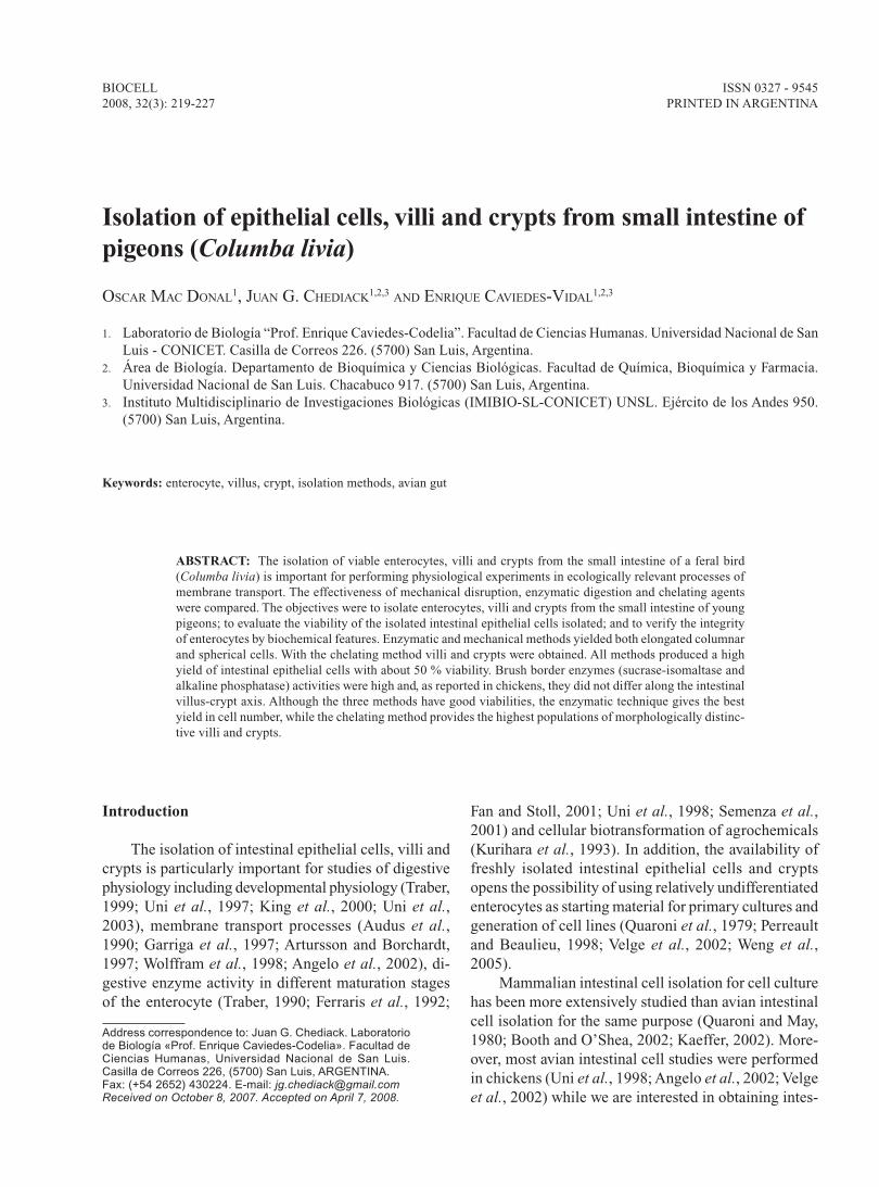
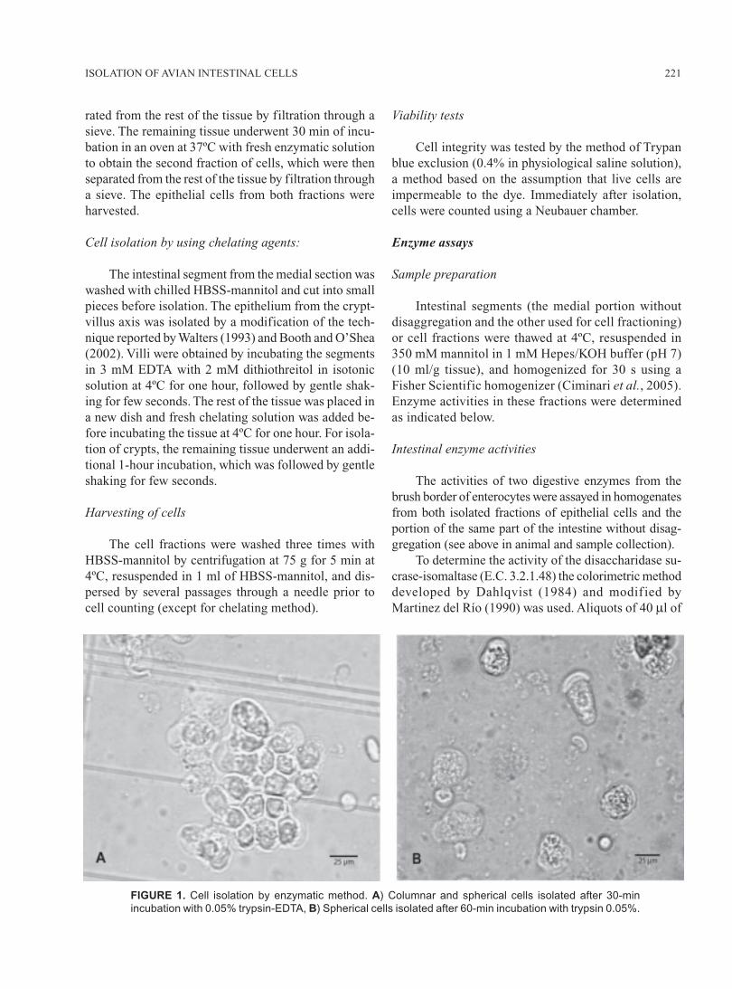
FIGURE 1. Cell isolation by enzymatic method. A) Columnar and spherical cells isolated after 30-min
incubation with 0.05% trypsin-EDTA, B) Spherical cells isolated after 60-min incubation with trypsin 0.05%.
OSCAR MAC DONAL et al.222
tissue or cell homogenate, appropriately diluted, wereincubated with 40 μl of 56 mM sucrose solution in 0.1M maleate/NaOH pH 6.5. After 10 min incubation at al40ºC the reaction was stopped by adding 1 ml ofGlicemia Enzimática reagent (Wiener LaboratoriosSAIC, Rosario-Argentina, with a sensitivity < 10 μg/ml). The enzymatic activity was determined by mea-suring at room temperature the absorbance at 505 nmwith a Spectronic 21D spectrophotometer.
Alkaline phosphatase (E.C. 3.1.3.1) activity wasdetermined spectrophotometrically by the rate of sodiumphenyl phosphate hydrolysis using an alkaline phos-phatase kit (Wiener Laboratorios SAIC, Rosario-Argen-tina) following the manufacturer’s recommendations.Briefly, aliquots of 40 μl of tissue or cell homogenate,appropriately diluted, were incubated with 40 μl of de-veloping reagent (sodium phenyl phosphate inaminomethyl propanol alkaline buffer). Hydrolyzedphenol was detected after a 10-min incubation with 4-aminoantipyrine and ferricyanide at 40ºC. The sensi-tivity of this method is 10-3 units/ml. The samples wereread immediately in a Spectronic 21D spectrophotom-eter at 520 nm.
Protein measurement
The protein concentration of samples was estimatedby Lowry’s method (1951) using the commercial kitProti2 Assay (EDTA/Cu Reagent, Wiener LaboratoriosSAIC, Rosario-Argentina). Absorbance was read at 540nm using bovine serum albumin as the standard, with asensitivity of 20 μg/ml.
Microphotographs of intestinal epithelial cells
Microphotographs of cells fractions, villi and cryptswere taken using an Olympus microscope model BX50and video camera Sony CCD-Iris. For measurementsof lengths and widths of isolated enterocytes, thefreeware software ImageJ (Rasband, 1997-2006) wasemployed.
Statistical analysis
Results are given as means ± 1 SEM; n is numberof individuals. Standard least-squares methods wereused to estimate parameters of linear regression. AStudent’s t-test was used to assess the statistical differ-ence of enzyme activities and protein content betweendifferent fractions of isolated cells. One way analysisof variance (ANOVA) was used to compare the viabil-ity of cells obtained with enzymatic and mechanicalmethods. The significance level was set at P < 0.05.
Results
Microscopic appearance of isolated epithelial cells
All three methods allowed isolation of morpho-logically identifiable intestinal epithelial cells. In bothfractions, the enzymatic and the mechanical methodyielded both elongated columnar and spherical cells.The columnar cells are typically from the villus tipand the spherical ones from mid- and lower villus
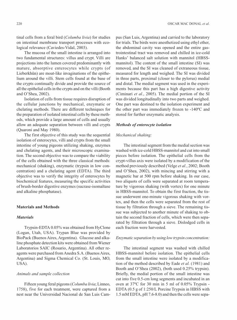
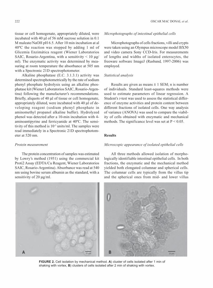
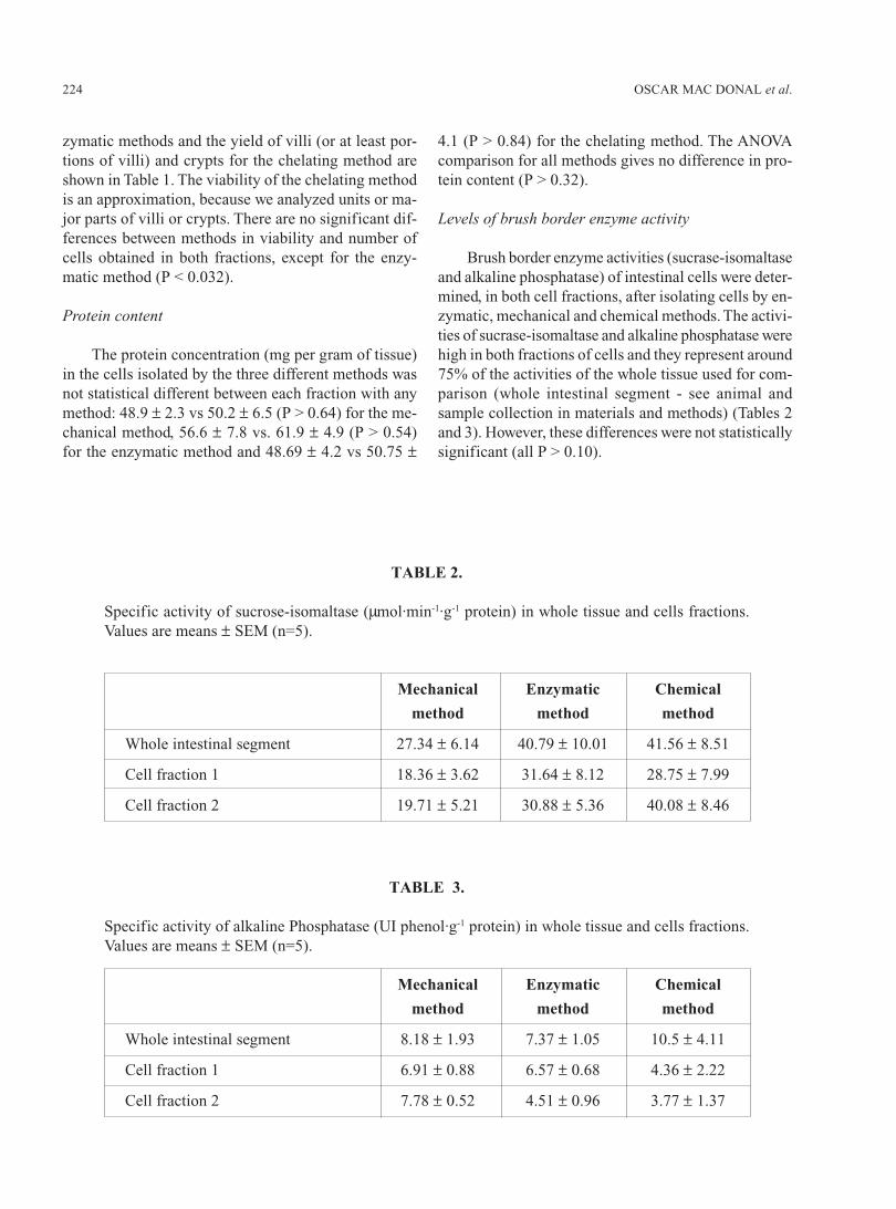
FIGURE 2. Cell isolation by mechanical method. A) cluster of cells isolated after 1 min of
shaking with vortex, B) clusters of cells isolated after 2 min of shaking with vortex.
223ISOLATION OF AVIAN INTESTINAL CELLS
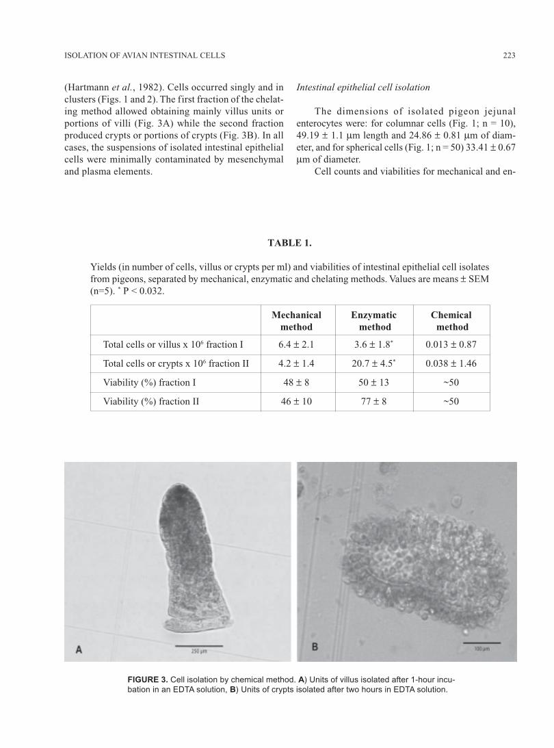
(Hartmann et al., 1982). Cells occurred singly and inclusters (Figs. 1 and 2). The first fraction of the chelat-ing method allowed obtaining mainly villus units orportions of villi (Fig. 3A) while the second fractionproduced crypts or portions of crypts (Fig. 3B). In allcases, the suspensions of isolated intestinal epithelialcells were minimally contaminated by mesenchymaland plasma elements.
Intestinal epithelial cell isolation
The dimensions of isolated pigeon jejunalenterocytes were: for columnar cells (Fig. 1; n = 10),49.19 ± 1.1 μm length and 24.86 ± 0.81 μm of diam-eter, and for spherical cells (Fig. 1; n = 50) 33.41 ± 0.67μm of diameter.
Cell counts and viabilities for mechanical and en-
TABLE 1.
Yields (in number of cells, villus or crypts per ml) and viabilities of intestinal epithelial cell isolatesfrom pigeons, separated by mechanical, enzymatic and chelating methods. Values are means ± SEM(n=5). * P < 0.032.
Mechanical Enzymatic Chemicalmethod method method
Total cells or villus x 106 fraction I 6.4 ± 2.1 3.6 ± 1.8* 0.013 ± 0.87
Total cells or crypts x 106 fraction II 4.2 ± 1.4 20.7 ± 4.5* 0.038 ± 1.46
Viability (%) fraction I 48 ± 8 50 ± 13 ~50
Viability (%) fraction II 46 ± 10 77 ± 8 ~50
FIGURE 3. Cell isolation by chemical method. A) Units of villus isolated after 1-hour incu-
bation in an EDTA solution, B) Units of crypts isolated after two hours in EDTA solution.
OSCAR MAC DONAL et al.224
zymatic methods and the yield of villi (or at least por-tions of villi) and crypts for the chelating method areshown in Table 1. The viability of the chelating methodis an approximation, because we analyzed units or ma-jor parts of villi or crypts. There are no significant dif-ferences between methods in viability and number ofcells obtained in both fractions, except for the enzy-matic method (P < 0.032).
Protein content
The protein concentration (mg per gram of tissue)in the cells isolated by the three different methods wasnot statistical different between each fraction with anymethod: 48.9 ± 2.3 vs 50.2 ± 6.5 (P > 0.64) for the me-chanical method, 56.6 ± 7.8 vs. 61.9 ± 4.9 (P > 0.54)for the enzymatic method and 48.69 ± 4.2 vs 50.75 ±
4.1 (P > 0.84) for the chelating method. The ANOVAcomparison for all methods gives no difference in pro-tein content (P > 0.32).
Levels of brush border enzyme activity
Brush border enzyme activities (sucrase-isomaltaseand alkaline phosphatase) of intestinal cells were deter-mined, in both cell fractions, after isolating cells by en-zymatic, mechanical and chemical methods. The activi-ties of sucrase-isomaltase and alkaline phosphatase werehigh in both fractions of cells and they represent around75% of the activities of the whole tissue used for com-parison (whole intestinal segment - see animal andsample collection in materials and methods) (Tables 2and 3). However, these differences were not statisticallysignificant (all P > 0.10).
TABLE 2.
Specific activity of sucrose-isomaltase (μmol·min-1·g-1 protein) in whole tissue and cells fractions.Values are means ± SEM (n=5).
Mechanical Enzymatic Chemical
method method method
Whole intestinal segment 27.34 ± 6.14 40.79 ± 10.01 41.56 ± 8.51
Cell fraction 1 18.36 ± 3.62 31.64 ± 8.12 28.75 ± 7.99
Cell fraction 2 19.71 ± 5.21 30.88 ± 5.36 40.08 ± 8.46
TABLE 3.
Specific activity of alkaline Phosphatase (UI phenol·g-1 protein) in whole tissue and cells fractions.Values are means ± SEM (n=5).
Mechanical Enzymatic Chemical
method method method
Whole intestinal segment 8.18 ± 1.93 7.37 ± 1.05 10.5 ± 4.11
Cell fraction 1 6.91 ± 0.88 6.57 ± 0.68 4.36 ± 2.22
Cell fraction 2 7.78 ± 0.52 4.51 ± 0.96 3.77 ± 1.37
225ISOLATION OF AVIAN INTESTINAL CELLS
Discussion
The small intestinal epithelium constitutes a sys-tem in constant renewal. Enterocytes comprise up to90% of epithelial cells in the crypt and over 95% ofvillus cells (Cheng and Leblond, 1974). The typicalenterocyte from the villus-tip is recognizable under lightmicroscopy by its oblong shape and conspicuous brushborder (Fig. 1B). The spherical cells are typical for mid-and lower villus and crypts (Fig. 1A; Hartmann et al.,1982). In the mechanical and enzymatic methods,spherical cells constituted the main cell type isolated inboth cell fractions (Figs. 1 and 2).
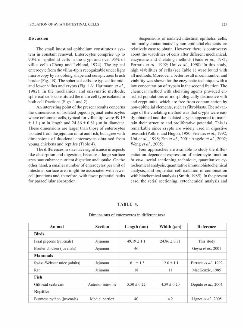
An interesting point of the present results concernsthe dimensions of isolated pigeon jejunal enterocyteswhere columnar cells, typical for villus-tip, were 49.19± 1.1 μm in length and 24.86 ± 0.81 μm in diameter.These dimensions are larger than those of enterocytesisolated from the jejunum of rat and fish, but agree withdimensions of duodenal enterocytes obtained fromyoung chickens and reptiles (Table 4).
The differences in size have significance in aspectslike absorption and digestion, because a large surfacearea may enhance nutrient digestion and uptake. On theother hand, a smaller number of enterocytes per unit ofintestinal surface area might be associated with fewercell junctions and, therefore, with fewer potential pathsfor paracellular absorption.
Suspensions of isolated intestinal epithelial cells,minimally contaminated by non-epithelial elements arerelatively easy to obtain. However, there is controversyabout the viabilities of cells after different mechanical,enzymatic and chelating methods (Eade et al., 1981;Ferraris et al., 1992; Uni et al., 1998). In this study,high viabilities of cells (see Table 1) were found withall methods. Moreover a better result in cell number andviability was shown for the enzymatic technique with alow concentration of trypsin in the second fraction. Thechemical method with chelating agents provided en-riched populations of morphologically distinctive villiand crypt units, which are free from contamination bynon-epithelial elements, such as fibroblasts. The advan-tage of the chelating method was that crypts were eas-ily obtained and the isolated crypts appeared to main-tain their structure and proliferative potential. This isremarkable since crypts are widely used in digestiveresearch (Pothier and Hugon, 1980; Ferraris et al., 1992;Uni et al., 1998; Fan et al., 2001; Angelo et al., 2002;Weng et al., 2005).
Four approaches are available to study the differ-entiation-dependent expression of enterocyte functionin vivo: serial sectioning technique, quantitative cy-tochemical analysis, quantitative immunohistochemicalanalysis, and sequential cell isolation in combinationwith biochemical analysis (Smith, 1985). In the presentcase, the serial sectioning, cytochemical analysis and
TABLE 4.
Dimensions of enterocytes in different taxa.
Animal Section Length (μm) Width (μm) Reference
Birds
Feral pigeons (juvenals) Jejunum 49.19 ± 1.1 24.86 ± 0.81 This study
Broiler chicken (juvenals) Jejunum 46 Geyra et al., 2001
Mammals
Swiss-Webster mice (adults) Jejunum 16.1 ± 1.5 12.0 ± 1.1 Ferraris et al., 1992
Rat Jejunum 18 11 MacKenzie, 1985
Fish
Gilthead seabream Anterior intestine 5.50 ± 0.22 4.59 ± 0.20 Dopido et al., 2004
Reptiles
Burmese python (juvenals) Medial portion 40 4.2 Lignot et al., 2005
OSCAR MAC DONAL et al.226
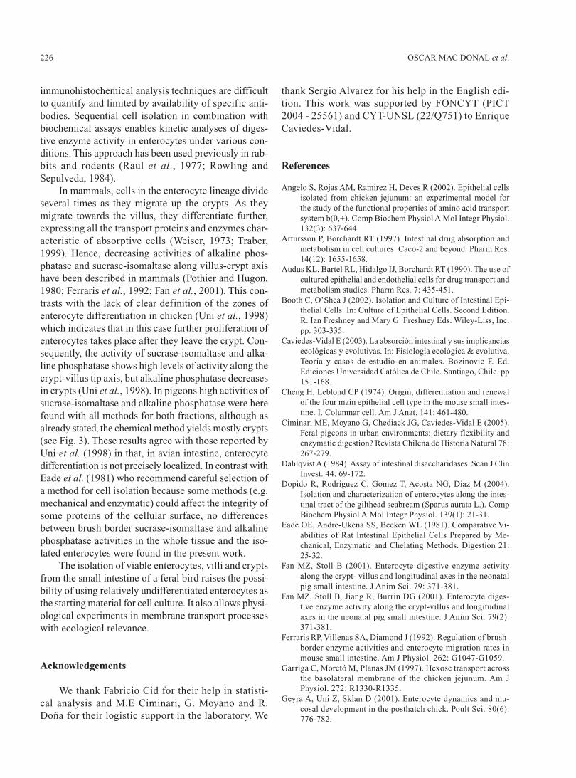
immunohistochemical analysis techniques are difficultto quantify and limited by availability of specific anti-bodies. Sequential cell isolation in combination withbiochemical assays enables kinetic analyses of diges-tive enzyme activity in enterocytes under various con-ditions. This approach has been used previously in rab-bits and rodents (Raul et al., 1977; Rowling andSepulveda, 1984).
In mammals, cells in the enterocyte lineage divideseveral times as they migrate up the crypts. As theymigrate towards the villus, they differentiate further,expressing all the transport proteins and enzymes char-acteristic of absorptive cells (Weiser, 1973; Traber,1999). Hence, decreasing activities of alkaline phos-phatase and sucrase-isomaltase along villus-crypt axishave been described in mammals (Pothier and Hugon,1980; Ferraris et al., 1992; Fan et al., 2001). This con-trasts with the lack of clear definition of the zones ofenterocyte differentiation in chicken (Uni et al., 1998)which indicates that in this case further proliferation ofenterocytes takes place after they leave the crypt. Con-sequently, the activity of sucrase-isomaltase and alka-line phosphatase shows high levels of activity along thecrypt-villus tip axis, but alkaline phosphatase decreasesin crypts (Uni et al., 1998). In pigeons high activities ofsucrase-isomaltase and alkaline phosphatase were herefound with all methods for both fractions, although asalready stated, the chemical method yields mostly crypts(see Fig. 3). These results agree with those reported byUni et al. (1998) in that, in avian intestine, enterocytedifferentiation is not precisely localized. In contrast withEade et al. (1981) who recommend careful selection ofa method for cell isolation because some methods (e.g.mechanical and enzymatic) could affect the integrity ofsome proteins of the cellular surface, no differencesbetween brush border sucrase-isomaltase and alkalinephosphatase activities in the whole tissue and the iso-lated enterocytes were found in the present work.
The isolation of viable enterocytes, villi and cryptsfrom the small intestine of a feral bird raises the possi-bility of using relatively undifferentiated enterocytes asthe starting material for cell culture. It also allows physi-ological experiments in membrane transport processeswith ecological relevance.
Acknowledgements
We thank Fabricio Cid for their help in statisti-cal analysis and M.E Ciminari, G. Moyano and R.Doña for their logistic support in the laboratory. We
thank Sergio Alvarez for his help in the English edi-tion. This work was supported by FONCYT (PICT2004 - 25561) and CYT-UNSL (22/Q751) to EnriqueCaviedes-Vidal.
References
Angelo S, Rojas AM, Ramirez H, Deves R (2002). Epithelial cellsisolated from chicken jejunum: an experimental model forthe study of the functional properties of amino acid transportsystem b(0,+). Comp Biochem Physiol A Mol Integr Physiol.132(3): 637-644.
Artursson P, Borchardt RT (1997). Intestinal drug absorption andmetabolism in cell cultures: Caco-2 and beyond. Pharm Res.14(12): 1655-1658.
Audus KL, Bartel RL, Hidalgo IJ, Borchardt RT (1990). The use ofcultured epithelial and endothelial cells for drug transport andmetabolism studies. Pharm Res. 7: 435-451.
Booth C, O’Shea J (2002). Isolation and Culture of Intestinal Epi-thelial Cells. In: Culture of Epithelial Cells. Second Edition.R. Ian Freshney and Mary G. Freshney Eds. Wiley-Liss, Inc.pp. 303-335.
Caviedes-Vidal E (2003). La absorción intestinal y sus implicanciasecológicas y evolutivas. In: Fisiología ecológica & evolutiva.Teoría y casos de estudio en animales. Bozinovic F. Ed.Ediciones Universidad Católica de Chile. Santiago, Chile. pp151-168.
Cheng H, Leblond CP (1974). Origin, differentiation and renewalof the four main epithelial cell type in the mouse small intes-tine. I. Columnar cell. Am J Anat. 141: 461-480.
Ciminari ME, Moyano G, Chediack JG, Caviedes-Vidal E (2005).Feral pigeons in urban environments: dietary flexibility andenzymatic digestion? Revista Chilena de Historia Natural 78:267-279.
Dahlqvist A (1984). Assay of intestinal disaccharidases. Scan J ClinInvest. 44: 69-172.
Dopido R, Rodriguez C, Gomez T, Acosta NG, Diaz M (2004).Isolation and characterization of enterocytes along the intes-tinal tract of the gilthead seabream (Sparus aurata L.). CompBiochem Physiol A Mol Integr Physiol. 139(1): 21-31.
Eade OE, Andre-Ukena SS, Beeken WL (1981). Comparative Vi-abilities of Rat Intestinal Epithelial Cells Prepared by Me-chanical, Enzymatic and Chelating Methods. Digestion 21:25-32.
Fan MZ, Stoll B (2001). Enterocyte digestive enzyme activityalong the crypt- villus and longitudinal axes in the neonatalpig small intestine. J Anim Sci. 79: 371-381.
Fan MZ, Stoll B, Jiang R, Burrin DG (2001). Enterocyte diges-tive enzyme activity along the crypt-villus and longitudinalaxes in the neonatal pig small intestine. J Anim Sci. 79(2):371-381.
Ferraris RP, Villenas SA, Diamond J (1992). Regulation of brush-border enzyme activities and enterocyte migration rates inmouse small intestine. Am J Physiol. 262: G1047-G1059.
Garriga C, Moretó M, Planas JM (1997). Hexose transport acrossthe basolateral membrane of the chicken jejunum. Am JPhysiol. 272: R1330-R1335.
Geyra A, Uni Z, Sklan D (2001). Enterocyte dynamics and mu-cosal development in the posthatch chick. Poult Sci. 80(6):776-782.
227ISOLATION OF AVIAN INTESTINAL CELLS
Hartmann F, Owen R, Bissell DM (1982). Characterization of iso-lated epithelial cells from rat small intestine. Am J Physiol.242(2): G147-155.
Kaeffer B (2002). Mammalian intestinal epithelial cells in primaryculture: a mini-review. In vitro Cell Dev Biol Anim. 38(3):123-134.
King DE, Asem EK, Adeola O (2000). Ontogenetic developmentof intestinal digestive functions in white Pekin ducks. J Nutr130: 57-62.
Kurihara N, Paulson GD, Otto S, Miyamoto J, Hollingworth RM(1993). Use of isolated cells to study the metabolism of agro-chemicals in animals. Pure & Appl Chem. 65(10): 2299-2312.
Lignot JH, Helmstetter C, Secor SM (2005). Postprandial morpho-logical response of the intestinal epithelium of the Burmesepython (Python molurus). Comp Biochem Physiol A MolIntegr Physiol. 141(3): 280-291.
Lowry OH, Rosebrough, NJ, Farr AL, Randall RJ (1951). Proteinmeasurement with the folin phenol agent. J Biol Chem. 193:265-275.
MacKenzie NM (1985). Comparison of the metabolic activities ofenterocytes isolated from different regions of the small intes-tine of the neonate. Biology of the Neonate. 48: 257-268.
Martínez del Río C (1990) Dietary, phylogenetic, and ecologicalcorrelates of intestinal sucrase and maltase activity in birds.Physiol Zool. 63: 987-1011.
Perreault N, Beaulieu JF (1998). Primary cultures of fully differen-tiated and pure human intestinal epithelial cells. Exp Cell Res.245: 34-42.
Pothier P, Hugon JS (1980). Characterization of isolated villus andcrypt cells from the small intestine of the adult mouse. CellTissue Res. 211(3): 405-418.
Quaroni A, May RJ (1980). Establishment and characterization ofintestinal epithelial cell cultures. Methods in Cell Biology.Volume 21B. Academic Press, Inc. 403-427.
Quaroni A, Wands J, Trelstad RL, Isselbacher KJ (1979). Epithe-lioid cell cultures from rat small intestine. Characterizationby morphologic and immunologic criteria. J Cell Biol 80: 248-265.
Rasband WS (1997-2006). ImageJ, U.S. National Institutes ofHealth, Bethesda, Maryland, USA, http://rsb.info.nih.gov/ij
Raul F, Simon P, Kedinger M, Haffen K (1977). Intestinal enzymesactivities in isolated villus and crypt cells during postnataldevelopment of the rat. Cell Tissue Res. 12; 176(2): 167-178.
Rowling PJ, Sepulveda FV (1984). The distribution of (Na+ + K+)-ATPase along the villus crypt-axis in the rabbit small intes-tine. Biochim Biophys Acta. 771(1): 35-41.
Semenza G, Auricchio S, Mantei N (2001). Small-intestinal disac-charidases In: Metab Molec Bases of Inherit Disease. Scriver,Ch.R., Beaudet, A.L., Sly, W.S., and Valle, D. (Eds.), Childs,B. Kinzler, K.W, and Vogelstein, B. (Ass. Eds.), 8th Edition,Chapt. 75; McGraw-Hill, New York, pp. 1623-1650.
Smith MW (1985). Expression of digestive and absorptive func-tion in differentiating enterocytes. Ann Rev Physiol. 47: 247-260.
Traber PG (1990). Regulation of sucrase-isomaltase gene expres-sion along the crypt-villus axis of rat small intestine. BiochemBiophys Res Commun. 173(3): 765-773.
Traber PG (1999). Development of brush border enzyme activity.Development of the Gastrointestinal Tract. Sanderson IR andWalker WA (Eds). Hamilton, Ontario. Canada. pp 103-122.
Trypan Blue. Mediatech Technical Information. www.cellgro.comUni Z, Ganot S, Sklan D (1997). Posthatch development of mu-
cosal function in the broiler small intestine. Poultry Science77: 75-82.
Uni Z, Platin R, Sklan D (1998). Cell proliferation in chicken in-testinal epithelium occurs both in the crypt and along the vil-lus. J Comp Physiol B. 168: 241-247.
Uni Z, Tako E, Gal-Garber, Sklan D (2003). Morphological, mo-lecular and functional changes in the chicken small intestineof the late-term embryo. Poult Sci. 82(11): 1747-1754.
Velge P, Bottreau E, Querré P, Pardon P, Nicolle JC, Morisson M,Bout D, Dimier I (2002). Establishment and characterizationof partially differentiated chicken enterocyte cell clones. EurJ Cell Biol. 81: 203-212.
Walters RJ (1993). Ion channel regulation in small intestinal crypts.Ph.D. Thesis. University of Cambridge. UK. http://www.cellscience.com/author.htm.
Weiser MM (1973). Intestinal epithelial cell surface membrane gly-coprotein synthesis. I. An indication of cellular differentia-tion. J Biol Chem. 248: 2536-2541.
Weng XH, Beyenbach KW, Quaroni A (2005). Cultured monolay-ers of the dog jejunum with the structural and functional prop-erties resembling the normal epithelium. Am J PhysiolGastrointest Liver Physiol. 288(4): G705-717.
Wolffram S, Grenacher B, Scharrer E (1998). H+-Coupled uphilltransport of the dipeptide glicylsarcosine by bovine intestinalbrush- border membrane vesicles. J Dairy Sci 81: 2595-2603.

Copyright © 2022 FDOKUMEN