Isolation and Purification of a New Kalimantacin/Batumin-Related Polyketide Antibiotic and...
11
Chemistry & Biology Article Isolation and Purification of a New Kalimantacin/ Batumin-Related Polyketide Antibiotic and Elucidation of Its Biosynthesis Gene Cluster Wesley Mattheus, 1 Ling-Jie Gao, 2 Piet Herdewijn, 2 Bart Landuyt, 3 Jan Verhaegen, 4 Joleen Masschelein, 1 Guido Volckaert, 1 and Rob Lavigne 1, * 1 Laboratory of Gene Technology 2 Interface Valorization Platform 3 Animal Physiology and Neurobiology Section 4 Experimental Laboratory Medicine Katholieke Universiteit Leuven, Leuven B-3000, Belgium *Correspondence: [email protected] DOI 10.1016/j.chembiol.2010.01.014 SUMMARY Kal/bat, a polyketide, isolated to high purity (>95%) is characterized by strong and selective antibacterial activity against Staphylococcus species (minimum inhibitory concentration, 0.05 mg/mL), and no resis- tance was observed in strains already resistant to commonly used antibiotics. The kal/bat biosynthesis gene cluster was determined to a 62 kb genomic region of Pseudomonas fluorescens BCCM_ID9359. The kal/bat gene cluster consists of 16 open reading frames (ORF), encoding a hybrid PKS-NRPS system, extended with trans-acting tailoring functions. A full model for kal/bat biosynthesis is postulated and experimentally tested by gene inactivation, structural confirmation (using NMR spectroscopy), and com- plementation. The structural and microbiological study of biosynthetic kal/bat analogs revealed the importance of the carbamoyl group and 17-keto group for antibacterial activity. The mechanism of self-resistance lies within the production of an inac- tive intermediate, which is activated in a one-step enzymatic oxidation upon export. The genetic basis and biochemical elucidation of the biosynthesis pathway of this antibiotic will facilitate rational engi- neering for the design of novel structures with improved activities. This makes it a promising new therapeutic option to cope with multidrug-resistant clinical infections. INTRODUCTION In the antibiotic resistance age, which is marked by an increasing need for novel compounds (Von Nussbaum et al., 2006), sec- ondary metabolites continue to play a major role in antibacterial drug discovery and development (Nathan, 2004). Their enor- mous structural complexity, linked with biological activity, forms an intrinsic advantage over synthetic compounds. Knowledge of their biosynthesis pathways substantially increases this advan- tage for the development of structural diversity through directed biosynthetic mutagenesis (Hopwood, 2004). Polyketides and nonribosomal peptides represent two of the major classes of antibiotics, generated on multimodular enzy- matic assembly lines. The modularity of the scaffold biosyn- thesis, together with the plurality of postassembly modifications by tailoring enzymes, offer prospects of creating novel antibi- otics with optimized properties toward activity, production, and pharmacokinetics. The kalimantacin antibiotics were isolated from a fermentation broth of Alcaligenes species YL-02632S and display a strong antistaphylococcal effect (Kamigiri et al., 1996; Tokunaga et al., 1996)(Figure 1). The antibiotic batumin, which has the same molecular composition as kalimantacin A, was isolated from a culture liquid of Pseudomonas batumici and showed similar antimicrobial activity (Smirnov et al., 2000). To date, no batumin-resistant clinical staphylococcus isolates have been reported (Klochko et al., 2008). Batumin was previ- ously shown to have only moderate activity against enterobacte- ria (Salmonella, Bordetella, Escherichia, and Klebsiella species) and no activity against micrococci and streptococci. Chemical- and UV-induced mutagenesis allowed the production of strains with 3.5-fold increased batumin production. During these exper- iments, histidine auxotrophy tended to be associated with loss of batumin production, hinting at a link between batumin biosyn- thesis and the histidine operon (Smirnov et al., 2000). The limited commercial availability and unsatisfying purity (<80%) of batumin hampers further research. Because the absolute con- figurations of the stereogenic carbon centers of all the above- mentioned compounds are not known, it is still unclear whether they are the same. In this article, we present the isolation and purification of kal/bat, the sequence of the kal/bat gene cluster, and a model for its biosynthesis and self-resistance. RESULTS Microbiological Aspects and Purification/ Characterization of the Antibiotic Compound During the screening for novel antibiotics, a strain obtained from the BCCM/LMG Bacteria Collection (ID9359) displayed strong Chemistry & Biology 17, 1–11, February 26, 2010 ª2010 Elsevier Ltd All rights reserved 1 CHBIOL 1643 Please cite this article in press as: Mattheus et al., Isolation and Purification of a New Kalimantacin/Batumin-Related Polyketide Antibiotic and Eluci- dation of Its Biosynthesis Gene Cluster, Chemistry & Biology (2010), doi:10.1016/j.chembiol.2010.01.014
Transcript of Isolation and Purification of a New Kalimantacin/Batumin-Related Polyketide Antibiotic and...

Please cite this article in press as: Mattheus et al., Isolation and Purification of a New Kalimantacin/Batumin-Related Polyketide Antibiotic and Eluci-dation of Its Biosynthesis Gene Cluster, Chemistry & Biology (2010), doi:10.1016/j.chembiol.2010.01.014
Chemistry & Biology
Article
Isolation and Purification of a New Kalimantacin/Batumin-Related Polyketide Antibiotic andElucidation of Its Biosynthesis Gene ClusterWesley Mattheus,1 Ling-Jie Gao,2 Piet Herdewijn,2 Bart Landuyt,3 Jan Verhaegen,4 Joleen Masschelein,1
Guido Volckaert,1 and Rob Lavigne1,*1Laboratory of Gene Technology2Interface Valorization Platform3Animal Physiology and Neurobiology Section4Experimental Laboratory Medicine
Katholieke Universiteit Leuven, Leuven B-3000, Belgium
*Correspondence: [email protected] 10.1016/j.chembiol.2010.01.014
SUMMARY
Kal/bat, a polyketide, isolated to high purity (>95%) ischaracterized by strong and selective antibacterialactivity against Staphylococcus species (minimuminhibitory concentration, 0.05 mg/mL), and no resis-tance was observed in strains already resistant tocommonly used antibiotics. The kal/bat biosynthesisgene cluster was determined to a 62 kb genomicregion of Pseudomonas fluorescens BCCM_ID9359.The kal/bat gene cluster consists of 16 open readingframes (ORF), encoding a hybrid PKS-NRPS system,extended with trans-acting tailoring functions. A fullmodel for kal/bat biosynthesis is postulated andexperimentally tested by gene inactivation, structuralconfirmation (using NMR spectroscopy), and com-plementation. The structural and microbiologicalstudy of biosynthetic kal/bat analogs revealed theimportance of the carbamoyl group and 17-ketogroup for antibacterial activity. The mechanism ofself-resistance lies within the production of an inac-tive intermediate, which is activated in a one-stepenzymatic oxidation upon export. The genetic basisand biochemical elucidation of the biosynthesispathway of this antibiotic will facilitate rational engi-neering for the design of novel structures withimproved activities. This makes it a promising newtherapeutic option to cope with multidrug-resistantclinical infections.
INTRODUCTION
In the antibiotic resistance age, which is marked by an increasing
need for novel compounds (Von Nussbaum et al., 2006), sec-
ondary metabolites continue to play a major role in antibacterial
drug discovery and development (Nathan, 2004). Their enor-
mous structural complexity, linked with biological activity, forms
an intrinsic advantage over synthetic compounds. Knowledge of
Chemistry & Biology 1
CHBIOL
their biosynthesis pathways substantially increases this advan-
tage for the development of structural diversity through directed
biosynthetic mutagenesis (Hopwood, 2004).
Polyketides and nonribosomal peptides represent two of the
major classes of antibiotics, generated on multimodular enzy-
matic assembly lines. The modularity of the scaffold biosyn-
thesis, together with the plurality of postassembly modifications
by tailoring enzymes, offer prospects of creating novel antibi-
otics with optimized properties toward activity, production, and
pharmacokinetics. The kalimantacin antibiotics were isolated
from a fermentation broth of Alcaligenes species YL-02632S
and display a strong antistaphylococcal effect (Kamigiri et al.,
1996; Tokunaga et al., 1996) (Figure 1). The antibiotic batumin,
which has the same molecular composition as kalimantacin A,
was isolated from a culture liquid of Pseudomonas batumici
and showed similar antimicrobial activity (Smirnov et al., 2000).
To date, no batumin-resistant clinical staphylococcus isolates
have been reported (Klochko et al., 2008). Batumin was previ-
ously shown to have only moderate activity against enterobacte-
ria (Salmonella, Bordetella, Escherichia, and Klebsiella species)
and no activity against micrococci and streptococci. Chemical-
and UV-induced mutagenesis allowed the production of strains
with 3.5-fold increased batumin production. During these exper-
iments, histidine auxotrophy tended to be associated with loss of
batumin production, hinting at a link between batumin biosyn-
thesis and the histidine operon (Smirnov et al., 2000). The limited
commercial availability and unsatisfying purity (<80%) of
batumin hampers further research. Because the absolute con-
figurations of the stereogenic carbon centers of all the above-
mentioned compounds are not known, it is still unclear whether
they are the same.
In this article, we present the isolation and purification of
kal/bat, the sequence of the kal/bat gene cluster, and a model
for its biosynthesis and self-resistance.
RESULTS
Microbiological Aspects and Purification/Characterization of the Antibiotic CompoundDuring the screening for novel antibiotics, a strain obtained from
the BCCM/LMG Bacteria Collection (ID9359) displayed strong
7, 1–11, February 26, 2010 ª2010 Elsevier Ltd All rights reserved 1
1643

Figure 1. Structure of Kalimantacins and Batumin
The molecules have a linear polyketidal backbone with an incorporated glycine, multiple methyl branches, and a characteristic carbamoyl-group.
Chemistry & Biology
Characterization of the kal/bat Gene Cluster
Please cite this article in press as: Mattheus et al., Isolation and Purification of a New Kalimantacin/Batumin-Related Polyketide Antibiotic and Eluci-dation of Its Biosynthesis Gene Cluster, Chemistry & Biology (2010), doi:10.1016/j.chembiol.2010.01.014
antibacterial activity against staphylococci. Sequence analysis of
the ribosomal genes revealed that the strain belongs to the species
Pseudomonas fluorescens. Fermentation experiments with
P. fluorescens strain BCCM_ID9359 showed an optimal growth
temperature of 28�C, whereas antibiotic production is significantly
higher at 16�C. Antibiotic production followed the cell growth and
reached a maximum after 48 hr (data not shown). HPLC analysis of
crude extracts shows a peak with a retention time of 20.8 min
(see Figure S1A available online); the extracts were purified and
shown to have antibiotic activity on a bacterial lawn plate assay.
HRMS revealed a molecular peak at [M + Na]/e = 571.3337,
indicating a molecular formula of C30H48N2O7Na (<1 ppm error)
(Figure S1B), identical to those of kalimantacin A and batumin,
which were independently isolated fromAlcaligenes speciesstrain
YL-02632S and P. batumici fermentations, respectively (Kamigiri
et al., 1996; Tokunaga et al., 1996; Smirnov et al., 2000). Large-
scale purification by silica gel column chromatography yielded
35 mg/L of over 95% pure compound, allowing further structural
and biological testing. 1H and 13C NMR spectra data verified the
polyketide structure ((1); Figure 1) (Figures S1C and S1D). HPLC
and HRMS analysis showed the remaining inpurity to be
a secondary compound with a slightly reduced retention time of
20.2 min and a molecular peak at [M + Na]/e = 573.3501, indicating
a molecular formula of C30H50N2O7Na (Figures S1A and S2A). In1H NMR spectra, all of the typical peaks (olefinicproton and methyl
CHBIOL 164
2 Chemistry & Biology 17, 1–11, February 26, 2010 ª2010 Elsevier Lt
protons) of the polyketide were present, which suggested that
the product is a 17-keto reduction product. The new multipeak
at 4.0 ppm indicated that an additional hydroxyl group was formed
in the molecule. In 13C NMR spectra, no peak corresponding to a
keto group was observed, but a new peak at 66.1 ppm (typical
chemical shift for secondary alcohol) appeared. These spectra
data further proved the deduced structure (7). A detailed interpre-
tation of 1H and 13C NMR spectra of 17-hydroxy kal/bat are shown
in the Table S1 and Figures S2B and S2C.
The high yield and purity levels achieved for the active
compound enabled accurate microbiological and chemical
testing. Minimal inhibitory concentrations (MICs) of kal/bat
were determined using NCCLS standards, confirming its rather
selective activity against Staphylococcus species and, to a lesser
extent, against enterobacteria (Table S2). During the course of
screening, no kal/bat-resistant clinical Staphylococcus strains
were discovered, and no resistance in strains already resistant
to commonly used antibiotics was observed, hinting at a novel
target or a strategy to bypass existing resistance.
Molecular Analysis of the Biosynthesis PathwayLocalization and identification of the kal/bat
gene cluster
The biosynthesis gene cluster of this predicted polyketide
was identified by random fragment sequencing and selection
3
d All rights reserved
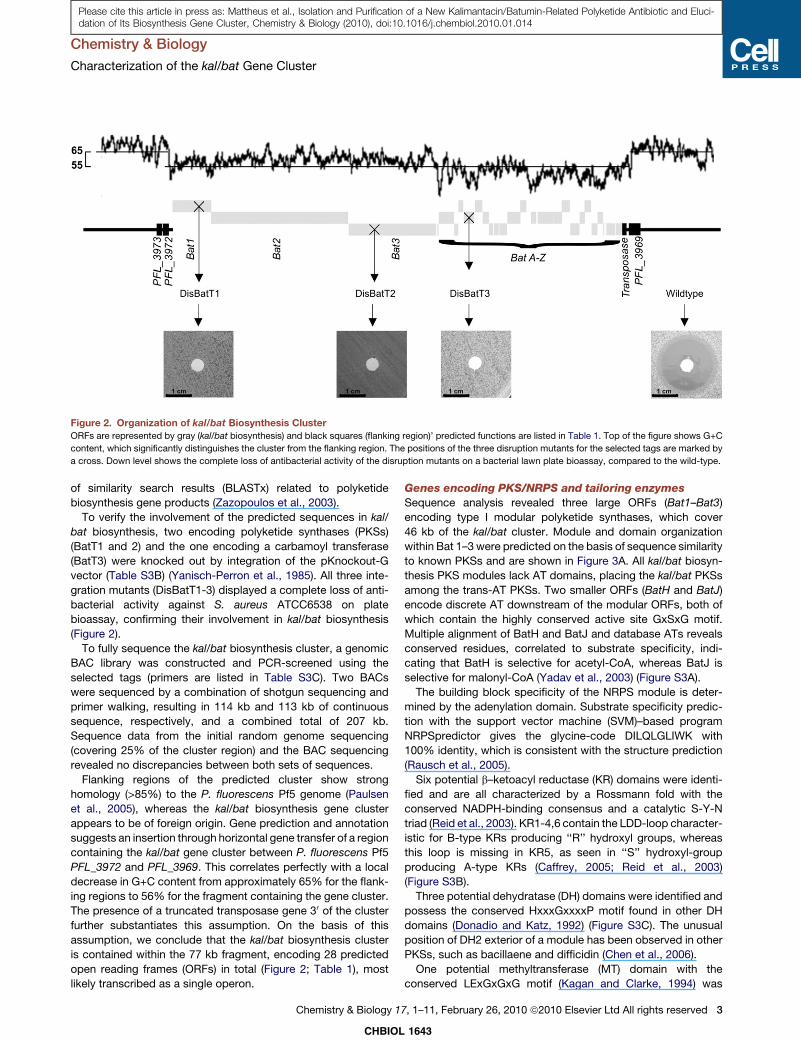
Figure 2. Organization of kal/bat Biosynthesis Cluster
ORFs are represented by gray (kal/bat biosynthesis) and black squares (flanking region)’ predicted functions are listed in Table 1. Top of the figure shows G+C
content, which significantly distinguishes the cluster from the flanking region. The positions of the three disruption mutants for the selected tags are marked by
a cross. Down level shows the complete loss of antibacterial activity of the disruption mutants on a bacterial lawn plate bioassay, compared to the wild-type.
Chemistry & Biology
Characterization of the kal/bat Gene Cluster
Please cite this article in press as: Mattheus et al., Isolation and Purification of a New Kalimantacin/Batumin-Related Polyketide Antibiotic and Eluci-dation of Its Biosynthesis Gene Cluster, Chemistry & Biology (2010), doi:10.1016/j.chembiol.2010.01.014
of similarity search results (BLASTx) related to polyketide
biosynthesis gene products (Zazopoulos et al., 2003).
To verify the involvement of the predicted sequences in kal/
bat biosynthesis, two encoding polyketide synthases (PKSs)
(BatT1 and 2) and the one encoding a carbamoyl transferase
(BatT3) were knocked out by integration of the pKnockout-G
vector (Table S3B) (Yanisch-Perron et al., 1985). All three inte-
gration mutants (DisBatT1-3) displayed a complete loss of anti-
bacterial activity against S. aureus ATCC6538 on plate
bioassay, confirming their involvement in kal/bat biosynthesis
(Figure 2).
To fully sequence the kal/bat biosynthesis cluster, a genomic
BAC library was constructed and PCR-screened using the
selected tags (primers are listed in Table S3C). Two BACs
were sequenced by a combination of shotgun sequencing and
primer walking, resulting in 114 kb and 113 kb of continuous
sequence, respectively, and a combined total of 207 kb.
Sequence data from the initial random genome sequencing
(covering 25% of the cluster region) and the BAC sequencing
revealed no discrepancies between both sets of sequences.
Flanking regions of the predicted cluster show strong
homology (>85%) to the P. fluorescens Pf5 genome (Paulsen
et al., 2005), whereas the kal/bat biosynthesis gene cluster
appears to be of foreign origin. Gene prediction and annotation
suggests an insertion through horizontal gene transfer of a region
containing the kal/bat gene cluster between P. fluorescens Pf5
PFL_3972 and PFL_3969. This correlates perfectly with a local
decrease in G+C content from approximately 65% for the flank-
ing regions to 56% for the fragment containing the gene cluster.
The presence of a truncated transposase gene 30 of the cluster
further substantiates this assumption. On the basis of this
assumption, we conclude that the kal/bat biosynthesis cluster
is contained within the 77 kb fragment, encoding 28 predicted
open reading frames (ORFs) in total (Figure 2; Table 1), most
likely transcribed as a single operon.
CHBIOL
Chemistry & Biology 1
Genes encoding PKS/NRPS and tailoring enzymes
Sequence analysis revealed three large ORFs (Bat1–Bat3)
encoding type I modular polyketide synthases, which cover
46 kb of the kal/bat cluster. Module and domain organization
within Bat 1–3 were predicted on the basis of sequence similarity
to known PKSs and are shown in Figure 3A. All kal/bat biosyn-
thesis PKS modules lack AT domains, placing the kal/bat PKSs
among the trans-AT PKSs. Two smaller ORFs (BatH and BatJ)
encode discrete AT downstream of the modular ORFs, both of
which contain the highly conserved active site GxSxG motif.
Multiple alignment of BatH and BatJ and database ATs reveals
conserved residues, correlated to substrate specificity, indi-
cating that BatH is selective for acetyl-CoA, whereas BatJ is
selective for malonyl-CoA (Yadav et al., 2003) (Figure S3A).
The building block specificity of the NRPS module is deter-
mined by the adenylation domain. Substrate specificity predic-
tion with the support vector machine (SVM)–based program
NRPSpredictor gives the glycine-code DILQLGLIWK with
100% identity, which is consistent with the structure prediction
(Rausch et al., 2005).
Six potential b–ketoacyl reductase (KR) domains were identi-
fied and are all characterized by a Rossmann fold with the
conserved NADPH-binding consensus and a catalytic S-Y-N
triad (Reid et al., 2003). KR1-4,6 contain the LDD-loop character-
istic for B-type KRs producing ‘‘R’’ hydroxyl groups, whereas
this loop is missing in KR5, as seen in ‘‘S’’ hydroxyl-group
producing A-type KRs (Caffrey, 2005; Reid et al., 2003)
(Figure S3B).
Three potential dehydratase (DH) domains were identified and
possess the conserved HxxxGxxxxP motif found in other DH
domains (Donadio and Katz, 1992) (Figure S3C). The unusual
position of DH2 exterior of a module has been observed in other
PKSs, such as bacillaene and difficidin (Chen et al., 2006).
One potential methyltransferase (MT) domain with the
conserved LExGxGxG motif (Kagan and Clarke, 1994) was
1643
7, 1–11, February 26, 2010 ª2010 Elsevier Ltd All rights reserved 3
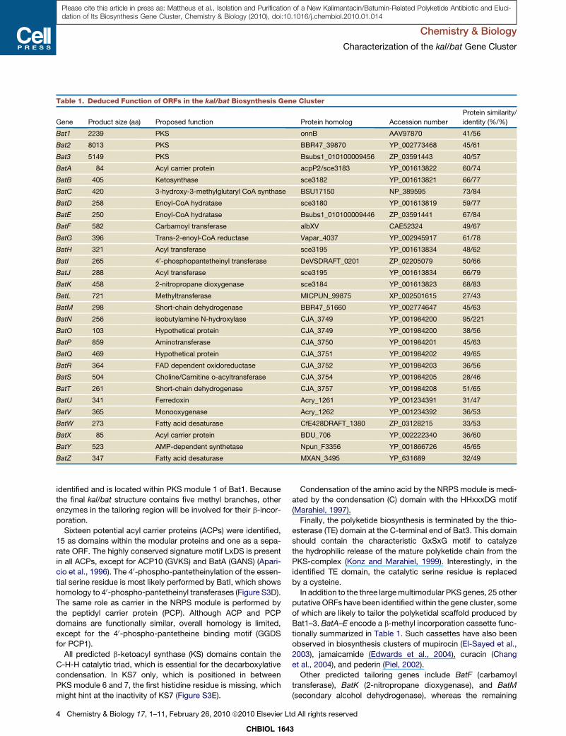
Table 1. Deduced Function of ORFs in the kal/bat Biosynthesis Gene Cluster
Gene Product size (aa) Proposed function Protein homolog Accession number
Protein similarity/
identity (%/%)
Bat1 2239 PKS onnB AAV97870 41/56
Bat2 8013 PKS BBR47_39870 YP_002773468 45/61
Bat3 5149 PKS Bsubs1_010100009456 ZP_03591443 40/57
BatA 84 Acyl carrier protein acpP2/sce3183 YP_001613822 60/74
BatB 405 Ketosynthase sce3182 YP_001613821 66/77
BatC 420 3-hydroxy-3-methylglutaryl CoA synthase BSU17150 NP_389595 73/84
BatD 258 Enoyl-CoA hydratase sce3180 YP_001613819 59/77
BatE 250 Enoyl-CoA hydratase Bsubs1_010100009446 ZP_03591441 67/84
BatF 582 Carbamoyl transferase albXV CAE52324 49/67
BatG 396 Trans-2-enoyl-CoA reductase Vapar_4037 YP_002945917 61/78
BatH 321 Acyl transferase sce3195 YP_001613834 48/62
BatI 265 40-phosphopantetheinyl transferase DeVSDRAFT_0201 ZP_02205079 50/66
BatJ 288 Acyl transferase sce3195 YP_001613834 66/79
BatK 458 2-nitropropane dioxygenase sce3184 YP_001613823 68/83
BatL 721 Methyltransferase MICPUN_99875 XP_002501615 27/43
BatM 298 Short-chain dehydrogenase BBR47_51660 YP_002774647 45/63
BatN 256 isobutylamine N-hydroxylase CJA_3749 YP_001984200 95/221
BatO 103 Hypothetical protein CJA_3749 YP_001984200 38/56
BatP 859 Aminotransferase CJA_3750 YP_001984201 45/63
BatQ 469 Hypothetical protein CJA_3751 YP_001984202 49/65
BatR 364 FAD dependent oxidoreductase CJA_3752 YP_001984203 36/56
BatS 504 Choline/Carnitine o-acyltransferase CJA_3754 YP_001984205 28/46
BatT 261 Short-chain dehydrogenase CJA_3757 YP_001984208 51/65
BatU 341 Ferredoxin Acry_1261 YP_001234391 31/47
BatV 365 Monooxygenase Acry_1262 YP_001234392 36/53
BatW 273 Fatty acid desaturase CfE428DRAFT_1380 ZP_03128215 33/53
BatX 85 Acyl carrier protein BDU_706 YP_002222340 36/60
BatY 523 AMP-dependent synthetase Npun_F3356 YP_001866726 45/65
BatZ 347 Fatty acid desaturase MXAN_3495 YP_631689 32/49
Chemistry & Biology
Characterization of the kal/bat Gene Cluster
Please cite this article in press as: Mattheus et al., Isolation and Purification of a New Kalimantacin/Batumin-Related Polyketide Antibiotic and Eluci-dation of Its Biosynthesis Gene Cluster, Chemistry & Biology (2010), doi:10.1016/j.chembiol.2010.01.014
identified and is located within PKS module 1 of Bat1. Because
the final kal/bat structure contains five methyl branches, other
enzymes in the tailoring region will be involved for their b-incor-
poration.
Sixteen potential acyl carrier proteins (ACPs) were identified,
15 as domains within the modular proteins and one as a sepa-
rate ORF. The highly conserved signature motif LxDS is present
in all ACPs, except for ACP10 (GVKS) and BatA (GANS) (Apari-
cio et al., 1996). The 40-phospho-pantetheinylation of the essen-
tial serine residue is most likely performed by BatI, which shows
homology to 40-phospho-pantetheinyl transferases (Figure S3D).
The same role as carrier in the NRPS module is performed by
the peptidyl carrier protein (PCP). Although ACP and PCP
domains are functionally similar, overall homology is limited,
except for the 40-phospho-pantetheine binding motif (GGDS
for PCP1).
All predicted b-ketoacyl synthase (KS) domains contain the
C-H-H catalytic triad, which is essential for the decarboxylative
condensation. In KS7 only, which is positioned in between
PKS module 6 and 7, the first histidine residue is missing, which
might hint at the inactivity of KS7 (Figure S3E).
CHBIOL 164
4 Chemistry & Biology 17, 1–11, February 26, 2010 ª2010 Elsevier Lt
Condensation of the amino acid by the NRPS module is medi-
ated by the condensation (C) domain with the HHxxxDG motif
(Marahiel, 1997).
Finally, the polyketide biosynthesis is terminated by the thio-
esterase (TE) domain at the C-terminal end of Bat3. This domain
should contain the characteristic GxSxG motif to catalyze
the hydrophilic release of the mature polyketide chain from the
PKS-complex (Konz and Marahiel, 1999). Interestingly, in the
identified TE domain, the catalytic serine residue is replaced
by a cysteine.
In addition to the three large multimodular PKS genes, 25 other
putative ORFs have been identified within the gene cluster, some
of which are likely to tailor the polyketidal scaffold produced by
Bat1–3. BatA–E encode a b-methyl incorporation cassette func-
tionally summarized in Table 1. Such cassettes have also been
observed in biosynthesis clusters of mupirocin (El-Sayed et al.,
2003), jamaicamide (Edwards et al., 2004), curacin (Chang
et al., 2004), and pederin (Piel, 2002).
Other predicted tailoring genes include BatF (carbamoyl
transferase), BatK (2-nitropropane dioxygenase), and BatM
(secondary alcohol dehydrogenase), whereas the remaining
3
d All rights reserved

Chemistry & Biology
Characterization of the kal/bat Gene Cluster
Please cite this article in press as: Mattheus et al., Isolation and Purification of a New Kalimantacin/Batumin-Related Polyketide Antibiotic and Eluci-dation of Its Biosynthesis Gene Cluster, Chemistry & Biology (2010), doi:10.1016/j.chembiol.2010.01.014
genes cannot be directly linked to the kal/bat biosynthesis and
may be involved in self-resistance, as discussed later.
Model for kal/bat biosynthesis and verification
by knockout analysis
Because 12 building blocks are required to assemble the kal/bat
backbone, the predicted modular constitution of Bat1–3 sug-
gests that kal/bat biosynthesis proceeds collinearly, with each
module performing only one chain extension per molecule. The
domain organization and proposed kal/bat biosynthesis model
are shown in Figure 3. To gain experimental insight into the kal/
bat biosynthesis and to test the proposed model, specific
in vivo gene inactivation experiments were performed using in
frame deletions of all kal/bat tailoring ORFs, minimizing the risk
of polar effects (Table 2). Phenotypes of the ORF-specific
mutants were examined by plate bioassay, HPLC purification,
FT-MS analysis, high-yield purification, and 1H, 13C NMR anal-
ysis and were functionally complemented in trans.
Deletion of BatC (HMG-CoA synthase) resulted in total loss of
antibacterial activity against S. aureus ATCC6538. The HPLC
profile revealed no detectable kal/bat or other related UV active
compounds (Figure 4). As part of the b-methyl incorporation
cassette, BatC is thought to introduce methyl-groups. This
b-branch incorporation is a polarity reversal, compared with
the S-adenosylmethionine–mediated methyl transfer seen in
module 1. This system expands the possible methylation sites
to both nucleophilic a-carbons and electrophilic b-carbons.
Analogous to the in vitro proven bacillaene and myxovirescin
pathways (Calderone et al., 2006) (Figure 3B), we propose that
BatJ loads the freestanding ACP BatA with malonyl-CoA.
BatB, with the missing cysteine of the C-H-H triad that is essen-
tial for condensation, decarboxylates malonyl-CoA-BatA to
generate acetyl-CoA-BatA. The HMG-CoA synthase homolog,
which is encoded by BatC, catalyzes the condensation between
this acetate and the acetoacetate-like moieties generated by
modules 5, 9, 10, and 11, respectively. Two members of the
enoyl-CoA hydratase family (BatD and BatE) contain the two
consensus motifs essential for the oxyanion hole that stabilizes
the enolate anions. These are hypothesized to catalyze the
subsequent dehydration and decarboxylation (Gu et al., 2006).
Knockout experiments of the corresponding HMG-CoA syn-
thase homolog found in the mupirocin cluster resulted in the
release of a truncated intermediate, mupirocin H (Wu et al.,
2007). Interestingly, mutations in any of the other genes of the
mupirocin b-methyl incorporation cassette gave the same
phenotypic results (Wu et al., 2008). Similar to these results,
the release of (4) (Figure 3A) is triggered, probably because the
inactivity of BatC blocks the normal flux of metabolites along
the multienzyme complex. As seen with mupirocin H, metabolite
production levels might be significantly reduced, hampering the
detection of intermediate (4). To verify that this phenotype is due
to knocking out BatC, rather than polar effects, we amplified and
inserted the BatC ORF into the broad-host-range IncQ expres-
sion vector pJH10. Expression in trans under control of the tac
promoter fully restored wild-type kal/bat production.
Inactivation of BatF (carbamoyl transferase) led to a severely
reduced antibacterial activity on plate bioassay. However,
HPLC analysis performed on extracts of this mutant revealed
production of a compound (25 mg/L) with a slightly shorter reten-
tion time of 20.6 min (Figure 4). Large-scale fermentation and
CHBIOL
Chemistry & Biology 1
purification of this compound ([M + Na]/e = 528.3287,
C29H47NO6Na [<1 ppm error]) confirms the presence of the
27-descarbamoyl product ((6); Figure 4). Indeed, besides the
chemical shifts and coupling constants for most of the typical
peaks (olefinic protons and methyl protons) identical to those
of kal/bat, only the chemical shift of 27-H shifted to up-field
(from 4.89 ppm to 3.86 ppm). In addition, 29 peaks were
observed in the 13C- NMR spectra (corresponding to 29 carbons
in the molecule), from which the peak corresponding to the
carbamoyl group had disappeared. The chemical shift of C-27
shifted to up-field (from 73.5 ppm to 69.9 ppm), whereas C-28
shifted to down-field (from 17.9 ppm to 21.2 ppm). The chemical
shifts of other carbons (far from C-27) are almost identical with
that of kal/bat. These spectra data further confirmed the absence
of a carbamoyl group (CONH2) at C27 (Figure S4). This finding is
consistent with the proposed role of BatF as O-carbamoyltrans-
ferase in the kal/bat biosynthesis. The detailed interpretation of1H and 13C NMR spectra of 27-descarbamoyl kal/bat are shown
in Table S1. Further examination of the antibacterial activity of
27-descarbamoyl kal/bat revealed an MIC of 0.512 mg/mL for
S. aureus ATCC6538, which is eight times that of wild-type kal/
bat (Table 2). This severely reduced activity of 27-descarbamoyl
kal/bat confirms the importance of the carbamoyl-group for the
antibacterial activity of kal/bat. As in the wild-type, a second
minor product was isolated (2.3 mg/L) and was shown to be
17-hydroxy-27-descarbamoyl kal/bat ((8); Figure S5) by 1H and13C NMR (Table S1). Complementation by expression of BatF
in trans fully restored wild-type kal/bat production.
BatK shows similarity to the C-terminal portion of the trans-
acting polyketide biosynthetic enzymes PksE and mmpIII (Chen
et al., 2006; El-Sayed et al., 2003), found in bacillaene and mupir-
ocin biosynthesis, respectively. This C-terminal moiety is pre-
dicted to encode a 2-nitropropane dioxygenase-like domain,
associated with the N-terminal acyltransferase domain. Lomakin
et al. (2007) reported strong structural similarity between the
2-nitropropane dioxygenase from Pseudomonas aeruginosa
and the enoyl-reductase domain of the yeast fatty acid synthase.
Bumpus et al. (2008) confirmed the trans-enoyl reductase activity
of PksE in vitro. In frame deletion of BatK resulted in the total loss
of antibacterial activity on plate bioassay, and no structurally
related compounds were detected on HPLC (data not shown).
On the basis of the homology with PksE, we hypothesize that
BatK is responsible for the reduction of the olefin structure (5)
during kal/bat biosynthesis. The inability to detect any unsatu-
rated dihydro-kal/bat could be due to blockage of the normal
flux of metabolites along the multienzyme complex or instability
of this compound.
Deletion of the putative secondary alcohol dehydrogenase
BatM resulted in a phenotype completely lacking antibacterial
activity against S. aureus ATCC6538 on plate assay (Figure 4).
HPLC, HRMS, and 1H-, 13C-NMR analysis on extracts showed
the production of 17-hydroxy kal/bat (7) at yields (13 mg/L) signif-
icantly higher than in the wild-type (5 mg/L) and a complete lack
of kal/bat production. Complementation by expression of BatM
in trans fully restored wild-type kal/bat production. Further
examination of the antibacterial activity of 17-hydroxy kal/bat
(7) revealed an MIC of 4 mg/mL for S. aureus ATCC6538, showing
that its activity is only 1.6% compared with that of kal/bat (Table
2). This strongly increased MIC value for 17-hydroxy kal/bat
1643
7, 1–11, February 26, 2010 ª2010 Elsevier Ltd All rights reserved 5

CHBIOL 1643
Chemistry & Biology
Characterization of the kal/bat Gene Cluster
6 Chemistry & Biology 17, 1–11, February 26, 2010 ª2010 Elsevier Ltd All rights reserved
Please cite this article in press as: Mattheus et al., Isolation and Purification of a New Kalimantacin/Batumin-Related Polyketide Antibiotic and Eluci-dation of Its Biosynthesis Gene Cluster, Chemistry & Biology (2010), doi:10.1016/j.chembiol.2010.01.014
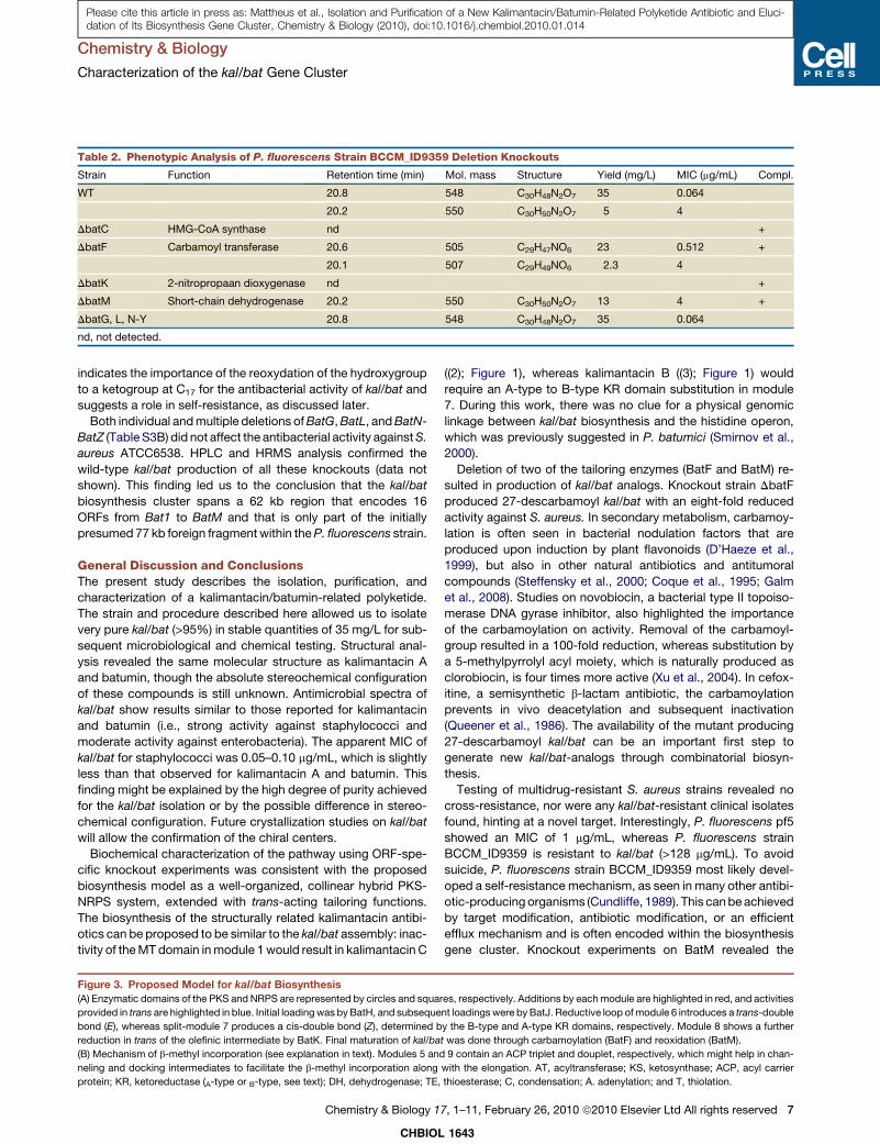
Table 2. Phenotypic Analysis of P. fluorescens Strain BCCM_ID9359 Deletion Knockouts
Strain Function Retention time (min) Mol. mass Structure Yield (mg/L) MIC (mg/mL) Compl.
WT 20.8 548 C30H48N2O7 35 0.064
20.2 550 C30H50N2O7 5 4
DbatC HMG-CoA synthase nd +
DbatF Carbamoyl transferase 20.6 505 C29H47NO6 23 0.512 +
20.1 507 C29H49NO6 2.3 4
DbatK 2-nitropropaan dioxygenase nd +
DbatM Short-chain dehydrogenase 20.2 550 C30H50N2O7 13 4 +
DbatG, L, N-Y 20.8 548 C30H48N2O7 35 0.064
nd, not detected.
Chemistry & Biology
Characterization of the kal/bat Gene Cluster
Please cite this article in press as: Mattheus et al., Isolation and Purification of a New Kalimantacin/Batumin-Related Polyketide Antibiotic and Eluci-dation of Its Biosynthesis Gene Cluster, Chemistry & Biology (2010), doi:10.1016/j.chembiol.2010.01.014
indicates the importance of the reoxydation of the hydroxygroup
to a ketogroup at C17 for the antibacterial activity of kal/bat and
suggests a role in self-resistance, as discussed later.
Both individual and multiple deletions of BatG, BatL, and BatN-
BatZ (Table S3B) did not affect the antibacterial activity against S.
aureus ATCC6538. HPLC and HRMS analysis confirmed the
wild-type kal/bat production of all these knockouts (data not
shown). This finding led us to the conclusion that the kal/bat
biosynthesis cluster spans a 62 kb region that encodes 16
ORFs from Bat1 to BatM and that is only part of the initially
presumed 77 kb foreign fragment within the P. fluorescens strain.
General Discussion and ConclusionsThe present study describes the isolation, purification, and
characterization of a kalimantacin/batumin-related polyketide.
The strain and procedure described here allowed us to isolate
very pure kal/bat (>95%) in stable quantities of 35 mg/L for sub-
sequent microbiological and chemical testing. Structural anal-
ysis revealed the same molecular structure as kalimantacin A
and batumin, though the absolute stereochemical configuration
of these compounds is still unknown. Antimicrobial spectra of
kal/bat show results similar to those reported for kalimantacin
and batumin (i.e., strong activity against staphylococci and
moderate activity against enterobacteria). The apparent MIC of
kal/bat for staphylococci was 0.05–0.10 mg/mL, which is slightly
less than that observed for kalimantacin A and batumin. This
finding might be explained by the high degree of purity achieved
for the kal/bat isolation or by the possible difference in stereo-
chemical configuration. Future crystallization studies on kal/bat
will allow the confirmation of the chiral centers.
Biochemical characterization of the pathway using ORF-spe-
cific knockout experiments was consistent with the proposed
biosynthesis model as a well-organized, collinear hybrid PKS-
NRPS system, extended with trans-acting tailoring functions.
The biosynthesis of the structurally related kalimantacin antibi-
otics can be proposed to be similar to the kal/bat assembly: inac-
tivity of the MT domain in module 1 would result in kalimantacin C
Figure 3. Proposed Model for kal/bat Biosynthesis
(A) Enzymatic domains of the PKS and NRPS are represented by circles and squar
provided in trans are highlighted in blue. Initial loading was by BatH, and subseque
bond (E), whereas split-module 7 produces a cis-double bond (Z), determined b
reduction in trans of the olefinic intermediate by BatK. Final maturation of kal/ba
(B) Mechanism of b-methyl incorporation (see explanation in text). Modules 5 and
neling and docking intermediates to facilitate the b-methyl incorporation along
protein; KR, ketoreductase (A-type or B-type, see text); DH, dehydrogenase; TE,
CHBIOL
Chemistry & Biology 1
((2); Figure 1), whereas kalimantacin B ((3); Figure 1) would
require an A-type to B-type KR domain substitution in module
7. During this work, there was no clue for a physical genomic
linkage between kal/bat biosynthesis and the histidine operon,
which was previously suggested in P. batumici (Smirnov et al.,
2000).
Deletion of two of the tailoring enzymes (BatF and BatM) re-
sulted in production of kal/bat analogs. Knockout strain DbatF
produced 27-descarbamoyl kal/bat with an eight-fold reduced
activity against S. aureus. In secondary metabolism, carbamoy-
lation is often seen in bacterial nodulation factors that are
produced upon induction by plant flavonoids (D’Haeze et al.,
1999), but also in other natural antibiotics and antitumoral
compounds (Steffensky et al., 2000; Coque et al., 1995; Galm
et al., 2008). Studies on novobiocin, a bacterial type II topoiso-
merase DNA gyrase inhibitor, also highlighted the importance
of the carbamoylation on activity. Removal of the carbamoyl-
group resulted in a 100-fold reduction, whereas substitution by
a 5-methylpyrrolyl acyl moiety, which is naturally produced as
clorobiocin, is four times more active (Xu et al., 2004). In cefox-
itine, a semisynthetic b-lactam antibiotic, the carbamoylation
prevents in vivo deacetylation and subsequent inactivation
(Queener et al., 1986). The availability of the mutant producing
27-descarbamoyl kal/bat can be an important first step to
generate new kal/bat-analogs through combinatorial biosyn-
thesis.
Testing of multidrug-resistant S. aureus strains revealed no
cross-resistance, nor were any kal/bat-resistant clinical isolates
found, hinting at a novel target. Interestingly, P. fluorescens pf5
showed an MIC of 1 mg/mL, whereas P. fluorescens strain
BCCM_ID9359 is resistant to kal/bat (>128 mg/mL). To avoid
suicide, P. fluorescens strain BCCM_ID9359 most likely devel-
oped a self-resistance mechanism, as seen in many other antibi-
otic-producing organisms (Cundliffe, 1989). This can be achieved
by target modification, antibiotic modification, or an efficient
efflux mechanism and is often encoded within the biosynthesis
gene cluster. Knockout experiments on BatM revealed the
es, respectively. Additions by each module are highlighted in red, and activities
nt loadings were by BatJ. Reductive loop of module 6 introduces a trans-double
y the B-type and A-type KR domains, respectively. Module 8 shows a further
t was done through carbamoylation (BatF) and reoxidation (BatM).
9 contain an ACP triplet and douplet, respectively, which might help in chan-
with the elongation. AT, acyltransferase; KS, ketosynthase; ACP, acyl carrier
thioesterase; C, condensation; A. adenylation; and T, thiolation.
1643
7, 1–11, February 26, 2010 ª2010 Elsevier Ltd All rights reserved 7

Figure 4. Phenotypic Analysis of P. fluorescens BCCM_ID9359 Knockouts(A) HPLC profile of CHCl3-extracts. Absorbance was measured at 228 nm. kal/bat and related analogs elute at approximately 20 min.
(B) Plate bioassay of purified kal/bat and analogs on S. aureus ATCC6538.
(C) HRMS and NMR spectroscopic confirmed structures of purified products.
Chemistry & Biology
Characterization of the kal/bat Gene Cluster
Please cite this article in press as: Mattheus et al., Isolation and Purification of a New Kalimantacin/Batumin-Related Polyketide Antibiotic and Eluci-dation of Its Biosynthesis Gene Cluster, Chemistry & Biology (2010), doi:10.1016/j.chembiol.2010.01.014
importance of the reoxidation at C17 for the antibacterial activity
of kal/bat. This one-step maturation converts an almost inactive
precursor 17-hydroxy kal/bat into the fully active mature kal/
bat. One may assume a regulation/resistance mechanism, given
the seemingly illogical reduction/reoxidation null-operation as
proposed by the model. The KR3 domain in module 4 first
reduces the C17 keto-group and is later reoxidized by BatM.
Indeed, together with the results of DBatM, there is no indication
that KR3 of module 4 is inactive. Dot plot analysis revealed that
KR3 is an exact copy of KR4 with conserved catalytic motifs.
Second, an inactive KR3 would generate an unwanted acetoace-
tyl-like substrate susceptible to the b-methyl incorporation
cassette. In addition, the intrinsic substrate specificity for b-
hydroxy-substituted substrates of KS4 suggests an active KR3
(Nguyen et al., 2008). Export of an inactive intermediate and
subsequent conversion to a toxic antibiotic has already been
reported and is predominant through N-acetylation of amino
groups or O-phosphorylation of hydroxyls (Cundliffe, 1989). The
CHBIOL 164
8 Chemistry & Biology 17, 1–11, February 26, 2010 ª2010 Elsevier Lt
conversion to the active molecule is performed by a secreted de-
acetylase or phosphatase, respectively. Signal peptide analysis
using SignalP-HMM predicted an N-terminal signal peptide in
BatM, with 0.83 probability. Therefore, we propose that the inac-
tive intermediate 17-hydroxy kal/bat is exported out of the cell for
subsequent activation by the secreted BatM. Although this
system is very similar to the previously mentioned systems, the
important difference is that the inactive hydroxy-kal/bat is synthe-
sized by the modular assembly line, rather than through the action
of a separate enzyme. These individual antibiotic-inactivating
enzymes (e.g., phosphotranferases and acetyltransferases)
have shown to be an immediate source of resistance for patho-
gens through horizontal gene transfer, substantially increasing
the rate of resistance development (D’Costa et al., 2006).
Producing the inactive 17-hydroxy kal/bat by implementation in
the multistep modular assembly circumvents this disadvantage.
Another possibility for the production of an inactive precursor
is the ‘‘feed-forward’’ phenomenon—that is, an inactive
3
d All rights reserved

Chemistry & Biology
Characterization of the kal/bat Gene Cluster
Please cite this article in press as: Mattheus et al., Isolation and Purification of a New Kalimantacin/Batumin-Related Polyketide Antibiotic and Eluci-dation of Its Biosynthesis Gene Cluster, Chemistry & Biology (2010), doi:10.1016/j.chembiol.2010.01.014
precursor acts as a signal to prepare the producing organism for
the later toxic levels of the antibiotic. Tahlan et al. (2008) showed
that an antibiotically inactive precursor of actinorhodin dere-
pressed transcription of the resistance pump gene at lower
molar concentration than did actinorhodin itself, thus ensuring
the full availability of the efflux system at the time the toxin is
produced. The significantly reduced production of kal/bat-
related compounds (13 mg/L of (7)) in DbatM, compared with
wild-type (35 mg/L of (1)), and the fact that BatM most likely is
active during export of kal/bat indicate a metabolite-regulated
production.
Within the cluster, only gene products BatG (showing similarity
to trans-enoyl-CoA reductases) and BatL (showing weak simi-
larity to RNA methylases) appear to be nonessential for kal/bat
production, making them plausible candidates to be involved
in self-resistance.
The genetic basis and biochemical elucidation of the biosyn-
thesis pathway of this novel type of antibiotic will facilitate rational
engineering for the design of novel structures with improved
activities. Indeed, a plurality of biosynthetic strategies combined
in the kal/bat pathway was shown here. The backbone is assem-
bled through a type 1 multimodular system with a combination of
trans-acting ‘‘AT-less’’ PKS modules and a cis-acting NRPS
module. Subsequent determination of the oxidation state
happens through cis-acting catalytic domains or a trans-enoyl
reductase involved in polyunsaturated fatty acid biosynthesis.
Carbon branching is achieved on both nucleophilic a-carbons
and electrophilic b-carbons through S-adenosylmethionine–
mediated methyl transfer and an isoprenoid-like logic, respec-
tively. This finding pinpoints once more that Nature’s ingenuity
for producing natural products is not restricted to classes of
biosynthetic systems (Muller, 2004).
SIGNIFICANCE
In the antibiotic resistance age, which is marked by an
increasing need for novel compounds, secondary metabo-
lites continue to play a major role in antibacterial drug
discovery and development. Polyketides represent a major
class of antibiotics because they are amenable to rational
antibiotic design by genetic modification within the modular
biosynthesis operon. Our research focuses on a promising
methicilin-resistant Staphylococcus aureus (a critical path-
ogen in hospital environments) antibiotic, synthesized by
a plurality of polyketide biosynthetic strategies. Analysis of
this molecule and its analogs shows its potential and
amenability toward engineering with improved activity.
EXPERIMENTAL PROCEDURES
Bacterial Strains, Plasmids, and Culture Conditions
P. fluorescens strain BCCM_ID9359 was used as the wild-type kal/bat
producer. The strain was grown at 28�C in tryptose-broth (Merck, Germany)
for liquid cultivation and on tryptose-agar (Merck) for solid cultivation. E. coli
Transformax� EC100� (Epicenter, US) was used for routine subcloning,
plasmid preparations, and BAC cloning. E. coli S17-1 (Simon et al., 1983)
was used for conjugal transfer of DNA into P. fluorescens strain
BCCM_ID9359. E. coli strains were grown at 37�C in LB broth and LB agar
(LB broth supplemented with 1.5% w/v agar). S. aureus ATCC6538 was
used in bioassays to monitor kal/bat production. Media were supplemented
CHBIOL
Chemistry & Biology 1
with appropriate antibiotic concentrations as follows: ampicillin (100 mg/L),
kanamycin (50 mg/L), triclosan (25 mg/L), chloramphenicol (15 mg/L), and
tetracycline (15 mg/L). Plasmids used during this work are listed in Table S3A.
Production, Isolation, and Analysis of kal/bat
P. fluorescens strain BCCM_ID9359 was seeded in 250 mL of Tryptose broth in
a 1 L Erlenmeyer flask and was incubated at 16�C on a rotary shaker (200 rpm)
for 48 hr. To isolate kal/bat, the culture was adjusted to pH 10 with NaOH, and
the cells were removed by centrifugation. After acidification with formic acid to
pH 3, the supernatant was extracted with chloroform (2 3 250 mL) and was
concentrated in vacuo. The extract was either used directly for HPLC analysis
or was further purified by silica gel column chromatography. The HPLC anal-
ysis was performed on an Alltima C-18 column (5 mL, 250 3 4.6 mm; Alltech).
The column was equilibrated with 100% solvent A (5% acetonitrile [ACN];
0.1% TFA) and was developed with the following program: 0–1 min, 100%
A; 1–30 min, a linear gradient from 100% A to 100% ACN; and 30–40 min,
linear gradient from 100% ACN to 100% A at a flow rate of 1 mL/min and
UV detection at 228 nm using a Shimadzu SPD-10A detector.
The silica gel column chromatography (20 3 250 mm; CH2Cl2/MeOH/
HCOOH; 100/4/0.1 to 100/10/0.1) was used for large scale purification for
subsequent NMR analysis and microbiological testing. MS analysis was per-
formed on a Bruker Daltonics Apex-Qe FT mass spectrometer, and NMR
was performed on a Bruker Ultrashield Avance instrument, operating at 600/
300MHz for 1H and 150/75MHz for 13C nuclei in CDCl3. The isolated yield
for pure kal/bat from the wild-type P. fluorescens strain BCCM_ID9359 was
35 mg/L of culture medium.
Bioassay and MIC Determination
Kal/bat production was monitored by using the disk-diffusion method (Bauer
et al., 1966). The Mueller-Hinton agar plate was uniformly inoculated with
S. aureus ATCC6538, and a paper disk was impregnated with a culture sample
or extrated product was placed on the agar surface. The size of the inhibition
zone after 24 hr incubation is a relative measure for kal/bat production. MICs
of kal/bat were determined using NCCLS standards (National Committee for
Clinical Laboratory Standards, document M7-A5).
Library Construction and Screening
of the kal/bat Biosynthesis Gene Cluster
A genomic random fragment library was constructed and sequenced. P. fluo-
rescens strain BCCM_ID9359 genomic DNA was isolated, randomly sheared
by ultrasonic vibration, and subsequently cloned in pUC19 using T4 DNA
ligase (Promega). Transformation to E. coli Transformax� EC100� by electro-
poration yielded a library of over 10,000 clones with average insert size of 1 kb.
Over 5000 clones were sequenced, resulting in a total of 3.5 Mb of raw data,
which represent approximately 24% of the approximately 7 Mb P. fluorescens
strain BCCM_ID9359 genome. Clones related to polyketide biosynthesis were
identified by keyword searches of the batchBLAST analysis.
BAC genomic library construction of P. fluorescens strain BCCM_ID9359
was performed essentially as described by Osoegawa et al. (1998). High-
molecular-weight DNA was isolated in plugs, partially digested with HindIII,
and size selected by pulsed-field gel electrophoresis. Subsequent cloning in
pIndigoBAC5 (Epicenter) and transformation to E. coli Transformax�EC100� were performed as recommended by the manufacturer and yielded
a library of over 7000 clones with average inserts of 100 kb, resulting in a
34 times coverage of the genome. This library was screened by PCR for the
presence of the kal/bat biosynthesis genes using the primers of the confirmed
tags (Table S3C). PCRs were performed on cell cultures using GoTaq�
DNA Polymerase (Promega) with an initial denaturation of 5 min for efficient
bacterial lysis.
DNA Sequencing and Analysis
Selected BAC-clones were subcloned and sequenced by a combination of
shotgun sequencing and primer walking. Sequencing reactions were run using
Big Dye Terminator mix (Applied Biosystems), cleaned, and analyzed on an
ABI 3130 genetic analyzer. Sequence assembly into contigs was performed
using Sequencher 4.8 software (Gene Codes Corporation, USA). ORF predic-
tions were made using comparative genomics approaches (tBLASTx) and the
Genemark.hmm algorithm (Borodovsky et al., 2003).
1643
7, 1–11, February 26, 2010 ª2010 Elsevier Ltd All rights reserved 9

Chemistry & Biology
Characterization of the kal/bat Gene Cluster
Please cite this article in press as: Mattheus et al., Isolation and Purification of a New Kalimantacin/Batumin-Related Polyketide Antibiotic and Eluci-dation of Its Biosynthesis Gene Cluster, Chemistry & Biology (2010), doi:10.1016/j.chembiol.2010.01.014
Targeted Inactivation and Complementation
DNA transfer to P. fluorescens strain BCCM_ID9359 was performed through
biparental mating with E. coli S17-1. A late exponential culture of E. coli with
the relevant plasmid and P. fluorescens strain BCCM_ID9359 were mixed on
a 0.45 mm sterile Millipore filter, placed on an LB-agar plate. After overnight
incubation at 28�C, the mixture was resuspended in 1 mL of saline solution
and spread on LB-agar plates supplemented with plasmid selective antibiotic
and triclosan. Cointegrant clones were picked and incubated in tryptose broth
without selection at 30�C overnight. Serial dilutions were spread on tryptose
plates containing 5% sucrose, to select for vector excision. Deletion knock-
outs were screened by replica plating and PCR analysis. Clean-cut deletions
were achieved by cloning two DNA fragments of approximately 300–500 bp
flanking the targeted gene into the suicide vector pAKE604 (El-Sayed et al.,
2001), whereas disruptions needed only 1 fragment into pKnockout-G suicide
vector (Windgassen et al., 2000).
For the complementation experiments with pJH10, the same biparental
mating procedure was used. Constructs prepared in this study are listed in
Table S3B.
ACCESSION NUMBER
Sequencing data are accessible at GenBank under accession number
GU479979.
SUPPLEMENTAL INFORMATION
Supplemental Information includes five figures and three tables and may be
found with this article online at doi:10.1016/j.chembiol.2010.01.014.
ACKNOWLEDGMENTS
We thank Christopher M. Thomas for supplying vectors pAKE604 and pJH10.
We also thank the Flemish Government for the financing of the mass spec-
trometry facility ProMeta.
Received: November 17, 2009
Revised: January 6, 2010
Accepted: January 14, 2010
Published: February 25, 2010
REFERENCES
Aparicio, J.F., Molanar, I., Schwecke, T., Koning, A., Haydock, S.F., Khaw,
L.E., Staunton, J., and Leadlay, P.F. (1996). Organization of the biosynthetic
gene cluster of rapamycin in Streptomyces hygroscopicus: analysis of the
enzymatic domains in the modular polyketide synthase. Gene 169, 9–16.
Bauer, A.W., Kirby, W.M., Sherris, J.C., and Turck, M. (1966). Antibiotic
susceptibility testing by a standardized single disk method. Am. J. Clin. Pathol.
45, 493–496.
Borodovsky, M., Mills, R., Besemer, J., and Lomsadze, A. (2003). Prokaryotic
gene prediction using GeneMark and GeneMark.hmm. Curr. Protoc. Bioinfor-
matics. Chapter 4, Unit 4.5.
Bumpus, S.B., Magarvey, N.A., Kelleher, N.L., Walsh, C.T., and Calderone,
C.T. (2008). Polyunsaturated fatty-acid-like trans-enoyl reductases utilized in
polyketide biosynthesis. J. Am. Chem. Soc. 130, 11614–11616.
Caffrey, P. (2005). The stereochemistry of ketoreduction. Chem. Biol. 12,
1060–1062.
Calderone, C.T., Kowtoniuk, W.E., Kelleher, N.L., Walsh, C.T., and Dorrestein,
P.C. (2006). Convergence of isoprene and polyketide biosynthetic machinery:
isoprenyl-S-carrier proteins in the pksX pathway of Bacillus subtilis. Proc. Natl.
Acad. Sci. USA 103, 8977–8982.
Chang, Z., Sitachitta, N., Rossi, J.V., Roberts, M.A., Flatt, P.M., Jia, J.,
Sherman, D.H., and Gerwick, W.H. (2004). Biosynthetic pathway and gene
cluster analysis of curacin A, an antitubulin natural product from the tropical
marine cyanobacterium Lyngbya majuscula. J. Nat. Prod. 67, 1356–1367.
CHBIOL 164
10 Chemistry & Biology 17, 1–11, February 26, 2010 ª2010 Elsevier L
Chen, X.H., Vater, J., Piel, J., Franke, P., Scholz, R., Schneider, K., Koumoutsi,
A., Hitzeroth, G., Grammel, N., Strittmatter, A.W., et al. (2006). Structural and
functional characterization of three polyketide synthase gene clusters in
Bacillus amyloliquefaciens FZB 42. J. Bacteriol. 188, 4024–4036.
Coque, J.J., Perez-Llarena, F.J., Enguita, F.J., Fuente, J.L., Martın, J.F., and
Liras, P. (1995). Characterization of the cmcH genes of Nocardia lactamdurans
and Streptomyces clavuligerus encoding a functional 30-hydroxymethylce-
phem O-carbamoyltransferase for cephamycin biosynthesis. Gene 162,
21–27.
Cundliffe, E. (1989). How antibiotic-producing organisms avoid suicide. Annu.
Rev. Microbiol. 43, 207–233.
D’Costa, V.M., McGrann, K.M., Hughes, D.W., and Wright, G.D. (2006).
Sampling the antibiotic resistome. Science 311, 374–377.
D’Haeze, W., Van Montagu, M., Prome, J.C., and Holsters, M. (1999).
Carbamoylation of azorhizobial Nod factors is mediated by NodU. Mol. Plant
Microbe Interact. 12, 68–73.
Donadio, S., and Katz, L. (1992). Organization of the enzymatic domains in
the multifunctional polyketide synthase involved in erythromycin formation in
Saccharopolyspora erythraea. Gene 111, 51–60.
Edwards, D.J., Marquez, B.L., Nogle, L.M., McPhail, K., Goeger, D.E.,
Roberts, M.A., and Gerwick, W.H. (2004). Structure and biosynthesis of the
jamaicamides, new mixed polyketide-peptide neurotoxins from the marine
cyanobacterium Lyngbya majuscula. Chem. Biol. 11, 817–833.
El-Sayed, A.K., Hothersall, J., and Thomas, C.M. (2001). Quorum-sensing-
dependent regulation of biosynthesis of the polyketide antibiotic mupirocin
in Pseudomonas fluorescens NCIMB 10586. Microbiology 147, 2127–2139.
El-Sayed, A.K., Hothersall, J., Cooper, S.M., Stephens, E., Simpson, T.J., and
Thomas, C.M. (2003). Characterization of the mupirocin biosynthesis gene
cluster from Pseudomonas fluorescens NCIMB 10586. Chem. Biol. 10, 419–
430.
Galm, U., Wang, L., Wendt-Pienkowski, E., Yang, R., Liu, W., Tao, M.,
Coughlin, J.M., and Shen, B. (2008). In vivo manipulation of the bleomycin
biosynthetic gene cluster in Streptomyces verticillus ATCC15003 revealing
new insights into its biosynthetic pathway. J. Biol. Chem. 283, 28236–28245.
Gu, L., Jia, J., Liu, H., Hakansson, K., Gerwick, W.H., and Sherman, D.H.
(2006). Metabolic coupling of dehydration and decarboxylation in the curacin
A pathway: functional identification of a mechanistically diverse enzyme pair.
J. Am. Chem. Soc. 128, 9014–9015.
Hopwood, D.A. (2004). Cracking the polyketide code. PLoS Biol. 2, e35.
Kagan, R.M., and Clarke, S. (1994). Widespread occurrence of three sequence
motifs in diverse S-adenosylmethionine-dependent methyltransferases
suggests a common structure for these enzymes. Arch. Biochem. Biophys.
310, 417–427.
Kamigiri, K., Suzuki, Y., Shibazaki, M., Morioka, M., Suzuki, K., Tokunaga, T.,
Setiawan, B., and Rantiatmodjo, R.M. (1996). Kalimantacins A, B and C, novel
antibiotics from Alcaligenes sp. YL-02632S. I. Taxonomy, fermentation, isola-
tion and biological properties. J. Antibiot. (Tokyo) 49, 136–139.
Klochko, V.V., Kiprianova, E.A., Churkina, L.N., and Avdeeva, L.V. (2008).
Antimicrobial spectrum of antibiotic batumin. Mikrobiol. Z. 70, 41–46.
Konz, D., and Marahiel, M.A. (1999). How do peptide synthetases generate
structural diversity? Chem. Biol. 6, R39–R48.
Lomakin, I.B., Xiong, Y., and Steitz, T.A. (2007). The crystal structure of yeast
fatty acid synthase, a cellular machine with eight active sites working together.
Cell 129, 319–332.
Marahiel, M.A. (1997). Protein templates for the biosynthesis of peptide antibi-
otics. Chem. Biol. 4, 561–567.
Muller, R. (2004). Don’t classify polyketide synthases. Chem. Biol. 11, 4–6.
Nathan, C. (2004). Antibiotics at the crossroads. Nature 431, 899–902.
Nguyen, T., Ishida, K., Jenke-Kodama, H., Dittmann, E., Gurgui, C., Hochmuth,
T., Taudien, S., Platzer, M., Hertweck, C., and Piel, J. (2008). Exploiting the
mosaic structure of trans-acyltransferase polyketide synthases for natural
product discovery and pathway dissection. Nat. Biotechnol. 26, 225–233.
3
td All rights reserved
Chemistry & Biology
Characterization of the kal/bat Gene Cluster
Please cite this article in press as: Mattheus et al., Isolation and Purification of a New Kalimantacin/Batumin-Related Polyketide Antibiotic and Eluci-dation of Its Biosynthesis Gene Cluster, Chemistry & Biology (2010), doi:10.1016/j.chembiol.2010.01.014
Osoegawa, K., Woon, P.Y., Zhao, B., Frengen, E., Tateno, M., Catanese, J.J.,
and de Jong, P.J. (1998). An improved approach for construction of bacterial
artificial chromosome libraries. Genomics 52, 1–8.
Paulsen, I.T., Press, C.M., Ravel, J., Kobayashi, D.Y., Myers, G.S., Mavrodi,
D.V., DeBoy, R.T., Seshadri, R., Ren, Q., Madupu, R., et al. (2005). Complete
genome sequence of the plant commensal Pseudomonas fluorescens Pf-5.
Nat. Biotechnol. 23, 873–878.
Piel, J. (2002). A polyketide synthase-peptide synthetase gene cluster from an
uncultured bacterial symbiont of Paederus beetles. Proc. Natl. Acad. Sci. USA
99, 14002–14007.
Queener, S.F., Webber, J.A., and Queener, S.W. (1986). Beta-Lactam Antibi-
otics for Clinical Use (New York: Marcel Dekker).
Rausch, C., Weber, T., Kohlbacher, O., Wohlleben, W., and Huson, D.H.
(2005). Specificity prediction of adenylation domains in nonribosomal peptide
synthetases (NRPS) using transductive support vector machines (TSVMs).
Nucleic Acids Res. 33, 5799–5808.
Reid, R., Piagentini, M., Rodriguez, E., Ashley, G., Viswanathan, N., Carney, J.,
Santi, D.V., Hutchinson, C.R., and McDaniel, R. (2003). A model of structure
and catalysis for ketoreductase domains in modular polyketide synthases.
Biochemistry 42, 72–79.
Simon, R., Priefer, U., and Puhler, A. (1983). A broad host range mobilization
system for in vivo genetic engineering: transposon mutagenesis in gram
negative bacteria. Nat. Biotechnol. 1, 784–791.
Smirnov, V.V., Churkina, L.N., Perepnikhatka, V.I., Mukvich, N.S., Garagulia,
A.D., Kiprianova, E.A., Kravets, A.N., and Dovzhenko, S.A. (2000). Isolation
of highly active strain producing the antistaphylococcal antibiotic batumin.
Prikl. Biokhim. Mikrobiol. 36, 55–58.
Steffensky, M., Muhlenweg, A., Wang, Z.X., Li, S.M., and Heide, L. (2000).
Identification of the novobiocin biosynthetic gene cluster of Streptomyces
spheroides NCIB 11891. Antimicrob. Agents Chemother. 44, 1214–1222.
Tahlan, K., Yu, Z., Xu, Y., Davidson, A.R., and Nodwell, J.R. (2008). Ligand
recognition by ActR, a TetR-like regulator of actinorhodin export. J. Mol.
Biol. 383, 753–761.
CHBIOL
Chemistry & Biology 17
Tokunaga, T., Kamigiri, K., Orita, M., Nishikawa, T., Shimizu, M., and Kaniwa,
H. (1996). Kalimantacin A, B, and C, novel antibiotics produced by Alcaligenes
sp. YL-02632S. II. Physico-chemical properties and structure elucidation.
J. Antibiot. (Tokyo) 49, 140–144.
Von Nussbaum, F., Brands, M., Hinzen, B., Weigand, S., and Habich, D. (2006).
Antibacterial natural products in medicinal chemistry—exodus or revival?
Angew. Chem. Int. Ed. Engl. 45, 5072–5129.
Windgassen, M., Urban, A., and Jaeger, K.-E. (2000). Rapid gene inactivation
in Pseudomonas aeruginosa. FEMS Microbiol. Lett. 193, 201–205.
Wu, J., Cooper, S.M., Cox, R.J., Crosby, J., Crump, M.P., Hothersall, J.,
Simpson, T.J., Thomas, C.M., and Willis, C.L. (2007). Mupirocin H, a novel
metabolite resulting from mutation of the HMG-CoA synthase analogue,
mupH in Pseudomonas fluorescens. Chem. Commun. (Camb.) 20, 2040–2042.
Wu, J., Hothersall, J., Mazzetti, C., O’Connell, Y., Shields, J.A., Rahman, A.S.,
Cox, R.J., Crosby, J., Simpson, T.J., Thomas, C.M., and Willis, C.L. (2008). In
vivo mutational analysis of the mupirocin gene cluster reveals labile points in
the biosynthetic pathway: the ‘‘leaky hosepipe’’ mechanism. ChemBioChem
9, 1500–1508.
Xu, H., Heide, L., and Li, S.M. (2004). New aminocoumarin antibiotics formed
by a combined mutational and chemoenzymatic approach utilizing the carba-
moyltransferase NovN. Chem. Biol. 11, 655–662.
Yadav, G., Gokhale, R.S., and Mohanty, D. (2003). Computational approach
for prediction of domain organization and substrate specificity of modular
polyketide synthases. J. Mol. Biol. 328, 335–363.
Yanisch-Perron, C., Vieira, J., and Messing, J. (1985). Improved M13 phage
cloning vectors and host strains: nucleotide sequences of the M13mp18 and
pUC19 vectors. Gene 33, 103–119.
Zazopoulos, E., Huang, K., Staffa, A., Liu, W., Bachmann, B.O., Nonaka, K.,
Ahlert, J., Thorson, J.S., Shen, B., and Farnet, C.M. (2003). A genomics-guided
approach for discovering and expressing cryptic metabolic pathways. Nat.
Biotechnol. 21, 187–190.
1643
, 1–11, February 26, 2010 ª2010 Elsevier Ltd All rights reserved 11