Biomass production of four Cynara cardunculus clones and lignin composition analysis
Upload
independentCategory
view
0download
0
Bangladesh Vet J (2010) 44(1-4):12-20
*Corresponding author: Telephone: +880 91 67401~6/ Ext. 2330; Fax: +880 91 61510; Cellphone: +88-01713815104; e-mail: [email protected] Copyright © Bangladesh Veterinary Association All right reserved 0030-9915/10
Isolation and Characterization of Intestinal Sub-epithelial Myofibroblast in Rat: An in vitro Tool for Bioresearch System
M. S. Islam1,2*, H. Ozaki2
1Department of Pharmacology, Faculty of Veterinary Science, Bangladesh Agricultural University, Mymensingh-2202, Bangladesh.
2Department of Veterinary Pharmacology, Graduate School of Agriculture and Life Sciences, The University of Tokyo, Tokyo, Japan.
ABSTRACT
Primary cell culture is a valuable tool that extensively used in the basic research of organ specific functions, physiology, pathology, pharmacology and so on. The aim of this study was to derive primary mesenchyme cell line representative of colon tissues. First, we identified the
location of sub-epithelial myofibroblast in the colon tissues in rat. -smooth muscle-actin (-SMA) positive myofibroblast found just beneath the epithelia of colon tissues. Accordingly, we isolated intestinal sub-epithelial myofibroblasts (ISEMFs) from colonic lamina propria of rat. The isolated cells were cultured and characterized by phase contrast, immunofluorescent and RT-PCR
analysis. The harvested cells expressed the specific markers such as -SMA, vimentin and were negative for desmin expression. Finally, we did trans-well migration assay to know the difficulties or convenience of this isolated cell culture experiment. The isolated rat ISEMFs grow well in culture media and easy to culture for the research purposes. Therefore, the primary ISEMF might be a useful alternative in vitro tool for bioresearch study.
Key words: Intestinal sub-epithelial myofibroblast, -SMA, Colon, Rat
INTRODUCTION
Sub-epithelial myofibroblasts are located at the interface between the epithelium and lamina propria in most mucosal tissues (Valentich et al., 1997). Ultrastructural features of ISEMFs demonstrate both smooth muscle cell and fibroblasts and are thought to be intermediate cell of smooth muscle cell and fibroblast cells (Andoh et al., 2005). Although the cells have been characterized in many ways but still the physiological roles of ISMFs

M S Islam and H Ozaki, 2010
13
are not fully understood. To analyze the roles of ISEMFs, primary culture ISEMFs are often used for experiments (Furuya et al., 2005a; Furuya et al., 2005b). Furthermore, rat is an easily available lab animal and primary sources of ISEMFs from rat is a convenient source and is an excellent model for study pathophysiological and pharmacological features of diseases and drugs.
During mucosal inflammation, a mixture of cells of different lineages including epithelial cells, lymphocytes, resident macrophages, sub-epithelial fibroblasts and fibroblast-like cells are involved in either pro-inflammatory or anti-inflammatory phenomena. Intestinal sub-epithelial myofibroblasts are inflammatory and secrete not only inflammatory cytokines, chemokines, growth factors, and lipid and gaseous inflammatory mediators but also many extracellular matrix proteins. Thus, myofibroblasts play an important role in organogenesis, oncogenesis, inflammation, and fibrosis in most organs and tissues (Valentich et al., 1997; Powell et al., 1999; Salas et al., 2003). During intestinal development, the fetal mesenchyme differentiates into various structures: the outer muscle layers, the submucosal connective tissue, and the lamina propria or mucosal connective tissue. Various fibroblast cell phenotypes can be observed in vivo in the lamina propria as sub-epithelial myofibroblasts (Plateroti et al., 1998) that form a regular network of stellate cells (Komuro et al., 1990). The aim of this work was to design a reproducible and well-defined in vitro model of intestinal sub-epithelial myofibroblast from rat colon.
MATERIALS AND METHODS
Animals
Animals were treated in accordance with the institutional guidelines of the University of Tokyo. Sprague-Dawley SD rats (5 weeks old, male) were used in this experiment. These rats were acclimatized and provided food and water ad libitum. ISEMFs were isolated from colon tissues.
Immunofluorescent staining
Colon specimens were collected from mice, washed and fixed in 4%
paraformaldehyde solution. The specimen was cut into 5m by microtome (Coldtome, Model CM-502, SAKURA, Japan). Sections were washed with PBS three times and air dried for 30 minutes. After that, specimens were permeabilized with Triton X-100 (0.2%), blocked by 10% normal goat serum and then incubated with the primary antibody, rat rabbit anti-αSMA (1:1000 dilution) over night at 4º C. Specimens were then washed with

Isolation of Intestinal sub-epithelial myofibroblast
14
PBS three times, each for 15 minutes and incubated with the appropriate secondary antibodies (1:1000 dilution) for 2hour under dark condition at the room temperature. Nuclei were further stained with DAPI (Molecular Probes). Images were obtained using an Eclipse E800 fluorescence microscope (Nikon, Tokyo, Japan).
Intestinal sub-epithelial myofibroblasts in rat
Rat was killed in accordance with the institutional guidance. Colon was collected feces of colon was washed gently with Hank`s balanced salt solution (HBSS). Colon was cut according to the mesenteric border. Muscle layer was separated from the mucosal layer under microscope. Mucosal sheets were treated with 1 mM ethylenediaminetetraacetic acid (EDTA) in Ca2+-free HBSS buffer three times at 37ºC for 30 minutes to remove the epithelial layer. The mucosal sheet without epithelial cells was then cut into small pieces and cultured in DMEM containing 10% fetal bovine serum (FBS) and 1% antimycotic/antibiotic at 37ºC in 5% CO2/95% O2. ISEMF cells have started to migrate from implants to the surroundings and after confluent the surrounding areas with migrated cells, the implants were taken off the dish. The confluent cells were then sub-cultured for subsequent study.
Immunohistochemistry of ISEMF
ISEMF cells were cultured onto the cover glass in DMEM. After cultured, the cells were washed with Ca2+-Mg free HBSS three times and fixed in 2% paraformaldehyde (PFA) for 30 minutes. Then then the cells were washed three times with phosphate buffered salt solution (PBS), each for 15 minutes. ISEMF were incubated with mouse monoclonal anti-αSMA (1:500), mouse anti-vimentin (1: 200), and rabbit anti-desmin (1:200) respectively over night at 4º C. The all specimen were washed with PBS three times, each for 15 minutes and then incubated with appropriate secondary secondary antibodies diluted 1:1000 respectively for 2hours room temperature in a dark chamber. Nuclei were stained with DAPI. Images were obtained using an Eclipse E800 fluorescence microscope (Nikon, Tokyo, Japan).
RNA extraction and quantification of αSMA, desmin and vimentin mRNA level
Total RNA was extracted by using Trizol reagent (Invitrogen, Tokyo, Japan). First-strand cDNA was synthesized by using a random nine-mer primer and ReverTra Ace at 30º C for 10 minutes, 42º C for 1hour, 99º C for 5 minutes and 4º C for 5 minutes. PCR

M S Islam and H Ozaki, 2010
15
amplification was performed by using ExTaq DNA polymerase. Primers used for PCR analysis are provided in Table 1. After an initial check, RT-PCR was performed in 32 cycles.
Table1. Sequence of the primers used for RT-PCR analysis
Primer sets Orientation Sequence (5′ to 3′) PCR product
Rat-αSMA (NM_031004)
Forward GGGAGTGATGGTTGGAATGG 197bp
Reverse CCGTTAGCAAGGTCGGATG
Rat-Vimentin (NM_031140)
Forward ACGAATACCGGAGACAGGTG 265bp
Reverse TCCAGCAGCCTTCCTGTAGGT
Rat-Desmin (NM_022531)
Forward ACCTGCGAGATTGATGCTCT 275bp
Reverse AAGGTCTGGATCGGAAGGTT
Rat-GAPDH (XM_576394)
Forward TCCCTCAAGATTGTCAGCAA 308bp
Reverse AGATCCACAACGGATACATT
Statistical analysis
Trans-well migration assays were performed four times (n=4), and values are expressed as mean ±SEM. Statistical significance (**P <0.01) was determined by using the Student’s t test.
RESULTS
Now a day, primary cell culture is an important tool in bio-research examination. Colon mucosa comprises a mixture of different lineages including epithelial cells, lymphocytes, resident macrophages, sub-epithelial myofibroblasts, fibroblasts, pericytes of vasculature, smooth muscle of the muscularis mucosae and smooth muscle surrounding the lymphatic lacteals. Each of the cells plays specific roles in the body homeostasis. Among the cells present in the intestine, sub-epithelial myofibroblast is considered most important cell in the colon tissues which play pivotal roles in gastrointestinal diseases and health. In this study, we have successfully isolated ISEMF from colon tissues in rat.
Isolation of Intestinal sub-epithelial myofibroblast
16
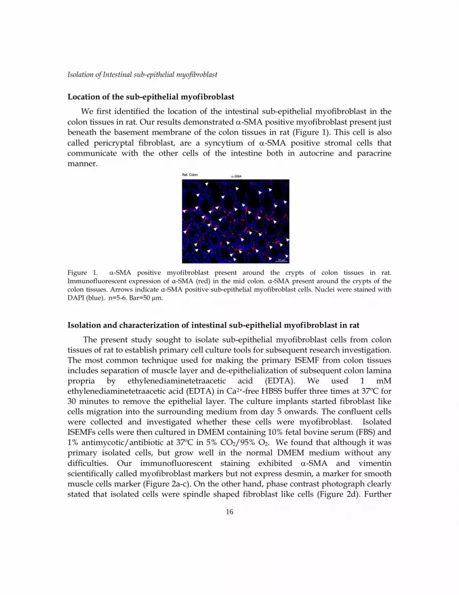
Location of the sub-epithelial myofibroblast
We first identified the location of the intestinal sub-epithelial myofibroblast in the
colon tissues in rat. Our results demonstrated -SMA positive myofibroblast present just beneath the basement membrane of the colon tissues in rat (Figure 1). This cell is also
called pericryptal fibroblast, are a syncytium of -SMA positive stromal cells that communicate with the other cells of the intestine both in autocrine and paracrine manner.
Figure 1. -SMA positive myofibroblast present around the crypts of colon tissues in rat. Immunofluorescent expression of α-SMA (red) in the mid colon. α-SMA present around the crypts of the colon tissues. Arrows indicate α-SMA positive sub-epithelial myofibroblast cells. Nuclei were stained with DAPI (blue). n=5-6. Bar=50 µm.
Isolation and characterization of intestinal sub-epithelial myofibroblast in rat
The present study sought to isolate sub-epithelial myofibroblast cells from colon tissues of rat to establish primary cell culture tools for subsequent research investigation. The most common technique used for making the primary ISEMF from colon tissues includes separation of muscle layer and de-epithelialization of subsequent colon lamina propria by ethylenediaminetetraacetic acid (EDTA). We used 1 mM ethylenediaminetetraacetic acid (EDTA) in Ca2+-free HBSS buffer three times at 37ºC for 30 minutes to remove the epithelial layer. The culture implants started fibroblast like cells migration into the surrounding medium from day 5 onwards. The confluent cells were collected and investigated whether these cells were myofibroblast. Isolated ISEMFs cells were then cultured in DMEM containing 10% fetal bovine serum (FBS) and 1% antimycotic/antibiotic at 37ºC in 5% CO2/95% O2. We found that although it was primary isolated cells, but grow well in the normal DMEM medium without any
difficulties. Our immunofluorescent staining exhibited -SMA and vimentin scientifically called myofibroblast markers but not express desmin, a marker for smooth muscle cells marker (Figure 2a-c). On the other hand, phase contrast photograph clearly stated that isolated cells were spindle shaped fibroblast like cells (Figure 2d). Further

M S Islam and H Ozaki, 2010
17
confirmation of ISEMF cells were done by RT-PCR analysis. In RT-PCR analysis, isolated
cells were positive for -SMA, vimentin but negative for desmin mRNA (Figure 3).
Figure 2. Morphology and characteristics of rat ISEMFs cells. Immunofluorescent staining shows ISEMF positive for (a) α-SMA (upper left panel); (b) vimentin (upper right panel) and negative for (c) desmin (lower left panel). Nuclei were stained with DAPI. Bar=20 µm. (c), representative phase contrast photograph shows the morphology of isolated primary ISEMF cells. ISEMF
Cells show spindle shaped morphology with healthy appearance.
Figure 3. RT-PCR analysis of rat ISEMF. ISEMFs were cultured and analyzed for αSMA, vimentin and desmin. Rat ISEMF expressed mRNA for αSMA (196 bp), vimentin (265 bp), but not desmin (275 bp). GAPDH (308bp). n=5-6.
Trans-well migration Assay
Migration is an important tool for research investigation particularly for inflammation and wound healing and reparative process concerned researches. Finally, we checked whether the isolated primary ISEMF is convenient for in vitro research investigation. In multicellular organism, chemotaxis is a critical phenomenon both for physiological as well as pathological situation. We did experiment using our isolated primary ISEMFs for trans-well migration assay. In this study, the chemotaxis activity of

Isolation of Intestinal sub-epithelial myofibroblast
18
fetal bovine serum (FBS) was investigated. 1% FBS in the lower chamber expressed strong chemotaxis comparing with the non-treated control (Figure 4). Our isolated cells showed easy to culture and research investigation. Besides these, the primary cells demonstrate some new function which can be concluded with established cancer cell line cells.
Figure 4. Fetal bovine serums (FBS) is a strong chemotaxis substance in rat ISEMF. A, ISEMFs cells were allowed 8h to migrate through trans-wells membranes- (a) control (no treatment) and (b) FBS 1% (Lower). B. Analytical data showing increased migration of cells at 8h of FBS treatment. **P<0.01. n=4 each.
DISCUSSION
Intestinal sub-epithelial myofibroblast present just beneath the basement membrane
of the colon tissues. This cell is also called pericryptal fibroblast, are a syncytium of -SMA positive stromal cells which reside subjacent to the basement membrane of the small and large intestine (Powell et al., 1999; Andoh et al., 2005). ISEMFs cells play important roles in the gastrointestinal pathophysiology and pharmacology by their secretory roles and supporting network by lamina propria. There is report that ISEMFs cells regulate epithelial cell function (Andoh et al., 2007). The ideal technique for isolation of ISEMF includes two major primary steps such as separation of muscle layer and de-epithelialization of colon lamina propria. We separated muscle layer from colon tissues under microscopic dissection process. Following separation of muscle layer, EDTA in 1mM concentration was used for complete separation of epithelia from processed lamina propria. The culture implants started fibroblast like cells migration into the surrounding medium from day 5 onwards. The confluent cells were collected and investigated whether these cells were myofibroblast. Isolated ISEMFs cells were then cultured in DMEM containing 10% fetal bovine serum (FBS) and 1% antimycotic/antibiotic at 37ºC in 5% CO2/95% O2. We found that although it was

M S Islam and H Ozaki, 2010
19
primary isolated cells, but grow well in the normal DMEM medium without any difficulties. Morphology of the cells was spindle shaped fibroblast like cells.
The primary isolated cells then examined using the myofibroblast specific markers
such as -SMA and vimentin. Isolated cells expressed both -SMA and vimentin protein. As it is said, there is a possibility of contamination of ISEMF with some neighbor cells such as smooth muscle cells. We therefore, investigated desmin, a marker for smooth muscle cells marker. Our isolated cells completely negative for desmin indicated no contamination. In accordance with our findings, other scientific reports also explained that ISEMF are specialized cells that exhibit ultrastructural structure of both fibroblast
and smooth muscle cells characters and expressed positive for -SMA and vimentin but negative for desmin (Andoh et al., 2007; Sappino et al., 1990). ISEMF are distinguished
from smooth muscle which express -SMA but are negative for vimentin. Although the cells have been characterized in many ways but still the physiological roles of ISEMFs are not fully understood. To analyze the roles of ISEMFs, primary culture ISEMFs are often used for experiments (Furuya et al., 2005a; Furuya et al., 2005b). Furthermore, rat is an easily available lab animal and primary sources of ISEMFs from rat is a convenient source and is an excellent model for study pathophysiological and pharmacological features of diseases and drugs. In multicellular organism, chemotaxis is a critical phenomenon both for physiological as well as pathological situation. Migration is an important tool for research investigation particularly for inflammation and wound healing and reparative process concerned researches. Our study suggested that FBS is a strong chemotaxis substance. Other scientist also reported that FBS is a strong chemotactic substance (Hong et al., 2008).
In summary, we successfully isolated ISEMF from rat colon, characterized their features and cultures. We also characterized their pharmacological respond in FBS treated trans-well migration assay. Thus, Rat ISEMFs might be a powerful and beneficial tool to study the physiological, pathological and pharmacological roles of myofibroblast cells in the body.
REFERENCES
Andoh A, Bamba S, Brittan M, Fujiyama Y, Wright NA, 2007. Role of intestinal subepithelial myofibroblasts in inflammation and regenerative response in the gut. Pharmacology and Therapeutics 114: 94-106.
Andoh A, Bamba S, Fujiyama Y, Brittan M, Wright NA, 2005. Colonic subepithelial myofibroblasts in mucosal inflammation and repair: contribution of bone marrow-

Isolation of Intestinal sub-epithelial myofibroblast
20
derived stem cells to the gut regenerative response. Journal of Gastroenterology 40: 1089-1099.
Furuya K, Sokabe M, Furuya S, 2005a. Characteristics of subepithelial fibroblasts as a mechano-sensor in the intestine: cell-shape-dependent ATP release and P2Y1 signaling. Journal of Cell Science 118: 3289-3304.
Furuya S, Furuya K, Sokabe M, Hiroe T, Ozaki T, 2005b. Characteristics of cultured subepithelial fibroblasts in the rat small intestine. II. Localization and functional analysis of endothelin receptors and cell-shape-independent gap junction permeability. Cell and Tissue Research 319: 103-119.
Hong X, Jiang F, Kalkanis SN, Zhang ZG, Zhang X, Zheng X, Jiang H, Mikkelsen T, Chopp M, 2008. Increased chemotactic migration and growth in heparanase-overexpressing human U251n glioma cells. Journal of Experimental and Clinical Cancer Research 27: 23-30.
Komuro T, Hashimoto Y, 1990. Three-dimensional structure of the rat intestinal wall (mucosa and submucosa). Archives of Histology and Cytology 53: 1-21.
Plateroti M, Rubin DC, Duluc I, Singh R, Foltzer-Jourdainne C, Freund JN, Kedinger M, 1998. Subepithelial fibroblast cell lines from different levels of gut axis display regional characteristics. The American Journal of Physiology 274: G945-954.
Powell DW, Mifflin RC, Valentich JD, Crowe SE, Saada JI, West AB, 1999. Myofibroblasts. II. Intestinal subepithelial myofibroblasts. The American Journal of Physiology 277: C183-201.
Salas A, Fernandez-Banares F, Casalots J, Gonzalez C, Tarroch X, Forcada P, Gonzalez G, 2003. Subepithelial myofibroblasts and tenascin expression in microscopic colitis. Histopathology 43: 48-54.
Sappino AP, Schurch W, Gabbiani G, 1990. Differentiation repertoire of fibroblastic cells: expression of cytoskeletal proteins as marker of phenotypic modulations. Laboratory Investigation 63: 144-161.
Valentich JD, Popov V, Saada JI, Powell DW, 1997. Phenotypic characterization of an intestinal subepithelial myofibroblast cell line. The American Journal of Physiology 272: C1513-1524.
Copyright © 2022 FDOKUMEN