Is cancer a disease of abnormal cellular metabolism ... - Nature
11
Is cancer a disease of abnormal cellular metabolism? New angles on an old idea Ralph J. DeBerardinis, MD, PhD In the 1920s, Otto Warburg observed that tumor cells consumed a large amount of glucose, much more than normal cells, and converted most of it to lactic acid. This phenomenon, now known as the “Warburg effect,” is the foundation of one of the earliest general concepts of cancer: that a fundamental disturbance of cellular metabolic activity is at the root of tumor formation and growth. In the ensuing decades, as it became apparent that abnormalities in chromosomes and eventually individual genes caused cancer, the “metabolic” model of cancer lost a good deal of its appeal, even as emerging technologies were exploiting the Warburg effect clinically to detect tumors in vivo. We now know that tumor suppressors and proto-oncogenes influence metabolism, and that mutations in these genes can promote a metabolic phenotype supporting cell growth and proliferation. Thus, these advances have unified aspects of the metabolic and genetic models of cancer, and have stimulated a renewed interest in the role of cellular metabolism in tumorigenesis. This review reappraises the notion that dysregulated cellular metabolism is a key feature of cancer, and discusses some metabolic issues that have escaped scrutiny over the years and now deserve closer attention. Genet Med 2008:10(11):767–777. Key Words: cancer, metabolism, Warburg effect, glutamine, glucose The Warburg effect, also known as aerobic glycolysis, is defined as a high rate of glucose utilization and lactate production despite the presence of sufficient oxygen to oxidize glucose carbon in the mitochondria. Recognition of this unusual metabolic phenome- non stems from experiments performed by the German physiol- ogist Otto Warburg, starting in the 1920s. 1,2 In those experiments, Warburg compared the metabolism of rapidly proliferating mouse ascites tumor cells with that of differentiated, quiescent cells from organs of the adult animal. He proposed that a funda- mental impairment of cellular respiratory capacity was the root cause of all cancer, a bold and controversial claim that was ulti- mately rejected despite his continued writing and lecturing on the subject for some 40 years. Nevertheless, appreciation of the War- burg effect as a feature of tumor cell metabolism has survived its namesake by a long stretch. Today, the glycolytic activity of tu- mors is not only accepted, but exploited clinically by 18 F-deoxy- glucose positron emission tomography (FDG-PET), which de- tects tumors precisely by virtue of their enhanced ability to take up and metabolize glucose compared with normal tissue. The War- burg effect remains the most frequently cited evidence that tu- mors display dysfunctional metabolism. Recently, interest in tumor metabolism has enjoyed a re- naissance as an ever-growing number of reports uncovers the molecular connections between transformation and cell me- tabolism, and as technological improvements increase the fea- sibility of studying tumor metabolism in vivo. The field seems poised to offer significant insights into tumor biology over the next decade. As such, it is worth re-examining the evidence for a bona fide connection between altered cellular metabolic state and tumorigenesis: does the notion of such a connection stand up to our current understanding of tumor biology and cancer genetics? If there is such a link, then the following should be true: 1. Tumor cells should have metabolic activities that differ from nontransformed, quiescent cells, and these activi- ties should be required for tumor growth; 2. The mutations in tumor suppressors and proto-onco- genes that promote cancer should regulate the metabolic activities observed in tumors; and 3. Mutations in metabolic enzymes should, in at least some cases, promote tumorigenesis. This review addresses how well these criteria are met and discusses some other issues relevant to tumor metabolism that may feature prominently in future research. DO TUMOR CELLS HAVE METABOLIC ACTIVITIES THAT ARE DIFFERENT FROM QUIESCENT CELLS AND ARE REQUIRED FOR TUMOR GROWTH? Most studies on tumor metabolism have been motivated by one of two general concepts about the way cell metabolism is regulated. The first is that tumor metabolism is primarily a response to stresses imposed upon cells during tumor growth. There is abundant evidence that some stresses, particularly hypoxia, exist in the tumor microenvironment and exert ef- From the Department of Pediatrics and The McDermott Center for Human Growth and Development, University of Texas Southwestern Medical Center, Dallas Texas. Ralph J. DeBerardinis, MD, PhD, 5323 Harry Hines Boulevard, Room K4.216, Dallas, TX 75390-9063. E-mail: [email protected]. Disclosure: The author declares no conflict of interest. Submitted for publication July 11, 2008. Accepted for publication August 20, 2008. DOI: 10.1097/GIM.0b013e31818b0d9b November 2008 Vol. 10 No. 11 review Genetics IN Medicine 767
-
Upload
khangminh22 -
Category
Documents
-
view
1 -
download
0
Transcript of Is cancer a disease of abnormal cellular metabolism ... - Nature
Is cancer a disease of abnormal cellularmetabolism? New angles on an old ideaRalph J. DeBerardinis, MD, PhD
In the 1920s, Otto Warburg observed that tumor cells consumed a large amount of glucose, much more than normal
cells, and converted most of it to lactic acid. This phenomenon, now known as the “Warburg effect,” is the foundation
of one of the earliest general concepts of cancer: that a fundamental disturbance of cellular metabolic activity is at the
root of tumor formation and growth. In the ensuing decades, as it became apparent that abnormalities in chromosomes
and eventually individual genes caused cancer, the “metabolic” model of cancer lost a good deal of its appeal, even as
emerging technologies were exploiting the Warburg effect clinically to detect tumors in vivo. We now know that tumor
suppressors and proto-oncogenes influence metabolism, and that mutations in these genes can promote a metabolic
phenotype supporting cell growth and proliferation. Thus, these advances have unified aspects of the metabolic and
genetic models of cancer, and have stimulated a renewed interest in the role of cellular metabolism in tumorigenesis. This
review reappraises the notion that dysregulated cellularmetabolism is a key feature of cancer, and discusses somemetabolic
issues that have escaped scrutiny over the years and now deserve closer attention. Genet Med 2008:10(11):767–777.
Key Words: cancer, metabolism, Warburg effect, glutamine, glucose
TheWarburg effect, alsoknownas aerobic glycolysis, is definedas a high rate of glucose utilization and lactate production despitethe presence of sufficient oxygen to oxidize glucose carbon in themitochondria. Recognition of this unusual metabolic phenome-non stems from experiments performed by the German physiol-ogistOttoWarburg, starting in the1920s.1,2 In those experiments,Warburg compared the metabolism of rapidly proliferatingmouse ascites tumor cells with that of differentiated, quiescentcells from organs of the adult animal. He proposed that a funda-mental impairment of cellular respiratory capacity was the rootcause of all cancer, a bold and controversial claim that was ulti-mately rejected despite his continuedwriting and lecturing on thesubject for some 40 years. Nevertheless, appreciation of theWar-burg effect as a feature of tumor cell metabolism has survived itsnamesake by a long stretch. Today, the glycolytic activity of tu-mors is not only accepted, but exploited clinically by 18F-deoxy-glucose positron emission tomography (FDG-PET), which de-tects tumorspreciselybyvirtueof their enhancedability to takeupand metabolize glucose compared with normal tissue. The War-burg effect remains the most frequently cited evidence that tu-mors display dysfunctional metabolism.Recently, interest in tumor metabolism has enjoyed a re-
naissance as an ever-growing number of reports uncovers the
molecular connections between transformation and cell me-tabolism, and as technological improvements increase the fea-sibility of studying tumor metabolism in vivo. The field seemspoised to offer significant insights into tumor biology over thenext decade. As such, it is worth re-examining the evidence for abona fide connection between altered cellularmetabolic state andtumorigenesis: does the notion of such a connection stand up toour current understanding of tumor biology and cancer genetics?If there is such a link, then the following should be true:
1. Tumor cells should have metabolic activities that differfrom nontransformed, quiescent cells, and these activi-ties should be required for tumor growth;
2. The mutations in tumor suppressors and proto-onco-genes that promote cancer should regulate the metabolicactivities observed in tumors; and
3. Mutations in metabolic enzymes should, in at least somecases, promote tumorigenesis.
This review addresses how well these criteria are met anddiscusses some other issues relevant to tumor metabolism thatmay feature prominently in future research.
DO TUMOR CELLS HAVE METABOLIC ACTIVITIES THATARE DIFFERENT FROM QUIESCENT CELLS AND AREREQUIRED FOR TUMOR GROWTH?
Most studies on tumor metabolism have been motivated byone of two general concepts about the way cell metabolism isregulated. The first is that tumor metabolism is primarily aresponse to stresses imposed upon cells during tumor growth.There is abundant evidence that some stresses, particularlyhypoxia, exist in the tumor microenvironment and exert ef-
From the Department of Pediatrics and The McDermott Center for Human Growth and
Development, University of Texas Southwestern Medical Center, Dallas Texas.
Ralph J. DeBerardinis, MD, PhD, 5323 Harry Hines Boulevard, Room K4.216, Dallas, TX
75390-9063. E-mail: [email protected].
Disclosure: The author declares no conflict of interest.
Submitted for publication July 11, 2008.
Accepted for publication August 20, 2008.
DOI: 10.1097/GIM.0b013e31818b0d9b
November 2008 � Vol. 10 � No. 11 r e v i e w
Genetics IN Medicine 767
fects on metabolism.3–5 But the high glycolytic flux in tumorscan appear even when oxygen is abundant, and the metabolicconsequences of hypoxia include specific impairments of pro-tein and lipid synthesis that are counterproductive to cellgrowth and proliferation.6–8 These observations suggest thatthe cellular responses to tumor hypoxia, including enhancedglycolysis, serve to facilitate tumor cell survival, not growth.This is also true in nontransformed cells, which rely on glyco-lysis to survive periods of hypoxia.9 When oxygen delivery im-proves and rapid cell growth resumes, the persistence of glyco-lysis is likely due to other factors.Alternatively, one can presume that tumor cell metabolic
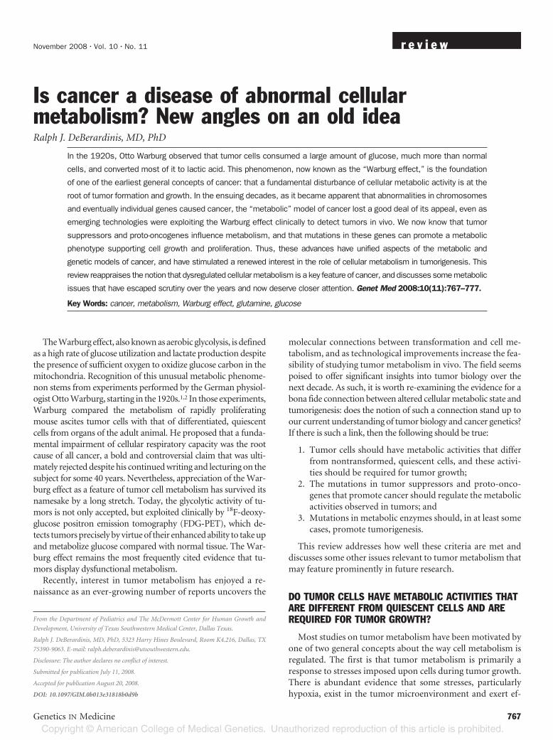
activities function primarily to support the unusually highrates of cell growth and proliferation found in tumors. Becauseeach round of replicative cell division requires a doubling ofprotein, lipids, and nucleic acids, it stands to reason that tumorcell metabolism must provide the energy and biosynthesisneeded to meet this challenge (Fig. 1). Rapid tumor growth
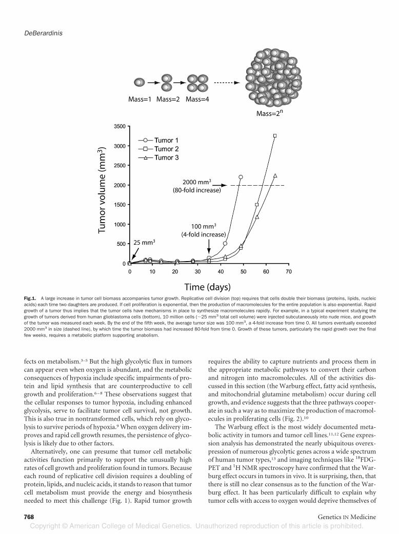
requires the ability to capture nutrients and process them inthe appropriate metabolic pathways to convert their carbonand nitrogen into macromolecules. All of the activities dis-cussed in this section (the Warburg effect, fatty acid synthesis,and mitochondrial glutamine metabolism) occur during cellgrowth, and evidence suggests that the three pathways cooper-ate in such a way as to maximize the production of macromol-ecules in proliferating cells (Fig. 2).10
The Warburg effect is the most widely documented meta-bolic activity in tumors and tumor cell lines.11,12 Gene expres-sion analysis has demonstrated the nearly ubiquitous overex-pression of numerous glycolytic genes across a wide spectrumof human tumor types,13 and imaging techniques like 18FDG-PET and 1H NMR spectroscopy have confirmed that theWar-burg effect occurs in tumors in vivo. It is surprising, then, thatthere is still no clear consensus as to the function of the War-burg effect. It has been particularly difficult to explain whytumor cells with access to oxygen would deprive themselves of
Fig.1. A large increase in tumor cell biomass accompanies tumor growth. Replicative cell division (top) requires that cells double their biomass (proteins, lipids, nucleicacids) each time two daughters are produced. If cell proliferation is exponential, then the production of macromolecules for the entire population is also exponential. Rapidgrowth of a tumor thus implies that the tumor cells have mechanisms in place to synthesize macromolecules rapidly. For example, in a typical experiment studying thegrowth of tumors derived from human glioblastoma cells (bottom), 10 million cells (�25 mm3 total cell volume) were injected subcutaneously into nude mice, and growthof the tumor was measured each week. By the end of the fifth week, the average tumor size was 100 mm3, a 4-fold increase from time 0. All tumors eventually exceeded2000 mm3 in size (dashed line), by which time the tumor biomass had increased 80-fold from time 0. Growth of these tumors, particularly the rapid growth over the finalfew weeks, requires a metabolic platform supporting anabolism.
DeBerardinis
768 Genetics IN Medicine
the majority of the ATP that can be produced from glucosemetabolism by instead converting pyruvate into lactate. How-ever, three points should be emphasized regarding the role ofthe Warburg effect in tumor cell metabolism. First, given thatthe Warburg effect is also observed during rapid proliferation
of primary cells, it is more accurately viewed as a general fea-ture of cell proliferation than as a symptom of transformation,and therefore can be assumed to contribute to anabolicmetab-olism.14,15 Second, rapid glucose metabolism also supplies in-termediates for biosynthetic pathways that arise from glycoly-
Fig. 2. Some metabolic activities are required for tumor growth. Among the various metabolic activities that have been observed in tumors or tumor cell lines, the three with themost compelling evidence for a required role in tumor growth are aerobic glycolysis (the Warburg effect), fatty acid/lipid synthesis and mitochondrial glutamine metabolism. Currentevidence suggests that these three pathways cooperate in a metabolic platform that supports cell growth and ultimately proliferation. The high rate of glycolysis, in addition toproducing ATP, generates glycerol and citrate to be used to synthesize membrane lipids. Meanwhile, mitochondrial metabolism of glutamine supplies the TCA cycle withintermediates to replace those exported for lipid synthesis and other anabolic processes. Cells using this form of metabolism secrete lactate produced from both glucoseand glutamine. Ammonia is also produced and secreted in abundance. Recent studies have shown that several of the enzymes participating in these pathways are requiredfor growth of tumors in mice (white ovals), while SDH and FH function genetically as tumor suppressors in humans. Abbreviations: Glc, glucose; DHAP, dihydroxyacetonephosphate; GA3P, glyceraldehyde 3-phosphate; Pyr, pyruvate; Lac, lactate; Ac-CoA, acetyl-CoA; Cit, citrate; �-KG, �-ketoglutarate; Succ, succinate; Fum, fumarate; Mal,malate; OAA, oxaloacetate; Mal-CoA, malonyl-CoA; Gln, glutamine; Glu, glutamate; NH4
�, ammonia; PK-M2, pyruvate kinase isoform M2; LDHA, lactate dehydrogenase-A;ACL, ATP citrate lyase; ACC�, acetyl-CoA carboxylase-�; FAS, fatty acid synthase; GLS, glutaminase; SDH, succinate dehydrogenase; FH, fumarate hydratase; PDH, pyruvatedehydrogenase; ME, malic enzyme.
The metabolism of tumor cells
November 2008 � Vol. 10 � No. 11 769
sis proximal to pyruvate, including ribose-5-phosphate andglycine for nucleotide biosynthesis, and glycerol for lipid syn-thesis. It has been suggested that one of the functions of theWarburg effect is tomaintain adequate sizes of these precursorpools to maximize cell growth.16 Third, cells engaged in aero-bic glycolysis do not convert 100% of their pyruvate into lac-tate. Rather, a measurable fraction of the pyruvate is metabo-lized in the tricarboxylic acid (TCA) cycle, providing energyand precursors for biosynthetic pathways that consume TCAcycle intermediates.15,17,18 Therefore, theWarburg effect servesboth bioenergetic and biosynthetic roles in proliferating cells.It may be that the Warburg effect is essentially a conse-
quence of an imbalance between maximum rates of glycolysisand pyruvate oxidation. The rate of pyruvate oxidation is con-trolled by the highly regulated pyruvate dehydrogenase (PDH)complex, which converts pyruvate to acetyl-CoA (Fig. 2). Dur-ing rapid cell proliferation, the glycolytic rate may exceed theVmax of PDH by more than 10-fold.19 If the PDH flux cannotmatch the glycolytic flux, cells must use other high-capacitysystems to avoid pyruvate accumulation. Chief among these islactate dehydrogenase-A (LDHA), which by converting pyru-vate to lactate also oxidizes NADH to NAD�. Because lactatecan be secreted and NAD� is required for glycolysis, expres-sion of LDHA allows proliferating cells to continue to reap thebenefits of a high glycolytic rate even in the face of a “maxed-out” PDH flux. This model is supported by studies performedin interleukin-3 (IL-3)-dependent hematopoietic cells.20 Inthose cells, the rates of cell proliferation, glucose consumption,and lactate production are directly proportional to the concen-tration of IL-3 in the medium. Strikingly, the ratio of lactateproduced to glucose consumed was positively regulated byIL-3, but the ratio of oxygen consumed to glycolysis declinedwith increasing IL-3. Thus, with progressive stimulation, cellscontinue to metabolize glucose beyond the point of maximalpyruvate oxidation, eventually reaching a state at which ahigher and higher fraction of glucose carbon is converted tolactate and the fraction of cellular ATP generated by glycolysisapproaches or even exceeds that from oxidative phosphoryla-tion, as in Warburg’s experiments.21
These data imply that factors influencing the Vmax of PDHand the balance between lactate production and pyruvate oxi-dation can impact the Warburg effect. Recent studies havedemonstrated that this balance is regulated by hypoxia induc-ible factor-1� (HIF-1�), a transcription factor with multipletargets involved in glucose metabolism. HIF-1� regulates ex-pression of pyruvate dehydrogenase kinase-1 (PDK-1), a ki-nase which limits PDH activity.22,23 Therefore, under hypoxicconditions, stabilization and transcriptional activity ofHIF-1�results in the enhancement of glycolysis and the suppression ofpyruvate oxidation by PDH. Thismechanism can also contrib-ute to the Warburg effect in well-perfused tumor cells, whichmay have aberrant/normoxic stabilization of HIF-1� becauseof a variety of genetic mechanisms.24 In these cells, the result-ing glycolysis is truly “aerobic.”Even under conditions of HIF-1� stabilization, however,
cell growth requires at least a small amount of PDH flux to
maintain TCA cycle activity, which supplies precursors for thesynthesis of fatty acids and other anabolic pathways (Fig. 2).Tumor cells often display rapid synthesis of fatty acids, choles-terol, and isoprenoids, presumably because a large fraction oftheir membrane lipids are synthesized de novo rather thanscavenged from extracellular sources.25 In a metabolic fluxstudy on human glioblastoma cells, fatty acid synthesis ac-counted for some 15% of cellular glucose consumption afterexcluding the fraction of glucose metabolized through theWarburg effect.18 Thus a significant fraction of the glucosecarbon that enters biosynthetic pathways is used to producefatty acids. Consistentwith that observation, the three enzymesrequired for fatty acid synthesis, ATP citrate lyase (ACL),acetyl-CoA carboxylase-� (ACC-�), and fatty acid synthase(FAS), are highly expressed inmany human cancers and tumorcell lines.26
Fatty acid synthesis is a paradigm of tumor biosyntheticpathways because it requires the use of a TCA cycle intermedi-ate (citrate) that might otherwise be oxidized in themitochon-dria (Fig. 2). The shunting of metabolites from the TCA cycleinto other pathways (cataplerosis) is part of the fundamentalbiochemistry of cell growth, and it emphasizes the versatility ofthe TCA cycle: rather than serving a purely oxidative functionas a source of reducing equivalents for the electron transportchain (ETC), it can also be used as a continuous source ofprecursor molecules for biosynthetic pathways. Early charac-terization of cataplerosis in highly lipogenic hepatoma cells ledto the concept of a “broken” or “truncated” TCA cycle becauseof an apparent impairment in citrate oxidation and the dem-onstration that the rate of citrate export was directly propor-tional to the rate of cell proliferation.27,28 These observa-tions underscore the importance of cataplerosis in tumorcell growth. In addition to lipid synthesis, catapleroticfluxes feed the synthesis of nucleotides and nonessentialamino acids, and are therefore used in the synthesis of allclasses of macromolecules.Glutamine metabolism is second only to theWarburg effect
in terms of historical significance to the study of tumor cellmetabolism. Glutamine is the most abundant amino acid inhuman plasma and participates in many metabolic pathwaysrequired for normal cell function. In addition to its role inprotein synthesis, it provides nitrogen for the synthesis of non-essential amino acids, purines, pyrimidines, and hexosamines,and is the major source of glutamate used for glutathione syn-thesis. Tumor cells have long been known to consume glu-tamine at high rates in vivo and to require high concentrationsof glutamine to survive and proliferate in vitro.29,30 Classicalstudies on tumor cell metabolism in culture demonstrated thatglutamine is an important carbon source since most of theglutamine consumed is used as a respiratory substrate in themitochondria rather than for protein synthesis.31 More recentexperiments have demonstrated that suppressing mitochon-drial glutamine metabolism can alter gene expression, ac-celerate apoptosis, and stimulate cellular differentia-tion.32,33 Therefore, glutamine metabolism has the potential
DeBerardinis
770 Genetics IN Medicine
to integrate a large number of cellular activities that supporttumorigenesis.A closer look at glutamine metabolism has revealed other
vital roles in proliferating cells (Fig. 2). First, partial oxidationof glutamine to lactate (glutaminolysis) uses the cytosolicmalic enzyme and therefore provides cells withNADPH for thereductive reactions of fatty acid andnucleotide biosynthesis. Insome cases, the rate of glutaminolysis can match or exceed therate of NADPH production by glucose-6-phosphate dehydro-genase (G6PD) in the pentose phosphate pathway.17,18 Second,glutamine’s conversion to �-ketoglutarate and entry into theTCA cycle can generate oxaolacetate (OAA), effectively replac-ing the metabolites that are removed from the cycle in cataple-rotic reactions (Fig. 2). This process, termed anaplerosis, is acritical component of growth metabolism because it allowscells to maintain TCA cycle function while withdrawing inter-mediates for biosynthetic reactions. In some cells, glutaminemetabolism is by far themost important source of anaplerosis,and depriving cells of glutamine rapidly depletes cellular poolsof TCA cycle intermediates.18,34,35 In cells simultaneously con-suming both glucose and glutamine, citrate production in-volves the condensation of two glucose-derived carbons (asacetyl-CoA) and four glutamine-derived carbons (as OAA).Although it is the glucose carbons that are ultimately trans-ferred to fatty acids, the process could not occur without thecontribution of glutamine-based anaplerosis.10
But how strong is the evidence that any of the enzymes par-ticipating in these three pathways are required for tumorgrowth? A number of studies using chemical inhibitors andmore recently RNA interference have addressed this questionin animal models of cancer (Fig. 2). The glycolysis inhibitor2-deoxyglucose, when given at doses that did not affect bodyweight or growth of the animal, significantly decreased carcin-ogen-induced mammary tumorigenesis in rats.36 RNA inter-ference against glycolytic enzymes or enzymes in de novo fattyacid synthesis also curtailed tumor growth in animal mod-els.37–39 Suppression of glutaminase, the first enzyme in mito-chondrial glutamine metabolism, using the chemical inhibitor6-diazo-5-oxo-L-norleucine (DON) or expression of an anti-sense mRNA limited tumor growth in mice.40,41 These studiesare the best evidence that the metabolic activities observed intumor cells in vitro are not simply artifacts of culture condi-tions, but are fundamental properties that contribute to tumorgrowth.It is interesting that in addition to the enhanced metabolic
rates described above, tumor cells can also display chronic sup-pression of the pathways that normally allow cells to utilizealternative fuels to survive periods of starvation. As a result, thecells have an increased reliance on specific fuels and a limitedability to compensate for fluctuations in nutrient availability.For example, constitutive activity of the oncogenic kinase Aktimpairs the activation of fatty acid oxidation, resulting inabrupt glioma cell death in low-glucose conditions.42 Loss ofthe tumor suppressor p53 diminishes the ability of colon can-cer cells to engage catabolic, energy-generating pathways likeautophagy and fatty acid oxidation.43 Glutamine depletion se-
lectively kills fibroblasts with enhanced c-Myc activity.35 Step-wise transformation of fibroblasts with multiple oncogenesprogressively increases the toxicity of a glycolysis inhibitor.44
These observations add credibility to the hope that inhibitingspecific metabolic pathways will be selectively toxic to tu-mor cells in vivo. Indeed, exploiting phenotypes of impairedmetabolic flexibility may prove to be more useful in cancertherapy than inhibiting growth-promoting activities likefatty acid synthesis, since these tend to be shared with nor-mal proliferating cells.
DO MUTATIONS IN PROTO-ONCOGENES AND TUMORSUPPRESSORS IMPACT CELL METABOLISM?
Mutations that promote tumorigenesis often reduce oreliminate cellular dependence on extrinsic signals to maintainsurvival, growth, and proliferation. These processes are usuallyunder control of growth factors and other signals that originateoutside of the cell and are transmitted inwards through signaltransduction pathways. In cancer, this “outside-in” regulationis diminished or lost, allowing cells to achieve self-sufficiencyin growth and proliferation.45 Cellular metabolism is also sub-ject to external control, because lineage-specific growth factorsand the signaling pathways they stimulate are required for cellsto activate anabolic pathways and suppress catabolic ones.46–48
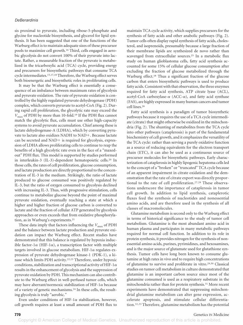
Because cell proliferation cannot occur without these meta-bolic activities, it is not surprising that tumor cells have in-creased autonomy in maintaining an anabolic phenotype. Alarge amount of evidence now supports the idea that tumorsuppressors and proto-oncogenes exert regulatory effects onmetabolism in normal cells, and that tumorigenicmutations inthese genes contribute to the metabolic autonomy observed intumor cells.49 Most of the regulatory mechanisms that havebeen described to date are focused on glucose metabolism andinvolve mutations in p53, phosphatidylinositol 3-kinase(PI3K) signaling, Ras and Myc, the most prevalent classes oftumorigenic mutations in humans (Fig. 3).Mutations in the tumor suppressor gene TP53 occur in ap-
proximately 50% of all human cancers. Recent studies haveuncovered multiple roles for p53 in glucose metabolism, re-vealing an inverse correlation between p53 activity and theWarburg effect. First, loss of p53 in primary fibroblasts en-hances glucose transport and metabolism through IKK andNF-�B.50 Second, the p53 transcriptional targetTIGAR (TP53-induced glycolysis and apoptosis regulator) decreases theabundance of the glycolytic activator fructose-2,6-bisphos-phate (F2,6biP), enabling cells with p53 activation after DNAdamage to divert glucose-6-phosphate into the oxidative pen-tose phosphate pathway, bolstering production of NADPHand ribose-5-phosphate for DNA repair.51 p53 also suppressesexpression of the glycolytic enzyme phosphoglycerate mutaseand enhances expression of the ETC assembly factor SCO2,effects that curtail glycolysis and maximize pyruvate oxida-tion.52,53 Therefore, loss of p53 function has multiple positiveeffects on aerobic glycolysis.
The metabolism of tumor cells
November 2008 � Vol. 10 � No. 11 771
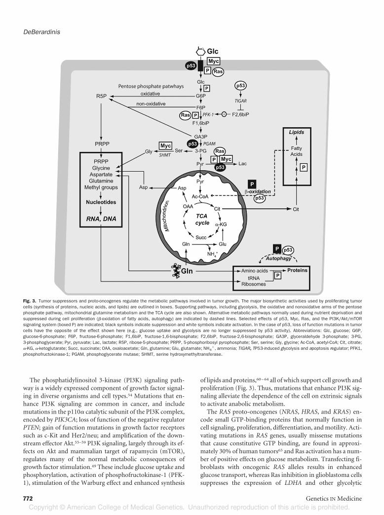
The phosphatidylinositol 3-kinase (PI3K) signaling path-way is a widely expressed component of growth factor signal-ing in diverse organisms and cell types.54 Mutations that en-hance PI3K signaling are common in cancer, and includemutations in the p110� catalytic subunit of the PI3K complex,encoded by PIK3CA; loss of function of the negative regulatorPTEN; gain of function mutations in growth factor receptorssuch as c-Kit and Her2/neu; and amplification of the down-stream effector Akt.55–59 PI3K signaling, largely through its ef-fects on Akt and mammalian target of rapamycin (mTOR),regulates many of the normal metabolic consequences ofgrowth factor stimulation.49 These include glucose uptake andphosphorylation, activation of phosphofructokinase-1 (PFK-1), stimulation of the Warburg effect and enhanced synthesis
of lipids and proteins,60–64 all of which support cell growth andproliferation (Fig. 3). Thus, mutations that enhance PI3K sig-naling alleviate the dependence of the cell on extrinsic signalsto activate anabolic metabolism.The RAS proto-oncogenes (NRAS, HRAS, and KRAS) en-
code small GTP-binding proteins that normally function incell signaling, proliferation, differentiation, andmotility. Acti-vating mutations in RAS genes, usually missense mutationsthat cause constitutive GTP binding, are found in approxi-mately 30% of human tumors65 and Ras activation has a num-ber of positive effects on glucose metabolism. Transfecting fi-broblasts with oncogenic RAS alleles results in enhancedglucose transport, whereas Ras inhibition in glioblastoma cellssuppresses the expression of LDHA and other glycolytic
Fig. 3. Tumor suppressors and proto-oncogenes regulate the metabolic pathways involved in tumor growth. The major biosynthetic activities used by proliferating tumorcells (synthesis of proteins, nucleic acids, and lipids) are outlined in boxes. Supporting pathways, including glycolysis, the oxidative and nonoxidative arms of the pentosephosphate pathway, mitochondrial glutamine metabolism and the TCA cycle are also shown. Alternative metabolic pathways normally used during nutrient deprivation andsuppressed during cell proliferation (�-oxidation of fatty acids, autophagy) are indicated by dashed lines. Selected effects of p53, Myc, Ras, and the PI3K/Akt/mTORsignaling system (boxed P) are indicated; black symbols indicate suppression and white symbols indicate activation. In the case of p53, loss of function mutations in tumorcells have the opposite of the effect shown here (e.g., glucose uptake and glycolysis are no longer suppressed by p53 activity). Abbreviations: Glc, glucose; G6P,glucose-6-phosphate; F6P, fructose-6-phosphate; F1,6biP, fructose-1,6-bisphosphate; F2,6biP, fructose-2,6-bisphosphate; GA3P, glyceraldehyde 3-phosphate; 3-PG,3-phosphoglycerate; Pyr, pyruvate; Lac, lactate; R5P, ribose-5-phosphate; PRPP, 5-phosphoribosyl pyrophosphate; Ser, serine; Gly, glycine; Ac-CoA, acetyl-CoA; Cit, citrate;�-KG, �-ketoglutarate; Succ, succinate; OAA, oxaloacetate; Gln, glutamine; Glu, glutamate; NH4
�, ammonia; TIGAR, TP53-induced glycolysis and apoptosis regulator; PFK1,phosphofructokinase-1; PGAM, phosphoglycerate mutase; SHMT, serine hydroxymethyltransferase.
DeBerardinis
772 Genetics IN Medicine
genes.66,67 Overexpression of mutant KRAS alleles associatedwith resistance to apoptosis can induce the Warburg effect infibroblasts.68 The effects of Ras on glycolysis require expressionof the gene for 6-phosphofructo-2-kinase/fructose 2,6-biphos-phatase (PFKFB3), which regulates abundance of the PFK-1activator F2,6-biP, suggesting that the effects are mediated atthe level of PFK-1 activity (Fig. 3).69,70 Interestingly, Ras mayalso enhance mitochondrial metabolism, as transformation ofbronchial epithelial cells with an oncogenic HRAS allele in-creases the entry of glucose carbon into the TCA cycle andvarious cataplerotic pathways.71
Members of the myc family of proto-oncogenes, especiallyc-myc, encode regulators of gene expression and are com-monly amplified in human tumor cells. As with the PI3K sig-naling pathway, c-Myc is required for the proliferation of nor-mal, nontransformed cells, but enhancement of its activity intumor cells can drive transcriptional effects independently ofexternal stimulation. Among c-Myc’s veritable host of tran-scriptional targets are genes involved in glucose metabolismand the Warburg effect, including LDHA and the glucosetransporter GLUT1. c-Myc also induces the expression of en-zymes involved in nucleotidemetabolism, including serine hy-droxymethyltransferase,72 which allows three carbon unitsfrom glycolysis to be used in purine and pyrimidine synthesis(Fig. 3). This distinguishes c-Myc frommost of the other driv-ers of the Warburg effect, which so far seem to lack directinfluence over nucleotide biosynthesis. Given c-Myc’s physio-logical role in facilitating the G1/S transition, it is likely thatthese transcriptional targets allow cells to produce the metab-olites needed to complete S phase successfully.
CAN MUTATIONS IN METABOLIC ENZYMES INFLUENCECANCER RISK?
Warburg hypothesized that the metabolism of all tumorcells was primarily because a consequence of irreversible de-fects in cellular respiration.2 This has not turned out to be thecase, as many tumor cell lines that have been carefully studiedretain the capacity for normal mitochondrial metabolism.11
However, there are now some very interesting exceptions inwhich genes for enzymes of the TCA cycle behave genetically astumor suppressors and are severely impaired in certain humantumors (Fig. 2). First,mutations in three of the four subunits ofthe succinate dehydrogenase (SDH) complex have been foundin pheochromocytomas and related tumors.73–75 In affectedfamilies, tumor risk is inherited as a dominant trait because ofloss-of-function mutations in SDH subunits, with the tumortissue displaying loss of heterozygosity and complete absenceof SDH enzyme activity.76 Similarly, mutations in fumaratehydratase (FH) cause a group of dominantly inherited cancersyndromes involving cutaneous and uterine lyomyomas.77
Precisely how dysfunction of these enzymes causes tumorgrowth is an area of active study. One contributing factor isthat thesemutations lead to the accumulation of succinate andfumarate, which interfere with degradation of HIF-1�, leadingto its normoxic stabilization.78 This would be predicted to in-
duce the Warburg effect, although the mechanisms of en-hanced tumor growth are not clear.The diverse roles of the mitochondria and the ETC in cell
metabolism, growth, survival, apoptosis, and production ofreactive oxygen species have led to the idea that mutations inmitochondrial DNA (mtDNA) might also influence cancerrisk. A great deal of work has attempted to correlate mutationsin the mitochondrial DNA with cancer. Germline sequencevariants in the mtDNA have been associated with invasivebreast cancer, endometrial cancer, and prostate cancer in cer-tain populations.79–81 Many other studies have reported asso-ciations of somatically acquired, tumor-specific mtDNA se-quence variants in ovarian, colon, bladder, head and neck, andother cancers.82 However, many of these variants had beenpreviously detected in large population studies and were pos-tulated to impart adaptive functions during migration of an-cestral human populations, making their contribution to tu-morigenesis less obvious. To determine the effect of a specificmtDNAmutation on tumor growth, Petros et al.81 introducedmitochondria harboring a known pathogenic mutation intohuman PC3 prostate cancer cells depleted of their own mito-chondrial DNA, and tested the resulting “cybrids” for tumorgrowth in nude mice. The cybrids containing the mutantmtDNA formed tumors much more rapidly than cybrids con-taining wild-type mtDNA. It should be noted that the mutantmtDNA was derived from a child with Leigh syndrome, notfrom a tumor, and that the recipient PC3 cells were alreadytransformed. Nevertheless, this study proves that a maximalcapacity for oxidative phosphorylation is dispensable for tu-mor growth, and is the strongest evidence to date that dysfunc-tionalmtDNA can impart a growth advantage to tumor cells invivo.Recently, a large-scale sequencing effort implicated muta-
tions in the gene for isocitrate dehydrogenase-1 (IDH1) in gli-oblastomamultiforme. Approximately half of the patients un-der 35 contained a somatically-acquired IDH1 mutation intheir tumor tissue. Strikingly, in all tumors harboring an IDH1mutation, the same codon within the isocitrate binding do-main was mutated, resulting in an amino acid substitution. Ineach case, the tumor tissue was heterozygous for the IDH1change and no obvious inactivating mutations were observed,raising the possibility of a gain of function for themutant allele.IDH1 is a cytosolic enzyme that decarboxylates isocitrate to�-ketoglutarate, reducing NADP� to NADPH. Thus IDH1mutations could influence cytosolic pools of any of these me-tabolites. Further study is needed to establish the relevance ofthese potentialmetabolic effects to cellular transformation andtumorigenesis.
WHERE DO WE GO FROM HERE?
At present, the data on tumor cell metabolismmake a com-pelling argument that during the process of transformation,cells acquire a fairly stereotyped set ofmetabolic characteristicsthat enable them to grow and proliferate with reduced depen-dence on extracellular signals, and that themutations in tumor
The metabolism of tumor cells
November 2008 � Vol. 10 � No. 11 773

suppressors and proto-oncogenes that cause cancer contributeto this “metabolic transformation.” There are a number of im-portant questions that still need to be addressed before we canappreciate fully the role of metabolism in the development,progression, and treatment of cancer. Below are six areas thatare in particular need of attention.First, there is still a large gap between our understanding
of tumor cell metabolism in vitro and tumor metabolism invivo. The inhibitor and RNA interference experiments citedabove demonstrate the requirement of certain enzymes fortumorigenesis, but the ability to observe the metabolism oflive tumors would provide a much richer understanding ofthe biochemical aspects of tumor growth and would sup-port more rigorous examination of the effect of tumorigenicmutations on metabolic flux. The use of 18FDG-PET andconventional NMR spectroscopy have provided some vali-dation of the Warburg effect and other pathways in livetumors, but are currently best suited to give snapshots ofmetabolism rather than robust measurements of metabolicflux. The application of more sensitive methods such as theuse of probes labeled with hyperpolarized 13C may ulti-mately support flux analysis in vivo.83
Second, the regulation of anaplerosis and of glutamine me-tabolism in general has not been carefully studied with respectto cell signaling. Although glutamine consumption is a generalmetabolic feature of cell proliferation, it is unknown whetherall tumor cells must use glutamine as the major source of car-bon for anaplerosis. In a few cases, cellular biosynthesis andgrowth have been correlated with the induction of alternativeanaplerotic pathways that do not involve glutamine metabo-lism,19,84 and thus the universality of glutamine-based anaple-rosis is debatable. Furthermore, while stimulation with mito-gens can enhance glutamine utilization in various cell types,neither the signaling pathways responsible for this activity northe specific pathways of glutamine metabolism (e.g., glu-taminolysis vs. glutamine-based anaplerosis) have beencharacterized.15,17 Resolving these issues will go a long waytoward integrating our understanding of cell signaling andthe metabolism of cell growth. Considering that glucose andglutamine play complementary roles in proliferating tumorcells, it will be interesting to test whether they are coregu-lated by the same signaling pathways and affected by thesame tumorigenic mutations.Third, many studies have sought to unravel the complex
relationships between oxidative stress, cellular transformationand cancer. At the level of cell metabolism, there are manyopen questions about how NADPH-producing systems areregulated and whether these mechanisms are altered in tumorcells. Rapidly proliferating tumor cells requireNADPHboth tomaintain pools of reduced glutathione and to support the re-ductive biosynthetic reactions needed for anabolic metabo-lism. The two NADPH-producing systems discussed above,G6PD and cytosolic malic enzyme, are expressed in tumors ofvarious histological types, and in some cases their activities arealmost equal in proliferating tumor cells, implying equivalentcontributions to the NADPH pool.17,18 It is interesting that
G6PD deficiency, a common X-linked condition, does not al-ter the risk of death from cancer.85 This suggests that malicenzyme or other systems can provide sufficient NADPH tosupport tumorigenesis in G6PD-deficient cells, and that selec-tive inhibition of malic enzyme in individuals with G6PD de-ficiency might suppress tumor growth. Conversely, maintain-ing an NADPH pool and a robust response against oxidativestress is critical in preventing tumorigenesis, as emphasized byrecent studies on the transcription factor NF-E2-related fac-tor-2 (Nrf2).86 Nrf2 is sequestered in the cytosol through asso-ciation with its repressor Keap1 (Kelch ECH associating pro-tein 1). Exposure to oxidative and other stresses allows Nrf2 totranslocate to the nucleus, where it heterodimerizes with smallMaf-family proteins and binds promoters containing antioxi-dant response elements, thus inducing expression of genes thatserve antioxidant functions. This protects cells from apoptosisduring exposure to oxidizing agents, UV irradiation, and otherstresses.87–89 Knockout mice lacking Nrf2 have enhanced sus-ceptibility to cancer when exposed to chemical mutagens.90,91
Future studies will be aimed at exploiting the Nrf2 pathway asa strategy to prevent cancer, and at understanding othermech-anisms by which normal cells and tumor cells respond to oxi-dative stress.Fourth, almost all previous studies on tumor cell metabo-
lism have been performed in unsynchronized populations ofcells distributed throughout the cell cycle, so that the resultingdata represent average cell metabolic activity throughout thecycle. The temptation is to assign the greatest biological impor-tance to the pathways that appear to be the most active, butsuch an interpretation could miss the significance of pathwaysthat are transiently induced at specific, critical points of the cellcycle. One can envision two ways to identify these activities: byperforming flux analysis in synchronized populations of cellsor by using inhibitors and genetic models to determine theeffect of disrupting candidate metabolic activities on progres-sion through the cell cycle. There is already strong evidencethat interfering with metabolism profoundly affects the cellcycle. In fibroblasts, glucose deprivation imposes an AMPK-and p53-dependent G1/S phase arrest,92 and in Drosophila,elimination of ETC components stimulates several distinct sig-naling pathways that culminate in arrest at the G1/S transi-tion.93,94 These efforts are relevant to metabolically directedcancer therapy, especially if such therapies are used as adju-vants to existing strategies that already exert cell cycle-specificeffects. For example, if ionizing radiation is most effective oncells at the G2/M transition, then ametabolic therapy targetinga G2-specific activity might increase the fraction of cells at thatstage, thereby increasing the overall efficacy of treatment.Fifth, it is possible that normal but genetically defined vari-
ation in the expression of metabolic enzymes could influencecancer risk and progression. Microarray analysis of gene ex-pression in cells from healthy individuals has shown that somemetabolic enzymes and transporters are highly variable in theirmRNA abundance, and that the variance is defined genetically.95
Currently, the association between disease states and varianceof expression in these genes is unknown. But the abundance of
DeBerardinis
774 Genetics IN Medicine
certain metabolic enzymes in tumors is correlated with poorclinical outcome in various forms of cancer.96,97 If an individ-ual’s cells have an unusually high expression of a key enzyme(s)at baseline, then the acquisition of a transforming mutation inthose cells might translate into enhanced growth and clinicallyaggressive features of the resulting tumor. Because the variancein expression of the enzyme would not independently causecancer, the genes responsible for the variance would probablynot be identified in linkage studies to find cancer-causinggenes.Finally, it will be interesting and important to fit tumor me-
tabolism back into the context of the entire cancer patient.Regardless of the increased metabolic autonomy of tumorcells, tumor growth still relies on the metabolism of the hostboth to provide nutrients and to remove secreted waste prod-ucts. Yet the issue of how tumors influence whole-body me-tabolism is still very much an open question, and one thatrelates directly to the health of cancer patients. For example,the phenomenon of cancer cachexia has been appreciated forcenturies, but its causes are only partially understood and in-clude processes that raise whole-body energy expenditure andstimulate catabolism in both the fat and muscle.98,99 Does thecatabolic response function in part to provide nutrients to thetumor? In rats, tumor growth was associated with progressiveincreases in the synthesis and release of glutamine from mus-cle, ultimately resulting in decreased glutamine stores in theface of relentless tumor growth, consistent with glutamine“steal” by the tumor.100 Other studies have demonstratedmarkedly enhanced Cori cycle metabolism to convert lactateback to glucose in the liver of cancer patients who were suffer-ing from progressive weight loss.101 These studies demonstratethe interconnected nature of whole-body metabolism in pa-tients with cancer. They also underscore the fact that any hopefor rational, metabolically directed cancer therapies depend onthe development of a comprehensive understanding of metab-olism in the host as well as in the tumor.
ACKNOWLEDGMENTS
This work was supported by National Institutes of HealthGrant K08 DK072565.The author thanks his mentors, John J. Tudor, Haig H.
Kazazian, and Craig B. Thompson for guidance and sup-port, and the William K. Bowes, Jr. Award Committee fortheir generosity.
References1. WarburgO.Uber den stoffwechsel der carcinomzelle.KlinWochenschr 1925;4:534–536.2. Warburg O. On respiratory impairment in cancer cells. Science 1956;124:269–270.3. KrohnKA, Link JM,MasonRP.Molecular imaging of hypoxia. J NuclMed 2008;49
(Suppl 2):129S–148S.4. Semenza GL. Targeting HIF-1 for cancer therapy.Nat Rev Cancer 2003;3:721–732.5. Gatenby RA, Gillies RJ. Why do cancers have high aerobic glycolysis? Nat Rev
Cancer 2004;4:891–899.6. Matsumoto S, Hyodo F, Subramanian S, et al. Low-field paramagnetic resonance
imaging of tumor oxygenation and glycolytic activity in mice. J Clin Invest 2008;118:1965–1973.
7. ArshamAM,Howell JJ, SimonMC.Anovel hypoxia-inducible factor-independenthypoxic response regulating mammalian target of rapamycin and its targets. J BiolChem 2003;278:29655–29660.
8. Lum JJ, Bui T, GruberM, et al. The transcription factor HIF-1alpha plays a criticalrole in the growth factor-dependent regulation of both aerobic and anaerobicglycolysis. Genes Dev 2007;21:1037–1049.
9. Malhotra R, Brosius FC III. Glucose uptake and glycolysis reduce hypoxia-inducedapoptosis in cultured neonatal rat cardiac myocytes. J Biol Chem 1999;274:12567–12575.
10. Deberardinis RJ, SayedN,DitsworthD, ThompsonCB. Brick by brick:metabolismand tumor cell growth. Curr Opin Genet Dev 2008;18:54–61.
11. Moreno-Sanchez R, Rodriguez-Enriquez S, Marin-Hernandez A, Saavedra E. En-ergy metabolism in tumor cells. FEBS J 2007;274:1393–1418.
12. Kim JW,Dang CV. Cancer’smolecular sweet tooth and theWarburg effect.CancerRes 2006;668927–8930.
13. Altenberg B, Greulich KO.Genes of glycolysis are ubiquitously overexpressed in 24cancer classes. Genomics 2004;84:1014–1020.
14. Wang T, Marquardt C, Foker J. Aerobic glycolysis during lymphocyte prolifera-tion. Nature 1976;261:702–705.
15. Brand K. Glutamine and glucose metabolism during thymocyte proliferation.Pathways of glutamine and glutamate metabolism. Biochem J 1985;228:353–361.
16. Newsholme EA, Crabtree B, Ardawi MS. The role of high rates of glycolysis andglutamine utilization in rapidly dividing cells. Biosci Rep 1985;5:393–400.
17. Forbes NS, Meadows AL, Clark DS, Blanch HW. Estradiol stimulates the biosyn-thetic pathways of breast cancer cells: detection by metabolic flux analysis.MetabEng 2006;8:639–652.
18. DeBerardinis RJ, Mancuso A, Daikhin E, et al. Beyond aerobic glycolysis: trans-formed cells can engage in glutaminemetabolism that exceeds the requirement forprotein and nucleotide synthesis. Proc Natl Acad Sci USA 2007;104:19345–19350.
19. Curi R, Newsholme P, Newsholme EA. Metabolism of pyruvate by isolated ratmesenteric lymphocytes, lymphocyte mitochondria and isolated mouse macro-phages. Biochem J 1988;250:383–388.
20. Bauer DE, Harris MH, Plas DR, et al. Cytokine stimulation of aerobic glycolysis inhematopoietic cells exceeds proliferative demand. FASEB J 2004;18:1303–1305.
21. Warburg O. On the origin of cancer cells. Science 1956;123:309–314.22. Kim JW, Tchernyshyov I, Semenza GL, Dang CV. HIF-1-mediated expression of
pyruvate dehydrogenase kinase: ametabolic switch required for cellular adaptationto hypoxia. Cell Metab 2006;3:177–185.
23. Papandreou I, Cairns RA, Fontana L, Lim AL, Denko NC. HIF-1 mediates adap-tation to hypoxia by actively downregulating mitochondrial oxygen consumption.Cell Metab 2006;3:187–197.
24. Gordan JD, SimonMC.Hypoxia-inducible factors: central regulators of the tumorphenotype. Curr Opin Genet Dev 2007;17:71–77.
25. OokhtensM, Kannan R, Lyon I, Baker N. Liver and adipose tissue contributions tonewly formed fatty acids in an ascites tumor. Am J Physiol 1984;247(1 Pt 2):R146–R153.
26. Kuhajda FP. Fatty-acid synthase and human cancer: new perspectives on its role intumor biology. Nutrition 2000;16:202–208.
27. Parlo RA, Coleman PS. Enhanced rate of citrate export from cholesterol-rich hep-atomamitochondria. The truncated Krebs cycle and othermetabolic ramificationsof mitochondrial membrane cholesterol. J Biol Chem 1984;259:9997–10003.
28. Parlo RA, Coleman PS. Continuous pyruvate carbon flux to newly synthesizedcholesterol and the suppressed evolution of pyruvate-generated CO2 in tumors:further evidence for a persistent truncated Krebs cycle in hepatomas. BiochimBiophys Acta 1986;886:169–176.
29. EagleH.Nutritionneeds ofmammalian cells in tissue culture. Science 1955;122:501–514.30. Kovacevic Z,McGivan JD.Mitochondrial metabolism of glutamine and glutamate
and its physiological significance. Physiol Rev 1983;63:547–605.31. Reitzer LJ, Wice BM, Kennell D. Evidence that glutamine, not sugar, is the major
energy source for cultured HeLa cells. J Biol Chem 1979;254:2669–2676.32. Mates JM, Segura JA, Alonso FJ,Marquez J. Pathways fromglutamine to apoptosis.
Front Biosci 2006;11:3164–3180.33. Donadio AC, Lobo C, Tosina M, et al. Antisense glutaminase inhibition modifies
the O-GlcNAc pattern and flux through the hexosamine pathway in breast cancercells. J Cell Biochem 2008;103:800–811.
34. Portais JC, Voisin P,MerleM,Canioni P.Glucose and glutaminemetabolism inC6glioma cells studied by carbon 13 NMR. Biochimie 1996;78:155–164.
35. Yuneva M, Zamboni N, Oefner P, Sachidanandam R, Lazebnik Y. Deficiency inglutamine but not glucose induces MYC-dependent apoptosis in human cells.J Cell Biol 2007;178:93–105.
36. Zhu Z, JiangW,McGinley JN, ThompsonHJ. 2-Deoxyglucose as an energy restric-tion mimetic agent: effects on mammary carcinogenesis and on mammary tumorcell growth in vitro. Cancer Res 2005;65:7023–7030.
37. Christofk HR, Vander Heiden MG, Harris MH, et al. The M2 splice isoform ofpyruvate kinase is important for cancer metabolism and tumour growth. Nature2008;452:230–233.
The metabolism of tumor cells
November 2008 � Vol. 10 � No. 11 775
38. Fantin VR, St-Pierre J, Leder P. Attenuation of LDH-A expression uncovers a linkbetween glycolysis, mitochondrial physiology, and tumor maintenance. CancerCell 2006;9:425–434.
39. Hatzivassiliou G, Zhao F, Bauer DE, et al. ATP citrate lyase inhibition can suppresstumor cell growth. Cancer Cell 2005;8:311–321.
40. Lobo C, Ruiz-BellidoMA, Aledo JC, Marquez J, Nunez De Castro I, Alonso FJ. In-hibition of glutaminase expression by antisense mRNA decreases growth and tu-mourigenicity of tumour cells. Biochem J 2000;348 (Pt 2):257–261.
41. Ovejera AA, Houchens DP, Catane R, Sheridan MA, Muggia FM. Efficacy of6-diazo-5-oxo-L-norleucine and N-[N-gamma-glutamyl-6-diazo-5-oxo-nor-leucinyl]-6-diazo-5-oxo-norleucine against experimental tumors in conventionaland nude mice. Cancer Res 1979; 39:3220–3224.
42. Buzzai M, Bauer DE, Jones RG, et al. The glucose dependence of Akt-transformedcells can be reversed by pharmacologic activation of fatty acid beta-oxidation.Oncogene 2005;24:4165–4173.
43. BuzzaiM, Jones RG, Amaravadi RK, et al. Systemic treatment with the antidiabeticdrug metformin selectively impairs p53-deficient tumor cell growth. Cancer Res2007;67:6745–6752.
44. RamanathanA,WangC, Schreiber SL. Perturbational profiling of a cell-linemodelof tumorigenesis by using metabolic measurements. Proc Natl Acad Sci USA 2005;102:5992–5997.
45. Hanahan D,Weinberg RA. The hallmarks of cancer.Cell 2000;100:57–70.46. Vander Heiden MG, Plas DR, Rathmell JC, Fox CJ, Harris MH, Thompson CB.
Growth factors can influence cell growth and survival through effects on glucosemetabolism.Mol Cell Biol 2001;21:5899–5912.
47. Lum JJ, Bauer DE, Kong M, et al. Growth factor regulation of autophagy and cellsurvival in the absence of apoptosis. Cell 2005;120:237–248.
48. Deberardinis RJ, Lum JJ, ThompsonCB. Phosphatidylinositol 3-kinase-dependentmodulation of carnitine palmitoyltransferase 1A expression regulates lipid metab-olism during hematopoietic cell growth. J Biol Chem 2006;281:37372–37380.
49. DeBerardinis RJ, Lum JJ, Hatzivassiliou G, Thompson CB. The biology of cancer:metabolic reprogramming fuels cell growth andproliferation.CellMetab 2008;7:11–20.
50. Kawauchi K, Araki K, Tobiume K, Tanaka N. p53 regulates glucose metabolismthrough an IKK-NF-kappaB pathway and inhibits cell transformation. Nat CellBiol 2008;10:611–618.
51. Bensaad K, Tsuruta A, SelakMA, et al. TIGAR, a p53-inducible regulator of glyco-lysis and apoptosis. Cell 2006;126:107–120.
52. Kondoh H, Lleonart ME, Gil J, et al. Glycolytic enzymes can modulate cellular lifespan. Cancer Res 2005;65:177–185.
53. Matoba S, Kang JG, Patino WD, et al. p53 regulates mitochondrial respiration.Science 2006;312:1650–1653.
54. Franke TF,Hornik CP, Segev L, ShostakGA, SugimotoC. PI3K/Akt and apoptosis:size matters. Oncogene 2003;22:8983–8998.
55. Samuels Y, Wang Z, Bardelli A, et al. High frequency of mutations of the PIK3CAgene in human cancers. Science 2004;304:554.
56. Chian R, Young S, Danilkovitch-Miagkova A, et al. Phosphatidylinositol 3 kinasecontributes to the transformation of hematopoietic cells by the D816V c-Kit mu-tant. Blood 2001;98:1365–1373.
57. Slamon DJ, Godolphin W, Jones LA, et al. Studies of the HER-2/neu proto-onco-gene in human breast and ovarian cancer. Science 1989;244:707–712.
58. Knobbe CB, Merlo A, Reifenberger G. Pten signaling in gliomas. Neuro Oncol2002;4:196–211.
59. Pedrero JM, Carracedo DG, Pinto CM, et al. Frequent genetic and biochemicalalterations of the PI 3-K/AKT/PTEN pathway in head and neck squamous cellcarcinoma. Int J Cancer 2005;114:242–248.
60. Barata JT, Silva A, Brandao JG, Nadler LM, Cardoso AA, Boussiotis VA. Activation ofPI3K is indispensable for interleukin 7-mediated viability, proliferation, glucose use,and growth of T cell acute lymphoblastic leukemia cells. J ExpMed 2004;200:659–669.
61. Rathmell JC, Fox CJ, Plas DR, Hammerman PS, Cinalli RM, Thompson CB. Akt-directed glucose metabolism can prevent Bax conformation change and promotegrowth factor-independent survival.Mol Cell Biol 2003;23:7315–7328.
62. Elstrom RL, Bauer DE, Buzzai M, et al. Akt stimulates aerobic glycolysis in cancercells. Cancer Res 2004;64:3892–3899.
63. BauerDE,HatzivassiliouG, Zhao F, Andreadis C, ThompsonCB. ATP citrate lyaseis an important component of cell growth and transformation.Oncogene 2005;24:6314–6322.
64. Gingras AC, Raught B, Sonenberg N. Regulation of translation initiation by FRAP/mTOR. Genes Dev 2001;15:807–826.
65. Roberts PJ, Der CJ. Targeting the Raf-MEK-ERKmitogen-activated protein kinasecascade for the treatment of cancer. Oncogene 2007;26:3291–3310.
66. Flier JS, Mueckler MM, Usher P, Lodish HF. Elevated levels of glucose transportand transporter messenger RNA are induced by ras or src oncogenes. Science 1987;235:1492–1495.
67. Blum R, Jacob-Hirsch J, Amariglio N, Rechavi G, Kloog Y. Ras inhibition in glio-blastoma down-regulates hypoxia-inducible factor-1alpha, causing glycolysisshutdown and cell death. Cancer Res 2005;65:999–1006.
68. Vizan P, Boros LG, Figueras A, et al. K-ras codon-specific mutations producedistinctive metabolic phenotypes in NIH3T3 mice [corrected] fibroblasts. CancerRes 2005;65:5512–5515.
69. Kole HK, Resnick RJ, Van Doren M, Racker E. Regulation of 6-phosphofructo-1-kinaseactivity in ras-transformed rat-1 fibroblasts.ArchBiochemBiophys 1991;286:586–590.
70. Telang S, Yalcin A, Clem AL, et al. Ras transformation requires metabolic controlby 6-phosphofructo-2-kinase. Oncogene 2006;25:7225–7234.
71. Telang S, Lane AN, Nelson KK, Arumugam S, Chesney J. The oncoprotein H-RasV12 increases mitochondrial metabolism.Mol Cancer 2007;6:77.
72. Nikiforov MA, Chandriani S, O’Connell B, et al. A functional screen for Myc-responsive genes reveals serine hydroxymethyltransferase, a major source of theone-carbon unit for cell metabolism.Mol Cell Biol 2002;22:5793–5800.
73. Astuti D, Latif F, Dallol A, et al. Gene mutations in the succinate dehydrogenasesubunit SDHB cause susceptibility to familial pheochromocytoma and to familialparaganglioma. Am J Hum Genet 2001;69:49–54.
74. Baysal BE, Ferrell RE, Willett-Brozick JE, et al. Mutations in SDHD, a mitochon-drial complex II gene, in hereditary paraganglioma. Science 2000;287:848–851.
75. Niemann S, Muller U. Mutations in SDHC cause autosomal dominant paragan-glioma, type 3. Nat Genet 2000;26:268–270.
76. Gimenez-Roqueplo AP, Favier J, Rustin P, et al. The R22Xmutation of the SDHDgene in hereditary paraganglioma abolishes the enzymatic activity of complex II inthemitochondrial respiratory chain and activates the hypoxia pathway.Am JHumGenet 2001;69:1186–1197.
77. Tomlinson IP, AlamNA, Rowan AJ, et al. Germlinemutations in FH predispose todominantly inherited uterine fibroids, skin leiomyomata and papillary renal cellcancer. Nat Genet 2002;30:406–410.
78. SelakMA,Armour SM,MacKenzie ED, et al. Succinate linksTCAcycle dysfunction tooncogenesis by inhibiting HIF-alpha prolyl hydroxylase.Cancer Cell 2005;7:77–85.
79. Canter JA, Kallianpur AR, Parl FF, Millikan RC. Mitochondrial DNA G10398Apolymorphism and invasive breast cancer inAfrican-Americanwomen.Cancer Res2005;65:8028–8033.
80. Liu VW, Wang Y, Yang HJ, et al. Mitochondrial DNA variant 16189T�C is asso-ciated with susceptibility to endometrial cancer. HumMutat 2003;22:173–174.
81. Petros JA, Baumann AK, Ruiz-Pesini E, et al. mtDNAmutations increase tumori-genicity in prostate cancer. Proc Natl Acad Sci USA 2005;102:719–724.
82. Brandon M, Baldi P, Wallace DC. Mitochondrial mutations in cancer. Oncogene2006;25:4647–4662.
83. Parsons DW, Jones S, Zhang X, et al. An integrated genomic analysis of humanglioblastoma multiforme. Science 2008;321:1807–1812.
84. Day SE, Kettunen MI, Gallagher FA, et al. Detecting tumor response to treatmentusing hyperpolarized 13Cmagnetic resonance imaging and spectroscopy.NatMed2007;13:1382–1387.
85. Kim BJ, Forbes NS. Flux analysis shows that hypoxia-inducible-factor-1-alphaminimally affects intracellular metabolism in tumor spheroids. Biotechnol Bioeng2007;96:1167–1182.
86. CoccoP,ToddeP, Fornera S,MancaMB,MancaP, SiasAR.Mortality in a cohort ofmenexpressing the glucose-6-phosphate dehydrogenase deficiency.Blood 1998;91:706–709.
87. Kensler TW, Wakabayashi N, Biswal S. Cell survival responses to environmentalstresses via the Keap1-Nrf2-ARE pathway. Annu Rev Pharmacol Toxicol 2007;47:89–116.
88. Lee HR, Cho JM, Shin DH, et al. Adaptive response to GSH depletion and resis-tance to L: -buthionine-(S,R)-sulfoximine: involvement of Nrf2 activation. MolCell Biochem 2008;318:23–31.
89. Kawachi Y, Xu X, Taguchi S, et al. Attenuation of UVB-induced sunburn reactionand oxidative DNA damage with no alterations in UVB-induced skin carcinogen-esis in Nrf2 gene-deficient mice. J Invest Dermatol 2008;128:1773–1779.
90. Cho JM,Manandhar S, LeeHR, ParkHM, KwakMK. Role of theNrf2-antioxidantsystem in cytotoxicity mediated by anticancer cisplatin: implication to cancer cellresistance. Cancer Lett 2008;260:96–108.
91. Iida K, Itoh K, Kumagai Y, et al. Nrf2 is essential for the chemopreventive efficacyof oltipraz against urinary bladder carcinogenesis.Cancer Res 2004;64:6424–6431.
92. Ramos-Gomez M, Kwak MK, Dolan PM, et al. Sensitivity to carcinogenesis isincreased and chemoprotective efficacy of enzyme inducers is lost in nrf2 tran-scription factor-deficient mice. Proc Natl Acad Sci USA 2001;98:3410–3415.
93. Jones RG, Plas DR, Kubek S, et al. AMP-activated protein kinase induces a p53-dependent metabolic checkpoint.Mol Cell 2005;18:283–293.
94. Mandal S, Guptan P,Owusu-Ansah E, BanerjeeU.Mitochondrial regulation of cellcycle progression during development as revealed by the tenured mutation inDrosophila. Dev Cell 2005;9:843–854.
95. Owusu-Ansah E, Yavari A, Mandal S, Banerjee U. Distinct mitochondrial retro-grade signals control the G1-S cell cycle checkpoint. Nat Genet 2008;40:356–361.
DeBerardinis
776 Genetics IN Medicine

96. Cheung VG, Conlin LK,Weber TM, et al. Natural variation in human gene expres-sion assessed in lymphoblastoid cells. Nat Genet 2003;33:422–425.
97. Langbein S, Zerilli M, Zur Hausen A, et al. Expression of transketolase TKTL1predicts colon and urothelial cancer patient survival:Warburg effect reinterpreted.Br J Cancer 2006;94:578–585.
98. Krockenberger M, Honig A, Rieger L, et al. Transketolase-like 1 expression corre-lates with subtypes of ovarian cancer and the presence of distant metastases. Int JGynecol Cancer 2007;17:101–106.
99. Bosaeus I, Daneryd P, Svanberg E, Lundholm K. Dietary intake and resting energy
expenditure in relation to weight loss in unselected cancer patients. Int J Cancer2001;93:380–383.
100. Tisdale MJ. Molecular pathways leading to cancer cachexia. Physiology (Bethesda)2005;20:340–348.
101. Chen MK, Espat NJ, Bland KI, Copeland EM, III, Souba WW. Influence of pro-gressive tumor growth on glutamine metabolism in skeletal muscle and kidney.Ann Surg 1993;217:655–666; discussion 666–657.
102. Holroyde CP, Gabuzda TG, Putnam RC, Paul P, Reichard GA. Altered glucosemetabolism in metastatic carcinoma. Cancer Res 1975;35:3710–3714.
The metabolism of tumor cells
November 2008 � Vol. 10 � No. 11 777