iron storage disease prevalence in captive ring-tailed lemurs
91
UNIVERSIDADE DE LISBOA Faculdade de Medicina Veterinária IRON STORAGE DISEASE PREVALENCE IN CAPTIVE RING-TAILED LEMURS MARIA DO CARMO LOURO VASSALO SANTOS ORIENTADOR Dr. João Inácio Louro Simões de Almeida CO-ORIENTADOR Doutor George Thomas Stilwell 2018 LISBOA CONSTITUIÇÃO DO JÚRI Doutor Rui José Branquinho de Bessa Doutor Fernando Ribeiro Alves Afonso Dr. João Inácio Louro Simões de Almeida
-
Upload
khangminh22 -
Category
Documents
-
view
0 -
download
0
Transcript of iron storage disease prevalence in captive ring-tailed lemurs

UNIVERSIDADE DE LISBOA
Faculdade de Medicina Veterinária
IRON STORAGE DISEASE PREVALENCE IN CAPTIVE RING-TAILED LEMURS
MARIA DO CARMO LOURO VASSALO SANTOS
ORIENTADOR
Dr. João Inácio Louro Simões de Almeida
CO-ORIENTADOR
Doutor George Thomas Stilwell
2018
LISBOA
CONSTITUIÇÃO DO JÚRI Doutor Rui José Branquinho de Bessa Doutor Fernando Ribeiro Alves Afonso Dr. João Inácio Louro Simões de Almeida


UNIVERSIDADE DE LISBOA
Faculdade de Medicina Veterinária
IRON STORAGE DISEASE PREVALENCE IN CAPTIVE RING-TAILED LEMURS
MARIA DO CARMO LOURO VASSALO SANTOS
DISSERTAÇÃO DE MESTRADO INTEGRADO EM MEDICINA VETERINÁRIA
ORIENTADOR
Dr. João Inácio Louro Simões de Almeida
CO-ORIENTADOR
Doutor George Thomas Stilwell
2018
LISBOA
CONSTITUIÇÃO DO JÚRI Doutor Rui José Branquinho de Bessa Doutor Fernando Ribeiro Alves Afonso Dr. João Inácio Louro Simões de Almeida

Anexo 2 - DECLARAÇÃO RELATIVA ÀS CONDIÇÕES DE REPRODUÇÃO DA TESE
DECLARAÇÃO Nome _________________________________________________________________
Endereço electrónico ______________________Telefone __________/__________
Número do Bilhete de Identidade__________________________
Título: Dissertação Tese
___________________________________________________________________
___________________________________________________________________
___________________________________________________________________
Orientador(es)
___________________________________________________________________
_________________________________________Ano de conclusão____________
Designação do Mestrado ou do ramo de conhecimento do Doutoramento
___________________________________________________________________
Nos exemplares das teses de doutoramento ou dissertações de mestrado entregues para a
prestação de provas na Universidade e dos quais é obrigatoriamente enviado um exemplar
para depósito legal na Biblioteca Nacional e pelo menos outro para a Biblioteca da FMV/UTL
deve constar uma das seguintes declarações:
1. É AUTORIZADA A REPRODUÇÃO INTEGRAL DESTA TESE/TRABALHO APENAS
PARA EFEITOS DE INVESTIGAÇÃO, MEDIANTE DECLARAÇÃO ESCRITA DO
INTERESSADO, QUE A TAL SE COMPROMETE.
2. É AUTORIZADA A REPRODUÇÃO PARCIAL DESTA TESE/TRABALHO (indicar, caso
tal seja necessário, nº máximo de páginas, ilustrações, gráficos, etc.) APENAS PARA
EFEITOS DE INVESTIGAÇÃO, MEDIANTE DECLARAÇÃO ESCRITA DO
INTERESSADO, QUE A TAL SE COMPROMETE.
3. DE ACORDO COM A LEGISLAÇÃO EM VIGOR, (indicar, caso tal seja necessário, nº
máximo de páginas, ilustrações, gráficos, etc.) NÃO É PERMITIDA A REPRODUÇÃO
DE QUALQUER PARTE DESTA TESE/TRABALHO.
Faculdade de Medicina Veterinária da UL, ___/___/_____
Assinatura:______________________________________________________________

I
Agradecimentos
Agradeço em primeiro lugar aos meus pais, pelos valores que me transmitiram e por todo o apoio ao longo da vida. Se hoje acabo este curso, é totalmente graças a eles. Aos meus irmãos Salvador, Kiko, Bigo e Verinha, por toda a paciência que sempre tiveram ao longo destes seis anos de curso, sobretudo na fase de exames. Obrigado por se interessarem e pela companhia no estudo. Ao António, por ser o meu melhor amigo e por nunca desistir de me dar garra ao longo deste tempo. Por me ter ajudado tanto a conseguir entrar na faculdade e por me mostrar tantas vezes que a minha vocação é esta. Ao Professor George Stilwell pelo grande apoio na realização deste trabalho. Ao meu primo e querido amigo João e à Isabel, por sempre me terem guiado por este caminho e me terem ensinado a dar os “primeiros passos” nesta área. Obrigado também por toda a ajuda no arranque desta tese. Ao meu querido amigo Nuno, pela grande companhia ao longo destes anos e por me ter ajudado tanto durante este estudo. Ao Já cá venho (Carlota, Cilinha, Daniela, Augusto, João, Manel, Nuno e Zuca), obrigado por terem tornado estes 6 anos numa fase de vida tão divertida onde se criaram grandes amizades. Vou guardar grandes memórias destes tempos, javalis! Ao V (Catarina, Carolina, Daniela, Marina, Mi e Nana), por todas as boas conversas, jantares, etc. Obrigado por todos os conselhos de quem esteve sempre “um passo à minha frente”. Aos meus amigos não veterinários Rita, Carminho, Teresinha, João, Nuninho, António e Francisco, por terem estado presentes mesmo quando não percebiam nada do assunto. Aos meus amigos e cobaias Pacha, Maccaw e Sabi, por terem sido os meus primeiros professores e por me ajudarem todos os dias a ser uma melhor veterinária. Finalmente, ao meu filho Salvador que vem a caminho, por me ter dado a determinação que precisava para acabar esta recta final.

II
Iron storage disease prevalence in captive ring-tailed lemurs
Abstract
The present study aims to evaluate the prevalence of iron storage disease in captive ring-
tailed lemurs’ populations housed in different zoological parks in Portugal and relate it to the
different diet regimens.
Eighteen animals were admitted to this study and then subdivided into three different groups,
according to their zoological institution. Blood transferrin saturation level was measured for
each animal. The diet given at each park was also analyzed and then related to the obtained
transferrin saturation values.
It was verified that transferrin saturation value is high in 89% of the animals and the mean
was higher than 55% (above the reference range) in all groups.
Despite the small sample size, it was evident that there is a high prevalence of iron storage
disease in captive ring-tailed lemurs, which seems to be strongly related to the captive diet
offered in zoological institutions.
Key words: Iron storage disease, hemosiderosis, ring-tailed lemurs, food, transferrin
saturation

III
Prevalência de iron storage disease em lémures de cauda anelada mantidos
em cativeiro
Resumo
O presente estudo tem como objectivo determinar a prevalência de iron storage disease em
lémures de cauda anelada mantidos em condições de cativeiro e relacioná-la com a dieta
fornecida aos lémures em diferentes parques zoológicos, em Portugal.
Para a realização do estudo, reuniu-se uma amostra de dezoito indivíduos divididos em três
grupos, conforme o parque de onde provinham. Procedeu-se à colheita de sangue de cada
animal com posterior análise da saturação de transferrina. Foi também analisada a dieta à
qual os lémures são sujeitos em cada parque, relacionando-a mais tarde com os níveis de
saturação de transferrina obtidos.
Foi constatado que o nível de saturação de transferrina dos indivíduos analisados é elevado
em 89% dos animais, encontrando-se acima de 55% (valor máximo do intervalo de
referência) em todos os grupos.
Apesar da reduzida amostra deste estudo, existe uma forte evidência da elevada
prevalência de iron storage disease nos lémures de cauda anelada mantidos em cativeiro, o
que parece estar fortemente relacionada com a dieta oferecida a esses animais nos
respectivos parques zoológicos.
Palavras-chave: Iron storage disease, hemossiderose, lémures de cauda anelada,
alimentação, saturação de transferrina

IV
Table of contents
Agradecimentos ........................................................................................................... I
Abstract ....................................................................................................................... II
Resumo ...................................................................................................................... III
Table of contents ....................................................................................................... IV
Table of figures .......................................................................................................... VI
List of tables ............................................................................................................. VII
List of graphics ......................................................................................................... VII
Acronyms ................................................................................................................ VIII
Internship report .......................................................................................................... 1
I. INTRODUCTION ................................................... Erro! Marcador não definido.
II. LITTERATURE REVIEW – Iron Storage Disease Prevalence in Captive Ring-
tailed Lemus ................................................................................................................ 5
1. Lemur 5
1.1. Origin 5
1.2. The ring-tailed lemur 7
1.3. Conservation 13
2. Captivity 14
2.1. Adaptation 14
2.2. Common problems in captivity 16
3. Pathophysiology of Hemosiderosis or Iron Storage Disease 19
3.1. Iron metabolism 19
3.2. Epidemiology 21
3.3. Aetiology 21
3.4. Symptomatology, post-mortem lesions and histopathology 24
3.5. Diagnosis 25
3.6. Treatment 28
3.7. Prevention 29
III. STUDY – Iron Storage Disease Prevalence in Captive Ring-tailed Lemurs 32
1. Goals 32

V
2. Material and methods 32
2.1. Sample 32
2.2. Institutions description 32
2.3. Sample collection 33
3. Statistical Analysis 35
4. Results 35
4.1. Sample description 35
4.2. Blood values 35
4.3. Diets description 38
5. Discussion 53
6. Study limitations and future considerations 55
Conclusion................................................................................................................. 57
References ................................................................................................................ 58
Appendix 1. ............................................................................................................... 65
Appendix 2. ............................................................................................................... 66
Appendix 3. ............................................................................................................... 67
Appendix 4. ............................................................................................................... 68
Appendix 5 ............................................................................................................... 69
Appendix 6. ............................................................................................................... 73
Appendix 7 ............................................................................................................... 77

VI
Table of figures
Figure 1. The ring-tailed lemur .................................................................................... 8
Figure 2. Lemur catta distribution ............................................................................... 9
Figure 3. Young lemur attached to mother's back .................................................... 11
Figure 4. Ring-tailed lemur eating vegetation ........................................................... 11
Figure 5. Attempt to mimic the wild habitat using a rope for young lemurs ............... 16
Figure 6. Iron absorption and transport scheme ....................................................... 20
Figure 7. Tamarindus indica tree .............................................................................. 23
Figure 8. Leaves, pods, seeds and barks from Tamarindus indica tree ................... 23
Figure 9. Deposits of hemosiderin found in the liver of a black-and-white ruffed
lemur ........................................................................................................................ 25
Figure 10. Histopathologic findings: Deposits of hemosiderin in the hepatocytes of a
black-and-white ruffed lemur ..................................................................................... 25
Figure 11. Zookeeper capturing a ring-tailed lemur .................................................. 34
Figure 12. Blood collection by external saphenous venipuncture ............................. 34

VII
List of tables
Table 1. Transferrin Saturation (%TS) baseline values in human medicine and their
interpretation ............................................................................................................. 28
Table 2. Individual transferrin saturation values ........................................................ 36
Table 3. Sample features for transferrin saturation ................................................... 37
Table 4. List of fruits, vegetables and cereals given to the lemurs at Park 1 ............ 39
Table 5. Diet analysis for ten animals in Park 1 ........................................................ 40
Table 6. List of fruits, vegetables and cereals given to the two lemurs at Park 2 ...... 43
Table 7. Diet analysis for two animals in Park 2........................................................ 44
Table 8. List of fruits, vegetables and cereals given to the lemurs at Park 3 ............ 48
Table 9. Foods avoided at Park 3 ............................................................................. 48
Table 10. Analytical composition of lemurs’ dry food - Marmoset / New World
monkeys, 4 mm pellets (Complete feed for Marmosets – NHP) ................................ 49
Table 11. Diet analysis for six animals in Park 3 ....................................................... 50
Table 12. Amount of iron and vitamin C given to the animals ................................... 52
List of graphics
Graphic 1. Average transferrin saturation values for the different groups ................ 36
Graphic 2. Results presentation per group ............................................................... 37
Graphic 3. Constituents of lemurs’ diet at Park 1 ..................................................... 38
Graphic 4. Constituents of lemurs’ diet at Park 2 ..................................................... 43
Graphic 5. Constituents of lemurs’ diet at Park 3 ..................................................... 49

VIII
Acronyms
AZA Association of Zoos and Aquariums
BW Body Weight
DFO Deferoxamine
DM Dry matter
EAZA European Association of Zoos and Aquariums
EEP European Endangered Species Programmes
ESB European Studbooks
ISD Iron Storage Disease
IUCN International Union for Conservation of Nature
RCP Regional Collection Plans
SF Serum Ferritin
SI Serum Iron
SSP Species Survival Plan
TAG Taxon Advisory Groups
TIBC Total Iron Binding Capacity
TS Transferrin Saturation

1
Internship report
The present study was developed during the 6th year of the Integrated Masters in Veterinary
Medicine of the Faculty of Veterinary Medicine, University of Lisbon, under the supervision of
Dr. João Almeida and Dr. George Stilwell.
The final masters’ internship was realised at Badoca Park for a period of four months - about
700 hours. The shifts had the duration of eight hours per day. During this time, I had the
opportunity to follow the park’s veterinary Dr. João Almeida on his daily routine. This included
the involvement in different fields of veterinary medicine, such as internal medicine, surgery,
medical imaging and mainly anaesthesiology.
It was possible to participate in numerous anaesthesia procedures with wild mammals such
as zebra, buffalo, wildebeest, oryx, wallaby, baboon, among others. Besides monitoring the
patients during these procedures, I also had the chance to: prepare and administer
subcutaneous, intramuscular and intravenous medication, including injectable anaesthetic
darts; collect samples of blood and faeces; place intravenous catheters; measure blood
pressure; realise a bronchoalveolar lavage, skin scraping, blood smears; wound cleansing
and debridement; realisation and interpretation of blood and urine analysis and cytology;
microscopically observe parasites.
Considering the field of internal medicine, I had the chance to participate in several medical
cases regarding wild mammals (primates, ruminants, equines, marsupials, etc), reptiles
(crocodiles and turtles) and numerous species of birds. It was possible to observe the
animals and identify the clinical signs, when showed; elaborate a list of differential diagnosis
and collaborate in the realisation of diagnostic methods and implementation of treatment.
Regarding medical imaging, I had the opportunity to help containing and positioning the
animals and to realise and interpret radiographic exams and ultrasound exams.
Additionally I also joined the veterinary in several surgery procedures such as: orchiectomy,
caesarean section, bone fractures resolution, nodule removal, tooth removal, among others.
Besides follow the responsible veterinary, I could also accompany the zookeepers on their
daily routine, preparing the animals’ food, feeding the animals and cleaning the animals’
interior installations. I had the chance to get closed to the ring-tailed lemurs and feed them at
least three days per week, during my internship.
Furthermore, I had the opportunity to take part in anaesthesiology formations for veterinary
students and professionals.
The idea of choosing the subject “Iron Storage Disease in captive ring-tailed lemurs” came
along with my special interest for this species. Additionally, the fact that this is a common but
barely known syndrome that affects numerous species such as lemurs, black rhinos, tapirs,
etc. made my supervisors and I consider it as an interesting opportunity to deepen this topic.

2
The overall internship gave me the chance to apply and enhance the veterinary knowledge
learned during the last six years. It also provided practical and theoretical tools, allowing the
development of notable scientific, technical and personal competences.

3
I. INTRODUCTION
Iron is an essential nutrient for all living organisms. Its multiple metabolic processes include
oxygen transport, DNA synthesis and electron transport (Conrad, Umbreit, & Moore, 1999).
Iron balance is normally controlled by iron absorption, since its excretion is insignificant.
The absorption occurs through the upper intestine portion, under intestinal mucosa regulation
(Gordeuk, Bacon, & Brittenham, 1987).
Despite being essential for most animals, iron can become toxic when excessive amounts of
this nutrient are absorbed, which can lead to hemosiderin accumulation in the liver, spleen,
lymph nodes, duodenum and other organs (Gonzales, Benirschke, Saltman, Roberts, &
Robinson, 1984), causing iron storage disease.
Therefore, once absorbed, iron must be bound to proteins to prevent free radical formation
(Conrad et al., 1999), since it can be associated with tissue damage and increased risk of
neoplasia (Gonzales et al., 1984).
Spelman, Osborn and Anderson (1989) hypothesized that the increased bioavailability of iron
in lemurs’ captive diet, compared to their natural diet, may be the critical factor for the high
incidence of hemosiderosis in this species.
However, despite the high levels of iron in the captive diets lead to an increased iron
absorption, there are several dietary components that can influence iron’s bioavailability.
Ascorbic acid is the most efficient enhancer of non-heme iron absorption (Teucher, Olivares
& Cori, 2004), therefore diet components such as citrus fruits and other substances with
vitamin C, will increase iron bioavailability and consequently, its absorption.
Contrary, phenolic compounds (polyphenols, tannins) are known to restrict iron absorption by
constituting a complex with this nutrient in the gastro-intestinal lumen, turning it less
bioavailable for cellular uptake (Brune, Rossander, & Hallberg, 1989).
For these reasons, several authors proposed a severe change in captive husbandry and diet
practices.
Numerous studies have been made in order to evaluate the incidence of hemosiderosis in
captive lemur populations, concluding that it is certainly a consistent postmortem finding in
different captive lemur species (Wood, Fang, Hunt, Streich, & Clauss, 2003). However, there
is still a lack of information about the ideal captive diet for this species. For this reason, it has
been recently proposed that new studies comparing multiple facilities should be elaborated
(Clauss & Paglia, 2012).
Considering this evidence, the present study compares the diet of three different zoological
facilities and relates it to the transferrin saturation values obtained from all the ring-tailed
lemurs housed at each institution.

4
Transferrin saturation was used to evaluate iron storage disease (ISD) status from each
lemur collection. As well as in human medicine, this parameter indicates if excessive
amounts of iron are being absorbed, prior to iron excessive deposition and tissue damage
(Wood et al., 2003).
Therefore, this study intends to reveal the impact of the captive lemurs’ diet on the transferrin
saturation values obtained at the respective institution.

5
II. LITTERATURE REVIEW – Iron Storage Disease Prevalence in Captive Ring-tailed Lemus
1. Lemur
1.1. Origin
Madagascar is the only place where members of the Superfamily Lemuroidea can be found
in the wild.
Situated off the southeast coast of Africa and separated from the continent by the 800 km-
wide Mozambique Channel, the island of Madagascar, in the southwestern Indian Ocean, is
the world’s fourth largest island (Swindler, 2002), after Greenland, New Guinea and Borneo
and the largest oceanic island (Schwitzer, Mittermeier, Davies, Johnson, Ratsimbazafy,
Razafindramanana, Louis & Rajaobelina, 2013).
It has been separated from other landmasses for at least 88 million years, and from mainland
Africa, its closest neighbour, for at least 130 million years (Schwitzer et al., 2013).
Madagascar is a 1650 km (1025 mi) long island split by a mountain chain running from north
to south.
According to Pastorini, Thalmann and Martin (2003) the island can be divided into eight
major zones of species distribution, each with distinctive climatic and vegetation
characteristics and/or delimited by physical barriers. The authors consider that these climatic,
vegetation and physical factors are important to understand the phylogeography of the
Malagasy lemurs.
With 581,540 km2, Madagascar’s total land area is only about 7% of Brazil, the world’s
richest country in primates, and yet its primate diversity is comparable and its endemism
much higher (Schwitzer et al., 2013).
Ganzhorn et al. (1999) suggest that a combination of long geographic isolation, poor soils,
and low plant productivity, in an erratic and severe climate, could have played a major role in
lemur evolution.
Until around 130 million years ago, Madagascar was attached to the African mainland as part
of the super continent Gondwanaland (formed by Africa, South America, Australia,
Antarctica, India, and Madagascar), but as Gondwanaland fractured, Madagascar moved
away from Africa (Wild Madagascar, 2017). The flora and fauna of Madagascar underwent
numerous adaptive radiations, due to the island’s long isolation and low rates of colonization
resulting in one of the world’s most diverse biotas with remarkable levels of endemism
(Myers, Mittermeier, Fonseca & Kent, 2000).
Endemism is extremely high, ranging from 55–100% at the species level, and at genus and
family level it far surpasses any other hotspot, with more than 480 genera and 26 families

6
endemic to this island (Schwitzer et al., 2013).
Madagascar’s endemic primates, the lemurs, are the most diversified element of a highly
unusual fauna that displays an adaptive variety surpassing that of any comparable primate
group, especially if the recently extinct “subfossil” forms are taken into account (Mertl-
Millhollen, 2008).
As stated by Martin (2000), although lemurs have remained relatively primitive in many
features, the adaptive array is remarkable. As an example, the author affirms that lemurs
show a greater spectrum of dental formulae and molar morphology than all other living
primates taken together, and there is also a considerable diversity in dietary habits, ranging
from insectivory through frugivory to folivory and including the special dietary adaptations of
the aye-aye.
The lemurs of Madagascar provide an excellent model for exploring evolutionary
diversification (Pastorini et al., 2003).
The first lemur-like primates on the fossil record appeared roughly 60 million years ago in
mainland Africa and crossed over to Madagascar shortly thereafter (Wild Madagascar,
2017).
As admitted by Schwitzer et al. (2013) the diversity of the lemur fauna of Madagascar is even
more impressive when one looks at the giant lemur species that disappeared since the
arrival of humans on the island some 2,000–2,500 years ago. These included at least 8
genera and 17 species, all of them larger than the surviving species.
Further, and of particular concern for conservation purposes, less than 10% of Madagascar
remains sufficiently intact to serve as habitat for wild lemur populations, meaning that all of
the country’s primate diversity is confined to an area of approximately 50–60,000 km2
(Mittermeier, Ganzhorn, Konstant, Glander, Tattersall, Groves & Rasoloarison, 2008).
Indeed, Madagascar is so important for primates that it is considered one of the four major
biogeographic regions for primates, together with South and Central America, mainland
Africa, and Asia, in spite of being only about 1.3–2.9% the size of each of the three
continental regions (Schwitzer et al., 2013).
The Malagasy lemurs constitute one of six major natural groups of living primates. Lemurs
species show remarkable diversity, both numerically and in terms of adaptation (Martin,
2000).
Classification within the Lemuriformes remains highly controversial and several different
taxonomic schemes have been proposed. However, according to Pastorini et al. (2003), the
living primates of Madagascar comprise five families: Lemuridae, Cheirogaleidae, Indriidae,
Daubentoniidae and Lepilemuridae.
The authors assume that at present, a tentative consensus accepts four genera (Eulemur,
Hapalemur, Lemur, and Varecia) in the family Lemuridae, which includes at least 10 species.
The Cheirogaleidae family is currently classified into five genera (Allocebus, Cheiro-galeus,

7
Microcebus, Mirza, and Phaner), containing at least 13 species. The family Indriidae
comprises at least seven species in three genera (Avahi, Indri, and Propithecus). The family
Daubentoniidae contains only one extant lemur species (Daubentonia madagascariensis).
Lepilemur is the only genus in the family Lepilemuridae and currently comprises a maximum
of seven species (Pastorini et al., 2003).
All of lemur species enounced are endemic to Madagascar and just a few of them are kept in
captivity in significant numbers. It means that the conservation of their unique habitat is an
imperative strategy to protect the future of most species that will remain in situ.
As cited by Schwitzer et al. (2013), looking at the importance of Madagascar’s primate fauna
in another way, although the country is only one of 91 countries having wild primate
populations, it alone is home to 15% of all primate taxa (103 of 682), 21% of all primate
species (99 of 480), 19% of all primate genera (15/77), and 29% of all primate families (5/17)
– a great responsibility for any one country and a concentration of unique primate species
unmatched by any other nation.
As has been stated many times, the survival of Madagascar’s unique biota, including its
primates and ultimately the well-being of its people, depends on the continued presence of
forests in the country (Harcourt & Thornback, 1990).
Madagascar, in the opinion of many, is the world’s single highest priority biodiversity hotspot
(Schwitzer et al., 2013).
1.2. The ring-tailed lemur
One of the most emblematic lemur species found in the island of Madagascar and the most
common in captivity belongs to Lemuridae family, and is usually called ring-tailed lemur
(Figure 1).
The scientific name of this small primate is Lemur catta. Lemur derives from the Latin word
lemurs, which means ghosts or spectres, a reference to the animal’s nocturnal habits and
silent movements. The specific epithet catta refers to the animal’s catlike form (Hanlon &
Wilson, 2010).

8
1.2.1. General characteristics
The conspicuous characteristic for which ring-tailed lemurs are known is their long tail,
measuring about 60cm that has alternating bands of black and white rings (Mittermeier,
Tatterson, Konstant, Meyers, & Mast, 1994).
Males and females are about the same size, in the wild measuring about 42.5cm from head
to rump and weighing between 2207 and 2213g on average (Sussman, 2000).
As evidenced by Kappeler (1991), ring-tailed lemurs weigh slightly more in captivity than their
wild counterparts with males weighing, on average, 2705g and females average 2678g.
Ring-tailed lemurs share unique dental characteristics with other members of the Superfamily
Lemuroidea. They have specialized teeth in their lower jaw that form a dental comb. These
long, narrow teeth project nearly straight forward from the jaw and this specialized dentition
is thought to aid in grooming (Swindler, 2002).
Males and females are minimally dimorphic. Males can be easily identified by their hairless
black scrotums and appear slightly larger in the head, upper arms, and shoulders. They have
well-developed wrist and brachial glands. Both sexes utilize anogenital glands for scent
marking (Cawthon, 2005).
The species is not considered territorial in a strict sense, but they will defend seasonal
resources against other ring-tailed lemur troops, as cited by Sauther & Sussman (1993).
They are diurnal and more terrestrial than other lemur species (Jolly, 1966).
Ring-tailed lemurs spend most of their time sleeping, sunbathing and resting, with males
engaging in these activities slightly more than females (Rasamimanana, Andrianome,
Rambeloarivony & Pasquet, 2006). The remainder of their time is spent feeding, moving,
traveling, and grooming.
Figure 1. The ring-tailed lemur (Original picture)

9
These animals live in social groups consisting of multiple males and females that are focused
around a single dominant female (Jolly, 1966). The average group size is 13 individuals but it
can range from 5 to 27 animals (Jolly, 1966; Sauther, Sussman, & Gould, 1999).
Females stay within their natal groups but males, begin to disperse when reaching three
years, and repeat the migration every 3.5 years (Shire, 2012).
In the wild, it is rare for female ring-tailed lemurs to live past 16 years of age and the oldest
known wild female was between 18 and 20 years old. Male life span is even less well-known,
because of the social system, but have been recorded living to at least 15 years of age
(Gould et al. 2003). In captivity, life span has reached 27 years (Jolly 2003).
1.2.2. Distribution
Lemur catta is found in the wild only in Madagascar (Hanlon & Wilson, 2010).
The diurnal ring-tailed lemur is found in dry brush, scrub and closed canopy forests of
southern and southwestern portion of this island (Figure 2), and is probably the most
terrestrial of all lemur taxa (Mittermeier, Konstant, Nicholl & Langrand, 1992).
Ring-tailed lemurs are patchily distributed throughout this portion of the island and they are
found in a variety of habitats up to altitudes of 2600m (Cawthon, 2005).
Rainfall in the southern domain is sparse and irregular, ranging from 300-800 mm and the
dry season is marked and very long (Mittermeier et al., 1994).
Because of the highly seasonal environment in which they live, wild ring-tailed lemurs must
exploit a wide variety of food sources throughout the year (see below Diet).
Figure 2. Lemur catta distribution (IUCN, 2014)

10
1.2.3. Reproduction
Lemurs are sexually mature when they reach eighteen months of age. However, young
males have a marked inferiority complex towards the older females and they do not usually
mate till the age of 2.5 years (Basilewsky, 1965).
In captivity, where food is not limited, mothers produce their first offspring at an earlier age,
have higher weight neonates, and shorter interbirth intervals, compared to mothers in the
wild (Wright, 1999). This suggests that body condition and mother’s nutritional state have a
strong impact on female’s reproduction.
Lemur’s reproductive cycle is strongly correlated to a particular season of the year. In the
wild births occur between August and November. The same happens in zoos in the southern
hemisphere. In the northern hemisphere the situation is different, occurring between March
and June.
The gestation period range from 130 to 144 days (Hanlon & Wilson, 2010).
In the first days, young animals are seen attached to their mothers’ bellies and at about
fifteen days, they start to climb on their mother’s back (Figure 3). When they reach one
month, young lemurs begin to share some food with their mothers.
At six months they are quite independent, though they may continue to suckle till about five
months and cling to their mother if danger threatens (Basilewsky, 1965).
Multiple births are rare in both wild and captive populations (Hanlon & Wilson, 2010).

11
Figure 4. Ring-tailed lemur eating vegetation (Source: http://etc.usf.edu)
Figure 3. Young lemur attached to mother's back (Original picture)
1.2.4. Diet
Lemur catta feeds primarily on fruit, but extensively on leaves as well (Hanlon & Wilson,
2010). Didiereaceae, an endemic plant family, and various species of Euphorbia are the
dominant plant forms in their habitat (Mittermeier et al., 1994) and constitute part of their diet.
One of the most important food sources for ring-tailed lemurs is the tamarind tree
(Tamarindus indica) which is abundant in gallery and more open forests away from rivers,
and produces fruits and leaves at alternating times of the year, providing a reliable food
source throughout the year (Jolly et al., 2002; Mertl-Millhollen et al., 2003). Tamarinds leaves
and seed pods can provide up to 50% of the total food consumed during particular seasons
of the year and are considered a keystone resource for ring-tailed lemurs (Sauther, 1998;
Jolly, 2003).
However, ring-tailed lemurs are best characterized as opportunistic omnivores so, besides
ripe fruits and leaves they eat leaf stems, flowers, flower stems, plant exudates, spiders,
spider webs, caterpillars, cicadas, insect cocoons, birds, chameleons, grasshoppers, and
even dirt from termite mounds (Oda, 1996; Sauther et al., 1999; Jolly, 2003). Like most
folivorous primates, ring-tailed lemurs supplement their diet by consuming soil, as it provides
a considerable sodium intake (Ganzhorn, 1987).
12
1.2.5. Threats
The primary threats to forests of the southern domain are the collection of firewood and
ornamental and medicinal plants, charcoal production and the uncontrolled use of the land
for livestock, especially cattle and goats (Mittermeier et al., 1994).
As evidenced by (Wright, 1999) the widespread annual burning, still engrained in the
Malagasy culture, has destroyed the seed banks and prevents restoration or recovery of tree
cover in many areas. Subsequent over-grazing and the falling of trees for charcoal
production further impact wild lemur populations (Andriaholinirina et al., 2014).
Additionally, much of their natural habitat has also been altered by human through
deforestation to create settlements.
Actual predation pressure on ring-tailed lemurs is unknown. However, some potential
predators include raptors, cat-like carnivores such as fossas and civets, various snakes, and
brown lemurs, which have been recorded capturing and eating infant ring-tailed lemurs.
Domestic cats introduced to Madagascar are also responsible for predation losses.
According to Kim Reuter (2016), two new independent studies estimate that there are only
between 2,000 and 2,400 ring-tailed lemurs — perhaps the most charismatic of
Madagascar’s animals, and a flagship species of the country — left in the wild. This is a 95%
decrease from the year 2000, when the last known population estimate was published. It also
means that at present there are more ring-tailed lemurs in zoos around the world than remain
in the wild.
In addition, the species is being extracted from the wild for the illegal pet trade, which
provides private households with pets and businesses with lemurs for foreign tourists’
amusement.
There is a suspected ring-tailed lemur population reduction of more than 50% over a three
generation period (36 years, estimating the generation length to be 12 years)
(Andriaholinirina et al., 2014).

13
Based on these premises, it is seriously necessary to implement conservation strategies that
may prevent this species extinction.
1.3. Conservation
Fortunately, as evidenced by Kim Reuter (2016) the illegal trade of live lemurs out of
Madagascar into the international market is strictly monitored. The author affirms that ring-
tailed lemurs in zoos across the world have not been the victims of this trade; rather, they
have been bred in captivity and are often incorporated in global breeding programs.
These breeding programs are part of a common conservation plan that integrates
responsible entities, in order to safeguard healthy populations, hoping that one day they can
be reintroduced in their wild habitat.
Habitat loss and hunting are the greatest causes of concern. Madagascar has undergone
major habitat destruction in the last millennium, resulting in a complete loss of 80% of
endemic habitat, and this deforestation has resulted in major erosion and drying of western
and central habitats (Wright, 1999).
The ring-tailed lemur has a strong preference for gallery forests and for Euphorbia bush, but
these habitats are already restricted in southern Madagascar and continue to decrease due
to annual burning practices that help create new pasture for livestock (Andriaholinirina et al.,
2014).
As affirmed by Jolly (2003), ring-tailed lemurs require some forest cover and are not
successful at resettling in secondary growth areas once they have been cleared, therefore
the total range occupied is large, but their distribution is patchy and dependent on forest
cover.
Ring-tailed lemurs are currently listed as Endangered, since 2014, on the IUCN Red
List (2017), and face a series of immediate threats from habitat loss and bushmeat hunting.
Satellite surveys of southern Madagascar indicate that Lemur catta habitat is disappearing at
an alarming rate, as indicated by Mittermeier et al. (1994). The same author states that more
surveys are needed to determine the distribution and sizes of remaining populations. Efforts
should also be made to link captive breeding programs with conservation programs in the
field.
Captive Lemur catta housed in zoos accredited by the Association of Zoos and Aquariums
(AZA) are managed under the AZA Species Survival Plan (SSP) (Shire, 2012). The main
goal of these plans is to use captive populations to ensure demographic stability and genetic
diversity of a species that is threatened or endangered in the wild through breeding and
management recommendations (AZA, 2017).
In addition, well-managed captive lemurs could contribute to global breeding programs
(Reuter & Schaefer, 2016).

14
EAZA (European Association of Zoos and Aquariums) member institutions have established
Taxon Advisory Groups (TAG) for all the different species of animals that are kept in zoos
and aquariums. One of the main tasks of the TAGs is to develop Regional Collection Plans
that describe which species are recommended to be kept, why, and how these species
should be managed. The Regional Collection Plans also identify which species need to be
managed in European Endangered Species Programmes (EEP) and European Studbooks
(ESB) (EAZA, 2017).
Those mentioned breeding programmes, EEP and ESB, as well as the Regional Collection
Plans (RCP), aim at conserving healthy populations of animals in captivity while
safeguarding the genetic health of the animals (EAZA, 2017).
The purpose of these programs is to protect the future of the world’s most vulnerable
species, such as the Lemur catta. This animal, listed as Endangered in IUCN Red List Status
2017, is included in ESB program (IUCN, 2017).
The studbook keeper collects all the data on births, deaths, transfers, etc., from all the EAZA
zoos and aquariums that keep the species in question. These data are entered in special
computer software programs, which allow the studbook keeper to carry out analyses of the
population (EAZA, 2017).
It is possible that EAZA zoos and aquariums may ask the studbook keepers for
recommendations on breeding or transfers. By collecting and analysing all the relevant
information on the species, the studbook keeper can judge if it is doing well in EAZA zoos
and aquariums, or if maybe a more rigid management is needed to maintain a healthy
population over the long term (EAZA, 2017).
These captive breeding programs can represent an important solution to restock the forested
areas of Madagascar, one of the world’s highest primate conservation priorities.
2. Captivity
The potential for zoos to contribute to conservation and education has increased, due to
fragmentation and destruction of natural habitats (Rabb, 2004).
Conservation and education are considered two main goals for most zoos (Patrick,
Matthews, Ayers & Tunnicliffe, 2007).
2.1. Adaptation
The import of ring-tailed lemurs into Europe started many decades ago, when these animals

15
were introduced in large numbers. As affirmed by Basilewsky (1965), even in the period
between the two World Wars it was rare for a ship to land at Marseilles, coming from
Madagascar, that did not have a pair of lemurs on board. At that time, there were lemurs in
practically every zoo in the world.
Lemurs are a commonly held species in captivity with an estimated 3,318 ring-tailed lemurs
housed in zoos and parks around the world, as registered in 2014 (Species 360, 2018), in
addition to many more in smaller roadside collections, laboratories, and pet trade. The
species is not only the most common lemur in captivity but indeed the most common of all
captive primates (Andriaholinirina et al., 2014).
Ring-tailed lemurs are the most intensely studied of all lemurs. In addition, they are also the
most easily recognizable lemurs.
The large number of captive individuals and the existence of ample literature covering
species-specific behaviors in the wild make this an ideal population to study.
The ring-tailed lemur is one of the most suitable candidates for captive conditions, when
talking about primates. The fact that they have relatively short generation times (compared to
anthropoid primates), increases the chance that permanent behavioural changes, as a result
of captivity, will become more widespread in the captive population in a shorter amount of
time (Shire, 2012).
Like many captive primates, ring-tailed lemurs are typically held in an environment that does
not mimic all the qualities of the wild habitat. Therefore, they do not have the full spectrum of
behavioural stimuli as wild ring-tailed lemurs (Hosey, 2005; Tarou, Bloomsmith & Maple,
2005).
Despite all the efforts to simulate the wild habitat, the zoo staff cannot replicate a completely
natural environment in captivity.
Hence, it is particularly important to study the requirements of individual species. The captive
environment should offer the appropriate conditions to encourage the entire behavioural
potential of an animal. This includes providing appropriate social conditions, which for
primate species play a particularly important role. Primates living in groups have a set of
cognitive capabilities and “emotional dispositions” (Netto & Van Hooff, 1986) that optimizes
their inclusive fitness. Artificial social systems, such as those found in captivity, are less
complex than those in the wild and may not provide adequate social interactions (Shire,
2012).
Therefore, social animals must be kept in an open-air enclosure living in a social group, with
its subsequent troop activity, and provided with sufficient space to live a normal healthy life.
To follow advice from field scientists on the lemurs' nutritional, social and territorial
requirements is essential (Mallinson, 1967).
As cited by Carlstead (2000), one of the primary challenges of captive species management
is assessing and coordinating husbandry protocols that facilitate the reproductive and

16
behavioural potential of all individuals in the captive population. The same author also
mentioned that standardizing methods to describe and quantify behaviour of animals housed
at different institutions is an essential tool for understanding intra-species behaviours.
Taking these facts into account, it is of singular importance that zoo vets and keepers have in
mind the necessity of follow guidelines that demonstrate the correct practices to keep these
animals in appropriate captive conditions.
When considering the effect of the zoo environment on a captive species, it is essential to
first consider the natural history of the species and the behaviour of conspecifics in the wild
(Hosey, Melfi & Pankhurst, 2013; Sherwen, Hemsworth, Butler, Franson & Magrath, 2015).
Ring-tailed lemurs are characterized by their behavioural flexibility and adaptability (Sauther
et al., 1999).
This species has proven to be easily kept in captivity and to breed readily in the right
conditions.
Zoos and wildlife parks might be the future for this species, whose adaptability, social
intelligence, opportunistic behaviour and ability to adjust to new environments, will certainly
be the appropriate attributes for the success in captivity.
Figure 5. Attempt to mimic the wild habitat using a rope for young lemurs (Original picture)
2.2. Common problems in captivity
2.2.1. Obesity
According to Mallinson (1967), the requirements of lemurs in captivity were hardly
17
considered, so animals that need a lot ofexercise, fresh air and sunshine were confined to
small heated indoors accommodation, and kept singly or in pairs. As a result, some of them
have become excessively fat and incapable of breeding, as affirmed by the author.
This is an important aspect to take into consideration, since one of the main health problems
of captive ring-tailed lemurs is obesity. This generally occurs due to the selective
consumption of preferred food items by alpha individuals in the group (Tyler, 2008).
Obesity is a major nutritional problem in captive lemurs and results from general overfeeding
or overfeeding of highly palatable foods (Junge, Williams & Campbell, 2009). These authors
suggest that providing excessive quantities of food, feeding inappropriate amounts of high-
sugar and high-starch foods relative to primate biscuit, the overuse of food for environment
enrichment, and the lack of exercise in captivity contribute to obesity.
It is quite common to see overweight or even obese lemurs in captivity (Goodchild, 2008).
Indeed, studies comparing wild and captive lemurs have concluded that those kept in
captivity are heavier than the ones found in the wild. The author states that this is a problem
that faces many collections, probably due to a lack of knowledge and information.
A key characteristic of lemur physiology is a low basal-metabolic rate, also reflected in their
behaviour. Folivorous species minimize energy expenditures to use a diet marginal in
energy. Sportive lemurs spend up to 85% of their time eating or resting, with a resting
metabolic rate that is among the lowest measured in mammals (Junge et al., 2009).
In addition, once obese lemurs become inactive and lethargic they do not burn off their
excess energy. As a result, they will gain even more weight, intensifying the situation.
When it comes to breeding problems, and considering obese females, the cycle may not
occur properly or the young are sometimes too large, which can carry serious difficulties for
females trying to give birth.
Periodic weighing paired with well-chosen girth and skinfold measurements with animals of
known linear dimensions would help zoos to monitor the effects of dietary and housing
adjustments, established to enhance reproduction, condition and welfare (Carolina, Keynes,
& Kingdom, 1995).
Besides causing breeding problems, obesity also leads to other health consequences.
Coronary heart disease and diabetes are two of the main problems that can derive from
obesity.
2.2.2. Diabetes
Information on the prevalence of diabetes mellitus in lemurs is limited. However, occasional
case reports mentioning a high prevalence of diabetes in lemurs can be found.
It is unknown if there is a species or genetic predisposition in lemurs for developing this
condition; however, obesity has been implicated as a risk factor in ring-tailed lemurs (Junge

18
et al., 2009).
Diamond (2003) suggested that “there is now a diabetes epidemic among captive
populations of many primate populations” and that can be attributed to their “zoo lifestyle”.
The most common diabetes type among lemurs is Type II diabetes mellitus. This condition
presupposes that there’s an inadequate use of insulin or the production of this hormone is
compromised. Therefore, high levels of blood glucose are usually found.
Type II Diabetes mellitus is related to obesity and insulin resistance (Kuhar, Fuller & Dennis,
2012) which in turn may be related to a number of factors in captive animal husbandry,
including stress, diet, lack of exercise, and lower fecundity (Wagner et al., 2006).
Nutritional management of diabetic lemurs consists of limiting the consumption of simple
sugars and starches, increasing dietary levels of fibre, fat, and protein, and spreading
feedings throughout the day to minimize fluctuation in blood glucose levels (Junge et al.,
2009).
In the early stages of diabetes, nutritional management alone may provide sufficient control;
however, as diabetes progresses, medical management with oral hypoglycemic agents or
insulin may be necessary in addition to dietary modifications and weight management to
control the condition (Junge et al., 2009).
2.2.3. Iron Storage Disease
When reflecting about common health problems in lemurs, it is extremely important and
almost inevitable to consider iron storage disease, a syndrome usually referred to as
hemosiderosis or hemochromatosis (Wood et al., 2003). This disease, has been studied
since many decades ago, due to a high prevalence in captive lemurs.
Iron storage disease in lemurs has been reported since as early as the 1960s, and in the
1980s was demonstrated to be a consistent finding in postmortem investigations of captive
lemurs (Wood et al., 2003). According to Junge et al. (2009), reports of hemosiderosis or
excess iron accumulation in tissues of lemurs at necropsy initially appeared in the literature
in the 1980s.
Excessive iron storage is a condition in which higher amounts of iron than normal are in
circulation, iron is deposited within the body, or both. Sometimes, the finding is directly
associated with clinical signs, disease, or mortality, but sometimes it is just an incidental
finding at necropsy without evident involvement in the cause of death (Clauss & Paglia,
2012).
Spelman et al. (1989) found that captive lemurs were extremely susceptible to excess
iron deposition (hemosiderosis) in the duodenum, liver and spleen, and they attributed this
disease to a diet rich in iron and ascorbic acid, and poor in tannins.
Whatever the reasons for ISD susceptibility, reducing dietary iron levels to maintenance
requirements of the species in question seems to be a logical preventive measure (Clauss &

19
Paglia, 2012).
3. Pathophysiology of Hemosiderosis or Iron Storage Disease
3.1. Iron metabolism
Iron is an essential mineral and is involved in many physiologic events, including oxygen
transport, electron transport and DNA synthesis (Abbaspour, Hurrell & Kelishadi, 2014).
However, when ingested in excessive quantities, this element can become toxic. This toxicity
involves many organs and can lead to a variety of serious diseases, such as liver disease,
heart disease, diabetes mellitus, hormonal abnormalities dysfunctional immune system, etc.
(Kang, 2001).
Iron is not easily excreted; therefore, absorption of dietary iron generally dictates body stores
(Beard, Dawson & Piñero, 1996).
When iron absorbed by the body exceeds amounts needed for normal physiologic functions,
the excess is stored in combination with the protein apoferritin, forming ferritin micelles.
(Williams, Junge, & Stalis, 2008; Kumar, Abbas & Aster, 2017). Hemosiderin is a granular
pigment that represents large aggregates of these ferritin micelles (Kumar et al., 2017). An
excessive systemic load of iron that is characterized by abundant hemosiderin in a variety of
tissues without impairment of the organ function is called hemosiderosis (Zachary, 2017).
Excessive accumulation of iron in tissues results in pathologic changes in those tissues, due
to ferrous ion’s (Fe2+) ability to catalyse reactions that generate toxic free radicals. In
addition, free iron readily damages tissues (Abbaspour et al., 2014).
Dietary iron contains both inorganic iron and the heme form.
This mineral is initially absorbed in the upper portion of duodenum. Iron is released from
heme by heme oxygenase and enters the plasma as inorganic iron, to be bound to a
transport protein, transferrin, which is responsible for iron’s blood transportation (Figure 6)
(Conrad, Umbreit & Moore, 1999).
Once absorbed, iron is recycled extensively so that iron losses are low (Conrad, Umbreit &
Moore, 1994).
Most forms of dietary iron occur in the oxidized ferric (Fe3+) state; however this form is
poorly absorbed unless it is either reduced or chelated, because ferric iron is insoluble in
aqueous solutions more alkaline than pH 3, whereas most ferrous iron remains soluble at
neutral pH (Conrad et al., 1999).
There are two major chemical forms of iron in a mixed diet, and each is absorbed by a
different mechanism (Morck & Cook, 1981). The same authors describe heme form as the

20
one found in haemoglobin and myoglobin and equivalent to 40% of the iron present in animal
tissue. On the other hand, the iron in non-heme form, which is correspondent to the
remaining 60%, must be released before it can be absorbed. Non-heme form can be found in
vegetables and its availability is affected by other dietary components.
Iron is stored primarily as 2 non-heme compounds: ferritin and hemosiderin. These are found
throughout the body but especially in the liver and spleen (Sheppard & Dierenfeld, 2002).
Factors affecting bioavailability and absorption are numerous and include the animal’s iron
status (i.e. iron deficient or replete), the animal’s age and sex, the chemical form of iron in
the diet, and levels of other dietary components including vitamins, minerals, fibre, and
secondary plant compounds (polyphenols, tannins, phytates, etc.) (PTAG, 2003). Dietary
constituents that solubilize iron enhance absorption, whereas compounds that either
precipitate or polymerize iron decrease absorption (Conrad et al., 1999).
The conversion of dietary Fe3+ to the more “bioavailable” Fe2+ is enhanced by ascorbic acid
(vitamin C), which provides enough reducing power to increase the absorption of non-heme
iron by three to five fold (Monsen, 1982).
The extent of vitamin C’s enhancement on iron absorption, however, depends on a number
of factors including dietary levels of fibre, phosphates, and phytates (PTAG, 2003).
High dietary levels of manganese, copper, cobalt, cadmium, and zinc decrease absorption of
iron, evidently by competing for binding sites (Sheppard & Dierenfeld, 2002). Therefore, high
levels of dietary iron may interfere directly with absorption of copper, leading to secondary
deficiencies of that nutrient.
In addition, plant polyphenols have been shown to decrease iron absorption by binding
dietary iron, making it unavailable for uptake (PTAG, 2003).
Figure 6. Iron absorption and transport scheme. Molecules shown in bold (DMT1. Ireg1, Tf R1
and ferritin) are regulated through iron-responsive elements (Templeton & Liu, 2003)

21
Literature indicates that the excess intake of dietary iron and ascorbic acid (citrus fruits)
combined with an insufficient amount of tannins ingested, can lead to an excessive
accumulation of iron in the animal tissues, also named hemosiderosis.
It is important to distinguish between hemosiderosis and hemochromatosis. Iron accumulates
first in the mucosal cells of the duodenum and then is preferentially stored in the liver,
spleen, and bone marrow as hemosiderin (Glenn, Campbell, Rotstein & Williams, 2006).
When hemosiderin is detected in tissues with no evidence of toxicosis the condition is termed
hemosiderosis. The term hemochromatosis is reserved for conditions in which there is
functional or morphologic evidence of iron toxicosis (Williams et al., 2008) and in which
pathologic changes have occurred (Junge et al., 2009). Hemochromatosis is an abnormally
increased storage of iron within the body that can cause hepatic dysfunction (Zachary, 2017).
This disease affects multiple tissues and can originate liver and heart disease, diabetes
mellitus, neurodegenerative disorders, organ fibrosis and an increased risk of cancer,
predominantly hepatic carcinomas.
3.2. Epidemiology
Excessive burden of iron, or iron storage disease, has been reported in a large variety of
captive mammal species, including browsing rhinoceroses; tapirs; fruit bats; lemurs;
marmosets and some other primates; sugar gliders; hyraxes; some rodents and lagomorphs;
dolphins; and some carnivores, including procyonids and pinnipeds (Clauss & Paglia, 2012).
The disease occurs most commonly in species that, in the wild, feed primarily on fruits and
insects, which are generally poor sources of dietary minerals (Sheppard & Dierenfeld, 2002).
The discovery of high susceptibility of lemurs to iron overload has generated a significant
concern regarding this species. With natural diets low in iron, this species may have
developed physiological mechanisms to compensate this scarcity, extracting dietary iron very
efficiently (Spelman et al., 1989; Dierenfeld, Pini & Sheppard, 1992).
The disease has been particularly investigated in lemurs and marmosets (Clauss & Paglia,
2012). The same authors concluded, based in several surveys of lemur pathology, that there
is a very high incidence of excessive iron storage in many lemur species with evident
differences between free-ranging and captive specimens.
However, although Lemur catta is considered susceptible to ISD, differences among lemur
species are evident and ring-tailed lemur tends to develop hemosiderosis to a lesser extent
in captivity than other lemur species (Glenn et al., 2006).
3.3. Aetiology
Iron overload disorders represent a heterogenous group of conditions resulting from inherited

22
and acquired causes (Siah, Ombiga, Adams, Trinder & Olynyk, 2006).
A syndrome of excessive iron accumulation, leading to hemosiderin deposits
(hemosiderosis) was first recognized in lemurs as early as the 1960’s, but descriptive reports
of the condition were not published until the 1980’s (PTAG, 2003).
Reports of hemosiderosis in captive lemurs published in the 1980s described excessive iron
deposits in tissues at necropsy in 67% to 100% of lemurs examined, whereas wild lemurs
dying within a month of importation had no detectable hemosiderin deposits (Williams et al.,
2008).
As referred, the aetiology of ISD can either be genetic, caused by heritable changes in iron
uptake and storage, or acquired. Although the causes for the high incidence of this disease
in lemurs were not completely clarified yet, there is strong evidence, based on several
experimental studies, that supports the hypothesis of being related to nutritional causes.
Lemurs have physiological mechanisms to compensate the scarcity of dietary iron to which
they are submitted in the wild. This can lead to an enhanced predisposition to ISD when the
levels of dietary iron are above those found in standard captive diets (Spelman et al., 1989;
Dierenfeld et al., 1992). One explanation is that the captive lemur ingests and stores more
dietary iron than it is genetically capable of utilizing (Spelman et al., 1989).
The natural diet of affected species provides them with lower levels of available iron than the
diet in captivity, meaning that those species did not have to develop mechanisms to protect
them against iron overload. This is particularly important, when animal by-products are used
as a protein source in commercial diets fed to frugivores and insectivores (Sheppard &
Dierenfeld, 2002).
Manufactured complete feeds often inadvertently contain high amounts of iron, not because
it is added deliberately, but because it is contained in various ingredients, especially in
sources of other minerals, such as calcium carbonate or phosphorus sources, and because
of small inevitable abrasions from the processing machinery (Clauss & Paglia, 2012). For
example, commercial monkey biscuits fed to lemurs provide 15 times the human requirement
of iron per kg (Gonzales et al., 1984).
As a link to diet has been proposed, some researchers have recommended altering the diets
of captive lemurs to more closely mimic the presumed diet of wild lemurs (Williams et al.,
2008).
Initial reports of iron overload in captive lemurs suggested that captive diets containing low
levels of natural iron-binding ingredients (such as tannins) and high levels of iron and vitamin
C were responsible for the high incidence of hemosiderosis observed.
Tannins are polyphenols that prevent the iron uptake by the duodenum mucosal cells. These
secondary plant compounds act by chelating transition metals such as iron, which is bound
to a hydroxyl group.
There are several plants that include these elements in their composition, as the tree

23
Figure 7. Tamarindus indica tree (Richard & Francis, 2016)
Figure 8. Leaves, pods, seeds and barks from Tamarindus indica tree
(Source: pfaf.org)
Tamarindus indica, predominant on the soils of Madagascar (Figure 7).
The tamarind dominates the gallery forest of western Madagascar and contains from 7 to
32% tannin in its leaves, pods, and bark (Figure 8) that are consumed by ring-tailed lemurs,
constituting almost 50% of their natural diet (Spelman et al., 1989).
Such plants with high concentrations of tannins are not included in the diet of captive lemurs.
In addition, the iron absorption and bioavailability is enhanced by ascorbic acid, also known
as vitamin C, provided in many fruits and vegetables offered in captive diets.
The high levels of dietary ascorbic acid also increase the formation of free-radical reactions
that produce the toxic effects of iron (Spelman et al., 1989).

24
3.4. Symptomatology, post-mortem lesions and histopathology
The disease can be directly associated with clinical signs or mortality, but sometimes it is just
an incidental finding at necropsy without evident involvement in the fatality (Clauss & Paglia,
2012).
Usually, the first clinical signs are caused by liver fibrosis, resulting in circulatory failure,
ascites and hypoalbuminemia (Sheppard & Dierenfeld, 2002).
Besides liver disease, excess tissue iron deposition can lead to an increased risk of cancer,
organ fibrosis, heart disease, decreased immunity, and diabetes mellitus (Glenn et al., 2006).
The histopathology of this condition has been well described and follows a characteristic
pattern: clusters of hemosiderin appear first in the phagocytic cells of the duodenum (the
normal site of iron absorption) and accumulates in liver, kidney, spleen, and bone marrow. In
severe cases, this pigment also accumulates in the parenchymal cells and interstitial areas of
various organs, particularly the liver (Spelman et al., 1989). In this situation, when iron
overload becomes more pronounced, hepatic cell necrosis and periportal fibrosis can occur.
At necropsy, considerable liver damage and disease are often observed in lemurs, with
subsequent staining revealing the presence of iron in the damaged liver and other organs
(Lowenstine & Munson, 1999; Dorrestein, Sa, Ratiarison & Mete, 2000; Smith, 2000).
Additionally, there are several reports that relate a high incidence of neoplasia in animals
with ISD, enouncing hepatocelullar carcinoma as the most common necropsy finding.
It is now clearly recognized that chronic iron storage disease can entail serious health

25
consequences for many captive wild mammals, such as lemurs. Therefore, the
implementation of prophylactic programs against ISD becomes particularly indispensable.
Figure 9. Deposits of hemosiderin found in the liver of a black-and-white ruffed lemur
(Adapted from Burnum, 2016)
Figure 10. Histopathologic findings: Deposits of hemosiderin in the hepatocytes of a black-
and-white ruffed lemur (Adapted from Burnum, 2016)
3.5. Diagnosis
Evaluating the iron status of lemurs is difficult, and hemosiderosis is most consistently
diagnosed at post-mortem examination (Williams et al., 2008).
Permission granted only for viewing on SEVPAC website.

26
The definitive antemortem diagnosis of iron storage disease can only be made by hepatic
biopsy (PTAG, 2003), a non-practical procedure to be done in zoos that would not be well
tolerated by the animals. The lack of a non-invasive method to evaluate iron status in captive
lemurs limits investigators’ ability to effectively screen animals for the presence of ISD, and
to detect the condition early when treatment protocols are most effective (Williams et al.,
2006). Nevertheless, a complete investigation of lemur iron status should be a recurrent
preventive strategy implemented in most lemur husbandry programs. This comprises
diagnostic tests, including total serum iron (SI), total iron binding capacity (TIBC), ferritin and
percent transferrin saturation (%TS).
Measurements of SI concentration, TIBC, ferritin concentration, and transferrin saturation are
routinely used to evaluate iron status in humans and various domestic animals (Smith, 1997;
Bassett, Halliday, Bryant, Dent, & Powell, 1988; Kang, 2001).
Although serum iron tests can be used to diagnose iron metabolism disorders, they are not
specific for iron storage disease. Additionally, it is important to take into consideration that
the time of the day, the recent ingestion of a meal, the regular consumption of iron-containing
supplements, chronic inflammation, infection, liver disease and malignancy can all influence
the measurement of iron analytes in serum (Williams et al., 2006).
Complicating this issue, serum ferritin concentration is usually measured using an
immunologic assay that is species specific. When ferritin assays are unavailable, transferrin
saturation is a useful and readily available backup option for monitoring iron status (Clauss &
Paglia, 2012).
Transferrin saturation is an assessment of tissue iron supply, calculated using serum iron
expressed as a percentage of total iron binding capacity (Williams et al., 2008). It represents
the percentage of all binding sites in the serum that contain iron; as it becomes saturated,
iron is deposited in the liver (Dutton, Junge, & Louis, 2003).
Transferrin saturation decreases in iron deficiency and in chronic disease states associated
with infection, inflammation, and malignant disease. Values increase in response to
increased iron absorption from the gastrointestinal tract and increased demand for
hematopoiesis (Williams et al., 2008).
The %TS value is regarded as a highly reliable diagnostic parameter in human
hemochromatosis (O’Hara, Cavanagh, Cassidy & Cullina, 2003). The %TS gives a quantified
indication of whether excessive amounts of iron are being absorbed from the diet. This can
reveal the problem prior to excessive iron deposition and the damage to internal organs that
would, over time, result from that excessive absorption (Wood et al., 2003).
Non-dietary factors should be considered as they can also affect %TS values. Recent blood
loss (e.g., from parturition, injury or phlebotomy), certain malignancies and inflammation
falsely decrease %TS, while hepatitis, pregnancy, contraceptive agents, hyperthyroidism and
recent ingestion of iron-containing supplements cause %TS to increase (Williams et al.,
27
2006)
Williams et al. (2008) concluded that transferrin saturation was positively correlated with
hepatic iron concentrations in ruffed and ring-tailed lemurs, but not in black lemurs, according
to their study.
While the number of studies regarding this subject is increasing, researchers are still facing a
lack of information concerning iron metabolism in lemurs.
Normal reference ranges for serum iron, TIBC and serum ferritin do not currently exist and
the utility of these tests as predictors of total body iron stores in lemurs remains to be
determined (PTAG, 2003).
Although Gonzales et al. (1984) previously used human standards to evaluate iron
parameters in lemurs, it has never been determined experimentally whether human
reference ranges for serum ferritin or %TS can be extrapolated to lemur species. In humans,
persistent elevations of ferritin 4400ng/mL and TS 45–70% are considered risk factors for
developing iron overload and warrant additional testing (Edwards, Griffen, Goldgar,
Drummond, Skolnick & Kushner 1988; Finch, Bellotti, Stray, Lipschitz, Cook, Pippard &
Huebers, 1986; Kang, 2001; Witte, Crosby, Edwards, Fairbanks & Mitros, 1996).
According to (Wood et al., 2003), the %TS in animals is normally above 15% and may rise to
50%, in dietary iron sufficiency.
Dutton et al. (2003) reported mean values of 71 g/dl for SI, 241 g/dl for TIBC and 41 ng/ml
for ferritin, measured in a population of ring-tailed lemurs in the Tsimanampetsotsa Strict
Nature Reserve in Madagascar. Using these values, it is possible to calculate an average of
approximately 30% for TS in this free-ranging population.
Lemurs with serum iron and transferrin saturation above 27 mol/L (150 g/dl) and 50%
respectively, but serum ferritin below 100 g/L (ng/ml), are considered to be at risk to have
accumulated toxic levels of iron (Crawford, Andrews, Chavey, Dunker, Garner & Sargent,
2005). In these cases, the authors recommend that every effort should be made to reduce
dietary iron uptake and iron indices, and physiologic parameters should be evaluated
frequently. However, considering the same values for SI and TS, but serum ferritin
consistently above 100 g/L (ng/ml), the authors consider that liver biopsies should be
performed to assess the extent of iron deposition and tissue damage, since the animals in
this case may have already accumulated excess iron.
Therefore, despite the lack of reliable information regarding reference ranges for %TS in
captive ring-tailed lemurs, it is commonly accepted, based on existing literature, that the
upper limit for these animals is 55%. As previously mentioned, it is important to remember
that the %TS is a useful iron status indicator, but not a definitive diagnostic method of ISD.
For this reason lowering the %TS in a lemur is not the same as treating it for ISD. If a lemur
has already developed considerable iron stores, lowering the %TS will stop it from increasing
more, but will only provide a very slow decrease of body’s overall iron storage (Wood et al.,

28
2003).
Taking this into account, it is important to consider the viable options of treatment for these
animals, in order to decrease the iron storage.
Table 1. Transferrin Saturation (%TS) baseline values in human medicine and their
interpretation (Wood et al., 2003).
3.6. Treatment
The treatment of affected individuals includes phlebotomy, the application of iron chelators,
and a reduction of (available) dietary iron levels (Clauss & Paglia, 2012).
Phlebotomy has been used in birds, rhinoceroses, dolphins and lemurs. It has been
established as an effective therapeutic measure for human genetic hemochromatosis (Witte,
1997). The periodic blood draws likely lowered the amount of iron available for storage in the
Lemur catta (Glenn et al., 2006).
Phlebotomy and chelation therapy have been performed with some success in individual
lemurs (Clauss & Paglia, 2012).
Chelation may be used alone or in conjunction with phlebotomy (Miller & Fowler, 2014).
The gold standard for chelation therapy is deferoxamine (DFO), but the fact that this drug
must be given parenterally, has spurred the search for oral agents capable of mobilizing iron
safely. Two such drugs have emerged, deferiprone and deferasirox (Beutler, 2007).
A combination of chelation (DFO 10mg/kg, IM, every other day for 4 weeks) and phlebotomy
(10ml/kg, weekly) resulted in decreased SI, serum ferritin (SF), TS, bilirubin and bile acids in
a lemur with severe hemochromatosis; however, the animal died of liver failure and
hepatocellular carcinoma (Miller & Fowler, 2014).
% TS Diagnosis Indicative of
<15% Dietary iron insufficiency Iron responsive anemia
15-25% Boundary low Boundary deficiency
25-44% Normal iron intake Normal healthy animal diet
45-55% Boundary high Boundary excess
>55% Iron overload Hemosiderosis/Hemochromatosis
29
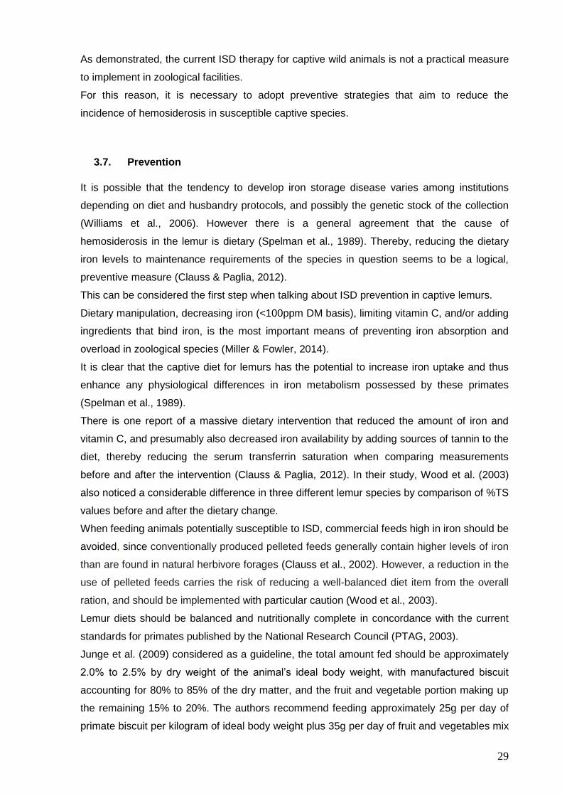
As demonstrated, the current ISD therapy for captive wild animals is not a practical measure
to implement in zoological facilities.
For this reason, it is necessary to adopt preventive strategies that aim to reduce the
incidence of hemosiderosis in susceptible captive species.
3.7. Prevention
It is possible that the tendency to develop iron storage disease varies among institutions
depending on diet and husbandry protocols, and possibly the genetic stock of the collection
(Williams et al., 2006). However there is a general agreement that the cause of
hemosiderosis in the lemur is dietary (Spelman et al., 1989). Thereby, reducing the dietary
iron levels to maintenance requirements of the species in question seems to be a logical,
preventive measure (Clauss & Paglia, 2012).
This can be considered the first step when talking about ISD prevention in captive lemurs.
Dietary manipulation, decreasing iron (<100ppm DM basis), limiting vitamin C, and/or adding
ingredients that bind iron, is the most important means of preventing iron absorption and
overload in zoological species (Miller & Fowler, 2014).
It is clear that the captive diet for lemurs has the potential to increase iron uptake and thus
enhance any physiological differences in iron metabolism possessed by these primates
(Spelman et al., 1989).
There is one report of a massive dietary intervention that reduced the amount of iron and
vitamin C, and presumably also decreased iron availability by adding sources of tannin to the
diet, thereby reducing the serum transferrin saturation when comparing measurements
before and after the intervention (Clauss & Paglia, 2012). In their study, Wood et al. (2003)
also noticed a considerable difference in three different lemur species by comparison of %TS
values before and after the dietary change.
When feeding animals potentially susceptible to ISD, commercial feeds high in iron should be
avoided, since conventionally produced pelleted feeds generally contain higher levels of iron
than are found in natural herbivore forages (Clauss et al., 2002). However, a reduction in the
use of pelleted feeds carries the risk of reducing a well-balanced diet item from the overall
ration, and should be implemented with particular caution (Wood et al., 2003).
Lemur diets should be balanced and nutritionally complete in concordance with the current
standards for primates published by the National Research Council (PTAG, 2003).
Junge et al. (2009) considered as a guideline, the total amount fed should be approximately
2.0% to 2.5% by dry weight of the animal’s ideal body weight, with manufactured biscuit
accounting for 80% to 85% of the dry matter, and the fruit and vegetable portion making up
the remaining 15% to 20%. The authors recommend feeding approximately 25g per day of
primate biscuit per kilogram of ideal body weight plus 35g per day of fruit and vegetables mix

30
per kilogram of ideal body weight.
Despite the metabolism and dietary requirements are undefined for most species, the
National Research Council estimates the adequate dietary iron concentration (dry matter
basis) for nonhuman primates to be 100 mg/kg (PTAG, 2003).
Iron concentrations as low as 65 ppm DM basis for lemurs have been recommended, but it is
difficult to formulate balanced diets this low (Miller & Fowler, 2014).
The fact that it is the iron-limited diets that are more difficult to produce and therefore
expensive, rather than iron-fortified diets, may contribute to a reluctance of changing feeding
regimes (Clauss & Paglia, 2012).
A safe upper limit for dietary iron has not been established for nonhuman primates. However,
special attention should be paid to the use of mineral supplements, in particular calcium
sources that may contain high iron levels, and mineral supplements without iron should be
used (Clauss & Paglia, 2012). Supplements are not needed if the animals are fed a well-
balanced diet in proper proportions and amounts (PTAG, 2003).
As previously mentioned the high dietary levels of vitamin C enhance the iron absorption and
can contribute to the development of hemosiderosis. For this reason, Spelman et al. (1989)
suggest that the use of citrus fruits in lemur diets should be critically assessed, as well as
other fruits and vegetables rich in vitamin C.
Feeding a well-balanced plant-based diet containing species-appropriate fibre levels will
likely minimize excessive iron absorption in diurnal lemurs (Junge et al., 2009), since dietary
levels of fibre, phosphates and phytates can influence the extent of vitamin C’s enhancement
on iron absorption.
The addition of locally available fresh leaves and browse is beneficial for diurnal species as a
means of increasing fibre in the diet, stimulating natural feeding behaviours, and providing
environment enrichment (Junge et al., 2009).
As referred before, it is known that wild lemur diets have higher amounts of tannins, when
compared to captive lemur diets. Therefore, iron-binding compounds should be given with
the main feed or within an hour after feeding, but not before (Wood et al., 2003).
Indeed, polyphenols such as tannic acid, in the form of tea (leaves or infusion) or tamarind
(pods or juice), has been used in birds, lemurs, and fruit bats. This was shown to be effective
in lemurs by following %TS (Miller & Fowler, 2014). However, tannins do not affect only iron
availability and their addition to the captive diet may lower the digestibility and nutrient
availability of other diet components as well, resulting in deficiencies in other micronutrients,
not having the desired impact on iron absorption (Zutrition, 2017).
While some investigators have recommended adding tea, beans, or other tannins sources to
captive lemur diets in an effort to reduce dietary iron absorption, there is currently insufficient
evidence to support these recommendations (Junge et al., 2009).
Hence, the deliberate inclusion of tannins or other polyphenol sources, although successful

31
in individual experiments and not evidently harmful in others, cannot be recommended at
present without further research (Clauss & Paglia, 2012).
Despite the results of many surveys, however, prophylactic programs against ISD have not
been implemented in most lemur husbandry programs (Wood et al., 2003).
Adjusting the dietary iron levels so that requirements are not exceeded would represent the
most practical approach if limiting iron in diets was not so difficult or against historical feeding
traditions (Clauss & Paglia, 2012). Nevertheless, changing captive diets is a crucial step to
take in most lemur husbandry programs but not the only key factor for a successful ISD
prevention.
Screening protocols based on blood measurements (in tandem with medical training
programs that reduce the need for anaesthetic intervention), should be instigated for
susceptible species (Crawford et al., 2005). Measurement of serum ferritin concentration in
conjunction with transferrin saturation may constitute a non-invasive approach for evaluating
iron status in lemurs (Williams et al., 2008).
Diagnosis of this disease has remained consistently at the point of necropsy, and routine
screenings that would detect the disease early enough to instigate therapeutic measures are
not part of a standard management protocol (Wood et al., 2003). In addition, the investigation
of ISD at necropsy of susceptible animals should be implemented as a routine procedure, to
estimate the incidence of this disease in the facilities and to increase documented cases of
negative findings.
Post that, although results indicate that further investigations should be conducted, there are
some practical measures to adopt that seem to be a logical first step.
Current feeding regimes should either be adjusted within reasonable boundaries right away,
such as reducing iron levels to maintenance recommendations of model species, with
accompanying health monitoring measures, or (further) adjusted on the basis of necropsy
findings or screening of live animals (Clauss & Paglia, 2012).
Studies of lemurs housed at multiple institutions are needed to gain a better understanding of
the prevalence, severity and clinical impact of iron overload in captive lemurs (Williams et al.,
2008).
32
III. STUDY – Iron Storage Disease Prevalence in Captive Ring-tailed Lemurs
The present study was carried out in three different zoological facilities and included all the
ring-tailed lemurs housed at these institutions.
In order to facilitate the analysis of the diet influence, the individuals were divided into three
different groups, according to the zoological facility in which they live.
1. Goals This master’s dissertation aims to evaluate the prevalence of ISD in ring-tailed lemurs
housed at several institutions from different regions of Portugal and relate it to their captive
diet.
The survey may contribute to better understand possible causes for the high prevalence of
this disease in susceptible species, such as lemurs. In addition, the present study intends to
expose potential strategies that can contribute to ISD prevention.
2. Material and methods
2.1. Sample
The present study was conducted to assess the prevalence of hemosiderosis in captive ring-
tailed lemurs in Portugal. The study was carried out in three different zoological facilities and
included all the ring-tailed lemurs housed at these institutions. In order to facilitate statistical
analysis, the individuals were categorised into three different groups, according to the
zoological facility in which they live.
The animals were divided into three groups, correspondent to the three different zoological
facilities. Each group had a different number of animals included:
Park 1: 10 animals
Park 2: 2 animals
Park 3: 6 animals
2.2. Institutions description
2.2.1. Park 1
Park 1 is situated in the southwestern region of Portugal in the Alentejo. It includes 600 wild
animals of 75 different species in an area of 90ha.

33
The park is home to ten ring-tailed lemurs, cohabiting with four red-bellied lemurs (Eulemur
rubriventer). All animals are kept in an open enclosure during the day, spending the night in
an indoor installation, separated by species.
The park is a member of the European Association for the Study and Conservations of
Lemurs. It is also a consortium of the European Zoological Gardens and Universities, aiming
to the implementation of research projects to promote the conservation of different lemur
species.
2.2.2. Park 2
This park is situated in the north region of Portugal and is the home to 100 different species
of animals, including mammals, reptiles, birds, amphibians and fishes.
Characterized by its pedagogical component, the zoo aims to collaborate in different
educational programs, promoting the realization of several activities that intend to sensitize
people for the protection of biodiversity.
Similarly, this zoological park contributes to different research programs, through the
implementation of partnerships with other zoos and universities.
The institution houses two ring-tailed lemurs living in an indoor enclosure.
2.2.3. Park 3
Located in the north of Alentejo region, in the southwest of Portugal, the park houses 350
animals from 60 different species, in an area of 20ha.
Nature conservation and protection, as well as education for sustainability are their main
purposes.
The park is home for eight ring-tailed lemurs, living in an open enclosure during the day. At
night, all the lemurs are closed in an indoor installation.
2.3. Sample collection
The blood was collected by external saphenous venipuncture to a serum separator tube and
then centrifuged for serum obtaining. The animals were not submitted to anaesthesia and all
the samples were collected under minimal stress conditions. Zookeepers from the institution
were responsible for animals’ capture, using an appropriate net (Figure 11). Immediately
after that, the animals were identified using a microchip scanner, and the blood was collected
while the zookeeper contained the animal (Figure 12).
The animals’ information was registered in an identification form (Appendix 4).
The laboratory analysis was duplicated to increase accuracy in animals from which it was
possible to collect more than 1ml of blood.
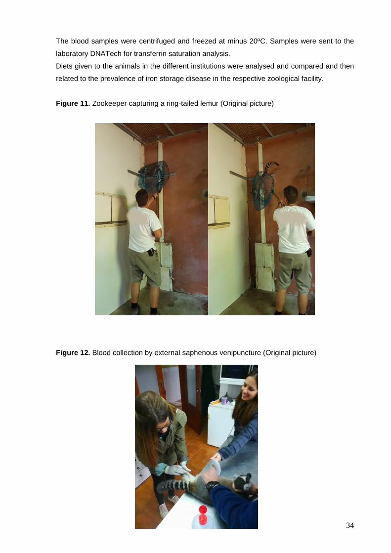
34
The blood samples were centrifuged and freezed at minus 20ºC. Samples were sent to the
laboratory DNATech for transferrin saturation analysis.
Diets given to the animals in the different institutions were analysed and compared and then
related to the prevalence of iron storage disease in the respective zoological facility.
Figure 11. Zookeeper capturing a ring-tailed lemur (Original picture)
Figure 12. Blood collection by external saphenous venipuncture (Original picture)

35
3. Statistical Analysis All data collected during the study were loaded onto the program Microsoft Office Excell
(Microsoft Office 2011 for Mac). The statistical analysis was performed using the extension
R Commander, from software R, version 3.0.1.
The one-way statistical test ANOVA was used to determine if there were any significant
differences between the analysed groups (Park 1, Park 2 and Park 3).
Statistical differences were set at p<0.05 for a 95% confidence interval.
The statistical analysis was conducted by calculating sample features of each group (mean,
standard deviation, maximum, minimum and median) for transferrin saturation level.
4. Results
4.1. Sample description
All the sample participants (n=18) were submitted to the same protocol, which estimated the
levels of transferrin saturation verified at the moment of blood collection.
The sample included 18 animals, 13 males and 6 females, all from the same species - Lemur
catta, living in captive conditions. Animals’ average age was 5.8 years (minimum 1 year and
maximum 14 years).
4.2. Blood values
The results obtained from all the individuals are registered in Table 2, separated by groups. It
was possible to identify the animal by its microchip identification number.
The average transferrin saturation values varied between parks. However, these differences
were not significant when comparing Park 1 with Park 2 (p = 0.8977) or between Park 2 and
Park 3 (p = 0.1161).
Only the difference between the groups Park 1 and Park 3 values were statistically different
(p = 0.0404).

36
Table 2. Individual transferrin saturation values (Reference values for this species:
20 – 55%). Blood samples were duplicated to increase accuracy when possible.
Institution Animal
identification Sample 1 Sample 2
Park 1
852 100.3%
764 83.2% 100%
891 91% 88.3%
784 89.3% 92.7%
451 48.9% 49.6%
455 68.9% 70.5%
588 103.2%
742 52.9%
751 85.1% 83.4%
757 56% 56.3%
Average 78.8%
Park 2
754 65%
759 70% 87%
Average 71.75%
Park 3
7630 99.7%
6250 71.8%
7621 176.8%
1108 145.2%
6387 70.8%
0443 157.6%
Average 120.32%

37
Graphic 1. Average transferrin saturation values for the different groups
4.2.1. Results presentation per group
All groups analysed demonstrated high average levels of transferrin saturation, considering
that the reference range is between 20% and 55% (Table 2 and Graphic 1).
The group Park 3 revealed the highest average level of transferrin saturation, with a mean of
120.32%. On the other hand, the group Park 2 was considered the one with the lowest
average level of transferrin saturation, with an average of 71.75%.
The group Park 1 showed intermediate values, with an average level of transferrin saturation
of 78.80%.
Table 3. Sample features for transferrin saturation
Transferrin saturation (%)
Mean Standard deviation Minimum Maximum Median
Park 1 78.80 20.16 49.25 103.2 86.95
Park 2 71.75 9.5 65.00 78.50 71.75
Park 3 120.32 45.67 70.80 176.80 122.45

38
Graphic 2. Results presentation per group
4.3. Diets description
Most of the food given to the lemurs at the parks is offered by supermarkets, as it is reaching
the expiry date. This is valid mainly for fruits and vegetables and also for some cereals and
supplements, with exception for dry food.
To have a detailed description of the animals’ diet, all the food given was minutely described
for 15 consecutive days (Table 5, 7 and 11). During this time, the zookeeper registered the
type and amount of fruit, vegetables or cereals given, as well as any eventual supplement
offered to the animals.
Considering this information, it was possible to elaborate a specific graphic for each park,
which allowed to observe the relative percentage of each class of food given to the animals.
4.3.1. Park 1
At Park 1, fruits are the main components of lemurs’ diet, representing 91.7% of the food
given to the animals. Although constituting a significant minor proportion, vegetables also

39
represent an important element of their diet (7.47%). The cereals given to the lemurs are
counted as just 0.83% of their feed.
It was possible to identify all the types of fruits, vegetables and cereals given to the animals
at the park (Table 4).
Table 4. List of fruits, vegetables and cereals given to the lemurs at Park 1
Graphic 3. Constituents of lemurs’ diet at Park 1
91.70%
7.47% 0.83%
Fruit Vegetables Cereals
Fruits Vegetables Cereals
Apple Mixed salad (lettuce and carrot) Seed bread
Banana Leek Raisin bread
Pear Beaded lettuce
Annona Endives
Melon Green salad
Mango Carrot
Rockmelon Spinach
Persimmons
Watermelon
Grapes
Pineapple
Strawberry
Plum

40
Table 5. Diet analysis for ten animals in Park 1
Type of food
Amount (pieces)
Unit weight (g)
Total weight (g)
Day 1 A.M. Red apple 5 130 650
Banana 5 115 575
Pear 5 230 1150
P.M. Seed bread 1 100 100
Banana 4 115 460
Mixed salad (lettuce and carrot) 2 175 350
Annona 3 220 660
Pink lady apple 2 185 370
Melon 0.50 2000 1000
Day 2 A.M. Red apple 5 130 650
Banana 5 115 575
Mango 2 500 1000
P.M. Leek 3 200 600
Beaded Lettuce 0.50 400 200
Mixed salad (lettuce and carrot) 1 175 175
Annona 2 220 440
Banana da Madeira 6 100 600
Rockmelon 0.50 1250 625
Day 3 A.M. Red Apple 3 130 390
Banana 6 115 690
Melon 0.25 2000 500
P.M. Raisin bread 1 100 100
Persimmons 4 300 1200
Endives 3 150 450
Mango 1 500 500
Melon 0.25 2000 500
Leek 4 200 800
Banana 4 115 460
Day 4 A.M. Red apple 5 130 650
Banana 2 115 230
Watermelon 0.25 2500 625
P.M. Red apple 6 130 780
Melon 0.25 2000 500
Brunches of grapes 2 400 800
Leek 4 200 800
Day 5 A.M. Melon 1 2000 2000
Red apple 5 130 650
Pineapple 0.50 1300 650
P.M. Rockmelon 0.50 1250 625

41
Strawberry 11 20 220
Pear 6 230 1380
Melon 0.25 2000 500
Red apple 4 130 520
Mango 3 500 1500
Green salad 2 175 350
Day 6 A.M. Melon 0.25 2000 500
Red apple 6 130 780
P.M. Persimmons 8 300 2400
Strawberry 30 20 600
Brunches of grapes 2.50 400 1000
Banana 4 115 460
Green salad 1 175 175
Day 7 A.M. Red apple 9 130 1170
Strawberry 24 20 480
Brunches of grapes 2.50 400 1000
P.M. Strawberry 10 20 200
Pear 5 230 1150
Melon 0.50 2000 1000
Red apple 4 130 520
Mango 3 500 1500
Banana 2 115 230
Green salad 2 175 350
Day 8 A.M. Annona 1 220 220
Red apple 4 130 520
Pineapple 0.13 1300 162.5
Banana 4 115 460
Mango 1 500 500
P.M. Mango 4 500 2000
Persimmons 5 300 1500
Annona 2 220 440
Pineapple 0.50 1300 650
Seed bread 1 100 100
Leek 5 200 1000
Day 9 A.M. Red apple 5 130 650
Plum 13 60 780
Melon 0.75 2000 1500
P.M. Pear 3 230 690
Seed bread 1 100 100
Rockmelon 0.50 1250 625
Mango 3 500 1500
Persimmons 8 300 2400
Day 10 A.M. Red apple 4 130 520
Plum 12 60 720

42
Rockmelon 0.50 1250 625
P.M. Carrot 5 150 750
Banana 5 115 575
Rockmelon 0.50 1250 625
Seed bread 1 100 100
Plum 10 60 600
Day 11 A.M. Melon 0.50 2000 1000
Pineapple 0.50 1300 650
Red apple 6 130 780
Plum 8 60 480
P.M. Pear 6 230 1380
Strawberry 12 20 240
Red apple 5 130 650
Pineapple 0.50 1300 650
Plum 10 60 600
Day 12 A.M. Mango 1 500 500
Red apple 7 130 910
Plum 10 60 600
P.M. Persimmons 6 300 1800
Red apple 4 130 520
Melon 0.50 2000 1000
Seed bread 0.50 100 50
Day 13 A.M. Banana 4 115 460
Melon 0.50 2000 1000
Red apple 4 130 520
Plum 2 60 120
P.M. Persimmons 5 300 1500
Mango 3 500 1500
Brunches of grapes 2 400 800
Red apple 4 130 520
Spinach 1 200 200
Day 14 A.M. Pear 5 230 1150
Brunches of grapes 1.50 400 600
Melon 0.25 2000 500
P.M. Mango 2 500 1000
Banana 5 115 575
Plum 8 60 480
Red apple 4 130 520
Persimmons 2 300 600
Seed bread 1 100 100
Day 15 A.M. Banana 4 115 460
Red apple 4 130 520
Rockmelon 0.75 1250 937.5
Pear 3 230 690

43
P.M. Leek and carrot soup 1 350 350
Mixed salad (lettuce and carrot) 1 175 175
Seed bread 1 100 100
Banana 4 115 460
4.3.2. Park 2
Fruit is also the main constituent of lemurs’ diet at Park 2, representing 64.12% of the food
given. At this park, vegetables are also an important food source, constituting almost 31.65%
of their diet.
The park also provides dry food to the animals, although in a much smaller proportion than
the other enounced food classes (0.84%).
Cereals represent the smallest proportion of the food given, calculated as just 0.25%.
Occasionally the lemurs receive food supplements that account as 3.13% of their diet.
As shown in Table 6, it was possible to identify all the types of fruits, vegetables, dry food,
cereals and supplements given to the animals at the park.
Table 6. List of fruits, vegetables and cereals given to the two lemurs at Park 2
Fruits Vegetables Dry food Cereals Supplements
Tomato Spinach Dog dry food Cerelac (baby food) Pine nuts
Strawberry Carrot Seeds mix
Apple Leek Cat wet food
Pear Pepper
Banana Endives
Rockmelon Cucumber
Grapes Lettuce
Mango
Fruit purees
Papaya
Passionfruit

44
Graphic 4. Constituents of lemurs’ diet at Park 2
Table 7. Diet analysis for two animals in Park 2
Type of food Amount Unit weight (g) Total weight (g)
Day 1 Tomato 1 100 100
Spinach 15 60 900
Strawberry 2 20 40
Apple 1 130 130
Pear 1 230 230
Banana 1.5 115 172.5
Rockmelon 0.25 1250 312.5
Carrot 2 150 300
Day 2 Brunch of grapes 1 400 400
Pear 0.5 230 115
Mango 0.5 500 250
Banana 2 115 230
Leek 2 200 400
Carrot 0.5 150 75
Tomato 1 100 100
Pepper 0.5 210 105
Apple 1 130 130
Seeds mix 1 100 100
Fruit purees 2 100 200
Day 3 Endive 1 150 150
Pear 1 230 230
Banana 2 115 230
Cucumber 0.5 350 175
64.12%
31.65%
0.25%0.84%
3.13%
Fruit Vegetables Cereals Dry food Supplements

45
Papaya 0.5 515 257.5
Pepper 0.25 210 52.5
Apple 1 130 130
Seeds mix 1 100 100
Cerelac pap 1 30 30
Day 4 Pepper 0.5 210 105
Banana 1.5 115 172.5
Apple 1 130 130
Pear 2 230 460
Carrot 2 150 300
Brunch of grapes 1 400 400
Tomato 1 100 100
Lettuce 1 430 430
Pine nuts 1 70 70
Day 5 Banana 2 115 230
Pear 1 230 230
Apple 1 130 130
Brunch of grapes 1 400 400
Rockmelon 0.5 1250 625
Mango 1 500 500
Carrot 1 150 150
Dog dry food 1 100 100
Day 6 Rockmelon 0.25 1250 312.5
Cucumber 0.5 350 175
Strawberry 3 20 60
Apple 1 130 130
Lettuce 1 430 430
Papaya 0.25 515 128.75
Mango 0.33 500 165
Passionfruit 1 160 160
Wet cat food 1 100 100
Day 7 Tomato 1 100 100
Spinach 15 60 900
Strawberry 2 20 40
Apple 1 130 130
Pear 1 230 230
Banana 1.5 115 172.5
Rockmelon 0.25 1250 312.5
Carrot 2 150 300
Day 8 Brunch of grapes 1 400 400
Pear 0.5 230 115
Mango 0.5 500 250
Banana 2 115 230
Leek 2 200 400

46
Carrot 0.5 150 75
Tomato 1 100 100
Pepper 0.5 210 105
Apple 1 130 130
Seeds mix 1 100 100
Fruit purees 2 100 200
Day 9 Endive 1 150 150
Pear 1 230 230
Banana 2 115 230
Cucumber 0.5 350 175
Papaya 0.5 515 257.5
Pepper 0.25 210 52.5
Apple 1 130 130
Seeds mix 1 100 100
Cerelac pap 1 30 30
Day 10 Pepper 0.5 210 105
Banana 1.5 115 172.5
Apple 1 130 130
Pear 2 230 460
Carrot 2 150 300
Brunch of grapes 1 400 400
Tomato 1 100 100
Lettuce 1 430 430
Pine nuts 1 70 70
Day 11 Banana 2 115 230
Pear 1 230 230
Apple 1 130 130
Brunch of grapes 1 400 400
Rockmelon 0.5 1250 625
Mango 1 500 500
Carrot 1 150 150
Dog dry food 1 100 100
Day 12 Rockmelon 0.25 1250 312.5
Cucumber 0.5 350 175
Strawberry 3 20 60
Apple 1 130 130
Lettuce 1 430 430
Papaya 0.25 515 128.75
Mango 0.33 500 165
Passionfruit 1 160 160
Wet cat food 1 100 100
Day 13 Tomato 1 100 100
Spinach 15 60 900
Strawberry 2 20 40

47
Apple 1 130 130
Pear 1 230 230
Banana 1.5 115 172.5
Rockmelon 0.25 1250 312.5
Carrot 2 150 300
Day 14 Brunch of grapes 1 400 400
Pear 0.5 230 115
Mango 0.5 500 250
Banana 2 115 230
Leek 2 200 400
Carrot 0.5 150 75
Tomato 1 100 100
Pepper 0.5 210 105
Apple 1 130 130
Seeds mix 1 100 100
Fruit purees 2 100 200
Day 15 Endive 1 150 150
Pear 1 230 230
Banana 2 115 230
Cucumber 0.5 350 175
Papaya 0.5 515 257.5
Pepper 0.25 210 52.5
Apple 1 130 130
Seeds mix 1 100 100
Cerelac pap 1 30 30
4.3.3. Park 3
At park 3, fruits and vegetables are also the main constituents of lemurs’ diet, representing
46.77% and 31.76% of their nutrition, respectively. However, at this park there is a significant
higher proportion of dry food (Table 10) given to the animals (11.34%), comparing to the
other parks. Additionally, cereals (3.84%) and variable supplements (6.28%) are also
provided to the animals in relatively high quantity.
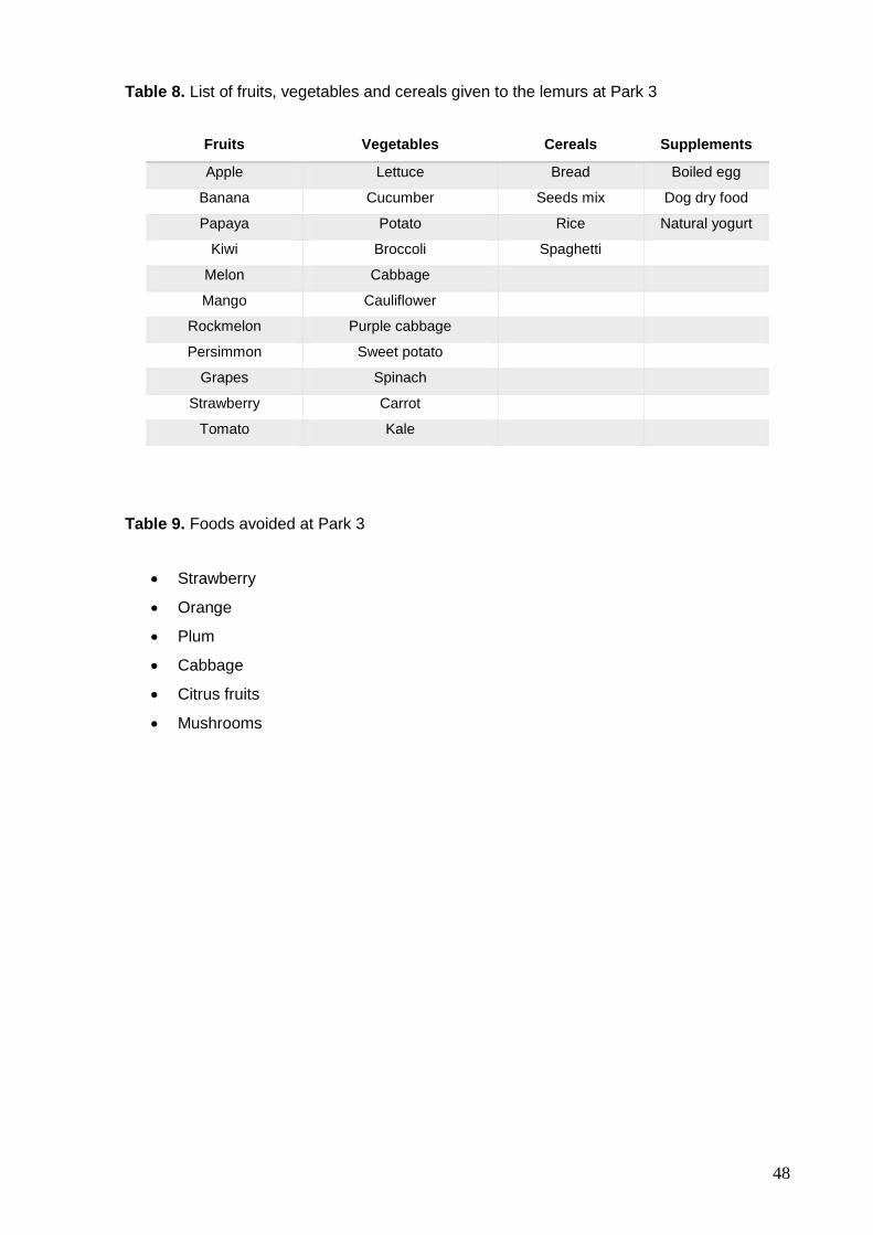
48
Table 8. List of fruits, vegetables and cereals given to the lemurs at Park 3
Fruits Vegetables Cereals Supplements
Apple Lettuce Bread Boiled egg
Banana Cucumber Seeds mix Dog dry food
Papaya Potato Rice Natural yogurt
Kiwi Broccoli Spaghetti
Melon Cabbage
Mango Cauliflower
Rockmelon Purple cabbage
Persimmon Sweet potato
Grapes Spinach
Strawberry Carrot
Tomato Kale
Table 9. Foods avoided at Park 3
Strawberry
Orange
Plum
Cabbage
Citrus fruits
Mushrooms

49
Table 10. Analytical composition of lemurs’ dry food - Marmoset / New World monkeys, 4 mm pellets (Complete feed for Marmosets – NHP)
Analytical constituents % Nutritional Additives Per kg
Crude Protein 26.10 Vitamin A (E 672) 18000 (IU)
Crude oils and fats 7.00 Vitamin D3 (E671) 3000 (IU)
Crude ash 6.80 Vitamin E 120 (mg)
Calcium 1.00
Vitamin C (stabilized; Ascorbyl
monophosphate calcium sodium
salt)
2900 (mg)
Phosphorus 0.70 Iron, Ferrous sulphate monohydrate
(E1) 100 (mg)
Sodium 0.20 Zinc, Zinc sulphate monohydrate
(E6) 50 (mg)
Lysine 1.70 Manganese, Manganous-(II)-
sulphate monohydrate (E5) 30 (mg)
Met + Cyst 1.10 Copper, Copper-(II)-sulphate
pentahydrate (E4) 5 (mg)
Selenium, Sodium selenite (E8) 0.1 (mg)
Energy MJ/kg Iodine, Calcium iodate anhydrous
(E2) 2.0 (mg)
ME 15.6 Cobalt, Cobaltous carbonate
monohydrate (E3) 2.0 (mg)
Composition: Grains and grain products, oil seed products, sugar, milk and dairy products,
vegetable oils, fruits (dried), minerals, flavours.

50
Graphic 5. Constituents of lemurs’ diet at Park 3
Table 11. Diet analysis for six animals in Park 3
Type of food Amount Unit weight
(g) Total weight (g)
Day 1 A.M. Cucumber 480
New World pellet 320
P.M. Apple 580
Melon 700
Cucumber 640
Boiled egg 4 50 200
Day 2 A.M. Lettuce 480
New World pellet 320
P.M. Grapes 425
Persimmons 855
Spinach 200
Carrots 440
Seeds mix 160
Day 3 A.M. Lettuce 480
New World pellet 320
P.M. Banana 400
Papaya 680
Grapes 200
Sweet potato 640
Bread 2 40 80
Day 4 A.M. Lettuce 480
New World pellet 320
P.M. Mango 530
Papaya 750
46.77%
31.76%
3.84%
11.34%
6.28%
Fruit Vegetables Cereals Dry food Supplements

51
Tomato 640
Dog dry food 200
Day 5 A.M. Lettuce 480
New World pellet 320
P.M. Grapes 400
Apple 880
Broccoli 640
Natural yogurt 4 120 480
Day 6 A.M. Potato 280
Broccoli 200
New World pellet 320
P.M. Banana 860
Kiwi 420
Lettuce 640
Rice 800
Day 7 A.M. Cabbage 480
New World pellet 320
P.M. Persimmons 350
Rockmelon 500
Apple 430
Lettuce 640
Seeds mix 500
Day 8 A.M. Cauliflower 480
New World pellet 320
P.M. Apple 530
Rockmelon 750
Tomato 640
Boiled egg 4 50 200
Day 9 A.M. Broccoli 480
New World pellet 320
P.M. Apple 700
Banana 580
Sweet potato 640
Seeds mix 160
Day 10 A.M. Tomato 480
New World pellet 320
P.M. Grapes 1280
Lettuce 640
Bread 2 40 80
Day 11 A.M. Purple cabbage 480
New World pellet 320
P.M. Apple 800
Tomato 480
Carrots 640

52
Dog dry food 200
Day 12 A.M. Cabbage 480
New World pellet 320
P.M. Kiwi 640
Apple 640
Cabbage 640
Natural yogurt 4 120 480
Day 13 A.M. Sweet potato 480
New World pellet 320
P.M. Papaya 700
Strawberries 580
Carrots 400
Broccoli 240
Spaghetti 800
Day 14 A.M. Sweet potato 480
New World pellet 320
P.M. Apple 1280
Lettuce 640
Seeds mix 500
Day 15 A.M. Cucumber 480
New World pellet 320
P.M. Grapes 780
Tomato 500
Lettuce 640
Boiled egg 4 50 200
4.4. Amount of iron and vitamin C
The content of iron and vitamin C were also discriminated for each type of food (Appendix
5, 6 and 7), which allowed the estimation of the total amount given to the animals,
regarding these components. It was calculated based on the “Nutritive value of foods”
(Gebhardt & Thomas, 2002) and analytical composition of the foods given, when available.
The assessment of tannin content in the food offered to the animals was not achievable,
since it is not defined for most fruits and vegetables.
Table 12. Amount of iron and vitamin C given to the animals
Amount per day Park 1 Park 2 Park 3
Iron (mg/kg BW) 3.44 9.7 9.4
Vitamin C (mg/kg food) 249 213 506
*Considering the ideal body weight for L. catta (2.6kg)

53
5. Discussion
The intention of the present study was to analyze the levels of transferrin saturation of all the
ring-tailed lemurs housed at three different zoological parks and relate the obtained values to
the diet which the animals are submitted to.
Hemosiderosis has been reported in captive lemurs since the decade of 1960s, as noticed by
pathologists at San Diego Zoo, when deposits of hemosiderin were found in the liver, spleen,
lymph nodes, duodenum and other organs (Gonzales et al., 1984). Subsequently,
researchers began to explore possible causes for the incidence of hemosiderosis in
susceptible animals.
It is commonly accepted that the main cause for the high prevalence of ISD in lemurs is
related to their captive diet, composed of several substances that may contribute to enhance
the iron absorption, such as the high dietary levels of iron and ascorbic acid (citrus fruits)
(Spelman et al., 1989).
Additionally, diets of captive lemurs have fewer substances that reduce iron availability, such
as tannins, compared to the foods that the animals find in the wild (Clauss & Paglia, 2012).
For these reasons, our study included a diet analysis, focusing on the amount of fruit,
vegetables, cereals, dry food and supplements given to the animals in each park, and its iron
and vitamin C content.
Although the diagnosis of this condition requires a liver biopsy, it is possible to evaluate the
animals’ iron status using blood tests such as ferritin and transferrin saturation, similarly to
human medicine (Williams et al., 2008). Transferrin saturation is an indicative of tissue iron
reserves and it has been demonstrated to be positively correlated with hepatic iron
deposition in ring-tailed lemurs (Williams et al., 2008).
Transferrin saturation represents the ratio between serum iron and total iron binding capacity
(TS=SI/TIBC) (Ghosh & Collier, 2012) and the TIBC corresponds to the maximum amount of
iron that can be bound to transferrin. Under physiologic circumstances, nearly all iron is
bound to plasma proteins. However, under excessive iron conditions, transferrin is fully
saturated and the plasma may contain iron which is not bound to plasma proteins (“free iron”)
(Zanen, Adriaansen, Bommel, Posthuma & Jong, 1996) leading to increased values of
transferrin saturation, occasionally over 100%.
The present study revealed a high prevalence of ISD in almost all the animals housed at all
the participant zoological facilities, considering that the reference range for transferrin
saturation varies between 20 and 55%.
Although some animals revealed normal values of transferrin saturation, the average
calculated for all the parks evaluated demonstrated a high prevalence of ISD in captive ring-
tailed lemurs housed at these parks in Portugal.

54
As demonstrated, the average of iron ingested per day at Park 1, Park 2 and Park 3 was
3.44 mg/kg BW, 9.7 mg/kg BW and 9.4 mg/kg BW, respectively.
According to the “Dietary References Intakes” from the Institute of Medicine of the National
Academies, U.S., the recommended daily allowance for a man weighing approximately 65 kg
is 8 mg of iron per day. Considering the ideal body weight for ring-tailed lemurs to be 2.6 kg,
it is reasonable to affirm by extrapolation that the lemurs housed at the enounced parks are
consuming high levels of dietary iron, particularly at Park 2 and Park 3.
Despite this evidence, there is a significant difference between the transferrin saturation
mean values obtained at Park 3 and the mean values from the other parks. One possible
explanation for the higher values obtained at Park 3 is related to the increased percentage of
dry food given to the animals housed at this park, leading to increased amounts of iron and
vitamin C offered to the lemurs.
According to the National Research Council, the appropriate concentration of dietary iron
should be 100 mg/kg DM (PTAG, 2003), which corresponds to the iron concentration in the
commercial diet given at Park 3 (Table 10). However, it has been recently proposed that the
iron concentrations for lemurs should be as low as 65 ppm (65 mg/kg), although it is difficult
to formulate diets with this concentration (Miller & Fowler, 2014).
Alternatively, it has been recommended to feed approximately 25 g of monkey pellets per
kilogram of ideal body weight per day (Junge et al., 2009). Considering the ideal body weight
of a captive ring-tailed lemur to be approximately 2.6 kg (Kappeler, 1991), and the total
amount of monkey biscuits given to the eight lemurs at Park 3 to be 320 g per day, it can be
considered as an adequate amount per animal (15.4 g per kilogram of ideal body weight).
However, even feeding the animals with appropriate amounts of dry food, it was still verified
high levels of transferrin saturation after blood analysis.
It is considered by the National Research Council that the recommended vitamin C
concentration for ring-tailed lemurs is 111 mg of vitamin C per kg of food (Moury & Campbell,
2001). This means that an excessive amount of vitamin C is being provided in the food from
all the studied ring-tailed lemurs, as evidenced in table 12. It is important to notice that this
concentration is significantly higher at Park 3, when comparing to the other parks. The
reason for this difference is the fact that the lemurs from the enounced park consume a
larger proportion of dry food in their diet (11.34%), which has a concentration of 2900 mg of
vitamin C per kg of food, leading to a substantial increment in the total amount of vitamin C
present in the food given to the lemurs.
Consequently, the higher values of %TS detected Park 3 may be related to both iron and
vitamin C concentration levels, incremented by the larger proportion of dry food offered to the
animals.

55
Therefore, it is essential to reformulate the diet given to the animals, in order to achieve
smaller levels of dietary iron and vitamin C.
Diets formulation should be based on species adaptation, considering their nutrition in the
wild and their common captive health problems. The fact that almost all zoological institutions
in Portugal feed their animals (especially primates) using food offered by supermarkets,
makes food selection more difficult, which leads to an inadequate diet.
For this reason, it is essential that the mentioned institutions follow strategies to implement a
species-specific diet for all the animals.
A list of avoided foods should be applied for each species, based on their metabolism and
nutrition adaptation in captive conditions. This could eventually be realized by a nutrition
specialist, preferably a veterinarian.
Considering ISD, it is crucial that lemurs’ diet formulation avoids iron rich foods (Appendix 1)
and vitamin C rich foods (Appendix 2) and contains high levels of tannin rich food sources
(Appendix 3). This should be considered the first step to reduce the incidence of
hemosiderosis in this species, thus reducing their mortality.
As previously mentioned, ISD in lemurs is associated with hemosiderin accumulation in liver,
kidney, spleen and bone marrow, often leading to a stage of generalized hemosiderosis in
interstitial tissue and parenchymal cells, producing extensive tissue damage (Spelman et al.,
1989), which can lead to an increased mortality risk (Mainous, Wells, Carek, Gill, & Geesey,
2004).
Therefore, it is essential to make zoological parks aware of the necessity of creating
balanced diets, adapted to the species in question, since it can effectively decrease animals’
mortality.
6. Study limitations and future considerations
The most preeminent limitation of this study was the sample size. Despite the initial intention
of gathering animals from five different zoological parks, there were two facilities that
discarded the participation on the present study due to animal husbandry possible
interference. Furthermore, the sample size was different for each institution.
Increasing the sample size could be beneficial to have more accurate results.
Diet analysis was another limitation almost as relevant as the previously mentioned. As the
fruit and vegetables are offered by supermarkets it is difficult to accurately define the diet, as
it varies according to availability. This variation makes it extremely challenging to analyze the
relative percentage of fruit, vegetables, cereals, dry food and supplements given to the
animals.

56
Additionally, the significant food variation made it extremely difficult to accurately analyze
relevant diet components, such as iron, ascorbic acid and tannins, considered essential
factors for the incidence of ISD.
A deeper investigation of diet components, quantifying the exact amount of iron, ascorbic
acid and tannins would be a useful approach to determine its influence on transferrin
saturation levels. However, the initial goal of this study was to provide a practical analysis of
the diet given and to suggest the respective revision based on comparative results.
The lack of information regarding this subject was considered an additional limitation.
Unfortunately, the number of studies including this species is limited, thus reference values
for hematological components are not completely defined yet.
Despite the initial goals of this study did not include a reassessment of the results obtained, it
would be interesting to analyze the levels of transferrin saturation after the implementation of
the suggested diet changes.
57
Conclusion It was extremely challenging to take specific conclusions from this study, due to the sample
size. However, it is strongly evident that ISD is currently present in almost all lemurs’
collections in Portugal.
Although the causes are not completely defined, it is reasonable to conclude that food is
probably the main risk factor for the high incidence of hemosiderosis in this species.
As referred before, it is known that ISD can carry serious health problems for susceptible
species, such as lemurs and it is also considered an important mortality cause for these
animals. For this reason, further research should be conducted in order to reformulate the
diets given to the animals, as an approach to reduce the incidence of ISD.
During this study, it was verified among zoos and wildlife parks that there was no
consciousness about the impact that ISD can have in lemurs’ health, neither the diet’s
influence on this disease.
It could be interesting to develop an awareness campaign to sensitize wildlife veterinarians
and zookeepers about this subject, in order to create strategies that decrease the prevalence
of ISD in captive populations.

58
References
Abbaspour, N., Hurrell, R. & Kelishadi, R. (2014). Review on iron and its importance for human health. Journal of Research in Medical Sciences : The Official Journal of Isfahan University of Medical Sciences, 19(2), 164–174.
Andriaholinirina, N., Baden, A., Blanco, M., Chikhi, L., Cooke, A., Davies, N., Dolch, R., Donati, G.,
Ganzhorn, J., Golden, C., Groeneveld, L.F., Hapke, A., Irwin, M., Johnson, S., Kappeler, P., King, T., Lewis, R., Louis, E.E., Markolf, M., Mass, V., Mittermeier, R.A., Nichols, R., Patel, E., Rabarivola, C.J., Raharivololona, B., Rajaobelina, S., Rakotoarisoa, G., Rakotomanga, B., Rakotonanahary, J., Rakotondrainibe, H., Rakotondratsimba, G., Rakotondratsimba, M., Rakotonirina, L., Ralainasolo, F.B., Ralison, J., Ramahaleo, T., Ranaivoarisoa, J.F., Randrianahaleo, S.I., Randrianambinina, B., Randrianarimanana, L., Randrianasolo, H., Randriatahina, G., Rasamimananana, H., Rasolofoharivelo, T., Rasoloharijaona, S., Ratelolahy, F., Ratsimbazafy, J., Ratsimbazafy, N., Razafindraibe, H., Razafindramanana, J., Rowe, N., Salmona, J., Seiler, M., Volampeno, S., Wright, P., Youssouf, J., Zaonarivelo, J. & Zaramody, A. (2014). Lemur catta, The IUCN Red List of Threatened Species 2014.
AZA. (2017). Global Species Management Plans. Accessed in: December, 2017. Retrieved from:
https://www.aza.org/global-species-management-plans.
Basilewsky, G. (1965). Keeping and breeding madagascan lemurs in captivity. International Zoo Yearbook, 5(1), 132–137
Bassett, M. L., Halliday, J. W., Bryant, S., Dent, O. & Powell, L. W. (1988). Screening for
Hemochromatosis. New England Journal of Medicine, 328(22), 1616–1620. https://doi.org/10.1056/NEJM199306033282208
Beard, J. L., Dawson, H., Piñero, D. J. (1996). Iron metabolism: a comprehensive review. Nutrition Reviews, 54:295-317.
Beutler, E. (2007). Iron storage disease: Facts, fiction and progress, 39(2), 140-147. Department of
Molecular and Experimental Medicine, The Scripps Research Institute, 10550 North Torrey Pines Road, La Jolla, CA, 92037, USA.
Brune, M., Rossander, L., & Hallberg, L. (1989). Iron absorption and phenolic compounds:
importance of different phenolic structures. European Journal of Clinical Nutrition, 43(8), 547—557.
Burnum, A. (2016). Hemochromatosis in a black-and-white ruffed lemur. University of Georgia,
College of Veterinary Medicine. Accessed in: January, 2018. Retrieved from: https://vet.uga.edu/images/uploads/pathology/sevpac/27-Burnum.pdf
Carlstead, K. (2000). Constructing behavior profiles of zoo animals: Incorporating behavioral
information into captive population management. AZA Behavior and Husbandry Group Oregon Zoo. 40 p.
Carolina, N., Keynes, M., & Kingdom, U. (1995). Organization of White Adipose Tissue in Lemuridae. American Journal of Primatology, 35(1): 1 – 13.
Cawthon, Lang KA. (2005). Primate Factsheets: Ring-tailed lemur (Lemur catta) Taxonomy,
Morphology, & Ecology. Accessed in December, 2017. Retrieved from: http://pin.primate.wisc.edu/factsheets/entry/ring-tailed_lemur
Clauss, M., Lechner-Doll, M., Hanichen, T., Hatt, J. M. (2002). Excessive iron storage in captive
mammalian herbivores: a hypothesis for its evolutionary etiopathology. In: Proceedings of 4th Scientific Meeting of the European Association of Zoo and Wildlife Veterinarians, Heidelberg,

59
Germany. p123-131.
Clauss, M., & Paglia, D. E. (2012). Iron storage disorders in captive wild mammals : the comparative evidence iron storage disorders in captive wild mammals . In Journal of Zoo and Wildlife Medicine, 43, S6 - 18.
Conrad, M. E., Umbreit, J., Moore, E. G. (1994). Iron absorption and cellular uptake of iron. In
Hershko, C., Konijn, A. M., Aisen, P., eds. Progress in iron research. New York, NY: Plenum Press:69-79.
Conrad, M. E., Umbreit, J. N., & Moore, E. G. (1999). Iron absorption and transport. The American Journal of the Medical Sciences, 318(4), 213–229. https://doi.org/10.1097/00000441-199910000-00002
Crawford, G. C., Andrews, G. A., Chavey, P. S., Dunker, F. H., Garner, M. M., Sargent, E. L. (2005).
Survey and clinical application of serum iron, total iron binding capacity, transferrin saturation, and serum ferritin in captive black and white ruffed lemurs (Varecia variegate variegate). In Journal of Zoo and Wildlife Medicine. 36: 653-660
Diamond, J. (2003). The double puzzle of diabetes. In Nature International Journal of Science, 423, 599 – 602.
Dierenfeld, E.S., Pini, M.T., Sheppard, C.D. (1992). Hemosiderosis and dietary iron in birds. In Journal of Nutrition; 124: 2685S–2686S
Dorrestein, G. M., de Sa, L., Ratiarison, S., Mete, A. (2000). Iron in the liver of animals in the zoo: a pathologist’s point of view. In: Nijboer, J., Hatt, J. M., Kaumanns, W., Beijnen, A., Ganslosser, U., editors. Zoo animal nutrition. Furth, Germany: Filander Verlag. p108-112.
Dutton, C. J., Junge, R. E., & Louis, E. E. (2003). Biomedical evaluation of free-ranging ring-tailed lemurs (Lemur catta) in Tsimanampetsotsa strict nature reserve, Madagascar. In Journal of Zoo and Wildlife Medicine, 34(1): 16 – 24.
EAZA (European Association of Zoos and Aquaria). (2017). Accessed in December 2017. Retrieved from: https://www.eaza.net/conservation/programmes/
Edwards, C. Q., Griffen, L. M., Goldgar, D., Drummond, C., Skolnick, M. H., Kushner, J. P. (1988). Prevalence of hemochromatosis among 11.065 presumably healthy blood donors. In New England Journal of Medicine. 318: 1355-1362.
Finch, C. A., Bellotti, V., Stray, S., Lipschitz, D. A., Cook, J. D., Pippard, M. J., Huebers, H. A. (1986). Plasma ferritin determination as a diagnostic tool. In West Journal of Medicine. 145: 657-663.
Ganzhorn, J. U. (1987) . Soil consumption of two groups of semi-free ranging lemurs (Lemur catta and Lemur fulvus). In Ethology 74 (2): 146–154.
Ganzhorn, J. U., Wright P. C., Ratsimbazafy J., (1999). Primate communities: Madagascar. In: Fleagle J. G., Janson, C., Reed, K. E., editors. Cambridge University Press. Cambridge, United Kingdom.
Gebhardt, S. & Thomas, R., (2002). Nutritive value of foods. United States Department of
Agriculture, Agricultural Research Service, Nutrient Data Laboratory, Beltsville, Maryland. Accessed in August, 2018. Retrieved from: https://www.ars.usda.gov/is/np/NutritiveValueofFoods/NutritiveValueofFoods.pdf
Ghosh, S. & Collier, A. (2012). In Churchill's Pocketbook of Diabetes (Second Edition). 1 – 49.

60
Glenn, K. M., Campbell, J. L., Rotstein, D. & Williams, C. V. (2006). Retrospective Evaluation of the Incidence and Severity of Hemosiderosis in a Large Captive Lemur Population. In American Journal of Primatology, 68(4) 369 - 381.
Gonzales, J., Benirschke, K., Saltman, P., Roberts, J. & Robinson, P. T. (1984). Hemosiderosis in
Lemurs. In Zoo Biology, 3, 255–265. Goodchild, B. Y. S. (2008). The problem of obesity in captive lemurs. In International Zoo News,
55(6), 353–357. Gordeuk, V. R., Bacon, B. R., & Brittenham, G. M. (1987). Iron overload: causes and consequences.
In Annual Review of Nutrition. 7: 485 – 508.
Gould, L., Sussman, R.W., Sauther, M.L. (2003). Demographic and life-history patterns in a population of ring-tailed lemurs (Lemur catta) at Beza Mahafaly Reserve, Madagascar: a 15-year perspective. In American Journal of Physical Anthropology. 120(2): 182-94.
Hanlon, E., & Wilson, D. E. (2010). Lemur catta (Primates: Lemuridae). In Mammalian Species.
42(1), 58–74. Harcourt, C., & Thornback, J. (1990). Lemurs of Madagascar and the Comoros. In The lUCN Red
Data Book. lUCN - The World Conservation Union Gland, Switzerland and Cambridge, U.K. 1990. Accessed in December, 2017. Retrieved from: https://portals.iucn.org/library/sites/library/files/documents/RD-1990-001.pdf
Hosey, G. R. (2005). How does the zoo environment affect the behaviour of captive primates? In
Applied Animal Behaviour Science. 90(2), 107–129. Hosey, G.R., Melfi, V.A. & Pankhurst, S. (2013). Zoo animals: Behavior, management and welfare.
New York: Oxford University Press. IUCN. (2017). In IUCN Red List of Threatened Species. Accessed in December 2017. Retrieved
from: http://www.iucnredlist.org/ Jolly, A. (1966). Lemur Behavior: A Madagascar Field Study. In Science. 157(3785) 181 – 182.
Chicago and Londo: University of Chicago.
Jolly, A., Dobson, A., Rasamimanana, H.M., Walker, J., O'Connor, S., Solberg, M. & Perel, V. (2002). Demography of Lemur catta at Berenty Reserve, Madagascar: effects of troop size, habitat and rainfall. In International Journal of Primatology 23(2): 327-55.
Jolly, A. (2003). Mammals: Lemur catta, ring-tailed lemur, Maky. In: Goodman, SM, Benstead JP,
editors. The natural history of Madagascar. Chicago: University of Chicago Pr. p 1329-31. Junge, R. E., Williams, C. V, & Campbell, J. (2009). Nutrition a nd Behavior of Lemurs. In Veterinary
Clinics of North America: Exotic Animal Practice. 12, 339–348. Kang, J. O. (2001). Chronic iron overload and toxicity: clinical chemistry perspective. In Clinical
Laboratory Science. 14(3): 209 - 219.
Kappeler, P.M. (1991). Patterns of sexual dimorphism in body weight among prosimian primates. In Folia Primatology 57(3): 132-46.
Kuhar, C. W., Fuller, G. A. & Dennis, P. M. (2012). Research article: A Survey of Diabetes
Prevalence. In Zoo-housed Primates, 7(March), 1–7.
61
Kumar, V., Abbas, A. K., Aster, J. C. (2017). Robbins and Cotran pathologic basis of disease (Tenth edition). Philadelphia, PA: Elsevier/Saunders.
Lowenstine, L. J. & Munson, L. (1999). Iron overload in the animal kingdom. In: Fowler, M. E.,
Murray, E., Miller, R. E., editors. Zoo and wild animal medicine. Current therapy 4th edition. Philadelphia: W. B. Saunders. p260-268.
Mainous, A. G., Wells, B., Carek, P. J., Gill, J. M. & Geesey, M. E. (2004). The Mortality Risk of Elevated Serum Transferrin Saturation and Consumption of Dietary Iron. In The Annals of Family Medicine. 2(2): 139–144.
Mallinson, B. J. J. (1967). Lemurs in Captivity. In Oryx Journal. 9(5): 335 – 336. Cambridge University Press.
Martin, R. D. (2000). Origins, diversity and relationships of lemurs. In International Journal of Primatology. 21(6), 1021–1049.
Mertl-Millhollen, A.S., Moret, E.S., Felantsoa, D., Rasamimanana, H.M., Blumenfeld-Jones K.C., & Jolly, A. (2003). Ring-tailed lemur home ranges correlate with food abundance and nutritional content at a time of environmental stress. In International Journal of Primatology. 24(5): 969 - 85.
Mertl-Millhollen, A. S. (2008). Book review: Lemurs: Ecology and Adaptation. In American Journal of
Physical Anthropology. 135(4), 491 – 492.
Miller, R. E. & Fowler, M. E. (2014). Update on iron overload in zoological species. In Fowler’s zoo and animal medicine. Volume 8, Chapter 69.
Mittermeier, R. a, Konstant, W. R., Nicholl, M. E. & Langrand, O. (1992). Lemurs of Madagascar: An action plan for their conservation 1993-1999. https://doi.org/10.2305/IUCN.CH.1992.SSC-AP.5.en
Mittermeier, R. A., Tatterson, I., Konstant, Wi. R., Meyers, D. M. & Mast, R. B. (1994). Lemurs of
Madagascar. In Conservation International Tropical Field Guide Series. 356p. Washington D.C.
Mittermeier, R. A., Ganzhorn, J. U., Konstant, W. R., Glander, K., Tattersall, I., Groves, C. P. & Rasoloarison, R. M. (2008). Lemur diversity in Madagascar. In International Journal of Primatology. https://doi.org/10.1007/s10764-008-9317-y
Monsen, E. (1982). Ascorbic acid: An enhancing factor in iron absorption. In Nutritional bioavailability of iron. 5: 85 – 95. Washington, D.C., American Chemical Society.
Morck, T. A. & Cook, J. D. (1981). Factors Affecting the Bioavailability of Dietary Iron. 26(12): 667 – 672. In American Association of Cereal Chemists. Department of Medicine, University of Kansas Medical Center.
Mowry, C. B. & Campbell, J. L. (2001). Nutrition. In: Ring-tailed lemur (Lemur catta) husbandry manual. American Association of Zoos and Aquariums. Accessed in August, 2018. Retrieved from: https://nagonline.net/wp-content/uploads/2014/02/Ring-Tailed-Lemur-Nutrition.pdf
Myers, N., Mittermeier, R.A., Mittermeier, C.G., da Fonseca, G.A.B. & Kent, J. (2000) Biodiversity hotspots for conservation priorities. In Nature, 403, 853–858.
Netto, W. & Van Hoff, J.A.R.A.M. (1986). Conflict interference and the development of dominance
relationships in immature Macaca fascicularis. In: Primate ontogeny, cognition and social behavior. Cambridge University Press, Cambridge. P. 291 – 300
62
O’Hara, R., Cavanagh, N., Cassidy, M., Cullina, M. (2003). The role of transferrin saturation as a screening test for hereditary haemochromatosis. In An Irish population seeking medical care. Annals of Clinical Biochemistry. 40:169-174.
Oda, R. (1996). Predation on a chameleon by a ring-tailed lemur (Lemur catta) in the Berenty Reserve, Madagascar. In Folia Primatology 67(1): 40-3.
Pastorini, J., Thalmann, U. & Martin, R. D. (2003). A molecular approach to comparative
phylogeography of extant Malagasy lemurs. In Proceedings of National Academy of Siences. 100(10): 5879 – 84.
Patrick, P.G., Matthews, C.E., Ayers, D.F. & Tunnicliffe, S.D. (2007). Conservation and education:
Proeminent themes in zoo mission statements. The Journal of Environmental Education. 38(3): 53.
Prosimian Taxon Advisory Group (PTAG). (2003). Iron storage disease in lemurs. Working paper, PTAG meeting on iron storage disease, 29 March, St. Louis Zoo, St. Louis, Missouri.
Rabb, G.B. (2004). The evolution of zoos from menageries to centres of conservation and caring. In
Curator: The Museum Journal, 47(3), 237-246.
Rao, B. S. N. & Prabhavathi, T. (1982). Tannin content of foods commonly consumed in India and its influence on ionisable iron. In Journal of the Science of Food and Agriculture. 33: 89 – 96. National Institute of Nutrition, Indian Council of Medical Research, India.
Rasamimanana, H., Andrianome, V.N., Rambeloarivony, H. & Pasquet, P. (2006). Male and Female Ring-tailed Lemurs' Energetic Strategy Does Not Explain Female Dominance. In: Jolly A, editor. Ring-tailed Lemur Biology: Lemur Catta in Madagascar. New York: Springer. p 271-295.
Reuter, K. E. & Schaefer, S. (2016). Captive Conditions of Pet Lemurs in Madagascar, 48–63. https://doi.org/10.1159/000444582
Sauther, M. L. & Sussman, R.W. (1993). A New Interpretation of the Social Organization and Mating System of the Ring-tailed Lemur (Lemur Catta). In: Kappeler PM, Ganzhorn JU, editors. Lemur Social Systems and Their Ecological Basis. New York and London: Plenum. p 111-121.
Sauther, M. L. (1998). Interplay of phenology and reproduction in ring-tailed lemurs: implications for ring-tailed lemur conservation. In Folia Primatology 69(1): 309 - 20.
Sauther, M. L., Sussman, R.W. & Gould, L. (1999). The socioecology of the ring-tailed lemur: Thirty-
five years of research. In Evolutionary Anthropology 8(4):120-132.
Schwitzer, C., Mittermeier, R. A., Davies, N., Johnson, S., Ratsimbazafy, J., Razafindramanana, J. & Rajaobelina, S. (2013). Lemurs of Madagascar: a strategy for their conservation 2013 – 2016. In Lemurs of Madagascar: a strategy for their conservation 2013 – 2016 (pp. 132–134). https://doi.org/10.1007/s13398-014-0173-7.2
Serrano, J., Puupponen-Pimia, R., Dauer, A., Aura, A. & Saura-Calixto, F. (2009). Tannins: Current knowledge of food sources, intake, bioavailability and biological effects. In Molecular Nutrition and Food Research, 53: 310 - 329.
Sheppard, C. & Dierenfeld, E. (2002). Iron Storage Disease in Birds: Speculation on Etiology and Implications for Captive Husbandry. In Journal of Avian Medicine and Surgery, 16(3), 192 – 197.
Sherwen, S.L., Hemsworth, P.H., Butler, K.L., Franson, K.V. & Magrath, M. J. (2015). Impacts of

63
visitor number on Kangaroos housed in free-range exhibits. In Zoo Biology, 34(4), 287-295.
Shire, T. (2012). Differences in behavior between captive and wild ring-tailed lemur (Lemur catta) populations: Implications for reintroduction and captive management. Graduate Theses and Dissertations. Paper 12459. Iowa State University.
Siah, C. W., Ombiga, J., Adams, L. A., Trinder, D. & Olynyk, J. K. (2006). Normal iron metabolism and the pathophysiology of iron overload disorders. In Clinical biochemist reviews, 27(1), 5-16.
Smith, J. E. (1997). Iron metabolism and its disorders. In Clinical biochemistry of domestic animals. 5th edition. Washington, D. C. Academic Press Inc. 223-239.
Smith, J. E. (2000). Naturally occurring iron overload in animals. In: Barton, J. C., Edwards, C. Q., editors. Hemochromatosis. Genetics, pathophysiology, diagnosis and treatment. Cambridge: Cambridge University Press. P508-518.
Species 360. (2018). Accessed in January, 2018. Retrieved from: https://www.species360.org/ Spelman, L. H., Osborn, K. G. & Anderson, M. P. (1989). Pathogenesis of hemosiderosis in lemurs:
role of dietary iron, tannin, and ascorbic acid. In Zoo Biology. 8:239–251.
Sussman, R.W. (2000). Primate ecology and social structure. In Lorises, lemurs and tarsiers. 1: 207p. Needham Heights (MA): Pearson Custom.
Swindler, D. R. (2002). Primate dentition: an introduction to the teeth of non-human primates,
p284. Cambridge University Press. Cambridge, United Kingdom. Tarou, L.R., Bloomsmith, M.A. & Maple, T. (2005). Survey of stereotypic behavior in prosimians. In
American Journal of Primatology. 65: 181–196.
Templeton, D. M. & Liu, Y. (2003). Genetic regulation of cell function in response to iron overload or chelation. In Biochimica et Biophysica Acta. 1619, 113–124.
Teucher, Olivares & Cori. (2004). Enhancers of Iron Absorption: Ascorbic Acid and other Organic Acids. In International Journal for Vitamin and Nutrition Research. 74(6): 403–419.
Tyler, M. (2008). Captive diet nutritional review of the Black and White Ruffed Lemurs Varecia variegata variegata and Ring-tailed Lemurs Lemur catta held at Perth Zoo.
Wagner, J. D., Kavanaugh, K., Ward, G. M., Auerbach, B. J., Harwood, J. & Kaplan, J. R. (2006).
Old World nonhuman primate models of Type II diabetes mellitus. In ILAR Journal, Institute of Laboratory Animal Resources. 47: 259 - 271.
Wild Madagascar. (2017). Accessed in December 2017. Retrieved from:
http://www.wildmadagascar.org/wildlife/lemurs.html Williams, C. V., Campbell, J. & Glenn, K. M. (2006). Comparison of Serum Iron , Total Iron Binding
Capacity , Ferritin , and Percent Transferrin Saturation in Nine Species of Apparently Healthy Captive Lemurs. In American Journal of Primatology. 68(5), 477 – 489.
Williams, C. V., Junge, R. E. & Stalis, I. H. (2008). Evaluation of iron status in lemurs by analysis of
serum iron and ferritin concentrations, total iron-binding capacity, and transferrin saturation. In Journal of the American Veterinary Medical Association. 232(4): 578 - 585.
Witte, D. L., Crosby, W. H., Edwards, C. Q., Fairbanks, V. F. & Mitros, F. A. (1996). Hereditary
hemochromatosis. In Clinica Chimica Acta. 245: 139 - 200.

64
Witte, D. L. (1997). Mild liver enzyme abnormalities: Eliminating hemochromatosis as cause. In Clinical Chemistry. 43: 1535 - 1538.
Wright, P. C. (1999). Lemur traits and Madagascar ecology: Coping with an island environment. In American Journal of Physical Anthropology, 110: 31-72. Department of Anthropology, State University of New York. Stony Brook, New York.
Wood, C., Fang, S. G., Hunt, A., Streich, W. J. & Clauss, M. (2003). Increased Iron Absorption in
Lemurs : Quantitative Screening and Assessment of Dietary Prevention. In American Journal of Primatology. 61(3), 101–110.
Zachary, J. F. (2017). Pathologic basis of veterinary disease (sixth edition). St Louis, Missouri:
Elsevier.
Zanen, A., Adriaansen, H., van Bommel E., Posthuma, R. and Jong, G. (1996). Oversaturation of transferrin after intravenous ferric gluconate (FerrIecitR) in haemodialysis patients. In Nephrology Dialysis Tranplantation, 11(5): 820 – 824. Departments of Internal Medicine, 2Clinical Chemistry, and 3Pharmacology, Drechtsteden Hospital, Dordrecht, The Netherlands.
Zutrition, (2017). Accessed in December, 2017. Retrieved from: http://www.zutrition.com/ring-tailed-
lemur-nutritional-disorders/

65
Appendix 1. List of iron-rich food sources (Source: http://healthline.com and
http://dietitians.ca)
Vegetables Beans
Lentils
Peas
Soybeans
Potatoes
Mushrooms
Palm hearts
Asparagus
Turnips
Beets
Spinach
Kale
Swiss chard
Collard greens
Beet greens
Fruits Prune
Olives
Mulberries
Dried apricots
Whole grain Quinoa
Oats
Nuts and seeds Pumpkin seeds
Sesame seeds
Cashews
Pine nuts
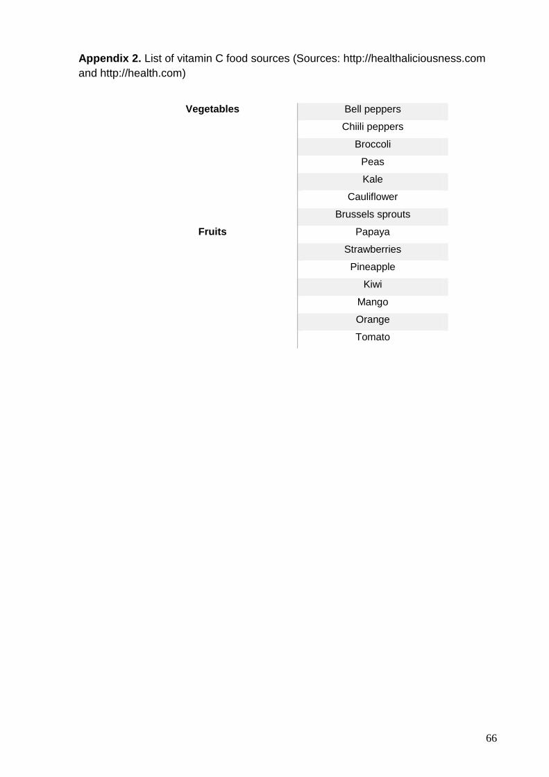
66
Appendix 2. List of vitamin C food sources (Sources: http://healthaliciousness.com
and http://health.com)
Vegetables Bell peppers
Chiili peppers
Broccoli
Peas
Kale
Cauliflower
Brussels sprouts
Fruits Papaya
Strawberries
Pineapple
Kiwi
Mango
Orange
Tomato

67
Appendix 3. List of tannin-rich food sources (Source: Rao & Prabhavathi, 1982;
Serrano, Puupponen-Pimia, Dauer, Aura & Saura-Calixto, 2009)
Vegetables Chickpea
Green gram
Red gram
Cowpea
Kidney bean
Lentils
Fruits Apple Mango
Apricot Medlar
Avocado Nectarine
Banana Peach
Blackberries Pear
Blueberries Persimmon
Cherries Pineapple
Cloudberries Plum
Cranberries Pomegranate
Currants Prune
Dates Quince
Grapes Raspberries
Lime Strawberries
Cereals Barley
Red sorghum
Condiments Coriander
Tamarind
Turmeric
Chili powder
Tea

68
Appendix 4. Animal identification form
Institution:
Animal ID:
Birth date: Sex:
Date collected: Time:
Food given: Yes/Time: No:
Comments:

69
Appendix 5. Iron and vitamin C content in the food offered at Park 1
Type of food
Iron content (mg/100g)
Total iron (mg)
Vit C content (mg/100g)
Total Vit C (mg)
Day 1 A.M. Red apple 0.1 0.65 6 39
Banana 0.4 2.3 8.7 50.025
Pear 0.1 1.15 4 46
P.M. Seed bread 1.67 1.67 0.2 0.2
Banana 0.4 1.84 8.7 40.02
Mixed salad (lettuce and carrot) 0.775 2.7125 9.4 32.9
Annona 0.6 3.96 16 105.6
Pink lady apple 0.1 0.37 6 22.2
Melon 0.2 2 42 420
Day 2 A.M. Red apple 0.1 0.65 6 39
Banana 0.4 2.3 8.7 50.025
Mango 0.2 2 28 280
P.M. Leek 1 6 12 72
Beaded Lettuce 0.9 1.8 9.2 18.4
Mixed salad (lettuce and carrot) 0.775 1.35625 9.4 16.45
Annona 0.6 2.64 16 70.4
Banana da Madeira 0.4 2.4 8.7 52.2
Rockmelon 0.2 1.25 42 262.5
Day 3 A.M. Red Apple 0.1 0.39 6 23.4
Banana 0.4 2.76 8.7 60.03
Melon 0.2 1 42 210
P.M. Raisin bread 1.67 1.67 0.2 0.2
Persimmons 2.5 30 66 792
Endives 0.8 3.6 6.5 29.25
Mango 0.2 1 28 140
Melon 0.2 1 42 210
Leek 1 8 12 96
Banana 0.4 1.84 8.7 40.02
Day 4 A.M. Red apple 0.1 0.65 6 39
Banana 0.4 0.92 8.7 20.01
Watermelon 0.2 1.25 10 62.5
P.M. Red apple 0.1 0.78 6 46.8
Melon 0.2 1 42 210
Brunches of grapes 0.3 2.4 11 88
Leek 1 8 12 96
Day 5 A.M. Melon 0.2 4 42 840
Red apple 0.1 0.65 6 39
Pineapple 0.3 1.95 15 97.5

70
P.M. Rockmelon 0.2 1.25 42 262.5
Strawberry 0.5 1.1 57 125.4
Pear 0.1 1.38 4 55.2
Melon 0.2 1 42 210
Red apple 0.1 0.52 6 31.2
Mango 0.2 3 28 420
Green salad 0.9 3.15 9.2 32.2
Day 6 A.M. Melon 0.2 1 42 210
Red apple 0.1 0.78 6 46.8
P.M. Persimmons 2.5 60 66 1584
Strawberry 0.5 3 57 342
Brunches of grapes 0.3 3 11 110
Banana 0.4 1.84 8.7 40.02
Green salad 0.9 1.575 9.2 16.1
Day 7 A.M. Red apple 0.1 1.17 6 70.2
Strawberry 0.5 2.4 57 273.6
Brunches of grapes 0.3 3 11 110
P.M. Strawberry 0.5 1 57 114
Pear 0.1 1.15 4 46
Melon 0.2 2 42 420
Red apple 0.1 0.52 6 31.2
Mango 0.2 3 28 420
Banana 0.4 0.92 8.7 20.01
Green salad 0.9 3.15 9.2 32.2
Day 8 A.M. Annona 0.6 1.32 16 35.2
Red apple 0.1 0.52 6 31.2
Pineapple 0.3 0.4875 15 24.375
Banana 0.4 1.84 8.7 40.02
Mango 0.2 1 28 140
P.M. Mango 0.2 4 28 560
Persimmons 2.5 37.5 66 990
Annona 0.6 2.64 16 70.4
Pineapple 0.3 1.95 15 97.5
Seed bread 1.67 1.67 0.2 0.2
Leek 1 10 12 120
Day 9 A.M. Red apple 0.1 0.65 6 39
Plum 0.2 1.56 10 78
Melon 0.2 3 42 630
P.M. Pear 0.1 0.69 4 27.6
Seed bread 1.67 1.67 0.2 0.2
Rockmelon 0.2 1.25 42 262.5

71
Mango 0.2 3 28 420
Persimmons 2.5 60 66 1584
Day 10 A.M. Red apple 0.1 0.52 6 31.2
Plum 0.2 1.44 10 72
Rockmelon 0.2 1.25 42 262.5
P.M. Carrot 0.4 3 10 75
Banana 0.4 2.3 8.7 50.025
Rockmelon 0.2 1.25 42 262.5
Seed bread 1.67 1.67 0.2 0.2
Plum 0.2 1.2 10 60
Day 11 A.M. Melon 0.2 2 42 420
Pineapple 0.3 1.95 15 97.5
Red apple 0.1 0.78 6 46.8
Plum 0.2 0.96 10 48
P.M. Pear 0.1 1.38 4 55.2
Strawberry 0.5 1.2 57 136.8
Red apple 0.1 0.65 6 39
Pineapple 0.3 1.95 15 97.5
Plum 0.2 1.2 10 60
Day 12 A.M. Mango 0.2 1 28 140
Red apple 0.1 0.91 6 54.6
Plum 0.2 1.2 10 60
P.M. Persimmons 2.5 45 66 1188
Red apple 0.1 0.52 6 31.2
Melon 0.2 2 42 420
Seed bread 1.67 0.835 0.2 0.1
Day 13 A.M. Banana 0.4 1.84 8.7 40.02
Melon 0.2 2 42 420
Red apple 0.1 0.52 6 31.2
Plum 0.2 0.24 10 12
P.M. Persimmons 2.5 37.5 66 990
Mango 0.2 3 28 420
Brunches of grapes 0.3 2.4 11 88
Red apple 0.1 0.52 6 31.2
Spinach 3.6 7.2 28 56
Day 14 A.M. Pear 0.1 1.15 4 46
Brunches of grapes 0.3 1.8 11 66
Melon 0.2 1 42 210
P.M. Mango 0.2 2 28 280
Banana 0.4 2.3 8.7 50.025
Plum 0.2 0.96 10 48

72
Red apple 0.1 0.52 6 31.2
Persimmons 2.5 15 66 396
Seed bread 1.67 1.67 0.2 0.2
Day 15 A.M. Banana 0.4 1.84 8.7 40.02
Red apple 0.1 0.52 6 31.2
Rockmelon 0.2 1.875 42 393.75
Pear 0.1 0.69 4 27.6
P.M. Leek and carrot soup 0.85 2.975 11.5 40.25
Mixed salad (lettuce and carrot) 0.775 1.35625 9.4 16.45
Seed bread 1.67 1.67 0.2 0.2
Banana 0.4 1.84 8.7 40.02

73
Appendix 6. Iron and vitamin C content in the food offered at Park 2
Type of food Iron content (mg/100g) Total iron (mg) Vit C content (mg/100g) Total Vit C (mg)
Day 1 Tomato 0.4 0.4 19 19
Spinach 3.6 32.4 28 252
Strawberry 0.5 0.2 57 22.8
Apple 0.1 0.13 6 7.8
Pear 0.1 0.23 4 9.2
Banana 0.4 0.69 8.7 15.0075
Rockmelon 0.2 0.625 42 131.25
Carrot 0.4 1.2 10 30
Day 2 Brunch of grapes 0.3 1.2 11 44
Pear 0.1 0.115 4 4.6
Mango 0.2 0.5 28 70
Banana 0.4 0.92 8.7 20.01
Leek 1 4 12 48
Carrot 0.4 0.3 10 7.5
Tomato 0.4 0.4 19 19
Pepper 0.43 0.4515 150 157.5
Apple 0.1 0.13 6 7.8
Seeds mix 8.8 8.8 1.7 1.7
Fruit purees 3 6 35 70
Day 3 Endive 0.8 1.2 6.5 9.75
Pear 0.1 0.23 4 9.2
Banana 0.4 0.92 8.7 20.01
Cucumber 0.2 0.35 4 7
Papaya 0.25 0.64375 62 159.65
Pepper 0.43 0.22575 130 68.25
Apple 0.1 0.13 6 7.8
Seeds mix 8.8 8.8 1.7 1.7
Cerelac pap 8.5 2.55 65 19.5
Day 4 Pepper 0.43 0.4515 150 157.5
Banana 0.4 0.69 8.7 15.0075
Apple 0.1 0.13 6 7.8
Pear 0.1 0.46 4 18.4
Carrot 0.4 1.2 10 30
Brunch of grapes 0.3 1.2 11 44
Tomato 0.4 0.4 19 19
Lettuce 0.9 3.87 9.2 39.56
Pine nuts 5.5 3.85 1.6 1.12
Day 5 Banana 0.4 0.92 8.7 20.01

74
Pear 0.1 0.23 4 9.2
Apple 0.1 0.13 6 7.8
Brunch of grapes 0.3 1.2 11 44
Rockmelon 0.2 1.25 42 262.5
Mango 0.2 1 28 140
Carrot 0.4 0.6 10 15
Dog dry food 8 8 20 20
Day 6 Rockmelon 0.2 0.625 42 131.25
Cucumber 0.4 0.7 4 7
Strawberry 0.5 0.3 57 34.2
Apple 0.1 0.13 6 7.8
Lettuce 0.9 3.87 9.2 39.56
Papaya 0.25 0.321875 62 79.825
Mango 0.2 0.33 28 46.2
Passionfruit 1.6 2.56 30 48
Wet cat food 0.4 0.4 9 9
Day 7 Tomato 0.4 0.4 19 19
Spinach 3.6 32.4 28 252
Strawberry 0.5 0.2 57 22.8
Apple 0.1 0.13 6 7.8
Pear 0.1 0.23 4 9.2
Banana 0.4 0.69 8.7 15.0075
Rockmelon 0.2 0.625 42 131.25
Carrot 0.4 1.2 10 30
Day 8 Brunch of grapes 0.3 1.2 11 44
Pear 0.1 0.115 4 4.6
Mango 0.2 0.5 28 70
Banana 0.4 0.92 8.7 20.01
Leek 1 4 12 48
Carrot 0.4 0.3 10 7.5
Tomato 0.4 0.4 19 19
Pepper 0.43 0.4515 150 157.5
Apple 0.1 0.13 6 7.8
Seeds mix 8.8 8.8 1.7 1.7
Fruit purees 3 6 35 70
Day 9 Endive 0.8 1.2 6.5 9.75
Pear 0.1 0.23 4 9.2
Banana 0.4 0.92 8.7 20.01
Cucumber 0.4 0.7 4 7
Papaya 0.25 0.64375 62 159.65
Pepper 0.43 0.22575 150 78.75

75
Apple 0.1 0.13 6 7.8
Seeds mix 8.8 8.8 1.7 1.7
Cerelac pap 8.5 2.55 65 19.5
Day 10 Pepper 0.43 0.4515 150 157.5
Banana 0.4 0.69 8.7 15.0075
Apple 0.1 0.13 6 7.8
Pear 0.1 0.46 4 18.4
Carrot 0.4 1.2 10 30
Brunch of grapes 0.3 1.2 11 44
Tomato 0.4 0.4 19 19
Lettuce 0.9 3.87 9.2 39.56
Pine nuts 5.5 3.85 1.6 1.12
Day 11 Banana 0.4 0.92 8.7 20.01
Pear 0.1 0.23 4 9.2
Apple 0.1 0.13 6 7.8
Brunch of grapes 0.3 1.2 11 44
Rockmelon 0.2 1.25 42 262.5
Mango 0.2 1 28 140
Carrot 0.4 0.6 10 15
Dog dry food 8 8 20 20
Day 12 Rockmelon 0.2 0.625 42 131.25
Cucumber 0.4 0.7 4 7
Strawberry 0.5 0.3 57 34.2
Apple 0.1 0.13 6 7.8
Lettuce 0.9 3.87 9.2 39.56
Papaya 0.25 0.321875 62 79.825
Mango 0.2 0.33 28 46.2
Passionfruit 1.6 2.56 30 48
Wet cat food 0.4 0.4 9 9
Day 13 Tomato 0.4 0.4 19 19
Spinach 3.6 32.4 28 252
Strawberry 0.5 0.2 57 22.8
Apple 0.1 0.13 6 7.8
Pear 0.1 0.23 4 9.2
Banana 0.4 0.69 8.7 15.0075
Rockmelon 0.2 0.625 42 131.25
Carrot 0.4 1.2 10 30
Day 14 Brunch of grapes 0.3 1.2 11 44
Pear 0.1 0.115 4 4.6
Mango 0.2 0.5 28 70
Banana 0.4 0.92 8.7 20.01
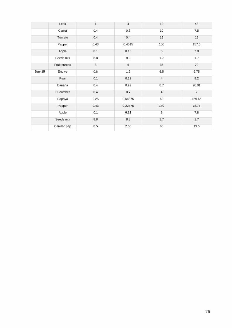
76
Leek 1 4 12 48
Carrot 0.4 0.3 10 7.5
Tomato 0.4 0.4 19 19
Pepper 0.43 0.4515 150 157.5
Apple 0.1 0.13 6 7.8
Seeds mix 8.8 8.8 1.7 1.7
Fruit purees 3 6 35 70
Day 15 Endive 0.8 1.2 6.5 9.75
Pear 0.1 0.23 4 9.2
Banana 0.4 0.92 8.7 20.01
Cucumber 0.4 0.7 4 7
Papaya 0.25 0.64375 62 159.65
Pepper 0.43 0.22575 150 78.75
Apple 0.1 0.13 6 7.8
Seeds mix 8.8 8.8 1.7 1.7
Cerelac pap 8.5 2.55 65 19.5

77
Appendix 7. Iron and vitamin C content in the food offered at Park 3
Type of food Iron content (mg/100g)
Total iron (mg) Vit C content (mg/100g)
Total Vit C (mg)
Day 1 A.M. Cucumber 0.4 1.92 4 19.2
New World pellet 10 32 290 928
P.M. Apple 0.1 0.58 6 34.8
Melon 0.2 1.4 42 294
Cucumber 0.4 2.56 4 25.6
Boiled egg 1.2 2.4 0.1 0.2
Day 2 A.M. Lettuce 0.9 4.32 9.2 44.16
New World pellet 10 32 290 928
P.M. Grapes 0.3 1.275 11 46.75
Persimmons 2.5 21.375 66 564.3
Spinach 3.6 7.2 28 56
Carrots 0.4 1.76 10 44
Seeds mix 8.8 14.08 1.7 2.72
Day 3 A.M. Lettuce 0.9 4.32 9.2 44.16
New World pellet 10 32 290 928
P.M. Banana 0.4 1.6 8.7 34.8
Papaya 0.25 1.7 62 421.6
Grapes 0.3 0.6 11 22
Sweet potato 0.6 3.84 2.4 15.36
Bread 1.7 1.36 0.1 0.08
Day 4 A.M. Lettuce 0.9 4.32 9.2 44.16
New World pellet 10 32 290 928
P.M. Mango 0.2 1.06 28 148.4
Papaya 0.25 1.875 62 465
Tomato 0.4 2.56 19 121.6
Dog dry food 8 16 20 40
Day 5 A.M. Lettuce 0.9 4.32 9.2 44.16
New World pellet 10 32 290 928
P.M. Grapes 0.3 1.2 11 44
Apple 0.1 0.88 6 52.8
Broccoli 0.7 4.48 89.2 570.88
Natural yogurt 0.1 0.48 0.9 4.32
Day 6 A.M. Potato 0.8 2.24 19.7 55.16
Broccoli 0.7 1.4 89.2 178.4
New World pellet 10 32 290 928
P.M. Banana 0.4 3.44 8.7 74.82
Kiwi 0.3 1.26 98 411.6
Lettuce 0.9 5.76 9.2 58.88
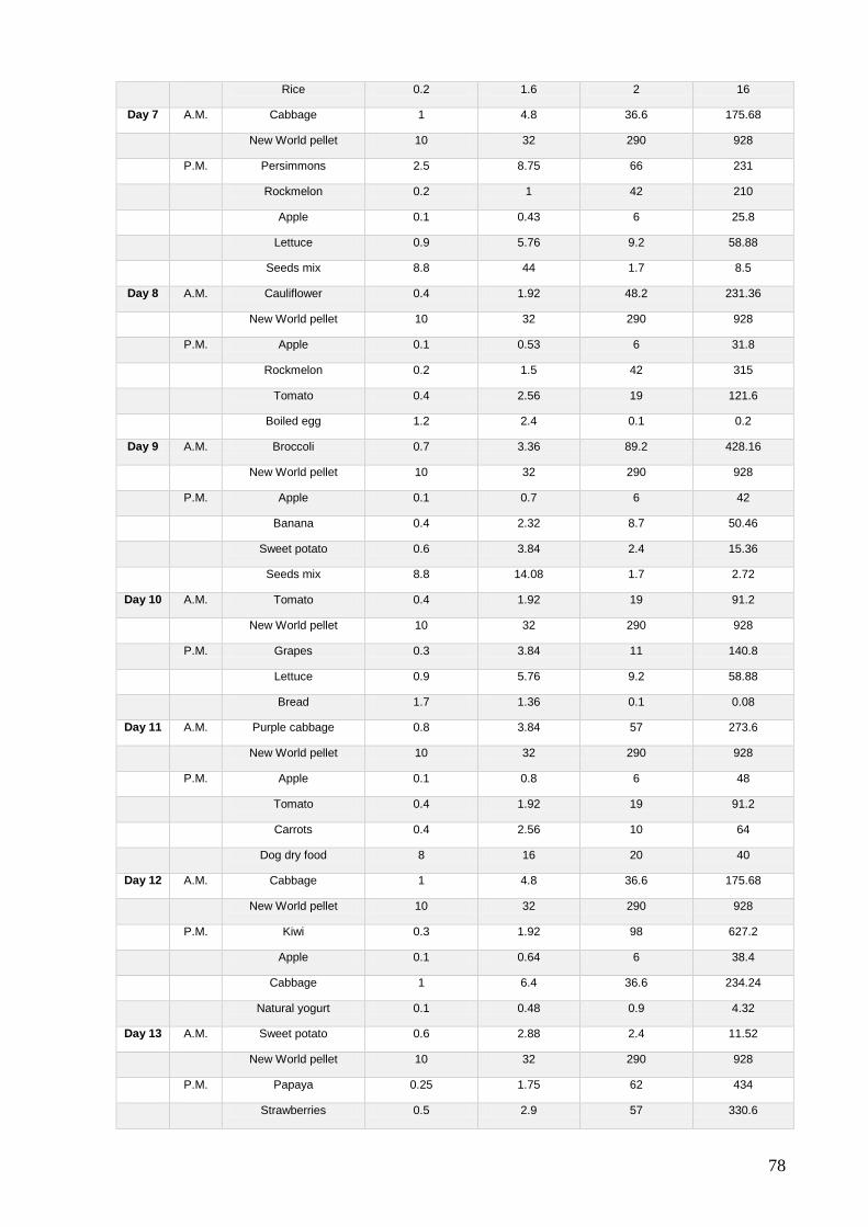
78
Rice 0.2 1.6 2 16
Day 7 A.M. Cabbage 1 4.8 36.6 175.68
New World pellet 10 32 290 928
P.M. Persimmons 2.5 8.75 66 231
Rockmelon 0.2 1 42 210
Apple 0.1 0.43 6 25.8
Lettuce 0.9 5.76 9.2 58.88
Seeds mix 8.8 44 1.7 8.5
Day 8 A.M. Cauliflower 0.4 1.92 48.2 231.36
New World pellet 10 32 290 928
P.M. Apple 0.1 0.53 6 31.8
Rockmelon 0.2 1.5 42 315
Tomato 0.4 2.56 19 121.6
Boiled egg 1.2 2.4 0.1 0.2
Day 9 A.M. Broccoli 0.7 3.36 89.2 428.16
New World pellet 10 32 290 928
P.M. Apple 0.1 0.7 6 42
Banana 0.4 2.32 8.7 50.46
Sweet potato 0.6 3.84 2.4 15.36
Seeds mix 8.8 14.08 1.7 2.72
Day 10 A.M. Tomato 0.4 1.92 19 91.2
New World pellet 10 32 290 928
P.M. Grapes 0.3 3.84 11 140.8
Lettuce 0.9 5.76 9.2 58.88
Bread 1.7 1.36 0.1 0.08
Day 11 A.M. Purple cabbage 0.8 3.84 57 273.6
New World pellet 10 32 290 928
P.M. Apple 0.1 0.8 6 48
Tomato 0.4 1.92 19 91.2
Carrots 0.4 2.56 10 64
Dog dry food 8 16 20 40
Day 12 A.M. Cabbage 1 4.8 36.6 175.68
New World pellet 10 32 290 928
P.M. Kiwi 0.3 1.92 98 627.2
Apple 0.1 0.64 6 38.4
Cabbage 1 6.4 36.6 234.24
Natural yogurt 0.1 0.48 0.9 4.32
Day 13 A.M. Sweet potato 0.6 2.88 2.4 11.52
New World pellet 10 32 290 928
P.M. Papaya 0.25 1.75 62 434
Strawberries 0.5 2.9 57 330.6

79
Carrots 0.4 1.6 10 40
Broccoli 0.7 1.68 89.2 214.08
Spaghetti 1.3 10.4 2 16
Day 14 A.M. Sweet potato 0.6 2.88 2.4 11.52
New World pellet 10 32 290 928
P.M. Apple 0.1 1.28 6 76.8
Lettuce 0.9 5.76 9.2 58.88
Seeds mix 8.8 44 1.7 8.5
Day 15 A.M. Cucumber 0.4 1.92 4 19.2
New World pellet 10 32 290 928
P.M. Grapes 0.3 2.34 11 85.8
Tomato 0.4 2 19 95
Lettuce 0.9 5.76 9.2 58.88
Boiled egg 1.2 2.4 0.1 0.2