Invasive and indigenous microbiota impact intestinal stem cell activity through multiple pathways in...
26
10.1101/gad.1827009 Access the most recent version at doi: 2009 23: 2333-2344 Genes Dev. Nicolas Buchon, Nichole A. Broderick, Sveta Chakrabarti, et al. Drosophila activity through multiple pathways in Invasive and indigenous microbiota impact intestinal stem cell Material Supplemental http://genesdev.cshlp.org/content/suppl/2009/10/02/23.19.2333.DC1.html References http://genesdev.cshlp.org/content/23/19/2333.full.html#related-urls Article cited in: http://genesdev.cshlp.org/content/23/19/2333.full.html#ref-list-1 This article cites 39 articles, 8 of which can be accessed free at: Related Content Genes Dev. October 1, 2009 23: 2260-2265 Won-Jae Lee homeostasis Bacterial-modulated host immunity and stem cell activation for gut service Email alerting click here top right corner of the article or Receive free email alerts when new articles cite this article - sign up in the box at the http://genesdev.cshlp.org/subscriptions go to: Genes & Development To subscribe to Copyright © 2009 by Cold Spring Harbor Laboratory Press Cold Spring Harbor Laboratory Press on October 6, 2009 - Published by genesdev.cshlp.org Downloaded from
-
Upload
johnshopkins -
Category
Documents
-
view
3 -
download
0
Transcript of Invasive and indigenous microbiota impact intestinal stem cell activity through multiple pathways in...
10.1101/gad.1827009Access the most recent version at doi: 2009 23: 2333-2344Genes Dev.
Nicolas Buchon, Nichole A. Broderick, Sveta Chakrabarti, et al.
Drosophilaactivity through multiple pathways in Invasive and indigenous microbiota impact intestinal stem cell
MaterialSupplemental http://genesdev.cshlp.org/content/suppl/2009/10/02/23.19.2333.DC1.html
References
http://genesdev.cshlp.org/content/23/19/2333.full.html#related-urlsArticle cited in:
http://genesdev.cshlp.org/content/23/19/2333.full.html#ref-list-1This article cites 39 articles, 8 of which can be accessed free at:
Related Content
Genes Dev. October 1, 2009 23: 2260-2265
Won-Jae LeehomeostasisBacterial-modulated host immunity and stem cell activation for gut
serviceEmail alerting
click heretop right corner of the article orReceive free email alerts when new articles cite this article - sign up in the box at the
http://genesdev.cshlp.org/subscriptions go to: Genes & DevelopmentTo subscribe to
Copyright © 2009 by Cold Spring Harbor Laboratory Press
Cold Spring Harbor Laboratory Press on October 6, 2009 - Published by genesdev.cshlp.orgDownloaded from
Invasive and indigenous microbiotaimpact intestinal stem cell activitythrough multiple pathways in Drosophila
Nicolas Buchon,2 Nichole A. Broderick, Sveta Chakrabarti, and Bruno Lemaitre1
Global Health Institute, Ecole Polytechnique Federale de Lausanne (EPFL), CH-1015 Lausanne, Switzerland
Gut homeostasis is controlled by both immune and developmental mechanisms, and its disruption can lead toinflammatory disorders or cancerous lesions of the intestine. While the impact of bacteria on the mucosal immunesystem is beginning to be precisely understood, little is known about the effects of bacteria on gut epitheliumrenewal. Here, we addressed how both infectious and indigenous bacteria modulate stem cell activity inDrosophila. We show that the increased epithelium renewal observed upon some bacterial infections isa consequence of the oxidative burst, a major defense of the Drosophila gut. Additionally, we provide evidencethat the JAK–STAT (Janus kinase–signal transducers and activators of transcription) and JNK (c-Jun NH2 terminalkinase) pathways are both required for bacteria-induced stem cell proliferation. Similarly, we demonstrate thatindigenous gut microbiota activate the same, albeit reduced, program at basal levels. Altered control of gutmicrobiota in immune-deficient or aged flies correlates with increased epithelium renewal. Finally, we show thatepithelium renewal is an essential component of Drosophila defense against oral bacterial infection. Altogether,these results indicate that gut homeostasis is achieved by a complex interregulation of the immune response, gutmicrobiota, and stem cell activity.
[Keywords: Intestinal stem cell; proliferation; gut microbiota; JAK–STAT; JNK; pathogenic bacteria]
Supplemental material is available at http://www.genesdev.org.
Received June 2, 2009; revised version accepted August 25, 2009.
The gut of all organisms is in constant exposure to en-vironmental and microbial aggressions (Sansonetti 2004).It is an active barrier, limiting invasion by pathogenicmicroorganisms and promoting tolerance to indigenousmicrobiota (Pedron and Sansonetti 2008). To maintainhomeostasis, the gut epithelium is constantly renewedthroughout an organism’s life by the division and differ-entiation of intestinal stem cells (ISCs). In Drosophila,ISCs are scattered along the basement membrane of themidgut (Micchelli and Perrimon 2006; Ohlstein andSpradling 2006). The division of an ISC generates botha new ISC and a post-mitotic enteroblast, which differ-entiates into either an absorptive enterocyte or a secre-tory enteroendocrine cell. In basal conditions, the adultgut epithelium is renewed in ;1–2 wk (Micchelli andPerrimon 2006).
We recently reported a global analysis of the changes ingene expression that occur in the Drosophila gut inresponse to infection by a Gram-negative bacterium,Erwinia carotovora carotovora 15 (Ecc15) (Buchon et al.2009). This study demonstrated that bacterial infec-
tion triggers expression of a combination of immune,stress, and developmental signaling pathways. Most im-mune genes—notably, antibacterial peptide genes (e.g.,Diptericin)—are induced in the gut by the Imd pathway.This pathway is activated upon recognition of peptido-glycan of Gram-negative bacteria by the transmembranereceptor PGRP-LC, and leads to activation of the NF-kBprotein Relish (Zaidman-Remy et al. 2006). Subsequentanalysis showed that the JAK–STAT (Janus kinase–signaltransducers and activators of transcription) signalingpathway was activated by damage in the gut of infectedflies and regulates the production of a subset of antimi-crobial peptides. Surprisingly, it was further observed thatingestion of Ecc15 strongly stimulates ISC division, pro-moting a rapid turnover of the gut epithelium (Buchonet al. 2009). These results highlighted an unexpected linkbetween oral bacterial infection and epithelial renewal.These data also suggested that, following infection, guthomeostasis is maintained by the balance between celldamage induced by infection and epithelial repair by ISCactivation. However, the precise causes of ISC prolifera-tion, as well as the signaling events regulating thisinduction, were unclear, as was the effect of indigenousor lethal bacteria on this mechanism.
Here we use the genetically tractable model Drosophilamelanogaster to study the link between gut bacteria, the
Corresponding authors.1E-MAIL [email protected]; FAX 41-21-693-17-90.2E-MAIL [email protected]; FAX 41-21-693-17-90.Article is online at http://www.genesdev.org/cgi/doi/10.1101/gad.1827009.
GENES & DEVELOPMENT 23:2333–2344 � 2009 by Cold Spring Harbor Laboratory Press ISSN 0890-9369/09; www.genesdev.org 2333
Cold Spring Harbor Laboratory Press on October 6, 2009 - Published by genesdev.cshlp.orgDownloaded from
gut immune response, and epithelium renewal. We showthat the oxidative burst, induced by the host consecutiveto Ecc15 infection, is a major inducer of ISC activation.Infection with this bacterium activates both JAK–STATand JNK (c-Jun NH2 terminal kinase) pathways in ISCs topromote proliferation. Indigenous bacteria induce thesame program and lead to a basal level of proliferation.Inversely, Pseudomonas entomophila, a lethal entomo-pathogenic bacterium, interferes with ISC activation,thereby disrupting gut homeostasis.
Results
Infection-induced oxidative burst is a major inducerof epithelium renewal
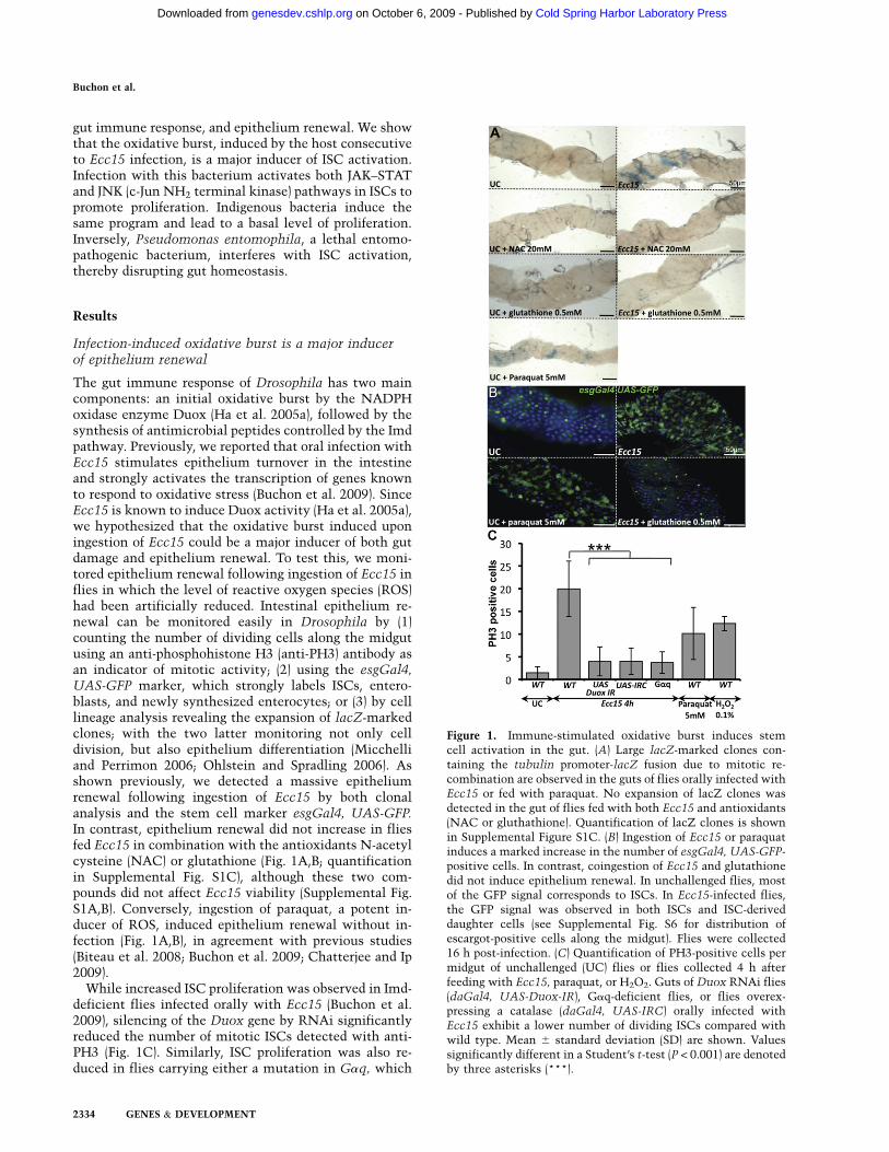
The gut immune response of Drosophila has two maincomponents: an initial oxidative burst by the NADPHoxidase enzyme Duox (Ha et al. 2005a), followed by thesynthesis of antimicrobial peptides controlled by the Imdpathway. Previously, we reported that oral infection withEcc15 stimulates epithelium turnover in the intestineand strongly activates the transcription of genes knownto respond to oxidative stress (Buchon et al. 2009). SinceEcc15 is known to induce Duox activity (Ha et al. 2005a),we hypothesized that the oxidative burst induced uponingestion of Ecc15 could be a major inducer of both gutdamage and epithelium renewal. To test this, we moni-tored epithelium renewal following ingestion of Ecc15 inflies in which the level of reactive oxygen species (ROS)had been artificially reduced. Intestinal epithelium re-newal can be monitored easily in Drosophila by (1)counting the number of dividing cells along the midgutusing an anti-phosphohistone H3 (anti-PH3) antibody asan indicator of mitotic activity; (2) using the esgGal4,UAS-GFP marker, which strongly labels ISCs, entero-blasts, and newly synthesized enterocytes; or (3) by celllineage analysis revealing the expansion of lacZ-markedclones; with the two latter monitoring not only celldivision, but also epithelium differentiation (Micchelliand Perrimon 2006; Ohlstein and Spradling 2006). Asshown previously, we detected a massive epitheliumrenewal following ingestion of Ecc15 by both clonalanalysis and the stem cell marker esgGal4, UAS-GFP.In contrast, epithelium renewal did not increase in fliesfed Ecc15 in combination with the antioxidants N-acetylcysteine (NAC) or glutathione (Fig. 1A,B; quantificationin Supplemental Fig. S1C), although these two com-pounds did not affect Ecc15 viability (Supplemental Fig.S1A,B). Conversely, ingestion of paraquat, a potent in-ducer of ROS, induced epithelium renewal without in-fection (Fig. 1A,B), in agreement with previous studies(Biteau et al. 2008; Buchon et al. 2009; Chatterjee and Ip2009).
While increased ISC proliferation was observed in Imd-deficient flies infected orally with Ecc15 (Buchon et al.2009), silencing of the Duox gene by RNAi significantlyreduced the number of mitotic ISCs detected with anti-PH3 (Fig. 1C). Similarly, ISC proliferation was also re-duced in flies carrying either a mutation in Gaq, which
Figure 1. Immune-stimulated oxidative burst induces stemcell activation in the gut. (A) Large lacZ-marked clones con-taining the tubulin promoter-lacZ fusion due to mitotic re-combination are observed in the guts of flies orally infected withEcc15 or fed with paraquat. No expansion of lacZ clones wasdetected in the gut of flies fed with both Ecc15 and antioxidants(NAC or gluthathione). Quantification of lacZ clones is shownin Supplemental Figure S1C. (B) Ingestion of Ecc15 or paraquatinduces a marked increase in the number of esgGal4, UAS-GFP-positive cells. In contrast, coingestion of Ecc15 and glutathionedid not induce epithelium renewal. In unchallenged flies, mostof the GFP signal corresponds to ISCs. In Ecc15-infected flies,the GFP signal was observed in both ISCs and ISC-deriveddaughter cells (see Supplemental Fig. S6 for distribution ofescargot-positive cells along the midgut). Flies were collected16 h post-infection. (C) Quantification of PH3-positive cells permidgut of unchallenged (UC) flies or flies collected 4 h afterfeeding with Ecc15, paraquat, or H2O2. Guts of Duox RNAi flies(daGal4, UAS-Duox-IR), Gaq-deficient flies, or flies overex-pressing a catalase (daGal4, UAS-IRC) orally infected withEcc15 exhibit a lower number of dividing ISCs compared withwild type. Mean 6 standard deviation (SD) are shown. Valuessignificantly different in a Student’s t-test (P < 0.001) are denotedby three asterisks (***).
Buchon et al.
2334 GENES & DEVELOPMENT
Cold Spring Harbor Laboratory Press on October 6, 2009 - Published by genesdev.cshlp.orgDownloaded from
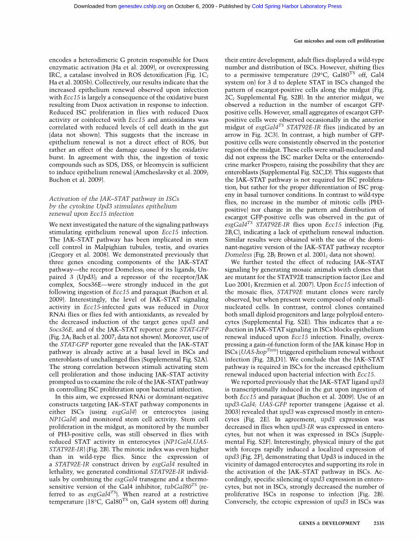
encodes a heterodimeric G protein responsible for Duoxenzymatic activation (Ha et al. 2009), or overexpressingIRC, a catalase involved in ROS detoxification (Fig. 1C;Ha et al. 2005b). Collectively, our results indicate that theincreased epithelium renewal observed upon infectionwith Ecc15 is largely a consequence of the oxidative burstresulting from Duox activation in response to infection.Reduced ISC proliferation in flies with reduced Duoxactivity or coinfected with Ecc15 and antioxidants wascorrelated with reduced levels of cell death in the gut(data not shown). This suggests that the increase inepithelium renewal is not a direct effect of ROS, butrather an effect of the damage caused by the oxidativeburst. In agreement with this, the ingestion of toxiccompounds such as SDS, DSS, or bleomycin is sufficientto induce epithelium renewal (Amcheslavsky et al. 2009;Buchon et al. 2009).
Activation of the JAK–STAT pathway in ISCsby the cytokine Upd3 stimulates epitheliumrenewal upon Ecc15 infection
We next investigated the nature of the signaling pathwaysstimulating epithelium renewal upon Ecc15 infection.The JAK–STAT pathway has been implicated in stemcell control in Malpighian tubules, testis, and ovaries(Gregory et al. 2008). We demonstrated previously thatthree genes encoding components of the JAK–STATpathway—the receptor Domeless; one of its ligands, Un-paired 3 (Upd3); and a repressor of the receptor/JAKcomplex, Socs36E—were strongly induced in the gutfollowing ingestion of Ecc15 and paraquat (Buchon et al.2009). Interestingly, the level of JAK–STAT signalingactivity in Ecc15-infected guts was reduced in DuoxRNAi flies or flies fed with antioxidants, as revealed bythe decreased induction of the target genes upd3 andSocs36E, and of the JAK–STAT reporter gene STAT-GFP(Fig. 2A; Bach et al. 2007; data not shown). Moreover, use ofthe STAT-GFP reporter gene revealed that the JAK–STATpathway is already active at a basal level in ISCs andenteroblasts of unchallenged flies (Supplemental Fig. S2A).The strong correlation between stimuli activating stemcell proliferation and those inducing JAK–STAT activityprompted us to examine the role of the JAK–STAT pathwayin controlling ISC proliferation upon bacterial infection.
In this aim, we expressed RNAi or dominant-negativeconstructs targeting JAK–STAT pathway components ineither ISCs (using esgGal4) or enterocytes (usingNP1Gal4) and monitored stem cell activity. Stem cellproliferation in the midgut, as monitored by the numberof PH3-positive cells, was still observed in flies withreduced STAT activity in enterocytes (NP1Gal4,UAS-STAT92E-IR) (Fig. 2B). The mitotic index was even higherthan in wild-type flies. Since the expression ofa STAT92E-IR construct driven by esgGal4 resulted inlethality, we generated conditional STAT92E-IR individ-uals by combining the esgGal4 transgene and a thermo-sensitive version of the Gal4 inhibitor, tubGal80TS (re-ferred to as esgGal4TS). When reared at a restrictivetemperature (18°C, Gal80TS on, Gal4 system off) during
their entire development, adult flies displayed a wild-typenumber and distribution of ISCs. However, shifting fliesto a permissive temperature (29°C, Gal80TS off, Gal4system on) for 3 d to deplete STAT in ISCs changed thepattern of escargot-positive cells along the midgut (Fig.2C; Supplemental Fig. S2B). In the anterior midgut, weobserved a reduction in the number of escargot GFP-positive cells. However, small aggregates of escargot GFP-positive cells were observed occasionally in the anteriormidgut of esgGal4TS STAT92E-IR flies (indicated by anarrow in Fig. 2C3). In contrast, a high number of GFP-positive cells were consistently observed in the posteriorregion of the midgut. These cells were small-nucleated anddid not express the ISC marker Delta or the enteroendo-crine marker Prospero, raising the possibility that they areenteroblasts (Supplemental Fig. S2C,D). This suggests thatthe JAK–STAT pathway is not required for ISC prolifera-tion, but rather for the proper differentiation of ISC prog-eny in basal turnover conditions. In contrast to wild-typeflies, no increase in the number of mitotic cells (PH3-positive) nor change in the pattern and distribution ofescargot GFP-positive cells was observed in the gut ofesgGal4TS STAT92E-IR flies upon Ecc15 infection (Fig.2B,C), indicating a lack of epithelium renewal induction.Similar results were obtained with the use of the domi-nant-negative version of the JAK–STAT pathway receptorDomeless (Fig. 2B; Brown et al. 2001; data not shown).
We further tested the effect of reducing JAK–STATsignaling by generating mosaic animals with clones thatare mutant for the STAT92E transcription factor (Lee andLuo 2001; Krezmien et al. 2007). Upon Ecc15 infection ofthe mosaic flies, STAT92E mutant clones were rarelyobserved, but when present were composed of only small-nucleated cells. In contrast, control clones containedboth small diploid progenitors and large polyploid entero-cytes (Supplemental Fig. S2E). This indicates that a re-duction in JAK–STAT signaling in ISCs blocks epitheliumrenewal induced upon Ecc15 infection. Finally, overex-pressing a gain-of-function form of the JAK kinase Hop inISCs (UAS-hopTum) triggered epithelium renewal withoutinfection (Fig. 2B,D1). We conclude that the JAK–STATpathway is required in ISCs for the increased epitheliumrenewal induced upon bacterial infection with Ecc15.
We reported previously that the JAK–STAT ligand upd3is transcriptionally induced in the gut upon ingestion ofboth Ecc15 and paraquat (Buchon et al. 2009). Use of anupd3-Gal4; UAS-GFP reporter transgene (Agaisse et al.2003) revealed that upd3 was expressed mostly in entero-cytes (Fig. 2E). In agreement, upd3 expression wasdecreased in flies when upd3-IR was expressed in entero-cytes, but not when it was expressed in ISCs (Supple-mental Fig. S2F). Interestingly, physical injury of the gutwith forceps rapidly induced a localized expression ofupd3 (Fig. 2F), demonstrating that Upd3 is induced in thevicinity of damaged enterocytes and supporting its role inthe activation of the JAK–STAT pathway in ISCs. Ac-cordingly, specific silencing of upd3 expression in entero-cytes, but not in ISCs, strongly decreased the number ofproliferative ISCs in response to infection (Fig. 2B).Conversely, the ectopic expression of upd3 in ISCs was
Gut microbes and stem cell proliferation
GENES & DEVELOPMENT 2335
Cold Spring Harbor Laboratory Press on October 6, 2009 - Published by genesdev.cshlp.orgDownloaded from
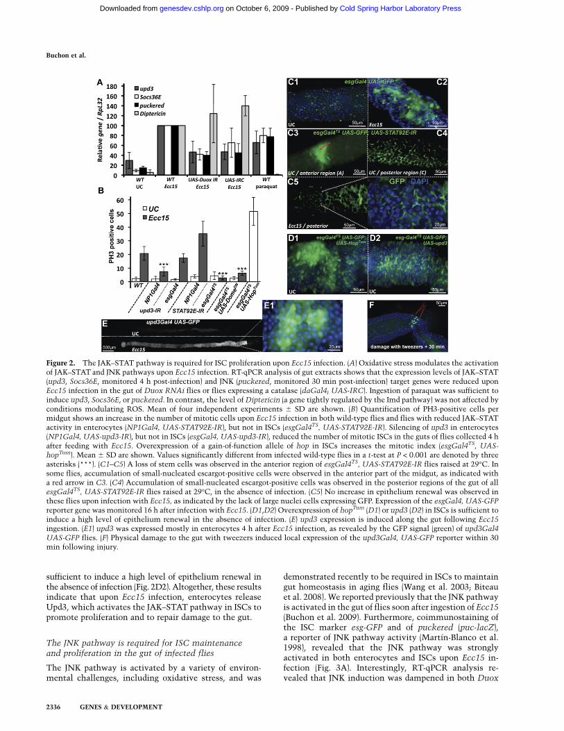
sufficient to induce a high level of epithelium renewal inthe absence of infection (Fig. 2D2). Altogether, these resultsindicate that upon Ecc15 infection, enterocytes releaseUpd3, which activates the JAK–STAT pathway in ISCs topromote proliferation and to repair damage to the gut.
The JNK pathway is required for ISC maintenanceand proliferation in the gut of infected flies
The JNK pathway is activated by a variety of environ-mental challenges, including oxidative stress, and was
demonstrated recently to be required in ISCs to maintaingut homeostasis in aging flies (Wang et al. 2003; Biteauet al. 2008). We reported previously that the JNK pathwayis activated in the gut of flies soon after ingestion of Ecc15(Buchon et al. 2009). Furthermore, coimmunostaining ofthe ISC marker esg-GFP and of puckered (puc-lacZ),a reporter of JNK pathway activity (Martın-Blanco et al.1998), revealed that the JNK pathway was stronglyactivated in both enterocytes and ISCs upon Ecc15 in-fection (Fig. 3A). Interestingly, RT-qPCR analysis re-vealed that JNK induction was dampened in both Duox
Figure 2. The JAK–STAT pathway is required for ISC proliferation upon Ecc15 infection. (A) Oxidative stress modulates the activationof JAK–STAT and JNK pathways upon Ecc15 infection. RT-qPCR analysis of gut extracts shows that the expression levels of JAK–STAT(upd3, Socs36E, monitored 4 h post-infection) and JNK (puckered, monitored 30 min post-infection) target genes were reduced uponEcc15 infection in the gut of Duox RNAi flies or flies expressing a catalase (daGal4; UAS-IRC). Ingestion of paraquat was sufficient toinduce upd3, Socs36E, or puckered. In contrast, the level of Diptericin (a gene tightly regulated by the Imd pathway) was not affected byconditions modulating ROS. Mean of four independent experiments 6 SD are shown. (B) Quantification of PH3-positive cells permidgut shows an increase in the number of mitotic cells upon Ecc15 infection in both wild-type flies and flies with reduced JAK–STATactivity in enterocytes (NP1Gal4, UAS-STAT92E-IR), but not in ISCs (esgGal4TS, UAS-STAT92E-IR). Silencing of upd3 in enterocytes(NP1Gal4, UAS-upd3-IR), but not in ISCs (esgGal4, UAS-upd3-IR), reduced the number of mitotic ISCs in the guts of flies collected 4 hafter feeding with Ecc15. Overexpression of a gain-of-function allele of hop in ISCs increases the mitotic index (esgGal4TS, UAS-hopTum). Mean 6 SD are shown. Values significantly different from infected wild-type flies in a t-test at P < 0.001 are denoted by threeasterisks (***). (C1–C5) A loss of stem cells was observed in the anterior region of esgGal4TS, UAS-STAT92E-IR flies raised at 29°C. Insome flies, accumulation of small-nucleated escargot-positive cells were observed in the anterior part of the midgut, as indicated witha red arrow in C3. (C4) Accumulation of small-nucleated escargot-positive cells was observed in the posterior regions of the gut of allesgGal4TS, UAS-STAT92E-IR flies raised at 29°C, in the absence of infection. (C5) No increase in epithelium renewal was observed inthese flies upon infection with Ecc15, as indicated by the lack of large nuclei cells expressing GFP. Expression of the esgGal4, UAS-GFP
reporter gene was monitored 16 h after infection with Ecc15. (D1,D2) Overexpression of hopTum (D1) or upd3 (D2) in ISCs is sufficient toinduce a high level of epithelium renewal in the absence of infection. (E) upd3 expression is induced along the gut following Ecc15
ingestion. (E1) upd3 was expressed mostly in enterocytes 4 h after Ecc15 infection, as revealed by the GFP signal (green) of upd3Gal4
UAS-GFP flies. (F) Physical damage to the gut with tweezers induced local expression of the upd3Gal4, UAS-GFP reporter within 30min following injury.
Buchon et al.
2336 GENES & DEVELOPMENT
Cold Spring Harbor Laboratory Press on October 6, 2009 - Published by genesdev.cshlp.orgDownloaded from
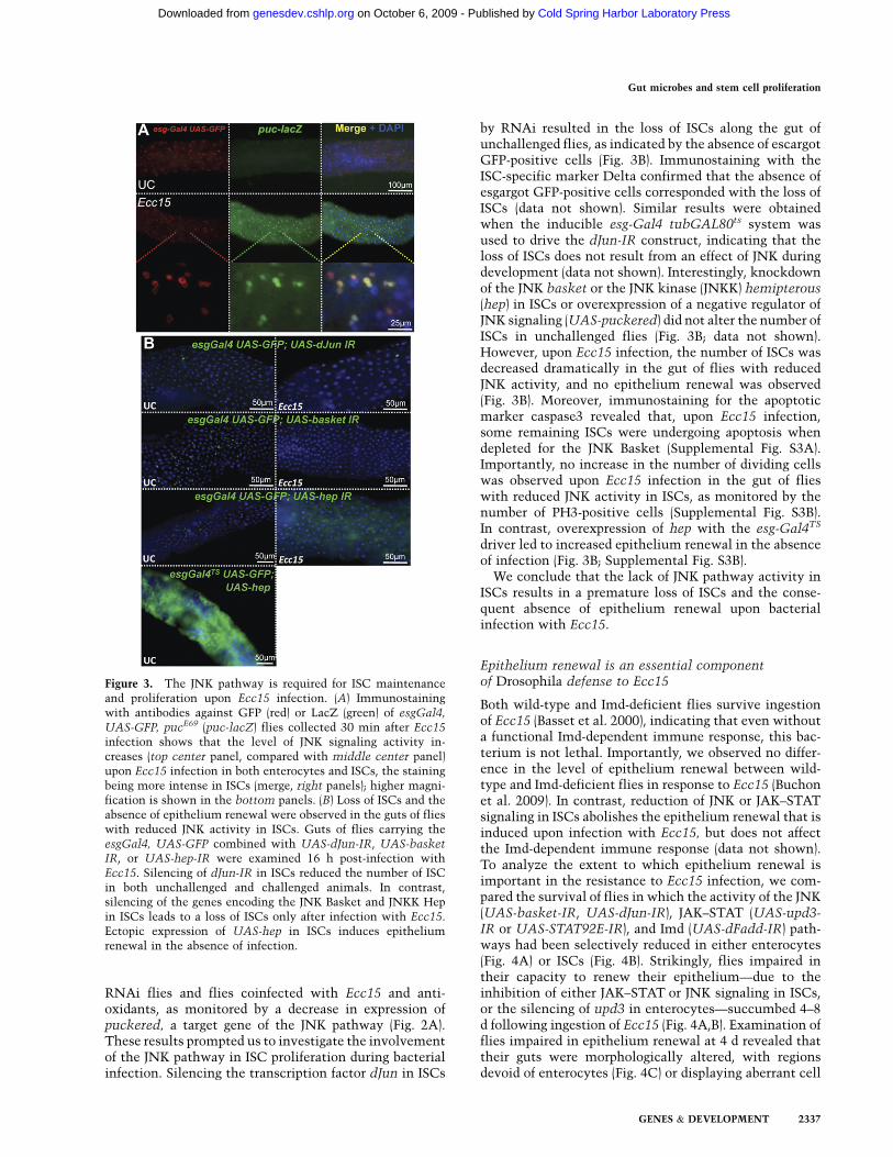
RNAi flies and flies coinfected with Ecc15 and anti-oxidants, as monitored by a decrease in expression ofpuckered, a target gene of the JNK pathway (Fig. 2A).These results prompted us to investigate the involvementof the JNK pathway in ISC proliferation during bacterialinfection. Silencing the transcription factor dJun in ISCs
by RNAi resulted in the loss of ISCs along the gut ofunchallenged flies, as indicated by the absence of escargotGFP-positive cells (Fig. 3B). Immunostaining with theISC-specific marker Delta confirmed that the absence ofesgargot GFP-positive cells corresponded with the loss ofISCs (data not shown). Similar results were obtainedwhen the inducible esg-Gal4 tubGAL80ts system wasused to drive the dJun-IR construct, indicating that theloss of ISCs does not result from an effect of JNK duringdevelopment (data not shown). Interestingly, knockdownof the JNK basket or the JNK kinase (JNKK) hemipterous(hep) in ISCs or overexpression of a negative regulator ofJNK signaling (UAS-puckered) did not alter the number ofISCs in unchallenged flies (Fig. 3B; data not shown).However, upon Ecc15 infection, the number of ISCs wasdecreased dramatically in the gut of flies with reducedJNK activity, and no epithelium renewal was observed(Fig. 3B). Moreover, immunostaining for the apoptoticmarker caspase3 revealed that, upon Ecc15 infection,some remaining ISCs were undergoing apoptosis whendepleted for the JNK Basket (Supplemental Fig. S3A).Importantly, no increase in the number of dividing cellswas observed upon Ecc15 infection in the gut of flieswith reduced JNK activity in ISCs, as monitored by thenumber of PH3-positive cells (Supplemental Fig. S3B).In contrast, overexpression of hep with the esg-Gal4TS
driver led to increased epithelium renewal in the absenceof infection (Fig. 3B; Supplemental Fig. S3B).
We conclude that the lack of JNK pathway activity inISCs results in a premature loss of ISCs and the conse-quent absence of epithelium renewal upon bacterialinfection with Ecc15.
Epithelium renewal is an essential componentof Drosophila defense to Ecc15
Both wild-type and Imd-deficient flies survive ingestionof Ecc15 (Basset et al. 2000), indicating that even withouta functional Imd-dependent immune response, this bac-terium is not lethal. Importantly, we observed no differ-ence in the level of epithelium renewal between wild-type and Imd-deficient flies in response to Ecc15 (Buchonet al. 2009). In contrast, reduction of JNK or JAK–STATsignaling in ISCs abolishes the epithelium renewal that isinduced upon infection with Ecc15, but does not affectthe Imd-dependent immune response (data not shown).To analyze the extent to which epithelium renewal isimportant in the resistance to Ecc15 infection, we com-pared the survival of flies in which the activity of the JNK(UAS-basket-IR, UAS-dJun-IR), JAK–STAT (UAS-upd3-IR or UAS-STAT92E-IR), and Imd (UAS-dFadd-IR) path-ways had been selectively reduced in either enterocytes(Fig. 4A) or ISCs (Fig. 4B). Strikingly, flies impaired intheir capacity to renew their epithelium—due to theinhibition of either JAK–STAT or JNK signaling in ISCs,or the silencing of upd3 in enterocytes—succumbed 4–8d following ingestion of Ecc15 (Fig. 4A,B). Examination offlies impaired in epithelium renewal at 4 d revealed thattheir guts were morphologically altered, with regionsdevoid of enterocytes (Fig. 4C) or displaying aberrant cell
Figure 3. The JNK pathway is required for ISC maintenanceand proliferation upon Ecc15 infection. (A) Immunostainingwith antibodies against GFP (red) or LacZ (green) of esgGal4,
UAS-GFP, pucE69 (puc-lacZ) flies collected 30 min after Ecc15infection shows that the level of JNK signaling activity in-creases (top center panel, compared with middle center panel)upon Ecc15 infection in both enterocytes and ISCs, the stainingbeing more intense in ISCs (merge, right panels); higher magni-fication is shown in the bottom panels. (B) Loss of ISCs and theabsence of epithelium renewal were observed in the guts of flieswith reduced JNK activity in ISCs. Guts of flies carrying theesgGal4, UAS-GFP combined with UAS-dJun-IR, UAS-basketIR, or UAS-hep-IR were examined 16 h post-infection withEcc15. Silencing of dJun-IR in ISCs reduced the number of ISCin both unchallenged and challenged animals. In contrast,silencing of the genes encoding the JNK Basket and JNKK Hepin ISCs leads to a loss of ISCs only after infection with Ecc15.Ectopic expression of UAS-hep in ISCs induces epitheliumrenewal in the absence of infection.
Gut microbes and stem cell proliferation
GENES & DEVELOPMENT 2337
Cold Spring Harbor Laboratory Press on October 6, 2009 - Published by genesdev.cshlp.orgDownloaded from
shapes, suggesting a disruption in the continuity of thegut and a lack of tissue integrity. The correspondingincrease in susceptibility demonstrates that epitheliumrenewal is necessary for the proper recovery of flies afterinfection with Ecc15 and is more critical than the Imdpathway in the response to this bacterium.
Indigenous microbiota impact epithelium renewalthrough ISC stimulation
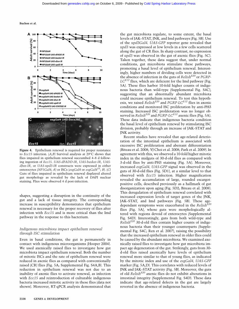
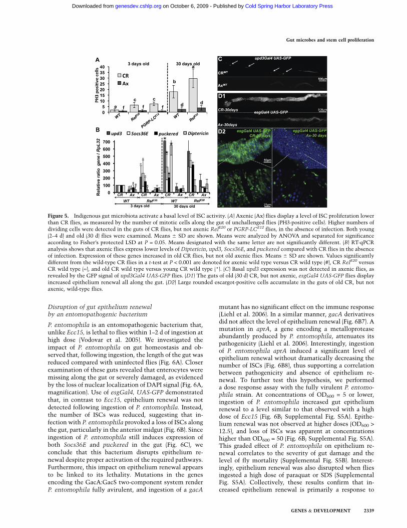
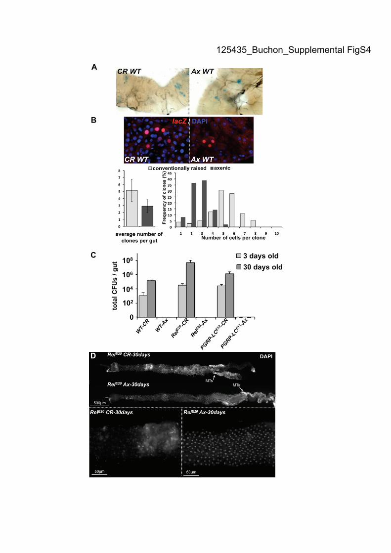
Even in basal conditions, the gut is permanently incontact with indigenous microorganisms (Hooper 2004).We used axenically raised flies to investigate how gutmicrobiota impact epithelium renewal. Both the numberof mitotic ISCs and the rate of epithelium renewal werereduced in axenic flies as compared with conventionallyraised (CR) flies (Fig. 5A; Supplemental Fig. S4A,B). Thisreduction in epithelium renewal was not due to aninability of axenic flies to activate renewal, as infectionwith Ecc15 and reintroduction of culturable indigenousbacteria increased mitotic activity in these flies (data notshown). Moreover, RT-qPCR analysis demonstrated that
the gut microbiota regulate, to some extent, the basallevels of JAK–STAT, JNK, and Imd pathways (Fig. 5B). Useof the upd3Gal4, UAS-GFP reporter gene revealed thatupd3 was expressed at low levels in a few cells scatteredalong the gut of CR flies. In sharp contrast, no expressionof upd3 was observed in the gut of axenic flies (Fig. 5C).Taken together, these data suggest that, under normalconditions, gut microbiota stimulate these pathways,promoting a basal level of epithelium renewal. Interest-ingly, higher numbers of dividing cells were detected inthe absence of infection in the guts of RelishE20 or PGRP-LCE12 flies, which are deficient for the Imd pathway (Fig.5A). These flies harbor 10-fold higher counts of indige-nous bacteria than wild-type (Supplemental Fig. S4C),suggesting that an abnormally abundant microbiotacould increase epithelium renewal. To test this hypoth-esis, we raised RelishE20 and PGRP-LCE12 flies in axenicconditions and monitored ISC proliferation by anti-PH3staining. Increased ISC proliferation was no longer ob-served in RelishE20 and PGRP-LCE12 axenic flies (Fig. 5A).These data indicate that indigenous bacteria conditionthe basal level of epithelium renewal by stimulating ISCdivision, probably through an increase of JAK–STAT andJNK activity.
Recent studies have revealed that age-related deterio-ration of the intestinal epithelium is associated withexcessive ISC proliferation and aberrant differentiation(Biteau et al. 2008; YJ Choi et al. 2008; Park et al. 2009). Inagreement with this, we observed a 10-fold higher mitoticindex in the midguts of 30-d-old flies as compared with3-d-old flies by anti-PH3 staining (Fig. 5A). Moreover,increased esgGal4, UAS-GFP signal was observed in theguts of 30-d-old flies (Fig. 5D1), at a similar level to thatobserved with Ecc15 infection. Higher magnificationrevealed the accumulation of large, rounded escargot-positive cells, described previously as a hallmark of gutdisorganization upon aging (Fig. 5D2; Biteau et al. 2008).This deregulation of epithelium renewal correlated withincreased expression levels of target genes of the JNK,JAK–STAT, and Imd pathways (Fig. 5B). These age-dependant symptoms were exacerbated in the RelishE20
flies (Fig. 5A), whose guts were morphologically al-tered with regions devoid of enterocytes (SupplementalFig. S4D). Interestingly, guts from both wild-type andRelishE20 30-d-old flies contain higher counts of indige-nous bacteria than their younger counterparts (Supple-mental Fig. S4C; Ren et al. 2007), raising the possibilitythat the increased epithelium renewal in older flies couldbe caused by the abundant microbiota. We examined axe-nically raised flies to investigate how gut microbiota im-pact age degeneration of the gut. Strikingly, guts from 30-d-old flies raised axenically have levels of epitheliumrenewal more similar to that of young flies, as indicatedby the mitotic index and use of the esgGal4, UAS-GFPmarker (Fig. 5A,D). This correlates with reduced levels ofJNK and JAK–STAT activity (Fig. 5B). Moreover, the gutsof old RelishE20 axenic flies do not exhibit alterations inintestinal integrity (Supplemental Fig. S4D). These dataindicate that age-related defects in the gut are largelyreverted in the absence of indigenous bacteria.
Figure 4. Epithelium renewal is required for proper resistanceto Ecc15 infection. (A,B) Survival analysis at 29°C shows thatflies impaired in epithelium renewal succumbed 4–8 d follow-ing ingestion of Ecc15. UAS-dFADD-IR, UAS-basket-IR, UAS-
dJun-IR, or UAS-upd3-IR constructs were expressed in eitherenterocytes (NP1Gal4; A) or ISCs (esgGal4 or esgGal4TS; B). (C)Guts of flies impaired in epithelium renewal displayed alteredgut morphology as revealed by the lack of DAPI nuclearstaining. Flies were observed 4 d post-infection.
Buchon et al.
2338 GENES & DEVELOPMENT
Cold Spring Harbor Laboratory Press on October 6, 2009 - Published by genesdev.cshlp.orgDownloaded from
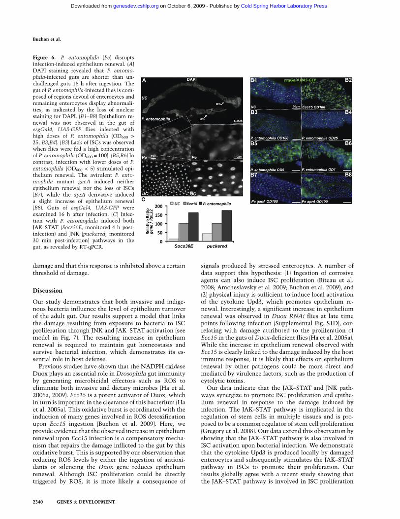
Disruption of gut epithelium renewalby an entomopathogenic bacterium
P. entomophila is an entomopathogenic bacterium that,unlike Ecc15, is lethal to flies within 1–2 d of ingestion athigh dose (Vodovar et al. 2005). We investigated theimpact of P. entomophila on gut homeostasis and ob-served that, following ingestion, the length of the gut wasreduced compared with uninfected flies (Fig. 6A). Closerexamination of these guts revealed that enterocytes weremissing along the gut or severely damaged, as evidencedby the loss of nuclear localization of DAPI signal (Fig. 6A,magnification). Use of esgGal4, UAS-GFP demonstratedthat, in contrast to Ecc15, epithelium renewal was notdetected following ingestion of P. entomophila. Instead,the number of ISCs was reduced, suggesting that in-fection with P. entomophila provoked a loss of ISCs alongthe gut, particularly in the anterior midgut (Fig. 6B). Sinceingestion of P. entomophila still induces expression ofboth Socs36E and puckered in the gut (Fig. 6C), weconclude that this bacterium disrupts epithelium re-newal despite proper activation of the required pathways.Furthermore, this impact on epithelium renewal appearsto be linked to its lethality. Mutations in the genesencoding the GacA:GacS two-component system renderP. entomophila fully avirulent, and ingestion of a gacA
mutant has no significant effect on the immune response(Liehl et al. 2006). In a similar manner, gacA derivativesdid not affect the level of epithelium renewal (Fig. 6B7). Amutation in aprA, a gene encoding a metalloproteaseabundantly produced by P. entomophila, attenuates itspathogenicity (Liehl et al. 2006). Interestingly, ingestionof P. entomophila aprA induced a significant level ofepithelium renewal without dramatically decreasing thenumber of ISCs (Fig. 6B8), thus supporting a correlationbetween pathogenicity and absence of epithelium re-newal. To further test this hypothesis, we performeda dose response assay with the fully virulent P. entomo-phila strain. At concentrations of OD600 = 5 or lower,ingestion of P. entomophila increased gut epitheliumrenewal to a level similar to that observed with a highdose of Ecc15 (Fig. 6B; Supplemental Fig. S5A). Epithe-lium renewal was not observed at higher doses (OD600 >12.5), and loss of ISCs was apparent at concentrationshigher than OD600 = 50 (Fig. 6B; Supplemental Fig. S5A).This graded effect of P. entomophila on epithelium re-newal correlates to the severity of gut damage and thelevel of fly mortality (Supplemental Fig. S5B). Interest-ingly, epithelium renewal was also disrupted when fliesingested a high dose of paraquat or SDS (SupplementalFig. S5A). Collectively, these results confirm that in-creased epithelium renewal is primarily a response to
Figure 5. Indigenous gut microbiota activate a basal level of ISC activity. (A) Axenic (Ax) flies display a level of ISC proliferation lowerthan CR flies, as measured by the number of mitotic cells along the gut of unchallenged flies (PH3-positive cells). Higher numbers ofdividing cells were detected in the guts of CR flies, but not axenic RelE20 or PGRP-LCE12 flies, in the absence of infection. Both young(2–4 d) and old (30 d) flies were examined. Means 6 SD are shown. Means were analyzed by ANOVA and separated for significanceaccording to Fisher’s protected LSD at P = 0.05. Means designated with the same letter are not significantly different. (B) RT-qPCRanalysis shows that axenic flies express lower levels of Diptericin, upd3, Socs36E, and puckered compared with CR flies in the absenceof infection. Expression of these genes increased in old CR flies, but not old axenic flies. Means 6 SD are shown. Values significantlydifferent from the wild-type CR flies in a t-test at P < 0.001 are denoted for axenic wild type versus CR wild type (#), CR RelE20 versusCR wild type (»), and old CR wild type versus young CR wild type (*). (C) Basal upd3 expression was not detected in axenic flies, asrevealed by the GFP signal of upd3Gal4 UAS-GFP flies. (D1) The guts of old (30 d) CR, but not axenic, esgGal4 UAS-GFP flies displayincreased epithelium renewal all along the gut. (D2) Large rounded escargot-positive cells accumulate in the guts of old CR, but notaxenic, wild-type flies.
Gut microbes and stem cell proliferation
GENES & DEVELOPMENT 2339
Cold Spring Harbor Laboratory Press on October 6, 2009 - Published by genesdev.cshlp.orgDownloaded from
damage and that this response is inhibited above a certainthreshold of damage.
Discussion
Our study demonstrates that both invasive and indige-nous bacteria influence the level of epithelium turnoverof the adult gut. Our results support a model that linksthe damage resulting from exposure to bacteria to ISCproliferation through JNK and JAK–STAT activation (seemodel in Fig. 7). The resulting increase in epitheliumrenewal is required to maintain gut homeostasis andsurvive bacterial infection, which demonstrates its es-sential role in host defense.
Previous studies have shown that the NADPH oxidaseDuox plays an essential role in Drosophila gut immunityby generating microbicidal effectors such as ROS toeliminate both invasive and dietary microbes (Ha et al.2005a, 2009). Ecc15 is a potent activator of Duox, whichin turn is important in the clearance of this bacterium (Haet al. 2005a). This oxidative burst is coordinated with theinduction of many genes involved in ROS detoxificationupon Ecc15 ingestion (Buchon et al. 2009). Here, weprovide evidence that the observed increase in epitheliumrenewal upon Ecc15 infection is a compensatory mecha-nism that repairs the damage inflicted to the gut by thisoxidative burst. This is supported by our observation thatreducing ROS levels by either the ingestion of antioxi-dants or silencing the Duox gene reduces epitheliumrenewal. Although ISC proliferation could be directlytriggered by ROS, it is more likely a consequence of
signals produced by stressed enterocytes. A number ofdata support this hypothesis: (1) Ingestion of corrosiveagents can also induce ISC proliferation (Biteau et al.2008; Amcheslavsky et al. 2009; Buchon et al. 2009), and(2) physical injury is sufficient to induce local activationof the cytokine Upd3, which promotes epithelium re-newal. Interestingly, a significant increase in epitheliumrenewal was observed in Duox RNAi flies at late timepoints following infection (Supplemental Fig. S1D), cor-relating with damage attributed to the proliferation ofEcc15 in the guts of Duox-deficient flies (Ha et al. 2005a).While the increase in epithelium renewal observed withEcc15 is clearly linked to the damage induced by the hostimmune response, it is likely that effects on epitheliumrenewal by other pathogens could be more direct andmediated by virulence factors, such as the production ofcytolytic toxins.
Our data indicate that the JAK–STAT and JNK path-ways synergize to promote ISC proliferation and epithe-lium renewal in response to the damage induced byinfection. The JAK–STAT pathway is implicated in theregulation of stem cells in multiple tissues and is pro-posed to be a common regulator of stem cell proliferation(Gregory et al. 2008). Our data extend this observation byshowing that the JAK–STAT pathway is also involved inISC activation upon bacterial infection. We demonstratethat the cytokine Upd3 is produced locally by damagedenterocytes and subsequently stimulates the JAK–STATpathway in ISCs to promote their proliferation. Ourresults globally agree with a recent study showing thatthe JAK–STAT pathway is involved in ISC proliferation
Figure 6. P. entomophila (Pe) disruptsinfection-induced epithelium renewal. (A)DAPI staining revealed that P. entomo-phila-infected guts are shorter than un-challenged guts 16 h after ingestion. Thegut of P. entomophila-infected flies is com-posed of regions devoid of enterocytes andremaining enterocytes display abnormali-ties, as indicated by the loss of nuclearstaining for DAPI. (B1–B8) Epithelium re-newal was not observed in the gut ofesgGal4, UAS-GFP flies infected withhigh doses of P. entomophila (OD600 >
25, B3,B4). (B3) Lack of ISCs was observedwhen flies were fed a high concentrationof P. entomophila (OD600 = 100). (B5,B6) Incontrast, infection with lower doses of P.
entomophila (OD600 < 5) stimulated epi-thelium renewal. The avirulent P. ento-
mophila mutant gacA induced neitherepithelium renewal nor the loss of ISCs(B7), while the aprA derivative induceda slight increase of epithelium renewal(B8). Guts of esgGal4, UAS-GFP wereexamined 16 h after infection. (C) Infec-tion with P. entomophila induced bothJAK–STAT (Socs36E, monitored 4 h post-infection) and JNK (puckered, monitored30 min post-infection) pathways in thegut, as revealed by RT-qPCR.
Buchon et al.
2340 GENES & DEVELOPMENT
Cold Spring Harbor Laboratory Press on October 6, 2009 - Published by genesdev.cshlp.orgDownloaded from
upon infection with a low dose of P. entomophila (Jianget al. 2009). This work and our study clearly demonstratethat the JAK–STAT pathway adjusts the level of epithe-lium renewal to ensure proper tissue homeostasis bylinking enterocyte damage to ISC proliferation. In thestudy by Jiang et al. (2009), they also uncover an ad-ditional role of this pathway in the differentiation ofenteroblasts during basal gut epithelium turnover. Theimplication of the JAK–STAT pathway in differentiationcould explain the accumulation of the small-nucleatedescargot-positive cells we observed in the gut of flies withreduced JAK–STAT signaling in ISCs (Fig. 2C). The JAK–STAT pathway was also shown previously to control theexpression of some antimicrobial peptides such as Dro-somycin 3 (Dro3) (Buchon et al. 2009). Therefore, theJAK–STAT pathway has a dual role in the gut uponinfection, controlling both the immune response andepithelium renewal.
Our data show that the lack of JNK pathway activity inISCs results in the loss of ISCs in guts infected withEcc15, thus preventing epithelium renewal. Our findingsare consistent with the attributed function of JNK at thecenter of a signal transduction network that coordinatesthe induction of protective genes in response to oxidativechallenge (Wang et al. 2003; Biteau et al. 2008). This
cytoprotective role against ROS would protect ISCs fromthe oxidative burst induced upon Ecc15 infection,explaining why ISCs die by apoptosis when JNK activityis reduced. As suggested by Biteau et al. (2008), it is likelythat JNK signaling is required not only to protect ISCsfrom oxidative stress, but also to induce stem cell pro-liferation to replace damaged differentiated cells. This issupported by the observation that overexpression of theJNKK Hep in ISCs is sufficient to trigger an epitheliumrenewal in the absence of infection. In addition, increasedJNK activity in ISCs of old flies has been linked tohyperproliferative states and age-related deterioration ofthe intestinal epithelium (Biteau et al. 2008). Our studyextends this observation by showing that JNK signaling isalso required for epithelium renewal upon Ecc15 infec-tion. Thus, infection with Ecc15 recapitulates in anaccelerated time frame the impacts of increased stressobserved in guts of aging flies
We observed that the inhibition of the dJun transcrip-tion factor in ISCs leads to a loss of stem cells in theabsence of infection, suggesting that this transcriptionfactor plays a critical role in ISC maintenance in the gut.We have no definitive explanation for why the dJun-IRconstruct behaves differently than the basket and hep-IRconstructs. We speculate that this could be due to (1)differences in the basal activity of the JNK pathway,which would be blocked only with the dJun-IR thattargets a terminal component of the pathway; (2) effectsof Jun in ISCs independent of the JNK pathway, asreported in other systems (Bates et al. 2008); or (3) sideeffects of the dJun-IR construct.
In contrast to the requirement of the JNK pathwayupon Ecc15 infection, Jiang et al. (2009) reported that oralingestion with a low dose of P. entomophila still inducedmitosis in the JNK-defective mutant hep1. In agreement,we found that inhibiting the JNK pathway in ISCs did notblock the induction of epithelium renewal by a low doseof P. entomophila (Supplemental Fig. 3C). This differencein the requirement of the JNK pathway may be explainedby the nature of these two pathogens. Whereas Ecc15damages the gut through an oxidative burst that activatesthe JNK pathway, the stimulation of epithelium renewalby P. entomophila could be due to a more direct effect ofthis bacterium on the gut. Altogether, Jiang et al. (2009)and our study point to an essential role of the JAK–STATpathway in modulation of epithelium renewal activity,while the role of JNK may be dependent on the infectiousagent and any associated oxidative stress. While it isknown that the JNK pathway is activated by a variety ofenvironmental challenges including ROS, the precisemechanism of activation of this pathway has not beenelucidated. Similarly, the molecular basis of upd3 in-duction in damaged enterocytes is not known. Futurework should decipher the nature of the signals thatactivate these pathways in both ISCs and enterocytes,as well as the possible cross-talk between the JNK andJAK–STAT pathways in ISC control.
The observation that flies unable to renew their gutepithelium eventually succumb to Ecc15 infection high-lights the importance of this process in the gut immune
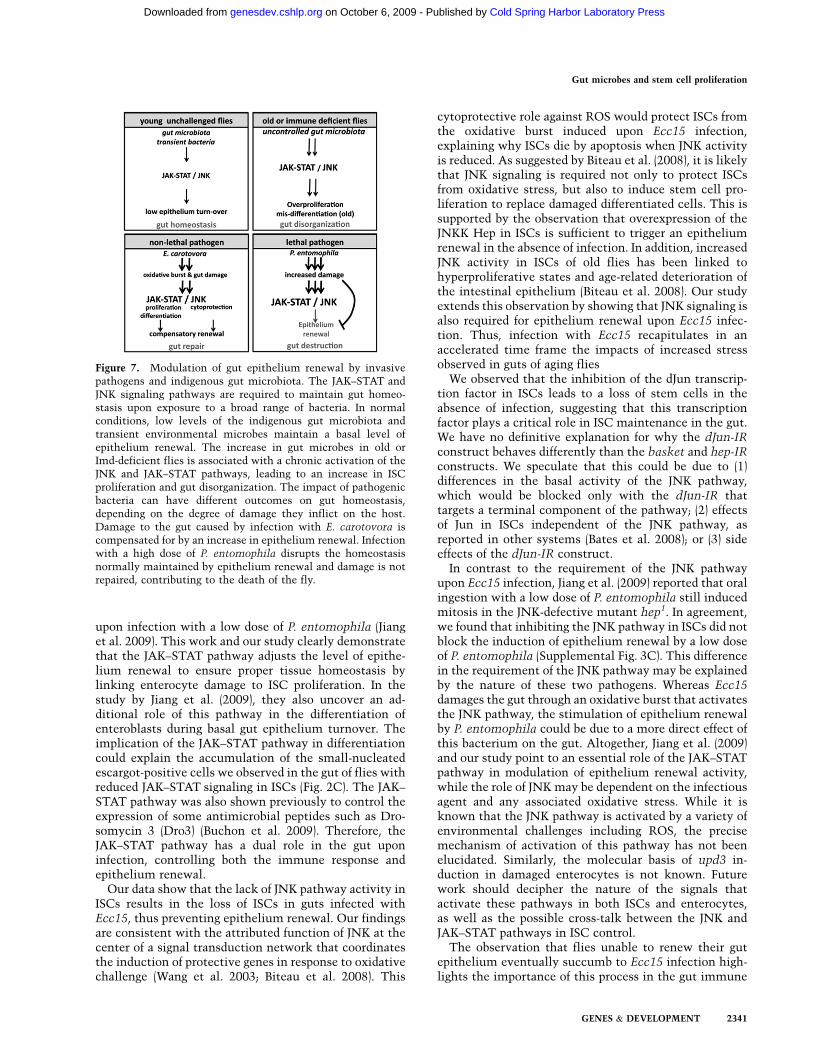
Figure 7. Modulation of gut epithelium renewal by invasivepathogens and indigenous gut microbiota. The JAK–STAT andJNK signaling pathways are required to maintain gut homeo-stasis upon exposure to a broad range of bacteria. In normalconditions, low levels of the indigenous gut microbiota andtransient environmental microbes maintain a basal level ofepithelium renewal. The increase in gut microbes in old orImd-deficient flies is associated with a chronic activation of theJNK and JAK–STAT pathways, leading to an increase in ISCproliferation and gut disorganization. The impact of pathogenicbacteria can have different outcomes on gut homeostasis,depending on the degree of damage they inflict on the host.Damage to the gut caused by infection with E. carotovora iscompensated for by an increase in epithelium renewal. Infectionwith a high dose of P. entomophila disrupts the homeostasisnormally maintained by epithelium renewal and damage is notrepaired, contributing to the death of the fly.
Gut microbes and stem cell proliferation
GENES & DEVELOPMENT 2341
Cold Spring Harbor Laboratory Press on October 6, 2009 - Published by genesdev.cshlp.orgDownloaded from
response. It is striking that defects in epithelium renewalare more detrimental to host survival than deficiency inthe Imd pathway, even though this pathway controlsmost of the intestinal immune-regulated genes inducedby Ecc15 (Buchon et al. 2009). Our results are in agree-ment with a previous study indicating that, in theDrosophila gastrointestinal tract, the Imd-dependent im-mune response is normally dispensable to most transientbacteria, but is provisionally crucial in the event that thehost encounters ROS-resistant microbes (Ryu et al. 2006).However, our study demonstrates that efficient and rapidclearance of bacteria in the gut by Duox is possible onlywhen coordinated with epithelium renewal to repairdamage caused by ROS. This finely tuned balance be-tween bacterial elimination by Duox activity and gutresistance to collateral damage induced by ROS is likelythe reason why flies normally survive infection by Ecc15.Yet, this calibration also exposes a vulnerability thatcould easily be manipulated or subverted by other path-ogens. Along this line, our work also exposes the range ofimpact different bacteria can have on stem cell activa-tion. We observed that infection with high doses of P.entomophila led to a loss of gut integrity, including theloss of stem cells. Moreover, the ability of P. entomophilato disrupt epithelium renewal correlates with damage tothe gut and the death of the host. Since both JNK andJAK–STAT pathways are activated upon infection with P.entomophila, this suggests that this bacterium activatesthe appropriate pathways necessary to repair the gut,but ISCs are unable to respond accordingly. Interestingly,a completely avirulent P. entomophila mutant (gacA)does not persist in the gut and does not induce epitheliumrenewal. In contrast, an attenuated mutant (aprA) some-what restores epithelium renewal. These observations,along with the dose response analysis using P. entomo-phila and corrosive agents, suggest that the virulencefactors of this entomopathogen disrupt epithelium re-newal through excessive damage to the gut. Of note,recent studies suggest that both Helicobacter pylori andShigella flexneri, two bacterial pathogens of the humandigestive tract, interfere with epithelium renewal to exerttheir pathological effects (Iwai et al. 2007; Mimuro et al.2007). This suggests that epithelium renewal could bea common target for bacteria that infect through the gut(Chatterjee and Ip 2009; Cronin et al. 2009; Jiang et al.2009). In this respect, the host defense to oral bacterialinfection could be considered as a bimodular response,composed of both immune and homeostatic processesthat require strict coordination. Disruption of eitherprocess results in the failure to resolve the infection andimpedes the return to homeostasis.
In contrast to the acute invasion by pathogenic bacte-ria, indigenous gut microbiota are in constant associationwith the gut epithelium, and thus may impact guthomeostasis. Using axenically raised flies, we establishedthat indigenous microbiota stimulate a basal level ofepithelium renewal that correlates with the level ofactivation of the JAK–STAT and JNK pathways. Thisraises the possibility that both indigenous and invasivebacteria, such as Ecc15, are capable of triggering epithe-
lium renewal by the same process. Additionally, our datasupport a novel homeostatic mechanism in which thedensity of indigenous bacteria is coupled to the level ofepithelium renewal. This is the first report that gutmicrobiota affect stem cell activation and epitheliumrenewal, concepts proposed previously in mammaliansystems (Hooper 2004) but never fully demonstrated.This also implies that variations in the level of epithe-lium renewal observed in different laboratory contextscould actually be due to impacts from gut microbes.
Importantly, in this context, we show that lack ofindigenous microbiota reverts most age-related deterio-ration of the gut. Aging of the gut is usually marked byboth hyperproliferation of ISCs and differentiation de-faults that lead to disorganization of the gut epithelium(Biteau et al. 2008; N Choi et al. 2008). These alterationshave been shown to be associated with activation of thePDGF- and VEGF-related factor 2 (Pvf2)/Pvr and JNKsignaling pathways directly in ISCs (Biteau et al. 2008;N Choi et al. 2008). Accordingly, inhibition of the JNKpathway in ISCs fully reverts the epithelium alterationsthat occur with aging (Biteau et al. 2008). This raises thepossibility that gut microbiota could exert their effectthrough prolonged activation of the JNK pathway. In-terestingly, immune-deficient flies, lacking the Imd path-way, also display hyperproliferative guts and have higherbasal levels of activation of the JNK and JAK–STATpathways. The observation that these flies also harborhigher numbers of indigenous bacteria further supportsa model in which failure to control gut microbiota leadsto an imbalance in gut epithelium turnover. Future workshould analyze the mechanisms by which gut microbiotaaffect epithelium renewal and whether this is due toa direct impact of bacteria on the gut or is mediatedindirectly through changes in fly physiology. Moreover,the correlation between higher numbers of indigenousbacteria and increased disorganization of the gut uponaging in flies lacking the Imd pathway raises the possi-bility that a main function of this pathway is to controlgut microbiota. This is in agreement with conceptsemerging in mammals that support an essential role ofthe gut immune response in maintaining the beneficialnature of the host–microbiota association (Hooper 2009).This function also parallels the theory of ‘‘controlledinflammation’’ described in mammals, where a low levelof immune activation is proposed to maintain gut barrierintegrity (Sansonetti 2004; Pedron and Sansonetti 2008).
In conclusion, this study unravels some of the complexinterconnections between the immune response, inva-sive and indigenous microbiota, and stem cell homeosta-sis in the gut of Drosophila. Based on the evolutionaryconservation of transduction pathways such as JNK andJAK–STAT between Drosophila and mammals, it is likelythat similar processes occur in the gut of mammalsduring infection. Interestingly, stimulation of stem cellactivity by invasive bacteria is proposed to favor thedevelopment of hyperproliferative states found in pre-cancerous lesions (Macdonald and Monteleone 2005;Radtke and Clevers 2005). Thus, Drosophila may providea more accessible model to elucidate host mechanisms to
Buchon et al.
2342 GENES & DEVELOPMENT
Cold Spring Harbor Laboratory Press on October 6, 2009 - Published by genesdev.cshlp.orgDownloaded from
maintain homeostasis and the impact of bacteria on thisprocess.
Materials and methods
Fly stocks and rearing
For description of the stock genotypes, specificities of the Gal4drivers, and efficiency of RNAi lines used in this study, see theSupplemental Material. Drosophila stocks were maintained at23°C using standard fly medium (maize flour, dead yeast, agar,and fruit juice) devoid of living yeast. RNAi constructs werevalidated by RT-qPCR (Supplemental Fig. S7). Axenic Canton,Oregon, esgGal4, UAS-GFP, RelE20, and PGRP-LCE12 stockswere generated by bleaching embryos and maintaining embryosand emerging flies on autoclaved fly medium. The presence ofbacteria in gut homogenates was examined by PCR amplifica-tion of 16S rRNA genes using eubacterial primers (27F and1492R), and by culturing the homogenates on mannitol agar or1/10-strength tryptic soy agar.
Bacterial strains
Ecc15 is a Gram-negative bacterium that induces a strong localimmune response (Basset et al. 2000; Buchon et al. 2009). P.entomophila is a Gram-negative bacterium that is lethal toDrosophila when ingested (Vodovar et al. 2005; Liehl et al. 2006).
Infection and compound feeding
Bacterial infections were performed as described elsewhere(Buchon et al. 2009). For oral infection, flies were incubated 2 hat 29°C in an empty vial and then placed in a fly vial with foodsolution and maintained at 29°C. The food solution was obtainedby mixing a pellet of an overnight culture of bacteria (OD600 =
200), 10 mM paraquat (Promega), or 0.2% H2O2 (Sigma) witha solution of 5% sucrose (1:1), added to a filter disk thatcompletely covered the surface of standard fly medium. Flieswere incubated for 2 d at 29°C on the contaminated filter, or 1d for survival analysis, after which they were transferred to freshvials. For antioxidant treatments, flies were fed a mixture ofbacteria (final OD600 = 100) and sucrose (2.5% final), to whichglutathione (0.5 mM final; Sigma) or NAC (20 mM final; Sigma)was added; control flies were fed bacteria and sucrose or sucrosealone.
Clonal analysis
The marked lineage system developed by Harrison and Perrimon(1993) was used to generate clones of lacZ-expressing cells(Ohlstein and Spradling 2006). See the Supplemental Materialfor details. Adults of the genotype yw, hs-FLP; X-15-33/X-15-29
were produced by standard crosses. To induce somatic recombi-nation, 3-d-old adult flies were heat-shocked for 60 min at 37°Con three consecutive days. A day after, flies were orally infectedwith Ecc15 or fed on sucrose. Guts were dissected 10 d afterinfection and stained with X-gal (Romeo and Lemaitre 2008).
Live imaging and immunofluorescence
For live imaging, guts were dissected at room temperature in 13
phosphate-buffered saline (PBS) and immediately mounted in theanti-fading agent AF1 (Citifluor). Samples were observed forfluorescence with an Axioplot imager Z1 and Axiocam mRMcamera (Zeiss). For immunofluorescence, guts were dissected in
13 PBS; fixed for 20 min in PBS, 0.1% Tween 20 (PBT), and 4%paraformaldehyde; and then stained with primary antibody (anti-PH3 [1/1000; Upstate Biotechnologies/Millipore], anti-cleavedCaspase-3 [1/500; Asp175, Cell signal], anti-GFP [1/1000; RocheDiagnostics], anti-b-galactosidase [1/500; Sigma], anti-Delta [1/5000], anti-Prospero [1/500] [Developmental Studies HybridomaBank]) in PBT + BSA. Secondary staining was performed withAlexa488 or Alexa594-coupled anti-mouse or anti-rabbit anti-bodies (Invitrogen). DNA was stained with DAPI (Sigma).
RT-qPCR
Total RNA was extracted from 40 dissected guts using Trizol(Invitrogen). RT-qPCR was performed using SYBR Green I(Roche) on a LightCycler 2.0 as described previously (Buchonet al. 2009). The amount of mRNA detected was normalized tocontrol RpL32 values. Primers used to monitor mRNA quanti-fication can be obtained on request.
Acknowledgments
We thank our colleague J.P. Boquete for technical support; M.Meister (Strasbourg) for stimulating discussions and the gift ofthe UAS-upd3 lines; W.-J. Lee (Seoul) for comments on the man-uscript; and R. Ueda (National Institute of Genetic, Mishima,Japan), M. Crozatier (Toulouse), A. Bardin (Paris), F. Leulier (Gif-sur-Yvette), J.E. Darnell (New York), and C.A. Micchelli (St.Louis) for fly stocks. This work was funded by the ERC AdvancedGrant and the Swiss National Fund (3100A0-12079/1).
References
Agaisse H, Petersen UM, Boutros M, Mathey-Prevot B, PerrimonN. 2003. Signaling role of hemocytes in Drosophila JAK/STAT-dependent response to septic injury. Dev Cell 5: 441–450.
Amcheslavsky A, Jiang J, Ip YT. 2009. Tissue damage-inducedintestinal stem cell division in Drosophila. Cell Stem Cell 4:49–61.
Bach EA, Ekas LA, Ayala-Camargo A, Flaherty MS, Lee H,Perrimon N, Baeg GH. 2007. GFP reporters detect theactivation of the Drosophila JAK/STAT pathway in vivo.Gene Expr Patterns 7: 323–331.
Basset A, Khush RS, Braun A, Gardan L, Boccard F, HoffmannJA, Lemaitre B. 2000. The phytopathogenic bacteria Erwinia
carotovora infects Drosophila and activates an immuneresponse. Proc Natl Acad Sci 97: 3376–3381.
Bates KL, Higley M, Letsou A. 2008. Raw mediates antagonismof AP-1 activity in Drosophila. Genetics 178: 1989–2002.
Biteau B, Hochmuth CE, Jasper H. 2008. JNK Activity insomatic stem cells causes loss of tissue homeostasis in theaging Drosophila gut. Cell Stem Cell 3: 442–455.
Brown S, Hu N, Hombria JC. 2001. Identification of the firstinvertebrate interleukin JAK/STAT receptor, the Drosophila
gene domeless. Curr Biol 11: 1700–1705.Buchon N, Broderick NA, Poidevin M, Pradervand S, Lemaitre
B. 2009. Drosophila intestinal response to bacterial infection:Activation of host defense and stem cell proliferation. Cell
Host Microbe 5: 200–211.Chatterjee M, Ip YT. 2009. Pathogenic stimulation of intestinal
stem cell response in Drosophila. J Cell Physiol 220: 664–671.
Choi N, Kim J, Yang D, Kim Y, Yoo M. 2008. Age-relatedchanges in Drosophila midgut are associated with PVF2,a PDGF/VEGF-like growth factor. Aging Cell 7: 318–334.
Choi YJ, Hwang MS, Park JS, Bae SK, Kim YS, Yoo MA. 2008.Age-related upregulation of Drosophila caudal gene via
Gut microbes and stem cell proliferation
GENES & DEVELOPMENT 2343
Cold Spring Harbor Laboratory Press on October 6, 2009 - Published by genesdev.cshlp.orgDownloaded from
NF-kB in the adult posterior midgut. Biochim Biophys Acta
1780: 1093–1100.Cronin SJ, Nehme NT, Limmer S, Liegeois S, Pospisilik JA,
Schramek D, Leibbrandt A, Simoes RD, Gruber S, Puc U,et al. 2009. Genome-wide RNAi screen identifies genesinvolved in intestinal pathogenic bacterial infection. Science325: 340–343.
Gregory L, Came PJ, Brown S. 2008. Stem cell regulation byJAK/STAT signaling in Drosophila. Semin Cell Dev Biol 19:407–413.
Ha E, Oh C, Bae YS, Lee WJ. 2005a. A direct role for dual oxidasein Drosophila gut immunity. Science 310: 847–850.
Ha E, Oh C, Ryu J, Bae YS, Kang SW, Jang IH, Brey P, Lee WJ.2005b. An antioxidant system required for host protec-tion against gut infection in Drosophila. Dev Cell 8: 125–132.
Ha EM, Lee K, Park SH, Kim SH, Nam HJ, Lee HY, Kang D, LeeWJ. 2009. Regulation of DUOX by the Gaq-phospholipaseCb-Ca2+ pathway in Drosophila gut immunity. Dev Cell 16:386–397.
Harrison DA, Perrimon N. 1993. Simple and efficient generationof marked clones in Drosophila. Curr Biol 3: 424–433.
Hooper LV. 2004. Bacterial contributions to mammalian gutdevelopment. Trends Microbiol 12: 129–134.
Hooper LV. 2009. Do symbiotic bacteria subvert host immunity?Nat Rev Microbiol 7: 367–374.
Iwai H, Kim M, Yoshikawa Y, Ashida H, Ogawa M, Fujita Y,Muller D, Kirikae T, Jackson PK, Kotani S, et al. 2007. Abacterial effector targets Mad2L2, an APC inhibitor, tomodulate host cell cycling. Cell 130: 611–623.
Jiang H, Patel PH, Kohlmaier A, Grenley MO, McEwen DG,Edgar BA. 2009. Cytokine/Jak/Stat signaling mediates re-generation and homeostasis in the Drosophila midgut. Cell137: 1343–1355.
Krzemien J, Dubois L, Makki R, Meister M, Vincent A,Crozatier M. 2007. Control of blood cell homeostasis inDrosophila larvae by the posterior signaling centre. Nature446: 325–328.
Lee T, Luo L. 2001. Mosaic analysis with a repressible cellmarker (MARCM) for Drosophila neural development.Trends Neurosci 24: 251–254.
Liehl P, Blight M, Vodovar N, Boccard F, Lemaitre B. 2006.Prevalence of local immune response against oral infectionin a Drosophila/Pseudomonas infection model. PLoS Pathog
2: e56. doi: 10.1371/journal. ppat.0020056.Macdonald TT, Monteleone G. 2005. Immunity, inflammation,
and allergy in the gut. Science 307: 1920–1925.Martın-Blanco E, Gampel A, Ring J, Virdee K, Kirov N, Tolkovsky
AM, Martinez-Arias A. 1998. puckered encodes a phospha-tase that mediates a feedback loop regulating JNK activityduring dorsal closure in Drosophila. Genes & Dev 12: 557–570.
Micchelli CA, Perrimon N. 2006. Evidence that stem cellsreside in the adult Drosophila midgut epithelium. Nature
439: 475–479.Mimuro H, Suzuki T, Nagai S, Rieder G, Suzuki M, Nagai T,
Fujita Y, Nagamatsu K, Ishijima N, Koyasu S, et al. 2007.Helicobacter pylori dampens gut epithelial self-renewal byinhibiting apoptosis, a bacterial strategy to enhance coloni-zation of the stomach. Cell Host Microbe 2: 250–263.
Ohlstein B, Spradling AC. 2006. The adult Drosophila posteriormidgut is maintained by pluripotent stem cells. Nature 439:470–474.
Park JS, Kim YS, Yoo MA. 2009. The role of p38b MAPK in age-related modulation of intestinal stem cell proliferation anddifferentiation in Drosophila. Aging 1: 637–651.
Pedron T, Sansonetti PJ. 2008. Commensals, bacterial pathogensand intestinal inflammation: An intriguing menage a trois.Cell Host Microbe 3: 344–347.
Radtke F, Clevers H. 2005. Self-renewal and cancer of the gut:Two sides of a coin. Science 307: 1904–1909.
Ren C, Webster P, Finkel SE, Tower J. 2007. Increased internaland external bacterial load during Drosophila aging withoutlife-span trade-off. Cell Metab 6: 144–152.
Romeo Y, Lemaitre B. 2008. Drosophila immunity: Methods formonitoring the activity of toll and imd signaling pathways.Methods Mol Biol 415: 379–394.
Ryu J, Ha E, Oh C, Seol J, Brey P, Jin I, Lee D, Kim J, Lee D, LeeWJ. 2006. An essential complementary role of NF-kB path-way to microbicidal oxidants in Drosophila gut immunity.EMBO J 25: 3693–3701.
Sansonetti PJ. 2004. War and peace at mucosal surfaces. Nat RevImmunol 4: 953–964.
Vodovar N, Vinals M, Liehl P, Basset A, Degrouard J, Spellman P,Boccard F, Lemaitre B. 2005. Drosophila host defense afteroral infection by an entomopathogenic Pseudomonas spe-cies. Proc Natl Acad Sci 102: 11414–11419.
Wang MC, Bohmann D, Jasper H. 2003. JNK signaling conferstolerance to oxidative stress and extends lifespan in Dro-
sophila. Dev Cell 5: 811–816.Zaidman-Remy A, Herve M, Poidevin M, Pilifloury S, Kim MO,
Blanot D, Oh BH, Ueda R, Mengin-Lecreulx D, Lemaitre B.2006. The Drosophila amidase PGRP-LB modulates the im-mune response to bacterial infection. Immunity 24: 463–473.
Buchon et al.
2344 GENES & DEVELOPMENT
Cold Spring Harbor Laboratory Press on October 6, 2009 - Published by genesdev.cshlp.orgDownloaded from
A Ecc15 OD100 Ecc15 OD75 Ecc15 OD50
Ecc15 OD25 Ecc15 OD5 Ecc15 OD1
CF
Us/m
L
of
infe
cti
on
mix
ture
B
Initial load After
2h incubation
125435_Buchon_Supplemental FigS1
* *
C
50μm
lacZ / DAPI
Ecc15 Ecc15 + glutathione
D
1014
1012
1010
108
106
104
102
0
Ecc15 Ecc15 + glutathione
daGal4; +
daGal4; UAS-Duox IR
average number of
clones per gut Number of cells per clone
Fre
qu
en
cy o
f clo
nes (
%)
A
125435_Buchon_Supplemental FigS2
6 days
GFP / aspecific
GFP DAPI
B esgGal4TS UAS-GFP; UAS-STAT92E-IR
esgGal4TS UAS-GFP UAS-STAT92E IR
esgGal4TS UAS-GFP UAS-STAT92E IR
GFP prospero GFP prospero DAPI
esgGal4 UAS-GFP
GFP delta
GFP delta
GFP delta DAPI
GFP delta DAPI
10XSTAT-GFP
esgGal4 UAS-lacZ ; STAT-GFP
50μm
10μm
10μm 50μm
20μm
F
UC UC Ecc15 Ecc15 UC Ecc15
E
120
100
20
40
60
80
0
STAT92E- GFP+ clones WT GFP+ clones
50μm
C
D
E1

esgGal4TS UAS-GFP UAS-basket IR, Ecc15 16h
GFP caspase3
DAPI GFP
caspase3
A
UC
Ecc15
B
125435_Buchon_Supplemental FigS3
50μm
C
esgGal4TS UAS-GFP UAS-basket IR
Ecc15 OD100 Pe OD5
125435_Buchon_Supplemental FigS4
C
A CR WT Ax WT
RelE20 CR-30days
RelE20 Ax-30days
RelE20 CR-30days RelE20 Ax-30days
DAPI D
B
500μm
MTs MTs
50μm 50μm
CR WT Ax WT
lacZ / DAPI
conventionally raised axenic
0
108
106
104
102
tota
l C
FU
s /
gu
t
3 days old
30 days old
average number of
clones per gut Number of cells per clone
Fre
qu
en
cy
of
clo
ne
s (
%)
Incre
asin
g d
ose
125435_Buchon_Supplemental FigS5
B
A
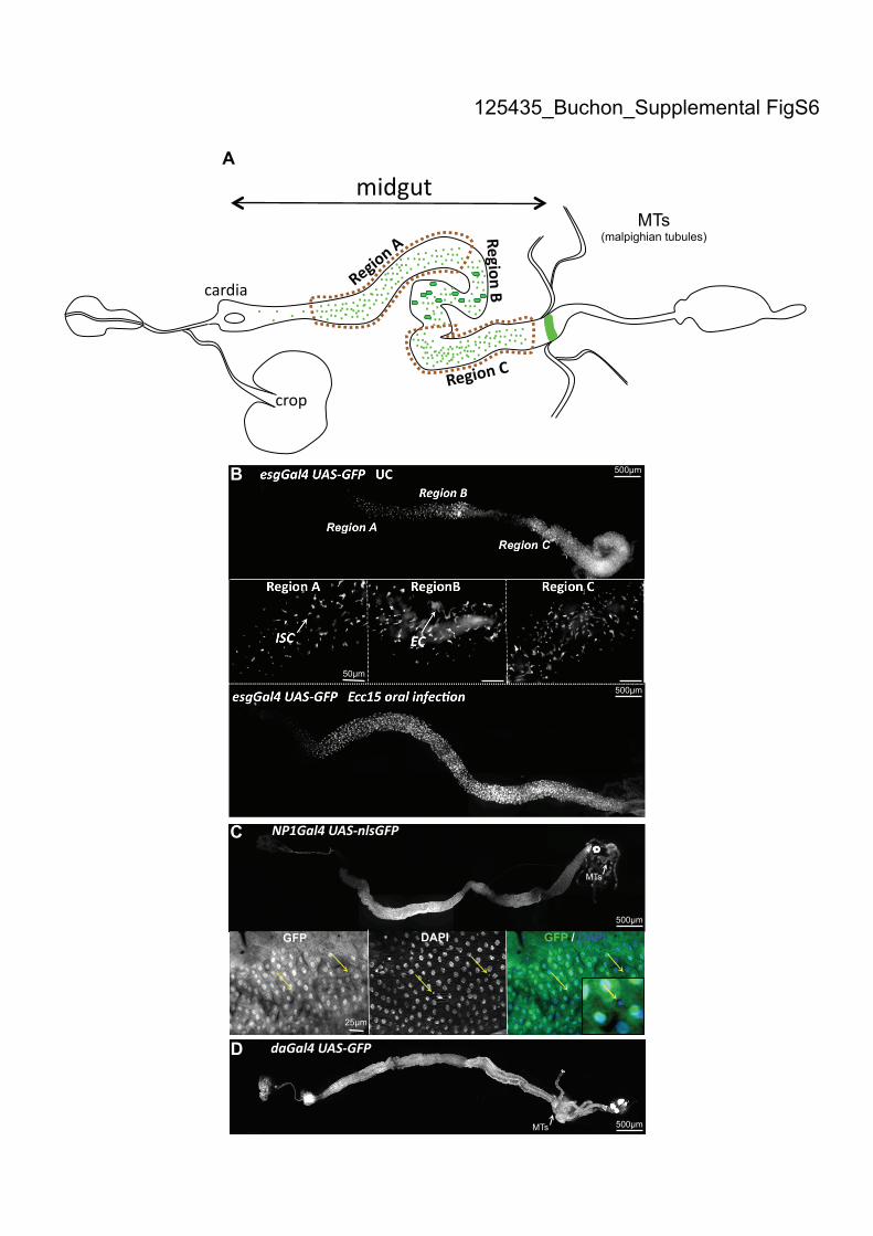
Region A
Region C
GFP DAPI GFP / DAPI
125435_Buchon_Supplemental FigS6
MTs (malpighian tubules)
25μm
500μm
500μm
500μm
500μm
50μm
B
C
A
MTs
MTs
D
125435_Buchon_Supplemental FigS7
C P. entomophila 4h
B P. entomophila 4h
A
Supplemental Materials and Methods
Fly stocks
OregonR flies or flies carrying one copy of the NP1Gal4 transgene were used as wild-
type controls as previously described (Zaidman-Remy et al. 2006). NP1Gal4,
esgGal4 and daGal4 express Gal4 in the midgut, ISCs or ubiquitously, respectively
(Supplemental Fig. S6; Buchon et al. 2009). UAS-RNAi transgenic fly lines of
dFADD (R1), STAT (R2), dJun (R1), basket (R1), Hemipterous (R1) were obtained
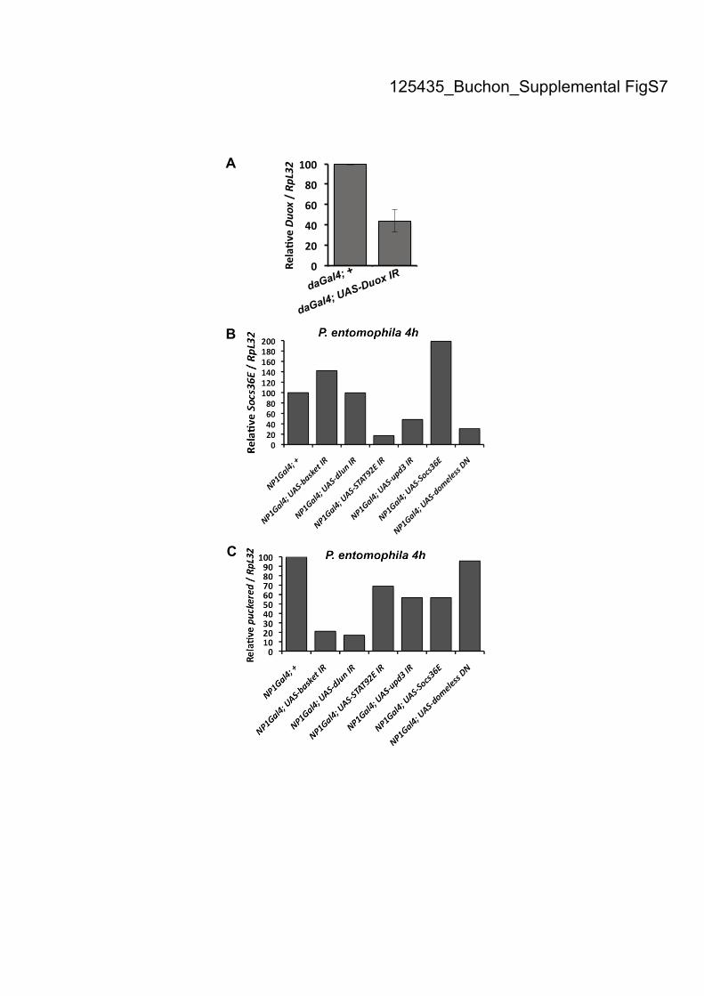
from R. Ueda (National Institute of Genetics, Mishima, Japan). The UAS-Duox-IR,
G q and UAS-IRC lines were obtained from W-J. Lee (Seoul, South Korea) (Ha et al.
2005a; Ryu et al. 2006). The extinction of the Duox gene expression in this context
has been validated by RT-qPCR (Supplemental Fig. S7A). pucE69
(puckered-lacZ),
upd3-IR; upd3Gal4, UAS-GFP; hopmsv-1
and UAS-hopTum
are described elsewhere
(Agaisse 2003). The UAS-DomelessDN
, UAS-Socs36E and UAS-upd3 fly lines were a
gift of M. Crozatier (Brown et al. 2001; Krzemie et al. 2007). The UAS-upd3 fly line
was generated by M. Meister (IBMC, Strasbourg). Adult flies carrying one copy of
the UAS-RNAi construct combined with one copy of the Gal4 driver were analyzed.
F1 progeny carrying both the UAS-RNAi (or UAS-Dominant negative) and the Gal4
driver were raised at 18°C during 3 days after adult eclosion and then transferred to
29°C for optimal efficiency of the UAS/GAL4 system. A functional validation of the
effect of the RNAi construct has been done showing that STAT92E-IR, upd3-IR, UAS-
Socs36E and UAS-DomelessDN
actually inhibit JAK-STAT activity (Supplemental
Fig. S7B). Similarly, we showed that basket-IR and dJun-IR inhibit JNK
(Supplemental Fig. S7C). The 10XSTAT-GFP line is described in (Bach et al. 2007).
The mutant lines PGRP-LCE12
, RelishE20
(Rel), have been described elsewhere
(Romeo and Lemaitre 2008). The esgGal4, UAS-GFP line is described in (Micchelli
and Perrimon 2006). Conditional esgGal4TS
animals were obtained by crossing virgin
females esgGal4, UAS-GFP; tubGal80TS
with males expressing a UAS construct. The
activity of the Gal4 system was controlled by placing 2-day-old F1 adults at either
restrictive (29°C, Gal80TS
off, Gal4 system on) or permissive (18°C, Gal80TS
on,
Gal4 system off) temperatures.
Clonal analysis
The marked lineage system developed by Harrison and Perrimon was used to
generate clones of lacZ expressing cells (Harrison and Perrimon 1993; Ohlstein and
Spradling 2006). Adults of the genotype yw, hs-FLP; X-15-33/X-15-29 were
produced by standard crosses. To induce somatic recombination, 3-day-old adult
flies were heat-shocked at 37°C for 60 min for 3 consecutive days. A day thereafter,
flies were orally infected with Ecc15 or fed on sucrose. Guts were dissected 10 d
(Fig. 1A), 2 days (Supplemental Fig. S1C) or 20 days (Supplemental Fig. S4A,B)
after infection and stained with X-gal (Romeo and Lemaitre 2008) or immunostained
against lacZ expression. In this system, post-mitotic recombination fuses the -
tubulin promoter to the lacZ gene allowing transcription of the marker. In absence of
heat-shock, X15 flies carry two inactive tubulin promoter-lacZ (X-15-29 and X-15-
33). Upon heat-shock, random cells undergo flippase-mediated recombination at the
flippase recombination target (FRT) region, generating an active lacZ transgene.
Thereafter, cells carrying the recombined lacZ transgene, as well as its progeny, will
be marked by constitutive production of -galactosidase. Upon infection, the size of
the marked clone is a direct measure of proliferation.
FRT/FLP clones: STAT92E06346
clones were generated using the FLP/FRT system as
previously described (Krzemie et al. 2007). Briefly, yw, hsFLP; FRT82B Ubi-GFP
MRPS flies were crossed with FRT82B, STAT92E flies. F1 progeny was raised at
18°C, and then heat-shocked 5 days after eclosion at 37°C for 60 min during 3 days
to induce somatic recombination. Guts were dissected 3 days after, and GFP was
detected by immunostaining.
MARCM system: STAT92E397
clones were generated using the MARCM system that
labels in GFP the marked progeny (Lee and Luo 2001). Briefly, yw, hsFLP UAS-
GFP tub-Gal4; +; FRT82B tub-Gal80 were crossed by either FRT82B flies or
FRT82B, STAT92E397
flies. F1 progenies were raised at 18°C, and then heat-shocked
5 days after eclosion at 37°C for 60 min during 3 days to induce somatic
recombination. Guts were dissected 2 days after, and GFP was detected by
immunostaining.
Supplemental Figure legends
Supplemental Figure S1. N-Acetyl Cysteine and glutathione inhibit the oxidative
burst but do not directly affect Ecc15. (A) Ingestion of lower concentrations of Ecc15
induced epithelium renewal in the gut of Drosophila. Increased levels of newly
synthesized enterocytes, as detected by the esgGal4, UAS-GFP reporter gene, were
detected when flies were fed bacteria at concentrations higher than OD600=25. (B)
The bacterial counts (CFUs/mL) of the feeding solutions used in A and Fig. 1A are
shown. Incubation of NAC or glutathione did not affect bacterial titers. * indicates
concentrations of Ecc15 that did not induce epithelium renewal. (C) Infection with
Ecc15 caused the expansion of lacZ-positive clones. The expansion of clones was
greatly reduced when flies were co-infected with Ecc15 and glutathione. Clones were
stained with an antibody against lacZ. Both the number of clones and number of cells
per clone were counted for 20 guts of each condition. Means ± SD are shown. (D)
Mitotic activity (PH3) is reduced in the midgut of Duox-IR flies 16 h post-infection
with Ecc15, but is increased at later time points following infection.
Supplemental Figure S2. The JAK-STAT pathway is activated in ISCs and is
required for epithelium renewal upon Ecc15 infection. (A) A STAT-GFP transgene
reveals that the transcription factor STAT92E is activated in ISCs of uninfected flies,
as detected by the co-localization of GFP signal in small escargot-positive (marked
with LacZ) cells scattered along the gut. Guts of esgGal4, UAS-lacZ; STAT-GFP
were stained with anti-lacZ and anti-GFP antibodies. (B) Accumulation of escargot-
positive cells was observed in the posterior midgut of esgGal4TS
, UAS-GFP; UAS-
STAT92E-IR flies. DAPI staining reveals that these escargot-positive cells had small
nuclei. (C, D) Escargot-positive cells in the posterior midgut of STAT92E-IR flies
were Prospero (C) and Delta (D) negative as revealed with immunostaining. (E) 3-
day-old hsFLP; FRT82B nlsGFP/FRT82B (left) or hsFLP; FRT82B nlsGFP/FRT82B
STAT9206346
(right) flies were heat shocked for 1h during three consecutive days to
induce FLP/FRT recombination. Guts were observed 2-3 d after oral infection with
Ecc15. While large wild-type GFP-negative clones were observed (red arrow, left),
no STAT92E06346
GFP-negative clones could be detected, indicating that STAT92E is
required for ISC proliferation upon Ecc15 infection. (E1) MARCM clones showing
that STAT92E397
mutant cell clones fail to enlarge in contrast to wild-type clone
(GFP-marked clones are indicated with red arrows). Guts of flies that were heat
shocked during three consecutive days were analyzed 2-3 d after oral infection with
Ecc15. (F) RT-qPCR analysis revealed that the expression of an upd3-IR construct in
enterocytes (using NP1Gal4), but not in ISCs (using esgGal4) reduced the level of
upd3 in the gut. Induction of the JAK-STAT pathways was monitored by RT-qPCR
analysis 4 h following oral infection with P. entomophila.
Supplemental Figure S3. Inhibition of the JNK pathway in ISCs alters epithelium
renewal following Ecc15 infection. (A) Ecc15 infection leads to a reduction in the
number of escargot-positive cells in flies with reduced JNK activity in ISCs, which
correlates with increased apoptotic signal in ISCs. Gut of esgGal4TS
, UAS-GFP; UAS-
basket-IR flies collected 16h after Ecc15 infection were stained with antibodies
against GFP and anti-caspase 3 activity. The rare escargot-positive cells were also
stained with the antibody against caspase 3. (B) Quantification of PH3-positive cells
per midgut shows an increase in the number of mitotic cells upon Ecc15 infection in
wild-type flies, but not in flies with reduced JNK activity in ISCs (esgGal4TS
combined with UAS-basket-IR or UAS-puckered). Additionally, overexpression of
hep in ISCs increases the mitotic index in unchallenged flies (esgGal4TS
, UAS-hep).
Mean ± SD are shown. (C) Inhibition of the JNK pathway in ISCs (esgGal4TS
; UAS-
GFP; UAS-basket-IR) blocks the induction of epithelium renewal following ingestion
of a high dose of Ecc15 (OD600=100), but not upon ingestion of a low dose of P.
entomophila (OD600=5). Flies were examined 16 h post-infection.
Supplemental Figure S4. Gut microbiota induce a basal level of epithelium renewal.
(A) Large lacZ marked clones (composed of 5-6 cells) containing the tubulin
promoter-lacZ fusion due to mitotic recombination were observed in the guts of flies
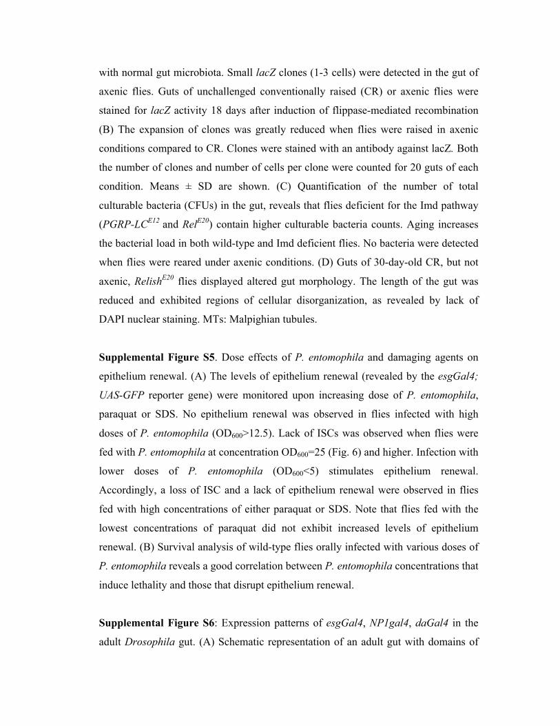
with normal gut microbiota. Small lacZ clones (1-3 cells) were detected in the gut of
axenic flies. Guts of unchallenged conventionally raised (CR) or axenic flies were
stained for lacZ activity 18 days after induction of flippase-mediated recombination
(B) The expansion of clones was greatly reduced when flies were raised in axenic
conditions compared to CR. Clones were stained with an antibody against lacZ. Both
the number of clones and number of cells per clone were counted for 20 guts of each
condition. Means ± SD are shown. (C) Quantification of the number of total
culturable bacteria (CFUs) in the gut, reveals that flies deficient for the Imd pathway
(PGRP-LCE12
and RelE20
) contain higher culturable bacteria counts. Aging increases
the bacterial load in both wild-type and Imd deficient flies. No bacteria were detected
when flies were reared under axenic conditions. (D) Guts of 30-day-old CR, but not
axenic, RelishE20
flies displayed altered gut morphology. The length of the gut was
reduced and exhibited regions of cellular disorganization, as revealed by lack of
DAPI nuclear staining. MTs: Malpighian tubules.
Supplemental Figure S5. Dose effects of P. entomophila and damaging agents on
epithelium renewal. (A) The levels of epithelium renewal (revealed by the esgGal4;
UAS-GFP reporter gene) were monitored upon increasing dose of P. entomophila,
paraquat or SDS. No epithelium renewal was observed in flies infected with high
doses of P. entomophila (OD600>12.5). Lack of ISCs was observed when flies were
fed with P. entomophila at concentration OD600=25 (Fig. 6) and higher. Infection with
lower doses of P. entomophila (OD600<5) stimulates epithelium renewal.
Accordingly, a loss of ISC and a lack of epithelium renewal were observed in flies
fed with high concentrations of either paraquat or SDS. Note that flies fed with the
lowest concentrations of paraquat did not exhibit increased levels of epithelium
renewal. (B) Survival analysis of wild-type flies orally infected with various doses of
P. entomophila reveals a good correlation between P. entomophila concentrations that
induce lethality and those that disrupt epithelium renewal.
Supplemental Figure S6: Expression patterns of esgGal4, NP1gal4, daGal4 in the
adult Drosophila gut. (A) Schematic representation of an adult gut with domains of
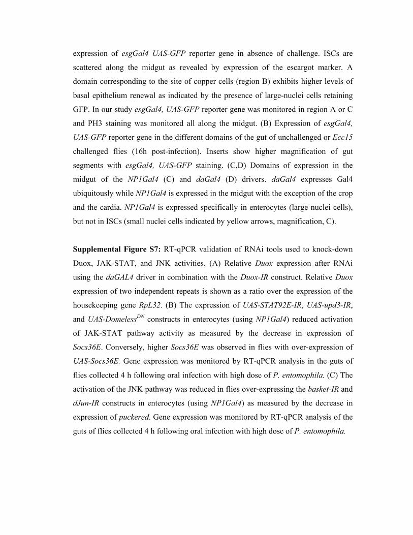
expression of esgGal4 UAS-GFP reporter gene in absence of challenge. ISCs are
scattered along the midgut as revealed by expression of the escargot marker. A
domain corresponding to the site of copper cells (region B) exhibits higher levels of
basal epithelium renewal as indicated by the presence of large-nuclei cells retaining
GFP. In our study esgGal4, UAS-GFP reporter gene was monitored in region A or C
and PH3 staining was monitored all along the midgut. (B) Expression of esgGal4,
UAS-GFP reporter gene in the different domains of the gut of unchallenged or Ecc15
challenged flies (16h post-infection). Inserts show higher magnification of gut
segments with esgGal4, UAS-GFP staining. (C,D) Domains of expression in the
midgut of the NP1Gal4 (C) and daGal4 (D) drivers. daGal4 expresses Gal4
ubiquitously while NP1Gal4 is expressed in the midgut with the exception of the crop
and the cardia. NP1Gal4 is expressed specifically in enterocytes (large nuclei cells),
but not in ISCs (small nuclei cells indicated by yellow arrows, magnification, C).
Supplemental Figure S7: RT-qPCR validation of RNAi tools used to knock-down
Duox, JAK-STAT, and JNK activities. (A) Relative Duox expression after RNAi
using the daGAL4 driver in combination with the Duox-IR construct. Relative Duox
expression of two independent repeats is shown as a ratio over the expression of the
housekeeping gene RpL32. (B) The expression of UAS-STAT92E-IR, UAS-upd3-IR,
and UAS-DomelessDN
constructs in enterocytes (using NP1Gal4) reduced activation
of JAK-STAT pathway activity as measured by the decrease in expression of
Socs36E. Conversely, higher Socs36E was observed in flies with over-expression of
UAS-Socs36E. Gene expression was monitored by RT-qPCR analysis in the guts of
flies collected 4 h following oral infection with high dose of P. entomophila. (C) The
activation of the JNK pathway was reduced in flies over-expressing the basket-IR and
dJun-IR constructs in enterocytes (using NP1Gal4) as measured by the decrease in
expression of puckered. Gene expression was monitored by RT-qPCR analysis of the
guts of flies collected 4 h following oral infection with high dose of P. entomophila.