A Pathobiont of the Microbiota Balances Host Colonization and Intestinal Inflammation
12
Cell Host & Microbe Article A Pathobiont of the Microbiota Balances Host Colonization and Intestinal Inflammation Janet Chow 1 and Sarkis K. Mazmanian 1, * 1 Division of Biology, California Institute of Technology, Pasadena, CA 91125, USA *Correspondence: [email protected] DOI 10.1016/j.chom.2010.03.004 SUMMARY The gastrointestinal tract harbors a diverse micro- biota that has coevolved with mammalian hosts. Though most associations are symbiotic or com- mensal, some resident bacteria (termed pathobionts) have the potential to cause disease. Bacterial type VI secretion systems (T6SSs) are one mechanism for forging host-microbial interactions. Here we reveal a protective role for the T6SS of Helicobacter hepati- cus, a Gram-negative bacterium of the intestinal microbiota. H. hepaticus mutants with a defective T6SS display increased numbers within intestinal epithelial cells (IECs) and during intestinal coloniza- tion. Remarkably, the T6SS directs an anti-inflamma- tory gene expression profile in IECs, and CD4+ T cells from mice colonized with T6SS mutants produce increased interleukin-17 in response to IECs present- ing H. hepaticus antigens. Thus, the H. hepaticus T6SS limits colonization and intestinal inflammation, promoting a balanced relationship with the host. We propose that disruption of such balances contributes to human disorders such as inflammatory bowel disease and colon cancer. INTRODUCTION The mammalian gastrointestinal (GI) tract represents an ecolog- ical niche of extraordinary microbial complexity (Hooper and Gordon, 2001). Humans and other mammals offer residence to 1000 microbial species, which form an interkingdom bionet- work known to provide nutritional, metabolic, and immunologic benefits to the host (Ley et al., 2006). Although the magnitude of this evolutionarily dynamic interaction is undeniably vast, surprisingly few molecules that mediate host-microbiota associ- ations have been described. Microbial secretion systems are universal mechanisms dedicated to molecular interactions between bacterial and host cells and can range from simple transporters to large, membrane-spanning structures. Type III secretion systems (T3SSs), T4SSs, and T6SSs function as multi- subunit complexes that translocate effector substrates across the double membrane of Gram-negative bacteria and act as a ‘‘needle and syringe’’ to inject molecules directly into eukary- otic cells (Raskin et al., 2006). The temporal and spatial profile of secreted molecules can have profound effects on host biology. Therefore, these secretion systems represent well- evolved microbial strategies capable of mediating molecular interactions between the microbiota and mammals. T6SS genes are present in over 25% of all sequenced bacterial genomes and have been largely associated with bacterial patho- genesis. In recent seminal studies, Mekalanos and colleagues reported that Vibrio cholerae requires T6SS genes to cause cytotoxicity in macrophages (Pukatzki et al., 2006). A set of mole- cules were identified as the first T6SS substrates: hemolysin cor- egulated protein (Hcp) and three valine-glycine-repeat (VgrG) proteins. Further evidence supporting a role for T6SSs in bacterial virulence has been demonstrated in other Gram-negative patho- gens including Pseudomonas aeruginosa (Mougous et al., 2006), enteroaggregative E. coli (Dudley et al., 2006), and Aeromonas hydrophila (Suarez et al., 2008). However, the function of bacterial T6SSs is not limited to eliciting disease. A growing body of research has shown that T6SSs may modulate virulence (Blader- groen et al., 2003; Das et al., 2002; Parsons and Heffron, 2005). Therefore, in some contexts T6SSs may modulate bacterial virulence in order to ‘‘balance’’ the relationship between microbe and host. T6SSs are almost exclusively found in a-, b-, and g-proteo- bacteria (Bingle et al., 2008). Helicobacter hepaticus is unique in that it appears to contain the only bacterial genome in the epsilon (3) subgroup of proteobacteria that encodes for a T6SS. H. hepaticus, a spiral microaerophilic bacterium, pro- motes inflammation in animal models of colon cancer and experimental colitis (inflammation of the colon). Interestingly, H. hepaticus only causes disease in immunocompromised animals that lack immune regulation and mount inflammatory responses toward intestinal bacteria (Erdman et al., 2009; Kull- berg et al., 2001). Pathologies elicited by H. hepaticus remark- ably resemble that of human disease based on molecular, cellular, and histological parameters. Furthermore, colonization with H. hepaticus causes a chronic inflammatory response similar to human inflammatory bowel disease (IBD), in contrast to infections with Citrobacter rodentium or Salmonella typhimu- rium, which induce acute and self-limiting infections. Therefore, the H. hepaticus-Mus musculus relationship represents a unique and invaluable tool in the study of intestinal pathologies highly similar to human disorders such as IBD and colon cancer. Interestingly, multiple studies have shown that H. hepaticus sustains long-term colonization of the lower GI tract of wild- type mice without causing intestinal disease (reviewed in Solnick and Schauer, 2001). In fact, H. hepaticus colonization is highly endemic in most laboratory animal facilities but is largely unno- ticed due to the absence of clinical disease. We have previously proposed that H. hepaticus acts as an intestinal pathobiont—i.e., Cell Host & Microbe 7, 265–276, April 22, 2010 ª2010 Elsevier Inc. 265
-
Upload
independent -
Category
Documents
-
view
6 -
download
0
Transcript of A Pathobiont of the Microbiota Balances Host Colonization and Intestinal Inflammation

Cell Host & Microbe
Article
A Pathobiont of the Microbiota BalancesHost Colonization and Intestinal InflammationJanet Chow1 and Sarkis K. Mazmanian1,*1Division of Biology, California Institute of Technology, Pasadena, CA 91125, USA
*Correspondence: [email protected]
DOI 10.1016/j.chom.2010.03.004
SUMMARY
The gastrointestinal tract harbors a diverse micro-biota that has coevolved with mammalian hosts.Though most associations are symbiotic or com-mensal, some resident bacteria (termed pathobionts)have the potential to cause disease. Bacterial type VIsecretion systems (T6SSs) are one mechanism forforging host-microbial interactions. Here we reveala protective role for the T6SS of Helicobacter hepati-cus, a Gram-negative bacterium of the intestinalmicrobiota. H. hepaticus mutants with a defectiveT6SS display increased numbers within intestinalepithelial cells (IECs) and during intestinal coloniza-tion. Remarkably, the T6SS directs an anti-inflamma-tory gene expression profile in IECs, and CD4+ T cellsfrom mice colonized with T6SS mutants produceincreased interleukin-17 in response to IECs present-ing H. hepaticus antigens. Thus, the H. hepaticusT6SS limits colonization and intestinal inflammation,promoting a balanced relationship with the host. Wepropose that disruption of such balances contributesto human disorders such as inflammatory boweldisease and colon cancer.
INTRODUCTION
The mammalian gastrointestinal (GI) tract represents an ecolog-
ical niche of extraordinary microbial complexity (Hooper and
Gordon, 2001). Humans and other mammals offer residence to
�1000 microbial species, which form an interkingdom bionet-
work known to provide nutritional, metabolic, and immunologic
benefits to the host (Ley et al., 2006). Although the magnitude
of this evolutionarily dynamic interaction is undeniably vast,
surprisingly few molecules that mediate host-microbiota associ-
ations have been described. Microbial secretion systems are
universal mechanisms dedicated to molecular interactions
between bacterial and host cells and can range from simple
transporters to large, membrane-spanning structures. Type III
secretion systems (T3SSs), T4SSs, and T6SSs function as multi-
subunit complexes that translocate effector substrates across
the double membrane of Gram-negative bacteria and act as
a ‘‘needle and syringe’’ to inject molecules directly into eukary-
otic cells (Raskin et al., 2006). The temporal and spatial profile
of secreted molecules can have profound effects on host
Cell
biology. Therefore, these secretion systems represent well-
evolved microbial strategies capable of mediating molecular
interactions between the microbiota and mammals.
T6SS genes are present in over 25% of all sequenced bacterial
genomes and have been largely associated with bacterial patho-
genesis. In recent seminal studies, Mekalanos and colleagues
reported that Vibrio cholerae requires T6SS genes to cause
cytotoxicity in macrophages (Pukatzki et al., 2006). A set of mole-
cules were identified as the first T6SS substrates: hemolysin cor-
egulated protein (Hcp) and three valine-glycine-repeat (VgrG)
proteins. Further evidence supporting a role for T6SSs in bacterial
virulence has been demonstrated in other Gram-negative patho-
gens including Pseudomonas aeruginosa (Mougous et al., 2006),
enteroaggregative E. coli (Dudley et al., 2006), and Aeromonas
hydrophila (Suarez et al., 2008). However, the function of bacterial
T6SSs is not limited to eliciting disease. A growing body of
research has shown that T6SSs may modulate virulence (Blader-
groen et al., 2003; Das et al., 2002; Parsons and Heffron, 2005).
Therefore, in some contexts T6SSs may modulate bacterial
virulence in order to ‘‘balance’’ the relationship between microbe
and host.
T6SSs are almost exclusively found in a-, b-, and g-proteo-
bacteria (Bingle et al., 2008). Helicobacter hepaticus is unique
in that it appears to contain the only bacterial genome in the
epsilon (3) subgroup of proteobacteria that encodes for a
T6SS. H. hepaticus, a spiral microaerophilic bacterium, pro-
motes inflammation in animal models of colon cancer and
experimental colitis (inflammation of the colon). Interestingly,
H. hepaticus only causes disease in immunocompromised
animals that lack immune regulation and mount inflammatory
responses toward intestinal bacteria (Erdman et al., 2009; Kull-
berg et al., 2001). Pathologies elicited by H. hepaticus remark-
ably resemble that of human disease based on molecular,
cellular, and histological parameters. Furthermore, colonization
with H. hepaticus causes a chronic inflammatory response
similar to human inflammatory bowel disease (IBD), in contrast
to infections with Citrobacter rodentium or Salmonella typhimu-
rium, which induce acute and self-limiting infections. Therefore,
the H. hepaticus-Mus musculus relationship represents a unique
and invaluable tool in the study of intestinal pathologies highly
similar to human disorders such as IBD and colon cancer.
Interestingly, multiple studies have shown that H. hepaticus
sustains long-term colonization of the lower GI tract of wild-
type mice without causing intestinal disease (reviewed in Solnick
and Schauer, 2001). In fact, H. hepaticus colonization is highly
endemic in most laboratory animal facilities but is largely unno-
ticed due to the absence of clinical disease. We have previously
proposed that H. hepaticus acts as an intestinal pathobiont—i.e.,
Host & Microbe 7, 265–276, April 22, 2010 ª2010 Elsevier Inc. 265

Cell Host & Microbe
Host-Microbiota Interactions by Type VI Secretion
a symbiont that is able to promote pathology only when specific
genetic or environmental conditions are altered in the host
(Mazmanian et al., 2008; Round and Mazmanian, 2009). The
concept of pathobionts is supported by clinical data which reveal
that in IBD patients with underlying genetic mutations, inflamma-
tion is targeted to specific members of the microbiota and not
to infectious pathogens (Packey and Sartor, 2009). T cell
responses and antibody reactivity during IBD target certain
subsets of symbiotic microbes such as Escherichia, Clostridium,
and Enterococcus species that are found in all people. Since
H. hepaticus displays a potentially pathogenic association with
its murine host similar to specific microbes found in humans,
and the T6SS appears to mediate both symbiotic and patho-
genic outcomes, we investigated a role for the H. hepaticus
T6SS during intestinal inflammation.
Herein we report that the T6SS of H. hepaticus mediates crit-
ical protective functions during association with its mammalian
host. In cell cultures, infection of intestinal epithelial cells (IECs)
with H. hepaticus T6SS mutants results in increased bacterial
association compared to wild-type bacteria. In animals, T6SS
mutants colonize the lower GI tract to a higher degree. Most
importantly, H. hepaticus defective in type VI secretion is unable
to restrain potent innate and adaptive immune responses in an
animal model of experimental colitis. Coculture experiments
with IECs presenting H. hepaticus antigens result in higher levels
of proinflammatory cytokines when incubated with T cells from
T6SS mutant-colonized animals compared to wild-type-colo-
nized animals. Thus, our findings reveal that H. hepaticus has
evolved a T6SS as a mechanism to actively maintain a nonpatho-
genic, symbiotic relationship in the GI tract by regulating bacte-
rial colonization and host inflammation. Disturbances in the
dynamic molecular interaction between gut bacteria and the
intestinal immune system therefore lead to exacerbated host
inflammation. As intestinal bacteria profoundly influence host
biology, our findings support an emerging hypothesis that alter-
ation in the composition of the microbiota, known as dysbiosis,
is a critical factor in various human inflammatory disorders
such as IBD and colon cancer.
RESULTS
H. hepaticus Possesses a Canonicaland Functional T6SSThe genome of Helicobacter hepaticus contains a 71 kb genomic
island (termed HHGI1 for Helicobacter hepaticus genomic island
1), which includes genes homologous to virulence factors (Suer-
baum et al., 2003). Isolates missing the pathogenicity island are
highly attenuated for inducing disease, and deletion of a 19 gene
segment of HHGI1 reduces virulence (Ge et al., 2008). However,
the functions of the gene products encoded within HHGI1 remain
unclear. In recent years, studies have identified a protein secre-
tion system referred to as a type VI secretion system (T6SS)
(Raskin et al., 2006). We report herein that HHGI1 encodes for
a set of T6SS genes that are homologous to those found in
V. cholerae, P. aeruginosa, and other Gram-negative microor-
ganisms (see Table S1 available online). H. hepaticus contains
12 homologous T6SS genes that are clustered and arranged
in a genomic organization similar to other T6SSs (Figure 1A).
A homolog of the icmF gene, thought to encode for a structural
266 Cell Host & Microbe 7, 265–276, April 22, 2010 ª2010 Elsevier In
transmembrane protein of virtually all identified T6SSs, lies
within this genomic locus (H. hepaticus gene HH0252). Further-
more, HHGI1 encodes for homologs of Hcp (HH0243) and three
VgrG proteins (HH0237, HH0242, HH0291), highly conserved
translocated substrates that play key roles in T6SSs.
We constructed insertional mutants in the icmF or hcp genes,
as previous studies in several pathogens have shown that dele-
tion of these genes leads to T6SS defects. An antibiotic selection
marker (erythromycin acetyl transferase, eryR) (Mehta et al.,
2007) was inserted within each open reading frame by homolo-
gous recombination. PCR amplification verified integration of
the eryR gene to create mutants DIcmF and DHcp (Figure 1B).
To validate a functional defect in the T6SS, in vitro cultured
bacteria were assayed by immunoblot for Hcp. In wild-type
bacteria, Hcp was detected in the culture supernatant and cell
pellet fraction as expected (Figure 1C). However, in the DIcmF
mutant, Hcp was absent from supernatant fractions and accu-
mulated to a higher degree in the cell pellet, indicating that
Hcp is still produced but not secreted in the absence of a func-
tional T6SS. Therefore, deletion of the icmF homolog results in
a secretion defect for Hcp, demonstrating functional inactivation
of the T6SS.
Bacteria possessing T6SSs bind host cells into which
substrates can be injected. Since H. hepaticus colonizes the
murine gut and is in close association with intestinal crypts
(see data below), we investigated the association of H. hepaticus
with murine IECs. H. hepaticus was cocultured with the mouse
IEC line MODE-K (Vidal et al., 1993) and examined for transloc-
alization of VgrG by confocal immunofluorescence microscopy.
Labeling of MODE-K cells with antisera specific for the T6SS
substrate VgrG showed diffuse staining upon incubation with
wild-type bacteria (Figures 1D and 1E). Conversely, VgrG
labeling of MODE-K cells incubated with DIcmF mutant showed
punctate staining patterns suggesting VgrG produced by T6SS
mutants is bacterially associated. As whole bacterial antisera
raised against H. hepaticus reveals a punctate staining pattern
as well (Figure 1D), the diffuse staining of VgrG in wild-type
bacteria suggests substrate translocation in the presence of
MODE-K cells (Figure 1E). Taken together, our results demon-
strate that deletion of T6SS genes results in mutants unable to
secrete effector substrates during bacterial growth and cocul-
ture with IECs.
H. hepaticus Is Internalized into Murine IECsMany enteric pathogens such as Listeria monocytogenes and
Shigella flexneri have been shown to enter IECs during infection.
In several cases, T3SSs either facilitate entry or are required for
internalization (Galan and Wolf-Watz, 2006). To determine if
H. hepaticus enters IECs, we infected bacteria with MODE-K
cells and observed that both wild-type and T6SS mutants were
capable of entering host cells (Figure 2A). Confocal microscopy
demonstrated that all strains were found associated with the cell
surface as well. Z stacks reconstructed from confocal images
further revealed significant numbers of intact bacteria within
the intracellular compartment of IECs (Movie S1). To determine
if uptake was an active process, we incubated wild-type and
T6SS mutants with MODE-K cells for 30 min in the presence
or absence of cytochalasin D, an inhibitor of actin polymeriza-
tion. Cells were subsequently treated with gentamicin to kill
c.

BWT ΔIcmFPellet Supernatant
Hcp
ΔHcp WT ΔIcmF ΔHcpC
A
WTΔIcmF ΔHcpWT4.0 kb3.0 kb
2.0 kb1.6 kb
1.0 kb
icmF hcp
D No bacteria WT ΔΔIcmF ΔΔVgrG
αα-VgrG
αα-H.hepaticus
HH0246HH0242 HH0243 HH0244 HH0245 HH0247 HH0248 HH0249 HH0250 HH0251 HH0252
IcmFHcpVgrG
HH0291HH0237
VgrG VgrG
HH0246HH0242 HH0243 HH0244 HH0245 HH0247 HH0248 HH0249 HH0250 HH0251 HH0252
IcmFHcpVgrG
HH0291HH0237
VgrG VgrG
E
αα-VgrG
WT ΔΔIcmF
Figure 1. H. hepaticus Encodes for a Functional T6SS
(A) Schematic diagram of the genetic organization of H. hepaticus T6SS genes. Gray arrows represent Hcp and VgrG genes, cross-hatched arrow indicates
IcmF homolog, black arrows represent T6SS homologs of unknown function, and white arrow indicates a gene nonhomologous to other T6SS genes. See
also Table S1.
(B) Genomic DNA collected from mid-log cultures of WT, DIcmF, or DHcp H. hepaticus was amplified by PCR using icmF-or hcp-specific primers. Insertion of the
eryR gene was detected by a 1.1 kb increase in the resulting PCR band.
(C) Hcp is undetected in supernatants from DIcmF and DHcp T6SS mutants. Equal amounts of mid-log bacterial cultures of WT, DIcmF, or DHcp were centrifuged
to separate bacterial pellets and supernatant. Supernatants were subsequently filtered to ensure removal of all bacteria. Bacterial pellets and supernatants were
analyzed by western blot. Membranes were blotted with anti-Hcp antibody.
(D and E) Confocal images of bacteria incubated with MODE-K cells. WT, DIcmF, or DVgrG (DHH0242) H. hepaticus were incubated with MODE-K cells for 5 hr.
MODE-K cells were subsequently rinsed with PBS, fixed in 4% PFA, and stained for the eukaryotic cell membrane marker wheat germ agglutinin (red) and either
H. hepaticus or VgrG (green). Outlined regions for WT and DIcmF in (D) are shown at higher magnification (E). Scale bar represents 20 mm.
Cell Host & Microbe
Host-Microbiota Interactions by Type VI Secretion
extracellular bacteria and plated for colony-forming units (CFUs).
Figure 2B reveals that while all strains could be recovered from
the intracellular compartment of MODE-K cells, cytochalasin D
Cell
inhibited bacterial uptake. Therefore, H. hepaticus appears to
be actively internalized into cultured IECs through a requirement
for actin rearrangement.
Host & Microbe 7, 265–276, April 22, 2010 ª2010 Elsevier Inc. 267

+ Cytochalasin D
WT ΔIcmF ΔHcp0.00
0.01
0.02
0.03B
ANo bacteria WT ΔΔIcmF ΔΔHcp
C0.5hr 3hr 6hr
**
0.000.050.100.150.200.250.300.350.40 **
0.00
0.03
0.06
0.09
0.12
0.15
0.18
Ratio
0.00
0.03
0.06
0.09
0.12
0.15
0.18
****
Intracellular
0.05.010.015.020.025.030.035.040.0
Ratio
0.05.010.015.020.025.030.035.040.0
0.05.010.015.020.025.030.035.040.0
*
*
Cell-associated
E35302520151050
**
**
Ratio
Ratio
D0.5hr 3hr 6hr
WT ΔIcmF ΔHcp WT ΔIcmF ΔHcp
WT ΔIcmF ΔHcp WT ΔIcmF ΔHcp WT ΔIcmF ΔHcp
WT ΔIcmF ΔHcp WT ΔIcmF ΔHcp WT ΔIcmF ΔHcp
Figure 2. T6SS Mutants Display Higher
Intracellular and Cell-Associated Accumu-
lation in MODE-K Cells
(A) Confocal image of bacteria inside MODE-K
cells. WT, DIcmF, or DHcp was incubated with
MODE-K for 6 hr. MODE-K cells were rinsed with
PBS, fixed in 4% PFA, and stained for H. hepaticus
(green) and the eukaryotic cell membrane marker
wheat germ agglutinin (red). Scale bar represents
30 mm. See also Movie S1.
(B) Cytochalasin D inhibits uptake of H. hepaticus.
Prior to incubation with bacteria, MODE-K cells
were treated with 10 mM cytochalasin D for 1 hr.
Bacteria were added at an moi of 100. After
0.5 hr incubation at 37�C under microaerophilic
conditions, cells were treated with 100 mg/ml
gentamicin and intracellular bacteria plated for
enumeration. Results are expressed as CFUs
of intracellular bacteria divided by number of
MODE-K cells. Error bars indicate SEM from three
experiments.
(C and D) Gentamicin protection assay in which
MODE-K cells were incubated with bacteria at an
moi of 100. After 0.5, 3, or 6 hr incubation, media
was replaced with 100 mg/ml gentamicin for
enumeration of intracellular bacteria (C) or without
gentamicin for cell-associated bacteria (D). Cells
were washed and bacteria plated for quantifica-
tion. Ratios are expressed as CFUs of bacteria
divided by number of MODE-K cells. Error bars
indicate SEM from three to five experiments.
*p < 0.05, **p < 0.01 versus WT.
(E) Increased adherence of T6SS mutants is not
dependent on bacterial internalization. Prior to
coculture, MODE-K cells were treated with 10 mM
cytochalasin D. Bacteria were added at an moi of
100 for 6 hr at 37�C under microaerophilic condi-
tions. Bacteria were plated for enumeration.
Results are expressed as CFUs of bacteria divided
by number of MODE-K cells. Error bars indicate
SD from two experiments. **p < 0.01 versus WT.
See also Figure S1.
Cell Host & Microbe
Host-Microbiota Interactions by Type VI Secretion
T6SS Mutants Display Increased CellAssociation within IECsWe sought to determine if the T6SS affects bacterial internaliza-
tion. MODE-K cells were incubated with H. hepaticus, treated
with (or without) gentamicin, and plated for bacterial enumera-
tion. At early time points (30 min), no differences were observed
in the proportions of intracellular and cell-associated bacteria
between wild-type and T6SS mutants (Figures 2C and 2D).
Remarkably, by 3 and 6 hr, DIcmF and DHcp had significantly
higher levels of intracellular bacteria (Figure 2C). In addition,
268 Cell Host & Microbe 7, 265–276, April 22, 2010 ª2010 Elsevier Inc.
T6SS mutants displayed greater numbers
of MODE-K-associated bacteria after 6 hr
(Figure 2D). Total numbers of bacteria
recovered from cocultures (which in-
cludes all non-cell-associated bacteria)
as well as cultures grown in the same
media without MODE-K cells were com-
parable (Figure S1A and data not shown),
demonstrating that the mutations did not
affect bacterial growth. These results reveal that the T6SS of
H. hepaticus limits intracellular bacterial numbers within IECs.
To investigate if the increased cell association by T6SS
mutants was due to increased adherence, we blocked internali-
zation of H. hepaticus into MODE-K cells. Cytochalasin D was
added for the duration of the coculture (6 hr). In the absence of
bacterial internalization, T6SS mutants still exhibited an increase
in adherence to the extracellular surface of MODE-K cells
(Figure 2E). An increase in cell adherence in the absence of inter-
nalization was also observed for T6SS mutants when MODE-K
3500
WT ΔIcmF ΔHcp
CFU/mg
050010001500200025003000
400045005000
**
*
B
0246810121416182022
Control WT ΔIcmF ΔHcp
RelativeUnits
**
**
Control WT ΔIcmF ΔHcp
**
*
Colon Cecum
0246810121416182022
A
C No bacteria WT ΔΔIcmF
D 0.025
0.020
0.015
0.010
0.005
0WT ΔIcmF
**
WT ΔIcmFControl
GentamicinControls
Ratio
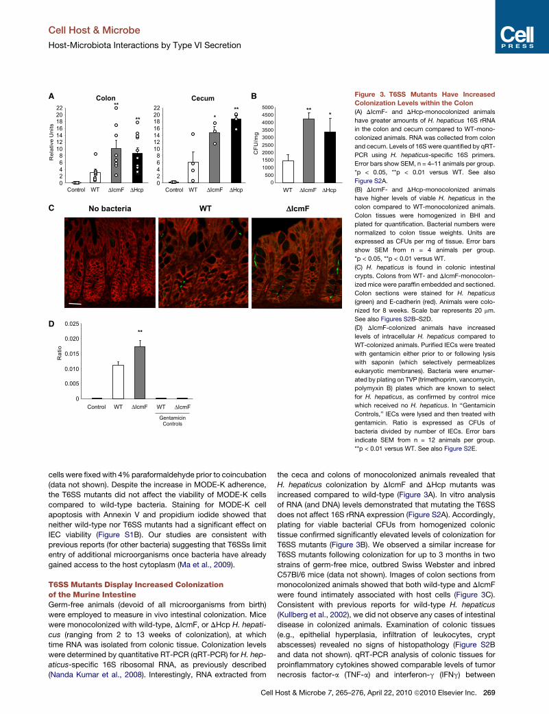
Figure 3. T6SS Mutants Have Increased
Colonization Levels within the Colon
(A) DIcmF- and DHcp-monocolonized animals
have greater amounts of H. hepaticus 16S rRNA
in the colon and cecum compared to WT-mono-
colonized animals. RNA was collected from colon
and cecum. Levels of 16S were quantified by qRT-
PCR using H. hepaticus-specific 16S primers.
Error bars show SEM, n = 4–11 animals per group.
*p < 0.05, **p < 0.01 versus WT. See also
Figure S2A.
(B) DIcmF- and DHcp-monocolonized animals
have higher levels of viable H. hepaticus in the
colon compared to WT-monocolonized animals.
Colon tissues were homogenized in BHI and
plated for quantification. Bacterial numbers were
normalized to colon tissue weights. Units are
expressed as CFUs per mg of tissue. Error bars
show SEM from n = 4 animals per group.
*p < 0.05, **p < 0.01 versus WT.
(C) H. hepaticus is found in colonic intestinal
crypts. Colons from WT- and DIcmF-monocolon-
ized mice were paraffin embedded and sectioned.
Colon sections were stained for H. hepaticus
(green) and E-cadherin (red). Animals were colo-
nized for 8 weeks. Scale bar represents 20 mm.
See also Figures S2B–S2D.
(D) DIcmF-colonized animals have increased
levels of intracellular H. hepaticus compared to
WT-colonized animals. Purified IECs were treated
with gentamicin either prior to or following lysis
with saponin (which selectively permeablizes
eukaryotic membranes). Bacteria were enumer-
ated by plating on TVP (trimethoprim, vancomycin,
polymyxin B) plates which are known to select
for H. hepaticus, as confirmed by control mice
which received no H. hepaticus. In ‘‘Gentamicin
Controls,’’ IECs were lysed and then treated with
gentamicin. Ratio is expressed as CFUs of
bacteria divided by number of IECs. Error bars
indicate SEM from n = 12 animals per group.
**p < 0.01 versus WT. See also Figure S2E.
Cell Host & Microbe
Host-Microbiota Interactions by Type VI Secretion
cells were fixed with 4% paraformaldehyde prior to coincubation
(data not shown). Despite the increase in MODE-K adherence,
the T6SS mutants did not affect the viability of MODE-K cells
compared to wild-type bacteria. Staining for MODE-K cell
apoptosis with Annexin V and propidium iodide showed that
neither wild-type nor T6SS mutants had a significant effect on
IEC viability (Figure S1B). Our studies are consistent with
previous reports (for other bacteria) suggesting that T6SSs limit
entry of additional microorganisms once bacteria have already
gained access to the host cytoplasm (Ma et al., 2009).
T6SS Mutants Display Increased Colonizationof the Murine IntestineGerm-free animals (devoid of all microorganisms from birth)
were employed to measure in vivo intestinal colonization. Mice
were monocolonized with wild-type, DIcmF, or DHcp H. hepati-
cus (ranging from 2 to 13 weeks of colonization), at which
time RNA was isolated from colonic tissue. Colonization levels
were determined by quantitative RT-PCR (qRT-PCR) for H. hep-
aticus-specific 16S ribosomal RNA, as previously described
(Nanda Kumar et al., 2008). Interestingly, RNA extracted from
Cell
the ceca and colons of monocolonized animals revealed that
H. hepaticus colonization by DIcmF and DHcp mutants was
increased compared to wild-type (Figure 3A). In vitro analysis
of RNA (and DNA) levels demonstrated that mutating the T6SS
does not affect 16S rRNA expression (Figure S2A). Accordingly,
plating for viable bacterial CFUs from homogenized colonic
tissue confirmed significantly elevated levels of colonization for
T6SS mutants (Figure 3B). We observed a similar increase for
T6SS mutants following colonization for up to 3 months in two
strains of germ-free mice, outbred Swiss Webster and inbred
C57Bl/6 mice (data not shown). Images of colon sections from
monocolonized animals showed that both wild-type and DIcmF
were found intimately associated with host cells (Figure 3C).
Consistent with previous reports for wild-type H. hepaticus
(Kullberg et al., 2002), we did not observe any cases of intestinal
disease in colonized animals. Examination of colonic tissues
(e.g., epithelial hyperplasia, infiltration of leukocytes, crypt
abscesses) revealed no signs of histopathology (Figure S2B
and data not shown). qRT-PCR analysis of colonic tissues for
proinflammatory cytokines showed comparable levels of tumor
necrosis factor-a (TNF-a) and interferon-g (IFNg) between
Host & Microbe 7, 265–276, April 22, 2010 ª2010 Elsevier Inc. 269
-2.0 0 2.0Log(Ratio)
WT ΔIcmFA
WT
2,176ΔΔIcmF195
Downregulated
17
WT
1,072ΔΔIcmF566
Upregulated
15
B
Downregulated Upregulated
Number of Genes150 100 50 0 50 100 150 WT
ΔIcmF
MHC I-mediated immunityDevelopmental processesDNA repairCell differentiationCell structure & motilityOrganelle biogenesisMHC II-mediated immunityT-cell mediated immunityCell cycleCytokine/chemokine signaling
C
WTGene TitleGeneFold Change
-2.07-1.52
-3.23
-4.13-4.05
-8.11
Tlr4Nfkb1
Tlr3
ApcIL17ra
Ki67
Toll-like receptor 4Nfkb1, p105
Toll-like receptor 3
Adenomatosis polyposis coliInterleukin 17 receptor A
Ki-67
D
RelativeUnits
Con WT ΔIcmF
TLR4
0
1
2
3
4
5
6
0123456789 APC
Con WT ΔIcmF0
1
2
3
4
5 IL-17RA
Con WT ΔIcmF
E
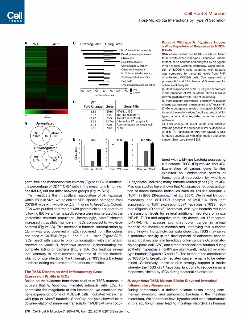
Figure 4. Wild-type H. hepaticus Induces
a Wide Repertoire of Responses in MODE-
K Cells
RNA was harvested from MODE-K cells incubated
for 6 hr with either wild-type H. hepaticus, DIcmF
mutant, or no bacteria and analyzed by an Agilent
Whole Mouse Genome Microarray. Gene expres-
sion of MODE-K cells incubated with bacteria
was compared to transcript levels from RNA
of untreated MODE-K cells. Only genes with a
p value <0.5 and fold change >1.5 were used for
subsequent analysis.
(A) Heat-map analysis of MODE-K gene expression
in the presence of WT or DIcmF shows massive
downregulation by wild-type H. hepaticus.
(B) Venn diagram showing up- and down regulation
of gene expression in the presence of WT or DIcmF.
(C) Gene ontogeny analysis of changes in MODE-K
transcript levels for various functional groups. Wild-
type bacteria downregulate numerous cellular
pathways.
(D) Fold change of select innate and adaptive
immune genes in the presence of WT H. hepaticus.
(E) qRT-PCR analysis of RNA from MODE-K cells
for genes associated with inflammation and colon
cancer. Error bars show SEM.
Cell Host & Microbe
Host-Microbiota Interactions by Type VI Secretion
germ-free and monocolonized animals (Figure S2C). In addition,
the percentage of CD4+TCRb+ cells in the mesenteric lymph no-
des (MLNs) did not differ between groups (Figure S2D).
To investigate the intracellular association of H. hepaticus
within IECs in vivo, we colonized SPF (specific pathogen-free)
C57Bl/6 mice with wild-type, DIcmF, or no H. hepaticus. Colonic
IECs were purified and treated with gentamicin either prior to or
following IEC lysis. Internalized bacteria were enumerated as the
gentamicin-resistant population. Interestingly, DIcmF showed
increased intracellular numbers in IECs compared to wild-type
bacteria (Figure 3D). This increase in bacterial internalization by
DIcmF was also observed in IECs recovered from the colons
and ceca of C57Bl/6 Rag1�/� and IL-10�/� mice (Figure S2E).
IECs lysed with saponin prior to incubation with gentamicin
showed no viable H. hepaticus bacteria, demonstrating the
complete killing of bacteria (Figure 3D). Our findings reveal
that, contrary to most secretion systems of enteric bacteria
which promote infections, the H. hepaticus T6SS limits bacterial
numbers during colonization of the mouse intestine.
The T6SS Directs an Anti-Inflammatory GeneExpression Profile in IECsBased on the evidence from these studies of T6SS mutants, it
appears that H. hepaticus intimately interacts with IECs. To
appreciate the magnitude of this interaction, we examined the
gene expression profile of MODE-K cells incubated with either
wild-type or DIcmF bacteria. GeneChip analysis showed clear
downregulation of numerous transcripts in MODE-K cells cocul-
270 Cell Host & Microbe 7, 265–276, April 22, 2010 ª2010 Elsevier Inc.
tured with wild-type bacteria possessing
a functional T6SS (Figures 4A and 4B).
Examination of various gene families
exhibited an unmistakable pattern of
transcriptional repression by wild-type
H. hepaticus, including many immune-related genes (Figure 4C).
Previous studies have shown that H. hepaticus reduces activa-
tion of innate immune molecules such as Toll-like receptor 4
(TLR4) in IECs (Sterzenbach et al., 2007). We reveal by both
microarray and qRT-PCR analysis of MODE-K RNA that
suppression of TLR4 expression by H. hepaticus is T6SS medi-
ated (Figures 4D and 4E). Moreover, wild-type bacteria reduced
the transcript levels for several additional mediators of innate
(NF-kB, TLR3) and adaptive immunity (interleukin-17 receptor,
IL-17RA). H. hepaticus promotes colon cancer in animal
models; the molecular mechanisms underlying this outcome
are unknown. Intriguingly, our data show that T6SS may serve
a protective activity in the development of colorectal cancer,
as a critical oncogene in hereditary colon cancers (Adenomato-
sis polyposis coli, APC) and a marker for cell proliferation during
epithelial hyperplasia (Ki-67) are significantly reduced by wild-
type bacteria (Figures 4D and 4E). The extent of the contribution
by T6SS to H. hepaticus-mediated cancer remains to be deter-
mined. Collectively, these studies strongly support a model
whereby the T6SS of H. hepaticus functions to reduce immune
responses elicited by IECs during bacterial colonization.
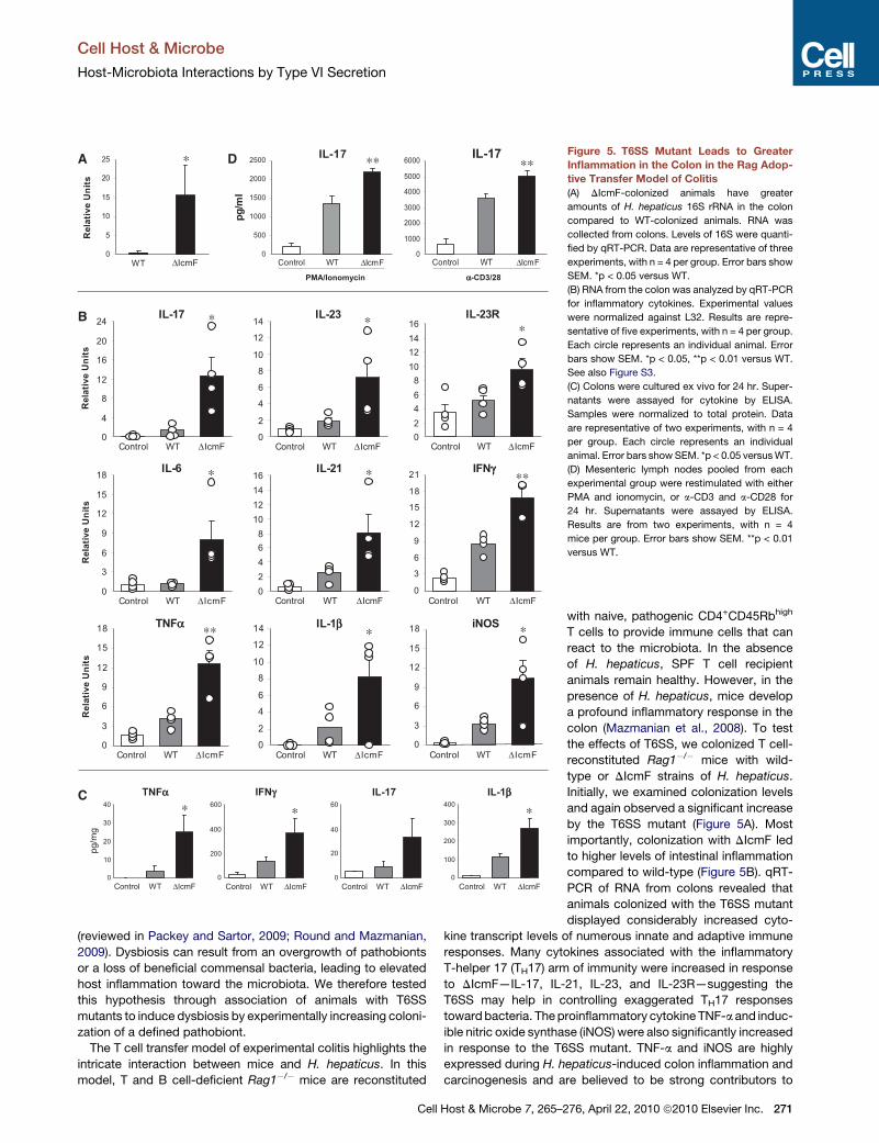
H. hepaticus T6SS Mutant Elicits Elevated IntestinalInflammatory ResponsesDuring homeostasis, a defined balance exists among com-
mensal, symbiotic, and pathobiotic bacteria in the intestinal
microbiota. We and others have hypothesized that disturbances
in this equilibrium may lead to intestinal disorders in humans
IL-23R
0246810121416
Control WT ΔIcmF
∗IL-17
0
4
8
12
16
20
24
Control WT ΔIcmF
RelativeUnits
∗
0
3
6
9
12
15
18 ∗∗
Control WT ΔIcmF
RelativeUnits
TNFαα
IL-21
0246810121416
Control WT ΔIcmF
∗
IL-23
0
2
4
6
8
10
12
14
Control WT ΔIcmF
∗
0
3
6
9
12
15
18 ∗
Control WT ΔIcmF
iNOS
0
3
6
9
12
15
18
Control WT
RelativeUnits
IL-6
ΔIcmF
∗
02
46
810
1214 ∗
IL-1ββ
Control WT ΔIcmF
0
3
6
9
12
15
18
21 ∗∗IFNγγ
Control WT ΔIcmF
0
5
10
15
20
25
WT
∗
ΔIcmFPMA/Ionomycin
0
500
1000
1500
2000
2500
Control WT ΔIcmF
IL-17
pg/ml
0
1000
2000
3000
4000
5000
6000
Control WT ΔIcmF
IL-17
αα-CD3/28
RelativeUnits
B
A D
C∗
0
10
20
30
40
pg/mg
TNFαα
Control WT ΔIcmF0
20
40
60IL-17
Control WT ΔIcmF
∗
0
200
400
600IFNγγ
Control WT ΔIcmF
∗
0
100
200
300
400IL-1ββ
Control WT ΔIcmF
∗∗ ∗∗Figure 5. T6SS Mutant Leads to Greater
Inflammation in the Colon in the Rag Adop-
tive Transfer Model of Colitis
(A) DIcmF-colonized animals have greater
amounts of H. hepaticus 16S rRNA in the colon
compared to WT-colonized animals. RNA was
collected from colons. Levels of 16S were quanti-
fied by qRT-PCR. Data are representative of three
experiments, with n = 4 per group. Error bars show
SEM. *p < 0.05 versus WT.
(B) RNA from the colon was analyzed by qRT-PCR
for inflammatory cytokines. Experimental values
were normalized against L32. Results are repre-
sentative of five experiments, with n = 4 per group.
Each circle represents an individual animal. Error
bars show SEM. *p < 0.05, **p < 0.01 versus WT.
See also Figure S3.
(C) Colons were cultured ex vivo for 24 hr. Super-
natants were assayed for cytokine by ELISA.
Samples were normalized to total protein. Data
are representative of two experiments, with n = 4
per group. Each circle represents an individual
animal. Error bars show SEM. *p < 0.05 versus WT.
(D) Mesenteric lymph nodes pooled from each
experimental group were restimulated with either
PMA and ionomycin, or a-CD3 and a-CD28 for
24 hr. Supernatants were assayed by ELISA.
Results are from two experiments, with n = 4
mice per group. Error bars show SEM. **p < 0.01
versus WT.
Cell Host & Microbe
Host-Microbiota Interactions by Type VI Secretion
(reviewed in Packey and Sartor, 2009; Round and Mazmanian,
2009). Dysbiosis can result from an overgrowth of pathobionts
or a loss of beneficial commensal bacteria, leading to elevated
host inflammation toward the microbiota. We therefore tested
this hypothesis through association of animals with T6SS
mutants to induce dysbiosis by experimentally increasing coloni-
zation of a defined pathobiont.
The T cell transfer model of experimental colitis highlights the
intricate interaction between mice and H. hepaticus. In this
model, T and B cell-deficient Rag1�/� mice are reconstituted
Cell Host & Microbe 7, 265–2
with naive, pathogenic CD4+CD45Rbhigh
T cells to provide immune cells that can
react to the microbiota. In the absence
of H. hepaticus, SPF T cell recipient
animals remain healthy. However, in the
presence of H. hepaticus, mice develop
a profound inflammatory response in the
colon (Mazmanian et al., 2008). To test
the effects of T6SS, we colonized T cell-
reconstituted Rag1�/� mice with wild-
type or DIcmF strains of H. hepaticus.
Initially, we examined colonization levels
and again observed a significant increase
by the T6SS mutant (Figure 5A). Most
importantly, colonization with DIcmF led
to higher levels of intestinal inflammation
compared to wild-type (Figure 5B). qRT-
PCR of RNA from colons revealed that
animals colonized with the T6SS mutant
displayed considerably increased cyto-
kine transcript levels of numerous innate and adaptive immune
responses. Many cytokines associated with the inflammatory
T-helper 17 (TH17) arm of immunity were increased in response
to DIcmF—IL-17, IL-21, IL-23, and IL-23R—suggesting the
T6SS may help in controlling exaggerated TH17 responses
toward bacteria. The proinflammatory cytokine TNF-a and induc-
ible nitric oxide synthase (iNOS) were also significantly increased
in response to the T6SS mutant. TNF-a and iNOS are highly
expressed during H. hepaticus-induced colon inflammation and
carcinogenesis and are believed to be strong contributors to
76, April 22, 2010 ª2010 Elsevier Inc. 271

Cell Host & Microbe
Host-Microbiota Interactions by Type VI Secretion
disease (Erdman et al., 2009). Interestingly, there was no marked
difference in intestinal pathology by measures of cell prolifera-
tion, cellular infiltrates, and abscess formation between wild-
type and DIcmF mutant-colonized animals (Figure S3A). Histopa-
thology analysis by a blinded pathologist verified similar colitis
scores between wild-type and mutant-colonized animals
(Figure S3B). Although more subtle phenotypes cannot be
excluded, the lack of increased disease by T6SS is not surprising
given the fact that wild-type bacteria elicit very pronounced
disease in the T cell transfer model. To measure inflammatory
protein levels, organ cultures in which unstimulated colon
sections are cultured ex vivo, and supernatants assayed by
ELISA showed an increase in the inflammatory molecules
TNF-a, IL-1b, and IL-17 in tissues harvested from DIcmF mutant-
colonized animals compared to wild-type (Figure 5C). Further-
more, MLN cells from DIcmF-colonized animals that were
restimulated in vitro with PMA/ionomycin or T cell stimuli
(a-CD3/a-CD28) released increased IL-17 during in vitro cultures
(Figure 5D). Analysis of the ceca also showed similar patterns of
increased T6SS mutant colonization and elevated proinflamma-
tory cytokine responses (Figures S3C–S3E). Collectively, these
results reveal that experimentally induced dysbiosis results in
increased inflammatory responses from both the innate and
adaptive immune systems, and the T6SS of H. hepaticus func-
tions to reduce intestinal inflammation during colonization.
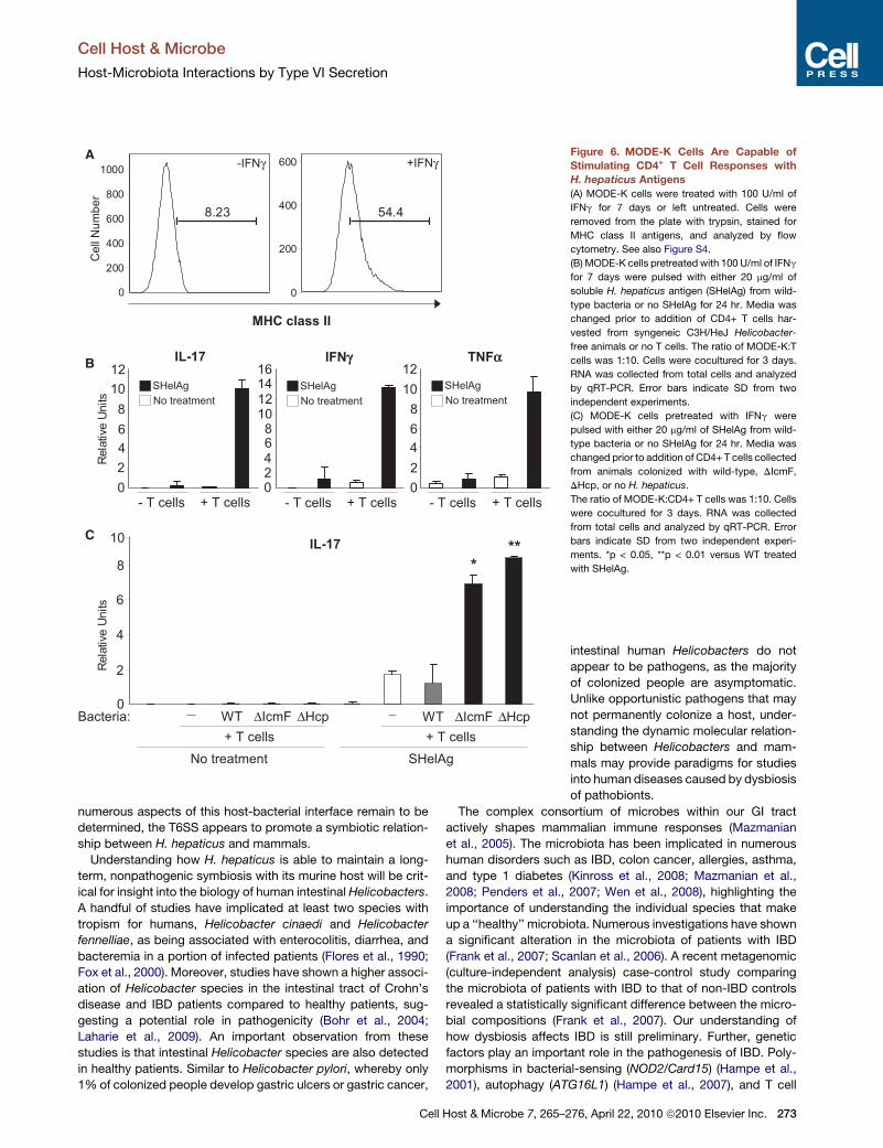
IECs Are Capable of Stimulating CD4+ T Cell Responseswith H. hepaticus AntigenIn the GI tract, IECs form a monolayer barrier that separates
luminal contents from underlying host cells. Though IECs con-
tribute to innate immunity by sensing through TLRs and secreting
antimicrobial peptides, IECs are generally not believed to mediate
adaptive immunity, unlike professional antigen-presenting cells
(APCs) such as dendritic cells and macrophages. Interestingly,
studies have shown that IECs are capable of expressing major
histocompatibility complex (MHC) class II proteins (Bland and
Warren, 1986; Mayer et al., 1991), and IEC presentation of
antigens to CD4+ T cells via MHC class II molecules can result
in lymphocyte proliferation (Westendorf et al., 2009; Vidal
et al., 1993). MODE-K cells increase MHC class II expression
in response to IFNg treatment, when stained with antibodies
directed against the I-Ak haplotype (Figure 6A) or to a nonpolymor-
phic region of the I-A molecule (Figure S4). The T cell activating
costimulatory molecules CD80 (B7.1) and CD86 (B7.2) were also
increased (data not shown). To determine if IECs could present
H. hepaticus antigens to T cells, MODE-K cells treated for
7 days with IFNg were pulsed with soluble Helicobacter antigens
(SHelAg). After 24 hr, MODE-K cells were washed and cocultured
with CD4+ T cells from MLNs of SPF Helicobacter-free C3H/HeJ
animals. Total RNA was analyzed by qRT-PCR for cytokine levels.
Cocultures that had been treatedwithSHelAg showed an increase
in numerous proinflammatory cytokine transcripts, such as IL-17,
IFNg, and TNF-a (Figure 6B). These responses are T cell specific,
as no cytokine production was observed in the absence of T cells.
Antigen-Specific CD4+ T Cell Responses Are Increasedin Animals Colonized with H. hepaticus T6SS MutantsWe investigated the hypothesis that IEC presentation of H. hep-
aticus antigens elicits increased proinflammatory responses
272 Cell Host & Microbe 7, 265–276, April 22, 2010 ª2010 Elsevier In
from CD4+ T cells from animals colonized with T6SS mutants.
MODE-K cells were pulsed with SHelAg or left untreated,
washed, and incubated with purified CD4+ T cells from mice
colonized with either wild-type, DIcmF, DHcp, or no H. hepati-
cus. In the presence of SHelAg, T cells from uncolonized and
wild-type H. hepaticus-colonized animals produced comparable
levels of IL-17 (Figure 6C). This illustrates that previous exposure
to H. hepaticus antigens does not augment immune responses
relative to naive animals, consistent with our findings that wild-
type bacteria do not induce inflammation. In contrast, CD4+
T cells harvested from both T6SS mutant-colonized animals
elicited significantly increased levels of IL-17 compared to cells
from wild-type-colonized animals. This response was antigen
specific, as no cytokine was produced in the absence of SHelAg.
Therefore, colonization of animals with T6SS mutants leads to
the generation of an increased Th17 cell response in the gut,
revealing that H. hepaticus evolved this molecular mechanism
to restrain unwanted intestinal inflammation. As Th17 cell
responses are important mediators of IBD and colon cancer in
experimental animals (Hue et al., 2006), our data suggest that
increased gut inflammation in reaction to dysbiosis of the micro-
biota may be crucial in the onset and/or progression of human
intestinal diseases.
DISCUSSION
Host-bacterial interactions (whether beneficial or harmful) are
defined by a dynamic exchange of molecules that mediate
various biological outcomes. We characterize herein that the
T6SS of H. hepaticus limits colonization of animals and actively
suppresses innate and adaptive immune responses. Although
T6SSs have been largely studied in the context of bacterial viru-
lence, growing evidence supports the notion that T6SSs may
have also evolved for nonpathogenic purposes in symbiotic
bacteria. Previous studies have shown that H. hepaticus can
reduce activation of TLR4 and TLR5 (Sterzenbach et al., 2007).
Intriguingly, Kullberg et al. demonstrated that anti-inflammatory
regulatory T cells (Tregs) from H. hepaticus-infected animals
are able to prevent intestinal inflammation when transferred to
naive mice (Kullberg et al., 2002). As Treg cells are known to
suppress Th1 and Th17 cell responses, these data correlate
well with our findings. However, the molecular mechanism(s)
underlying these observations has remained elusive. A large
deletion of the HHGI1 has been reported to reduce inflammation
caused by H. hepaticus resulting in the absence of typhocolitis in
IL-10�/� mice (Ge et al., 2008). Though differences arising from
animal models cannot be excluded, we speculate that additional
virulence factors (not T6SS components) within HHGI1 may
elicit proinflammatory responses. In support of this notion,
several HHGI1 genes bear homology to known toxins (data
not shown). Prospective studies will determine the precise func-
tion of gene products within HHGI1 and their effects on the
induction of pathology in animal hosts. Collectively, however,
we reveal herein that type VI secretion functions to attenuate
both innate and adaptive immunity to H. hepaticus. A summary
of these findings is depicted in Figure 7, illustrating that the
T6SS of H. hepaticus may shape an immunologically tolerant
host immune system (i.e., reducing TLR, Th17 responses and
promoting Tregs) through its interaction with IECs. Though
c.
CellNumber
MHC class II
A
200
400
600
800
1000
8.23
0
-IFNγ
0
200
400
600
54.4
+IFNγ
C
8
0
2
4
6
+ T cellsSHelAg
WT ΔIcmF ΔHcp+ T cells
No treatment
*
**
RelativeUnits
IL-17
WT ΔIcmF ΔHcpBacteria:
B IL-17
No treatmentSHelAg
024681012
RelativeUnits
- T cells + T cells
IFNγγ
0246810121416
TNFαα
0246
1012
8No treatmentSHelAg
No treatmentSHelAg
- T cells + T cells - T cells + T cells
10
Figure 6. MODE-K Cells Are Capable of
Stimulating CD4+ T Cell Responses with
H. hepaticus Antigens
(A) MODE-K cells were treated with 100 U/ml of
IFNg for 7 days or left untreated. Cells were
removed from the plate with trypsin, stained for
MHC class II antigens, and analyzed by flow
cytometry. See also Figure S4.
(B) MODE-K cells pretreated with 100 U/ml of IFNg
for 7 days were pulsed with either 20 mg/ml of
soluble H. hepaticus antigen (SHelAg) from wild-
type bacteria or no SHelAg for 24 hr. Media was
changed prior to addition of CD4+ T cells har-
vested from syngeneic C3H/HeJ Helicobacter-
free animals or no T cells. The ratio of MODE-K:T
cells was 1:10. Cells were cocultured for 3 days.
RNA was collected from total cells and analyzed
by qRT-PCR. Error bars indicate SD from two
independent experiments.
(C) MODE-K cells pretreated with IFNg were
pulsed with either 20 mg/ml of SHelAg from wild-
type bacteria or no SHelAg for 24 hr. Media was
changed prior to addition of CD4+ T cells collected
from animals colonized with wild-type, DIcmF,
DHcp, or no H. hepaticus.
The ratio of MODE-K:CD4+ T cells was 1:10. Cells
were cocultured for 3 days. RNA was collected
from total cells and analyzed by qRT-PCR. Error
bars indicate SD from two independent experi-
ments. *p < 0.05, **p < 0.01 versus WT treated
with SHelAg.
Cell Host & Microbe
Host-Microbiota Interactions by Type VI Secretion
numerous aspects of this host-bacterial interface remain to be
determined, the T6SS appears to promote a symbiotic relation-
ship between H. hepaticus and mammals.
Understanding how H. hepaticus is able to maintain a long-
term, nonpathogenic symbiosis with its murine host will be crit-
ical for insight into the biology of human intestinal Helicobacters.
A handful of studies have implicated at least two species with
tropism for humans, Helicobacter cinaedi and Helicobacter
fennelliae, as being associated with enterocolitis, diarrhea, and
bacteremia in a portion of infected patients (Flores et al., 1990;
Fox et al., 2000). Moreover, studies have shown a higher associ-
ation of Helicobacter species in the intestinal tract of Crohn’s
disease and IBD patients compared to healthy patients, sug-
gesting a potential role in pathogenicity (Bohr et al., 2004;
Laharie et al., 2009). An important observation from these
studies is that intestinal Helicobacter species are also detected
in healthy patients. Similar to Helicobacter pylori, whereby only
1% of colonized people develop gastric ulcers or gastric cancer,
Cell Host & Microbe 7, 265–2
intestinal human Helicobacters do not
appear to be pathogens, as the majority
of colonized people are asymptomatic.
Unlike opportunistic pathogens that may
not permanently colonize a host, under-
standing the dynamic molecular relation-
ship between Helicobacters and mam-
mals may provide paradigms for studies
into human diseases caused by dysbiosis
of pathobionts.
The complex consortium of microbes within our GI tract
actively shapes mammalian immune responses (Mazmanian
et al., 2005). The microbiota has been implicated in numerous
human disorders such as IBD, colon cancer, allergies, asthma,
and type 1 diabetes (Kinross et al., 2008; Mazmanian et al.,
2008; Penders et al., 2007; Wen et al., 2008), highlighting the
importance of understanding the individual species that make
up a ‘‘healthy’’ microbiota. Numerous investigations have shown
a significant alteration in the microbiota of patients with IBD
(Frank et al., 2007; Scanlan et al., 2006). A recent metagenomic
(culture-independent analysis) case-control study comparing
the microbiota of patients with IBD to that of non-IBD controls
revealed a statistically significant difference between the micro-
bial compositions (Frank et al., 2007). Our understanding of
how dysbiosis affects IBD is still preliminary. Further, genetic
factors play an important role in the pathogenesis of IBD. Poly-
morphisms in bacterial-sensing (NOD2/Card15) (Hampe et al.,
2001), autophagy (ATG16L1) (Hampe et al., 2007), and T cell
76, April 22, 2010 ª2010 Elsevier Inc. 273

Hcp/VgrG
Treg
T
T
Th17Th1
Treg??
Wild-typeH. hepaticus
H. hepaticus
T6SS mutant Inflammation
Regulation
iNOSTNFαIL-1β
TLR4TLR5
NOD2
ATG16L1
IRGM
IL-23R
Figure 7. Proposed Interactions between H. hepaticus T6SS and the
Intestinal Immune Response during Colonization
During prolonged intestinal colonization of animals, H. hepaticus intimately
contacts the epithelium and uses its T6SS to create a tolerogenic immune
environment (possibly through downregulating TLR expression and/or
promoting Treg development). Crosstalk between host and bacteria maintains
a balanced symbiotic interaction. This balance can be disturbed by genetic
mutations associated with IBD (NOD2, ATG16L1, IRGM, IL-23R) and/or dys-
biosis caused by external disturbances (e.g., antibiotics, enteric infections,
diet, etc.), which may result in elevated immune responses (increased Th17)
in genetically susceptible hosts. Based on our and previous studies, it appears
that the combination of host genotype and microbial status contributes to
intestinal disease.
Cell Host & Microbe
Host-Microbiota Interactions by Type VI Secretion
immunity (IL-23R) (Duerr et al., 2006) genes have highlighted
the connection between microbes and inflammation in IBD
(Figure 7). However, genetic variations appear to predispose,
but not predict, disease development as concordance rates
between monozygotic twins are only 30% for IBD, and many
people with polymorphisms for IBD-related genes are healthy.
Thus, environmental factors play a significant role in disease.
Mounting evidence predicts that IBD, at least in part, results
from dysbiosis of the normal microbiota (O’Hara and Shanahan,
2006). The convergent contributions of the host genetic land-
scape and epigenetic variables (i.e., the microbiota) should
therefore be considered in evaluating the cause of complex
immunologic diseases in humans. Our findings predict that
discrete and identifiable bacterial species of the microbiota
can drive intestinal inflammation if their ‘‘balance’’ with the
host is altered. From this perspective, therapeutics which selec-
tively target pathobionts may prove invaluable as a treatment for
intestinal diseases such as IBD and colon cancer.
274 Cell Host & Microbe 7, 265–276, April 22, 2010 ª2010 Elsevier In
EXPERIMENTAL PROCEDURES
Bacterial Strains and Growth Conditions
Helicobacter hepaticus ATCC51449 (ATCC) was cultured on Brucella agar
plates with 5% sheep’s blood (Teknova) or in BHI with 10% FBS. Cultures
were grown at 37�C in 1% O2, 10% CO2, and 10% H2. For construction of
mutants, �2 kb fragments of HH0252 (IcmF), HH0243 (Hcp), and HH0242
(VgrG) were PCR amplified and ligated into pGEMT (Promega). An erythro-
mycin resistance gene digested from pSLB167 (Mehta et al., 2007) was
inserted within the ORFs. Plasmid construction was carried out using E. coli
JM109 with erythromycin and ampicillin used at 150 and 100 mg/ml, respec-
tively. Plasmid was introduced into H. hepaticus by electroporation. Mutants
were selected on plates with 5 mg/ml erythromycin.
MODE-K Cell Culture
MODE-K cells were cultured in DMEM supplemented with 10% FBS, 2 mM
L-glutamine, 50 U/ml penicillin, 50 mg/ml streptomycin, and 10 mM HEPES
at 37�C in 5% CO2 incubator. Cells were passaged using trypsin-EDTA. For
bacterial incubations, media were changed to UltraDOMA-PF media (Lonza)
supplemented with 10% FBS, 2 mM L-glutamine, 10 mM HEPES, nonessential
amino acids, 1 mM sodium pyruvate, and 0.5 mM b-mercaptoethanol. Cyto-
chalasin D (Sigma) was used at 10 mM and added 1 hr prior. For gentamicin
protection assays, bacteria were added at an moi of 100. Incubations were
carried at 37�C in 1% O2. MODE-K cells were washed in PBS and media
added with or without 100 mg/ml gentamicin for 2 hr at 37�C. MODE-K cells
were rinsed with PBS, lysed with 0.1% saponin for 15 min at 25�C, and plated
for bacterial quantification.
Generation of H. hepaticus Antibodies
Fragments (�900 bp) HH0243 (Hcp) and HH0242 (VgrG) were cloned into
pQE30 6xHis-tagged expression vector (QIAGEN) and transformed into
E. coli JM109. E. coli were grown at 25�C with 0.5 mM IPTG for 5 hr. Peptides
were purified using Ni-NTA columns (QIAGEN) and injected into chickens for
antibody production (QED Bioscience). Antibodies were collected from eggs
using the EGGstract IgY kit (Promega).
Immunohistochemistry
MODE-K cells were fixed in 4% PFA. Paraffin-embedded tissues were depar-
affinized in xylene and rehydrated in ethanol. Antigen retrieval in 10 mM sodium
citrate (pH 6.0) was carried out for 20 min at 95�C. PBS with 5% FBS was used
to block and dilute antibodies. Wheat germ agglutinin conjugated to tetrame-
thylrhodamine was used at 1:1000 for 1 hr at 4�C (Invitrogen). Anti-H. hepaticus
and anti-VgrG were incubated at 20 mg/ml overnight at 4�C. Rabbit anti-mouse
E-cadherin antibody was diluted at 1:250 (Santa Cruz Biotech). Samples were
imaged using a Zeiss LSM 510 Upright confocal microscope.
Microarray Hybridization and Data Analysis
RNA was prepared using TRIzol. RNA was labeled and hybridized to Agilent
microarrays (Whole Mouse Genome Microarray) following the manufacturer’s
instructions. Microarrays were scanned using an Agilent DNA Microarray
Scanner G2565CA, and data were acquired using Agilent’s Feature Extraction
Software version 10.1.1.1. Significant genes were selected based on p < 0.05
and fold change > 1.5. For enrichment analysis of biological process ontology,
probe lists were analyzed in DAVID and selected based on p < 0.01.
Animal Housing
Seven- to ten-week-old animals were used for all experiments. SPF C57Bl/6
and C3H/HeJ mice were purchased from Taconic Farms and Jackson Labora-
tories, respectively. SPF C57Bl/6 Rag1�/�and C57Bl/6 IL-10�/�micewere bred
and maintained in our facilities. Germ-free Swiss Webster and C57Bl/6 mice
were kept in sterile isolators. Germ-free animals were screened weekly for
bacterial, viral, and fungal contamination. Animals were cared for under estab-
lished protocols and IACUC guidelines of California Institute of Technology.
IEC Isolation
Colons were cut longitudinally, and 1 cm fragments were incubated twice in
HBSS (�Ca2+, Mg2+) with 5 mM EDTA and 10 mM HEPES for 20 min at
37�C with gentle agitation.
c.
Cell Host & Microbe
Host-Microbiota Interactions by Type VI Secretion
Cells were then treated with 5% FBS, 3 U/ml dispase, and 100 ug/ml DNase
for 30 min at 37�C. IECs were subsequently treated with 200 ug/ml gentamicin
for 2 hr at 37�C, lysed with 0.5% saponin, and plated on TVP plates for selec-
tion of H. hepaticus.
Adoptive Cell Transfer
Single-cell suspensions of spleens from sex-matched mice were treated with
red blood cell lysing buffer (Sigma). CD4+ T cells were isolated using a negative
selection CD4+ isolation kit (Miltenyi Biotec). Cells were stained with 5 mg/ml
anti-CD4-FITC and 2 mg/ml anti-CD45Rb-PE (eBioscience). CD4+CD45Rbhi
cells were isolated by fluorescence activated cell sorting (FACS). Cells
(2 3 105) were injected intraperitoneally into Rag1�/� animals. Two weeks,
later mice were orally gavaged with 1 3 108 wild-type, DIcmF, or DHcp
H. hepaticus. Animals were sacrificed 2–4 weeks after. Colon tissues were
fixed in Bouin’s fixative and sent out for paraffin-embedded sectioning and
H&E staining (Pacific Pathology, San Diego).
Quantitative Real-Time PCR
RNA was extracted using TRIzol. RNA was treated with DNase (Sigma) prior to
cDNA conversion using iScript cDNA synthesis kit (Bio-Rad). qRT-PCR was
performed using iQ SYBR Green Supermix (Bio-Rad). Reactions were carried
out on a Bio-Rad iCycler IQ5. For cytokine analysis, samples were normalized
to the housekeeping gene L32.
Colon Organ Culture and MLN Restimulation
Colon tissues were washed in PBS and cultured in 48-well plates in serum-free
complete RPMI for 24 hr. Supernatants were collected and normalized to total
protein concentration using Bradford reagent. Samples were analyzed by
ELISA (eBioscience). For restimulation assays, MLNs were disrupted into
single-cell suspensions and cultured in 48-well plates at 1 3 106 cells/ml in
complete RPMI. Cell stimulants were added: PMA at 50 ng/ml, ionomycin at
500 ng/ml, anti-CD3 at 2 mg/ml, and anti-CD28 at 2 mg/ml. Supernatants
were collected after 1 day and analyzed by ELISA.
MODE-K Antigen Presentation
MODE-K cells were treated with 100 U/ml of IFNg for 7 days prior to experi-
ments. MODE-K cells were pulsed with 20 mg/ml of SHelAg for 24 hr. Cells
were rinsed prior to the addition of CD4+ T cells collected from the MLNs of
SPF C3H/HeJ mice colonized with wild-type, DIcmF, DHcp, or no H. hepaticus
for 2 weeks. The ratio of MODE-K to T cells was 1:10. After 72 hr, RNA was
collected and cytokine levels assayed by qRT-PCR. Preparation of SHelAg
consisted of sonicating wild-type bacteria and centrifuging lysate to remove
insoluble material. MODE-K cells were stained with anti-MHC class II anti-
bodies obtained from eBioscience or ATCC (10.2.16) and analyzed by flow
cytometry.
Statistical Analysis
Student’s t test was used for evaluating statistical significance. p < 0.05 was
considered significant.
ACCESSION NUMBERS
Microarray data have been deposited in the GEO database with the accession
number GSE20434.
SUPPLEMENTAL INFORMATION
Supplemental Information includes one table, four figures, Supplemental
Experimental Procedures, and four movies and can be found with this article
at doi:10.1016/j.chom.2010.03.004.
ACKNOWLEDGMENTS
We thank Diana Perez and Rochelle Diamond for help with cell sorting, Vijaya
Rao and Igor Antoshechkin of the Millard and Muriel Jacobs Genetics and
Genomics Laboratory for the microarray studies, and the Beckman Imaging
Center at Caltech for use of microscopes. We are grateful to Dr. Rob Maier
Cell
(University of Georgia) and Stephane Benoit (University of Georgia) for the
kind gift of the pSLB167 plasmid, Dr. Dominique Kaiserlian (INSEM, France)
for the generous gift of MODE-K cells, and Dr. Vincent T. Young (University
of Michigan) for advice on generating mutants. Histopathology analysis was
performed by Dr. Roderick T. Bronson (Harvard Medical School). We are
grateful to members of the Mazmanian laboratory for their critical review of
the manuscript. J.C. is supported by a predoctoral training grant (National
Institutes of Health [NIH] GM007616). S.K.M. is a Searle Scholar. This work
is supported by funding from the NIH/NIDDK (DK078938), Emerald Founda-
tion, Damon Runyon Cancer Research Foundation, and the Crohn’s and Colitis
Foundation of America to S.K.M.
Received: October 18, 2009
Revised: February 2, 2010
Accepted: March 1, 2010
Published: April 21, 2010
REFERENCES
Bingle, L.E., Bailey, C.M., and Pallen, M.J. (2008). Type VI secretion: a begin-
ner’s guide. Curr. Opin. Microbiol. 11, 3–8.
Bladergroen, M.R., Badelt, K., and Spaink, H.P. (2003). Infection-blocking
genes of a symbiotic Rhizobium leguminosarum strain that are involved in
temperature-dependent protein secretion. Mol. Plant Microbe Interact. 16,
53–64.
Bland, P.W., and Warren, L.G. (1986). Antigen presentation by epithelial cells
of the rat small intestine. I. Kinetics, antigen specificity and blocking by anti-Ia
antisera. Immunology 58, 1–7.
Bohr, U.R., Glasbrenner, B., Primus, A., Zagoura, A., Wex, T., and Malfer-
theiner, P. (2004). Identification of enterohepatic Helicobacter species in
patients suffering from inflammatory bowel disease. J. Clin. Microbiol. 42,
2766–2768.
Das, S., Chakrabortty, A., Banerjee, R., and Chaudhuri, K. (2002). Involvement
of in vivo induced icmF gene of Vibrio cholerae in motility, adherence to epithe-
lial cells, and conjugation frequency. Biochem. Biophys. Res. Commun. 295,
922–928.
Dudley, E.G., Thomson, N.R., Parkhill, J., Morin, N.P., and Nataro, J.P. (2006).
Proteomic and microarray characterization of the AggR regulon identifies
a pheU pathogenicity island in enteroaggregative Escherichia coli. Mol. Micro-
biol. 61, 1267–1282.
Duerr, R.H., Taylor, K.D., Brant, S.R., Rioux, J.D., Silverberg, M.S., Daly, M.J.,
Steinhart, A.H., Abraham, C., Regueiro, M., Griffiths, A., et al. (2006).
A genome-wide association study identifies IL23R as an inflammatory bowel
disease gene. Science 314, 1461–1463.
Erdman, S.E., Rao, V.P., Poutahidis, T., Rogers, A.B., Taylor, C.L., Jackson,
E.A., Ge, Z., Lee, C.W., Schauer, D.B., Wogan, G.N., et al. (2009). Nitric oxide
and TNF-alpha trigger colonic inflammation and carcinogenesis in Helico-
bacter hepaticus-infected, Rag2-deficient mice. Proc. Natl. Acad. Sci. USA
106, 1027–1032.
Flores, B.M., Fennell, C.L., Kuller, L., Bronsdon, M.A., Morton, W.R., and
Stamm, W.E. (1990). Experimental infection of pig-tailed macaques (Macaca
nemestrina) with Campylobacter cinaedi and Campylobacter fennelliae. Infect.
Immun. 58, 3947–3953.
Fox, J.G., Chien, C.C., Dewhirst, F.E., Paster, B.J., Shen, Z., Melito, P.L.,
Woodward, D.L., and Rodgers, F.G. (2000). Helicobacter canadensis sp.
nov. isolated from humans with diarrhea as an example of an emerging
pathogen. J. Clin. Microbiol. 38, 2546–2549.
Frank, D.N., St Amand, A.L., Feldman, R.A., Boedeker, E.C., Harpaz, N., and
Pace, N.R. (2007). Molecular-phylogenetic characterization of microbial
community imbalances in human inflammatory bowel diseases. Proc. Natl.
Acad. Sci. USA 104, 13780–13785.
Galan, J.E., and Wolf-Watz, H. (2006). Protein delivery into eukaryotic cells by
type III secretion machines. Nature 444, 567–573.
Ge, Z., Sterzenbach, T., Whary, M.T., Rickman, B.H., Rogers, A.B., Shen, Z.,
Taylor, N.S., Schauer, D.B., Josenhans, C., Suerbaum, S., et al. (2008).
Host & Microbe 7, 265–276, April 22, 2010 ª2010 Elsevier Inc. 275
Cell Host & Microbe
Host-Microbiota Interactions by Type VI Secretion
Helicobacter hepaticus HHGI1 is a pathogenicity island associated with
typhlocolitis in B6.129-IL10 tm1Cgn mice. Microbes Infect. 10, 726–733.
Hampe, J., Cuthbert, A., Croucher, P.J., Mirza, M.M., Mascheretti, S., Fisher,
S., Frenzel, H., King, K., Hasselmeyer, A., MacPherson, A.J., et al. (2001).
Association between insertion mutation in NOD2 gene and Crohn’s disease
in German and British populations. Lancet 357, 1925–1928.
Hampe, J., Franke, A., Rosenstiel, P., Till, A., Teuber, M., Huse, K., Albrecht,
M., Mayr, G., De La Vega, F.M., Briggs, J., et al. (2007). A genome-wide asso-
ciation scan of nonsynonymous SNPs identifies a susceptibility variant for
Crohn disease in ATG16L1. Nat. Genet. 39, 207–211.
Hooper, L.V., and Gordon, J.I. (2001). Commensal host-bacterial relationships
in the gut. Science 292, 1115–1118.
Hue, S., Ahern, P., Buonocore, S., Kullberg, M.C., Cua, D.J., McKenzie, B.S.,
Powrie, F., and Maloy, K.J. (2006). Interleukin-23 drives innate and T cell-medi-
ated intestinal inflammation. J. Exp. Med. 203, 2473–2483.
Kinross, J.M., von Roon, A.C., Holmes, E., Darzi, A., and Nicholson, J.K.
(2008). The human gut microbiome: implications for future health care. Curr.
Gastroenterol. Rep. 10, 396–403.
Kullberg, M.C., Rothfuchs, A.G., Jankovic, D., Caspar, P., Wynn, T.A.,
Gorelick, P.L., Cheever, A.W., and Sher, A. (2001). Helicobacter hepaticus-
induced colitis in interleukin-10-deficient mice: cytokine requirements for the
induction and maintenance of intestinal inflammation. Infect. Immun. 69,
4232–4241.
Kullberg, M.C., Jankovic, D., Gorelick, P.L., Caspar, P., Letterio, J.J., Cheever,
A.W., and Sher, A. (2002). Bacteria-triggered CD4(+) T regulatory cells
suppress Helicobacter hepaticus-induced colitis. J. Exp. Med. 196, 505–515.
Laharie, D., Asencio, C., Asselineau, J., Bulois, P., Bourreille, A., Moreau, J.,
Bonjean, P., Lamarque, D., Pariente, A., Soule, J.C., et al. (2009). Association
between entero-hepatic Helicobacter species and Crohn’s disease:
a prospective cross-sectional study. Aliment. Pharmacol. Ther. 30, 283–293.
Ley, R.E., Peterson, D.A., and Gordon, J.I. (2006). Ecological and evolutionary
forces shaping microbial diversity in the human intestine. Cell 124, 837–848.
Ma, A.T., McAuley, S., Pukatzki, S., and Mekalanos, J.J. (2009). Translocation
of a Vibrio cholerae type VI secretion effector requires bacterial endocytosis by
host cells. Cell Host Microbe 5, 234–243.
Mayer, L., Eisenhardt, D., Salomon, P., Bauer, W., Plous, R., and Piccinini, L.
(1991). Expression of class II molecules on intestinal epithelial cells in humans.
Differences between normal and inflammatory bowel disease. Gastroenter-
ology 100, 3–12.
Mazmanian, S.K., Liu, C.H., Tzianabos, A.O., and Kasper, D.L. (2005). An
immunomodulatory molecule of symbiotic bacteria directs maturation of the
host immune system. Cell 122, 107–118.
Mazmanian, S.K., Round, J.L., and Kasper, D.L. (2008). A microbial symbiosis
factor prevents intestinal inflammatory disease. Nature 453, 620–625.
Mehta, N.S., Benoit, S.L., Mysore, J., and Maier, R.J. (2007). In vitro and in vivo
characterization of alkyl hydroperoxide reductase mutant strains of Helico-
bacter hepaticus. Biochim. Biophys. Acta 1770, 257–265.
Mougous, J.D., Cuff, M.E., Raunser, S., Shen, A., Zhou, M., Gifford, C.A.,
Goodman, A.L., Joachimiak, G., Ordonez, C.L., Lory, S., et al. (2006). A viru-
lence locus of Pseudomonas aeruginosa encodes a protein secretion
apparatus. Science 312, 1526–1530.
Nanda Kumar, N.S., Balamurugan, R., Jayakanthan, K., Pulimood, A.,
Pugazhendhi, S., and Ramakrishna, B.S. (2008). Probiotic administration alters
276 Cell Host & Microbe 7, 265–276, April 22, 2010 ª2010 Elsevier In
the gut flora and attenuates colitis in mice administered dextran sodium
sulfate. J. Gastroenterol. Hepatol. 23, 1834–1839.
O’Hara, A.M., and Shanahan, F. (2006). The gut flora as a forgotten organ.
EMBO Rep. 7, 688–693.
Packey, C.D., and Sartor, R.B. (2009). Commensal bacteria, traditional and
opportunistic pathogens, dysbiosis and bacterial killing in inflammatory bowel
diseases. Curr. Opin. Infect. Dis. 22, 292–301.
Parsons, D.A., and Heffron, F. (2005). sciS, an icmF homolog in Salmonella
enterica serovar Typhimurium, limits intracellular replication and decreases
virulence. Infect. Immun. 73, 4338–4345.
Penders, J., Stobberingh, E.E., van den Brandt, P.A., and Thijs, C. (2007). The
role of the intestinal microbiota in the development of atopic disorders. Allergy
62, 1223–1236.
Pukatzki, S., Ma, A.T., Sturtevant, D., Krastins, B., Sarracino, D., Nelson, W.C.,
Heidelberg, J.F., and Mekalanos, J.J. (2006). Identification of a conserved
bacterial protein secretion system in Vibrio cholerae using the Dictyostelium
host model system. Proc. Natl. Acad. Sci. USA 103, 1528–1533.
Raskin, D.M., Seshadri, R., Pukatzki, S.U., and Mekalanos, J.J. (2006). Bacte-
rial genomics and pathogen evolution. Cell 124, 703–714.
Round, J.L., and Mazmanian, S.K. (2009). The gut microbiota shapes intestinal
immune responses during health and disease. Nat. Rev. Immunol. 9, 313–323.
Scanlan, P.D., Shanahan, F., O’Mahony, C., and Marchesi, J.R. (2006).
Culture-independent analyses of temporal variation of the dominant fecal
microbiota and targeted bacterial subgroups in Crohn’s disease. J. Clin.
Microbiol. 44, 3980–3988.
Solnick, J.V., and Schauer, D.B. (2001). Emergence of diverse Helicobacter
species in the pathogenesis of gastric and enterohepatic diseases. Clin.
Microbiol. Rev. 14, 59–97.
Sterzenbach, T., Lee, S.K., Brenneke, B., von Goetz, F., Schauer, D.B., Fox,
J.G., Suerbaum, S., and Josenhans, C. (2007). Inhibitory effect of enterohe-
patic Helicobacter hepaticus on innate immune responses of mouse intestinal
epithelial cells. Infect. Immun. 75, 2717–2728.
Suarez, G., Sierra, J.C., Sha, J., Wang, S., Erova, T.E., Fadl, A.A., Foltz, S.M.,
Horneman, A.J., and Chopra, A.K. (2008). Molecular characterization of a func-
tional type VI secretion system from a clinical isolate of Aeromonas hydrophila.
Microb. Pathog. 44, 344–361.
Suerbaum, S., Josenhans, C., Sterzenbach, T., Drescher, B., Brandt, P., Bell,
M., Droge, M., Fartmann, B., Fischer, H.P., Ge, Z., et al. (2003). The complete
genome sequence of the carcinogenic bacterium Helicobacter hepaticus.
Proc. Natl. Acad. Sci. USA 100, 7901–7906.
Vidal, K., Grosjean, I., evillard, J.P., Gespach, C., and Kaiserlian, D. (1993).
Immortalization of mouse intestinal epithelial cells by the SV40-large T gene.
Phenotypic and immune characterization of the MODE-K cell line. J. Immunol.
Methods 166, 63–73.
Wen, L., Ley, R.E., Volchkov, P.Y., Stranges, P.B., Avanesyan, L., Stone-
braker, A.C., Hu, C., Wong, F.S., Szot, G.L., Bluestone, J.A., et al. (2008).
Innate immunity and intestinal microbiota in the development of type 1 dia-
betes. Nature 455, 1109–1113.
Westendorf, A.M., Fleissner, D., Groebe, L., Jung, S., Gruber, A.D., Hansen,
W., and Buer, J. (2009). CD4+Foxp3+ regulatory T cell expansion induced
by antigen-driven interaction with intestinal epithelial cells independent of local
dendritic cells. Gut 58, 211–219.
c.