Interleukin1β mediates GDNF up-regulation upon dopaminergic injury in ventral midbrain cell cultures
Upload
independentCategory
view
0download
0
Intravenous treatment of experimental Parkinson’s disease inthe mouse with an IgG-GDNF fusion protein that penetrates theblood-brain barrier
Ailing Fua, Qing-Hui Zhoua, Eric Ka-Wai Huib, Jeff Zhiqiang Lub, Ruben J. Boadoa,b, andWilliam M. Pardridgea,*
a Department of Medicine UCLA, Los Angeles, CA 90024b ArmaGen Technologies, Inc., Santa Monica, Ca 90401
AbstractGlial derived neurotrophic factor (GDNF) is a trophic factor for the nigra-striatal tract inexperimental Parkinson’s disease (PD). The neurotrophin must be administered by intra-cerebralinjection, because GDNF does not cross the blood-brain barrier (BBB). In the present study,GDNF was re-engineered to enable receptor-mediated transport across the BBB following fusionof GDNF to the heavy chain of a chimeric monoclonal antibody (MAb) against the mousetransferrin receptor (TfR), and this fusion protein is designated cTfRMAb-GDNF. This fusionprotein had been previously shown to retain low nM binding constants for both the GDNFreceptor and the mouse TfR, and to rapidly enter the mouse brain in vivo following intravenousadministration. Experimental PD in mice was induced by the intra-striatal injection of 6-hydroxydopamine, and mice were treated with either saline or the cTfRMAb-GDNF fusion proteinevery other day for 3 weeks, starting 1 hour after toxin injection. Fusion protein treatment caused a44% decrease in apomorphine-induced rotation, a 45% reduction in amphetamine-inducedrotation, a 121% increase in the vibrissae-elicited forelimb placing test, and a 272% increase instriatal tyrosine hydroxylase (TH) enzyme activity at 3 weeks after toxin injection. Fusion proteintreatment caused no change in TH enzyme activity in either the contralateral striatum or the frontalcortex. In conclusion, following fusion of GDNF to a BBB molecular Trojan horse, GDNF trophiceffects in brain in experimental PD are observed following intravenous administration.
Keywordsblood-brain barrier; Parkinson’s disease; GDNF; monoclonal antibody
1. IntroductionGlial-derived neurotrophic factor (GDNF) is a neurotrophin active in the nigral-striatal tractof the brain. The intra-cerebral injection of the GDNF recombinant protein (Hoffer et al,1994; Lapchak et al, 1997) has both protective and rescue properties in experimentalParkinson’s disease (PD). However, the bilateral trans-cranial infusion of GDNF into the
*Corresponding author: Dr. William M. Pardridge, UCLA Warren Hall 13-164, 900 Veteran Ave., Los Angeles, CA 90024, Ph: (310)825-8858, Fax: (310) 206-5163, [email protected]'s Disclaimer: This is a PDF file of an unedited manuscript that has been accepted for publication. As a service to ourcustomers we are providing this early version of the manuscript. The manuscript will undergo copyediting, typesetting, and review ofthe resulting proof before it is published in its final citable form. Please note that during the production process errors may bediscovered which could affect the content, and all legal disclaimers that apply to the journal pertain.
NIH Public AccessAuthor ManuscriptBrain Res. Author manuscript; available in PMC 2011 September 17.
Published in final edited form as:Brain Res. 2010 September 17; 1352: 208–213. doi:10.1016/j.brainres.2010.06.059.
NIH
-PA Author Manuscript
NIH
-PA Author Manuscript
NIH
-PA Author Manuscript
putamen in human PD was not therapeutic (Lang et al, 2006). This failure of GDNF inhuman PD was shown to be due to poor penetration of GDNF into brain tissue followingintra-cerebral infusion. A steep logarithmic decrease in the brain GDNF concentration wasobserved over a distance of just 6 mm removed from the infusion catheter in the primatebrain, and it was estimated that only 2–9% of the GDNF infused directly into the brainpenetrated the putamen (Salvatore et al, 2006).
The alternative approach to brain drug delivery of GDNF is the trans-vascular route acrossthe blood-brain barrier (BBB). Owing to the short distances, 40 μm, between capillaries inthe brain, drug distributes to all brain cells following delivery across the BBB (Pardridge,2002). However, GDNF does not cross the BBB in either the mouse (Kastin et al, 2003) orthe Rhesus monkey (Boado and Pardridge, 2009). Consequently, GDNF must be re-engineered to enable delivery across the BBB, and this is possible with molecular Trojanhorse (MTH) technology (Pardridge, 2010). A MTH is an endogenous peptide orpeptidomimetic monoclonal antibody (MAb) that is transported on an endogenous BBBreceptor, such as the insulin receptor or the transferrin receptor (TfR). The most potent MTHknown is a genetically engineered MAb against the human insulin receptor (HIR), and afusion protein of the HIRMAb and human GDNF has been genetically engineered (Boado etal, 2008). The HIRMAb-GDNF fusion protein is a dual receptor-specific protein with lownM binding constants for both the HIR, to mediate delivery across the BBB, and the GDNFreceptor (GFR)α1, to mediate GDNF trophic effects on neurons behind the BBB. However,the HIRMAb-GDNF fusion protein cannot be tested in rodent models of PD, because theHIRMAb does not cross react with the rodent insulin receptor (Pardridge et al, 1995), andthere is no known MAb against the rodent insulin receptor that could be used as a MTH inthe mouse or rat. Consequently, a surrogate MTH for use in the mouse has been engineered,which is a chimeric MAb against the mouse TfR, designated the cTfRMAb (Boado et al,2009). Recently, a fusion protein of human GDNF and the cTfRMAb, designated thecTfRMAb-GDNF fusion protein, has been engineered and expressed in stably transfectedChinese hamster ovary (CHO) cells (Zhou et al, 2010). The cTfRMAb-GDNF fusion proteinis bi-functional, and binds both the GFRα1 and the mouse TfR with low nM bindingconstants. The cTfRMAb-GDNF fusion protein is rapidly transported across the mouseBBB, and the brain uptake is 3.1 ± 0.2% of the injected dose (ID)/gram brain (Zhou et al,2010). The present investigation is an extension of the prior study describing the cTfRMAb-GDNF fusion protein (Zhou et al, 2010), and was designed to test the therapeutic effects ofchronic dosing in a murine model of PD. Experimental PD is produced in mice with theintra-cerebral injection of 6-hydroxydopamine (Lundblad et al, 2004). The therapeutic effectof fusion protein treatment was evaluated with 3 models of neurobehavior in the mice, aswell as striatal tyrosine hydroxylase (TH) enzyme activity. TH was used as a GDNF-dependent striatal function, as TH gene expression is regulated by the GDNF receptor(GFR) linked c-ret kinase (Xiao et al, 2002).
2. ResultsExperimental PD was induced in mice by the intra-striatal injection of 6-hydroxydopamineon day 1 of the study. The mice were then treated every other day with 1 mg/kg ofcTfRMAb-GDNF fusion protein given intravenously (IV), starting 1 hour after toxininjection. Mice were tested for apomorphine-induced and amphetamine-induced rotation at1, 2, and 3 weeks after toxin administration, and were then euthanized for measurement ofstriatal TH enzyme activity. The mice treated with saline showed an increase inapomporphine-induced contralateral rotation at 1 through 3 weeks after toxin injection(Figure 1). However, the mice treated with the cTfRMAb-GDNF fusion protein exhibited a44% decrease in apomorphine-induced rotation at both 2 and 3 weeks after toxinadministration. The mice treated with saline similarly showed an increase in amphetamine-
Fu et al. Page 2
Brain Res. Author manuscript; available in PMC 2011 September 17.
NIH
-PA Author Manuscript
NIH
-PA Author Manuscript
NIH
-PA Author Manuscript

induced ipsilateral rotation at 1 through 3 weeks after toxin injection (Figure 2). However,the mice treated with the cTfRMAb-GDNF fusion protein demonstrated a 62% and 45%reduction in amphetamine-induced rotation behavior at 2 and 3 weeks, respectively, aftertoxin administration (Figure 2). At the end of the study, the mice were evaluated with thevibrissae-elicited forelimb placing test. The saline and fusion protein treated mice bothshowed maximal placing scores on the right side, which is ipsilateral to the side of toxininjection (Figure 3). The mice treated with saline showed a reduction in placing score from169 ± 2, on the non-lesioned side, to 34 ± 4, on the lesioned side (mean ± SE, n=9 mice/group). The mice treated with the fusion protein showed a 121% increase in placing score,relative to the saline treated mice, on the lesioned side (Figure 3).
The striatal TH enzyme activity in the left, or non-lesioned striatum, 5289 ± 422 pmol/hr/mgprotein, in the saline treated mice was not significantly different from the TH enzymeactivity in the left striatum for the fusion protein treated mice, 5919 ± 275 pmol/hr/mgprotein (mean ± SE, n=9 mice/group) (Figure 4). Similarly, there was no difference in theTH enzyme activity in the frontal cortex for the saline treated mice, 203 ± 13 pmol/hr/mgprotein, vs the fusion protein treated mice, 209 ± 11 pmol/hr/mg protein (Figure 4).However, the fusion protein treatment caused a 272% increase in striatal TH enzymeactivity in the striatum on the lesioned side, from 1135 ± 192 pmol/hr/mg protein, in thesaline treated mice, to 4221 ± 725 pmol/hr/mg protein, in the fusion protein treated mice(Figure 4).
3. DiscussionThe results of this investigation demonstrate the known neuroprotective properties of intra-cerebrally injected GDNF in experimental PD (Hoffer et al, 1994; Lapchak et al, 1997) canbe replicated with intravenous administration of the neurotrophin, providing the GDNF isfused to a BBB molecular Trojan horse. GDNF alone does not cross the BBB, which hasbeen shown in both mice (Kastin et al, 2003) and Rhesus monkeys (Boado and Pardridge,2009). Owing to the BBB problem, the systemic administration of GDNF alone is nottherapeutic in experimental PD (Dietz et al, 2006). Therefore, if GDNF is to exerttherapeutic effects in brain following systemic administration, the neurotrophin must be re-engineered to cross the BBB. This is possible by fusion of the GDNF to a BBB MTH. TheMTH used in the present study is a chimeric MAb against the mouse TfR, which isdesignated the cTfRMAb. The constant regions of the cTfRMAb are derived from mouseIgG1/kappa, and the variable regions of the cTfRMAb are derived from the rat 8D3 MAbagainst the mouse TfR (Boado et al, 2009). Prior work has shown that the penetration of theBBB by the 8D3 MAb is receptor-mediated and saturable (Lee et al, 2000).
The properties of a pharmacologically active MTH fusion protein have been recentlyreviewed (Pardridge, 2010). First, the affinity of the GDNF for the GFRα1 must be retainedfollowing fusion to the MTH. Fusion of GDNF to either the HIRMAb or the cTfRMAb hasno effect on the affinity of GDNF for the GFRα1, and has no effect on the biological activityof GDNF in a bio-assay using a human neural cell line (Boado et al, 2008; Zhou et al, 2010).Second, fusion of the GDNF to the MTH must not impair binding of the MTH to theendogenous BBB receptor that initiates transport across the BBB. Fusion of GDNF to thecTfRMAb has no effect on fusion protein binding to the mouse TfR (Zhou et al, 2010). ThecTfRMAb-GDNF fusion protein binds both the GFRα1 and the mouse TfR with low nMbinding constants. Third, the brain uptake of the MTH must be sufficiently high so as toallow for pharmacological effects in brain at low systemic doses of the fusion protein. In thecase of the cTfRMAb-GDNF fusion protein, the brain uptake, 3.1% ID/g, is high, owing tothe high affinity of the cTfRMAb part of the fusion protein for the BBB TfR (Zhou et al,2010). Fourth, the BBB receptor transport system that is targeted by the MTH must be a
Fu et al. Page 3
Brain Res. Author manuscript; available in PMC 2011 September 17.
NIH
-PA Author Manuscript
NIH
-PA Author Manuscript
NIH
-PA Author Manuscript

transcytosis system, not an endocytosis system. Potential MTHs that are activelyendocytosed by cultured cells in vitro may not be transported across the BBB in vivo. Thecationic tat import peptide is actively taken up by cultured cells but is not transported acrossthe BBB in vivo (Lee and Pardridge, 2001). The poor in vivo transport properties of the tatpeptide may explain the failure to observe neuroprotection in a mouse model of PDfollowing systemic administration of a fusion protein of GDNF and the tat peptide (Dietz etal, 2006).
The use of a receptor-specific MAb as the BBB Trojan horse has certain advantages overnon-IgG Trojan horses. Fusion of the amino terminus of the mature GDNF to the carboxylterminus of the heavy chain of the targeting IgG places the GDNF in a dimeric configuration(Boado and Pardridge, 2008), which replicates the GDNF dimer that activates the GFRα1(Eketjall et al, 1999). The fusion of GDNF to an IgG such as the HIRMAb, for humans, orthe cTfRMAb, for mice, takes advantage of the presence of amino acid sequences, calledTregitopes, within the constant region of the IgG heavy chain that induce immune tolerance(DeGroot et al, 2008). The HIRMAb-GDNF fusion protein has been administered to Rhesusmonkeys at high doses of 50 mg/kg IV, and no toxicity or any effect on glycemic controlwas observed (Pardridge and Boado, 2009).
The efficiency of the trans-vascular intravenous (IV) delivery of GDNF to brain in PD isillustrated by comparison of the dose of GDNF administered IV in this study with the doseof GDNF administered by trans-cranial intra-cerebral injection in experimental PD. The IVinjection dose of the cTfRMAb-GDNF fusion protein administered in this study is 30 ug/mouse (Methods). Since the GDNF moiety comprises 17% of the cTfRMAb-GDNF fusionprotein (Zhou et al, 2010), the dose of GDNF in this study is 5 ug/mouse IV. In contrast, theintra-cerebral injection dose of GDNF is 10 ug/brain in a mouse model of PD (Date et al,1998), and is 100–1000 ug/brain in a rat model of PD (Hoffer et al, 1994; Lapchak et al,1997). The low efficiency of trans-cranial drug delivery is attributed to the diffusionlimitations within brain (Salvatore, et al, 2006). Conversely, owing to the short distances,e.g. 40 microns, between capillaries within the brain, diffusion plays no role in drugdistribution in brain following trans-vascular delivery (Pardridge, 2002).
GDNF is delivered to all regions of brain following trans-vascular drug delivery to the brain.However, GDNF delivery to off-target regions of brain will generate biological effects onlyif the GDNF receptor, GFRα1, and its associated tyrosine kinase, c-ret, are expressed in theoff-target region. While the GFRα1 is widely up-regulated following stroke or brain trauma,there is minimal expression of GFRα1 in normal brain (Arvidsson et al, 2001; Cheng et al,2008). The GDNF target kinase, c-ret, is not detectable in control human brain, although itsexpression in control rat brain is measureable (Yang et al, 2006). In the present study, thetrophic effects of trans-vascular GDNF are limited to the lesioned striatum, and there is nochange in TH enzyme activity in either the frontal cortex or the non-lesioned striatum(Figure 4). These results show that trans-vascular GDNF delivery to brain does not result inoff-target effects on TH expression.
In summary, the cTfRMAb-GDNF fusion protein is brain-penetrating form of GDNF that isspecifically active in the mouse. A prior study showed a high brain uptake of the fusionprotein of 3.1% ID/gram (Zhou et al, 2010). The present study demonstrates that chronicintravenous administration of the cTfRMAb-GDNF fusion protein in a mouse model ofexperimental PD is therapeutic.
Fu et al. Page 4
Brain Res. Author manuscript; available in PMC 2011 September 17.
NIH
-PA Author Manuscript
NIH
-PA Author Manuscript
NIH
-PA Author Manuscript

4. Experimental procedures4.1 6-hydroxydopamine model and treatment
All procedures were approved by the UCLA Animal Research Committee. Adult maleC57BL/6J mice (Jackson Labs, Bar Harbor, ME) weighing 26–34 g were anesthetized withketamine (100 mg/kg) and xylazine (10 mg/kg) intraperitoneally. Animals received aunilateral intra-cerebral injection of a total of 12 μg of 6-hydroxydopamine·HBr (SigmaChemical Co.) dissolved in 0.02% ascorbic acid in 0.9% saline. The 6-hydroxydopamine (6μg in 2 μL) was injected into the right striatum at 2 locations as described previously(Lundblad et al, 2004). The toxin was injected into the striatum at sites with the followingstereotaxic coordinates: +1.0 mm relative to bregma, 2.1 mm relative to midline, 2.9 mmbelow the skull surface (site 1); +0.3 mm relative to bregma, 2.3 mm relative to midline, 2.9mm below the skull surface (site 2). Mice were treated intravenously with either saline (n=9mice), or the cTfRMAb-GDNF fusion protein, 1.0 mg/kg (n=9 mice), every 2 days over thefollowing 3 weeks, with the first dose given 1 hour after toxin injection into the brain. Drugwas injected IV via the tail vein in a volume of 50 uL/mouse. The mice were euthanized at 3weeks following toxin administration for measurement of striatal TH enzyme activity.
The genetic engineering of the cTfRMAb-GDNF fusion protein has been describedpreviously (Zhou et al, 2010). The 134 amino acid mature human GDNF was fused to thecarboxyl terminus of the heavy chain of the IgG, as described by Boado et al (2008). Thereis a 93% amino acid identity between murine mature GDNF (Genbank NP_034405) andhuman mature GDNF (Genbank P39905), and a 93% amino acid identity between themurine GFRα1 extracellular domain (Genbank P97785) and the human GFRα1 extracellulardomain (Genbank NP_665736). The cTfRMAb-GDNF fusion protein was expressed bystably transfected CHO cells cultured in serum free medium, and was purified by protein Gaffinity chromatography, as described previously (Zhou et al, 2010). The residual CHO hostprotein content in the purified fusion protein was measured by ELISA (CygnusTechnologies, Southport, NC), and was 24 ng per mg fusion protein, which is also expressedas parts per million (PPM). The DNA content in the fusion protein was measured by realtime PCR using CHO cell genomic DNA as the assay standard, and was <0.05 PPM. Thefusion protein was formulated in 0.01 M Tris buffered saline, pH=5.5, and was stored eithersterile filtered at 4ºC or at −70ºC.
4.2 Behavioral testingBeginning 1 week after the toxin administration, mice were tested weekly for apomorphine-and amphetamine-induced rotation, which was performed on separate days. For theapomorphine testing, mice were administered apomorphine (0.6 mg/kg) injectedsubcutaneously. Full (360°), contralateral rotations only were counted over 20 min, starting5 min after apomorphine administration. For the amphetamine testing, mice wereadministered amphetamine (2.5 mg/kg) injected intraperitoneally. Full (360°), ipsilateralrotations only were counted over 20 min, starting 5 min after amphetamine administration.Mice were individually identified so that the rotations per minute (RPM) before and afterfusion protein therapy could be compared for each mouse.
A vibrissae-elicited forelimb placing trial was carried out as described previously for rats(Anstrom et al, 2007). Each session included 120 trials (60 left side and 60 right side) inwhich a forelimb motor response to ipsilateral facial whisker stimulation was scored. Intrials scored as a “3”, paw pads made full contact with table top. In trials scored as a “2”,paw pads do not make contact with the table. In trials scored as a “1” the limb movesforward only. In trials scored as a “0”, the limb does not move. A maximal possible score is180, wherein the paw pad makes full contact with the table top in all 60 trials.
Fu et al. Page 5
Brain Res. Author manuscript; available in PMC 2011 September 17.
NIH
-PA Author Manuscript
NIH
-PA Author Manuscript
NIH
-PA Author Manuscript

4.3 Tyrosine hydroxylase enzyme activityTH enzyme activity in mouse brain striatum (left and right side) and in frontal cortex wasmeasured with [3,5-3H]-L-tyrosine (Perkin Elmer, Boston, MA) as substrate. The labeled[3H]-water and L-DOPA was separated with a charcoal separation technique, as describedpreviously (Zhang et al, 2003). Left and right striatum, and frontal cortex were removed andhomogenized in 1.0 mL of 5 mM KH2PO4/pH=6.2/0.2% Triton X-100, followed bycentrifugation. An aliquot of the supernatant was removed for protein content, which wasmeasured with the bicinchoninic acid assay (Pierce Protein Research Products, Rockford,IL). Dithiothreitol was added to the supernatant to 0.1 mM, and the supernatant was storedat −20ºC. The final buffer composition of the TH enzyme activity assay included 0.4 mg/mLβ-NADPH (Sigma N1630), 1 mM (6R)-5,6,7,8-tetrahydrobiopterin (Sigma T4425), 20,000units/mL bovine liver catalase (Sigma C9322), 20 uM Fe(NH4)2(SO4)2 (Sigma F1543), 10uM L-tyrosine, and 13.3 uCi/mL of 3H-L-tyrosine in 0.03 M KH2PO4/pH=6.2. TH enzymeactivity was measured at 37ºC for 30 min, and expressed as pmol/hour/mg protein.
4.4 Statistical analysisStatistical differences between saline and fusion protein treated mice were determined withStudent’s t-test.
AcknowledgmentsThis research was supported by NIH grant NS065917.
Abbreviations
BBB blood-brain barrier
GDNF glial derived neurotrophic factor
PD Parkinson’s disease
MAb monoclonal antibody
HIR human insulin receptor
TfR transferrin receptor
cTfRMAb chimeric MAb against the mouse TfR
TH tyrosine hydroxylase
GFR GDNF receptor
CHO Chinese hamster ovary
IV intravenous
MTH molecular Trojan horse
ID injected dose
PPM parts per million
RPM revolutions per minute
ReferencesAnstrom KK, Schallert T, Woodlee MT, Shattuck A, Roberts DC. Repetitive vibrissae-elicited
forelimb placing before and immediately after unilateral 6-hydroxydopamine improves outcome ina model of Parkinson's disease. Behav Brain Res. 2007; 179:183–191. [PubMed: 17374405]
Fu et al. Page 6
Brain Res. Author manuscript; available in PMC 2011 September 17.
NIH
-PA Author Manuscript
NIH
-PA Author Manuscript
NIH
-PA Author Manuscript
Arvidsson A, Kokaia Z, Airaksinen MS, Saarma M, Lindvall O. Stroke induces widespread changes ofgene expression for glial cell line-derived neurotrophic factor family receptors in the adult rat brain.Neuroscience. 2001; 106:27–41. [PubMed: 11564414]
Boado RJ, Pardridge WM. Comparison of blood-brain barrier transport of glial-derived neurotrophicfactor (GDNF) and an IgG-GDNF fusion protein in the rhesus monkey. Drug Metab Dispos. 2009;37:2299–2304. [PubMed: 19741036]
Boado RJ, Zhang Y, Wang Y, Pardridge WM. GDNF fusion protein for targeted-drug delivery acrossthe human blood-brain barrier. Biotechnol Bioeng. 2008; 100:387–396. [PubMed: 18080333]
Boado RJ, Zhang Y, Wang Y, Pardridge WM. Engineering and expression of a chimeric transferrinreceptor monoclonal antibody for blood-brain barrier delivery in the mouse. Biotechnol Bioeng.2009; 102:1251–1258. [PubMed: 18942151]
Cheng Q, Di Liberto V, Caniglia G, Mudo G. Time-course of GDNF and its receptor expression afterbrain injury in the rat. Neurosci Lett. 2008; 439:24–29. [PubMed: 18501516]
Date I, Aoi M, Tomita S, Collins F, Ohmoto T. GDNF administration induces recovery of thenigrostriatal dopaminergic system both in young and aged parkinsonian mice. NeuroReport. 1998;9:2365–2369. [PubMed: 9694229]
De Groot AS, Moise L, McMurry JA, Wambre E, Van Overtvelt L, Moingeon P, Scott DW, Martin W.Activation of natural regulatory T cells by IgG Fc-derived peptide “Tregitopes”. Blood. 2008;112:3303–3311. [PubMed: 18660382]
Dietz GP, Valbuena PC, Dietz B, Meuer K, Mueller P, Weishaupt JH, Bahr M. Application of a blood-brain-barrier-penetrating form of GDNF in a mouse model for Parkinson's disease. Brain Res. 2006;1082:61–66. [PubMed: 16703672]
Eketjall S, Fainzilber M, Murray-Rust J, Ibanez CF. Distinct structural elements in GDNF mediatebinding to GFRalpha1 and activation of the GFRalpha1-c-Ret receptor complex. EMBO J. 1999;18:5901–5910. [PubMed: 10545102]
Hoffer BJ, Hoffman A, Bowenkamp K, Huettl P, Hudson J, Martin D, Lin LF, Gerhardt GA. Glial cellline-derived neurotrophic factor reverses toxin-induced injury to midbrain dopaminergic neuronsin vivo. Neurosci Lett. 1994; 182:107–111. [PubMed: 7891873]
Kastin AJ, Akerstrom V, Pan W. Glial cell line-derived neurotrophic factor does not enter normalmouse brain. Neurosci Lett. 2003; 340:239–241. [PubMed: 12672550]
Lang AE, Gill S, Patel NK, Lozano A, Nutt JG, Penn R, Brooks DJ, Hotton G, Moro E, Heywood P,Brodsky MA, Burchiel K, Kelly P, Dalvi A, Scott B, Stacy M, Turner D, Wooten VG, Elias WJ,Laws ER, Dhawan V, Stoessl AJ, Matcham J, Coffey RJ, Traub M. Randomized controlled trial ofintraputamenal glial cell line-derived neurotrophic factor infusion in Parkinson disease. AnnNeurol. 2006; 59:459–466. [PubMed: 16429411]
Lapchak PA, Miller PJ, Collins F, Jiao S. Glial cell line-derived neurotrophic factor attenuatesbehavioural deficits and regulates nigrostriatal dopaminergic and peptidergic markers in 6-hydroxydopamine-lesioned adult rats: comparison of intraventricular and intranigral delivery.Neuroscience. 1997; 78:61–72. [PubMed: 9135089]
Lee HJ, Engelhardt B, Lesley J, Bickel U, Pardridge WM. Targeting rat anti-mouse transferrinreceptor monoclonal antibodies through the blood-brain barrier in the mouse. J Pharmacol ExpTher. 2000; 292:1048–1052. [PubMed: 10688622]
Lee HJ, Pardridge WM. Pharmacokinetics and delivery of tat and tat-protein conjugates to tissues invivo. Bioconjug Chem. 2001; 12:995–999. [PubMed: 11716691]
Lundblad M, Picconi B, Lindgren H, Cenci MA. A model of L-DOPA-induced dyskinesia in 6-hydroxydopamine lesioned mice: relation to motor and cellular parameters of nigrostriatalfunction. Neurobiol Dis. 2004; 16:110–123. [PubMed: 15207268]
Pardridge WM. Drug and gene delivery to the brain: the vascular route. Neuron. 2002; 36:555–558.[PubMed: 12441045]
Pardridge WM. Biopharmaceutical drug targeting to the brain. J Drug Target. 2010; 18:157–167.[PubMed: 20064077]
Pardridge WM, Boado RJ. Pharmacokinetics and safety in rhesus monkeys of a monoclonal antibody-GDNF fusion protein for targeted blood-brain barrier delivery. Pharm Res. 2009; 26:2227–2236.[PubMed: 19609743]
Fu et al. Page 7
Brain Res. Author manuscript; available in PMC 2011 September 17.
NIH
-PA Author Manuscript
NIH
-PA Author Manuscript
NIH
-PA Author Manuscript
Pardridge WM, Kang YS, Buciak JL, Yang J. Human insulin receptor monoclonal antibody undergoeshigh affinity binding to human brain capillaries in vitro and rapid transcytosis through the blood-brain barrier in vivo in the primate. Pharm Res. 1995; 12:807–816. [PubMed: 7667183]
Salvatore MF, Ai Y, Fischer B, Zhang AM, Grondin RC, Zhang Z, Gerhardt GA, Gash DM. Pointsource concentration of GDNF may explain failure of phase II clinical trial. Exp Neurol. 2006;202:497–505. [PubMed: 16962582]
Xiao H, Hirata Y, Isobe K, Kiuchi K. Glial cell line-derived neurotrophic factor up-regulates theexpression of tyrosine hydroxylase gene in human neuroblastoma cell lines. J Neurochem. 2002;82:801–808. [PubMed: 12358785]
Yang C, Hutto D, Sah DW. Distribution of GDNF family receptor alpha3 and RET in rat and humannon-neural tissues. J Mol Histol. 2006; 37:69–77. [PubMed: 16773224]
Zhang Y, Calon F, Zhu C, Boado RJ, Pardridge WM. Intravenous nonviral gene therapy causesnormalization of striatal tyrosine hydroxylase and reversal of motor impairment in experimentalparkinsonism. Hum Gene Ther. 2003; 14:1–12. [PubMed: 12573054]
Zhou QH, Boado RJ, Lu JZ, Hui EK, Pardridge WM. Monoclonal antibody-glial-derived neurotrophicfactor fusion protein penetrates the blood-brain barrier in the mouse. Drug Metab Dispos. 2010;38:566–572. [PubMed: 20075191]
Fu et al. Page 8
Brain Res. Author manuscript; available in PMC 2011 September 17.
NIH
-PA Author Manuscript
NIH
-PA Author Manuscript
NIH
-PA Author Manuscript
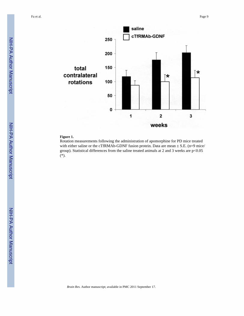
Figure 1.Rotation measurements following the administration of apomorphine for PD mice treatedwith either saline or the cTfRMAb-GDNF fusion protein. Data are mean ± S.E. (n=9 mice/group). Statistical differences from the saline treated animals at 2 and 3 weeks are p<0.05(*).
Fu et al. Page 9
Brain Res. Author manuscript; available in PMC 2011 September 17.
NIH
-PA Author Manuscript
NIH
-PA Author Manuscript
NIH
-PA Author Manuscript
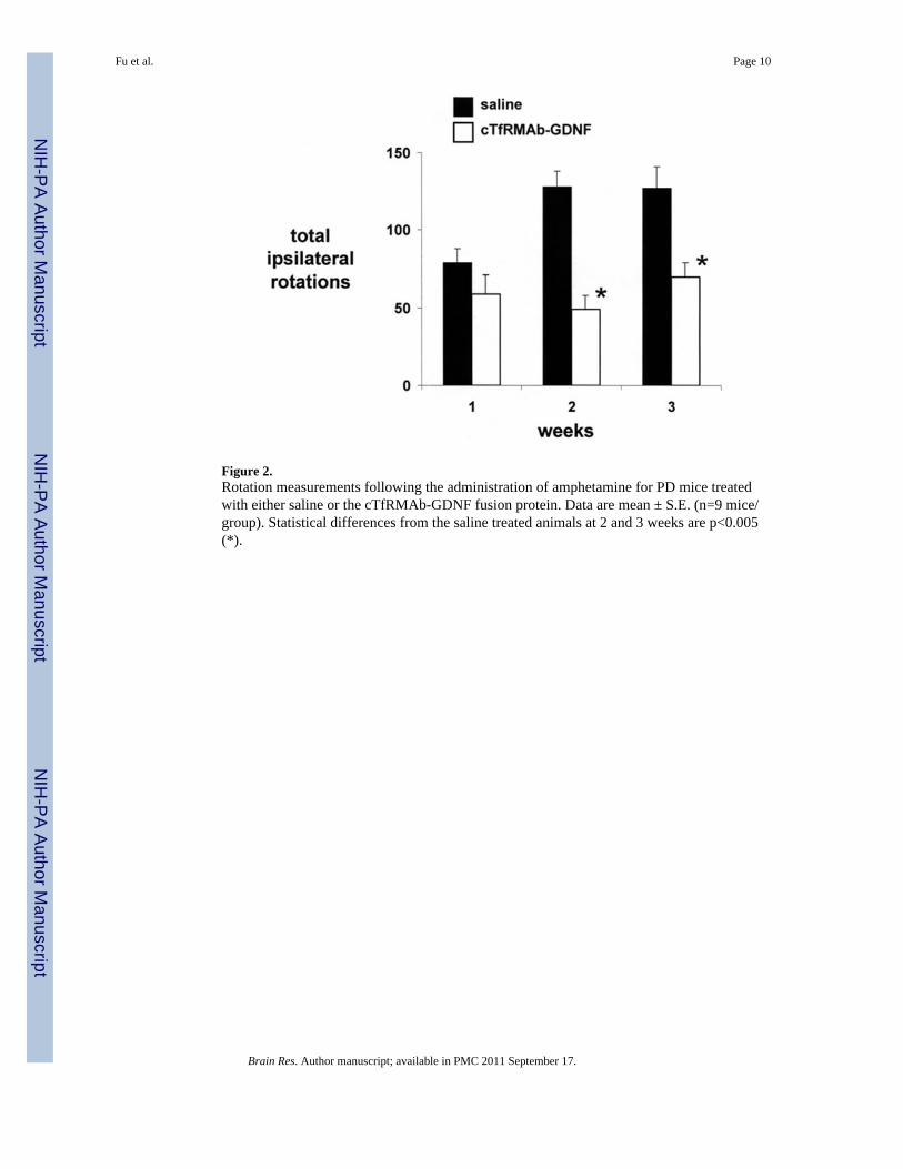
Figure 2.Rotation measurements following the administration of amphetamine for PD mice treatedwith either saline or the cTfRMAb-GDNF fusion protein. Data are mean ± S.E. (n=9 mice/group). Statistical differences from the saline treated animals at 2 and 3 weeks are p<0.005(*).
Fu et al. Page 10
Brain Res. Author manuscript; available in PMC 2011 September 17.
NIH
-PA Author Manuscript
NIH
-PA Author Manuscript
NIH
-PA Author Manuscript
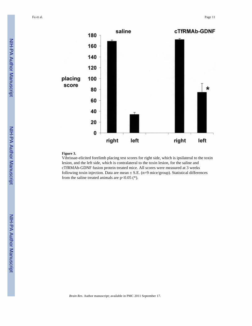
Figure 3.Vibrissae-elicited forelimb placing test scores for right side, which is ipsilateral to the toxinlesion, and the left side, which is contralateral to the toxin lesion, for the saline andcTfRMAb-GDNF fusion protein treated mice. All scores were measured at 3 weeksfollowing toxin injection. Data are mean ± S.E. (n=9 mice/group). Statistical differencesfrom the saline treated animals are p<0.05 (*).
Fu et al. Page 11
Brain Res. Author manuscript; available in PMC 2011 September 17.
NIH
-PA Author Manuscript
NIH
-PA Author Manuscript
NIH
-PA Author Manuscript
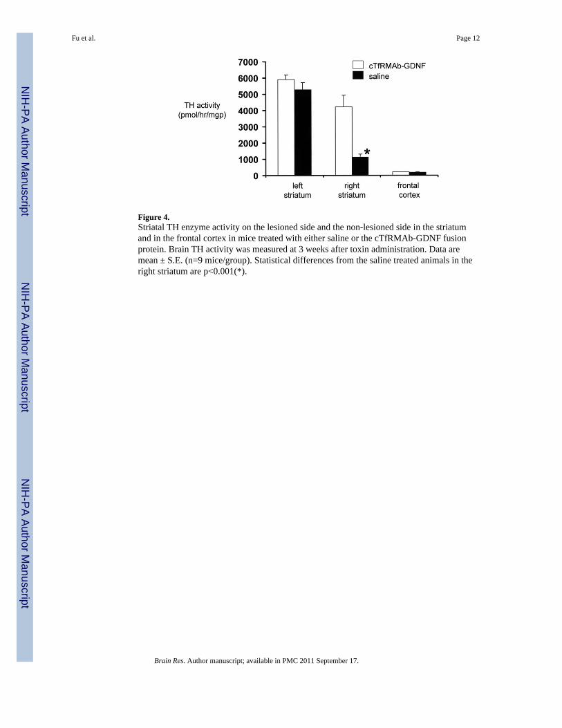
Figure 4.Striatal TH enzyme activity on the lesioned side and the non-lesioned side in the striatumand in the frontal cortex in mice treated with either saline or the cTfRMAb-GDNF fusionprotein. Brain TH activity was measured at 3 weeks after toxin administration. Data aremean ± S.E. (n=9 mice/group). Statistical differences from the saline treated animals in theright striatum are p<0.001(*).
Fu et al. Page 12
Brain Res. Author manuscript; available in PMC 2011 September 17.
NIH
-PA Author Manuscript
NIH
-PA Author Manuscript
NIH
-PA Author Manuscript
Copyright © 2022 FDOKUMEN