Intrathecal Coadministration of D-APV and Morphine Is Maximally Effective in a Rat Experimental...
7
Anesthesiology 2003; 98:734 – 40 © 2003 American Society of Anesthesiologists, Inc. Lippincott Williams & Wilkins, Inc. Intrathecal Coadministration of D-APV and Morphine Is Maximally Effective in a Rat Experimental Pancreatitis Model Ying Lu, M.D.,* Louis P. Vera-Portocarrero, B.S.,† Karin N. Westlund, Ph.D.‡ Background: Many studies have demonstrated that either glu- tamate N-methyl-D-aspartate (NMDA) receptor antagonists or opioid receptor agonists provide antinociception. Spinal coad- ministration of an NMDA receptor antagonist and morphine has an additive action for control of various pain states in animal models. The current study examined spinal coadminis- tration of low doses of NMDA receptor antagonist, D-(-)-2-Ami- no-5-phosphonovalerate (D-APV), and -opioid receptor ago- nist, morphine sulfate (MS), in reducing visceral nociception using an acute bradykinin induced pancreatitis model in rats. Methods: An intrathecal catheter was surgically inserted into the subarachnoid space for spinal drug administration in Sprague-Dawley rats. A laparotomy was performed for ligation and cannulation of the bile-pancreatic duct. Rats were pre- treated intrathecally with artificial cerebrospinal fluid (aCSF), D-APV, MS, or combined administration of D-APV and MS. These treatments were given 30 min before noxious visceral stimula- tion with bradykinin injected through the bile-pancreatic cath- eter. Spontaneous behavioral activity tests, including cage crossing, rearing, and hind limb extension, were conducted before and after bradykinin injection into the bile-pancreatic duct to assess visceral nociception. Results: Spinal pretreatment of D-APV or low doses of MS partially reduced visceral pain behaviors in this model. Pre- treatments with combinations of low doses of MS (0.05– 0.5 g) and D-APV (1 g) were maximally effective in returning all spontaneous behavioral activities to baseline. Conclusions: Spinal administration of combined doses of NMDA receptor antagonist, D-APV, and MS reversed three be- havioral responses to induction of an acute pancreatitis model. These results suggest that in the clinical management of visceral pain, such as pancreatitis, restricted usage of glutamate antag- onists might be useful as adjuvant potentiation at the onset of morphine therapy. PAIN is the primary cause of suffering in patients with visceral organ disorders. Pain arising from visceral or- gans has not been as widely investigated as pain arising from somatic tissues. In the past decade, studies of peripheral and central nervous system mechanisms in- volved in the generation and maintenance of visceral pain have used various animal models. 1–13 Visceral pain is still poorly understood from a pharmacologic point of view as well. The lack of information hinders efforts to develop better treatment strategies for clinical use. There is growing evidence that N-methyl-D-aspartate (NMDA) receptors are important in the transmission of nociceptive signals from the periphery via the spinal cord to the brain, and that NMDA receptors are involved in the induction and maintenance of the central sensitiza- tion for visceral structures. 14 –16 Use of NMDA receptor antagonists as antinociceptive agents has been successfully demonstrated in visceral pain models. Enhanced responses to colorectal distention in the presence of colonic inflam- mation were attenuated by NMDA receptor blocker MK- 801 and non-NMDA receptor antagonist DNQX. 17 The NMDA receptor antagonist (APV) has been used to block the effects of turpentine sensitization 18 and all NMDA-pro- duced nociceptive effects 16 in the visceromotor response to colorectal distension. This evidence provides support for the hypothesis that spinal glutamate receptors, particularly those of the NMDA subtype, play an important role in regulating spinal encoding of afferent information after visceral tissue inflammation or injury. Opioid receptor agonists can exert their action on both pre- and postsynaptic sites on primary afferent endings and have been widely used for controlling pain, particularly pain arising from viscera. Both and opi- oid receptor agonists have been shown to inhibit the visceromotor response to colorectal distension. 19 –21 Side effects, however, often limit clinical usefulness of -receptor agonists for long-term use in pain treatment. Tolerance-induced decreases in analgesia also limit their therapeutic efficacy. The NMDA receptor antagonists have limited usefulness in the clinic because of their side effects, including dysphoria, nightmares, motor distur- bance, and sedation. These side effects can decrease in combination with other drugs. 22,23 Some investigations demonstrate that NMDA receptor antagonists can pre- vent morphine tolerance. 24,25 Recently, investigators have found that spinal coadministration of glutamate receptor (NMDA or AMPA) antagonists and a low dose of morphine have synergistic antinociceptive effects in var- ious pain states. 26 –30 The experiments described in this article include anal- ysis of the behavioral manifestations of acute experimen- tal pancreatic pain induced by bile-pancreatic duct liga- tion and infusion of bradykinin in the conscious rat. The hypothesis tested was that glutamate-related mecha- nisms have a role in visceral nociceptive transmission from the inflamed pancreas and that combined use of an NMDA receptor antagonist and a -receptor agonist en- * Assistant Professor, † Graduate Assistant, ‡ Professor. Received from the Department of Anatomy and Neurosciences, The University of Texas Medical Branch, Galveston, Texas. Submitted for publication June 26, 2002. Accepted for publication October 25, 2002. Support was provided by RO1 grant 309041 from the National Institutes of Health, Bethesda, Maryland. Presented at the 31st Annual Meeting of the Society for Neuroscience, November 13, 2001, San Diego, California. Address reprint requests to Dr. Westlund: Professor, Department of Anatomy and Neurosciences, University of Texas Medical Branch, 301 University Boulevard Galveston, Texas 77555-1043. Address electronic mail to: [email protected]. Indi- vidual article reprints may be purchased through the Journal Web site, www.anesthesiology.org. Anesthesiology, V 98, No 3, Mar 2003 734
Transcript of Intrathecal Coadministration of D-APV and Morphine Is Maximally Effective in a Rat Experimental...
Anesthesiology 2003; 98:734–40 © 2003 American Society of Anesthesiologists, Inc. Lippincott Williams & Wilkins, Inc.
Intrathecal Coadministration of D-APV and Morphine IsMaximally Effective in a Rat Experimental PancreatitisModelYing Lu, M.D.,* Louis P. Vera-Portocarrero, B.S.,† Karin N. Westlund, Ph.D.‡
Background: Many studies have demonstrated that either glu-tamate N-methyl-D-aspartate (NMDA) receptor antagonists oropioid receptor agonists provide antinociception. Spinal coad-ministration of an NMDA receptor antagonist and morphinehas an additive action for control of various pain states inanimal models. The current study examined spinal coadminis-tration of low doses of NMDA receptor antagonist, D-(-)-2-Ami-no-5-phosphonovalerate (D-APV), and �-opioid receptor ago-nist, morphine sulfate (MS), in reducing visceral nociceptionusing an acute bradykinin induced pancreatitis model in rats.
Methods: An intrathecal catheter was surgically inserted intothe subarachnoid space for spinal drug administration inSprague-Dawley rats. A laparotomy was performed for ligationand cannulation of the bile-pancreatic duct. Rats were pre-treated intrathecally with artificial cerebrospinal fluid (aCSF),D-APV, MS, or combined administration of D-APV and MS. Thesetreatments were given 30 min before noxious visceral stimula-tion with bradykinin injected through the bile-pancreatic cath-eter. Spontaneous behavioral activity tests, including cagecrossing, rearing, and hind limb extension, were conductedbefore and after bradykinin injection into the bile-pancreaticduct to assess visceral nociception.
Results: Spinal pretreatment of D-APV or low doses of MSpartially reduced visceral pain behaviors in this model. Pre-treatments with combinations of low doses of MS (0.05–0.5 �g)and D-APV (1 �g) were maximally effective in returning allspontaneous behavioral activities to baseline.
Conclusions: Spinal administration of combined doses ofNMDA receptor antagonist, D-APV, and MS reversed three be-havioral responses to induction of an acute pancreatitis model.These results suggest that in the clinical management of visceralpain, such as pancreatitis, restricted usage of glutamate antag-onists might be useful as adjuvant potentiation at the onset ofmorphine therapy.
PAIN is the primary cause of suffering in patients withvisceral organ disorders. Pain arising from visceral or-gans has not been as widely investigated as pain arisingfrom somatic tissues. In the past decade, studies ofperipheral and central nervous system mechanisms in-volved in the generation and maintenance of visceralpain have used various animal models.1–13 Visceral painis still poorly understood from a pharmacologic point of
view as well. The lack of information hinders efforts todevelop better treatment strategies for clinical use.
There is growing evidence that N-methyl-D-aspartate(NMDA) receptors are important in the transmission ofnociceptive signals from the periphery via the spinalcord to the brain, and that NMDA receptors are involvedin the induction and maintenance of the central sensitiza-tion for visceral structures.14–16 Use of NMDA receptorantagonists as antinociceptive agents has been successfullydemonstrated in visceral pain models. Enhanced responsesto colorectal distention in the presence of colonic inflam-mation were attenuated by NMDA receptor blocker MK-801 and non-NMDA receptor antagonist DNQX.17 TheNMDA receptor antagonist (APV) has been used to blockthe effects of turpentine sensitization18 and all NMDA-pro-duced nociceptive effects16 in the visceromotor responseto colorectal distension. This evidence provides support forthe hypothesis that spinal glutamate receptors, particularlythose of the NMDA subtype, play an important role inregulating spinal encoding of afferent information aftervisceral tissue inflammation or injury.
Opioid receptor agonists can exert their action onboth pre- and postsynaptic sites on primary afferentendings and have been widely used for controlling pain,particularly pain arising from viscera. Both � and � opi-oid receptor agonists have been shown to inhibit thevisceromotor response to colorectal distension.19–21
Side effects, however, often limit clinical usefulness of�-receptor agonists for long-term use in pain treatment.Tolerance-induced decreases in analgesia also limit theirtherapeutic efficacy. The NMDA receptor antagonistshave limited usefulness in the clinic because of their sideeffects, including dysphoria, nightmares, motor distur-bance, and sedation. These side effects can decrease incombination with other drugs.22,23 Some investigationsdemonstrate that NMDA receptor antagonists can pre-vent morphine tolerance.24,25 Recently, investigatorshave found that spinal coadministration of glutamatereceptor (NMDA or AMPA) antagonists and a low dose ofmorphine have synergistic antinociceptive effects in var-ious pain states.26–30
The experiments described in this article include anal-ysis of the behavioral manifestations of acute experimen-tal pancreatic pain induced by bile-pancreatic duct liga-tion and infusion of bradykinin in the conscious rat. Thehypothesis tested was that glutamate-related mecha-nisms have a role in visceral nociceptive transmissionfrom the inflamed pancreas and that combined use of anNMDA receptor antagonist and a �-receptor agonist en-
* Assistant Professor, † Graduate Assistant, ‡ Professor.
Received from the Department of Anatomy and Neurosciences, The Universityof Texas Medical Branch, Galveston, Texas. Submitted for publication June 26,2002. Accepted for publication October 25, 2002. Support was provided by RO1grant 309041 from the National Institutes of Health, Bethesda, Maryland.Presented at the 31st Annual Meeting of the Society for Neuroscience, November13, 2001, San Diego, California.
Address reprint requests to Dr. Westlund: Professor, Department of Anatomy andNeurosciences, University of Texas Medical Branch, 301 University BoulevardGalveston, Texas 77555-1043. Address electronic mail to: [email protected]. Indi-vidual article reprints may be purchased through the Journal Web site,www.anesthesiology.org.
Anesthesiology, V 98, No 3, Mar 2003 734
hances antinociceptive effects in this animal model.These studies provide insights for improvement of cur-rent therapy for intractable pain caused by visceraldisease.
Materials and Methods
All experiments were approved by the Animal Careand Use Committee at our institution and are consistentwith the guidelines of this committee as well as thepolicies on the Ethical Treatment of Research Animalspublished by the International Association for the Studyof Pain. Ninety male Sprague-Dawley rats (250–300 g)were used for the study. The animals were housed in aroom with a constant ambient temperature of 22°C, a12 h light–dark cycle, and free access to food and water.
General ProceduresAnimals received an intrathecal catheter. After 4 to 5
days of recovery, a laparotomy for the bile-pancreaticduct ligation and cannulation was performed for deliveryof bradykinin. Bradykinin served as a noxious visceralstimulus that evoked visceral nociceptive responses andinduced severe acute pancreatitis that was evidenthistologically.
The next day rats received intrathecal aCSF, NMDAreceptor antagonist (D-APV), �-opioid receptor agonistMS, or a combination of both drugs. Thirty minutes later,0.5 ml of bradykinin (10�5
M, Sigma, St. Louis, MO)10 inlactated Ringer’s solution (Baxter Healthcare Corp.,Deerfield, IL) was injected into the pancreas. Based on aprevious physiologic study, 10�10-10�4
M bradykinin iseffective in producing nociceptive responses in thismodel without producing desensitization.10 Spontane-ous behavioral activity of the rats was observed duringthe first 10 min in a novel cage and quantified (1) beforeany surgery (naïve), (2) after intrathecal surgery, (3) thenext day after laparotomy (baseline), and (4) after nox-ious activation of pancreatic afferent fibers with brady-kinin and intrathecal aCSF or drug treatment. Histologicsamples were taken immediately following the lastbehavioral test.
Intrathecal and Intraductal Catheter PlacementsRats were anesthetized with methohexital sodium
(brevital sodium, 40 mg/kg, intraperitoneal) prior toreceiving an intrathecal catheter for administration ofdrugs according to Yaksh and Rudy.31 A 4.5 cm intrathe-cal catheter (32-guage, ReCathCo, Allicon Park, PA) wasimplanted intrathecally through an opening in the atlan-to-occipital membrane to the spinal T6–7 level. Thecatheter was linked to soft polyethylene tubing (PE10)(i.d. 0.28 mm, o.d. 0.61 mm; Clay Adams Brand, BectonDickinson Primary Care Diagnostics Becton Dickinsonand Company) and then connected to PE20 tubing,
which was tunneled under the skin, and remained sub-cutaneous until use. Rats were allowed to recover for 4to 5 days. After the recovery period, spontaneous behav-ioral activities of rats returned to levels observed in naïverats. Rats with behavioral deficits were excluded fromthe study (n � 5).
Rats were reanesthetized with methohexital sodium asdescribed previously for a ventral midline laparotomy.The PE10 tubing catheter connecting to PE20 tubing wasinserted into the common bile-pancreatic duct.6 ThePE20 tubing was tunneled under the skin to exit at thenape of the neck, and the exposed end of the tubing wassealed. The rats were allowed to recover overnight. Anyrat with a swollen abdomen or that became completelyinactive compared to before laparotomy was excludedfrom the study (n � 5).
Behavioral TestingBehavioral tests were conducted during the first 10 min
after rats were introduced to a novel, clear transparentcage. The timing of the behavioral observations wasdetermined through consideration of results of previousstudies in our laboratory,10 including physiology exper-iments that found that cell firing was sustained for 3–6 minafter bradykinin stimulation. Three spontaneous activi-ties observed were (1) cage crossing (forward locomo-tion across the centerline of the cage), (2) exploratoryrearing behavior (standing on the hind limb with orwithout support of the cage walls) and (3) hind limbextension (stretching or twisting of the hind limbs be-hind or under the body), which is a specific pain relatedbehavior. The number of times each behavior occurredspontaneously in the novel cage environment wascounted and recorded for every 10-s block of time, con-tinuing for 10 min.10 The mean of the counts for eachbehavioral measure was used as the animal’s behavioralresponse to induction of pancreatitis and response to pre-treatment with D-APV, MS, and combined treatment. Theexperimenter analyzing animal behavior was blind towhether the animals had received drug treatment oraCSF.32
Intrathecal Administration of DrugsAn NMDA receptor antagonist, D-APV (Tocris Cookson
Inc., Ellisville, MO) and MS (Paddock Laboratories Inc.,Minneapolis, MN), a �-opioid receptor agonist, wereassessed in this study. Drugs were dissolved in saline anddiluted in aCSF to their final concentrations (in 10 �l).The drugs were administered intrathecally with a Ham-ilton syringe (Reno, NV), followed by 10 �l of aCSF toflush the catheter. A dose response curve for each drugwas generated by examining the effects of pretreatmentwith the drug in animals with pancreatitis. In the D-APVpretreatment group, the drug doses injected intrathe-cally were of 0.5 (n � 6), 1 (n � 7), and 2 �g (n � 6).In the MS pretreatment group, the doses used were 0.05
735SPINAL D-APV AND MORPHINE FOR VISCERAL PAIN
Anesthesiology, V 98, No 3, Mar 2003
�g (n � 6), 0.1 �g (n � 6), 0.5 �g (n � 6), and 1 �g(n � 7). In the drug control group, 20 �l of aCSF wasinfused intrathecally (n � 7). After the dose responsecurve was generated for D-APV, a single dose of D-APV(1 �g) was combined with various doses of MS (0.01 �g,n � 6; 0.05 �g, n � 7; 0.1 �g, n � 6; 0.5 �g, n � 6 and1 �g, n � 6;) to examine the effects of coadministrationof both drugs.
Histopathology of the PancreasRats received an overdose of sodium pentobarbital and
the pancreatic tissue was collected from naïve rats (n � 3),from rats 1 day after laparotomy and cannulation (n � 3),and from rats 10 min after bradykinin injection (n � 3). Thetissue was cut into small blocks (about 3 � 3 � 2 mm) andwashed with phosphate buffer saline (PBS, pH 0.1 M),followed by block fixation in 4% buffered paraformalde-hyde for 2 to 3 days. The tissue was embedded in paraffinand cut at 4 �m thickness sections, and stained with he-matoxylin/eosin (HE). The pancreatic tissues were exam-ined and photographed with an Olympus microscope(Melville, NY) equipped with an advanced SPOT digitalcamera system for histopathologic analysis.
Statistical AnalysisComparisons tested included behavior of rats with
bradykinin-induced pancreatitis with and without drugpretreatments. The results of the behavioral testing werenot normally distributed and were therefore analyzedusing nonparametric statistics. Pairwise comparisonswithin groups for values before (baseline) and after drugtreatment were analyzed using the Wilcoxon test. Com-parisons were made between groups using the Mann–
Whitney U test. A P value of less than 0.05 was consid-ered a significant difference. The data were expressed asaverage � SEM. As a matter of interest, analysis of vari-ance (ANOVA) was also performed using Newman-Keulspost hoc comparisons and the results were identical. ThePrizm software program was used to determine the ED50
from the dose response curve.
Results
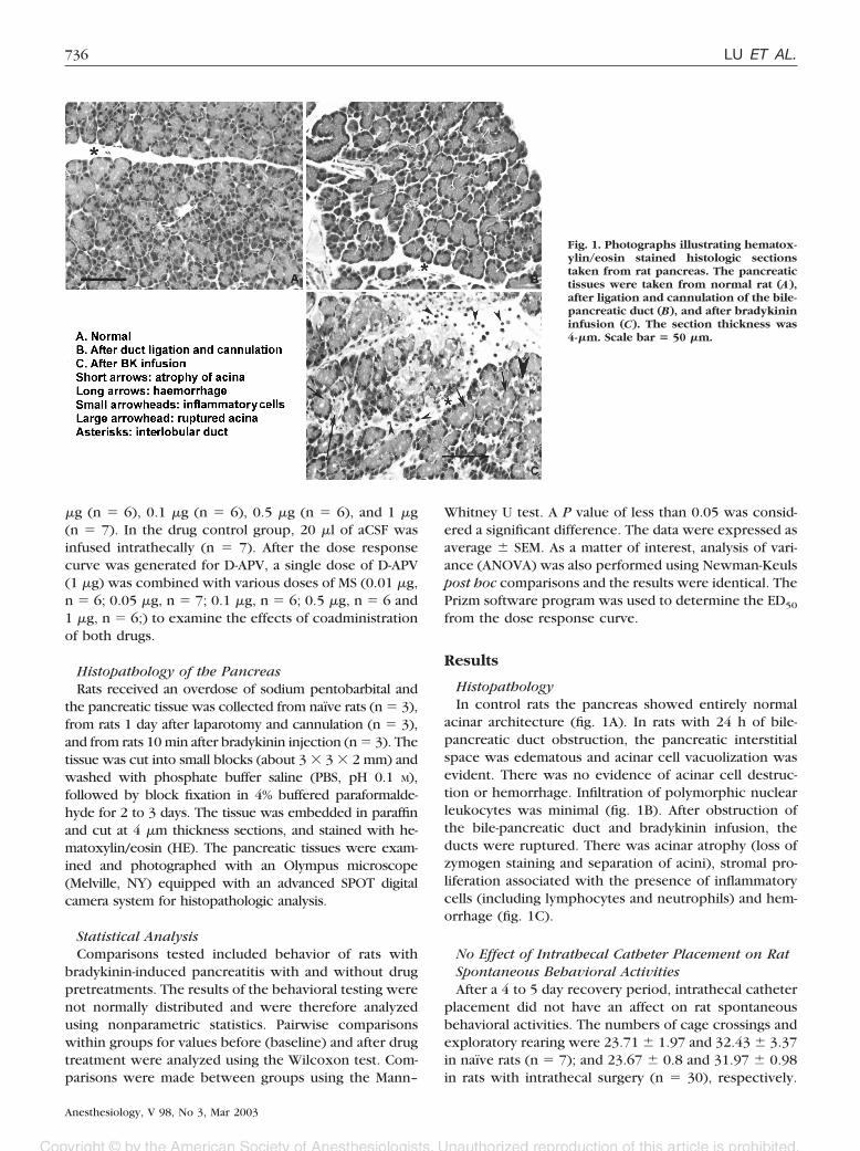
HistopathologyIn control rats the pancreas showed entirely normal
acinar architecture (fig. 1A). In rats with 24 h of bile-pancreatic duct obstruction, the pancreatic interstitialspace was edematous and acinar cell vacuolization wasevident. There was no evidence of acinar cell destruc-tion or hemorrhage. Infiltration of polymorphic nuclearleukocytes was minimal (fig. 1B). After obstruction ofthe bile-pancreatic duct and bradykinin infusion, theducts were ruptured. There was acinar atrophy (loss ofzymogen staining and separation of acini), stromal pro-liferation associated with the presence of inflammatorycells (including lymphocytes and neutrophils) and hem-orrhage (fig. 1C).
No Effect of Intrathecal Catheter Placement on RatSpontaneous Behavioral ActivitiesAfter a 4 to 5 day recovery period, intrathecal catheter
placement did not have an affect on rat spontaneousbehavioral activities. The numbers of cage crossings andexploratory rearing were 23.71 � 1.97 and 32.43 � 3.37in naïve rats (n � 7); and 23.67 � 0.8 and 31.97 � 0.98in rats with intrathecal surgery (n � 30), respectively.
Fig. 1. Photographs illustrating hematox-ylin/eosin stained histologic sectionstaken from rat pancreas. The pancreatictissues were taken from normal rat (A),after ligation and cannulation of the bile-pancreatic duct (B), and after bradykinininfusion (C). The section thickness was4-�m. Scale bar � 50 �m.
736 LU ET AL.
Anesthesiology, V 98, No 3, Mar 2003

Hind limb extension was not evident in either naive ratsor in rats after intrathecal surgery. Since there were nosignificant differences in the spontaneous behavioral ac-tivities between the two groups of rats (P � 0.05), thegroup with intrathecal surgery was included in the con-trol group for comparisons with other groups in thisstudy.
Spontaneous Behavioral Activity Subsequent toObstruction of the Bile-Pancreatic DuctBile-pancreatic duct ligation and cannulation resulted
in significant decreases in crossing and rearing behaviorsand development of hind limb extension after 24 hcompared with control rats (P � 0.01). The numbers ofcage crossing and rearing events were 23.67 � 0.79(range 13–35) and 31.97 � 0.98 (range 20–43) in con-trol rats and 12.54 � 0.56 (range 6–22) and 13.68 � 0.68(range 5–27) in rats with duct obstruction, respectively.Hind limb extension was not present in control rats, butwas evident in rats with ductal obstruction (5.04 � 0.51,range 0–16). Spontaneous behavioral activities in ratswith ductal ligation prior to chemical stimulation withbradykinin were used as their own baseline control mea-surements for comparison with drug pretreatments inthe following studies.
Effect of Bradykinin Infusion into Pancreas on RatSpontaneous Behavioral ActivitiesAfter bradykinin infusion into the pancreas, a signifi-
cant further reduction in cage crossing and rearing be-haviors occurred compared with their own baseline
(P � 0.05, figs. 2, 3). While further increases in hind limbextensions following bradykinin were observed, theywere not significantly higher than after ductal obstruc-tion by cannulation.
Effect of D-APV Pretreatment on SpontaneousBehavioral ActivitiesRats that received intrathecal aCSF pretreatment (0.0 �g
drug control group) as well as bradykinin infusion were
Fig. 2. Dose-response curve illustrating the antinociceptive ef-fect of intrathecal administration of D-APV on spontaneousbehavioral activities in rat with acute pancreatitis induced bybradykinin infusion into the pancreas. Values are representedas the mean � SEM. Comparisons were made within groupsusing Wilcoxon matched pairs test. The 0.0 �g dose representsaCSF treated rats (drug control group). * � P < 0.05 (vs. drugcontrol group). The ED50 for D-APV is indicated for each behav-ioral measure.
Fig. 3. Dose-response curves illustrating the antinociceptive ef-fects of intrathecal morphine (dash-dot) and coadministrationof D-APV (1 �g) with various low doses of MS (0.01, 0.05, 0.1, 0.5,or 1.0 �g; [dash-triangle]) on spontaneous behavioral activitiesin rats with pancreatitis induced by bradykinin infusion intothe pancreas. Values are represented as the mean � SEM. Com-parisons were made within groups using Wilcoxon matchedpairs test. Comparisons were made between groups usingMann–Whitney U test. The 0.0 �g dose represents the aCSFtreated rats (drug control group). # and ## � P < 0.05 and 0.01(vs. baseline). * and ** � P < 0.05 and 0.01 (vs. drug controlgroup). � and �� � P < 0.05 and 0.01 (vs. MS treated group).
737SPINAL D-APV AND MORPHINE FOR VISCERAL PAIN
Anesthesiology, V 98, No 3, Mar 2003

used in comparisons with drug-pretreated animals. Com-pared to the spontaneous activity of the aCSF treateddrug control group, intrathecal preadministration of lowdoses (0.5–1 �g) of D-APV significantly increased thenumber of cage crossings (P � 0.05). Increases in rear-ing were nonsignificant after D-APV, and hind limb ex-tension was decreased. The ED50 was 0.80 �g for cagecrossing, 0.96 �g for rearing, and 0.98 �g for hind limbextension (fig. 2). The hind limb extension behavior wasattenuated significantly in rats pretreated with the high-est dose of D-APV (2 �g), but side effects were evident,suggestive of sedation including flaccid paralysis andlack of activity. Behavioral data reported here for D-APV,as well as that for morphine and combined doses re-ported below, were similar at later time points (45 and60 min) extending through the peak effect of theseagents in pilot studies (data not shown).
Effect of MS Pretreatment on SpontaneousBehavioral ActivitiesThe MS significantly attenuated numbers of hind limb
extensions (P � 0.05–0.01), but did not restore cagecrossing and rearing, compared to that of the aCSFtreated drug control group. Only the maximum dose ofMS (1 �g) significantly increased cage crossing and rear-ing above that of the aCSF treated drug control group(P � 0.05–0.01, fig. 3). The ED50 for low doses of MSwere higher than the dose range used (up to 1 �g), thusMS had no significant effect on crossing and rearingbehaviors. There was a significant effect on hind limbextension in this model at all doses. The ED50 was9.84 �g for cage crossing, 9.25 �g for rearing, and 0.67 �gfor hind limb extension.
Interaction Between D-APV and MSIntrathecal coadministration of low doses of MS (0.05,
0.1, and 0.5 �g) and D-APV (1 �g), prevented both thedevelopment of significant pain-related behavioral activ-ity observed in rats with pancreatitis and restored spon-taneous exploratory behaviors (fig. 3). Thus, the lowdose of NMDA receptor antagonist, D-APV, combinedwith low doses of MS improved all behavioral measures.In the absence of bradykinin, behavior was unaffectedby the same doses of coadministered D-APV and MS (n �3; 1 �g � 0.05 �g).
Discussion
In the past decade, several animal models have beenemployed to investigate peripheral and central nervoussystem (CNS) mechanisms underlying pain arising fromvisceral organs. Nonetheless, the knowledge of neuronalmechanisms as well as clinical management of visceralpain states remains unsatisfactory.
In this study we investigated the role of NMDA andopioid receptors in an acute experimental pancreatitismodel in rats. The results indicate that spinal adminis-tration of a low dose of NMDA receptor antagonist,D-APV or low doses of MS (0.05–0.5 �g) were partiallyeffective for antinociception in this model. Higher dosesresulted in development of motoric deficits and/or seda-tion. This is despite the intrathecal administration di-rectly onto thoracic spinal levels (T6-T11) innervated bypancreatic afferents. The particular measures of painrelated behaviors observed in this model were resistantto D-APV unlike other visceral pain models in which ithas been found to be effective.3,16,18
Coadministration of low doses of MS with D-APV (1 �g)resulted in elimination of the pain-related behaviors in-duced in this acute pancreatitis model in rats. Potenti-ated action of these two agents brought the two spon-taneous behavioral activities back to baseline as well astotally eliminating hind limb extension, indicative ofsuccessful visceral antinociception.
Histopathological Change Correlated withBehavioral ChangeThe manipulation of the pancreas, including surgical
placement of the catheter into the pancreatic duct in thismodel, resulted in acute edematous histopathologicchanges in the pancreas. This effect of induced acutepancreatitis has been reported by Merriam et al.33 Thereduction of normal, spontaneous behavioral activitiesand onset of hind limb extension, a specific pain-relatedbehavior, in the conscious rat with ligation-induced pan-creatitis indicated that ligation of the bile-pancreaticduct itself produced visceral nociception arising fromthe pancreas.
The secondary bradykinin insult further contributes toa severe acute pancreatitis. Exposure to bradykinin sec-ondary to the catheter-induced changes included rup-tured pancreatic ducts, acinar atrophy, infiltration ofinflammatory cells, as well as hemorrhage. These his-topathologic changes after bradykinin stimulation wereparalleled by more severe alterations in nociceptive be-haviors, including significant reduction of cage crossingand rearing behaviors compared to the histopathologicand behavioral changes produced by ductal ligation(baseline). A nonsignificant increase in the number ofhind limb extensions also occurred. These spontaneousbehaviors have been reported previously to be associ-ated with increased visceral nociception.6,10,34,35
NMDA Receptor AntagonistsPrevious studies demonstrated that NMDA receptor
antagonists provide significant antinociceptive effects invisceral pain models involving hollow organs.17,18,36 Theactivation of NMDA and non-NMDA receptors in thespinal cord differentially modulates visceral nociceptiveinput. Spinal NMDA receptor antagonist, APV, blocked
738 LU ET AL.
Anesthesiology, V 98, No 3, Mar 2003

all NMDA-produced facilitation of visceromotor re-sponses to noxious CRD stimulation, while AMPA recep-tor antagonist, DNQX, blocked quisqualic acid-producedinhibitory effects.16 Intrathecal administration of APVprevented hyperreflexia after urinary bladder inflamma-tion3 and blocked the effect of turpentine sensitizationon visceromotor response to CRD.18However, the effec-tiveness of the NMDA receptor antagonist for antinoci-ception in the pancreas, a solid endodermally derivedorgan, was limited. We found that only the reduction incrossing following bradykinin was abrogated by intrathe-cal NMDA receptor antagonist D-APV. Spinal D-APV didnot bring all of spontaneous behavioral activities back tobaseline, providing a limited antinociceptive effect inthis pain model by this agent alone. Since this is incontrast to the reported effectiveness of D-APV in othervisceral pain models, this may denote differences in themodel used and/or methodological, species, or organdifferences.
Morphine and NMDA Receptor AntagonistsOpioids have been widely and successfully used for
management of visceral pain in patients and in animalstudies. Many previous studies have shown that �-opioidreceptors can modulate visceral nociceptive transmissionin the spinal cord. Intrathecal administration of morphineproduces a dose-dependent attenuation of visceromotorresponses to noxious colorectal distension.19,20,37
It has been shown that NMDA receptor activationincreases excitation in neurons leading to an inability ofopioids to effectively reduce somatic nociceptive trans-mission.38 Thus, it has been proposed that NMDA recep-tor antagonism enhances the antinociceptive effect ofmorphine and thus may improve the analgesic efficacy ofmorphine.30,39 The current study, using a visceral painmodel, supports the proposal that antagonism of NMDAreceptor action potentiates the effects of morphine. Theresults of the current study showed that spinal coadmin-istration of low doses (0.05–0.5 �g) of the opioid recep-tor agonist morphine sulfate, and a 1 �g dose of NMDAreceptor antagonist D-APV, produced antinociception.Coadministration of these two agents maximally in-creased spontaneous behavioral activities including elim-inating hind limb extension in this rat model of acutepancreatitis. These results concur with findings of pre-vious studies in somatic pain models demonstrating thatthe combined administration of morphine with NMDAreceptor antagonists can produce powerful potentiationof morphine’s actions on nociceptive events.27–30,40
In conclusion, our results imply that spinal administra-tion of the NMDA receptor antagonist, D-APV, can beused as a therapeutic adjuvant producing potent antino-ciception when combined with low doses of morphine.Coadministration of D-APV (1 �g) potentiated the anal-gesic effect of low doses of morphine in this model.These findings may be important to developing im-
proved strategies for pain control and adjuvant potenti-ation of morphine therapy for treatment of developingvisceral pain states.
References
1. Birder LA, de Groat WC: The effect of glutamate antagonists on c-fosexpression induced in spinal neurons by irritation of the lower urinary tract.Brain Res 1992; 580:115–20
2. Birder LA, de Groat WC: Induction of c-fos expression in spinal neurons bynociceptive and nonnociceptive stimulation of LUT. Am J Physiol 1993; 265(2 Pt2):R326–33
3. Rice AS, McMahon SB: Pre-emptive intrathecal administration of an NMDAreceptor antagonist (AP-5) prevents hyper-reflexia in a model of persistent vis-ceral pain. Pain 1994; 57:335–40
4. Sengupta JN, Gebhart GF: Mechanosensitive afferent fibers in the gastroin-testinal and lower urinary tracts. In Visceral Pain. Edited by Gebhart GF. Seattle:IASP Press; 1995:75–98
5. Avelino A, Cruz F, Coimbra A: Sites of renal pain processing in the rat spinalcord. A c-fos study using a percutaneous method to perform ureteral obstruction.J Auton Nerv Syst 1997; 6:60–6
6. Houghton AK, Kadura S, Westlund KN: Dorsal column lesions reverse thereduction of home cage activity in rats with pancreatitis. NeuroReport 1997;8:3795–800
7. Laird JM, Roza C, Olivar T: Antinociceptive activity of metamizol in rats withexperimental ureteric calculosis: central and peripheral components. InflammRes 1998; 47:389–95
8. Roza C, Laird JM, Cervero F: Spinal mechanisms underlying persistent painand referred hyperalgesia in rats with an experimental ureteric stone. J Neuro-physiol 1998; 79:1603–12
9. Boucher M, Meen M, Codron JP, Coudore F, Kemeny JL, Eschalier A:Cyclophosphamide-induced cystitis in freely-moving conscious rats: behavioralapproach to a new model of visceral pain. J Urol 2000; 164:203–8
10. Houghton AK, Wang CC, Westlund KN: Do nociceptive signals from thepancreas travel in the dorsal column? Pain 2001; 89:207–20
11. Laird JM, Martinez-Caro L, Garcia-Nicas E, Cervero F: A new model ofvisceral pain and referred hyperalgesia in the mouse. Pain 2001; 92:335–42
12. Lu Y, Westlund KN: Effects of baclofen on colon inflammation-inducedFos, CGRP and SP expression in spinal cord and brainstem. Brain Res 2001;889:118–30
13. Ness TJ, Gebhart GF: Inflammation enhances reflex and spinal neuronresponses to noxious visceral stimulation in rats. Am J Physiol - Gastrointestinal& Liver Physiology 2001; 80:G649–57
14. Olivar T and Laird JM: Differential effects of N-methyl-D-aspartate receptorblockade on somatic and visceral reflexes. Pain 1999; 79:67–73
15. Urban MO, Gebhart GF: Supraspinal contributions to hyperalgesia. ProcNatl Acad Sci U S A 1999; 96:7687–92
16. Kolhekar R, Gebhart GF: NMDA and quisqualate modulation of visceralnociception in the rat. Brain Res 1994; 651:215–26
17. Coutinho SV, Meller ST, Gebhart GF: Intracolonic zymosan producesvisceral hyperalgesia in the rat that is mediated by spinal NMDA and non-NMDAreceptors. Brain Res 1996; 736:7–15
18. Ide Y, Maehara Y, Tsukahara S, Kitahata LM, Collins JG: The effect of anintrathecal NMDA antagonist (APV) on the behavioral changes induced by colo-rectal inflammation with turpentine in rats. Life Sci 1997; 60:1359–63
19. Danzebrink RM, Green SA, Gebhart GF: Spinal mu and delta, but notkappa, opioid-receptor agonists attenuate responses to noxious colorectal dis-tension in the rat. Pain 1995; 63:39–47
20. Borgbjerg FM, Frigast C, Madsen JB, Mikkelsen LF: The effect of intrathecalopioid-receptor agonists on visceral noxious stimulation in rabbits. Gastroenter-ology 1996; 110:139–46
21. Gebhart GF, Su X, Joshi S, Ozaki N, Sengupta JN: Peripheral opioidmodulation of visceral pain. Ann NY Acad Sci 2000; 909:41–50
22. Nishiyama T, Gyermek L, Lee C, Kawasaki-Yatsugi S, Yamaguchi T,Hanaoka K: The analgesic interaction between intrathecal clonidine and gluta-mate receptor antagonists on thermal and formalin-induced pain in rats. AnesthAnalg 2001; 92:725–32
23. Advokat C, Rhein FQ: Potentiation of morphine-induced antinociceptionin acute spinal rats by the NMDA antagonist dextrophan. Brain Res 1995;699:157–60
24. Trujillo KA, Akil H: Inhibition of opiate tolerance by non-competitiveN-methyl-D-aspartate receptor antagonists. Brain Res 1994; 633:178–88
25. Wong CS, Cherng CH, Luk HN, Ho ST, Tung CS: Effects of NMDA receptorantagonists on inhibition of morphine tolerance in rats: binding at mu-opioidreceptors. Eur J Pharmacol 1996; 297:27–33
26. Chapman V. Dickenson AH: The combination of NMDA antagonism andmorphine produces profound antinociception in the rat dorsal horn. Brain Res1992; 573:321–3
739SPINAL D-APV AND MORPHINE FOR VISCERAL PAIN
Anesthesiology, V 98, No 3, Mar 2003

27. Dickenson AH: NMDA receptor antagonists: interactions with opioids.Acta Anaesthesiol Scand 1997; 41(1 Pt 2):112–5
28. Nishiyama T, Yaksh TL, Weber E: Effects of intrathecal NMDA and non-NMDA antagonists on acute thermal nociception and their interaction withmorphine. ANESTHESIOLOGY 1998; 89:715–22
29. Plesan A, Hedman U, Xu XJ, Wiesenfeld-Hallin, Z: Comparison of ketamineand dextromethorphan in potentiating the antinociceptive effect of morphine inrats. Anesth Analg 1998; 86:825–29
30. Nishiyama T: Interaction between intrathecal morphine and glutamatereceptor antagonists in formalin test. Eur J Pharmacol 2000; 395:203–10
31. Yaksh TL, Rudy TA: Chronic catheterization of the spinal subarachnoidspace. Physiol Behav 1976; 17:1031–6
32. McAdoo DJ, Xu GY, Robak G and Hughes MG: Changes in amino acidconcentrations over time and space around an impact injury and their diffusionthrough the spinal cord. Exp Neurol 1999; 159:538–44
33. Merriam LT, Wilcockson D, Samuel I, Joehl RJ: Ligation-induced acutepancreatitis increases pancreatic circulating trypsinogen activation peptides.J Surg Res 1996; 60:417–21
34. Craft RM, Carlisi VJ, Mattia A, Herman RM, Porreca F: Behavioral charac-
terization of the excitatory and desensitizing effects of intravesicalar capsaicinand resiniferatoxin in the rat. Pain 1993; 55:205–15
35. Wesselmann U, Czakanski PP, Affaitati G, Giamberardino MA: Uterineinflammation as a noxious visceral stimulus: behavioral characterization in therat. Neurosci Lett 1998; 246:73–6
36. Song XJ, Zhao ZQ: Involvement of NMDA and non-NMDA receptors intransmission of spinal visceral nociception in cat. Chung-Kuo Yao Li Hsueh Pao- Acta Pharmacologica Sinica 1999; 20:308–12
37. Harada Y, Nishioka K, Kitahata LM, Nakatani K, Collins JG: Contrastingactions of intrathecal U50, 488H, morphine, or [D-Pen2, D-Pen5] enkephalin orintravenous U50, 488H on the visceromotor response to colorectal distension inthe rat. ANESTHESIOLOGY 1995; 83:336–43
38. Dickenson AH: Neurophysiology of opioid poorly responsive pain [re-view]. Cancer Surveys 1994; 21:5–16
39. Dickenson AH: NMDA receptor antagonists: interactions with opioids.Acta Anaesthesiol Scand 1997; 41(1 Pt 2):112–5
40. Grass S, Hoffmann O, Xu XJ, Wiesenfeld-Hallin Z: N-methyl-D-aspartatereceptor antagonists potentiate morphine’s antinociceptive effect in the rat. ActaPhysiol Scand 1996; 158:269–73
740 LU ET AL.
Anesthesiology, V 98, No 3, Mar 2003