Interplay between high energy impulse noise (blast) and antioxidants in the lung
12
Interplay between high energy impulse noise (blast) and antioxidants in the lung Nabil M. Elsayed *, Nikolai V. Gorbunov Department of Respiratory Research, Division of Military Casualty Research, Walter Reed Army Institute of Research, Silver Spring, MD, USA Abstract High-energy impulse noise (BLAST) is a physical event characterized by an abrupt rise in atmospheric pressure above ambient lasting for a very short period, but potentially causing significant material and biological damage. Exposure to high-level BLAST can be destructive and lethal. Low-level BLAST similar to what is encountered repeatedly by military personnel during training and combat from detonation of munitions and firing of large caliber weapons, and during occupational use of explosives and some heavy machinery, can also cause significant injury. Globally, civilians are increasingly exposed to BLAST resulting from terrorist bombings or abandoned unmarked mines following numerous wars and conflicts. We have shown previously in several animal models that exposure to non-lethal BLAST results in pathological changes, mostly to the hollow organs characterized in the lungs, the most sensitive organ, by rupture of alveolar septa, and pulmonary hemorrhage and edema. These events potentially can cause alveolar flooding, respiratory insufficiency and adult respiratory distress syndrome (ARDS), leading to varying degrees of hypoxia, antioxidant depletion and oxidative damage. We have also observed progressive formation of nitric oxide in blood and other tissues. The totality of these observations supports our general hypothesis that exposure to BLAST can lead to antioxidant depletion and oxidative damage. Understanding the mechanism(s) of BLAST-induced oxidative stress may have important implications that include a potential beneficial role for antioxidants as a prophylaxis or as secondary treatment of injury after exposure alongside other protective and therapeutic modalities. In addition, it suggests a role for endogenous nitric oxide in the injury. This report reviews experimental evidence of BLAST-induced antioxidant depletion, and the potential benefit from antioxidant supplementation before exposure. # 2003 Elsevier Science Ireland Ltd. All rights reserved. Keywords: Impulse noise; Blast overpressure; Terrorist bombings; Mines; Antioxidants; Oxidative stress; Nitric oxide; Adult respiratory distress syndrome (ARDS) The opinions and conclusions contained in this report are those of the authors and do not reflect the views of the Department of the Army or Department of Defense. The research presented was conducted under institutional approved protocols, and funded by the US Army Medical Research and Materiel Command. All research was conducted in accordance with the NIH’s ‘‘Guide for the Care and Use of Laboratory Animals (NRC, 1996)’’. * Corresponding author. Present address: Hurley Consulting Associates, One Main Street, Chatham, NJ 07928, USA. Tel.: /1-973- 635-9898; fax: /1-973-635-9881. E-mail address: [email protected] (N.M. Elsayed). Toxicology 189 (2003) 63 /74 www.elsevier.com/locate/toxicol 0300-483X/03/$ - see front matter # 2003 Elsevier Science Ireland Ltd. All rights reserved. doi:10.1016/S0300-483X(03)00153-7
Transcript of Interplay between high energy impulse noise (blast) and antioxidants in the lung
Interplay between high energy impulse noise (blast) andantioxidants in the lung�
Nabil M. Elsayed *, Nikolai V. Gorbunov
Department of Respiratory Research, Division of Military Casualty Research, Walter Reed Army Institute of Research, Silver Spring,
MD, USA
Abstract
High-energy impulse noise (BLAST) is a physical event characterized by an abrupt rise in atmospheric pressure above
ambient lasting for a very short period, but potentially causing significant material and biological damage. Exposure to
high-level BLAST can be destructive and lethal. Low-level BLAST similar to what is encountered repeatedly by military
personnel during training and combat from detonation of munitions and firing of large caliber weapons, and during
occupational use of explosives and some heavy machinery, can also cause significant injury. Globally, civilians are
increasingly exposed to BLAST resulting from terrorist bombings or abandoned unmarked mines following numerous
wars and conflicts. We have shown previously in several animal models that exposure to non-lethal BLAST results in
pathological changes, mostly to the hollow organs characterized in the lungs, the most sensitive organ, by rupture of
alveolar septa, and pulmonary hemorrhage and edema. These events potentially can cause alveolar flooding, respiratory
insufficiency and adult respiratory distress syndrome (ARDS), leading to varying degrees of hypoxia, antioxidant
depletion and oxidative damage. We have also observed progressive formation of nitric oxide in blood and other
tissues. The totality of these observations supports our general hypothesis that exposure to BLAST can lead to
antioxidant depletion and oxidative damage. Understanding the mechanism(s) of BLAST-induced oxidative stress may
have important implications that include a potential beneficial role for antioxidants as a prophylaxis or as secondary
treatment of injury after exposure alongside other protective and therapeutic modalities. In addition, it suggests a role
for endogenous nitric oxide in the injury. This report reviews experimental evidence of BLAST-induced antioxidant
depletion, and the potential benefit from antioxidant supplementation before exposure.
# 2003 Elsevier Science Ireland Ltd. All rights reserved.
Keywords: Impulse noise; Blast overpressure; Terrorist bombings; Mines; Antioxidants; Oxidative stress; Nitric oxide; Adult
respiratory distress syndrome (ARDS)
�The opinions and conclusions contained in this report are those of the authors and do not reflect the views of the Department of
the Army or Department of Defense. The research presented was conducted under institutional approved protocols, and funded by the
US Army Medical Research and Materiel Command. All research was conducted in accordance with the NIH’s ‘‘Guide for the Care
and Use of Laboratory Animals (NRC, 1996)’’.
* Corresponding author. Present address: Hurley Consulting Associates, One Main Street, Chatham, NJ 07928, USA. Tel.: �/1-973-
635-9898; fax: �/1-973-635-9881.
E-mail address: [email protected] (N.M. Elsayed).
Toxicology 189 (2003) 63�/74
www.elsevier.com/locate/toxicol
0300-483X/03/$ - see front matter # 2003 Elsevier Science Ireland Ltd. All rights reserved.
doi:10.1016/S0300-483X(03)00153-7
1. Introduction
High-energy impulse noise (BLAST) is a term
used to describe the shock waves that result from a
wide range of events encountered in certain
military and civilian environments accompanying
many activities. Militarily, they range from explo-
sion of regular and specialized types of explosives
and ordnance such as concussion grenades, high-
yield and thermobaric munitions to firing of large
caliber weapons and culminating in nuclear deto-
nations, which produce massive destructive
BLAST waves. It should be noted that blast
overpressure is another term often used inter-
changeably in the literature to describe high-
energy impulse noise resulting from the same
events. Occupationally, BLAST exposures may
result from intentional or accidental explosions,
and during some manufacturing processes, and
from operation of certain types of machinery.
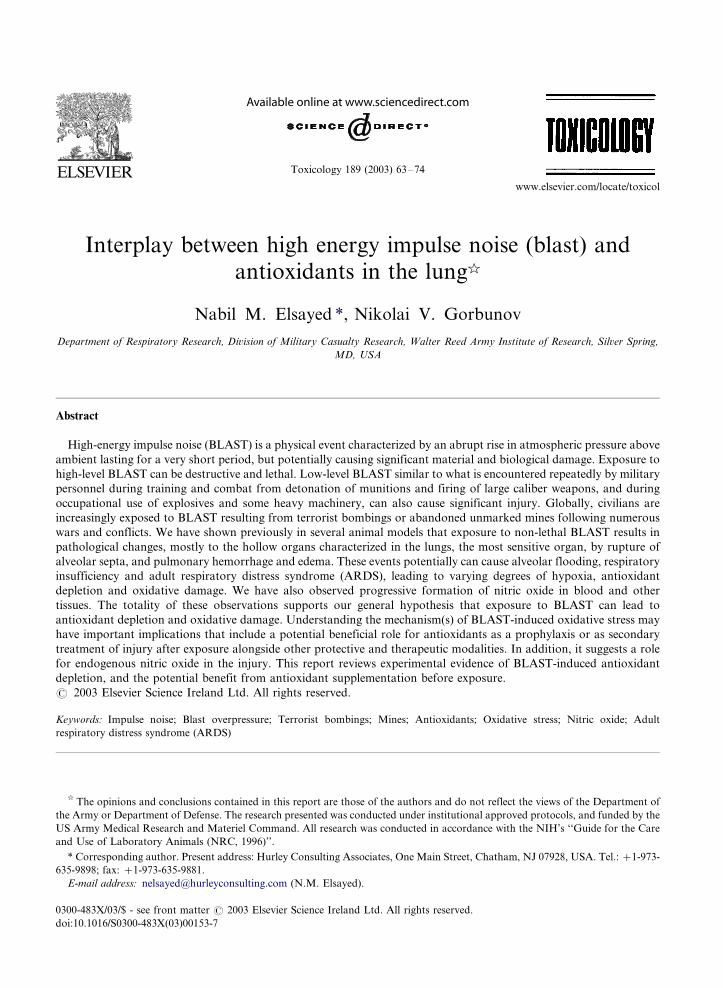
When BLAST magnitude is very large, the air
becomes compressed significantly such that it can
be seen with the naked eye as illustrated in Fig. 1.
The risk of exposure to BLAST that was
previously confined to the battlefield and military
training and exercise, or during certain industrial
processes, has expanded in recent years to include
the civilian population at large as they became
increasingly at risk of exposure to terrorist bomb-
ings (Abenhaim et al., 1992; Brismar and Bergen-
wald, 1982; Elsayed, 1997a; Frykberg and Tepas,
1988; Hadden et al., 1978; Hiss and Kahana, 1998;
Katz et al., 1989; Pfefferbaum, 2001; Phillips and
Richmond, 1991; Rignault and Deligny, 1989;
Fig. 1. Section from a still photograph of a visible air BLAST shock wave front from a large chemical explosion.
N.M. Elsayed, N.V. Gorbunov / Toxicology 189 (2003) 63�/7464
Thach et al., 2000). Another major source ofBLAST affecting civilians is the unmarked mines
abandoned after major wars and numerous regio-
nal military conflicts such as those found in the
deserts of Egypt after World War II (largest
concentration of mines in the world), in Vietnam
and other South East Asian countries, and in
former Yugoslavia to name a few.
2. Mechanism(s) of BLAST-induced injury
To study the mechanism(s) of BLAST injury
and to develop or test new therapeutic modalities,
as well as construct mathematical models for
injury prediction, BLAST waves has to be gener-
ated under controlled conditions either in a free-
field environment by detonation of known chargesof explosives or through BLAST simulation. One
such device that we have been using to simulate
BLAST waves in our laboratory at Walter Reed
Army Institute of Research is the shock tube
which was described in detail earlier (Elsayed,
1997a). This design is based on the reports of
Cassen et al. (1950) and Celander et al. (1950).
Basically, the Walter Reed shock tube we used is atwo-compartment horizontal, circular steel tube,
525 cm (17.5 ft) long, and 30 cm (12 in.) in
diameter. The shorter of the two, a 75 cm (2.5 ft),
compression compartment, can be separated from
the longer, a 450 cm (15 ft), expansion compart-
ment, by one or more polyester MylarTM sheets
(Du Pont Co., Wilmington, DE), of defined
thickness selected to produce a specific BLASTpeak pressure. Air is forced into the compression
compartment until the membrane bursts creating a
high-energy impulse noise wave that grows as it
travels through the expansion compartment until
it exits from the muzzle where a sample material or
a ‘‘deeply anesthetized’’ animal would be placed.
The BLAST wave peak pressure can be measured
by means of piezoelectric time�/pressure gaugesplaced at the nozzle or in specific locations around
it. The signal produced is then recorded using a
computer. In general, each BLAST wave gener-
ated produces a distinctive time�/pressure histo-
gram (signature). A typical BLAST histogram
would have a positive (above ambient) and may
have also a negative (below ambient) componentthat is equally injurious (Phillips and Richmond,
1991; Zhang et al., 1996). In enclosures, BLAST
shock waves are reflected off the walls or objects
becoming more complex and destructive (Phillips
and Richmond, 1991; Richmond, 1991). Although
the overpressure segment of the wave that exists
above ambient level lasts only for a very short
period measured in micro- to milli-seconds, it cancause significant physical and biological damage
to structures and living organisms. The magnitude
of the damage is dependent upon many interre-
lated factors including, shock wave characteristics
and magnitude, distance from the source, body
orientation, open space versus enclosure, etc.
(Benzinger, 1950; Elsayed, 1997a; Mellor, 1992;
Phillips and Richmond, 1991; Richmond, 1991;Schardin, 1950; Stuhmiller et al., 1991). The
potential for lethal internal injuries produced by
BLAST exposure was recognized during World
War I and was described as ‘‘the invisible sword
that can kill without blood’’ (Phillips and Rich-
mond, 1991).
Injury from BLAST is generally described as
primary blast injury if it results from exposure tothe BLAST waves alone; secondary if it results
from exposure to objects propelled by the initial
BLAST, and tertiary if it results from the body
being propelled against an object. Establishing an
accurate correlation between BLAST magnitude
and the resulting injuries either in experimental
animal models or BLAST casualties can provide
important and valuable information about theextent of injury, and hence the required treatment
as well as the size and/or nature of the explosive
device used to produce the BLAST.
For a number of years, we have been studying
the biological consequences of exposure to
BLAST, focusing mostly on low-level, non-lethal
single and multiple BLAST exposures. Our studies
agreed with other observations suggesting that thehollow organs such as the auditory and respiratory
systems are the most sensitive organs to BLAST
exposure (Benzinger, 1950; Brown et al., 1993;
Clemedson, 1956; Clemedson and Hultman, 1954;
Phillips and Richmond, 1991; Sharpnack et al.,
1991). BLAST-induced injury was observed also to
affect the cardiopulmonary system (Dodd et al.,
N.M. Elsayed, N.V. Gorbunov / Toxicology 189 (2003) 63�/74 65
1997), central nervous system (Cernak et al., 2001),
to cause visual system degeneration (Petras et al.,
1997), to damage the gastrointestinal system
(Clifford et al., 1984), and disrupt food intake
and exercise performance (Bauman et al., 1997;
Mundie et al., 2000).
We have further observed using different animal
models including rats, rabbits and sheep that
exposure to low-level BLAST waves can cause
oxidative stress (Elsayed et al., 1996; Elsayed,
1997a,b; Elsayed et al., 1997a,b; Gorbunov et al.,
1997a,b). It is well recognized that the auditory
system is very sensitive to BLAST sustaining
acoustic trauma, but the ears can be easily
protected, particularly when BLAST exposure is
anticipated in military training or occupational
settings (Yang et al., 1996; Ylikoski et al., 1995).
Impulse noise-induced acoustic trauma was found
to be associated with free radical production,
decreased strial blood flow hypoxia, and hearing
loss was shown to improve significantly after
treatment with antioxidants (Kopke et al., 2000,
2002; Seidman et al., 1993; Yamane et al., 1995).
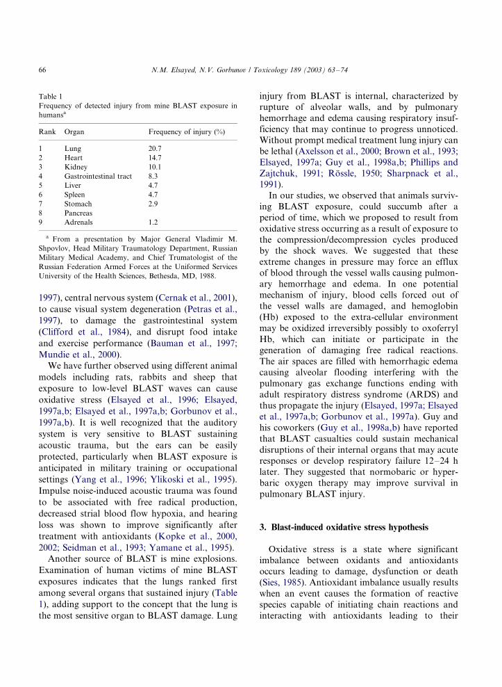
Another source of BLAST is mine explosions.
Examination of human victims of mine BLAST
exposures indicates that the lungs ranked first
among several organs that sustained injury (Table
1), adding support to the concept that the lung is
the most sensitive organ to BLAST damage. Lung
injury from BLAST is internal, characterized byrupture of alveolar walls, and by pulmonary
hemorrhage and edema causing respiratory insuf-
ficiency that may continue to progress unnoticed.
Without prompt medical treatment lung injury can
be lethal (Axelsson et al., 2000; Brown et al., 1993;
Elsayed, 1997a; Guy et al., 1998a,b; Phillips and
Zajtchuk, 1991; Rossle, 1950; Sharpnack et al.,
1991).In our studies, we observed that animals surviv-
ing BLAST exposure, could succumb after a
period of time, which we proposed to result from
oxidative stress occurring as a result of exposure to
the compression/decompression cycles produced
by the shock waves. We suggested that these
extreme changes in pressure may force an efflux
of blood through the vessel walls causing pulmon-ary hemorrhage and edema. In one potential
mechanism of injury, blood cells forced out of
the vessel walls are damaged, and hemoglobin
(Hb) exposed to the extra-cellular environment
may be oxidized irreversibly possibly to oxoferryl
Hb, which can initiate or participate in the
generation of damaging free radical reactions.
The air spaces are filled with hemorrhagic edemacausing alveolar flooding interfering with the
pulmonary gas exchange functions ending with
adult respiratory distress syndrome (ARDS) and
thus propagate the injury (Elsayed, 1997a; Elsayed
et al., 1997a,b; Gorbunov et al., 1997a). Guy and
his coworkers (Guy et al., 1998a,b) have reported
that BLAST casualties could sustain mechanical
disruptions of their internal organs that may acuteresponses or develop respiratory failure 12�/24 h
later. They suggested that normobaric or hyper-
baric oxygen therapy may improve survival in
pulmonary BLAST injury.
3. Blast-induced oxidative stress hypothesis
Oxidative stress is a state where significantimbalance between oxidants and antioxidants
occurs leading to damage, dysfunction or death
(Sies, 1985). Antioxidant imbalance usually results
when an event causes the formation of reactive
species capable of initiating chain reactions and
interacting with antioxidants leading to their
Table 1
Frequency of detected injury from mine BLAST exposure in
humansa
Rank Organ Frequency of injury (%)
1 Lung 20.7
2 Heart 14.7
3 Kidney 10.1
4 Gastrointestinal tract 8.3
5 Liver 4.7
6 Spleen 4.7
7 Stomach 2.9
8 Pancreas
9 Adrenals 1.2
a From a presentation by Major General Vladimir M.
Shpovlov, Head Military Traumatology Department, Russian
Military Medical Academy, and Chief Trumatologist of the
Russian Federation Armed Forces at the Uniformed Services
University of the Health Sciences, Bethesda, MD, 1988.
N.M. Elsayed, N.V. Gorbunov / Toxicology 189 (2003) 63�/7466
depletion. Under normal conditions, sufficient
concentrations of endogenous antioxidants as
well as redundant protective systems exist to
protect from environmental oxidant attacks. How-
ever, repeated exposure to environmental oxidants
such as air pollution, smoking, disease states, or
BLAST exposure, can result in accelerated rate of
antioxidant depletion tipping the balance from
sufficiency to deficiency producing oxidative
stress.
Oxidants produced in these processes include
radical and non-radicals reactive species, for
example, oxygen-containing molecules that are
more active than respirable molecular oxygen are
described as reactive oxygen species (ROS) (No-
guchi and Niki, 1999). Free radicals formed in the
biological systems are chemical entities with un-
paired electron on their outer most orbits. They
include superoxide anion radical (O2�+), hydroxyl
radical (+OH), nitrogen dioxide (NO2+), nitric
oxide (+NO), and thiyl radical (RS+). Radicals
may interact with each other to form more potent
complex moieties identifiable or unidentifiable. An
example is the reaction between O2�+ and +NO to
form peroxynitrite radical (+OONO). In addition,
some non-reactive antioxidants can be converted
to reactive radicals during the process of ‘‘anti-
oxidation,’’ under certain circumstances or disease
states. For example tocopherols in the process of
quenching a free radical can give rise to tocopheryl
radical that would abstracts hydrogen from ascor-
bic acid, and in the process, ascorbyl radicals are
formed. Non-radical reactive species include hy-
drogen peroxide (H2O2), singlet oxygen (1O2), lipid
hydroperoxide (LOOH), iron-oxygen complexes
(Fe�/O), and hypochloric acid (HOCl). Although
some ROS, such as O2�+ and HOCl, play im-
portant beneficial physiological roles such as
fighting infection through oxidative burst, others,
such as LOOH, and Fe�/O, play a destructive toxic
role. Some others play both roles, and can act as
oxidants or antioxidants depending on the circum-
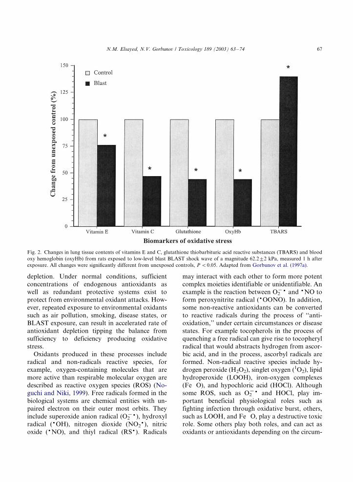
Fig. 2. Changes in lung tissue contents of vitamins E and C, glutathione thiobarbituric acid reactive substances (TBARS) and blood
oxy hemoglobin (oxyHb) from rats exposed to low-level blast BLAST shock wave of a magnitude 62.29/2 kPa, measured 1 h after
exposure. All changes were significantly different from unexposed controls, P B/0.05. Adapted from Gorbunov et al. (1997a).
N.M. Elsayed, N.V. Gorbunov / Toxicology 189 (2003) 63�/74 67
stances such as +NO, which will be discussed in
more detail later in this review. It was observed
that NO can act as an antioxidant and modulates
BLAST-induced oxidative stress. The reactions of
ROS with other molecules occur via different
reactions that include abstraction, addition, sub-
stitution, b-scission, and coupling reactions (No-
guchi and Niki, 1999). While hydrogen abstraction
reaction is an important step in lipid peroxidation,+NO addition reaction potentially plays an im-
portant antioxidant role in BLAST-mediated he-
moglobin (Hb) oxidation (Elsayed et al., 1997b;
Gorbunov et al., 1997a).In this review, experimental evidence of endo-
genous antioxidant depletion following BLAST
exposure leading to oxidative stress, then some
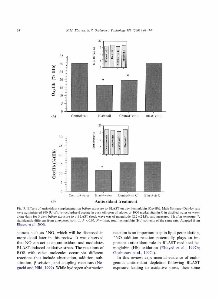
Fig. 3. Effects of antioxidant supplementation before exposure to BLAST on oxy hemoglobin (OxyHb). Male Sprague�/Dawley rats
were administered 800 IU of D-a-tocohpheryl acetate in corn oil, corn oil alone, or 1000 mg/kg vitamin C in distilled water or water
alone daily for 3 days before exposure to a BLAST shock wave was of magnitude 62.29/2 kPa, and measured 1 h after exposure. *,
significantly different from unexposed control, P B/0.05, N�/Inset, total hemoglobin (Hb) contents of the same rats. Adapted from
Elsayed et al. (2000).
N.M. Elsayed, N.V. Gorbunov / Toxicology 189 (2003) 63�/7468
beneficial effects of supplementation with phar-macological doses of antioxidants prior to BLAST
exposure (assessed by examining several biomar-
kers of oxidative stress) will be presented. Finally,
a proposed cascade outlining the events associated
with BLAST-induced lung injury and the potential
sites where antioxidants can act to break that
chain of events will be discussed.
4. BLAST and antioxidant depletion
In a series of studies (Elsayed, 1997a,b; Elsayedet al., 1996, 1997a,b; Gorbunov, et al., 1997a), in
which animals were exposed to different levels of
BLAST and examined at different times after
exposure it was observed that BLAST injury is
associated with a significant depletion of both
water- and oil-soluble antioxidants including vita-
min E, vitamin C and glutathione, a decline in
blood oxygenation, and a concomitant increase inlipid peroxidation. Fig. 2, summarizes some of
these results observed in rats 1 h after a single
exposure to low-level BLAST shock wave of �/62
kPa magnitude.
5. BLAST and antioxidant supplementation
In another set of studies (Armstrong et al., 1998;
Elsayed et al., 2000), preloading rats with phar-
macological doses of vitamin E, vitamin C, or a-
lipoic acid administered via oral gavage for 3 daysprior to BLAST exposure and examined 1 h after
BLAST exposure, resulted in improved blood
oxygenation, and survival of exposed animals
(Fig. 3). The pathological manifestation of BLAST
injury was also improved (data not shown) com-
pared with control rats that received the vehicle
alone. In addition, administration of allupurinol,
and superoxide dismutase, to rats (Seidman et al.,1993), defroxamine mesylate alone or in combina-
tion with glial cell line-derived neurotropic factor
(Yamasoba et al., 1999), acetyl-L-carnitine, N-
methyl-D-aspartate, reduced glutathione (Ohinata
et al., 2000), salicylate and N -L-acetylcysteine
combination (Kopke et al., 2000, 2002), were
reported to reduce impulse-noise induced hearingloss.
6. BLAST and nitric oxide
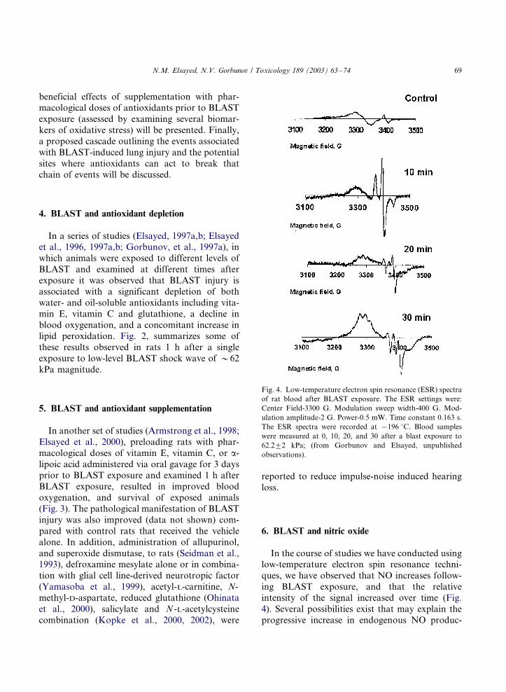
In the course of studies we have conducted usinglow-temperature electron spin resonance techni-
ques, we have observed that NO increases follow-
ing BLAST exposure, and that the relative
intensity of the signal increased over time (Fig.
4). Several possibilities exist that may explain the
progressive increase in endogenous NO produc-
Fig. 4. Low-temperature electron spin resonance (ESR) spectra
of rat blood after BLAST exposure. The ESR settings were:
Center Field-3300 G. Modulation sweep width-400 G. Mod-
ulation amplitude-2 G. Power-0.5 mW. Time constant 0.163 s.
The ESR spectra were recorded at �/196 8C. Blood samples
were measured at 0, 10, 20, and 30 after a blast exposure to
62.29/2 kPa; (from Gorbunov and Elsayed, unpublished
observations).
N.M. Elsayed, N.V. Gorbunov / Toxicology 189 (2003) 63�/74 69
tion over time following BLAST exposure. Resultsof in vitro studies from our laboratory in colla-
boration with Dr Kagan at the University of
Pittsburg (Gorbounov et al., 1995; Gorbunov et
al., 1996, 1997a,b, 1998; Kagan et al., 1996)
suggested that NO may act as an antioxidant
preventing oxidative damage by tert-butyl hydro-
peroxide-induced formation of alloy and proxy
radical. NO was proposed to act as an antioxidant
earlier (Hogg et al., 1993; Kanner et al., 1991), andis further supported by observations from a study
of compression injury to rat’s spinal cord (Ha-
mada et al., 1996) in which endogenous NO was
associated with a decline in lipid peroxidation.
When NO synthesis was blocked by the nitric
oxide syntheses inhibitor NG-nitro-L-argentine
methyl ester (L-NAME), lipid peroxidation in-
creased significantly (Fig. 5). A recent report by
Zunic et al. (2000) observed that pulmonaryBLAST injury is associated with NO overproduc-
tion in rabbit lungs 30 min after exposure coupled
with arterial depletion of arginine.
It is possible that increased NO content in blood
and tissue reflects an antioxidant mechanism by
which NO blocks oxoferryl Hb formation through
redox reactions and thus prevents propagation of
free-radical mediated reactions. Another possibi-lity is that NO alone or in conjunction with carbon
monoxide is acting as a molecular switch in a
signal transduction cascade in response to the
injury (Maulik et al., 1996). It is also possible
that it is involved in a mechanism not yet known.
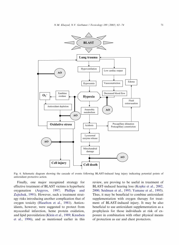
7. BLAST and injury cascade in the lung
The totality of the observation presented in thisreport suggest that BLAST exposure causes lung
damage and that it interferes with the blood’s
oxygen carrying capacity leading to varying de-
grees of hypoxia. Consequently, oxidative stress
manifested in antioxidant depletion, and lipid
peroxidation occurs. These events can possibly be
prevented or its severity reduced by means of
antioxidant supplementation. The schematic dia-gram presented in Fig. 6 summarizes the possible
cascade of events that would occur in the lung
following BLAST exposure. It is based on the
proposal of Taylor and Collins (1988) for manage-
ment of hemorrhage incorporating the experimen-
tal observations from our BLAST studies.
Fig. 5. Effect of nitric oxide (NO) and pretreatment with 30 mg/kg of the NO synthase inhibitor NG-nitro-L-arginine methylester (L-
NAME) on lipid peroxidation measured as thiobarbituric acid reactive substances (TBARS) following spinal cord injury in rats. NO
was measured in the injured and adjacent central regions directly by electron spin resonance (ESR) spin-trapping technique using iron
(Fe2�) and diethyldithiocarbamate (DETC) (adapted from Hamada et al., 1996).
N.M. Elsayed, N.V. Gorbunov / Toxicology 189 (2003) 63�/7470
Finally, one major recognized strategy for
effective treatment of BLAST victims is hyperbaric
oxygenation (Argyros, 1997; Phillips and
Zajtchuk, 1991). However, such a treatment strat-
egy risks introducing another complication that of
oxygen toxicity (Haselton et al., 1981). Antiox-
idants, however, were suggested to protect from
myocardial infarction, heme protein oxidation,
and lipid peroxidation (Klein et al., 1989; Knudsen
et al., 1996), and as mentioned earlier in this
review, are proving to be useful in treatment of
BLAST-induced hearing loss (Kopke et al., 2002,
2000; Seidman et al., 1993; Yamane et al., 1995).
Thus, it may be beneficial to combine antioxidant
supplementation with oxygen therapy for treat-
ment of BLAST-induced injury. It may be also
beneficial to use antioxidant supplementation as a
prophylaxis for those individuals at risk of ex-
posure in combination with other physical means
of protection as ear and chest protectors.
Fig. 6. Schematic diagram showing the cascade of events following BLAST-induced lung injury indicating potential points of
antioxidant protective action.
N.M. Elsayed, N.V. Gorbunov / Toxicology 189 (2003) 63�/74 71
In conclusion, the population at risk of exposureto BLAST has expanded in recent years. In an
attempt to devise new modalities for treatment
and/or protection, antioxidant supplementation
may have an important role to play. Moreover,
the role of NO in BLAST-induced injury process is
not completely understood, and therefore, needs to
be further explored. Such a role may offer an
additional or an alternative therapeutic modalityto the management of BLAST-induced injury.
Acknowledgements
The authors thank Dr Valerian Kagan, Dr
Anatoly Osipov, Dr Yulina Turina, Dr Vladimir
Turin, and Dr Billy Day at the University of
Pittsburg for their contribution in the early stages
of research; Majs Karen Armstrong and MaryCooper, and Dr Adolph Januszkiewicz for their
participation in the studies; Jennifer Morris and
Sgt Myron Williams for excellent technical assis-
tance; and COLs Charles McQueen and Maria
Mayorga for their support and constructive criti-
cism.
References
Abenhaim, L., Dab, W., Salmi, L.R., 1992. Study of civilian
victims of terrorist attacks (France 1982�/1987). J. Clin.
Epidemiol. 45, 103�/109.
Argyros, G.J., 1997. Management of primary blast injury.
Toxicology 121, 105�/115.
Armstrong, K.L., Cooper, M.F., Williams, M.T., Elsayed,
N.M., 1998. Vitamin E and lipoic acid, but not vitamin C
improve blood oxygenation after high-energy impulse noise
(blast) exposure. Biochem. Biophys. Res. Commun. 253,
114�/118.
Axelsson, H., Hjelmqvist, H., Medin, A., Persson, J.K.,
Suneson, A., 2000. Physiological changes in pigs exposed
to a blast wave from a detonating high-explosive charge.
Mil. Med. 165, 119�/126.
Bauman, R.A., Elsayed, N.M., Petras, J.M., Widholm, J., 1997.
Sublethal blast overpressure disrupts the food intake and
exercise performance in rats. Toxicology 121, 65�/79.
Benzinger, T., 1950. Physiological effects of blast in air and
water. In: German Aviation Medicine, World War II, vol. 2.
US Government Printing Office, Washington, DC, pp.
1225�/1259.
Brismar, B., Bergenwald, L., 1982. The terrorist bomb explo-
sion in Bologna, Italy 1980: an analysis of the effects and
injuries sustained. J. Trauma 22, 216�/220.
Brown, R.F.R., Cooper, G.J., Maynard, R.L., 1993. The
ultrastructure of rat lung following acute primary blast
injury. Int. J. Exp. Pathol. 74, 151�/162.
Cassen, B., Curtis, L., Kistler, K., 1950. Initial studies of the
effect of laboratory produced air blast on animals. J. Aviat.
Med. 21, 38�/47.
Celander, H., Clemedson, C., Erickson, U., Hultman, H., 1950.
The use of compressed air-operated shock tube for physio-
logical blast research. Acta Physiol. Scand. 32, 6�/13.
Cernak, I., Wang, Z., Jiang, J., Bian, X., Savic, J., 2001.
Ultrastructural and functional characteristics of blast in-
jury-induced neurotrauma. J. Trauma 50, 695�/706.
Clemedson, C.J., 1956. Blast injury. Physiol. Rev. 36, 336�/354.
Clemedson, C.J., Hultman, H., 1954. Air embolism and the
cause of death in blast injury. Mil. Surg. 114, 424�/437.
Clifford, C.B., Moe, J.B., Jaeger, J.J., Hess, J.L., 1984.
Gastrointestinal lesions in lambs due to multiple low level
blast overpressure exposure. Mil. Med. 149, 491�/495.
Dodd, K.T., Mundie, T.G., Lagutchik, M.S., Morris, J.R.,
1997. Cardiopulmonary effects of high impulse noise
exposure. J. Trauma 43, 656�/666.
Elsayed, N.M., Tyurina, Y.Y., Tyurin, V.A., Menshikova,
E.V., Kisin, E.R., Kagan, V.E., 1996. Antioxidant deple-
tion, lipid peroxidation, and impairment of calcium trans-
port induced by air blast overpressure in rat lungs. Exp.
Lung Res. 22, 179�/200.
Elsayed, N.M., 1997a. Toxicology of blast overpressure.
Toxicology 121, 1�/15.
Elsayed, N.M., 1997b. Antioxidant depletion and lipid perox-
idation initiated by blast overpressure. In: Baskin, S., Salem,
H. (Eds.), Oxidants, Antioxidants, and Free radicals. Taylor
and Francis, Washington, DC, pp. 315�/326.
Elsayed, N.M., Gorbunov, N.V., Kagan, V.E., 1997a. A
proposed biochemical mechanism for blast overpressure-
induced hemorrhagic injury. Toxicology 121, 81�/90.
Elsayed, N.M., Fitzpatrick, T.M., Dodd, K.T., 1997b. Free
radical-associated response in blood of sheep, rabbits, and
rats after a single exposure to high energy impulse noise
(blast). Environ. Nutr. Interact. 1, 11�/22.
Elsayed, N.M., Armstrong, K.L., Williams, M.T., Cooper,
M.F., 2000. Antioxidant loading reduced oxidative stress
induced by high-energy impulse (blast) exposure. Toxicol-
ogy 155, 91�/99.
Frykberg, E.R., Tepas, J.J., 1988. Terrorist bombings. Lessons
learned from Belfast to Beirut. Ann. Surg. 208, 569�/576.
Gorbounov, N.V., Osipov, A.N., Day, B.W., Zyas-Rivera, B.,
Kagan, V.E., Elsayed, N.M., 1995. Reduction of ferrylmyo-
globin and ferrylhemoglobin by nitric oxide: a protective
mechanism against ferryl hemoprotein-induced oxidations.
Biochemistry 34, 6689�/6699.
Gorbunov, N.V., Osipov, A.N., Sweetland, M.A., Day, B.W.,
Elsayed, N.M., Kagan, V.E., 1996. NO-redox paradox:
Direct oxidation of a-tocopherol and a-tocopherol
N.M. Elsayed, N.V. Gorbunov / Toxicology 189 (2003) 63�/7472
mediated oxidation of ascorbate. Biochem. Biophys. Res.
Commun. 219, 835�/841.
Gorbunov, N.V., Elsayed, N.M., Kisin, E.R., Kozlov, A.V.,
Kagan, V.E., 1997a. Air blast overpressure induces oxida-
tive stress in rat lungs: Interplay between hemoglobin,
antioxidants, and lipid peroxidation. Am. J. Physiol. 272,
L320�/L334.
Gorbunov, N.V., Yalowich, J.C., Gaddam, A., Thampatty, P.,
Ritov, V.B., Kisin, E.R., Elsayed, N.M., Kagan, V.E.,
1997b. Nitric oxide prevents oxidative damage by tert-butyl
hydroperoxide in erythroleukemia cells via nitrosylation of
heme and non-hem iron. Electron paramagnetic resonance
evidence. J. Biol. Chem. 272, 12249�/12880.
Gorbunov, N.V., Turina, Y.Y., Salama, G., Day, B., Argyros,
G., Elsayed, N.M., Kagan, V.E., 1998. Nitric oxide prevents
oxidative damage by tert-butyl hydroperoxide-induced for-
mation of alkoxyl and peroxyl radicals and oxidative
damage to cardiac monocytes. Biochem. Biophys. Res.
Commun. 244, 647�/651.
Guy, R.J., Glover, M.A., Cripps, N.P., 1998a. The pathophy-
siology of primary blast injury and its implications for
treatment. Part I: the thorax. J. R. Nav. Med. Serv. 84, 79�/
86.
Guy, R.J., Kirkman, E., Watkins, P.E., Cooper, G.J., 1998b.
Physiologic responses to primary blast. J. Trauma 45, 983�/
987.
Hadden, W.A., Rutherford, W.H., Merrett, J.D., 1978. The
injuries of terrorist bombing: a study of 1532 consecutive
patients. Br. J. Surg. 65, 525�/531.
Hamada, Y., Ikatya, T., Katoh, S., Tsuchiya, K., Niwa, M.,
Tsutsumishita, Y., Fukuzawa, K., 1996. Roles of nitric
oxide in compression injury of rat spinal cord. Free Radic.
Biol. Med. 20, 1�/9.
Haselton, P.S., Penna, P., Torry, J., 1981. Effect of oxygen on
the lungs after blast injury and burns. J. Clin. Pathol. 34,
1147�/1154.
Hiss, J., Kahana, T., 1998. Suicide bombers in Israel. Am. J.
Forensic Med. Pathol. 19, 63�/66.
Hogg, N., Kalyanaraman, B., Joseph, J., Struck, A., Parthasar-
athy, S., 1993. Inhibition of low-density lipoprotein oxida-
tion by nitric oxide. Potential role in atherogenesis. FEBS
Lett. 34, 170�/174.
Kagan, V.E., Day, B.W., Elsayed, N.M., Gorbunov, N.V.,
1996. Dynamics of nitrosylated haemoglobin in blood.
Nature 383, 30�/31.
Kanner, J., Harel, S., Granit, R., 1991. Nitric oxide as an
antioxidant. Arch. Biochem. Biophys. 289, 130�/136.
Katz, E., Ofek, B., Adler, J., Abramowitz, H.B., Krausz, M.M.,
1989. Primary blast injury after a bomb explosion in a
civilian bus. Ann. Surg. 209, 484�/488.
Klein, H.H., Pitch, S., Lindert, S., Nebendahal, K., Niedmann,
P., Kreuzer, H., 1989. Combined treatment with vitamin E
and C in experimental myocardial infarction in pigs. Am.
Heart J. 118, 667�/673.
Knudsen, C.A., Tappel, A.A., North, J.A., 1996. Multiple
antioxidants protects against heme protein and lipid oxida-
tion in kidney tissue. Free Radic. Biol. Med. 20, 165�/173.
Kopke, R.D., Weisskopf, P.A., Boone, J.L., Jackson, R.L.,
Wester, D.C., Hoffer, M.E., Lambert, D.C., Charon, C.C.,
Ding, D.L., McBride, D., 2000. Reduction of noise-induced
hearing loss using L-NAC and salicylate in the chinchilla.
Hear. Res. 149, 138�/146.
Kopke, R.D., Coleman, J.K., Liu, J., Campbell, K.C., Riffen-
burgh, R.H., 2002. Candidate’s thesis: enhancing intrinsic
cochlear stress defenses to reduce noise-induced hearing
loss. Laryngoscope 112, 1515�/1532.
Maulik, N., Engelman, D.T., Watanabe, M., Engelman, R.M.,
Rousou, J.A., Flack, J.E., III, Deaton, D.W., Gorbunov,
N.V., Elsayed, N.M., Kagan, V.E., Das, D.K., 1996. Nitric
oxide/carbon monoxide. A molecular switch for myocardial
preservation during ischemia. Circulation 94 (Suppl. II), II-
398�/II-406.
Mellor, S.G., 1992. The relationship of blast loading to death
and injury from explosion. World J. Surg. 16, 893�/898.
Mundie, T.G., Dodd, K.T., Lagutchik, M.S., Morris, J.R.,
Martin, D., 2000. Effects of blast exposure on exercise
performance in sheep. J. Trauma 48, 1115�/1121.
Noguchi, N., Niki, E., 1999. Chemistry of active oxygen species
and antioxidants. In: Papas, A.M. (Ed), Antioxidants
Status, Diet, Nutrition, and Health. CRC Press, Boca
Raton, FL, pp. 3�/20.
Ohinata, Y., Yamasoba, T., Schachtm, J., Miller, J.M., 2000.
Glutathione limits noise-induced hearing loss. Hear. Res.
146, 28�/34.
Petras, J.M., Bauman, R.A., Elsayed, N.M., 1997. Visual
system degeneration: primary blast overpressure-induced
brain injury. Toxicology 121, 41�/49.
Pfefferbaum, B., 2001. The impact of the Oklahoma city
bombing on children in the community. Mil. Med. 166
(Suppl.), 49�/50.
Phillips, Y.Y., Richmond, D.R., 1991. Primary blast injury and
basic research: a brief history. In: Bellamy, R., Zajtchuk, R.
(Eds.), Textbook of Military Medicine. Part 1. Conven-
tional Warfare Ballistic, Blast, and Burn Injuries, vol. 5.
Office of the Surgeon General, Department of the Army,
USA, Washington, DC, pp. 221�/240.
Phillips, Y.Y., Zajtchuk, J.T., 1991. The management of
primary blast injury. In: Bellamy, R., Zajtchuk, R. (Eds.),
Textbook of Military Medicine. Part 1. Conventional
Warfare, Ballistic, Bast, and Burn injuries, vol. 5. Office
of the Surgeon General, Department of the Army, USA,
Washington, DC, pp. 295�/335.
Richmond, D.R., 1991. Blast criteria for open spaces and
enclosures. Scand. Audiol. Suppl. 34, 49�/76.
Rignault, D.P., Deligny, M.C., 1989. The 1986 Terrorist
bombing experience in Paris. Ann. Surg. 209, 368�/373.
Rossle, R., 1950. Pathology of blast effects. In: German
Aviation Medicine, World War II, vol. 2. USA Government
Printing Office, Washington, DC, pp. 1260�/1273.
Schardin, H., 1950. The physical principles of the effects of a
detonation. In: German Aviation Medicine, World War II,
vol. 2. USA Government Printing Office, Washington, DC,
pp. 1207�/1224.
N.M. Elsayed, N.V. Gorbunov / Toxicology 189 (2003) 63�/74 73
Seidman, M.D., Shivapuja, B.G., Quirk, W.S., 1993. The
protective effects of allopurinol and superoxide dismutase
on noise-induced cochlear damage. Otolaryngol Head Neck
Surg. 109, 1052�/1056.
Sharpnack, D.D., Johnson, A.J., Philllips, Y.Y., 1991. The
pathology of primary blast injury. In: Bellamy, R.,
Zajtchuk, R. (Eds.), Textbook of Military Medicine. Part
1. Conventional Warfare, Ballistic, Blast, and Burn Injuries,
vol. 5. Office of the Surgeon General, Department of the
Army, USA, Washington, DC, pp. 271�/294.
Sies, H., 1985. Oxidative Stress: Introductory Remarks. In:
Sies, H. (Ed). Oxidative Stress. Academic Press, London,
pp. 1�/8.
Stuhmiller, J.H., Phillips, Y.Y., Richmond, D.R., 1991. The
physics and mechanisms of primary blast injury. In:
Bellamy, R., Zajtchuk, R. (Eds.), Textbook of Military
Medicine, Part 1. Conventional Warfare, Ballistic, Blast,
and Burn Injuries, vol. 5. Office of the Surgeon General,
Department of the Army, USA, Washington, DC, pp. 241�/
270.
Taylor, B.L., Collins, C., 1988. The management of massive
haemorrhage. Br. J. Hosp. Med. 40, 105�/110.
Thach, A.B., Ward, T.P., Hollifield, R.D., Cockerham, K.,
Birdsong, R., Kramer, K.K., 2000. Eye injuries in a terrorist
bombing: Dhahran, Saudi Arabia, June 25, 1996. Ophthal-
mology 107, 844�/847.
Yamane, H., Nakai, Y., Takayama, M., Iguchi, H., Nakagawa,
T., Kojima, A., 1995. Appearance of free radicals in the
guinea pig inner ear after noise-induced acoustic trauma.
Eur. Arch. Otorhinolaryngol. 252, 504�/508.
Yamasoba, T., Schacht, J., Shoji, F., Miller, J.M., 1999.
Attenuation of cochlear damage from noise trauma by an
iron chelator, a free radical scavenger and glial cell line-
derived neurotrophic factor in vivo. Brain Res. Jan
9;815(2):317�/325.
Yang, Z., Wang, Z., Tang, C., Ying, Y., 1996. Biological effects
of weak blast waves and safety limits for internal organ
injury in the human body. J. Trauma 40, S81�/S84.
Ylikoski, M., Pekkarinen, J.O., Starck, J.P., Paakkonen, R.J.,
Ylikoski, J.S., 1995. Physical characteristics of gunfire
impulse noise and its attenuation by hearing protectors.
Scand Audiol. 24, 3�/11.
Zhang, J., Wang, Z., Leng, H., Yang, Z., 1996. Studies on lung
injuries caused by blast underpressure. J. Trauma 40, S77�/
S80.
Zunic, G., Pavlovic, R., Malicevic, Z., Savic, V., Cernak, I.,
2000. Pulmonary blast injury increases nitric oxide produc-
tion, disturbs arginine metabolism, and alters the plasma
free amino acid pool in rabbits during the early posttrau-
matic period. Nitric Oxide 4, 123�/128.
N.M. Elsayed, N.V. Gorbunov / Toxicology 189 (2003) 63�/7474