Interactive simulator for e-Learning environments - BioMedical ...
18
RESEARCH Open Access Interactive simulator for e-Learning environments: a teaching software for health care professionals Claudio De Lazzari 1,2* , Igino Genuini 3,2 , Domenico M Pisanelli 4,2 , Alessandra D’Ambrosi 3,2 and Francesco Fedele 3,2 * Correspondence: [email protected] 1 CNR, Institute of Clinical Physiology, UOS of Rome, Via S.M. della Battaglia, 44, 00185 Rome, Italy 2 National Institute for Cardiovascular Research, Bologna, Italy Full list of author information is available at the end of the article Abstract There is an established tradition of cardiovascular simulation tools, but the application of this kind of technology in the e-Learning arena is a novel approach. This paper presents an e-Learning environment aimed at teaching the interaction of cardiovascular and lung systems to health-care professionals. Heart-lung interaction must be analyzed while assisting patients with severe respiratory problems or with heart failure in intensive care unit. Such patients can be assisted by mechanical ventilatory assistance or by thoracic artificial lung. “In silico” cardiovascular simulator was experimented during a training course given to graduate students of the School of Specialization in Cardiology at ‘Sapienza’ University in Rome. The training course employed CARDIOSIM © : a numerical simulator of the cardiovascular system. Such simulator is able to reproduce pathophysiological conditions of patients affected by cardiovascular and/or lung disease. In order to study the interactions among the cardiovascular system, the natural lung and the thoracic artificial lung (TAL), the numerical model of this device has been implemented. After having reproduced a patient’s pathological condition, TAL model was applied in parallel and hybrid model during the training course. Results obtained during the training course show that TAL parallel assistance reduces right ventricular end systolic (diastolic) volume, but increases left ventricular end systolic (diastolic) volume. The percentage changes induced by hybrid TAL assistance on haemodynamic variables are lower than those produced by parallel assistance. Only in the case of the mean pulmonary arterial pressure, there is a percentage reduction which, in case of hybrid assistance, is greater (about 40%) than in case of parallel assistance (20-30%). At the end of the course, a short questionnaire was submitted to students in order to assess the quality of the course. The feedback obtained was positive, showing good results with respect to the degree of students’ learning and the ease of use of the software simulator. Keywords: e-Learning, Numerical model, Cardiovascular system, Thoracic artificial lung assistance, Haemodynamic, Ontologies © 2014 De Lazzari et al.; licensee BioMed Central. This is an Open Access article distributed under the terms of the Creative Commons Attribution License (http://creativecommons.org/licenses/by/4.0), which permits unrestricted use, distribution, and reproduction in any medium, provided the original work is properly credited. The Creative Commons Public Domain Dedication waiver (http://creativecommons.org/publicdomain/zero/1.0/) applies to the data made available in this article, unless otherwise stated. De Lazzari et al. BioMedical Engineering OnLine 2014, 13:172 http://www.biomedical-engineering-online.com/content/13/1/172
-
Upload
khangminh22 -
Category
Documents
-
view
0 -
download
0
Transcript of Interactive simulator for e-Learning environments - BioMedical ...
De Lazzari et al. BioMedical Engineering OnLine 2014, 13:172http://www.biomedical-engineering-online.com/content/13/1/172
RESEARCH Open Access
Interactive simulator for e-Learning environments:a teaching software for health care professionalsClaudio De Lazzari1,2*, Igino Genuini3,2, Domenico M Pisanelli4,2, Alessandra D’Ambrosi3,2 and Francesco Fedele3,2
* Correspondence:[email protected], Institute of ClinicalPhysiology, UOS of Rome, Via S.M.della Battaglia, 44, 00185 Rome, Italy2National Institute for CardiovascularResearch, Bologna, ItalyFull list of author information isavailable at the end of the article
Abstract
There is an established tradition of cardiovascular simulation tools, but theapplication of this kind of technology in the e-Learning arena is a novel approach.This paper presents an e-Learning environment aimed at teaching the interaction ofcardiovascular and lung systems to health-care professionals. Heart-lung interactionmust be analyzed while assisting patients with severe respiratory problems or withheart failure in intensive care unit. Such patients can be assisted by mechanicalventilatory assistance or by thoracic artificial lung.“In silico” cardiovascular simulator was experimented during a training course givento graduate students of the School of Specialization in Cardiology at ‘Sapienza’University in Rome.The training course employed CARDIOSIM©: a numerical simulator of the cardiovascularsystem. Such simulator is able to reproduce pathophysiological conditions of patientsaffected by cardiovascular and/or lung disease. In order to study the interactionsamong the cardiovascular system, the natural lung and the thoracic artificial lung (TAL),the numerical model of this device has been implemented. After having reproduced apatient’s pathological condition, TAL model was applied in parallel and hybrid modelduring the training course.Results obtained during the training course show that TAL parallel assistance reducesright ventricular end systolic (diastolic) volume, but increases left ventricular end systolic(diastolic) volume. The percentage changes induced by hybrid TAL assistance onhaemodynamic variables are lower than those produced by parallel assistance. Only inthe case of the mean pulmonary arterial pressure, there is a percentage reductionwhich, in case of hybrid assistance, is greater (about 40%) than in case of parallelassistance (20-30%).At the end of the course, a short questionnaire was submitted to students in order toassess the quality of the course. The feedback obtained was positive, showing goodresults with respect to the degree of students’ learning and the ease of use of thesoftware simulator.
Keywords: e-Learning, Numerical model, Cardiovascular system, Thoracic artificial lungassistance, Haemodynamic, Ontologies
© 2014 De Lazzari et al.; licensee BioMed Central. This is an Open Access article distributed under the terms of the CreativeCommons Attribution License (http://creativecommons.org/licenses/by/4.0), which permits unrestricted use, distribution, andreproduction in any medium, provided the original work is properly credited. The Creative Commons Public Domain Dedicationwaiver (http://creativecommons.org/publicdomain/zero/1.0/) applies to the data made available in this article, unless otherwisestated.

De Lazzari et al. BioMedical Engineering OnLine 2014, 13:172 Page 2 of 18http://www.biomedical-engineering-online.com/content/13/1/172
BackgroundThe e-Learning industry is developing at a fast pace. In the medical context, in particular,
it is going to offer “face to face” classes, like those proposed in university rooms.
Unfortunately tutors mainly employ static contents (e.g. PowerPoint or Word files
or Virtual Learning Environments (VLEs) or websites). Such contents require no
real interaction by the users [1]. Simulators can increase the degree of interaction
in health care education, for example by realizing a “virtual patient”, implemented
by means of interactive computer simulations [1-3]. In this way the learner will take profit
of a various range of learning activities. Other notable experiences include for instance
Collaborative Tutoring Systems to determine clinical-reasoning skills of students [4].
In this paper we present our experience of an e-Learning course realized using the
numerical simulator of the cardiovascular system CARDIOSIM© [5]. This interactive
software is able to reproduce physiological and pathophysiological conditions of
patients affected by cardiovascular and/or lung disease [6,7]. In the simulator, several
different ventricular pumps and devices was implemented in order to assist one or
both ventricles in parallel or in series [8-11]. With respect to previous CARDIOSIM©
version, thoracic artificial lung (TAL) assistance simulation was implemented too.
This new version is also able to reproduce the cardiopulmonary pathological conditions
of a patient and to study the interaction among the cardiovascular system, the natural
lung and the TAL.
During a training course held for graduate students of the School of Specialization
in Cardiology at the Department of Cardiovascular, Respiratory, Nephrological,
Anesthesiological and Geriatric Sciences (hereinafter CRNAGS Department), “Sapienza”
University of Rome, heart-lung interaction was studied when thoracic artificial lung
assistance was applied.
Heart-lung interaction must be analyzed when patients with heart failure or with
severe respiratory problems are assisted in Intensive Care Cardiac Unit. Some pulmonary
diseases can be the result of haemodynamic pathology, most notably an increase in
pulmonary vascular resistance. In these cases the right ventricle (RV) is overloaded and
cardiac output is reduced to problematic levels. In many pulmonary diseases, both gas
exchange and haemodynamic disturbances are present to some degree. Therefore an
accurate analysis of the heart-lung interaction must be done in those patients with severe
respiratory problems or with heart failure [12].
Chronic respiratory failure or severe pulmonary infections can be treated using
thoracic artificial lung that do not use a pump [13] but rather can be attached to
the natural lung (NL) in series, in parallel or in hybrid mode [14].
During the course the choice of patients used for reproducing pathological conditions
has been done using on a set of pathological clinical patients. In these pathological
conditions (the baseline conditions for students who start the interaction with the
simulation tool), it is possible to observe the following effects: an increase of right
ventricular end systolic volume, an increase of right ventricular end diastolic volume and
an important increase of pulmonary arterial pressure. Starting from baseline conditions,
students have been trained to manage the virtual patient’s pathology by acting in different
ways. At first, students applied parallel TAL assistance and set pulmonary arterial
peripheral resistance to high value in order to induce a pathological overload in the right
ventricle. In a second moment, the same simulations were performed setting pulmonary

De Lazzari et al. BioMedical Engineering OnLine 2014, 13:172 Page 3 of 18http://www.biomedical-engineering-online.com/content/13/1/172
arterial peripheral resistance to physiological value in order to simulate the effects induced
by drugs that permit to download the work of the right ventricle. Finally students
reproduced the same simulations using the hybrid TAL assistance. Results obtained
by student’s simulations showed that parallel TAL assistance:
� increases both left ventricular and end systolic and end diastolic volume;
� reduces right ventricular and end systolic and end diastolic volume;
� reduces mean pulmonary arterial pressure.
All students’ simulations have been performed in an interactive way. By interacting
with the tool, students were able to put into practice concepts learned during their
previous studies. Sometime they made mistakes that have been highlighted by the
simulator. By simulating an inappropriate drug administration, students have been
able to verify how the conditions of the virtual patient worsened.
During our experience, it has been proved that cardiovascular interactive software
simulator can be a useful tool for studying and training cardiologists, anaesthesiologists,
nurses and medical graduate and undergraduate students in the management of mechanical
circulatory and ventilatory devices [15-17].
Materials and methodsThe software simulator developed by the Institute of Clinical Physiology of the Italian
National Research Council is able to reproduce pathophysiological conditions of patients
affected by cardiovascular and/or lung diseases. The simulator implements many different
cardiovascular pumps in order to assist one or both ventricles in parallel or in series way
[6,8-10]. The blood pump implemented can reproduce pulsatile or continuous flow.
The lung assistance is realized by a new software module implementing a thoracic
artificial lung. TAL can be connected to the NL in parallel, in series and in hybrid mode
[14,18,19]. CARDIOSIM© has a graphical and a numerical interface which shows the
instantaneous waveforms during the cardiac cycle and the mean values of pressures,
volumes and flow.
The training course, held for graduate students, consisted of different exercises in
which the pathophysiological conditions of patients hospitalized in CRNAGS Department
were reproduced using the interactive software simulator.
In this paper we describe our experience gained during the simulations cycle held in
a classroom with 25 students. Each pair of students was equipped with a workstation
on which the CARDIOSIM© software was installed. The exercises were focused on
the study of the effects induced on some haemodynamic variables by the heart-lung
interaction in presence of TAL assistance. Figure 1 (panel A) shows the software interface
used during the e-Learning course relative to a first level of choice that permits to select
different districts of cardiocirculatory network, different mechanical assist devices
and different mechanical ventilatory assistance. Panels B and C show two different
possible networks choose. Starting from baseline conditions, reproducing the
pathological conditions of hospitalized patient, the effects of different TAL assistances (in
parallel and hybrid mode) were reproduced by changing TAL compliances and resistances
[13,14], in presence of different pulmonary vascular resistance values. The selection of

Figure 1 Software interface used during the e-Learning training course. Window [A] represents a firstlevel of choice that permits to select among different districts of cardiocirculatory network, differentmechanical assist devices and different mechanical ventilatory assistance. Panels [B] and [C] show twodifferent possible networks choose. In panel [B] is represented a network with a simple implementation ofsystemic and pulmonary arterial sections and with a parallel pneumatic left ventricular assist device (LVAD).In panel [C] is represented a network with a complex implementation of systemic and pulmonary arterialtree and with “in series” rotary left ventricular assist device (Hemopump).
De Lazzari et al. BioMedical Engineering OnLine 2014, 13:172 Page 4 of 18http://www.biomedical-engineering-online.com/content/13/1/172
patients undergoing this type of therapy had been previously carried out by the students
on the basis of an ontology based on the evaluation of measured haemodynamic
parameter values.
The cardiovascular model
The overall modular configuration of the software simulator is reported in Figure 2.
Each block of the circulatory network can be implemented as combinations of “n”
different sub-blocks. The generic block (or sub-block) can be implemented using
different combinations of the three electrical networks reported in figure. Each
block (or sub-block) has two inputs and two outputs representing flow (Q) and
pressure (P). Panels B, C of Figure 1 are two possible circulatory network representations
obtained starting by the configuration presented in Figure 2. In this figure also mechanical
assist device (MAD) and thoracic artificial lung are represented as block having
two inputs and two outputs. This kind of assistances can be connected to the circulatory
network in different way (e.g. MAD can be connected in series or in parallel to
both ventricles). All single sub-blocks of the network can be implemented using
one of the three electrical representations realised by resistance (R), inductance (L)
and compliance (C). Each sub-block can be implemented numerically by first-order
partial differential equations. In the software simulator, the entire equation system
is solved by Euler’s method.

Systemic Arterial
LeftAtrium
LeftVentricle
PulmonaryVenous
Q1
P1
Q2
P2
Q3
P3
P Q
SectionAtrium Ventricle
Section P1 P2 P3
Q9 P9Mechanical
Pm
Qm
Coronary Section
Q4
P4
P8Q8
MechanicalAssistDevice
SystemicRi hRi h
Pulmonary P5P6P7
Q10 P10
Pm+
1
Qm
+1
yVenousSection
RightAtrium
RightVentricle
yArterialSection
P5
Q5
P6
Q6
P7
Q7
ThoracicQ QThoracicArtificialLung
QT
PT
QT+1
PT+1
RR L
C PQ
R
C PQ
R
PQ
R
[A] [B] [C]
C PQ C PQ
C
Q
RLC network RC network RC network
Figure 2 Overall modular configuration of the software simulator. Each block of the circulatorynetwork, enclose “Mechanical Assist Device” and “Thoracic Artificial Lung” blocks, can be implemented as acombinations of “n” different sub-blocks. The generic block (or sub-block) can be implemented using differentcombinations of the three electrical networks A, B, or C realised by resistance (R), inductance (L) and compliance(C). Each block (or sub-block) has two inputs and two outputs representing flow (Q) and pressure (P).
De Lazzari et al. BioMedical Engineering OnLine 2014, 13:172 Page 5 of 18http://www.biomedical-engineering-online.com/content/13/1/172
CARDIOSIM© configuration used during the course held for the graduate students
of the CRNAGS Department is reported in Figure 3 (Table 1). The electrical
analogue of the cardiovascular system (CVS) shows the left and right ventricles
(atrium), coronary section, systemic arterial and venous sections, main and small
pulmonary artery sections, pulmonary arteriole and capillary sections, pulmonary
venous section and thoracic artificial lung section.
The behaviour of both ventricles, atrium and septum, were reproduced by means
of variable elastance model [20-23]. The atrial septum is assumed to be rigid. This
representation allows reproducing the Starling’s law of the heart [24]. The different
sections of the circulatory network were implemented using lumped parameter
models:
a) Systemic arterial section was reproduced using a modified windkessel with a
variable peripheral resistance [6,25].
b) Systemic venous section was implemented by compliance and variable resistance
[7,9,25].
c) Main and small pulmonary artery sections were reproduced by RLC model
[14,26,27].
d) Pulmonary arteriole and capillary sections were modelled by a single resistance [14].
e) Pulmonary venous section was reproduced by RC element [26,27].
f ) The behaviour of the coronary network was implemented by lumped parameter
model based on the intramyocardial pump concept [11,28-30].

Rvs
Systemic
Pulmonary CapillarySection
Left HeartCvs
RloQTALala Qli
Pulmonary VenousSection
Systemic ArterialSection & Variable
Peripheral
SystemicVenous Section
ria
Rli
Pt Pt
Pla Plv
Qli Resistance [Ras]
Coronary Section
QrPt Pt
Rri
Rro
Rpc
Rvp
Cvp
Pvp
Qlia
Qri
Right Heart
Pt Pt
Prv PraPt
RpamLpam PpamRpasLpas PpasRpar
Rpc
Pt
RTALpabLTALan
RTALan
PTALin
QTALpab
QTA
Lan
Right Heart
Thoracic ArtificialLung Section
Pt
Rpam
Cpam
Lpam PpamRpasLpas Ppas
Cpas
PtMain Pulmonary
Artery SectionSmall Pulmonary
Artery Section
RTALin
CTALadCTALap
RTALad
RTALap
RTALb
LTALin
CTALin
Pulmonary ArterioleSection
QTALin
QTALRin
PTALadPTALala
QTALap
Pt
RTALalaLTALalaQTALala
Figure 3 Electrical analog model of the cardiovascular simulator including the thoracic artificiallung. The cardiovascular model is divided in the following sections: left (right) heart, systemic arterialsection (with variable peripheral resistance), systemic venous section, main (small) pulmonary artery section,pulmonary arteriole (capillary) section, pulmonary venous section and coronary circulation. TAL assistance isconnected between the input to the pulmonary artery and the natural lung/left atrial section. Table 1summarizes pressures, flows and RLC elements.
De Lazzari et al. BioMedical Engineering OnLine 2014, 13:172 Page 6 of 18http://www.biomedical-engineering-online.com/content/13/1/172
The thoracic artificial lung model
The electrical analogue of the new module implemented into CARDIOSIM© software is
shown in Figure 3. TAL assistance can be connected to NL in “series mode”, in “parallel
mode” or in “hybrid mode” (Figures 3 and 4) [14,18,19]. In “parallel mode” the blood flow is
routed from the pulmonary artery through the TAL and then returned to the left atrium
(Figure 4). This configuration reduces impedance of the TAL and natural lung system. In
“series mode”, the blood flow is routed from the proximal pulmonary artery through the
TAL and then back to the distal pulmonary artery, where the blood then flows through the
NL and finally back to the left atrium (Figure 4). In “hybrid mode” the blood flow is routed
in part to the NL through the TAL and in part to the left atrium through the TAL (Figure 4).
In the numerical simulator, TAL was implemented using a lumped parameter model
(Figure 3). Five parameters (RTALap, RTALad, LTALap, LTALad and RTALb) were used to
model the TAL assistance. Hybrid TAL configuration is realized connecting the
pulmonary circulation through the resistance (RTALpab) and linking the left atrium
through the RL elements (RTALala and LTALala). In “series mode” was implemented
setting the resistances RTALpab and RTALala to infinity value. Parallel TAL configuration
was realised setting the resistance RTALan to infinity value.
Software interface simulator
Figure 5 shows the interface windows presented to students during some phases of a
tutorial in which a simulated pathological patient was assisted by means of a TAL
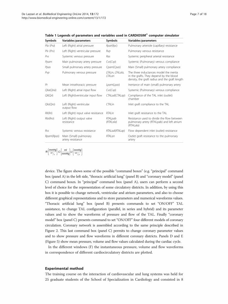
Table 1 Legends of parameters and variables used in CARDIOSIM© computer simulator
Symbols Variables/parameters Symbols Variables/parameters
Pla (Pra) Left (Right) atrial pressure Rpar(Rpc) Pulmonary arteriole (capillary) resistance
Plv (Prv) Left (Right) ventricular pressure Rvp Pulmonary venous resistance
Pvs Systemic venous pressure Ras Systemic peripheral arterial resistance
Ppam Main pulmonary artery pressure Cvs(Cvp) Systemic (Pulmonary) venous compliance
Ppas Small pulmonary artery pressure Cpam(Cpas) Main (Small) pulmonary artery compliance
Pvp Pulmonary venous pressure LTALin, LTALala,LTALan
The three inductances model the inertiain the grafts. They depend by the blooddensity, the graft radius and the graft length
Pt Mean intrathoracic pressure Lpam(Lpas) Inertance of main (small) pulmonary artery
Qlia(Qria) Left (Right) atrial input flow Cvs(Cvp) Systemic (Pulmonary) venous compliance
Qli(Qri) Left (Right)ventricular input flow CTALad(CTALap) Compliance of the TAL inlet (outlet)chamber
Qlo(Qro) Left (Right) ventricularoutput flow
CTALin Inlet graft compliance to the TAL
Rli(Rri) Left (Right) input valve resistance RTALin Inlet graft resistance to the TAL
Rlo(Rro) Left (Right) output valveresistance
RTALpab(RTALala)
Resistance used to divide the flow betweenpulmonary artery (RTALpab) and left atrium(RTALala)
Rvs Systemic venous resistance RTALad(RTALap) Flow dependent inlet (outlet) resistance
Rpam(Rpas) Main (Small) pulmonaryartery resistance
RTALan Outlet graft resistance to the pulmonaryartery
RimmHgml
s=
� �Ci
mlmmHg
� �Li
mmHgml
s2=
� �
De Lazzari et al. BioMedical Engineering OnLine 2014, 13:172 Page 7 of 18http://www.biomedical-engineering-online.com/content/13/1/172
device. The figure shows some of the possible “command boxes” (e.g. “principal” command
box (panel A) in the left side, “thoracic artificial lung” (panel B) and “coronary model” (panel
C) command boxes. In “principal” command box (panel A), users can perform a second
level of choice for the representation of some circulatory districts. In addition, by using this
box it is possible to change network, ventricular and atrium parameters, and also to choose
different graphical representations and to store parameters and numerical waveforms values.
“Thoracic artificial lung” box (panel B) presents commands to set “ON/OFF” TAL
assistance, to change TAL configuration (parallel, in series and hybrid) and its parameter
values and to show the waveforms of pressure and flow of the TAL. Finally “coronary
model” box (panel C) presents command to set “ON/OFF” four different models of coronary
circulation. Coronary network is assembled according to the same principle described in
Figure 2. This last command box (panel C) permits to change coronary parameter values
and to show pressure and flow waveforms in different coronary districts. Panels D and E
(Figure 5) show mean pressure, volume and flow values calculated during the cardiac cycle.
In the different windows (F) the instantaneous pressure, volume and flow waveforms
in correspondence of different cardiocirculatory districts are plotted.
Experimental methodThe training course on the interaction of cardiovascular and lung systems was held for
25 graduate students of the School of Specialization in Cardiology and consisted in 8

TAL
RightVentricle
NaturalLung
LeftAtrium
Flow occluderP l A Parallel modePulmonary Artery
TAL
Parallel mode
RightVentricle
NaturalLung
LeftAtrium
Flow occluderFlow occluderPulmonary Artery Series mode
TAL
RightVentricle
NaturalLung
LeftAtriumg
Flow occluder
Pulmonary Artery Hybrid mode
Figure 4 Shows the possible TAL connections: parallel mode (upper panel), in series mode (centralpanel) and hybrid mode (lower panel). In all connections TAL assistance takes blood from pulmonaryartery. In parallel (in series) mode TAL ejects blood into left atrium (natural lung). Finally in hybridconnection TAL ejects blood in both natural lung and left atrium. In this last connection the amount ofblood ejected into the natural lung depends from the value of resistance RTALpab (Figure 3).
De Lazzari et al. BioMedical Engineering OnLine 2014, 13:172 Page 8 of 18http://www.biomedical-engineering-online.com/content/13/1/172
two-hour sessions. During such sessions, cardiocirculatory pathological conditions of
patients have been reproduced. The reproduced conditions were comparable with those
measured on the patients analyzed and described in our previous papers [7,10,11].
The patients selection were done on the ontological concept basis applied on
haemodynamic measured variables. By using the numerical simulator, the students
simulated various drug therapies aimed at improving the patients’ cardiocirculatory
conditions [7]. The training course took into account also pathological cardiocirculatory
conditions which, in order to be solved, need: ventricular assist devices (VADs) [6,8,9],
intra-aortic balloon pump (IABP) [31], biventricular pacemakers [10], mechanical
ventilatory assistance [8,9] and so on. In these kinds of exercises students use
ontology in order to choose the mechanical circulatory support systems which best
suits to solve the simulated patient’s pathology [32].
In the following we describe a case study carried out during the seminars. Using data
measured on patients (admitted to the CRNAGS Department), students reproduced a
pathological condition that induced the following effects on the haemodynamic variables:
an increase of right ventricular end systolic volume, an increase of right ventricular end
diastolic volume and an important increase of pulmonary arterial pressure.
Starting by these circulatory conditions (baseline conditions), the experiments were
divided in different steps. In the first step parallel TAL assistance was applied, and the

Figure 5 Interface windows presented to the students during some phases of an exercise whichwas held during the training course. Panel A represents the “principal” command box, panel B the“thoracic artificial lung” command box and panel C the “coronary model” command box. Users can performa second level of choice for the representation of some circulatory districts from panel A. In addition usingthis command box it is possible changes network, ventricular and atrium parameters, it is possible choosedifferent graphical representations and to store parameters and numerical waveforms values. Panel Bpresents commands to set “ON/OFF” TAL assistance, to change TAL configuration and its parameter valuesand to show the waveforms of pressure and flow of the TAL. Finally panel C presents commands to set“ON/OFF” four different representation of coronary circulation. Coronary network is assembled according tothe same principle described in Figure 2. Panel C permits to change coronary parameter values and toshow pressure and flow waveforms in different coronary districts. Panels D and E show the mean pressure,volume and flow values. In all other windows (F) the instantaneous pressure, volume and flow waveformscalculated in the various districts of the network are plotted.
De Lazzari et al. BioMedical Engineering OnLine 2014, 13:172 Page 9 of 18http://www.biomedical-engineering-online.com/content/13/1/172
pulmonary arterial peripheral resistance was set to 240 [g·cm−4·sec−1], TAL compliances
were set to CTALin = 1.5 and 0.4 [ml/mmHg] and CTALad = CTALap = 2.0 and 0.1
[ml/mmHg]. This high value of pulmonary resistance induces an overload in the
right ventricle. By imposing such a high value, only TAL device induces some effects on
the cardiopulmonary circulation. In the second step the same simulations were performed
setting pulmonary arterial peripheral resistance to 120 [g·cm−4·sec−1]. This low value of
pulmonary resistance (induced by drug administration) permits to download the right
ventricle. By imposing such a low value, students can observe the double effects induced
by TAL assistance and by the administration of drugs that improves pulmonary
circulation and download the right ventricle. Finally in the third step, the same
simulations were reproduced for the hybrid TAL assistance. Different combination
of TAL compliances permits to evaluate the performances of the assistance [14].
In all simulations, conduced in interactive mode, the analyzed variables were: left
ventricular end diastolic volume (LVEDV), left ventricular end systolic volume
(LVESV), right ventricular end diastolic volume (RVEDV), right ventricular end systolic
volume (RVESV), cardiac output (CO), mean pulmonary arterial pressure, mean left
atrial pressure, mean systemic venous pressure and the value of the area of coronary

De Lazzari et al. BioMedical Engineering OnLine 2014, 13:172 Page 10 of 18http://www.biomedical-engineering-online.com/content/13/1/172
blood flow-aortic pressure loop (CBP-AoP) [11,33,34]. The mean values were calculated
during the cardiac cycle.
At the end of the training course, a questionnaire was submitted to students in order
to evaluate the quality of the training course. The evaluation of the answers provided
by students is reported in the next section. Such a precious feedback gave us an indication
of the quality of CARDIOSIM© and it will allow us to focus even better on users’ needs in
the future releases of the tool.
Results and discussionFigure 6 shows results obtained in the conditions described in the first step of “Experimental
method” section. Starting from the application of baseline condition, students applied the
TAL assistance in parallel and hybrid model using the numerical simulator. In Figure 6,
panel A shows the percentage changes obtained applying the parallel TAL assistance and
setting pulmonary arterial peripheral resistance to 240 [g·cm−4·sec−1]. TAL assistance
reduces RVEDV and RVESV, but increases LVEDV and LVESV [35]. The reduction of
RVEDV and RVESV allows the right ventricular pressure-volume (P-V) loop to move to the
left in the pressure-volume plane [36]. According with literature data, parallel assistance can
produce an increase in cardiac output [37,38]. A reduction of mean pulmonary arterial
pressure and an increase of CBF-AoP area have been produced by parallel TAL mode. In
this tutorial, students have verified that parallel attachment can significantly reduce
pulmonary pressures and unload the right ventricle [38,39]. Panel B shows the effects
induced by hybrid TAL assistance when peripheral resistance was set to 240 [g·cm−4·sec−1].
In both panels we reported results obtained for different values of TAL compliances.
Students using different combinations of TAL compliance values verified the different
effects induced on haemodynamic variables [13,40]. The percentage changes induced by
hybrid TAL assistance on some haemodynamic variables are lower than those produced
by parallel assistance. Only in the case of the mean pulmonary arterial pressure, there is a
percentage reduction which, in case of hybrid assistance, is greater (about 40%) than in
case of parallel assistance (20-30%). Finally, students have observed that TAL assistance
increases the mean left atrial pressure, as described in literature [37].
Figure 7 shows one of the different screen outputs produced by students during the
training course. In the central windows there are plotted the left ventricular (upper)
and the right ventricular (lower) P-V loops [21]. In Figure 7, students reproduced the
evolution of the left and right ventricular P-V loop, when TAL assistance was applied
in parallel mode starting from a baseline condition. By analysing the figure, it is possible to
observe how the left ventricular pressure-volume loop moves to the right (increasing
LVEDV and LVESV) in the P-V plane (upper window) from position A (baseline conditions)
to position B (assisted conditions). The right ventricular P-V loop (in lower window) moves
towards the right side in the P-V plane from position A (baseline conditions) to position B
(assisted conditions). This effect produces a reduction of RVEDV and RVESV and
an increase of right ventricular efficiency.
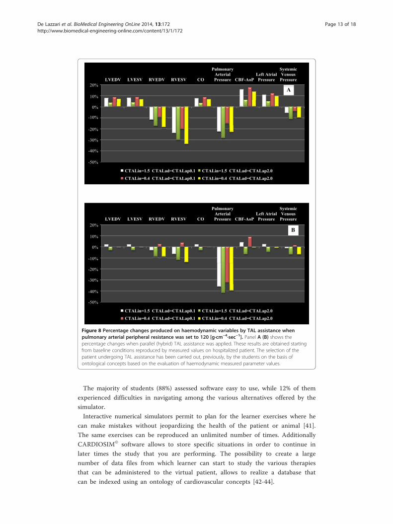
Figure 8 shows the percentage changes of some haemodynamic variables when the
pulmonary arterial peripheral resistance was set to 120 [g·cm−4·sec−1] in order to reproduce
the effects of drug administration. Panel A shows the percentage changes when parallel TAL
assistance was applied. The trend presented in Figure 8 (panel A) is the same presented in

30%LVEDV LVESV RVEDV RVESV CO
PulmonaryArterialPressure CBF-AoP
Left Atrial Pressure
SystemicVenous
Pressure
-10%
0%
10%
20%A
-50%
-40%
-30%
-20%
CTALin=1.5 CTALad=CTALap0.1 CTALin=1.5 CTALad=CTALap2.0CTALin=0.4 CTALad=CTALap0.1 CTALin=0.4 CTALad=CTALap2.0
LVEDV LVESV RVEDV RVESV CO
PulmonaryArterialPressure CBF AoP
Left Atrial Pressure
SystemicVenous
Pressure
10%
0%
10%
20%
30%LVEDV LVESV RVEDV RVESV CO Pressure CBF-AoP Pressure Pressure
B
-50%
-40%
-30%
-20%
-10%
CTALin=1.5 CTALad=CTALap0.1 CTALin=1.5 CTALad=CTALap2.0CTALin=0.4 CTALad=CTALap0.1 CTALin=0.4 CTALad=CTALap2.0
Figure 6 Percentage changes produced by parallel TAL assistance (panel A) on left/right ventricularend diastolic volume (LVED/RVED), left/right ventricular end systolic volume (LVESV/RVESV), cardiacoutput (CO), mean pulmonary arterial pressure, coronary blood flow-aortic pressure (CBF-AoP) area,mean left atrial pressure and mean systemic venous pressure. In panel B are reported the percentagechanges induced by hybrid TAL assistance. These results are obtained starting from baseline conditionsreproduced by measured values on hospitalized patient. The selection of the patient undergoing TALassistance has been carried out, previously, by the students on the basis of ontological concepts based onthe evaluation of haemodynamic measured parameter values. During the simulations pulmonary arterialperipheral resistance was set to 240 [g·cm-4·sec−1].
De Lazzari et al. BioMedical Engineering OnLine 2014, 13:172 Page 11 of 18http://www.biomedical-engineering-online.com/content/13/1/172
Figure 6 (panel A), but the percentage changes on CO, CBF-AoP and mean left
atrial pressure are more relevant in this second figure. Percentage changes on
LVESV and LVED (panel A) are higher in Figure 6 than in Figure 8. As far as hybrid TAL
assistance is concerned, panel B of Figure 8 shows an opposite trend (with respect to panel
B, Figure 6) for the percentage changes in some variables in correspondence with some
combinations of TAL compliances value. This phenomenon occurs in LVESV, LVEDV,
RVESV, RVEDV, cardiac output, mean left atrial pressure and in CBF-AoP area. The
reduced pulmonary peripheral resistance value during hybrid TAL assistance apparently
does not affect percentage changes in LVESV, LVEDV, CO and mean left atrial pressure.

A
B
A
AB
Figure 7 Possible screen output produced by student during the training course using theinteractive CARDIOSIM© software. Starting from baseline conditions the applications of parallel TALassistance induces a shift of both left (upper window) and right (lower window) P-V ventricular loops.Right P-V ventricular loop shift from A (baseline conditions) to B (assisted conditions). Left P-V ventricularloop shift (in the right side) from A to B. In the right box are reported some haemodynamic mean(calculated during the cardiac cycle) values assumed during the parallel TAL assistance.
De Lazzari et al. BioMedical Engineering OnLine 2014, 13:172 Page 12 of 18http://www.biomedical-engineering-online.com/content/13/1/172
The cardiac output is one of the variables on which researchers focus attention during
TAL assistance. Results presented in Figure 9 have been also obtained during the
training course held for students of the School of Specialization in Cardiology at
the CRNAGS Department. In this figure we present the percentage changes (with
respect to baseline conditions) of CO when TAL assistance was applied in different mode,
with different pulmonary arterial peripheral resistance values and with different TAL
compliance values.
Panel A (pulmonary arterial peripheral resistance was set to 240 [g·cm−4·sec−1])
emphasizes that, in the case of TAL parallel assistance, independently from the
values assumed by TAL compliances, cardiac output is more or less double with
respect to hybrid assistance. Panel B (pulmonary arterial peripheral resistance was
set to 120 [g·cm−4·sec−1]) shows that parallel TAL assistance causes an increase in
CO, whereas hybrid assistance can yield into a reduction in CO [37].
Finally, the analysis of the questionnaire submitted to students showed that:
� 80% of the students claimed to have had a good degree of learning in relation to
what is explained and exercises made during the training course;
� 8% claimed to have had a sufficient degree of learning;
� 12% claimed to have had a low degree of learning.
20%LVEDV LVESV RVEDV RVESV CO
PulmonaryArterialPressure CBF-AoP
Left Atrial Pressure
SystemicVenous
Pressure
-20%
-10%
0%
10%A
-50%
-40%
-30%
CTALin=1.5 CTALad=CTALap0.1 CTALin=1.5 CTALad=CTALap2.0
PulmonaryArterial Left Atrial
SystemicVenous
CTALin=0.4 CTALad=CTALap0.1 CTALin=0.4 CTALad=CTALap2.0
-10%
0%
10%
20%LVEDV LVESV RVEDV RVESV CO Pressure CBF-AoP Pressure Pressure
B
-50%
-40%
-30%
-20%
CTALin=1.5 CTALad=CTALap0.1 CTALin=1.5 CTALad=CTALap2.0CTALin=0.4 CTALad=CTALap0.1 CTALin=0.4 CTALad=CTALap2.0
Figure 8 Percentage changes produced on haemodynamic variables by TAL assistance whenpulmonary arterial peripheral resistance was set to 120 [g·cm−4·sec−1]. Panel A (B) shows thepercentage changes when parallel (hybrid) TAL assistance was applied. These results are obtained startingfrom baseline conditions reproduced by measured values on hospitalized patient. The selection of thepatient undergoing TAL assistance has been carried out, previously, by the students on the basis ofontological concepts based on the evaluation of haemodynamic measured parameter values.
De Lazzari et al. BioMedical Engineering OnLine 2014, 13:172 Page 13 of 18http://www.biomedical-engineering-online.com/content/13/1/172
The majority of students (88%) assessed software easy to use, while 12% of them
experienced difficulties in navigating among the various alternatives offered by the
simulator.
Interactive numerical simulators permit to plan for the learner exercises where he
can make mistakes without jeopardizing the health of the patient or animal [41].
The same exercises can be reproduced an unlimited number of times. Additionally
CARDIOSIM© software allows to store specific situations in order to continue in
later times the study that you are performing. The possibility to create a large
number of data files from which learner can start to study the various therapies
that can be administered to the virtual patient, allows to realize a database that
can be indexed using an ontology of cardiovascular concepts [42-44].

14%Cardiac Output
Hyb. TAL [CTALin=1.5 CTALad=CTALap0.1]
AA
8%
10%
12% Hyb. TAL [CTALin=0.4 CTALad=CTALap0.1]Hyb. TAL [CTALin=1.5 CTALad=CTALap2.0]Hyb. TAL [CTALin=0.4 CTALad=CTALap2.0]Par. TAL [CTALin=1.5
2%
4%
6% CTALad=CTALap0.1]Par. TAL [CTALin=0.4 CTALad=CTALap0.1]Par. TAL [CTALin=1.5 CTALad=CTALap2.0]Par. TAL [CTALin=0.4 CTALad=CTALap2.0]
0%
10%Cardiac Output
Hyb. TAL [CTALin=1.5
4%
6%
8%CTALad=CTALap0.1]Hyb. TAL [CTALin=0.4 CTALad=CTALap0.1]Hyb. TAL [CTALin=1.5 CTALad=CTALap2.0]Hyb. TAL [CTALin=0.4 CTALad=CTALap2.0]
B
-4%
-2%
0%
2% Par. TAL [CTALin=1.5 CTALad=CTALap0.1]Par. TAL [CTALin=0.4 CTALad=CTALap0.1]Par. TAL [CTALin=1.5 CTALad=CTALap2.0]Par TAL [CTALin=0 4
-6%
-4% Par. TAL [CTALin 0.4CTALad=CTALap2.0]
A
Figure 9 Percentage changes produced on cardiac output by different TAL assistance when pulmonaryarterial peripheral resistance was set to 240 [g·cm−4·sec−1] (panel A) and to 120 [g·cm-4·sec−1] (panel B).In both panels students obtained the percentage changes setting (in the interactive human numerical simulator)different TAL compliance values. During the assistance compliances were set to CTALin = 1.5 and 0.4 [ml/mmHg]and CTALad = CTALap = 2.0 and 0.1 [ml/mmHg].
De Lazzari et al. BioMedical Engineering OnLine 2014, 13:172 Page 14 of 18http://www.biomedical-engineering-online.com/content/13/1/172
During the described course, in same cardiocirculatory conditions, students applying
TAL assistance induced an increase of LVEDV, LVESV and a reduction of CO. These
results induced a decrease in the left ventricular efficiency. In order to improve the left
ventricular conditions a possible chooses consisted to help the left ventricle with a
mechanical circulatory assist device. To choose the better mechanical circulatory assist
device according to the virtual patient pathological conditions, students were assisted
by an ontology applied to mechanical circulatory support systems [32].
Following the same trend presented in literature data, this e-Learning experience
produced the following benefits:
✓ opportunities for accelerated learning (as proved by analysing students’
questionnaires) [3];

De Lazzari et al. BioMedical Engineering OnLine 2014, 13:172 Page 15 of 18http://www.biomedical-engineering-online.com/content/13/1/172
✓ lower costs with respect to in vivo experiments;
✓ increased attention span, due to interactive nature of the tool which is an added
value compared to traditional textbook learning [45];
✓ no medical related accidents [41,46];
In general, the interactive simulator can also be used to teach and practice the
management of complex perioperative medical events and emergencies [47].
The cardiovascular system is very complex and is characterized by the presence
of numerous control mechanisms that can occur under certain conditions.
A limitation in the use of these simulators is linked to the impossibility to reproduce
the behaviour of the entire CVS and its control mechanisms in the various circulatory
conditions that may arise. Over the years, researchers have developed several numerical
models of the CVS. Each model, developed in a more or less complex mode, has the
characteristic to be rigid and not flexible since it was developed according to the type
of study and/or experiment that the user wanted to perform [47-52].
CARDIOSIM© has a different approach: it is a peculiar software platform with unique
characteristics that we sketch out in the following.
First of all, unlike other in silico simulator [53-57], it is based on a modular
approach and it is equipped with a library of numerical models of the various
sections of the CVS. The library can be increased with other numerical models
and can be continually updated. The various models can be easily assembled, from
researchers, teachers or students, among them according to the experiment that
the user wants to reproduce. This is a first feature that distinguishes CARDIOSIM©
from other models that despite being already available in the network, do not have
the flexibility that is required to adapt to the type of pathology experiment that
the user want to study.
For each set of parameters, CARDIOSIM© permits to choose an initial set of values
inside libraries that contains also patient or animal measured value parameters. The
software simulator allows to save in Excel format the numerical values of the various
waveforms reproduced during the simulations. In this way it is possible to do any kind
of “off-line” analysis.
Another peculiarity of the software platform is the possibility of using several
ventricular assist devices reproducing continuous or pulsatile flow. These VADs
can be connected in series or in parallel mode to the left or right (or both) ven-
tricles. VADs implemented in CARDIOSIM© are pneumatic or rotary blood pump
(i.e. Hemopump HP31 [9], PUCA pump [58]). Each VAD can be synchronized or
not synchronized with the heart’s electrical activity or can operate in “full-empty”
mode. Also intraaortic balloon pump (IABP) and biventricular pacemaker devices
can be used with the numerical simulator when pathological conditions are simulated.
Unlike the previously presented tools, our software platform allows to choose
among different types of mechanical ventilatory assistance when VADs, IABP or
biventricular pacemaker are applied or not. This option allows to study the effects
produced on haemodynamic and energetic variables by the interaction among the
CVS, VAD and MVA or TAL.
Some limitations concerning this training course experience (the first of such a kind
for us) are described below:

De Lazzari et al. BioMedical Engineering OnLine 2014, 13:172 Page 16 of 18http://www.biomedical-engineering-online.com/content/13/1/172
� the questionnaire consisted of a small number of simple qualitative questions. In
the future we intend to submit to students a more detailed questionnaire from
which one can infer more analytical considerations;
� in the students’ use of the simulator, we observed they have no sufficient
confidence with such kind of tools: in particular, it is very difficult for them to
understand how a set of RLC elements can simulate a blood vessel;
� a limitation of our simulator lies the fact that its use is not intuitive and presents
difficulties for students not familiar with IT tools.
ConclusionsAn e-Learning environment can take profit from the employment of a software simulator
such as CARDIOSIM©.
Among the advantages of using a simulator, we have to consider that the same
pathophysiological conditions can be replicated several times by the students without any
damage for the virtual patient.
In particular, it is possible to simulate:
– Drug administration and their effects on haemodynamic variables
– Circulatory mechanical and ventilatory support devices
– The effects of choosing, configuring and activating one of such devices
– The effects of possible adjustments of device parameters
Currently, the e-Learning tool that we implemented is used in a local environment in
a traditional face-to-face interaction between teachers and students. In the future, we
plan to extend the e-Learning features in a telematic virtual environment by means of
an internet based platform.
We also believe that not only simulation software, but also an ontology-based model-
ling of the relevant concepts involved in the e-Learning domain is a valid approach in
order to reuse an environment in other domains or for other students’ profiles (e.g.
general practitioners, anesthesiologists, nurses).
Competing interestsThe authors declare that they have no competing interests.
Authors’ contributionsCD developed the numerical simulator CARDIOSIM© using Visual Basic program language. In this work heimplemented the numerical model of thoracic artificial lung (TAL). He organized, coordinated and participated in therealization of courses for the students. He acted as a tutor during the use of CARDIOSIM© by students. He analyzeddata obtained from simulations and joined the evaluation of the questionnaires produced by students. He drafted themanuscript. IG organized the questionnaire and participated in the realization of courses for the students. Heexplained to the students the theory on the cardiovascular system and the use of TAL. He set up the questionnaireand analyzed data obtained from simulations. DMP participated in the realization of courses for the students. He alsoacted as tutor during the use of CARDIOSIM© by students. He analyzed of data obtained from simulations. He draftedthe manuscript. AD explained to the students the theory on the cardiovascular system and the use of TAL. She actedas a tutor during the use of CARDIOSIM© by students. She set up the questionnaire. She analyzed data obtained fromsimulations and joined the evaluation of the questionnaires produced by students. FF explained to the students thetheory on the cardiovascular system and the use of TAL. He hold the introductory lesson of the course. He analyzeddata obtained from simulations. He drafted the manuscript. All authors read and approved the final manuscript.
Author details1CNR, Institute of Clinical Physiology, UOS of Rome, Via S.M. della Battaglia, 44, 00185 Rome, Italy. 2National Institute forCardiovascular Research, Bologna, Italy. 3Department of Cardiovascular, Respiratory, Nephrological, Anesthesiologicaland Geriatric Sciences, “Sapienza” University of Rome, Rome, Italy. 4CNR, Institute of Cognitive Sciences andTechnology (ISTC), Rome, Italy.

De Lazzari et al. BioMedical Engineering OnLine 2014, 13:172 Page 17 of 18http://www.biomedical-engineering-online.com/content/13/1/172
Received: 23 September 2014 Accepted: 5 December 2014Published: 18 December 2014
References
1. Trondsen E: Games and Simulations in E-Learning. SRI Business Intelligence Consultig. ; 2001.2. Chi D, Kokkevis E, Ogunyemi O, Bindiganavale R, Hollick M, Clarke J, Webber B, Badler N: Simulated casualtiesand medics for emergency training. In Medicine Meets Virtual Reality. Edited by Morgan KS, Hoffman HM,Stredney D, Weghorst SJ. Amsterdam: IOS Press; 1997:486–494.
3. Okuda Y, Bryson EO, DeMaria S, Jacobson L, Quinones J, Shen B, Levine AI: The utility of simulation in medicaleducation: what is the evidence? Mt Sinai J Med 2009, 76(4):330–343.
4. Suebnukarn S, Haddawy P: COMET: a collaborative tutoring system for medical problem-based learning.IEEE Intell Syst 2007, 22(4):70–77.
5. De Lazzari C: CARDIOSIM© - cardiovascular software simulator. Italy: C.N.R. National Research Council; 2011[https://cardiosim.dsb.cnr.it/]
6. De Lazzari C, Ferrari G, Mimmo R, Tosti G, Ambrosi D: A desk top computer model of the circulatory system forheart assistance simulation: effect of an LVAD on energetic relationships inside the left ventricle. Med EngPhys 1994, 16(2):97–103.
7. De Lazzari C, L’Abbate A, Micalizzi M, Trivella MG, Neglia D: Effects of amlodipine and adenosine on coronaryhaemodynamics: in vivo study and numerical simulation. Comput Meth Biomech Biomed Eng 2014,17(15):42–52.
8. De Lazzari C, Darowski M, Ferrari G, Pisanelli DM, Tosti G: The impact of rotary blood pump in conjunction withmechanical ventilation on ventricular energetic parameters: numerical simulation. Methods Inf Med 2006,45:574–583.
9. De Lazzari C, Darowski M, Ferrari G, Pisanelli DM, Tosti G: Modelling in the study of interaction of Hemopumpdevice and artificial ventilation. Comput Biol Med 2006, 36:1235–1251.
10. De Lazzari C, D’Ambrosi A, Tufano F, Fresiello L, Garante M, Sergiacomi R, Stagnitti F, Caldarera CM, Alessandri N:Cardiac resynchronization therapy: could a numerical simulator be a useful tool in order to predict theresponse of the biventricular pacemaker synchronization? Eur Rev Med Pharmacol Sci 2010, 14(11):969–978.
11. De Lazzari C, Del Prete E, Genuini I, Fedele F: In silico study of the haemodynamic effects induced bymechanical ventilation and biventricular pacemaker. Comput Methods Prog Biomed 2013, 110(3):519–527.
12. Perme SC, Southard RE, Joyce DL, Noon GP, Loebe M: Early mobilization of LVAD recipients who requireprolonged mechanical ventilation. Tex Heart Inst J 2006, 33(2):130–133.
13. McGillicuddy JW, Chambers SD, Galligan DT, Hirschl RB, Bartlett RH, Cook KE: In vitro fluid mechanical effects ofthoracic artificial lung compliance. ASAIO J 2005, 51(6):789–794.
14. Boschetti F, Perlman CE, Cook KE, Mockros LF: Hemodynamic effects of attachment modes and device design athoracic artificial lung. ASAIO J 2000, 46(1):42–48.
15. Colquitt RB, Colquhoun DA, Thiele RH: In silico modeling of physiologic systems. Best Pract Res Clin Anaesthesiol2011, 25:499–510.
16. Sinz E: Simulation-based education for cardiac, thoracic, and vascular anesthesiology. Semin Cardiothorac VascAnesth 2005, 9(4):291–307.
17. Wildhaber RA, Verrey F, Wenger RH: A graphical simulation software for instruction in cardiovascularmechanics physiology. Biomed Eng Online 2011, 10:8. doi:10.1186/1475-925X-10-8.
18. Zwischenberger JB, Anderson CM, Cook KE, Lick SD, Mockros LF, Bartlett RH: Development of an implantableartificial lung: challenges and progress. ASAIO J 2001, 47(4):316–320.
19. Federspiel WJ, Svitek RG: Artificial lungs: current research and future directions. In Encyclopedia of Biomaterialsand Biomedical Engineering. Edited by Wnek GE, Bowlin GL. New York: Marcel Dekker, Inc; 2004:922–931.
20. Maughan WL, Sunagawa K, Sagawa K: Ventricular systolic interdependence: volume elastance model inisolated canine hearts. Am J Physiol Heart Circ Physiol 1987, 253:H1381–H1390.
21. Sagawa K, Maughan WL, Suga H, Sunagawa K: Cardiac Contraction and the Pressure-Volume Relationships.New York: Oxford University Press; 1988.
22. Korakianitis T, Shi Y: A concentrated parameter model for the human cardiovascular system including heartvalve dynamics and atrioventricular interaction. Med Eng Phys 2006, 28:613–628.
23. De Lazzari C: Interaction between the septum and the left (right) ventricular free wall in order to evaluate theeffects on coronary blood flow: numerical simulation. Comput Meth Biomech Biomed Eng 2012,15(12):1359–1368.
24. Starling EH: The Linacre Lecture on the Law of the Heart. London: Longmans, Green & Co; 1918:1–27.25. Guyton AC, Jones CE, Coleman TG: Computer analysis of total circulatory function and of cardiac output
regulation. In Circulatory Physiology: Cardiac Utput and its Regulation. Philadelphia: Saunders WB Company; 1973.26. Frasch HF, Kresh JY, Noordergraaf A: Two-port analysis of microcirculation: an extension of windkessel. Am J
Physiol 1996, 270:H376–H385.27. Shi Y, Lawford P, Hose R: Review of zero-D and 1-D models of blood flow in the cardiovascular system. BioMed
Eng Online 2011, 10:33. doi:10.1186/1475-925X-10-33.28. Downey JM, Kirk ES: Inhibition of coronary blood flow by a vascular waterfall mechanism. Circ Res 1975,
36:753–760.29. Spaan JA, Nreuls NP, Laird JD: Diastolic–systolic coronary flow differences are caused by intramyocardial pump
action in the anesthetized dog. Circ Res 1981, 49:584–593.30. Spaan JA, Nreuls NP, Laird JD: Forward coronary flow normally seen in systole is the result of both forward
and concealed back flow. Basic Res Cardiol 1981, 76:582–586.31. De Lazzari C, Darowski M, Ferrari G, Clemente F, Guaragno M: Ventricular energetics during mechanical
ventilation and intraaortic balloon pumping – Computer simulation. J Med Eng Technol 2001, 25(3):103–111.

De Lazzari et al. BioMedical Engineering OnLine 2014, 13:172 Page 18 of 18http://www.biomedical-engineering-online.com/content/13/1/172
32. De Lazzari C, Guerrieri E, Pisanelli DM: A domain ontology for mechanical circulatory support systems.In Proceeding of the 30TH Annual Conference of Computers in Cardiology: 21–24 Sept. 2003. Edited by Murray A.Thessaloniki: IEEE Press; 2003:417–419.
33. Di Mario C, Kramas R, Gil R, Serruys PW: Slope of the instantaneous hyperemic diastolic CFV-pressure relation.A new index for assessment of the physiological significance of coronary stenosis in humans. Circulation 1994,90(3):1215–1224.
34. Krams R, Ten Cate FJ, Carlier SG, van der Steen AFW, Serruys PW: Diastolic coronary vascular reserve: a newindex to detect changes in the coronary microcirculation in hypertrophic cardiomyopathy. J Am Coll Cardiol2003, 43(4):670–677.
35. Lick SD, Zwischenberger JB, Wang D, Deyo DL, Alpard SK, Chambers SD: Improved right heart function with acompliant inflow artificial lung in series with the pulmonary circulation. Ann Thorac Surg 2001, 72(3):899–904.
36. Haft JW, Montoya P, Alnajjar O, Posner SR, Bull LL, Iannettoni MD, Bartlett RH, Hirschl RB: An artificial lungreduces pulmonary impedance and improves right ventricular efficiency in pulmonary hypertension. J ThoracCardiovasc Surg 2001, 122:1094–1100.
37. Perlman CE, Cook KE, Seipelt R, Mavroudis C, Backer CL, Mockros LF: In vivo hemodynamic responses to artificiallung attachment. ASAIO J 2005, 51(4):412–425.
38. Akay B, Reoma JL, Camboni D, Pohlmann JR, Albert JM, Kawatra A, Gouch AD, Bartlett RH, Cook KE: In-parallelartificial lung attachment at high flows in normal and pulmonary hypertension models. Ann Thorac Surg 2010,90(1):259–265.
39. Akay B, Foucher JA, Camboni D, Koch KL, Kawatra A, Cook KE: Hemodynamic design requirements for in seriesthoracic artificial lung attachment in a model of pulmonary hypertension. ASAIO J 2012, 58(4):426–431.
40. Haft J, Bull JL, Rose R, Katsra J, Grotberg JB, Bartlett RH, Hirschl RB: Design of an artificial lung compliancechamber for pulmonary replacement. ASAIO J 2003, 49(1):35–40.
41. Ahlberg G, Enochsson L, Gallagher AG, Hedman L, Hogman C, McClusky DA 3rd, Ramel S, Smith CD, Arvidsson D:Proficiency-based virtual reality training significantly reduces the error rate for residents during their first10 laparoscopic cholecystectomies. Am J Surg 2007, 193:797–804.
42. Backhaus M, Chung JD, Cowan BR, Tao W, Young AA: The cardiac atlas project: towards a map of the heart.In Patient-Specific Modeling of the Cardiovascular System, Volume 1. Edited by Kerckhoffs R. Springer Heidelberg;2010:113–129.
43. Eccher C, Scipioni A, Miller AA, Ferro A, Pisanelli DM: An ontology of cancer therapies supportinginteroperability and dataconsistency in EPRs. Comput Biol Med 2013, 43:822–832.
44. Eccher C, Purin B, Pisanelli DM, Battaglia M, Apolloni I, Forti S: Ontologies supporting continuity of care: the caseof heart failure. Comput Biol Med 2006, 36(7–8):789–801.
45. Wayne DB, Didwania A, Feinglass J, Fudala MJ, Barsuk JH, McGaghie WC: Simulation-based education improvesquality of care during cardiac arrest team responses at an academic teaching hospital: a case–control study.Chest 2008, 133(1):56–61.
46. Vincent C, Moorthy K, Sarker SK, Chang A, Darzi AW: Systems approaches to surgical quality and safety: fromconcept to measurement. Ann Surg 2004, 239:475–482.
47. Chakravarthy B, ter Haar E, Bhat SS, McCoy CE, Denmark TK, Lotfipour S: Simulation in medical school education:review for emergency medicine. Western J Emerg Med 2011, 12(4):461–466.
48. Heldt T, Shim EB, Kamm RD, Mark RG: Computational modeling of cardiovascular response to orthostaticstress. J Appl Physiol 2002, 92:1239–1254.
49. Mukkamala R, Cohen RJ: A forward model-based validation of cardiovascular system identification. Am JPhysiol Heart Circ Physiol 2001, 281(6):H2714–H2730.
50. Mukkamala R, Kim JK, Li Y, Sala-Mercado J, Hammond RL, Scislo TJ, O’Leary DS: Estimation of arterial andcardiopulmonary total peripheral resistance baroreflex gain values: validation by chronic arterial baroreceptordenervation. Am J Physiol Heart Circ Physiol 2006, 290(5):H1830–H1836.
51. Sheffer L, Santamore WP, Barnea O: Cardiovascular simulation toolbox. Cardiovasc Eng 2007, 7:81–88.52. Brunberg A, Heinke S, Spillner J, Autschbach R, Abel D, Leonhardt S: Modeling and simulation of the
cardiovascular system: a review of applications, methods, and potentials. Biomed Tech (Berlin) 2009, 54(5):233–244.53. Goldberger AL, Amaral LAN, Glass L, Hausdorff JM, Ivanov PC, Mark RG, Mietus JE, Moody GB, Peng CK, Stanley HE:
PhysioBank, PhysioToolkit, and PhysioNet: components of a new research resource for complex physiologicsignals. Circulation 2000, 101(23):e215–e220.
54. Bassingthwaighte JB: Strategies for the physiome project strategies for the physiome project. Ann Biomed Eng2000, 28:1043–1058.
55. Garny A, Nickerson DP, Cooper J, dos Santos RW, Miller AK, McKeever S, Nielsen PMF, Hunter PJ: CellML andassociated tools and techniques. Philos Trans A Math Phys Eng Sci 2008, 366(1978):3017–3043.
56. Lian J: Open source modeling of cardiovascular system: a brief overview. TOPETJ 2010, 3:1–3.57. Barnea O: Open-source programming of cardiovascular pressure-flow dynamics using SimPower toolbox in
Matlab and Simulink. Open Pacing Electrophysiol Ther J 2010, 3:55–59.58. De Lazzari C, Genuini I, Quatember B, Fedele F: Mechanical ventilation and thoracic artificial lung assistance
during mechanical circulatory support with PUCA pump: in silico study. Comput Methods Programs Biomed2014, 113(2):642–654.
doi:10.1186/1475-925X-13-172Cite this article as: De Lazzari et al.: Interactive simulator for e-Learning environments: a teaching software forhealth care professionals. BioMedical Engineering OnLine 2014 13:172.