Interactions of Metal—Metal-Bonded Antitumor Active Complexes with DNA Fragments and DNA
11
Interactions of Metal-Metal-Bonded Antitumor Active Complexes with DNA Fragments and DNA HELEN T. CHIFOTIDES AND KIM R. DUNBAR* Department of Chemistry, Texas A&M University, College Station, Texas 77843 Received September 1, 2004 ABSTRACT This Account summarizes the DNA binding properties of anticancer active dinuclear Rh, Re, and Ru compounds. The combined results of NMR spectroscopy, X-ray crystallography, and various biochemi- cal tools provide incontrovertible evidence that dinuclear com- plexes are favorably poised to bind to purine nucleobases, DNA fragments, and double-stranded DNA. Moreover, direct DNA photocleavage in vitro is effected by dirhodium compounds in the presence of electron acceptors in solution or directly attached to the dirhodium core. This research has provided valuable insight in the interactions of dinuclear compounds with DNA, knowledge that is an excellent backdrop for rational design of promising dinuclear drugs. Introduction The medicinal properties of inorganic compounds are well-known, dating to ancient times, with the earliest recognition being traced back to the Egyptians who used copper to sterilize water in 3000 B.C. Presently, metal compounds that exhibit antiarthritic, antibacterial, anti- cancer, antidepressant, and anti-hypertensive properties are in routine clinical use. Interest in metal antitumor compounds stems from the extraordinary effectiveness of cisplatin (cis-[Pt(NH 3 ) 2 Cl 2 ] or cis-DDP) 1-3 and related complexes for the treatment of ovarian, testicular, head, neck, esophageal, and lung carcinomas, with a cure rate greater than 90% in the case of testicular cancer. Extensive investigations of this potent antitumor agent have estab- lished DNA as its primary intracellular target. The intras- trand d(GpG) head-to-head (H-H) cross-links that are formed contribute to a cascade of events, including transcription inhibition and repair shielding of cisplatin- DNA lesions, which lead to cell death. 1-3 The success of cisplatin notwithstanding, it is important to continue the search for new anticancer active metal compounds with different activities and resistances as well as lower tox- icities. Among the recognized non-platinum antitumor agents are dinuclear carboxylate species of rhodium (Rh), 4 rhe- nium (Re), 5,6 and ruthenium (Ru). 7 The basic paddlewheel structure of these compounds, which consists of a metal- metal-bonded fragment with at least two bridging ligands (Chart 1), appears to defy most of the accepted guidelines for metal anticancer agents. The recognition of the anticancer activity of tetracarboxylate compounds spawned a number of investigations in the 1970s regarding plau- sible cellular targets, 8,9 but research in this area steadily declined, in part, because the compounds did not surpass the anticancer activity of cisplatin. The revival of this research field in our laboratories over the past decade has been directed towards elucidating the biological activity of metal-metal-bonded systems vis-a `-vis interactions with DNA, which is the primary target of platinum antican- cer agents. Our findings provide valuable insight in the viable substitution pathways and binding modes of di- metal units with DNA and pave the way for rational design of promising anticancer and photochemotherapeutic candidates. Prior to highlighting our results, a brief overview of the biological activity of each class of com- pounds is presented. Biological Activity of the Compounds Dirhodium. Studies of the biological activity of dirhodium complexes conducted in the 1970s support the conclusion that tetracarboxylate compounds Rh 2 (μ-O 2 CR) 4 (R ) Me, Et, Pr) exhibit significant in vivo antitumor activity against L1210 tumors, 10 Ehrlich ascites, 8,9,11,12 sarcoma 180, and P388 tumor lines. 4 Although the precise antitumor mech- anism of dirhodium carboxylate compounds has not been elucidated, it is known that they bind to DNA 8,9,13-15 and inhibit DNA replication and protein synthesis 16-18 in a manner akin to cisplatin. A systematic variation of the axial (ax) and equatorial (eq) ligands has shed light on the structure-activity relationships in this family of compounds. Of particular note is the fact that the antitumor activity increases in the series Rh 2 (μ-O 2 CR) 4 (R ) Me, Et, Pr) (Chart 1, struc- ture a) with the lipophilicity of the R group. 8,10,11 The compounds Rh 2 (μ-O 2 CCF 3 ) 4 (Chart 1, structure a) and Rh 2 (μ-HNCOCF 3 ) 4 (Chart 1, structure b) have been re- ported to significantly increase the survival rate of mice bearing Ehrlich ascites cells and have LD 50 values on the same order as that of cisplatin. 19,20 The most active member of the methoxyphenylphosphine series is the * To whom correspondence should be addressed. E-mail: [email protected]. Kim R. Dunbar was born in Mount Pleasant, PA, and received a B.S. from Westminster College in 1980 and a Ph.D. from Purdue University in 1984. She joined the faculty of Michigan State University in 1987 and moved to Texas A&M University in 1999 where she holds the position of Davidson Chair of Science. Her research interests span topics in synthetic and structural inorganic chemistry with a focus on materials and bioinorganic chemistry. She has been named a Fellow of the Alfred P. Sloan Foundation and the American Association for the Advancement of Science and a Camille and Henry Dreyfus Teacher -Scholar. She is an Associate Editor for the journal Inorganic Chemistry and Secretary of the ACS Division of Inorganic Chemistry. Helen T. Chifotides was born in Providence, RI, and received her B.S. and Ph.D. degrees in Chemistry from the University of Athens, Greece. After a NATO Postdoctoral Fellowship at Michigan State University (1994-1995), she was appointed as a Lecturer in Chemistry at Oregon State University (1995-1997). She held a senior staff scientist position in the Biochemistry Laboratory of the Pulmonary Hospital ‘Sotiria’ in Athens, Greece, until 2001. Currently, she is a Senior Research Associate at Texas A&M University. Her research interests focus on the interactions of mononuclear and dinuclear metal complexes with DNA and other biologically active molecules. Acc. Chem. Res. 2005, 38, 146-156 146 ACCOUNTS OF CHEMICAL RESEARCH / VOL. 38, NO. 2, 2005 10.1021/ar0302078 CCC: $30.25 2005 American Chemical Society Published on Web 02/15/2005
Transcript of Interactions of Metal—Metal-Bonded Antitumor Active Complexes with DNA Fragments and DNA
Interactions ofMetal-Metal-Bonded AntitumorActive Complexes with DNAFragments and DNAHELEN T. CHIFOTIDES AND KIM R. DUNBAR*Department of Chemistry, Texas A&M University,College Station, Texas 77843
Received September 1, 2004
ABSTRACTThis Account summarizes the DNA binding properties of anticanceractive dinuclear Rh, Re, and Ru compounds. The combined resultsof NMR spectroscopy, X-ray crystallography, and various biochemi-cal tools provide incontrovertible evidence that dinuclear com-plexes are favorably poised to bind to purine nucleobases, DNAfragments, and double-stranded DNA. Moreover, direct DNAphotocleavage in vitro is effected by dirhodium compounds in thepresence of electron acceptors in solution or directly attached tothe dirhodium core. This research has provided valuable insightin the interactions of dinuclear compounds with DNA, knowledgethat is an excellent backdrop for rational design of promisingdinuclear drugs.
IntroductionThe medicinal properties of inorganic compounds arewell-known, dating to ancient times, with the earliestrecognition being traced back to the Egyptians who usedcopper to sterilize water in 3000 B.C. Presently, metalcompounds that exhibit antiarthritic, antibacterial, anti-cancer, antidepressant, and anti-hypertensive propertiesare in routine clinical use. Interest in metal antitumorcompounds stems from the extraordinary effectiveness ofcisplatin (cis-[Pt(NH3)2Cl2] or cis-DDP)1-3 and relatedcomplexes for the treatment of ovarian, testicular, head,neck, esophageal, and lung carcinomas, with a cure rategreater than 90% in the case of testicular cancer. Extensiveinvestigations of this potent antitumor agent have estab-lished DNA as its primary intracellular target. The intras-
trand d(GpG) head-to-head (H-H) cross-links that areformed contribute to a cascade of events, includingtranscription inhibition and repair shielding of cisplatin-DNA lesions, which lead to cell death.1-3 The success ofcisplatin notwithstanding, it is important to continue thesearch for new anticancer active metal compounds withdifferent activities and resistances as well as lower tox-icities.
Among the recognized non-platinum antitumor agentsare dinuclear carboxylate species of rhodium (Rh),4 rhe-nium (Re),5,6 and ruthenium (Ru).7 The basic paddlewheelstructure of these compounds, which consists of a metal-metal-bonded fragment with at least two bridging ligands(Chart 1), appears to defy most of the accepted guidelinesfor metal anticancer agents. The recognition of theanticancer activity of tetracarboxylate compounds spawneda number of investigations in the 1970s regarding plau-sible cellular targets,8,9 but research in this area steadilydeclined, in part, because the compounds did not surpassthe anticancer activity of cisplatin. The revival of thisresearch field in our laboratories over the past decade hasbeen directed towards elucidating the biological activityof metal-metal-bonded systems vis-a-vis interactions withDNA, which is the primary target of platinum antican-cer agents. Our findings provide valuable insight in theviable substitution pathways and binding modes of di-metal units with DNA and pave the way for rational designof promising anticancer and photochemotherapeuticcandidates. Prior to highlighting our results, a briefoverview of the biological activity of each class of com-pounds is presented.
Biological Activity of the CompoundsDirhodium. Studies of the biological activity of dirhodiumcomplexes conducted in the 1970s support the conclusionthat tetracarboxylate compounds Rh2(µ-O2CR)4 (R ) Me,Et, Pr) exhibit significant in vivo antitumor activity againstL1210 tumors,10 Ehrlich ascites,8,9,11,12 sarcoma 180, andP388 tumor lines.4 Although the precise antitumor mech-anism of dirhodium carboxylate compounds has not beenelucidated, it is known that they bind to DNA8,9,13-15 andinhibit DNA replication and protein synthesis16-18 in amanner akin to cisplatin.
A systematic variation of the axial (ax) and equatorial(eq) ligands has shed light on the structure-activityrelationships in this family of compounds. Of particularnote is the fact that the antitumor activity increases inthe series Rh2(µ-O2CR)4 (R ) Me, Et, Pr) (Chart 1, struc-ture a) with the lipophilicity of the R group.8,10,11 Thecompounds Rh2(µ-O2CCF3)4 (Chart 1, structure a) andRh2(µ-HNCOCF3)4 (Chart 1, structure b) have been re-ported to significantly increase the survival rate of micebearing Ehrlich ascites cells and have LD50 values on thesame order as that of cisplatin.19,20 The most activemember of the methoxyphenylphosphine series is the
* To whom correspondence should be addressed. E-mail:[email protected].
Kim R. Dunbar was born in Mount Pleasant, PA, and received a B.S. fromWestminster College in 1980 and a Ph.D. from Purdue University in 1984. Shejoined the faculty of Michigan State University in 1987 and moved to Texas A&MUniversity in 1999 where she holds the position of Davidson Chair of Science.Her research interests span topics in synthetic and structural inorganic chemistrywith a focus on materials and bioinorganic chemistry. She has been named aFellow of the Alfred P. Sloan Foundation and the American Association for theAdvancement of Science and a Camille and Henry Dreyfus Teacher-Scholar.She is an Associate Editor for the journal Inorganic Chemistry and Secretaryof the ACS Division of Inorganic Chemistry.
Helen T. Chifotides was born in Providence, RI, and received her B.S. and Ph.D.degrees in Chemistry from the University of Athens, Greece. After a NATOPostdoctoral Fellowship at Michigan State University (1994-1995), she wasappointed as a Lecturer in Chemistry at Oregon State University (1995-1997).She held a senior staff scientist position in the Biochemistry Laboratory of thePulmonary Hospital ‘Sotiria’ in Athens, Greece, until 2001. Currently, she is a SeniorResearch Associate at Texas A&M University. Her research interests focus onthe interactions of mononuclear and dinuclear metal complexes with DNA andother biologically active molecules.
Acc. Chem. Res. 2005, 38, 146-156
146 ACCOUNTS OF CHEMICAL RESEARCH / VOL. 38, NO. 2, 2005 10.1021/ar0302078 CCC: $30.25 2005 American Chemical SocietyPublished on Web 02/15/2005

oxygen-metalated complex Rh2(µ-O2CCH3)3[µ-(o-OC6H4)P-(o-OMeC6H4)2](HOCCH3) (Chart 1, structure c), whichexhibits higher antitumor activity than cisplatin.21 More-over, cationic compounds of general formulae [Rh2-(µ-O2CCH3)2(N-N)2(H2O)2]2+ (N-N ) 2,2′-bipyridine (bpy)or 1,10-phenanthroline) (Chart 1, structure d) exhibitanticancer activity against human oral carcinoma KB cell
lines comparable to Rh2(µ-O2CCH3)4.22 The compound cis-Rh2(DTolF)2(O2CCF3)2(H2O)2 (Chart 1, structure e; DTolF-
) N,N′-p-tolylformamidinate) with two robust formamidi-nate and two labile trifluoroacetate bridging groupsrepresents a favorable compromise between antitumoractivity and toxic side effects. The latter was evaluated forefficacy against Yoshida ascites and T8 sarcomas and wasfound to exhibit considerably reduced toxicity with com-parable antitumor activity to cisplatin.23 The homolepticpaddlewheel compound Rh2(DTolF)4, however, exhibits noappreciable biological activity,24 presumably due to stericfactors that preclude access of biological targets to the axand eq sites of the dirhodium core. Other strategies toimprove dirhodium antitumor activity include the use ofwater-soluble ligands such as carbohydrate and cyclo-phosphamide derivatives,25 adducts attached to carrierligands such as isonicotinic acid,26 and cyclodextrinencapsulated compounds; the latter allow for localizedand controlled release of the drug with fewer side effects.27
Dirhenium. Dirhenium compounds constitute a prom-ising class of inorganic molecules for clinical development,given the noted lack of Re toxicity compared to other“heavy” metals (no LD50 has been reported).5 The com-pounds Re2(µ-O2CC2H5)2Br4(H2O)2
6 (Chart 1, structure f)and the water-soluble [Re2(µ-O2CC2H5)4](SO4),5 with qua-druple Re-Re bonds, exhibit considerable antitumoractivity against B-16 melanoma and sarcoma 180, respec-tively. Although no additional biological testings of dirhe-nium compounds have been reported since the studiesby Eastland and co-workers,5,6 it is postulated that theyinhibit DNA replication and protein synthesis similarly todirhodium complexes.
Diruthenium. Diruthenium compounds have beenmuch less investigated than their dirhodium counterpartsvis-a-vis antitumor properties, but their biological modeof action is thought to be comparable. Mixed-valentcarboxylato complexes of the type Ru2(µ-O2CR)4Cl (R )Me, Et) (Chart 1, structure g) were found to be moderatelyactive against P388 lymphocytic leukemia cell lines.4 Thein vitro antineoplastic activity of diruthenium compoundsis greatly enhanced, however, for the highly water-solubleseries M3[Ru2(µ-O2CR)4(H2O)2]‚4H2O, R ) m- or p-C6H4-SO3
- and M ) Na+ or K+ (Chart 1, structure h),7 a factthat reinforces the importance of solubility in increasingthe biological activity of these potential drugs.
Diplatinum. The study of diplatinum complexes con-taining cis-[Pt(NH3)2]2+ entities originated from the highactivity of “platinum blues” against the ascites S-180tumor system and their low toxicity, but there appears tobe no consensus regarding the nature of their interactionswith DNA.28
Reactions of Dinuclear Rhodium Complexeswith Nitrogen Donor ChelatesIn an early effort to elucidate the possible binding modesof DNA to the dirhodium core, efforts in our laboratorieswere aimed at studying dirhodium interactions withbidentate nitrogen chelates, for example, bpy, which may
Chart 1
Interactions of Dinuclear Antitumor Complexes with DNA Chifotides and Dunbar
VOL. 38, NO. 2, 2005 / ACCOUNTS OF CHEMICAL RESEARCH 147
be considered mimics of adjacent DNA bases. Althoughthe analogy was not fully appreciated until much later,we reasoned that if bpy could enter the coordinationsphere of “lantern-type” structures, it should be possiblefor DNA bases to behave likewise. Indeed, the adductRh2(µ-O2CCH3)3(η2-O2CCH3)(bpy)29,30 (Figure 1), as well asthe related species [Rh2(µ-O2CCH3)2(bpy)(NCCH3)4](BF4)2
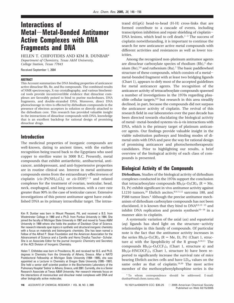
and Rh2(µ-O2CCF3)2(η1-O2CCF3)2(bpy)(THF)(H2O) (Figure2),30 exhibit structures with one chelating bpy moleculein ax-eq and eq-eq positions, respectively. By piecing to-gether the data obtained from these collective studies, amechanism for how nitrogen-containing chelates enterthe coordination sphere of the dirhodium core wasproposed (Scheme 1). The reaction involves an initialnucleophilic attack of the base at an ax site of the dimetalunit to afford the axially bound monodentate adduct a,followed by formation of a chelate ring by attack of asecond donor atom at an eq site (b; ax-eq adducts) andconversion to the final eq-eq adducts c.30 The rearrange-ment from ax to eq positions is a key feature of thischemistry and a major factor in dictating the outcome ofpurine reactions with dirhodium units.
Interactions with Nucleobases and Nucleos(t)-idesAxial Nucleobases. A perusal of the literature reveals that,unlike cisplatin, dirhodium compounds bind strongly topolyadenylic acids and adenine nucleos(t)sides.8,9,31-33
These DNA binding preferences are considered to be
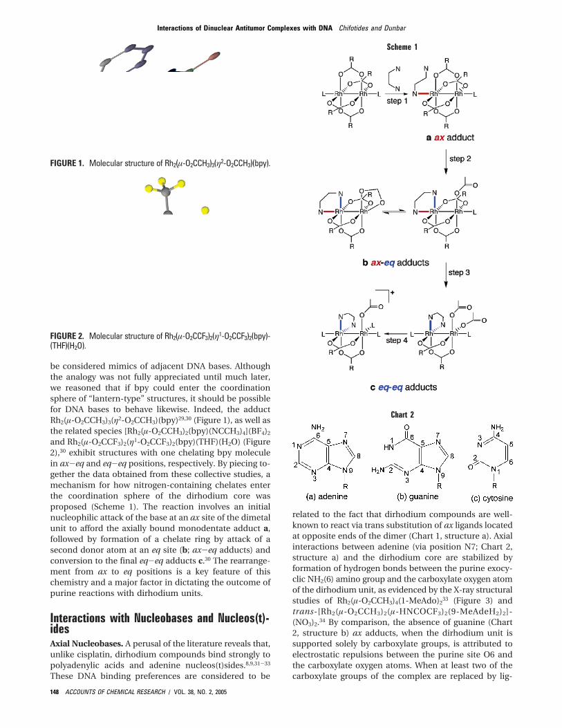
related to the fact that dirhodium compounds are well-known to react via trans substitution of ax ligands locatedat opposite ends of the dimer (Chart 1, structure a). Axialinteractions between adenine (via position N7; Chart 2,structure a) and the dirhodium core are stabilized byformation of hydrogen bonds between the purine exocy-clic NH2(6) amino group and the carboxylate oxygen atomof the dirhodium unit, as evidenced by the X-ray structuralstudies of Rh2(µ-O2CCH3)4(1-MeAdo)2
33 (Figure 3) andtrans-[Rh2(µ-O2CCH3)2(µ-HNCOCF3)2(9-MeAdeH2)2]-(NO3)2.34 By comparison, the absence of guanine (Chart2, structure b) ax adducts, when the dirhodium unit issupported solely by carboxylate groups, is attributed toelectrostatic repulsions between the purine site O6 andthe carboxylate oxygen atoms. When at least two of thecarboxylate groups of the complex are replaced by lig-
FIGURE 1. Molecular structure of Rh2(µ-O2CCH3)3(η2-O2CCH3)(bpy).
FIGURE 2. Molecular structure of Rh2(µ-O2CCF3)2(η1-O2CCF3)2(bpy)-(THF)(H2O).
Scheme 1
Chart 2
Interactions of Dinuclear Antitumor Complexes with DNA Chifotides and Dunbar
148 ACCOUNTS OF CHEMICAL RESEARCH / VOL. 38, NO. 2, 2005
ands with hydrogen-bonding donor moieties, how-ever, ax binding of guanine via N7 as in trans-[Rh2-(µ-O2CCH3)2(µ-NHCOCF3)2(9-EtGuaH)2] (Figure 4) andRh2(µ-NHCOCF3)4(dGuo)2, is favored.34 The argument ofunfavorable ax binding of guanine to dirhodium carboxy-late units is supported by the fact that although theguanine analogue, theophylline, binds axially to Rh2-(µ-O2CCH3)4 via N9, in [Rh2(HNCOCH3)4(theophylline)2]-NO3, the NH hydrogen-donor groups on the acetamidatemoieties favor binding via position N7.35 Axial binding ofcytosine via N3 (Chart 2, structure c) to Rh2(HNCOCH3)4
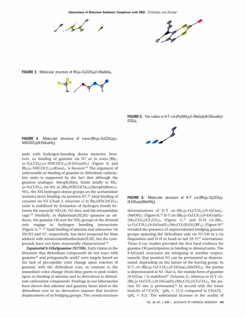
units is stabilized by formation of hydrogen bonds be-tween the exocyclic NH2(4), O2 sites, and the tetraamidatecage.36 Similarly, in diplatinum(III,III)-guanine ax ad-ducts, the guanine O6 and the NH3 groups of the dimetalunit engage in hydrogen bonding interactions(Figure 5).37-39 Axial binding of adenine and adenosine viaN9/N3 and N7, respectively, has been proposed for theiradducts with tetraacetatodiruthenium(II,III), but the com-pounds have not been structurally characterized.40
Equatorial 9-Ethylguanine (N7/O6). Early claims in theliterature that dirhodium compounds do not react withguanine32 and polyguanylic acids8 were largely based onthe lack of perceptible color change upon reaction ofguanine with the dirhodium core, in contrast to theimmediate color change (from blue-green to pink-violet)upon ax binding of adenine and its derivatives to dirhod-ium carboxylate compounds. Findings in our laboratorieshave shown that adenine and guanine bases bind to thedirhodium core in an alternative manner that involvesdisplacement of eq bridging groups. The crystal structure
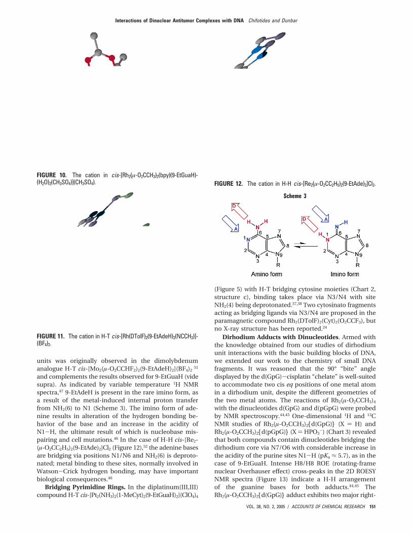
determinations of H-T cis-[Rh2(µ-O2CCH3)2(9-EtGua)2-(MeOH)2] (Figure 6),41 H-T cis-[Rh2(µ-O2CCF3)2(9-EtGuaH)2-(Me2CO)2](CF3CO2)2 (Figure 7),41 and H-H cis-[Rh2-(µ-O2CCH3)2(9-EtGuaH)2(Me2CO)(H2O)](BF4)2 (Figure 8)42
revealed the presence of unprecedented bridging guaninegroups spanning the dirhodium unit via N7/O6 in a cisdisposition and H-H or head-to-tail (H-T)43 orientations.These X-ray studies provided the first hard evidence forguanine O6 participation in binding to dimetal units. The9-EtGuaH structures are intriguing in another respect,namely, that position N1 can be protonated or deproto-nated, depending on the nature of the leaving group. InH-T cis-[Rh2(µ-O2CCH3)2(9-EtGua)2(MeOH)2], the purineis deprotonated at N1, that is, the enolate form of guanine(9-EtGua-) is stabilized41 (Scheme 2), whereas in H-T cis-[Rh2(µ-O2CCF3)2(9-EtGuaH)2(Me2CO)2](CF3CO2)2, the pu-rine N1 site is protonated,41 in accord with the lowerbasicity of CF3CO2
- (pKb ≈ 13.5) compared to CH3CO2-
(pKb ≈ 9.2). The substantial increase in the acidity of
FIGURE 3. Molecular structure of Rh2(µ-O2CCH3)4(1-MeAdo)2.
FIGURE 4. Molecular structure of trans-[Rh2(µ-O2CCH3)2(µ-NHCOCF3)2(9-EtGuaH)2].
FIGURE 5. The cation in H-T cis-[Pt2(NH3)2(1-MeCyt)2(9-EtGuaH)2]-(ClO4)4.
FIGURE 6. Molecular structure of H-T cis-[Rh2(µ-O2CCH3)2-(9-EtGua)2(MeOH)2].
Interactions of Dinuclear Antitumor Complexes with DNA Chifotides and Dunbar
VOL. 38, NO. 2, 2005 / ACCOUNTS OF CHEMICAL RESEARCH 149
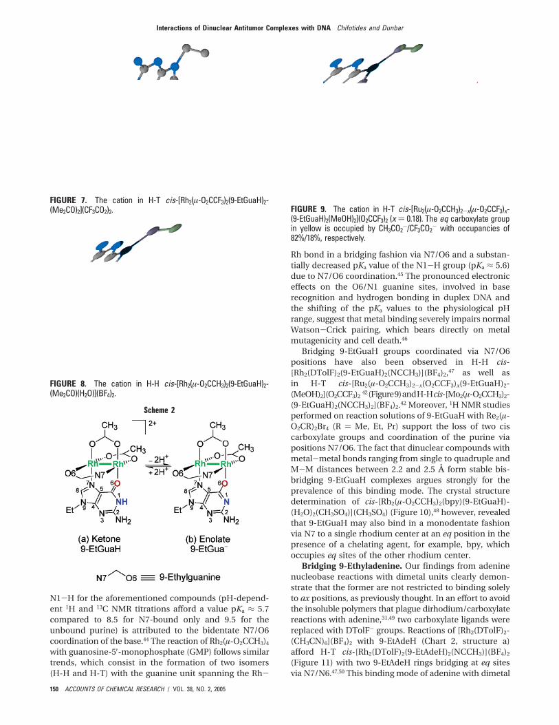
N1-H for the aforementioned compounds (pH-depend-ent 1H and 13C NMR titrations afford a value pKa ≈ 5.7compared to 8.5 for N7-bound only and 9.5 for theunbound purine) is attributed to the bidentate N7/O6coordination of the base.44 The reaction of Rh2(µ-O2CCH3)4
with guanosine-5′-monophosphate (GMP) follows similartrends, which consist in the formation of two isomers(H-H and H-T) with the guanine unit spanning the Rh-
Rh bond in a bridging fashion via N7/O6 and a substan-tially decreased pKa value of the N1-H group (pKa ≈ 5.6)due to N7/O6 coordination.45 The pronounced electroniceffects on the O6/N1 guanine sites, involved in baserecognition and hydrogen bonding in duplex DNA andthe shifting of the pKa values to the physiological pHrange, suggest that metal binding severely impairs normalWatson-Crick pairing, which bears directly on metalmutagenicity and cell death.46
Bridging 9-EtGuaH groups coordinated via N7/O6positions have also been observed in H-H cis-[Rh2(DTolF)2(9-EtGuaH)2(NCCH3)](BF4)2,47 as well asin H-T cis-[Ru2(µ-O2CCH3)2-x(O2CCF3)x(9-EtGuaH)2-(MeOH)2](O2CCF3)2
42(Figure9)andH-Hcis-[Mo2(µ-O2CCH3)2-(9-EtGuaH)2(NCCH3)2](BF4)2.42 Moreover, 1H NMR studiesperformed on reaction solutions of 9-EtGuaH with Re2(µ-O2CR)2Br4 (R ) Me, Et, Pr) support the loss of two ciscarboxylate groups and coordination of the purine viapositions N7/O6. The fact that dinuclear compounds withmetal-metal bonds ranging from single to quadruple andM-M distances between 2.2 and 2.5 Å form stable bis-bridging 9-EtGuaH complexes argues strongly for theprevalence of this binding mode. The crystal structuredetermination of cis-[Rh2(µ-O2CCH3)2(bpy)(9-EtGuaH)-(H2O)2(CH3SO4)](CH3SO4) (Figure 10),48 however, revealedthat 9-EtGuaH may also bind in a monodentate fashionvia N7 to a single rhodium center at an eq position in thepresence of a chelating agent, for example, bpy, whichoccupies eq sites of the other rhodium center.
Bridging 9-Ethyladenine. Our findings from adeninenucleobase reactions with dimetal units clearly demon-strate that the former are not restricted to binding solelyto ax positions, as previously thought. In an effort to avoidthe insoluble polymers that plague dirhodium/carboxylatereactions with adenine,31,49 two carboxylate ligands werereplaced with DTolF- groups. Reactions of [Rh2(DTolF)2-(CH3CN)6](BF4)2 with 9-EtAdeH (Chart 2, structure a)afford H-T cis-[Rh2(DTolF)2(9-EtAdeH)2(NCCH3)](BF4)2
(Figure 11) with two 9-EtAdeH rings bridging at eq sitesvia N7/N6.47,50 This binding mode of adenine with dimetal
FIGURE 7. The cation in H-T cis-[Rh2(µ-O2CCF3)2(9-EtGuaH)2-(Me2CO)2](CF3CO2)2.
FIGURE 8. The cation in H-H cis-[Rh2(µ-O2CCH3)2(9-EtGuaH)2-(Me2CO)(H2O)](BF4)2.
Scheme 2
FIGURE 9. The cation in H-T cis-[Ru2(µ-O2CCH3)2-x(µ-O2CCF3)x-(9-EtGuaH)2(MeOH)2](O2CCF3)2 (x ) 0.18). The eq carboxylate groupin yellow is occupied by CH3CO2
-/CF3CO2- with occupancies of
82%/18%, respectively.
Interactions of Dinuclear Antitumor Complexes with DNA Chifotides and Dunbar
150 ACCOUNTS OF CHEMICAL RESEARCH / VOL. 38, NO. 2, 2005
units was originally observed in the dimolybdenumanalogue H-T cis-[Mo2(µ-O2CCHF2)2(9-EtAdeH)2](BF4)2
51
and complements the results observed for 9-EtGuaH (videsupra). As indicated by variable temperature 1H NMRspectra,47 9-EtAdeH is present in the rare imino form, asa result of the metal-induced internal proton transferfrom NH2(6) to N1 (Scheme 3). The imino form of ade-nine results in alteration of the hydrogen bonding be-havior of the base and an increase in the acidity ofN1-H, the ultimate result of which is nucleobase mis-pairing and cell mutations.46 In the case of H-H cis-[Re2-(µ-O2CC2H5)2(9-EtAde)2]Cl2 (Figure 12),52 the adenine basesare bridging via positions N1/N6 and NH2(6) is deproto-nated; metal binding to these sites, normally involved inWatson-Crick hydrogen bonding, may have importantbiological consequences.46
Bridging Pyrimidine Rings. In the diplatinum(III,III)compound H-T cis-[Pt2(NH3)2(1-MeCyt)2(9-EtGuaH)2](ClO4)4
(Figure 5) with H-T bridging cytosine moieties (Chart 2,structure c), binding takes place via N3/N4 with siteNH2(4) being deprotonated.37,38 Two cytosinato fragmentsacting as bridging ligands via N3/N4 are proposed in theparamagnetic compound Rh2(DTolF)2(Cyt)2(O2CCF3), butno X-ray structure has been reported.24
Dirhodium Adducts with Dinucleotides. Armed withthe knowledge obtained from our studies of dirhodiumunit interactions with the basic building blocks of DNA,we extended our work to the chemistry of small DNAfragments. It was reasoned that the 90° “bite” angledisplayed by the d(GpG)-cisplatin “chelate” is well-suitedto accommodate two cis eq positions of one metal atomin a dirhodium unit, despite the different geometries ofthe two metal atoms. The reactions of Rh2(µ-O2CCH3)4
with the dinucleotides d(GpG) and d(pGpG) were probedby NMR spectroscopy.44,45 One-dimensional 1H and 13CNMR studies of Rh2(µ-O2CCH3)2{d(GpG)} (X ) H) andRh2(µ-O2CCH3)2{d(pGpG)} (X ) HPO3
-) (Chart 3) revealedthat both compounds contain dinucleotides bridging thedirhodium core via N7/O6 with considerable increase inthe acidity of the purine sites N1-H (pKa ≈ 5.7), as in thecase of 9-EtGuaH. Intense H8/H8 ROE (rotating-framenuclear Overhauser effect) cross-peaks in the 2D ROESYNMR spectra (Figure 13) indicate a H-H arrangementof the guanine bases for both adducts.44,45 TheRh2(µ-O2CCH3)2{d(GpG)} adduct exhibits two major right-
FIGURE 10. The cation in cis-[Rh2(µ-O2CCH3)2(bpy)(9-EtGuaH)-(H2O)2(CH3SO4)](CH3SO4).
FIGURE 11. The cation in H-T cis-[Rh(DTolF)2(9-EtAdeH)2(NCCH3)]-(BF4)2.
FIGURE 12. The cation in H-H cis-[Re2(µ-O2CC2H5)2(9-EtAde)2]Cl2.
Scheme 3
Interactions of Dinuclear Antitumor Complexes with DNA Chifotides and Dunbar
VOL. 38, NO. 2, 2005 / ACCOUNTS OF CHEMICAL RESEARCH 151
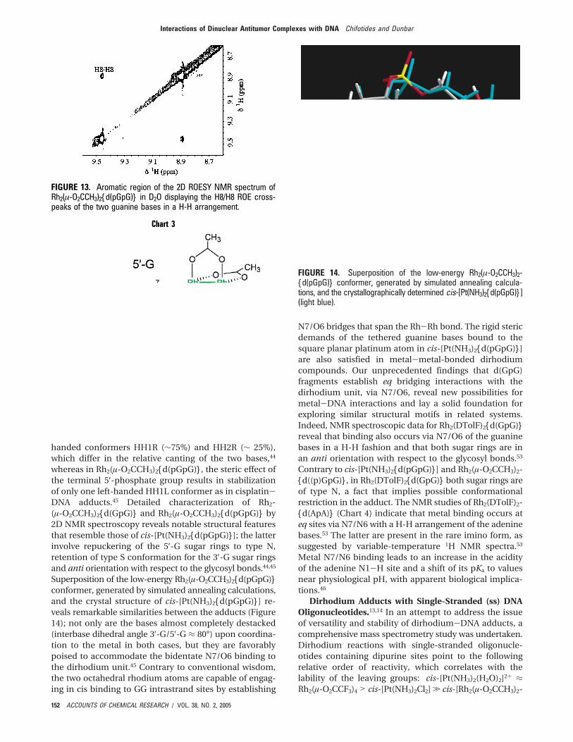
handed conformers HH1R (∼75%) and HH2R (∼ 25%),which differ in the relative canting of the two bases,44
whereas in Rh2(µ-O2CCH3)2{d(pGpG)}, the steric effect ofthe terminal 5′-phosphate group results in stabilizationof only one left-handed HH1L conformer as in cisplatin-DNA adducts.45 Detailed characterization of Rh2-(µ-O2CCH3)2{d(GpG)} and Rh2(µ-O2CCH3)2{d(pGpG)} by2D NMR spectroscopy reveals notable structural featuresthat resemble those of cis-[Pt(NH3)2{d(pGpG)}]; the latterinvolve repuckering of the 5′-G sugar rings to type N,retention of type S conformation for the 3′-G sugar ringsand anti orientation with respect to the glycosyl bonds.44,45
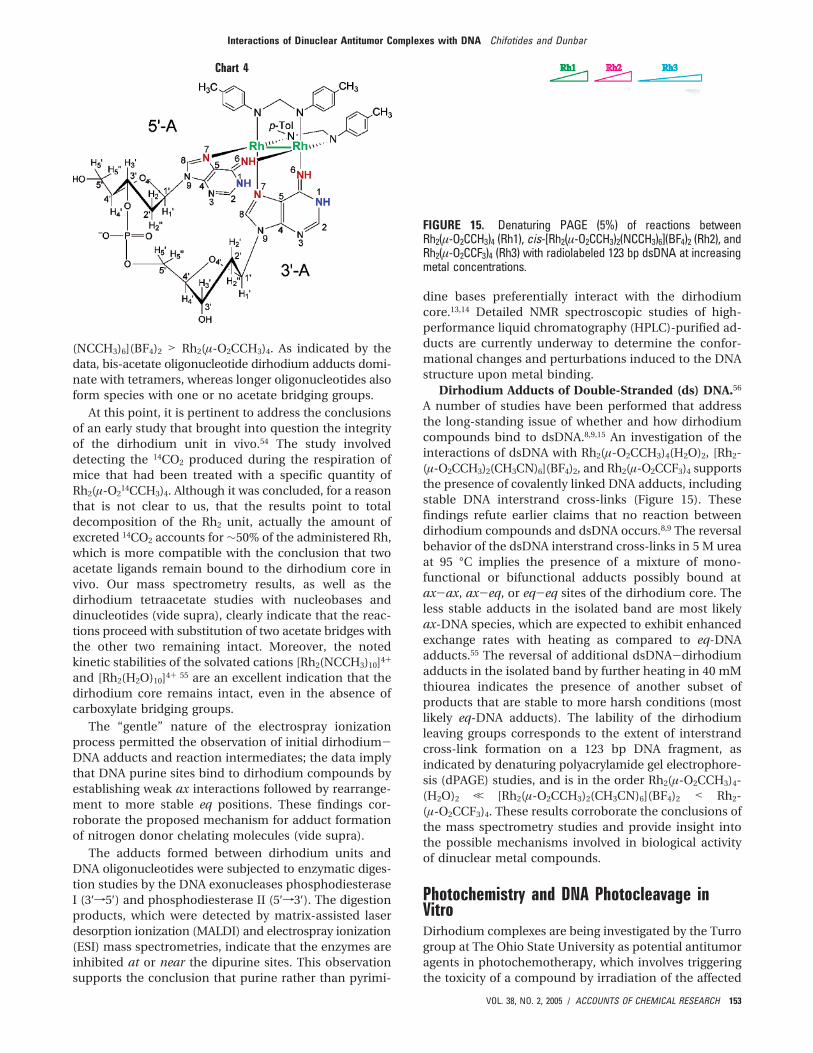
Superposition of the low-energy Rh2(µ-O2CCH3)2{d(pGpG)}conformer, generated by simulated annealing calculations,and the crystal structure of cis-[Pt(NH3)2{d(pGpG)}] re-veals remarkable similarities between the adducts (Figure14); not only are the bases almost completely destacked(interbase dihedral angle 3′-G/5′-G ≈ 80°) upon coordina-tion to the metal in both cases, but they are favorablypoised to accommodate the bidentate N7/O6 binding tothe dirhodium unit.45 Contrary to conventional wisdom,the two octahedral rhodium atoms are capable of engag-ing in cis binding to GG intrastrand sites by establishing
N7/O6 bridges that span the Rh-Rh bond. The rigid stericdemands of the tethered guanine bases bound to thesquare planar platinum atom in cis-[Pt(NH3)2{d(pGpG)}]are also satisfied in metal-metal-bonded dirhodiumcompounds. Our unprecedented findings that d(GpG)fragments establish eq bridging interactions with thedirhodium unit, via N7/O6, reveal new possibilities formetal-DNA interactions and lay a solid foundation forexploring similar structural motifs in related systems.Indeed, NMR spectroscopic data for Rh2(DTolF)2{d(GpG)}reveal that binding also occurs via N7/O6 of the guaninebases in a H-H fashion and that both sugar rings are inan anti orientation with respect to the glycosyl bonds.53
Contrary to cis-[Pt(NH3)2{d(pGpG)}] and Rh2(µ-O2CCH3)2-{d((p)GpG)}, in Rh2(DTolF)2{d(GpG)} both sugar rings areof type N, a fact that implies possible conformationalrestriction in the adduct. The NMR studies of Rh2(DTolF)2-{d(ApA)} (Chart 4) indicate that metal binding occurs ateq sites via N7/N6 with a H-H arrangement of the adeninebases.53 The latter are present in the rare imino form, assuggested by variable-temperature 1H NMR spectra.53
Metal N7/N6 binding leads to an increase in the acidityof the adenine N1-H site and a shift of its pKa to valuesnear physiological pH, with apparent biological implica-tions.46
Dirhodium Adducts with Single-Stranded (ss) DNAOligonucleotides.13,14 In an attempt to address the issueof versatility and stability of dirhodium-DNA adducts, acomprehensive mass spectrometry study was undertaken.Dirhodium reactions with single-stranded oligonucle-otides containing dipurine sites point to the followingrelative order of reactivity, which correlates with thelability of the leaving groups: cis-[Pt(NH3)2(H2O)2]2+ ≈Rh2(µ-O2CCF3)4 > cis-[Pt(NH3)2Cl2] . cis-[Rh2(µ-O2CCH3)2-
FIGURE 13. Aromatic region of the 2D ROESY NMR spectrum ofRh2(µ-O2CCH3)2{d(pGpG)} in D2O displaying the H8/H8 ROE cross-peaks of the two guanine bases in a H-H arrangement.
Chart 3
FIGURE 14. Superposition of the low-energy Rh2(µ-O2CCH3)2-{d(pGpG)} conformer, generated by simulated annealing calcula-tions, and the crystallographically determined cis-[Pt(NH3)2{d(pGpG)}](light blue).
Interactions of Dinuclear Antitumor Complexes with DNA Chifotides and Dunbar
152 ACCOUNTS OF CHEMICAL RESEARCH / VOL. 38, NO. 2, 2005
(NCCH3)6](BF4)2 > Rh2(µ-O2CCH3)4. As indicated by thedata, bis-acetate oligonucleotide dirhodium adducts domi-nate with tetramers, whereas longer oligonucleotides alsoform species with one or no acetate bridging groups.
At this point, it is pertinent to address the conclusionsof an early study that brought into question the integrityof the dirhodium unit in vivo.54 The study involveddetecting the 14CO2 produced during the respiration ofmice that had been treated with a specific quantity ofRh2(µ-O2
14CCH3)4. Although it was concluded, for a reasonthat is not clear to us, that the results point to totaldecomposition of the Rh2 unit, actually the amount ofexcreted 14CO2 accounts for ∼50% of the administered Rh,which is more compatible with the conclusion that twoacetate ligands remain bound to the dirhodium core invivo. Our mass spectrometry results, as well as thedirhodium tetraacetate studies with nucleobases anddinucleotides (vide supra), clearly indicate that the reac-tions proceed with substitution of two acetate bridges withthe other two remaining intact. Moreover, the notedkinetic stabilities of the solvated cations [Rh2(NCCH3)10]4+
and [Rh2(H2O)10]4+ 55 are an excellent indication that thedirhodium core remains intact, even in the absence ofcarboxylate bridging groups.
The “gentle” nature of the electrospray ionizationprocess permitted the observation of initial dirhodium-DNA adducts and reaction intermediates; the data implythat DNA purine sites bind to dirhodium compounds byestablishing weak ax interactions followed by rearrange-ment to more stable eq positions. These findings cor-roborate the proposed mechanism for adduct formationof nitrogen donor chelating molecules (vide supra).
The adducts formed between dirhodium units andDNA oligonucleotides were subjected to enzymatic diges-tion studies by the DNA exonucleases phosphodiesteraseI (3′f5′) and phosphodiesterase II (5′f3′). The digestionproducts, which were detected by matrix-assisted laserdesorption ionization (MALDI) and electrospray ionization(ESI) mass spectrometries, indicate that the enzymes areinhibited at or near the dipurine sites. This observationsupports the conclusion that purine rather than pyrimi-
dine bases preferentially interact with the dirhodiumcore.13,14 Detailed NMR spectroscopic studies of high-performance liquid chromatography (HPLC)-purified ad-ducts are currently underway to determine the confor-mational changes and perturbations induced to the DNAstructure upon metal binding.
Dirhodium Adducts of Double-Stranded (ds) DNA.56
A number of studies have been performed that addressthe long-standing issue of whether and how dirhodiumcompounds bind to dsDNA.8,9,15 An investigation of theinteractions of dsDNA with Rh2(µ-O2CCH3)4(H2O)2, [Rh2-(µ-O2CCH3)2(CH3CN)6](BF4)2, and Rh2(µ-O2CCF3)4 supportsthe presence of covalently linked DNA adducts, includingstable DNA interstrand cross-links (Figure 15). Thesefindings refute earlier claims that no reaction betweendirhodium compounds and dsDNA occurs.8,9 The reversalbehavior of the dsDNA interstrand cross-links in 5 M ureaat 95 °C implies the presence of a mixture of mono-functional or bifunctional adducts possibly bound atax-ax, ax-eq, or eq-eq sites of the dirhodium core. Theless stable adducts in the isolated band are most likelyax-DNA species, which are expected to exhibit enhancedexchange rates with heating as compared to eq-DNAadducts.55 The reversal of additional dsDNA-dirhodiumadducts in the isolated band by further heating in 40 mMthiourea indicates the presence of another subset ofproducts that are stable to more harsh conditions (mostlikely eq-DNA adducts). The lability of the dirhodiumleaving groups corresponds to the extent of interstrandcross-link formation on a 123 bp DNA fragment, asindicated by denaturing polyacrylamide gel electrophore-sis (dPAGE) studies, and is in the order Rh2(µ-O2CCH3)4-(H2O)2 , [Rh2(µ-O2CCH3)2(CH3CN)6](BF4)2 < Rh2-(µ-O2CCF3)4. These results corroborate the conclusions ofthe mass spectrometry studies and provide insight intothe possible mechanisms involved in biological activityof dinuclear metal compounds.
Photochemistry and DNA Photocleavage inVitroDirhodium complexes are being investigated by the Turrogroup at The Ohio State University as potential antitumoragents in photochemotherapy, which involves triggeringthe toxicity of a compound by irradiation of the affected
Chart 4
FIGURE 15. Denaturing PAGE (5%) of reactions betweenRh2(µ-O2CCH3)4 (Rh1), cis-[Rh2(µ-O2CCH3)2(NCCH3)6](BF4)2 (Rh2), andRh2(µ-O2CCF3)4 (Rh3) with radiolabeled 123 bp dsDNA at increasingmetal concentrations.
Interactions of Dinuclear Antitumor Complexes with DNA Chifotides and Dunbar
VOL. 38, NO. 2, 2005 / ACCOUNTS OF CHEMICAL RESEARCH 153
area with low-energy light. It has been reported that Rh2-(µ-O2CCH3)4 exhibits a long-lived excited state (τ ) 3.5 µs)that can be accessed with visible light (λexc ≈ 350-600 nm)and undergoes energy and electron transfer with a varietyof acceptors.57 Irradiation of Rh2(µ-O2CCH3)4 with visiblelight (λirr ) 400-610 nm), in the presence of electronacceptors, results in DNA photocleavage by the mixed-valent cation [Rh2(µ-O2CCH3)4]+.58 Unlike Rh2(µ-O2CCH3)4,which requires an electron acceptor in solution,58
Rh24+ complexes with dppz (dppz ) dipyrido[3,2-
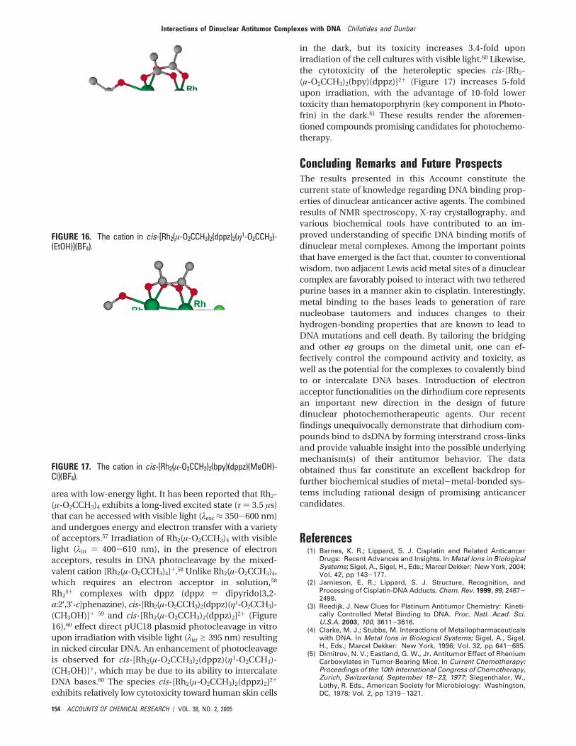
a:2′,3′-c]phenazine), cis-[Rh2(µ-O2CCH3)2(dppz)(η1-O2CCH3)-(CH3OH)]+ 59 and cis-[Rh2(µ-O2CCH3)2(dppz)2]2+ (Figure16),60 effect direct pUC18 plasmid photocleavage in vitroupon irradiation with visible light (λirr g 395 nm) resultingin nicked circular DNA. An enhancement of photocleavageis observed for cis-[Rh2(µ-O2CCH3)2(dppz)(η1-O2CCH3)-(CH3OH)]+, which may be due to its ability to intercalateDNA bases.60 The species cis-[Rh2(µ-O2CCH3)2(dppz)2]2+
exhibits relatively low cytotoxicity toward human skin cells
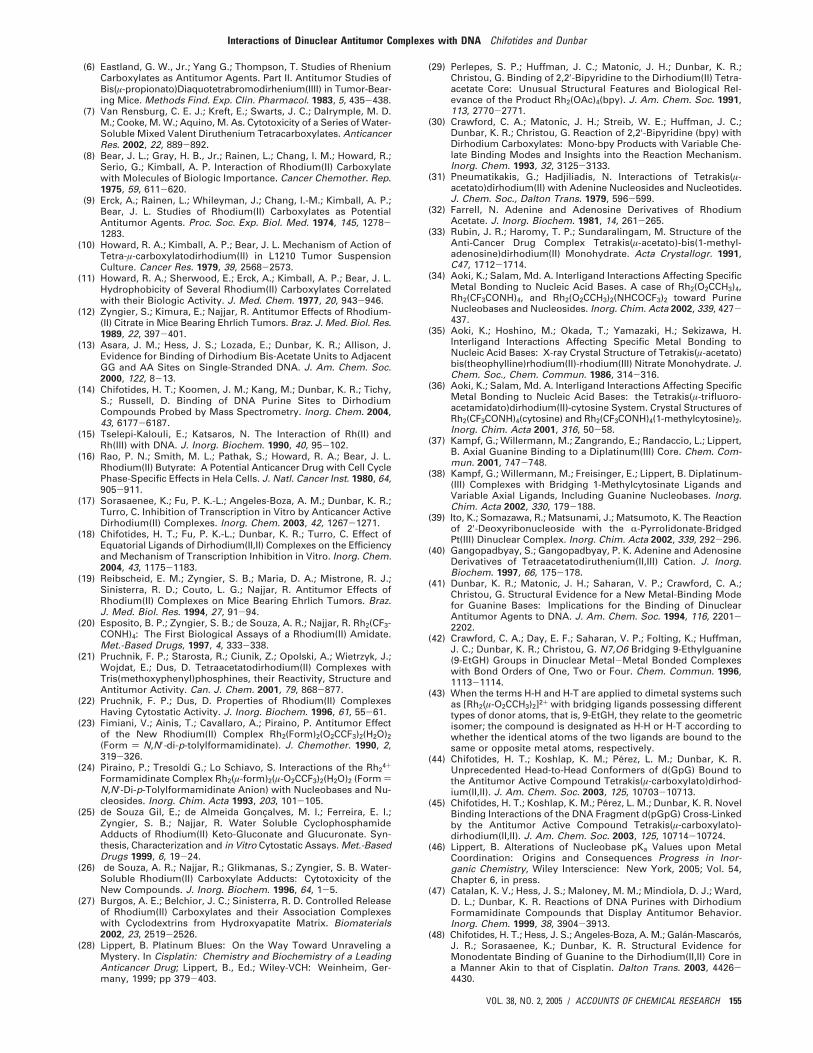
in the dark, but its toxicity increases 3.4-fold uponirradiation of the cell cultures with visible light.60 Likewise,the cytotoxicity of the heteroleptic species cis-[Rh2-(µ-O2CCH3)2(bpy)(dppz)]2+ (Figure 17) increases 5-foldupon irradiation, with the advantage of 10-fold lowertoxicity than hematoporphyrin (key component in Photo-frin) in the dark.61 These results render the aforemen-tioned compounds promising candidates for photochemo-therapy.
Concluding Remarks and Future ProspectsThe results presented in this Account constitute thecurrent state of knowledge regarding DNA binding prop-erties of dinuclear anticancer active agents. The combinedresults of NMR spectroscopy, X-ray crystallography, andvarious biochemical tools have contributed to an im-proved understanding of specific DNA binding motifs ofdinuclear metal complexes. Among the important pointsthat have emerged is the fact that, counter to conventionalwisdom, two adjacent Lewis acid metal sites of a dinuclearcomplex are favorably poised to interact with two tetheredpurine bases in a manner akin to cisplatin. Interestingly,metal binding to the bases leads to generation of rarenucleobase tautomers and induces changes to theirhydrogen-bonding properties that are known to lead toDNA mutations and cell death. By tailoring the bridgingand other eq groups on the dimetal unit, one can ef-fectively control the compound activity and toxicity, aswell as the potential for the complexes to covalently bindto or intercalate DNA bases. Introduction of electronacceptor functionalities on the dirhodium core representsan important new direction in the design of futuredinuclear photochemotherapeutic agents. Our recentfindings unequivocally demonstrate that dirhodium com-pounds bind to dsDNA by forming interstrand cross-linksand provide valuable insight into the possible underlyingmechanism(s) of their antitumor behavior. The dataobtained thus far constitute an excellent backdrop forfurther biochemical studies of metal-metal-bonded sys-tems including rational design of promising anticancercandidates.
References(1) Barnes, K. R.; Lippard, S. J. Cisplatin and Related Anticancer
Drugs: Recent Advances and Insights. In Metal Ions in BiologicalSystems; Sigel, A., Sigel, H., Eds.; Marcel Dekker: New York, 2004;Vol. 42, pp 143-177.
(2) Jamieson, E. R.; Lippard, S. J. Structure, Recognition, andProcessing of Cisplatin-DNA Adducts. Chem. Rev. 1999, 99, 2467-2498.
(3) Reedijk, J. New Clues for Platinum Antitumor Chemistry: Kineti-cally Controlled Metal Binding to DNA. Proc. Natl. Acad. Sci.U.S.A. 2003, 100, 3611-3616.
(4) Clarke, M. J.; Stubbs, M. Interactions of Metallopharmaceuticalswith DNA. In Metal Ions in Biological Systems; Sigel, A., Sigel,H., Eds.; Marcel Dekker: New York, 1996; Vol. 32, pp 641-685.
(5) Dimitrov, N. V.; Eastland, G. W., Jr. Antitumor Effect of RheniumCarboxylates in Tumor-Bearing Mice. In Current Chemotherapy:Proceedings of the 10th International Congress of Chemotherapy,Zurich, Switzerland, September 18-23, 1977; Siegenthaler, W.,Luthy, R. Eds., American Society for Microbiology: Washington,DC, 1978; Vol. 2, pp 1319-1321.
FIGURE 16. The cation in cis-[Rh2(µ-O2CCH3)2(dppz)2(η1-O2CCH3)-(EtOH)](BF4).
FIGURE 17. The cation in cis-[Rh2(µ-O2CCH3)2(bpy)(dppz)(MeOH)-Cl](BF4).
Interactions of Dinuclear Antitumor Complexes with DNA Chifotides and Dunbar
154 ACCOUNTS OF CHEMICAL RESEARCH / VOL. 38, NO. 2, 2005
(6) Eastland, G. W., Jr.; Yang G.; Thompson, T. Studies of RheniumCarboxylates as Antitumor Agents. Part II. Antitumor Studies ofBis(µ-propionato)Diaquotetrabromodirhenium(IIII) in Tumor-Bear-ing Mice. Methods Find. Exp. Clin. Pharmacol. 1983, 5, 435-438.
(7) Van Rensburg, C. E. J.; Kreft, E.; Swarts, J. C.; Dalrymple, M. D.M.; Cooke, M. W.; Aquino, M. As. Cytotoxicity of a Series of Water-Soluble Mixed Valent Diruthenium Tetracarboxylates. AnticancerRes. 2002, 22, 889-892.
(8) Bear, J. L.; Gray, H. B., Jr.; Rainen, L.; Chang, I. M.; Howard, R.;Serio, G.; Kimball, A. P. Interaction of Rhodium(II) Carboxylatewith Molecules of Biologic Importance. Cancer Chemother. Rep.1975, 59, 611-620.
(9) Erck, A.; Rainen, L.; Whileyman, J.; Chang, I.-M.; Kimball, A. P.;Bear, J. L. Studies of Rhodium(II) Carboxylates as PotentialAntitumor Agents. Proc. Soc. Exp. Biol. Med. 1974, 145, 1278-1283.
(10) Howard, R. A.; Kimball, A. P.; Bear, J. L. Mechanism of Action ofTetra-µ-carboxylatodirhodium(II) in L1210 Tumor SuspensionCulture. Cancer Res. 1979, 39, 2568-2573.
(11) Howard, R. A.; Sherwood, E.; Erck, A.; Kimball, A. P.; Bear, J. L.Hydrophobicity of Several Rhodium(II) Carboxylates Correlatedwith their Biologic Activity. J. Med. Chem. 1977, 20, 943-946.
(12) Zyngier, S.; Kimura, E.; Najjar, R. Antitumor Effects of Rhodium-(II) Citrate in Mice Bearing Ehrlich Tumors. Braz. J. Med. Biol. Res.1989, 22, 397-401.
(13) Asara, J. M.; Hess, J. S.; Lozada, E.; Dunbar, K. R.; Allison, J.Evidence for Binding of Dirhodium Bis-Acetate Units to AdjacentGG and AA Sites on Single-Stranded DNA. J. Am. Chem. Soc.2000, 122, 8-13.
(14) Chifotides, H. T.; Koomen, J. M.; Kang, M.; Dunbar, K. R.; Tichy,S.; Russell, D. Binding of DNA Purine Sites to DirhodiumCompounds Probed by Mass Spectrometry. Inorg. Chem. 2004,43, 6177-6187.
(15) Tselepi-Kalouli, E.; Katsaros, N. The Interaction of Rh(II) andRh(III) with DNA. J. Inorg. Biochem. 1990, 40, 95-102.
(16) Rao, P. N.; Smith, M. L.; Pathak, S.; Howard, R. A.; Bear, J. L.Rhodium(II) Butyrate: A Potential Anticancer Drug with Cell CyclePhase-Specific Effects in Hela Cells. J. Natl. Cancer Inst. 1980, 64,905-911.
(17) Sorasaenee, K.; Fu, P. K.-L.; Angeles-Boza, A. M.; Dunbar, K. R.;Turro, C. Inhibition of Transcription in Vitro by Anticancer ActiveDirhodium(II) Complexes. Inorg. Chem. 2003, 42, 1267-1271.
(18) Chifotides, H. T.; Fu, P. K.-L.; Dunbar, K. R.; Turro, C. Effect ofEquatorial Ligands of Dirhodium(II,II) Complexes on the Efficiencyand Mechanism of Transcription Inhibition in Vitro. Inorg. Chem.2004, 43, 1175-1183.
(19) Reibscheid, E. M.; Zyngier, S. B.; Maria, D. A.; Mistrone, R. J.;Sinisterra, R. D.; Couto, L. G.; Najjar, R. Antitumor Effects ofRhodium(II) Complexes on Mice Bearing Ehrlich Tumors. Braz.J. Med. Biol. Res. 1994, 27, 91-94.
(20) Esposito, B. P.; Zyngier, S. B.; de Souza, A. R.; Najjar, R. Rh2(CF3-CONH)4: The First Biological Assays of a Rhodium(II) Amidate.Met.-Based Drugs, 1997, 4, 333-338.
(21) Pruchnik, F. P.; Starosta, R.; Ciunik, Z.; Opolski, A.; Wietrzyk, J.;Wojdat, E.; Dus, D. Tetraacetatodirhodium(II) Complexes withTris(methoxyphenyl)phosphines, their Reactivity, Structure andAntitumor Activity. Can. J. Chem. 2001, 79, 868-877.
(22) Pruchnik, F. P.; Dus, D. Properties of Rhodium(II) ComplexesHaving Cytostatic Activity. J. Inorg. Biochem. 1996, 61, 55-61.
(23) Fimiani, V.; Ainis, T.; Cavallaro, A.; Piraino, P. Antitumor Effectof the New Rhodium(II) Complex Rh2(Form)2(O2CCF3)2(H2O)2(Form ) N,N′-di-p-tolylformamidinate). J. Chemother. 1990, 2,319-326.
(24) Piraino, P.; Tresoldi G.; Lo Schiavo, S. Interactions of the Rh24+
Formamidinate Complex Rh2(µ-form)2(µ-O2CCF3)2(H2O)2 (Form )N,N′-Di-p-Tolylformamidinate Anion) with Nucleobases and Nu-cleosides. Inorg. Chim. Acta 1993, 203, 101-105.
(25) de Souza Gil, E.; de Almeida Goncalves, M. I.; Ferreira, E. I.;Zyngier, S. B.; Najjar, R. Water Soluble CyclophosphamideAdducts of Rhodium(II) Keto-Gluconate and Glucuronate. Syn-thesis, Characterization and in Vitro Cytostatic Assays. Met.-BasedDrugs 1999, 6, 19-24.
(26) de Souza, A. R.; Najjar, R.; Glikmanas, S.; Zyngier, S. B. Water-Soluble Rhodium(II) Carboxylate Adducts: Cytotoxicity of theNew Compounds. J. Inorg. Biochem. 1996, 64, 1-5.
(27) Burgos, A. E.; Belchior, J. C.; Sinisterra, R. D. Controlled Releaseof Rhodium(II) Carboxylates and their Association Complexeswith Cyclodextrins from Hydroxyapatite Matrix. Biomaterials2002, 23, 2519-2526.
(28) Lippert, B. Platinum Blues: On the Way Toward Unraveling aMystery. In Cisplatin: Chemistry and Biochemistry of a LeadingAnticancer Drug; Lippert, B., Ed.; Wiley-VCH: Weinheim, Ger-many, 1999; pp 379-403.
(29) Perlepes, S. P.; Huffman, J. C.; Matonic, J. H.; Dunbar, K. R.;Christou, G. Binding of 2,2′-Bipyridine to the Dirhodium(II) Tetra-acetate Core: Unusual Structural Features and Biological Rel-evance of the Product Rh2(OAc)4(bpy). J. Am. Chem. Soc. 1991,113, 2770-2771.
(30) Crawford, C. A.; Matonic, J. H.; Streib, W. E.; Huffman, J. C.;Dunbar, K. R.; Christou, G. Reaction of 2,2′-Bipyridine (bpy) withDirhodium Carboxylates: Mono-bpy Products with Variable Che-late Binding Modes and Insights into the Reaction Mechanism.Inorg. Chem. 1993, 32, 3125-3133.
(31) Pneumatikakis, G.; Hadjiliadis, N. Interactions of Tetrakis(µ-acetato)dirhodium(II) with Adenine Nucleosides and Nucleotides.J. Chem. Soc., Dalton Trans. 1979, 596-599.
(32) Farrell, N. Adenine and Adenosine Derivatives of RhodiumAcetate. J. Inorg. Biochem. 1981, 14, 261-265.
(33) Rubin, J. R.; Haromy, T. P.; Sundaralingam, M. Structure of theAnti-Cancer Drug Complex Tetrakis(µ-acetato)-bis(1-methyl-adenosine)dirhodium(II) Monohydrate. Acta Crystallogr. 1991,C47, 1712-1714.
(34) Aoki, K.; Salam, Md. A. Interligand Interactions Affecting SpecificMetal Bonding to Nucleic Acid Bases. A case of Rh2(O2CCH3)4,Rh2(CF3CONH)4, and Rh2(O2CCH3)2(NHCOCF3)2 toward PurineNucleobases and Nucleosides. Inorg. Chim. Acta 2002, 339, 427-437.
(35) Aoki, K.; Hoshino, M.; Okada, T.; Yamazaki, H.; Sekizawa, H.Interligand Interactions Affecting Specific Metal Bonding toNucleic Acid Bases: X-ray Crystal Structure of Tetrakis(µ-acetato)bis(theophylline)rhodium(II)-rhodium(III) Nitrate Monohydrate. J.Chem. Soc., Chem. Commun. 1986, 314-316.
(36) Aoki, K.; Salam, Md. A. Interligand Interactions Affecting SpecificMetal Bonding to Nucleic Acid Bases: the Tetrakis(µ-trifluoro-acetamidato)dirhodium(II)-cytosine System. Crystal Structures ofRh2(CF3CONH)4(cytosine) and Rh2(CF3CONH)4(1-methylcytosine)2.Inorg. Chim. Acta 2001, 316, 50-58.
(37) Kampf, G.; Willermann, M.; Zangrando, E.; Randaccio, L.; Lippert,B. Axial Guanine Binding to a Diplatinum(III) Core. Chem. Com-mun. 2001, 747-748.
(38) Kampf, G.; Willermann, M.; Freisinger, E.; Lippert, B. Diplatinum-(III) Complexes with Bridging 1-Methylcytosinate Ligands andVariable Axial Ligands, Including Guanine Nucleobases. Inorg.Chim. Acta 2002, 330, 179-188.
(39) Ito, K.; Somazawa, R.; Matsunami, J.; Matsumoto, K. The Reactionof 2′-Deoxyribonucleoside with the R-Pyrrolidonate-BridgedPt(III) Dinuclear Complex. Inorg. Chim. Acta 2002, 339, 292-296.
(40) Gangopadbyay, S.; Gangopadbyay, P. K. Adenine and AdenosineDerivatives of Tetraacetatodiruthenium(II,III) Cation. J. Inorg.Biochem. 1997, 66, 175-178.
(41) Dunbar, K. R.; Matonic, J. H.; Saharan, V. P.; Crawford, C. A.;Christou, G. Structural Evidence for a New Metal-Binding Modefor Guanine Bases: Implications for the Binding of DinuclearAntitumor Agents to DNA. J. Am. Chem. Soc. 1994, 116, 2201-2202.
(42) Crawford, C. A.; Day, E. F.; Saharan, V. P.; Folting, K.; Huffman,J. C.; Dunbar, K. R.; Christou, G. N7,O6 Bridging 9-Ethylguanine(9-EtGH) Groups in Dinuclear Metal-Metal Bonded Complexeswith Bond Orders of One, Two or Four. Chem. Commun. 1996,1113-1114.
(43) When the terms H-H and H-T are applied to dimetal systems suchas [Rh2(µ-O2CCH3)2]2+ with bridging ligands possessing differenttypes of donor atoms, that is, 9-EtGH, they relate to the geometricisomer; the compound is designated as H-H or H-T according towhether the identical atoms of the two ligands are bound to thesame or opposite metal atoms, respectively.
(44) Chifotides, H. T.; Koshlap, K. M.; Perez, L. M.; Dunbar, K. R.Unprecedented Head-to-Head Conformers of d(GpG) Bound tothe Antitumor Active Compound Tetrakis(µ-carboxylato)dirhod-ium(II,II). J. Am. Chem. Soc. 2003, 125, 10703-10713.
(45) Chifotides, H. T.; Koshlap, K. M.; Perez, L. M.; Dunbar, K. R. NovelBinding Interactions of the DNA Fragment d(pGpG) Cross-Linkedby the Antitumor Active Compound Tetrakis(µ-carboxylato)-dirhodium(II,II). J. Am. Chem. Soc. 2003, 125, 10714-10724.
(46) Lippert, B. Alterations of Nucleobase pKa Values upon MetalCoordination: Origins and Consequences Progress in Inor-ganic Chemistry, Wiley Interscience: New York, 2005; Vol. 54,Chapter 6, in press.
(47) Catalan, K. V.; Hess, J. S.; Maloney, M. M.; Mindiola, D. J.; Ward,D. L.; Dunbar, K. R. Reactions of DNA Purines with DirhodiumFormamidinate Compounds that Display Antitumor Behavior.Inorg. Chem. 1999, 38, 3904-3913.
(48) Chifotides, H. T.; Hess, J. S.; Angeles-Boza, A. M.; Galan-Mascaros,J. R.; Sorasaenee, K.; Dunbar, K. R. Structural Evidence forMonodentate Binding of Guanine to the Dirhodium(II,II) Core ina Manner Akin to that of Cisplatin. Dalton Trans. 2003, 4426-4430.
Interactions of Dinuclear Antitumor Complexes with DNA Chifotides and Dunbar
VOL. 38, NO. 2, 2005 / ACCOUNTS OF CHEMICAL RESEARCH 155
(49) Koutsodimou, A.; Katsaros, N. Reactions of the Rhodium Trifluo-roacetate Dimer with Nucleosides and Nucleotides. J. Coord.Chem. 1996, 39, 169-197.
(50) Catalan, K. V.; Mindiola, D. J.; Ward, D. L.; Dunbar, K. R. A NovelDirhodium Compound with Neutral, Bridging 9-EthyladenineLigands. Inorg. Chem. 1997, 36, 2458-2460.
(51) Day, E. F.; Crawford, C. A.; Folting, K.; Dunbar, K. R.; Christou, G.A New Metal Binding Mode for Adenine: A Bidentate (N6, N7)Bridging Mode in the Complex [Mo2(O2CCHF2)2(9-EtAH)2][BF4]2‚2MeCN. J. Am. Chem. Soc. 1994, 116, 9339-9340.
(52) Prater, M. E.; Mindiola, D. J.; Ouyang, X.; Dunbar, K. R. AQuadruply-Bonded Dirhenium Complex Bridged by Two N1/N6Adeninate Ligands. Inorg. Chem. Commun. 1998, 1, 475-477.
(53) Chifotides, H. T.; Dunbar, K. R. Head-to-Head Cross-Links Betweenthe DNA Fragments d(GpG), d(ApA) and an Antitumor ActiveN,N′-Di-p-Tolylformamidinate Dirhodium(II,II) Compound. Manu-script in preparation, 2005.
(54) Erck, A.; Sherwood, E.; Bear, J. L.; Kimball, A. P. The Metabolismof Rhodium(II) Acetate in Tumor-Bearing Mice. Cancer Res. 1976,36, 2204-2209.
(55) Pittet, P.-A.; Dadci, L.; Zbinden, P.; Abou-Hamdan, A.; Merbach,A. E. High-pressure Proton NMR Study of Acetonitrile ExchangeKinetics on [Rh2(CH3CN)10]4+ and 17O NMR Investigation ofAqueous Solutions of [Rh2(H2O)10]4+. Inorg. Chim. Acta 1993, 206,135-140.
(56) Dunham, S. U.; Chifotides, H. T.; Mikulski, S.; Burr, A. E.; Dunbar,K. R. Covalent Binding and Interstrand Cross-Linking of DuplexDNA by Dirhodium(II,II) Carboxylate Compounds. Biochemistry,2005, 44, 996-1003.
(57) Bradley, P. M.; Bursten, B. E.; Turro, C. Excited-State Propertiesof Rh2(O2CCH3)4(L)2 (L ) CH3OH, THF, PPh3, py). Inorg. Chem.2001, 40, 1376-1379.
(58) Fu, P. K.-L.; Bradley, P. M.; Turro, C. DNA Cleavage by Photo-generated [Rh2(O2CCH3)4(L)2]+. Inorg. Chem. 2001, 40, 2476-2477.
(59) Bradley, P. M.; Angeles-Boza, A. M.; Dunbar, K. R.; Turro, C. DirectDNA Photocleavage by a New Intercalating Dirhodium(II,II) Com-plex: Comparison to Rh2(O2CCH3)4. Inorg. Chem. 2004, 43, 2450-2452.
(60) Angeles-Boza, A. M.; Bradley, P. M.; Fu, P. K.-L.; Wicke, S.E.; Bacsa, J.; Dunbar, K. R.; Turro, C. DNA Binding and Photo-cleavage in vitro by New Dppz Dirhodium(II,II) Complexes:Correlation to Cytotoxicity and Phototoxicity. Inorg. Chem. 2004,43, 8510-8519.
(61) Angeles-Boza, A. M.; Bradley, P. M.; Fu, P. K.-L.; Hilfiger, M. G.;Shatruk, M.; Dunbar, K. R.; Turro, C. Phototoxicity of a NewRh2(II,II) Complex: Increase in Cytotoxicity upon IrradiationSimilar to PDT Agent Hematoporphyrin. Manuscript in prepara-tion, 2005.
AR0302078
Interactions of Dinuclear Antitumor Complexes with DNA Chifotides and Dunbar
156 ACCOUNTS OF CHEMICAL RESEARCH / VOL. 38, NO. 2, 2005