
Interactions at the bilayer interface and receptor site induced by the novel synthetic pyrrolidinone...
11
Author's personal copy Interactions at the bilayer interface and receptor site induced by the novel synthetic pyrrolidinone analog MMK3 C. Fotakis a , S. Gega a , E. Siapi b , C. Potamitis a,b , K. Viras a , P. Moutevelis-Minakakis a , C.G. Kokotos a , S. Durdagi b,c,d , S. Golic Grdadolnik e , B. Sartori f , M. Rappolt f, ⁎, T. Mavromoustakos a, ⁎ a Chemistry Department, National and Kapodistrian University of Athens, Panepistimioupolis Zographou 15771, Greece b Institute of Organic and Pharmaceutical Chemistry, National Hellenic Research Foundation, Vas. Constantinou 48, Athens 11635, Greece c Department of Biology, Chemistry and Pharmacy, Free University of Berlin, 14195 Berlin, Germany d Department of Biological Sciences, Institute for Biocomplexity and Informatics, University of Calgary, 2500 University Dr, Calgary, Alberta, T2N1N4 Canada e Laboratory of Biomolecular Structure, National Institute of Chemistry, 1001 Ljubljana, Slovenia f Institute of Biophysics and Nanosystems Research, Austrian Academy of Sciences, 8042 Graz, Austria abstract article info Article history: Received 23 July 2009 Received in revised form 7 November 2009 Accepted 10 November 2009 Available online 17 November 2009 Keywords: Partial interdigitation Lipid bilayer MMK3 AT1 receptor Dipalmitoylphosphatidylcholine This work presents a thorough investigation of the interaction of the novel synthetic pyrrolidinone analog MMK3 with the model membrane system of dipalmitoylphosphatidylcholine (DPPC) and the receptor active site. MMK3 has been designed to exert antihypertensive activity by functioning as an antagonist of the angiotensin II receptor of subtype 1 (AT 1 ). Its low energy conformers were characterized by 2D rotating-frame Overhauser effect spectroscopy (ROESY) in combination with molecular dynamics (MD) simulations. Docking study of MMK3 shows that it fits to the AT 1 receptor as SARTANs, however, its biological activity appears to be lower. Thus, differential scanning calorimetry (DSC), Raman spectroscopy and small angle X-ray scattering (SAXS) experiments on the interaction of MMK3 with DPPC bilayers were carried out and results demonstrate that the drug is well incorporated into the membrane leaflets and furthermore causes partial bilayer interdigitation, although less effective than SARTANs. Thus, it appears that the nature of the bilayer matrix and the stereoelectronic active site requirements of the receptor are responsible for the low bioactivity of MMK3. © 2009 Elsevier B.V. All rights reserved. 1. Introduction The renin angiotensin system (RAS) constitutes the major system that regulates blood pressure. Therefore, it is the main target in the drug design for developing novel synthetic antihypertensive drugs. The first rationale behind the synthesis of novel angiotensin analogs was to block the formation of vasoconstrictive hormone angiotensin II (Ang II). Renin and angiotensin converting enzymes are responsible for transforming in the body angiotensinogen to angiotensin I (Ang I) and subsequently to Ang II. The blocking of angiotensin converting enzyme was crowned with success and the market experienced captopril and its congeners as beneficial molecules in blood regulation. However, their side effects of dry mouth and angioedema precluded them from being the ideal drugs for hypertension. Recently, the synthetic molecule aliskiren entered the market with trade name Tekturna (Novartis) and is the only available inhibitor from the renin inhibitor class [1]. The second class of synthetic molecules aims to block Ang II at the AT 1 receptor [2]. The first peptide analogs synthesized for this purpose, although not successful, provided molecular models for further rational drug design. To comprehend the stereoelectronic requirements for receptor binding, the stereochemical features of Ang II and its peptide antagonists sarmesin and sarilesin were explored [3–12]. These synthetic peptide analogues and other non-peptide AT 1 antagonists (commercially available and novel compounds) are designed to mimic the C-terminal part of Ang II. In this regard, losartan was the first successful peptidomimetic analog to be marketed. Furthermore, angiotensin receptor blockers (ARBs) have been developed to produce a more complete blockade of the action of angiotensin II as compared to other drug classes as well as decrease of their side effects [13–18]. Based on the molecular characteristics of these antagonists, a new avenue was explored in an attempt to design and synthesize novel AT 1 antagonists. Thus, (5S)-1-benzyl-5-(1H-imidazol-1-yl-methyl)-2- pyrrolidinone) denoted as MMK1 was synthesized to possess pyrrolidinone as template instead of biphenyltetrazole. However, MMK1 did not show the desired biological properties as the in vitro and in vivo studies demonstrated [19]. For this purpose, we proceeded with the synthesis of a derivative of MMK1, the (5S)-1-benzyl-5-(1H- Biochimica et Biophysica Acta 1798 (2010) 422–432 ⁎ Corresponding authors. M. Rappolt is to be contacted at the Institute of Biophysics and Nanosystems Research, Austrian Academy of Science, 8042 Graz, Austria. Tel.: +39 040 375 8708; fax: +39 040 375 8029. T. Mavromoustakos, Chemistry Department, National and Kapodistrian University of Athens, Panepistimioupolis Zographou 15771, Greece. Tel.: +30 2107274293; fax: +30 2107274261, +30 2107274293. E-mail addresses: [email protected] (M. Rappolt), [email protected] (T. Mavromoustakos). 0005-2736/$ – see front matter © 2009 Elsevier B.V. All rights reserved. doi:10.1016/j.bbamem.2009.11.009 Contents lists available at ScienceDirect Biochimica et Biophysica Acta journal homepage: www.elsevier.com/locate/bbamem
-
Upload
independent -
Category
Documents
-
view
5 -
download
0
Transcript of Interactions at the bilayer interface and receptor site induced by the novel synthetic pyrrolidinone...

Author's personal copy
Interactions at the bilayer interface and receptor site induced by the novel syntheticpyrrolidinone analog MMK3
C. Fotakis a, S. Gega a, E. Siapi b, C. Potamitis a,b, K. Viras a, P. Moutevelis-Minakakis a, C.G. Kokotos a,S. Durdagi b,c,d, S. Golic Grdadolnik e, B. Sartori f, M. Rappolt f,⁎, T. Mavromoustakos a,⁎a Chemistry Department, National and Kapodistrian University of Athens, Panepistimioupolis Zographou 15771, Greeceb Institute of Organic and Pharmaceutical Chemistry, National Hellenic Research Foundation, Vas. Constantinou 48, Athens 11635, Greecec Department of Biology, Chemistry and Pharmacy, Free University of Berlin, 14195 Berlin, Germanyd Department of Biological Sciences, Institute for Biocomplexity and Informatics, University of Calgary, 2500 University Dr, Calgary, Alberta, T2N1N4 Canadae Laboratory of Biomolecular Structure, National Institute of Chemistry, 1001 Ljubljana, Sloveniaf Institute of Biophysics and Nanosystems Research, Austrian Academy of Sciences, 8042 Graz, Austria
a b s t r a c ta r t i c l e i n f o
Article history:Received 23 July 2009Received in revised form 7 November 2009Accepted 10 November 2009Available online 17 November 2009
Keywords:Partial interdigitationLipid bilayerMMK3AT1 receptorDipalmitoylphosphatidylcholine
This work presents a thorough investigation of the interaction of the novel synthetic pyrrolidinone analogMMK3 with the model membrane system of dipalmitoylphosphatidylcholine (DPPC) and the receptor activesite. MMK3 has been designed to exert antihypertensive activity by functioning as an antagonist of theangiotensin II receptor of subtype 1 (AT1). Its low energy conformers were characterized by 2D rotating-frameOverhauser effect spectroscopy (ROESY) in combination withmolecular dynamics (MD) simulations. Dockingstudy of MMK3 shows that it fits to the AT1 receptor as SARTANs, however, its biological activity appears to belower. Thus, differential scanning calorimetry (DSC), Raman spectroscopy and small angle X-ray scattering(SAXS) experiments on the interaction of MMK3with DPPC bilayers were carried out and results demonstratethat the drug is well incorporated into the membrane leaflets and furthermore causes partial bilayerinterdigitation, although less effective than SARTANs. Thus, it appears that the nature of the bilayer matrix andthe stereoelectronic active site requirements of the receptor are responsible for the low bioactivity of MMK3.
© 2009 Elsevier B.V. All rights reserved.
1. Introduction
The renin angiotensin system (RAS) constitutes the major systemthat regulates blood pressure. Therefore, it is the main target in thedrug design for developing novel synthetic antihypertensive drugs.The first rationale behind the synthesis of novel angiotensin analogswas to block the formation of vasoconstrictive hormone angiotensin II(Ang II). Renin and angiotensin converting enzymes are responsiblefor transforming in the body angiotensinogen to angiotensin I (Ang I)and subsequently to Ang II. The blocking of angiotensin convertingenzyme was crowned with success and the market experiencedcaptopril and its congeners as beneficial molecules in bloodregulation. However, their side effects of dry mouth and angioedemaprecluded them from being the ideal drugs for hypertension. Recently,the synthetic molecule aliskiren entered the market with trade name
Tekturna (Novartis) and is the only available inhibitor from the renininhibitor class [1].
The second class of synthetic molecules aims to block Ang II at theAT1 receptor [2]. The first peptide analogs synthesized for this purpose,althoughnot successful, providedmolecularmodels for further rationaldrug design. To comprehend the stereoelectronic requirements forreceptor binding, the stereochemical features of Ang II and its peptideantagonists sarmesin and sarilesin were explored [3–12]. Thesesynthetic peptide analogues and other non-peptide AT1 antagonists(commercially available and novel compounds) are designed to mimicthe C-terminal part of Ang II. In this regard, losartan was the firstsuccessful peptidomimetic analog to be marketed. Furthermore,angiotensin receptor blockers (ARBs) have been developed to produceamore complete blockade of the action of angiotensin II as compared toother drug classes as well as decrease of their side effects [13–18].
Based on the molecular characteristics of these antagonists, a newavenuewas explored in an attempt to design and synthesize novel AT1antagonists. Thus, (5S)-1-benzyl-5-(1H-imidazol-1-yl-methyl)-2-pyrrolidinone) denoted as MMK1 was synthesized to possesspyrrolidinone as template instead of biphenyltetrazole. However,MMK1 did not show the desired biological properties as the in vitroand in vivo studies demonstrated [19]. For this purpose, we proceededwith the synthesis of a derivative of MMK1, the (5S)-1-benzyl-5-(1H-
Biochimica et Biophysica Acta 1798 (2010) 422–432
⁎ Corresponding authors. M. Rappolt is to be contacted at the Institute of Biophysicsand Nanosystems Research, Austrian Academy of Science, 8042 Graz, Austria. Tel.: +39040 375 8708; fax: +39 040 375 8029. T. Mavromoustakos, Chemistry Department,National and Kapodistrian University of Athens, Panepistimioupolis Zographou 15771,Greece. Tel.: +30 2107274293; fax: +30 2107274261, +30 2107274293.
E-mail addresses: [email protected] (M. Rappolt), [email protected](T. Mavromoustakos).
0005-2736/$ – see front matter © 2009 Elsevier B.V. All rights reserved.doi:10.1016/j.bbamem.2009.11.009
Contents lists available at ScienceDirect
Biochimica et Biophysica Acta
j ourna l homepage: www.e lsev ie r.com/ locate /bbamem

Author's personal copy
benzimidazol-1-yl-methyl)-2-pyrrolidinone named MMK3 (Fig. 1),which differs from MMK1 in two aspects: (a) it has a methoxy groupat 11 position and (b) it contains a benzimidazole ring instead of aimidazole ring. The rationale behind these structural modificationswas first to mimic in part the Ang II antagonist sarmesin (Sar) andsecond to extend the aromaticity of the molecule. We note, that thekey requirement for antagonist activity of sarmesin is its methoxygroup. In fact, the superagonist Sar[AngII] is identical to sarmesinexcept that it contains a methoxy group instead of phenolic hydroxylgroup at Tyr4. Detailed synthesis of this molecule and biological dataare reported elsewhere [20]. However, MMK3 as did MMK1, appearednot to have the desired activity both in vitro and in vivo. To someextent this was a surprising outcome, since the initial molecularmodeling certified good binding properties of MMK3 with the activesite of the AT1 receptor [19, 20].
The cell membrane is believed to play an important role in thecause and progression of hypertension. On the basis of extensivestudies of the antagonist losartan, our laboratory has proposed a two-step model, in which this antihypertensive drug is first incorporatedinto the bilayers through the lipid–water interface and then laterallydiffuses to reach the active site of the AT1 receptor [21]. A two-stepmechanism has also been suggested for other amphiphilic moleculessuch as cannabinoids and calcium channel antagonists [22–25]. Forthe above reason it was decided to study the interaction of MMK3with lipid model membranes.
The lowest energy conformer of MMK3 was derived from 2DROESY data in combination with molecular modeling. Additionally,since new models of the AT1 receptor have been published [26], wehave re-examined the interactions of MMK3 in the receptor site, thistime in a lipid environment by applying MD simulations. Then, in asecond part, the MMK3:bilayer interactions in great detail werecharacterized by analyzing differential scanning calorimetry (DSC),Raman spectroscopy and small angle X-ray diffraction experiments.
Hydrated DPPC lipids are used because they spontaneously formmultilamellar bilayers whose dynamic and thermotropic propertieshave been extensively studied by various biophysicalmethods [27–36].Moreover, phosphatidylcholines are themost abundant lipid species insarcolemma cardiac membranes. The most frequently found amongthem are PCs with oleic and linoleic chains, and further DPPC [37].Another studypoints out that partition coefficient of DPPC, especially inthe fluid state, resembles that of natural cardiac membranes [38].
These methodologies allows to learn about the thermodynamicchanges in the presence of MMK3, to determine chain fluidity andmobility alterations, and finally to correlate theses results with thestructural modifications on a molecular level. A concrete model forpartial bilayer interdigitation is presented and some potential scenariosin the framework of the two-step reaction model are discussed.
2. Materials and methods
2.1. Sample preparation
MMK3 was synthesized as described previously [19,20]. Lα-dipalmitoylphosphatidylcholine (Lα-DPPC, 99+%) was purchasedfrom Avanti Polar Lipids Inc (Alabaster, AL) and spectroscopic gradeCHCl3 from Sigma Aldrich (St. Louis, MO). For nuclear magneticresonance (NMR) measurements, the MMK3 concentration used was10 mM dissolved in CDCl3. For DSC measurements, appropriateamounts of DPPC and MMK3 diluted in chloroformwere mixed, driedunder stream of N2 and then stored under vacuum overnight. Afterdispersing in water (50% w/w), portions of the samples (ca. 5 mg)were sealed in stainless steel capsules obtained from Perkin-Elmer(Norwalk, CT). The same sample preparation was carried out for theRaman spectroscopy measurements. The amount of sample used wasca. 40 mg. For X-ray scattering experiments aqueous dispersions ofmultilamellar vesicles were prepared according to the above protocol
Fig. 1. Chemical structures of MMK3 and the carboxyl terminal segment of sarmesin. For MMK3 the critical dihedral angles that determine its conformational properties are defined.Equivalent aromatic rings of MMK3 and sarmesin are labeled with the same letters A–C.
423C. Fotakis et al. / Biochimica et Biophysica Acta 1798 (2010) 422–432

Author's personal copy
with a final lipid weight concentrations of 25%. The drug concentra-tions used for the different experiments were x=0.01 (99% molarratio of phospholipid and 1% molar ratio of drug), x=0.05 (95%molar ratio of phospholipid and 5% molar ratio of drug) and x=0.20(80% molar ratio of phospholipid and 20% molar ratio of drug).
2.2. NMR spectroscopy
NMR spectra were recorded on a Varian INOVA 600 MHzspectrometer (Palo Alto, CA) at 25 °C. The Double Quantum FilterCorrelation Spectroscopy (DQF-COSY), Heteronuclear Single Quan-tum Coherence (1H–13C gHSQC) and Heteronuclear Multiple BondCorrelation (1H–13C gHMBC) were performed with pulsed fieldgradients. The offset compensated Rotating Overhauser SpectroscopY(ROESY) experiment was performed using a mixing time of 150 msand a 4 kHz spin-locking field strength [39]. The 1H and 13C spectralwindows used were 6000 Hz and 30,000 Hz, respectively. Thehomonuclear proton spectra were acquired with 4096 data points int2 dimension, 4–32 scans, 256–512 complex points in t1 dimensionand with a relaxation delay of 1–1.5 s. The 1H–13C heteronuclearexperiments were acquired with 1024–4096 data points in t2dimension, 32–64 scans and 128–1024 complex points in t1dimension. Experimental data were processed using Varian VNMRsoftware. Spectra were zero-filled two times and apodized with asquared sine bell function shifted by π/2 in both dimensions.Interproton distances were calculated from integrated and normal-ized cross-peak intensities in ROESY spectra. The distance betweenadjacent aromatic protons (2.46 Å) was used for calibration. Theresulted distances were corrected for the frequency offset effects to beeliminated. Upper and lower limit constraints were estimated as±10% of the resulted values.
2.3. Molecular mechanics (MM) conformational analysis studies
Molecular modeling analysis was performed on a Silicon GraphicsO2 workstation using QUANTA software (MSI, London, UK) andCHARMm force field. The dielectric constant (ɛ) was set to 1 tosimulate CDCl3 used in the NMR studies. The first step in theconformational analysis of MMK3 was to construct a preliminary 3Dmodel which was minimized using the first order minimizationalgorithms, steepest descents, conjugate gradient, and NewtonRaphson with 0.01 Kcal/(mol.A) as the convergence criterion. Thisconformer was further subjected to random sampling obtaining 1000low energy conformers. Cluster analysis led to 11 clusters using adihedral angle RMSD threshold criterion. The lowest energy con-former of each cluster was further minimized. Among them, onlythree conformers satisfied the interatomic distances measured by theROESY spectrum were selected.
2.4. “In silico” docking studies
Molecular docking simulations using the FlexX algorithm of SYBYL[40] have been employed to the three lowest energy conformers ofMMK3 obtained by a combination of experimental and molecularmodeling results. FlexX uses a fast docking method that allowsflexibility in the ligands, keeping the receptor rigid, and it uses anincremental construction algorithm in order to place flexible ligandsinto a fully specified binding site. The default FlexX scoring functionwas used in the calculations. FlexX uses formal charges, which wereturned on during docking.
2.5. Molecular dynamics (MD) simulations
MD simulations have been carried out in order to examine thestability of ligand inside the binding pocket, and understand thebinding interactions between receptor and ligand.
The coordinate of theMMK3 ligandwas submitted to PRODRG [41]algorithm to obtain Gromacs topologies. The DPPC lipid bilayersmodel for the MD simulations was taken from Karttunen [42] (itincludes a 128 DPPC lipids and 3655 water molecules coordinate filederived from 100 ns MD simulations [43]). The MD simulations wereperformed with the GROMACS 3.3.1 software package [44] using theGROMOS96 force field [45]. Simulations were run in the NPTensemble at 300 K and 1 bar with periodic boundary conditions.During equilibration the Berendsen barostat and thermostat algo-rithms [46] were applied. Electrostatic interactions were calculatedusing the particle mesh Ewald method [47]. Cutoff distances for thecalculation of Coulomb and van der Waals interactions were 1.0 and1.4 nm, respectively. Prior to the dynamics simulation, energyminimization was applied to the full system without constraintsusing the steepest descent integrator for 2000 steps with the initialstep size of 0.01 Å (the minimization tolerance was set to 1000 kJ/(mol nm)). The system was then equilibrated via 250 ps simulationwith a time step of 2 fs, subsequently a 2.5 ns simulation wasperformed at 300 K and 1 bar with a time step of 2 fs using theBerendsen thermostat [46] and Parrinello–Rahman barostat algo-rithms [48]. All bonds were constrained using the linear constraintsolver (LINCS) algorithm [49]. Visualization of the dynamics trajec-tories was performed with the visual molecular dynamics (VMD)software package [50] and the Origin 6.0 program (OriginLabCorporation, Northampton, MA) was used for the plots.
2.6. Differential scanning calorimetry
Thermal scans were carried out using Perkin-Elmer DSC-7calorimeter (Norwalk, CT). All samples were scanned from 10 to60 °C until identical thermogramswere obtained using a scanning rateof 2.5 °C/min. The temperature scale of the calorimeter was calibratedusing indium (Tm=156.6 °C) and DPPC bilayers (Tm=41.2 °C). Thefollowing diagnostic parameters were used for the study of drug tomembrane interactions: Tm (maximum of the recorded heat capacity),Tonset (the starting temperature of the phase transition) and Tm1/2 (thehalf-height width of the phase transition). An empty pan for the baseline and a sample containing double distilled water were run for thetemperature range of 10–60 °C as a reference for the background. Thisbackground was subtracted from each thermal scan of the samples.The area under the peak, represents the enthalpy change during thetransition (ΔΗ). The mean values of ΔΗ of three identical scans weretabulated.
2.7. Raman spectroscopy
The Raman spectra were obtained with 4 cm−1 resolution from3500 to 400 cm−1 with interval 2 cm−1 using a Perkin-Elmer NIR FT-spectrometer (Spectrum GX II, Norwalk, CT) equipped with CCDdetector (Norwalk, CT). The measurements were performed at atemperature range of 27–50 °C. The laser power (a Nd:YAG at1064 nm, Norwalk, CT) was controlled to be constant within 400 mWduring the experiments. 1500 scans were accumulated and backscattering light was collected.
2.8. X-ray diffraction
Small angle X-ray scattering (SAXS) experiments were performedwith a small- and wide angle X-ray scattering camera with Kratkycollimation [51] (SWAXS, Hecus X-ray Systems, Graz, Austria)mounted on a sealed-tube generator (Philips PW 1729, Philips,Holland) operating at 2 kW. Cu-Kα radiation (λ=1.54 Å) was selectedusing a tungsten filter. A linear one-dimensional position-sensitivedetector (PSD 50-M, Hecus X-ray Systems, Graz, Austria) covered theq-range of interest from 0.004 to 0.5 Å−1. For the measurements, thesample was transferred in a 1.5 mm capillary and measured at 25 and
424 C. Fotakis et al. / Biochimica et Biophysica Acta 1798 (2010) 422–432

Author's personal copy
50 °C, respectively. The same capillary filled with water wasmeasuredas background. The exposure time was fixed to one hour and at eachtemperature the sample was exposed 10 times.
After background subtraction the scattering patterns were ana-lyzed by the global analysis program (GAP) [52]. For the latticecontributions of the lamellar fluid phase the Caillé theory [53,54] andfor the gel phase the paracrystalline theory [55] were considered. Forthe bilayer contribution a simple 4 parametermodel was applied. Thismodel uses one Gaussian for the head-groups and another for thehydrophobic core. A more detailed description of this model is givenin Pabst's recent review [56].
3. Results
3.1. Structure assignment and conformational analysis of MMK3
2D NMR COSY, ROESY, HSQC and HMBC experiments confirmedthe chemical structure of MMK3 given in Fig. 1. The 1H and 13C NMRchemical shift assignments for 1H and 13C are given in Table 1. Thespectra of MMK3 are obtained at CDCl3 environment which has alipophilic nature and simulates the lipophilic environment of lipidbilayers. Although micelle and especially liposomes are morecommonly used mediums for simulating the lipid environment [57],our previous studies had shown that AT1 antagonists in CDCl3,micellar or liposomal environment adopt almost identical conforma-tions [58]. These results justify in our present study the usage of thedeuterated chloroform solvent.
Structure elucidation of MMK3 was achieved based on thepublished structure of MMK1 which as mentioned in the introductioncontains the identical ring A, the methylene bridge and ring C withoutmethoxy group [19]. MMK3 bears benzimidazole ring instead ofimidazole ring. Briefly, H15 is resonated at 7.26 ppm as a singlet like inMMK1 [19]. Additionally, the 2D ROESY experiment allowed todifferentiate between H19/22 and H20/21, because H19/22 showedROE effect with H13/13′. The deshielded chemical shift values of H19/22 relatively to H20/21 confirmed the ROE results. The protons of themethoxy group are resonating as a singlet at 3.78 ppm. The easyassignment of primary, secondary and tertiary carbon chemical shiftswas achieved using 2D HSQC experiment and quaternary carbonsusing 2D HMBC experiment. The 2D ROESY experiment showed thethrough space connectivities between vicinal spatial protons pre-
sented in Table 2. Among the observed ROEs the critical one is thatbetween 4′ and protons 20/21, because it determines the bend of themolecule and the spatial vicinity between pyrrolidinone andbenzimidazole rings. In order to determine the lowest energyconformers, compatible with the critical ROEs, first the moleculewas optimized using different energy minimization algorithms suchas steepest descents, conjugate gradient, and Newton Raphson untilΕi−Εi−1 was b0.001 kcal/mol. Random sampling was applied to findeven lower energy structures using only the critical ROE H4′-Η20/21as a constraint. The obtained different conformers were againminimized using steepest descent and conjugate gradient algorithmsuntil Εi−Εi−1 was b0.001 kcal/mol and classified into clusters. Table3 describes the average structures, defined as conformers A–C of threeclusters that are compatible with the ROE constraints. In conformer Athe rings of benzimidazole and phenyl are far away from each other. Inconformer B the two rings are almost perpendicular and in closeproximity to each other. The close proximity is preserved inconformer C, but the two rings are almost parallel. These three lowenergy conformers are docked in AT1 receptor and the best scoredbinding pose is shown in Fig. 2. Panel A in Fig. 2 shows the structuraldetails of MMK3 and the surrounding amino acids of the active sitewhile panel B focuses on the lipophilic profile of the active site. Themajor characteristic of the docking is that benzimidazole ring issurrounded by the lipophilic moieties of Val108, Leu112 and Trp253.The carbonyl of pyrrolidinone is hydrogen bonded with Tyr113 andthe aromatic ring C is situated between the lipophilic aromatic rings(shown in brown color) of PHE182 and TYR113. Interestingly, themajor part of the cavity that surrounds MMK3 is lipophilic, some bearintermediate polarity (green color). Only barely in the depth of thecavity a small hydrophilic segment is observable (blue color).
The best-docked complex among the three lowest energyconformers was used as input in the MD simulations using the AT1receptor surrounded by a lipid bilayer environment. MMK3 keepsidentical conformation with that found in CDCl3 and receptor activesite at lipid bilayers environment.
Table 1Structure elucidation of MMK3. Chemical shifts obtained with 1H and 13C NMR spectraand 1H–13C couplings through HSQC and HMBC.
Proton Chemicalshift (ppm)
Carbon Chemicalshift (ppm)
HSQC HMBC
3, 3′ 1.85 (m),2.09 (m)
3 21.13 Η3, Η3′ Η4, Η4′, Η2, Η13, Η13′
4, 4′ 2.39 m),2.50 (m)
4 29.73 Η4, Η4′ Η3′
2 3.63 (m) 2 55.26 Η2 Η3, Η4, Η13, Η13′, Η66, 6′ 3.75 (d),
4.88 (d)6 44.47 H6, H6′ Η12, Η8
23 3.78 (s) 23 55.25 Η2313, 13′ 3.95 (q),
4.02 (q)13 68.91 Η13, Η13′ Η3, Η3′, Η2
12 6.68 (s) 12 113.57 Η12 Η6, Η6′, Η8, Η108 6.71(d) 8 120.26 H8 Η6, Η6′, Η12, Η1010 6.80 (m) 10 113.24 Η109 7.20 (t) 9 130.05 Η919/22 7.74 (d) 19/22 127.93 Η19/22 Η20/21, Η19/2215 7.26 (s) 15 145.33 – H19/2220/21 7.37 (d) 20/21 130.06 Η20/21 Η20/21
17/18 132.42 – Η20/217 137.63 – Η6, Η6′, Η9
11 159.97 – Η23, Η12, Η10, Η95 175.02 – Η3, Η4, Η4′, Η2, Η6, Η6′
Table 2Interproton distances of ΜΜK3 as they were calculated from volumes of ROEs.Numbering used in the table is provided in Fig. 1.
Protons Distances (Å)
Η12–Η6 2.40Η12–Η6′ 2.74Η8–Η6 2.74Η8–Η6′ 2.40Η23–Η10 2.81Η13–Η2 1.89Η13′–Η2 1.89Η13–Η3 2.57Η13′–Η3 2.69Η4′–Η20/21 3.00Η13′–Η19/22 3.47Η13–Η19/22 3.18
Table 3Values of dihedral angles (defined in Fig. 1) of low energy conformer of MMK3 andconformers A–C. Derived conformer A has a relative value of energy −32.3, conformerB −43.3 and conformer C −40.3 kcal/mol.
Dihedralangles
Valuesof dihedralangles (°) for thestarting conformer
Valuesof dihedralangles (°) forconformer A
Valuesof dihedralangles (°) forconformer B
Valuesof dihedralangles (°) forconformer C
τ1 101.4 77.6 85.6 −63.5τ2 −61.9 58.7 −152.4 60.9τ3 −67.6 −83.9 91.0 −89.3τ4 116.0 −107.0 65.1 −175.1τ5 173.4 5.6 −4.1 42.8τ6 −58.5 −64.4 −59.7 −68.6
425C. Fotakis et al. / Biochimica et Biophysica Acta 1798 (2010) 422–432

Author's personal copy
A representative snapshot of the MD simulations is shown in theFig. 3. The ligand (shown with bold sticks) at the active site of thereceptor (yellow helices) was merged to DPPC and water molecules(shown with sticks). After the MD simulations, a conformationalanalysis was performed to six rotatable bonds (defined in thecorresponding Fig. 1) in ligand. The torsional angle values of thesesix dihedral angles were screened throughout theMD simulations andshowed to be stable.
Fig. 4A shows the conformations of MMK3 used as inputcoordinate for the ligand (derived from docking studies) before theMD simulations and panel B of Fig. 4 the critical interactions of MMK3
obtained after applying MD simulations. The hydrogen bondingbetween the oxygen of the carbonyl ring of pyrrolidinone is nowshifted to the amide proton of GLN257. Another hydrogen bonding isobserved between the nitrogen of imidazole ring and hydrogen ofhydroxyl group of Ser109. The aromatic ring bearing the methoxygroup is surrounded by a lipophilic core (Lys199, Asn200 and Tyr184),while the aromatic ring of benzimidazole by the amino acids Val108,Ser109 and Phe110.
3.2. Differential scanning calorimetry
The thermal changes in the pure DPPC/water system as well as theinfluenceof different concentrated inclusions ofMMK3 inDPPCbilayersare shown in Fig. 5. Without any drug (top curve) two characteristicendothermic peaks are visible referring to the pre- and the maintransition, respectively. The DPPC molecules form below the pre-transition the well organized lamellar gel phase, Lβ′, while above themain transition temperature thefluid lamellar phase, Lα, is apparent. Anintermediate phase, Pβ′, is also observed, in which the bilayers aremodulated by a periodic ripple (ripple phase). The recorded transitiontemperatures and enthalpies are in good agreement with literaturevalues [27] (Table 4). In the presence of the drug MMK3 the followingobservationshavebeenmade. Already, at only1mol%ofMMK3 thepre-transition is suppressed indicating aneffect in the head-group regimeofthe drug molecule. Further, with increasing drug concentration themain transition temperature and the transition cooperativity decreasemonotonously. This shows that drug molecules exert an additionaleffect in the alkyl chains, when the concentration is increased. Theenthalpy of the main transition increases slightly from 7.5 to 8.1 kcal/mol for x=0.01 to 0.20 and it is above that of DPPC (7.4 kcal/mol).However, the total ΔΗ remains below the total enthalpy of the pureDPPC bilayers (8.5 kcal/mol). As we will outline later this enthalpyincrease indicates a partial interdigitation of the alkyl chains.
3.3. Raman spectroscopy
Raman spectra of DPPC bilayers alone and in the presence ofx=0.20MMK3were obtained in a temperature range of 27–50 °C andwere recorded in a range of 500–3500 cm−1. In order to characterizethe transition behavior, especially the C–C and C–H stretching modes,respectively, have been analyzed in greater detail.
Fig. 2.Docking of MMK3 in AT1 receptor using different views. Panel A shows the structural details of MMK3 and the surrounding amino acids of the active site. Panel B focuses on thelipophilic profile of the active site. Colors of blue represent the hydrophilic, brown the lipophilic and green the intermediate polarity segments of the receptor active site.
Fig. 3. Docking of MMK3 in AT1 receptor surrounded by lipid bilayers. Yellow colorrepresents the seven helices of AT1 receptor. Lipid bilayers are constituted with 128DPPC lipids and 3655 waters.
426 C. Fotakis et al. / Biochimica et Biophysica Acta 1798 (2010) 422–432

Author's personal copy
The C–C stretching mode region in the 1050–1150 cm−1 spectralinterval reflects directly intramolecular trans–gauche conformationalchanges within the hydrocarbon chain region of the lipid matrix.Especially, the temperature profiles of the peak height intensity ratioI1090/1130 allows the direct comparison of the bilayers disorder–ordercharacteristics between bilayers preparations without or with drugincorporation [34, 35]. Fig. 6 shows the changes in I1090/1130 intensityratio caused by MMK3 when it is incorporated in DPPC bilayers. Thetransition temperatures compare well to the results found from the
calorimetric measurement, and it is clearly seen that MMK3 induceslowering of the gauche/trans ratio, i.e. across the gel to fluid phasetransition ΔI drops from N0.8 to about 0.2. In summary, the gel phaseregion displays a greater fluidity in the presence of MMK3 and on theother side, the lipid chains in the fluid chain region exhibit less transto gauche isomerizations.
The methylene C–H stretching mode region 2800–3100 cm−1
provides the most intense bands in the Raman spectrum of lipidsamples and is commonly used to monitor changes in the lateralpacking properties and mobility of the lipid chain in both gel andliquid crystalline bilayer systems. In particular, the 2935/2880intensity ratio measures effects originating from changes both ininterchain and intrachain order–disorder processes in the bilayer acylchains. Although the C–H stretching mode region consists of manysuperimposed vibrational transitions, the peak height intensity ratiodescribed above provides a sensitive probe to monitor the lipid phasetransitions [59–61]. Fig. 7 shows changes in 2935/2880 peak heightintensity ratio caused by MMK3, when incorporated in DPPC bilayers.Although the effect is not as strongly expressed as in the I1090/1130intensity ratio, ΔI drops about 20–30% during the gel to fluid phasetransition indicating that the entropy changes of the melting isincreased under the influence of MMK3.
Other characteristic band alterations give evidence for theincorporation of MKK3 in the DPPC bilayers (data not shown). First,an additional band around 1600 cm−1 was observed, which isattributed to an alteration of the stretch vibration of the amide bond.Second, at 714 cm−1 corresponding to C–N stretch vibration, a shift to
Fig. 4. Panel A shows the conformation of MMK3 before the MD simulations and panel B the critical interactions of MMK3 obtained after applying MD simulations.
Fig. 5. DSC scans of DPPC bilayers containing MMK3 at molar ratios x=0.01, 0.05 and0.20. The thermal scan attributed to DPPC bilayers shows two distinct thermal events.The incorporation of the drug eliminates the small endothermic event (pre-transition).The higher the incorporated concentration of the drug, the broader is the transitionwidth and the lower the phase transition temperature.
Table 4Tonset, Tm, Tm1/2 and ΔΗ of DPPC alone and with incorporated MMK3 at molar ratiosx=0.01, 0.05 and 0.20.
Samples Tonset (°C) Tm (°C) Tm1/2 (°C) ΔΗ (kcal/mol)
DPPC (32.1) 39.4 (35.9) 41.2 (2.0) 1.0 (1.12±0.07) 7.36±0.05DPPC/MMK3(x=0.01)
38.7 40.2 1.3 7.50±0.07
DPPC/MMK3(x=0.05)
38.0 40.0 1.8 7.88±0.10
DPPC/MMK3(x=0.20)
36.7 38.6 1.8 8.10±0.17
Values in brackets are given for the pre-transition of DPPC. The table refers toexperiments displayed in Fig. 5.R data (compare also Tables 2 and 3).
427C. Fotakis et al. / Biochimica et Biophysica Acta 1798 (2010) 422–432

Author's personal copy
higher values was observed, whenMMK3 is present in themembrane.This indicates that MMK3 interacts with head-groups. Third, MMK3caused a shift and line-shape changes at 1296 cm−1, whichcorrespond to stretching vibrations of the (CH)2 region of the DPPCbilayers. This is a direct evidence of the interaction of MMK3 with(CH)2 region of the DPPC bilayers.
3.4. X-ray diffraction
Static small angle X-ray scattering experiments were carried outon DPPC/MKK3multilamellar vesicles to elucidate the influence of thedrug onto the model membrane system (Fig. 8). Remarkably, alreadyin the gel phase, MMK3 has a strong influence on the DPPCmembranes. Visible by naked eye the usually observed quasi longrange order of the membrane stacking is not preserved any more.Instead of recording the first five diffraction peaks as seen for pureDPPC liposomes in the gel phase [62], only the first order reflection isrelatively well expressed (Fig. 8A). The global fitting analysis revealstwo reasons for the rather diffuse scattering pattern. First, theaveraged number of correlated membranes is very low, i.e. thecrystallite size is limited to only about 3 lamellae, and second, the rootmean square fluctuation σ of the membranes is about 3 times higherthan normal (Table 5). The bilayer thickness as seen in thecorresponding electron density profile (Fig. 8C) is only slightlyinfluenced by the presence of MKK3, whereas the inter-membranedistance is about 7 Å increased when it is compared to the pure DPPC/
water system (Table 5). In the fluid lamellar phase at 50 °C thediffraction pattern appears common (Fig. 8B): the quasi long rangeorder and the root mean square fluctuation σ in the multilamellarsystem are very similar to those found in pure DPPC [63–67] (Table 5).However, the fitting results show that the bilayer thickness is clearlyreduced (about −4 Å) (Fig. 8D).
4. Discussion
MMK3 is a synthetic molecule designed rationally to mimic theantihypertensive effects of AT1 antagonists. Its lower activityrelatively to the prototype of AT1 antagonist losartan led us to studythoroughly its conformational properties both in the receptor site andin a lipid environment. MMK3 fits nicely within the active site of thecavity as reported for other AT1 antagonists [68]. However, it does nottightly interact with critical amino acids of Lys199 and His256 as it isreported with the AT1 antagonists. This may explain in part itsrelatively low activity.
Since AT1 antagonists act in the active site of AT1 receptor localizedin the transmembrane segment, we postulated an important role intheir action with the membrane itself. For this reason, we have alsostudied the effects of MMK3 within lipid bilayers to reveal theirpossible role in the drug action.
DSC results showed that already at low concentrations (x=0.01)MMK3 as losartan suppresses the pre-transition, a first hint for itspolar interface activity. At higher concentrations, MMK3 causes Tmlowering, decrease of cooperativity and slight increase of enthalpy
Fig. 6. I1090/1130 vs. temperature graphs for (A) DPPC alone and (B) DPPC bilayerscontaining x=0.20 of MMK3. Note, that the presence of drug lowers the phasetransition temperature and decreases ΔΙ, which is accompanied with a broadening ofphase transition temperature in agreement with DSC data (Fig. 5).
Fig. 7. Ι2935/2880 vs. temperature graphs for (A) DPPC alone and (B) DPPC bilayerscontaining x=0.20 of MMK3. Note, that the presence of drug lowers the phasetransition temperature in agreement with DSC data (Fig. 5).
428 C. Fotakis et al. / Biochimica et Biophysica Acta 1798 (2010) 422–432

Author's personal copy
change of main transition. AT1 antagonist losartan has shown to exertsimilar, but more pronounced, thermal effects on DPPC bilayers [69].Tm values of DPPC bilayers containing losartan at identical concentra-tions of x=0.05 and x=0.20 are 38.9 and 35.3 °C, respectively, andare lower as compared to values of 40.0 and 38.6 °C observed inbilayers containing MMK3. ΔΗ for DPPC bilayers containing x=0.05of either MMK3 or losartan is identical, but at higher concentration ofx=0.20 ΔΗ is higher for the bilayers containing losartan. Thisindicates that both drugs either significantly increase the trans:gaucheisomerization and/or enhance the van der Waals interactions duringthe main phase transition with losartan being more effective.
Raman Spectroscopy results confirmed and complemented thoseobtained by DSC. In particular, Raman results showed that the gelphase in the presence of MMK3 appeared more fluid and the fluidphase less fluid in comparison with DPPC bilayers alone. The trans:gauche isomerization reduction points out that the enthalpy increaseobserved in DSC experiments is solely attributed to the increase of vander Waals interactions, giving a hint of partial interdigitation effect.Again, similar but more pronounced results are obtained with DPPCbilayers containing losartan [69].
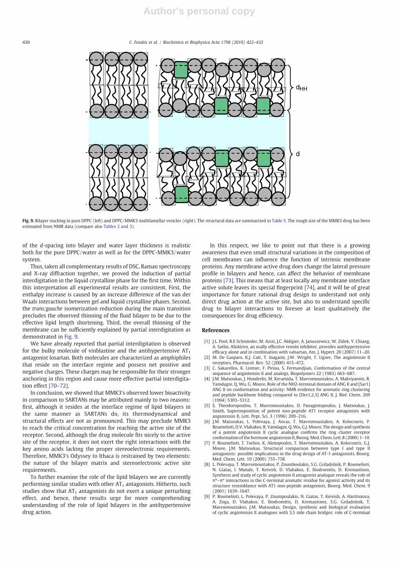
X-ray diffraction results show that incorporated MMK3 enhancesthe inter-membrane distance, d−dHH, in a range from 4 to 7 Å (seeTable 5), inducing some additional steric repulsion betweenadjacent membranes. Possibly, throughout its interfacial activity itsoftens the bilayer and hence causes increased undulation of themembrane. This view is further supported by the observed increaseof the root mean square fluctuation σ especially in the gel phase(25 °C). The analysis of membrane thickness reveals a differentpicture, here the main changes take place in the fluid lamellarphase, i.e. the bilayer reduces about 4 Å in the presence of MMK3. Apossible interpretation is outlined in Fig. 9. Throughout the insertionof MMK3 into on leaflet of the membrane voids are induced thatneed to be filled by lipids of the opposite leaflet. This in turn causesneighboring lipid molecules to interdigitate partially. We note thatthe scheme bases on experimental X-ray data, i.e. the decomposition
Fig. 8. X-ray scattering curves of DPPC/MMK3multilamellar vesicles at 25 °C (A) and 50 °C (B), and their corresponding electron density profiles (C), (D). (Top) The full red lines givethe global fit to the data. (Bottom) dHH defines the head to head-group distance and d the lattice repeat distance. For clarity in panel D two lipidmolecule models are superimposed tothe electron density profile of the bilayer. The most significant structural parameters are summarized in Table 5.
Table 5Structural data on pure DPPC bilayers and DPPC with 20 mol% MMK3 (x=0.20).
DPPC (20 °C) DPPC (50 °C) DPPC/MMK3(25 °C)
DPPC/MMK3(50 °C)
d (Å) 63.5a 67.0a 67.7 64.7dHH (Å) 44.2a 38.3a 42 33d−dHH (Å) 19.3a 28.7a 26 32σ (Å) 1b 6c 3 7
a Structural data taken from the review [67].b Estimated value from global data analysis (data not shown).c Data concerning the root mean square fluctuation in pure DPPC rely on data from
[65,66].
429C. Fotakis et al. / Biochimica et Biophysica Acta 1798 (2010) 422–432
Author's personal copy
of the d-spacing into bilayer and water layer thickness is realisticboth for the pure DPPC/water as well as for the DPPC-MMK3/watersystem.
Thus, taken all complementary results of DSC, Raman spectroscopyand X-ray diffraction together, we proved the induction of partialinterdigitation in the liquid crystalline phase for the first time. Withinthis interpretation all experimental results are consistent. First, theenthalpy increase is caused by an increase difference of the van derWaals interactions between gel and liquid crystalline phases. Second,the trans:gauche isomerization reduction during the main transitionprecludes the observed thinning of the fluid bilayer to be due to theeffective lipid length shortening. Third, the overall thinning of themembrane can be sufficiently explained by partial interdigitation asdemonstrated in Fig. 9.
We have already reported that partial interdigitation is observedfor the bulky molecule of vinblastine and the antihypertensive AT1antagonist losartan. Both molecules are characterized as amphiphilesthat reside on the interface regime and possess net positive andnegative charges. These charges may be responsible for their strongeranchoring in this region and cause more effective partial interdigita-tion effect [70–72].
In conclusion, we showed that MMK3's observed lower bioactivityin comparison to SARTANs may be attributed mainly to two reasons:first, although it resides at the interface regime of lipid bilayers inthe same manner as SARTANs do, its thermodynamical andstructural effects are not as pronounced. This may preclude MMK3to reach the critical concentration for reaching the active site of thereceptor. Second, although the drug molecule fits nicely to the activesite of the receptor, it does not exert the right interactions with thekey amino acids lacking the proper stereoelectronic requirements.Therefore, MMK3's Odyssey to Ithaca is restrained by two elements:the nature of the bilayer matrix and stereoelectronic active siterequirements.
To further examine the role of the lipid bilayers we are currentlyperforming similar studies with other AT1 antagonists. Hitherto, suchstudies show that AT1 antagonists do not exert a unique perturbingeffect, and hence, these results urge for more comprehendingunderstanding of the role of lipid bilayers in the antihypertensivedrug action.
In this respect, we like to point out that there is a growingawareness that even small structural variations in the composition ofcell membranes can influence the function of intrinsic membraneproteins. Any membrane active drug does change the lateral pressureprofile in bilayers and hence, can affect the behavior of membraneproteins [73]. This means that at least locally any membrane interfaceactive solute leaves its special fingerprint [74], and it will be of greatimportance for future rational drug design to understand not onlydirect drug action at the active site, but also to understand specificdrug to bilayer interactions to foresee at least qualitatively theconsequences for drug efficiency.
References
[1] J.L. Pool, R.E Schmieder, M. Azizi, J.C. Aldigier, A. Januszewicz, W. Zidek, Y. Chiang,A. Satlin, Aliskiren, an orally effective rennin inhibitor, provides antihypertensiveefficacy alone and in combination with valsartan, Am. J. Hypert. 20 (2007) 11–20.
[2] M. De Gasparo, K.J. Catt, T. Inagami, J.W. Wright, T. Ugner, The angiotensin IIreceptors, Pharmacol. Rev. 52 (2000) 415–472.
[3] C. Sakarellos, K. Lintner, F. Piriou, S. Fermandjian, Conformation of the centralsequence of angiotensin II and analogs, Biopolymers 22 (1983) 663–687.
[4] J.M. Matsoukas, J. Hondrelis, M. Keramida, T. Mavromoustakos, A. Makriyannis, R.Yamdagni, Q.Wu, G. Moore, Role of the NH2-terminal domain of ANG II and [Sar1]ANG II on conformation and activity: NMR evidence for aromatic ring clusteringand peptide backbone folding compared to [Des1,2,3] ANG II, J. Biol. Chem. 269(1994) 5303–5312.
[5] E. Theodoropoulou, T. Mavromoustakos, D. Panagiotopoulos, J. Matsoukas, J.Smith, Superimposition of potent non-peptide AT1 receptor antagonists withangiotensin II, Lett. Pept. Sci. 3 (1996) 209–216.
[6] J.M. Matsoukas, L. Polevaya, J. Ancas, T. Mavromoustakos, A. Kolocouris, P.Roumelioti, D.V. Vlahakos, R. Yamdagni, Q.Wu, G.J.Moore, The design and synthesisof a potent angiotensin II cyclic analogue confirms the ring cluster receptorconformationof thehormone angiotensin II, Bioorg.Med. Chem. Lett. 8 (2000)1–10.
[7] P. Roumelioti, T. Tselios, K. Alexopoulos, T. Mavromoustakos, A. Kolocouris, G.J.Moore, J.M. Matsoukas, Structural comparison between type I and type IIantagonists: possible implications in the drug design of AT-1 antagonists, Bioorg.Med. Chem. Lett. 10 (2000) 755–758.
[8] L. Polevaya, T. Mavromoustakos, P. Zoumboulakis, S.G. Grdadolnik, P. Roumelioti,N. Giatas, I. Mutule, T. Keivish, D. Vlahakos, E. Iliodromitis, D. Kremastinos,Synthesis and study of cyclic angiotensin II antagonist analogue reveals the role ofπ⁎–π⁎ interactions in the C-terminal aromatic residue for agonist activity and itsstructure resemblance with AT1 non-peptide antagonists, Bioorg. Med. Chem. 9(2001) 1639–1647.
[9] P. Roumelioti, L. Polevaya, P. Zoumpoulakis, N. Giatas, T. Keivish, A. Haritonova,A. Zoga, D. Vlahakos, E. Iliodromitis, D. Kremastinos, S.G. Grdadolnik, T.Mavromoustakos, J.M. Matsoukas, Design, synthesis and biological evaluationof cyclic angiotensin II analogues with 3,5 side chain bridges: role of C-terminal
Fig. 9. Bilayer stacking in pure DPPC (left) and DPPC/MMK3multilamellar vesicles (right). The structural data are summarized in Table 5. The rough size of the MMK3 drug has beenestimated from NMR data (compare also Tables 2 and 3).
430 C. Fotakis et al. / Biochimica et Biophysica Acta 1798 (2010) 422–432

Author's personal copy
aromatic residue and ring cluster for activity and implications in the drug designof AT1 non peptide antagonists, Bioorg. Med. Chem. Lett. 12 (2002) 2627–2633.
[10] M.A.C. Preto, A.M. Hermani, L.S. Maia, T. Mavromoustakos, M.J. Ramos, Moleculardynamics simulations of angiotensin II in aqueous and dimethylsulfoxideenvironments, J. Phys. Chem. B 109 (2005) 17743–17751.
[11] J.M. Matsoukas, G. Agelis, A. Wahhab, J. Hondrelis, D. Panagiotopoulos, R.Yamdagni, Q. Wu, T. Mavromoustakos, H.L.S. Maia, R. Ganter, G.J. Moore,Differences in backbone structure between angiotensin II agonists and type Iantagonists, J. Med. Chem. 38 (1995) 4660–4669.
[12] T. Mavromoustakos, E. Theodoropoulou, C. Dimitriou, J. Matsoukas, D. Panagio-topoulos, A. Makriyannis, Interactions of angiotensin II with membranes using acombination of differential scanning calorimetry and 31P-NMR spectroscopy, LettPept. Sci. 3 (1996) 175–180.
[13] T. Mavromoustakos, A. Kolocouris, M. Zervou, P. Roumelioti, J. Matsoukas, R.Weisemann, An effort to understand the molecular basis of hypertension throughthe study of conformational analysis of losartan and sarmesin using a combinationof Nuclear Magnetic Resonance spectroscopy and theoretical calculations, J. Med.Chem. 42 (1999) 1714–1722.
[14] P. Zoumpoulakis, S.G. Grdadolnik, J.M. Matsoukas, T. Mavromoustakos, Structureelucidation and conformational properties of eprosartan a non peptide Angio-tensin II AT1 antagonist, J. Pharm. Biomed. Anal. 28 (2002) 125–135.
[15] P. Zoumpoulakis, A. Zoga, P. Roumelioti, N. Giatas, S.G. Grdadolnik, E. Iliodromitis,D. Vlahakos, D. Kremastinos, J.M. Matsoukas, T. Mavromoustakos, Conformationaland biological studies for a pair of novel synthetic AT1 antagonists. Stereo-electronic requirements for antihypertensive efficacy, J. Pharm. Biomed. Anal. 31(2003) 833–844.
[16] P. Zoumpoulakis, A. Politi, S.G. Grdadolnik, J.M. Matsoukas, T. Mavromoustakos,Structure elucidation and conformational study of V8. A novel synthetic nonpeptide AT1 antagonist, J. Pharm. Biomed. Anal. 40 (2006) 1097–1104.
[17] T. Mavromoustakos, P. Zoumpoulakis, I. Kyrikou, A. Zoga, E. Siapi, M. Zervou, I.Daliani, D. Dimitriou, A. Pitsas, C. Kamoutsis, P. Laggner, Efforts to understand themolecular basis of hypertension trough drug:membrane interactions, Curr. Top.Med. Chem. 4 (2004) 445–459.
[18] T. Mavromoustakos, M. Zervou, P. Zoumpoulakis, I. Kyrikou, N.P. Benetis, L.Polevaya, P. Roumelioti, N. Giatas, A. Zoga, P.M. Minakakis, A. Kolocouris, D.Vlahakos, S.G. Grdadolnik, J.M Matsoukas, Conformation and bioactivity. Designand discovery of novel antihypertensive drugs, Curr. Top. Med. Chem. 4 (2004)385–401.
[19] P.Moutevelis-Minakakis,M. Gianni, H. Stougiannou, P. Zoumpoulakis, A. Zoga, A.D.Vlahakos, E. Iliodromitis, T. Mavromoustakos, Design and synthesis of novelantihypertensive drugs, Bioorg. Med. Chem. Lett 13 (2003) 1737–1740.
[20] T. Mavromoustakos, P.M. Minakakis, C.G. Kokotos, P. Kontogianni, A. Politi, P.Zoumpoulakis, J. Findlay, A. Cox, A. Balmforth, A. Zoga, E. Iliodromitis, Synthesis,binding studies, and in vivo biological evaluation of novel non-peptideantihypertensive analogs, Bioorg. Med. Chem. 14 (2006) 4353–4360.
[21] P. Zoumpoulakis, I. Daliani, M. Zervou, I. Kyrikou, E. Siapi, G. Lamprinidis, E.Mikros, T. Mavromoustakos, Losartan's molecular basis of interaction withmembranes and AT1 receptor, Chem. Phys. Lipids 125 (2003) 13–25.
[22] L.G. Herbette, Pharmacokinetic and pharmacodynamic design of lipophilic drugsbased on a structural model for drug-interactions with biological membranes,J. Pestic. Sci. 35 (1992) 363–368.
[23] H.S Young, V. Skita, R.P. Mason, L.G. Herbette, Molecular-basis for the inhibition of1,4-dihydropyridine calcium-channel drugs binding to their receptors by a non-specific site interaction mechanism, Biophys. J. 61 (1992) 1244–1255.
[24] D.G. Rhodes, R. Newton, R. Butler, L. Herbette, Equilibrium and kinetic studies ofthe interactions of selmetrol with membrane bilayers, Mol. Pharmacol. 42 (1992)596–602.
[25] T. Mavromoustakos, D.P. Yang, A. Makriyannis, Small angle X-ray diffraction anddifferential scanning calorimetric studies on O-methyl-(−)-Δ8-Tetrahydro-cannabinol and its 5′ iodinated derivative in membrane bilayers, Biochim.Biophys. Acta 1237 (1995) 183–188.
[26] T. Tuccinardi, P.L. Ferrarini, C. Manera, G. Ortore, G. Saccomanni, A. Martinelli,Cannabinoid CB2/CB1 selectivity. Recep-1118 tor modeling and automateddocking analysis, J. Med. Chem. 49 (2006) 984–994.
[27] K. Rumiana, M. Caffrey, Phases and phase transitions of the phosphatidylcholines,Biochim. Biophys. Acta 1376 (1998) 91–145.
[28] S. Tristram-Nagle, M.C. Wiener, C.P. Yang, J.F. Nagle, Kinetics of the subtransitionin diplamitoylphosphatidylcholine dispersions, Biochemistry 26 (1987)4288–4294.
[29] M.D. Houslay, K.K. Stanley, Dynamics of Biological Membranes, John Wiley andSons, New York, 1982.
[30] R.P. Rand, D. Chapman, K. Larsson, Tilted hydrocarbon chains of dipalmitoyllecithin become perpendicular to the bilayer before melting, Biophys. J. 15 (1975)1117–1124.
[31] J. Stamatoff, B. Feuer, H.J. Guggenheim, G. Tellez, T. Yamane, Amplitude of ripplingin the PB phase of dipalmitoylphosphatidylcholine bilayers, Biophys. J. 38 (1982)217–226.
[32] T.J. McIntosh, Differences in hydrocarbon chain tilt between hydrated phospha-tidylethanolamine and phosphatidylcholine bilayers: a molecular packing model,Biophys. J. 29 (1980) 237–246.
[33] N.B. Colthup, L.H. Daly, S.E. Wiberley, Introduction to Infrared and RamanSpectroscopy, Third Ed.Academic Press, New York, 1990.
[34] C. Huang, J.R. Lapides, I.W. Levin, Phase-transition behaviour of saturated,symmetric chain phospholipid bilayers dispersions determined by Ramanspectroscopy: correlation between spectral and thermodynamic parameters,J. Am. Chem. Soc. 104 (1982) 5926–5930.
[35] I.W. Levin, E.N. Lewis, Fourier transform Raman spectroscopy of biologicalmaterials, Anal. Chem. 62 (1990) 1101A–1111A.
[36] G. Pabst, M. Rappolt, H. Amenitsch, P. Laggner, Structural information frommultilamellar liposomes at full hydration: full q-range fitting with high qualityX-ray data, Phys. Rev. E 62 (2000) 4000–4009.
[37] T.J. Netticadan, T.F. Ashavaid, K.G. Nair, Characterisation of the canine cardiacsarcolemma in experimental mzocardial ischemia, Indian J. Clin. Biochem. 12 (1)(1997) 49–54.
[38] M. Trumbore, W. Chester, J. Moring, D. Rhodes, Structure and location ofamiodarone in a membrane bilayer as determined by molecular mechanics andquantitative X-ray diffraction, Biophys. J. 54 (1988) 535–543.
[39] C. Griesinger, R.R. Ernst, Frequency offset effects and their elimination in NMRrotating-frame cross-relaxation spectroscopy, J. Magn. Reson. 75 (1987) 261–271.
[40] Sybyl Molecular Modeling Software Package ver. 6.8; Tripos Inc.: St. Louis, MO63144, 2001.
[41] A.W. Schuettelkopf, D.M.F. van Aalten, A tool 1049 for high-throughputcrystallography of protein–ligand complexes, Acta Cryst.D 60 (2004) 1355–1363.
[42] M. Patra, M. Karttunen, M. Hyvönen, E. Falck, P. Vattulainen, Lipid bilayers drivento a wrong lane in molecular dynamics simulations by subtle changes in long-range electrostatic interactions, J. Phys. Chem. B 108 (2004) 4485–4494.
[43] M. Patra, M. Karttunen, M. Hyvonen, E. Falck, P. Lindqvist, I. Vattulainen, Moleculardynamics simulations of lipid bilayers: major artifacts due to truncatingelectrostatic interactions, Biophys. J. 84 (2003) 3636–3645.
[44] E. Lindhal, B. Hess, D. van der Spoel, GROMACS 3.0: a package for molecularsimulation and trajectory analysis, J. Mol. Model. 7 (2001) 306–317.
[45] W.F. van Gunsteren, S.R. Billeter, A.A. Eising, P.H. Hunenberger, P. Kruger, A.E.Mark, W.R.P. Scott, I.G. Tironi, Biomolecular simulation: the GROMOS96 manualand user guide, Vdf Hochschulverlag AG an der ETH Zurich, Zurich, 1996.
[46] H.J.C. Berendsen, J.P.M. Postma,W.F. van Gunsteren, A. Dinola, J.R. Haak, Moleculardynamics with coupling to an external bath, J. Comput. Chem. 81 (1984)3684–3690.
[47] U. Essmann, L. Perera, M.L. Berkowitz, T. Darden, H. Lee, L.G. Pedersen, A smoothparticle mesh Ewald method, J. Chem. Phys. 103 (1995) 8577–8592.
[48] M. Parrinello, A. Rahman, Polymorphic transitions in single crystals: a newmolecular dynamics method, Jpn. J. Appl. Phys. 52 (1981) 7182–7190.
[49] B. Hess, H. Bekker, H.J.C. Berendsen, J.G.E.M. Fraaije, LINCS: a linear constraintsolver for molecular simulations, J. Comput. Chem. 18 (1997) 1463–1472.
[50] W. Humphrey, A. Dalke, K. Schulten, VMD: visual molecular dynamics, J. Mol.Graph. 14 (1996) 33–38.
[51] P. Laggner, H. Mio, SWAX, a dual-detector camera for simulations small and wide-angle X-ray diffraction in polymer and liquid crystal research, Nucl. Instrum.Methods, Phys. Res. A 323 (1992) 86–90.
[52] G. Pabst, M. Rappolt, H. Amenitsch, P. Laggner, Structural information formultilamellar liposomes at full hydration: full q-range fitting with high-qualityX-ray data, Phys. Rev. E 62 (2000) 4000–4009.
[53] A. Caillé, Remarques sur la diffusion des rayons X dans les smectiques, A. C. R.Acad. Sci. Sér. B 274 (1972) 891–893.
[54] R. Zhang, N.S. Tristram, W. Sun, R.L. Headrick, T.C. Irving, R.M. Suter, J.F. Nagle,Small-angle X-ray scattering from lipid bilayers is well described by modifiedCaillé theory but not by paracrystalline theory, Biophys. J. 70 (1996) 349–357.
[55] R. Hosemann, S.N. Bagchi, Direct Analysis of Diffraction by Matter, North-Hollandpublishing Co, Amsterdam, 1962.
[56] G. Pabst, Global properties of biomimetic membranes: perspectives on molecularfeatures, Biophys. Rev. Lett. 1 (2006) 57–84.
[57] A. Siarheyeva, J.J. Lopez, C. Glaubitz, Localization of multidrug transportersubstrates with model membranes, Biochemistry 45 (2006) 6203–6211.
[58] T. Mavromoustakos, M. Zervou, P. Zoumpoulakis. Design and discovery of novelantihypertensive drugs through conformation and bioactivity studies. BenthamScience Publishers Ltd. Co-Editors:Atta-ur-Rahman, Allen B. Reitz. AssociateEditors: M. Iqbal Choundhary, Cheryl P. Kordik. Ur Frontiers in MedicinalChemistry 3 (2006), 87–113.
[59] J.R. Silvius, M. Lyons, P.L Yeagle, T.J. O'Leary, Thermotropic properties of bilayerscontaining branched-chain phospholipids, calorimetric, Raman and 31P NMRstudies, Biochemistry 24 (1985) 5388–5395.
[60] T.J. O' Leary, P.D. Ross, I.W Levin, Effects of anaesthetic and nonanaestheticsteroids on dipalmitoylphosphatidylcholine liposome: a calorimetric and Ramanspectroscopic investigation, Biochemistry 23 (1984) 4636–4641.
[61] T.J. O' Leary, I.W. Levin, Raman spectroscopic study of an interdigitated lipidbilayer dipalmitoylphosphatidylcholine dispersed in glycerol, Biochim. Biophys.Acta 776 (1984) 185–189.
[62] M.C. Wiener, R.M. Suter, J.F. Nagle, Structure of the fully hydrated gel phase ofdiplamitoylphosphatidylcholine, Biophys. J. 55 (1989) 315–325.
[63] J.W. Torbet, M.H.F. Wilkins, X-ray diffraction studies of lecithin bilayers, J. Theor.Biol. 62 (1976) 447–458.
[64] T.J. McIntosh, S.A. Simon, Hydration force and bilayer deformation: a reevaluation,Biochemistry 25 (1986) 4048–4066.
[65] H.I. Petrache, N. Gouliaev, N.S. Tristram, R. Zhang, R.M. Suter, J.F. Nagle,Interbilayer interactions from high-resolution X-ray scattering, Phys. Rev. E 57(1998) 7014–7024.
[66] G. Pabst, H. Amenitsch, D.P. Kharakoz, P. Laggner, M. Rappolt, Structure andfluctuations of phosphatidylcholines in the vicinity of the main phase transition,Phys. Rev. E 70 (2004) 021908.
[67] J.F. Nagle, N.S. Tristram, Structure of lipid bilayers, Biochim. Biophys. Acta 1469(2000) 159–195.
[68] C. Potamitis,M Zervou, V. Katsiaras, P Zoumpoulakis, S. Durdagi,M. Papadopoulos, J.Hayes, S. Grdadolnik, I. Kyrikou, D. Argyropoulos, G. Vatougia, T. Mavromoustakos,
431C. Fotakis et al. / Biochimica et Biophysica Acta 1798 (2010) 422–432

Author's personal copy
Antihypertensive drug valsartan in solution and at the AT1 receptor: conforma-tional analysis, dynamic NMR spectroscopy, in silico docking and moleculardynamics simulations, J. Chem. Inf. Mod. 49 (2009) 726–739.
[69] C. Photakis, D. Christodouleas, P. Chatzigeorgiou, M. Zervou, N.P. Benetis, K. Vyras,T. Mavromoustakos, Application of a novel CP-31P NMRmethodology to study thepossible interdigitation effect of losartan in phospholipids bilayers. Comparisonwith Raman spectroscopy data, Biophys. J. 96 (2009) 2227–2236.
[70] I. Kyrikou, I. Daliani, T. Mavromoustakos, H. Maswadeh, C. Demetzos, S.Xatziantoniou, S. Giatrellis, G. Nounesis, The modulation of thermal and dynamicproperties of vinblastine by cholesterol in membrane bilayers, Biochim. Biophys.Acta 1661 (2004) 1–8.
[71] H. Maswadeh, C. Demetzos, I. Daliani, I. Kyrikou, T. Mavromoustakos, A. Tsortos, G.Nounesis, A molecular basis explanation of the dynamic and thermal effects ofvinblastine sulfate upon dipalmitoylphosphatidylcholine bilayer membranes,Biochim. Biophys. Acta 1567 (2002) 49–55.
[72] I. Kyrikou, S. Xadjikakou, D.D. Kovala, K. Viras, T. Mavromoustakos, Effects of nonsteroid anti-inflammatory drugs in membrane bilayers containing cholesterol,Chem. Phys. Lipids 132 (2004) 157–169.
[73] R.S. Cantor, The lateral pressure profile in bilayers, Biophys. J. 76 (1999) 2625–2639.[74] C. Xing, O.H.S. Ollila, I. Vattulainen, R. Olli, Asymmetric nature of lateral pressure
profiles in supported lipid membranes and its implications for membrane proteinfunctions, Soft Matter 5 (2009) 3258–3261.
432 C. Fotakis et al. / Biochimica et Biophysica Acta 1798 (2010) 422–432