Interaction between 24-hydroxycholesterol, oxidative stress, and amyloid-β in amplifying neuronal...
15
Interaction between 24-hydroxycholesterol, oxidative stress, and amyloid-b in amplifying neuronal damage in Alzheimer’s disease: three partners in crime Paola Gamba, 1 * Gabriella Leonarduzzi, 1 * Elena Tamagno, 2 Michela Guglielmotto, 2 Gabriella Testa, 1 Barbara Sottero, 1 Simona Gargiulo, 1 Fiorella Biasi, 1 Alessandro Mauro, 3 Jose ´ Vin ˜a 4 and Giuseppe Poli 1 1 Department of Clinical and Biological Sciences, Faculty of Medicine San Luigi Gonzaga, University of Turin, Turin, Italy 2 Department of Experimental Medicine and Oncology, University of Turin, Turin, Italy 3 Division of Neurology and Neurorehabilitation, IRCCS Istituto Auxologico Italiano, Verbania, Italy and Department of Neurosciences, University of Turin, Turin, Italy 4 Department of Physiology, Faculty of Medicine, University of Valencia, Valencia, Spain Summary All three cholesterol oxidation products implicated thus far in the pathogenesis of Alzheimer’s disease, 7b-hy- droxycholesterol, 24-hydroxycholesterol, and 27-hydro- xycholesterol, markedly enhance the binding of amyloid- beta (Ab) to human differentiated neuronal cell lines (SK-N-BE and NT-2) by up-regulating net expression and synthesis of CD36 and b1-integrin receptors. However, only 24-hydroxycholesterol markedly potentiates the pro- apoptotic and pro-necrogenic effects of Ab 1–42 peptide on these cells: 7b-hydroxycholesterol and 27-hydroxycholes- terol, like unoxidized cholesterol, show no potentiating effect. This peculiar behavior of 24-hydroxycholesterol at physiologic concentrations (1 lM) depends on its strong enhancement of the intracellular generation of NADPH oxidase–dependent reactive oxygen species (ROS), mainly H 2 O 2 , and the consequent impairment of neuronal cell redox equilibrium, measured in terms of the GSSG ⁄ GSH ratio. Cell incubation with antioxidants quercetin or geni- stein prevents 24-hydroxycholesterol’s pro-oxidant effect and potentiation of Ab-induced necrosis and apoptosis. Thus, the presence of 24-hydroxycholesterol in the close vicinity of amyloid plaques appears to enhance the adhe- sion of large amounts of Ab to the plasma membrane of neurons and then to amplify the neurotoxic action of Ab by locally increasing ROS steady-state levels. This report further supports a primary involvement of altered brain cholesterol metabolism in the complex pathogenesis of Alzheimer’s disease. Key words: cholesterol; Alzheimer’s disease; oxysterols; amyloid-b; oxidative stress; neurotoxicity. Introduction Alzheimer’s disease (AD), a protein misfolding disorder, is the commonest type of dementia in developed countries; it is char- acterized by progressive neuronal degeneration, gliosis, and the accumulation of intracellular inclusions (neurofibrillary tangles) and extracellular deposits of amyloid-b (senile plaques) in the discrete regions of the basal forebrain, hippocampus, and asso- ciation cortices (Selkoe, 1991; Hashimoto et al., 2003). Amy- loid-b (Ab) is also deposited in the cerebral vessels. Accumulation of Ab peptides is thought to be an early and caus- ative event in AD pathogenesis and increases markedly during disease progression (Selkoe, 1994). Several intertwined causative factors lead to the development of AD, including increased release and polymerization of Ab, hypertension, advanced glycation end-products, oxidative stress, inflammation, and hypercholesterolemia (Querfurth & LaFerla, 2010). At least three important lines of evidence have implicated cholesterol in AD: (i) hypercholesterolemia is unani- mously recognized to be a risk factor for sporadic AD, a form that accounts for the great majority of cases (Puglielli et al., 2003; Panza et al., 2007); (ii) epidemiological studies have shown homozygosity for the APO-E4 allele (APO-E e4 genotype) to be associated with an increased risk of AD (Corder et al., 1993; Evans et al., 2004); (iii) feeding cholesterol and copper to rabbits produces some of the pathological signs of AD including amyloid-like plaques (Sparks & Schreurs, 2003). In addition, early epidemiological studies indicated that cholesterol-lowering agents belonging to the family of statins reduce the prevalence of AD (Jick et al., 2000), a conclusion not yet fully accepted, because of contradictory results of prospective clinical studies (Kandiah & Feldman, 2009). The brain is the organ with the highest concentration of cho- lesterol, which is essential for its normal function; most of the cholesterol is present in the free form and derives from de novo synthesis by astrocytes, as plasma lipoproteins cannot cross the blood–brain barrier (Puglielli et al., 2003). Cholesterol can only efflux to the plasma in normal conditions if it is transformed into oxysterols, cholesterol oxidation products that are thus impor- Correspondence Giuseppe Poli, MD, Department of Clinical and Biological Sciences, Univer- sity of Torino, San Luigi Gonzaga Hospital, Regione Gonzole 10, 10043 Orbassano, Torino, Italy. Tel.: +39 011 6705422; fax: +39 011 6705424; e-mail: [email protected] *These authors contributed equally to the work. Accepted for publication 4 January 2011 ª 2011 The Authors Aging Cell ª 2011 Blackwell Publishing Ltd/Anatomical Society of Great Britain and Ireland 403 Aging Cell (2011) 10, pp403–417 Doi: 10.1111/j.1474-9726.2011.00681.x Aging Cell
-
Upload
independent -
Category
Documents
-
view
2 -
download
0
Transcript of Interaction between 24-hydroxycholesterol, oxidative stress, and amyloid-β in amplifying neuronal...

Interaction between 24-hydroxycholesterol, oxidativestress, and amyloid-b in amplifying neuronal damagein Alzheimer’s disease: three partners in crime
Paola Gamba,1* Gabriella Leonarduzzi,1* ElenaTamagno,2 Michela Guglielmotto,2 Gabriella Testa,1
Barbara Sottero,1 Simona Gargiulo,1 Fiorella Biasi,1
Alessandro Mauro,3 Jose Vina4 and Giuseppe Poli1
1Department of Clinical and Biological Sciences, Faculty of
Medicine San Luigi Gonzaga, University of Turin, Turin, Italy2Department of Experimental Medicine and Oncology, University
of Turin, Turin, Italy3Division of Neurology and Neurorehabilitation, IRCCS Istituto
Auxologico Italiano, Verbania, Italy and Department of
Neurosciences, University of Turin, Turin, Italy4Department of Physiology, Faculty of Medicine, University of
Valencia, Valencia, Spain
Summary
All three cholesterol oxidation products implicated thus
far in the pathogenesis of Alzheimer’s disease, 7b-hy-
droxycholesterol, 24-hydroxycholesterol, and 27-hydro-
xycholesterol, markedly enhance the binding of amyloid-
beta (Ab) to human differentiated neuronal cell lines
(SK-N-BE and NT-2) by up-regulating net expression and
synthesis of CD36 and b1-integrin receptors. However,
only 24-hydroxycholesterol markedly potentiates the pro-
apoptotic and pro-necrogenic effects of Ab1–42 peptide on
these cells: 7b-hydroxycholesterol and 27-hydroxycholes-
terol, like unoxidized cholesterol, show no potentiating
effect. This peculiar behavior of 24-hydroxycholesterol at
physiologic concentrations (1 lM) depends on its strong
enhancement of the intracellular generation of NADPH
oxidase–dependent reactive oxygen species (ROS), mainly
H2O2, and the consequent impairment of neuronal cell
redox equilibrium, measured in terms of the GSSG ⁄ GSH
ratio. Cell incubation with antioxidants quercetin or geni-
stein prevents 24-hydroxycholesterol’s pro-oxidant effect
and potentiation of Ab-induced necrosis and apoptosis.
Thus, the presence of 24-hydroxycholesterol in the close
vicinity of amyloid plaques appears to enhance the adhe-
sion of large amounts of Ab to the plasma membrane of
neurons and then to amplify the neurotoxic action of Ab
by locally increasing ROS steady-state levels. This report
further supports a primary involvement of altered brain
cholesterol metabolism in the complex pathogenesis of
Alzheimer’s disease.
Key words: cholesterol; Alzheimer’s disease; oxysterols;
amyloid-b; oxidative stress; neurotoxicity.
Introduction
Alzheimer’s disease (AD), a protein misfolding disorder, is the
commonest type of dementia in developed countries; it is char-
acterized by progressive neuronal degeneration, gliosis, and the
accumulation of intracellular inclusions (neurofibrillary tangles)
and extracellular deposits of amyloid-b (senile plaques) in the
discrete regions of the basal forebrain, hippocampus, and asso-
ciation cortices (Selkoe, 1991; Hashimoto et al., 2003). Amy-
loid-b (Ab) is also deposited in the cerebral vessels.
Accumulation of Ab peptides is thought to be an early and caus-
ative event in AD pathogenesis and increases markedly during
disease progression (Selkoe, 1994).
Several intertwined causative factors lead to the development
of AD, including increased release and polymerization of Ab,
hypertension, advanced glycation end-products, oxidative
stress, inflammation, and hypercholesterolemia (Querfurth &
LaFerla, 2010). At least three important lines of evidence have
implicated cholesterol in AD: (i) hypercholesterolemia is unani-
mously recognized to be a risk factor for sporadic AD, a form
that accounts for the great majority of cases (Puglielli et al.,
2003; Panza et al., 2007); (ii) epidemiological studies have
shown homozygosity for the APO-E4 allele (APO-E e4 genotype)
to be associated with an increased risk of AD (Corder et al.,
1993; Evans et al., 2004); (iii) feeding cholesterol and copper to
rabbits produces some of the pathological signs of AD including
amyloid-like plaques (Sparks & Schreurs, 2003). In addition, early
epidemiological studies indicated that cholesterol-lowering
agents belonging to the family of statins reduce the prevalence
of AD (Jick et al., 2000), a conclusion not yet fully accepted,
because of contradictory results of prospective clinical studies
(Kandiah & Feldman, 2009).
The brain is the organ with the highest concentration of cho-
lesterol, which is essential for its normal function; most of the
cholesterol is present in the free form and derives from de novo
synthesis by astrocytes, as plasma lipoproteins cannot cross the
blood–brain barrier (Puglielli et al., 2003). Cholesterol can only
efflux to the plasma in normal conditions if it is transformed into
oxysterols, cholesterol oxidation products that are thus impor-
Correspondence
Giuseppe Poli, MD, Department of Clinical and Biological Sciences, Univer-
sity of Torino, San Luigi Gonzaga Hospital, Regione Gonzole 10, 10043
Orbassano, Torino, Italy. Tel.: +39 011 6705422; fax: +39 011 6705424;
e-mail: [email protected]
*These authors contributed equally to the work.
Accepted for publication 4 January 2011
ª 2011 The AuthorsAging Cell ª 2011 Blackwell Publishing Ltd/Anatomical Society of Great Britain and Ireland
403
Aging Cell (2011) 10, pp403–417 Doi: 10.1111/j.1474-9726.2011.00681.xAg
ing
Cell

tant to balance the local synthesis of sterols (Lutjohann et al.,
1996).
The major oxysterol involved in this excretion mechanism
appears to be 24-hydroxycholesterol (24-OH), also known as
cerebrosterol, which is produced almost exclusively in the brain
by cholesterol 24-hydroxylase (cytochrome P450-46A1) (Bjork-
hem & Meaney, 2004; Bjorkhem, 2006). Another oxysterol,
27-hydroxycholesterol (27-OH), has been found to be produced
in situ in the brain by the cytochrome P450-27A1, or even, unlike
its parent compound, to flow from the circulation into the brain
(Heverin et al., 2005). A third compound, 7b-hydroxycholesterol
(7b-OH), may derive in the brain from the oxidation of choles-
terol by Ab and, to a lesser extent, by amyloid precursor protein
(Nelson & Alkon, 2005).
Notably, an increased concentration of 24-OH has been
detected in the cerebrospinal fluid of patients with AD
(Schonknecht et al., 2002), while decreased 24-OH and
increased 27-OH are consistently found in autoptic brain sam-
ples from patients with AD (Heverin et al., 2004). Further, an
abnormal pattern of cholesterol hydroxylases has been observed
in the AD brain, with a prominent expression of 24-hydroxylase
in astrocytes and around amyloid plaques (Brown et al., 2004).
Hence, changes in the brain cholesterol ⁄ oxysterol balance, with
pathological accumulation of cholesterol oxides in the Central
Nervous System, may be the missing link between hypercholes-
terolemia and AD.
Oxysterols, which may either originate in the body or derive
from the diet, are 27-carbon molecules produced by the
oxidation of cholesterol. It is widely accepted that both enzy-
matic and nonenzymatic reactions are responsible for oxyster-
ol generation in the body (Leonarduzzi et al., 2002; Poli
et al., 2009). Enzymatic processes mainly involve the side
chain of cholesterol yielding 24-OH and 27-OH. On the con-
trary, oxidation of the sterol nucleus, which primarily occurs
at position 7 or at the 5,6 double bond, is commonly a non-
enzymatic (autooxidative) reaction and generates, among oth-
ers, 7b-OH of probable interest in the pathogenesis of
neurodegenerative diseases. A typical nonenzymatic oxidation
process of cholesterol is that mediated by inflammatory cells
undergoing oxidative burst. Oxysterols have been shown to
exert several in vitro and in vivo biochemical activities of both
physiologic and pathologic relevance (Sottero et al., 2009).
Particularly relevant is the strong pro-inflammatory action
shown by several components of this class of compounds
(Vejux & Lizard, 2009). Thus, inflammation readily generates
oxysterols that in turn up-regulate the flogistic process.
To determine how the oxysterols 24-OH, 27-OH, and 7b-OH
could modulate and perhaps amplify the expression of AD, we
investigated the potential interaction between these cholesterol
oxides and Ab, whose extracellular accumulation in neuritic pla-
ques is one of the hallmarks of AD. A possible manner of interac-
tion could be by enhancing binding to Ab toxic peptides by
vicinal neuronal cells. To date, this event has been demonstrated
to occur for another cell type, namely microglia: human THP-1
monocytes and murine microglial cells have been shown to
readily bind to the fibrillar form of Ab, through a receptor com-
plex involving CD36, a6b1-integrin, and CD47 (Bamberger
et al., 2003).
On the basis of our recent demonstration that certain choles-
terol oxidation products, unlike the parent compound, markedly
up-regulate both the expression and the synthesis of CD36 in
the cells of the macrophage lineage (Leonarduzzi et al., 2008),
we decided to investigate whether oxysterols also exert the
same effect on neuronal cells.
All three cholesterol oxidation products considered strongly
enhanced the binding of Ab to human differentiated neuronal
cell lines (SK-N-BE and NT-2), by strongly up-regulating expres-
sion and synthesis not only of CD36 but also of b1-integrin
receptors. However, only the combination of 24-OH plus the
Ab1–42 peptide exerted net neurotoxicity. The reason for this
selective behavior of 24-OH turned out to be dependent on its
strong pro-oxidant action.
Results
27-OH, 7b-OH, and 24-OH up-regulate the expression
of the CD36 and b1-integrin genes, but not that of
the CD47 gene, in SK-N-BE and NT-2 neuronal cells
The three oxysterols considered as being potentially implicated
in the pathogenesis of Alzheimer’s disease were first checked
for their ability to modulate the CD36 ⁄ b1-integrin ⁄ CD47 recep-
tor complex in two differentiated human neuronal cell lines,
namely SK-N-BE and NT-2 cells.
In SK-N-BE cells, a statistically significant increase (about
1.5 to 2-fold induction, P < 0.001–0.05) in CD36 mRNA and
b1-integrin mRNA levels was evident after 5 h cell incubation
with any of the three oxysterols in biologically compatible con-
centrations (1 lM) compared with control. Of interest, this over-
expression appeared to occur in a synchronous manner and was
not inducible even minimally when oxysterols were replaced by
an identical amount of unoxidized cholesterol (Fig. 1A). A trend
to increased CD36 mRNA and b1-integrin mRNA levels was con-
sistently observed also in NT-2 neuronal cells after 4 h cell incu-
bation with 7b-OH or 24-OH, even if statistically significant
(about 2-fold induction compared with control, P < 0.001 and
P < 0.01) only in the latter case. As regards 27-OH, it appeared
to increase the expression of b1-integrin and not that of CD36
(Fig. 1B). Expression of the CD47 gene was in no case modu-
lated by any of the three oxysterols considered in this study,
neither in SK-N-BE nor in NT-2 cells (Fig. 2).
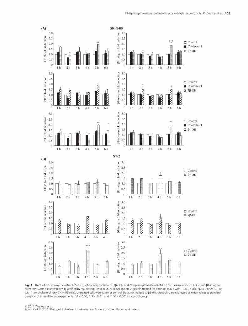
27-OH, 7b-OH, and 24-OH increase CD36 and b1-
integrin synthesis
A statistically significant increase (P < 0.001 and P < 0.05 vs.
control) in the synthesis of the two receptors corresponded to
the over-expression of CD36 and b1-integrin genes, in both
SK-N-BE and NT-2 neuronal cells, measured by Western blotting
after 48 h treatment with 27-OH, 7b-OH, or 24-OH. Of note,
24-Hydroxycholesterol potentiates amyloid-beta neurotoxicity, P. Gamba et al.
ª 2011 The AuthorsAging Cell ª 2011 Blackwell Publishing Ltd/Anatomical Society of Great Britain and Ireland
404
SK-N-BE
** *** Control2.0
2.5
3.0
2.0
2.5
3.0
Cholesterol
27-OH
CD
36 f
old
indu
ctio
n
1 h
β1-i
nteg
rin
fold
indu
ctio
n
CD
36 f
old
indu
ctio
n
β1-i
nteg
rin
fold
indu
ctio
n
CD
36 f
old
indu
ctio
n
β1-i
nteg
rin
fold
indu
ctio
n
CD
36 f
old
indu
ctio
n
β1-i
nteg
rin
fold
indu
ctio
n
CD
36 f
old
indu
ctio
n
β1-i
nteg
rin
fold
indu
ctio
n
CD
36 f
old
indu
ctio
n
β1-i
nteg
rin
fold
indu
ctio
n
0
0.5
1.0
1.5
2.5
3.0
0
0.5
1.0
1.5
2.5
3.0
* * *
0
0.5
1.0
1.5
2.0
3.0
0
0.5
1.0
1.5
2.0
3.0
CholesterolControl
7β-OH
** **
0
0.5
1.0
1.5
2.0
2.5
0
0.5
1.0
1.5
2.0
2.5
CholesterolControl
24-OH
NT-2
Control27-OH
1.5
2.0
2.5
3.0
1.5
2.0
2.5
3.0
0
0.5
1.0
2.0
2.5
3.0
0
0.5
1.0
2.0
2.5
3.0
Control7β-OH
*** **
0
0.5
1.0
1.5
2.5
3.0
0
0.5
1.0
1.5
2.5
3.0
2 h 3 h 4 h 5 h 6 h 1 h 2 h 3 h 4 h 5 h 6 h
1 h 2 h 3 h 4 h 5 h 6 h
1 h 2 h 3 h 4 h 5 h 6 h
1 h 2 h 3 h 4 h 5 h 6 h
1 h 2 h 3 h 4 h 5 h 6 h
1 h 2 h 3 h 4 h 5 h 6 h
1 h 2 h 3 h 4 h 5 h 6 h
1 h 2 h 3 h 4 h 5 h 6 h
1 h 2 h 3 h 4 h 5 h 6 h
1 h 2 h 3 h 4 h 5 h 6 h
1 h 2 h 3 h 4 h 5 h 6 h0
0.5
1.0
1.5
2.0
0
0.5
1.0
1.5
2.0Control24-OH
(A)
(B)
Fig. 1 Effect of 27-hydroxycholesterol (27-OH), 7b-hydroxycholesterol (7b-OH), and 24-hydroxycholesterol (24-OH) on the expression of CD36 and b1-integrin
receptors. Gene expression was quantified by real-time RT–PCR in SK-N-BE (A) and NT-2 (B) cells treated for times up to 6 h with 1 lM 27-OH, 7b-OH, or 24-OH or
with 1 lM cholesterol (only SK-N-BE cells). Untreated cells were taken as control. Data, normalized to b2-microglobulin, are expressed as mean values ± standard
deviation of three different experiments. *P < 0.05, **P < 0.01, and ***P < 0.001 vs. control group.
24-Hydroxycholesterol potentiates amyloid-beta neurotoxicity, P. Gamba et al.
ª 2011 The AuthorsAging Cell ª 2011 Blackwell Publishing Ltd/Anatomical Society of Great Britain and Ireland
405

the effects of the three oxysterols on CD36 and b1-integrin syn-
thesis appeared quantitatively quite similar: about 150–190%
(SK-N-BE) and 300% (NT-2) the increment of CD36 levels and
180–250% (SK-N-BE) and 250% (NT-2) the increment of
b1-integrin levels vs. control taken as 100% (Fig. 3).
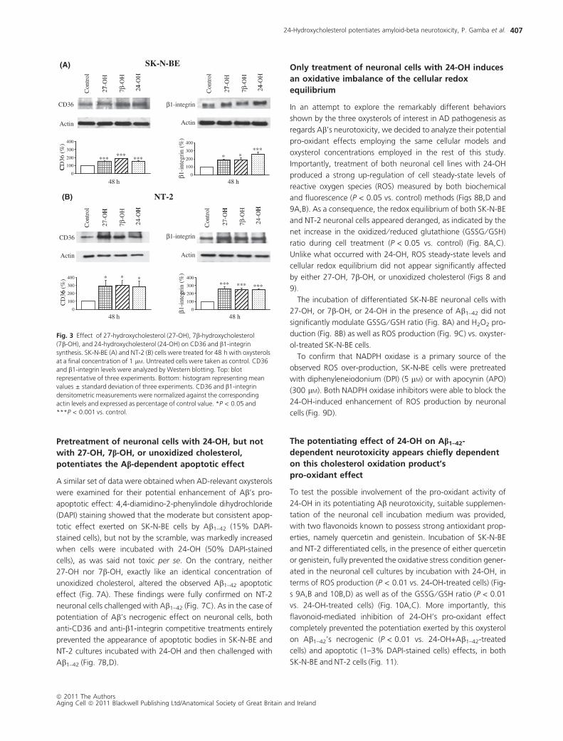
AD-relevant oxysterols strongly enhance the
adhesion of neuronal cells to amyloid-b; the
effect is mediated by up-regulation of CD36
and b1-integrin receptors
The consequence of increased availability of CD36 and b1-inte-
grin receptors, in terms of binding to Ab, was investigated on
SK-N-BE neuronal cells incubated for 48 h in the presence of 27-
OH, 7b-OH, or 24-OH and then challenged for 8 h with the
Ab1–42 peptide. The binding of Ab1–42 (detected by confocal
microscopy) to neuronal cells was greatly stimulated by cell pre-
treatment with any of the three oxysterols vs. that recovered in
cells either untreated or simply incubated with unoxidized cho-
lesterol (Fig. 4). This marked enhancement of Ab1–42 peptide
binding was fully prevented by the addition of either anti-CD36-
or anti-b1-integrin-specific antibodies to the incubation medium
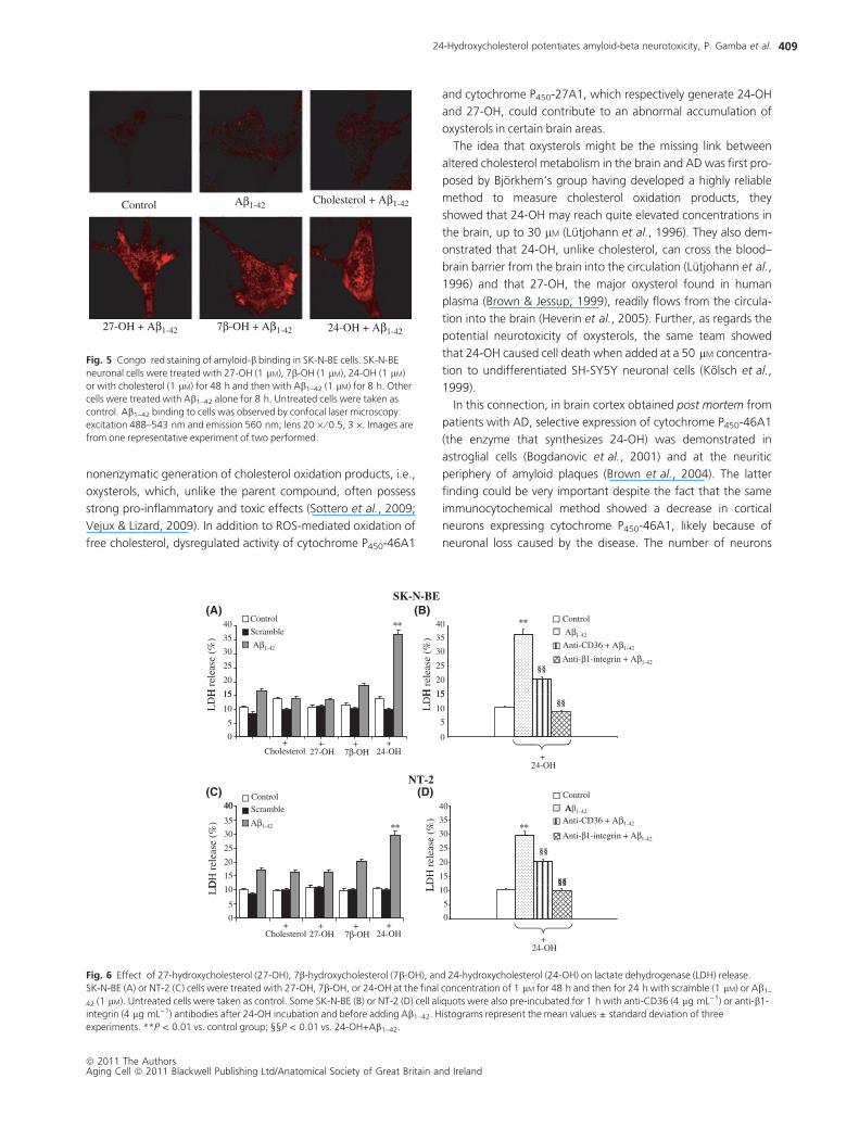
before the challenge with the Ab1–42 peptide (Fig. 4). At Congo
red staining, SK-N-BE neuronal cells treated with the oxysterols
and then with Ab1–42 showed highly stimulated Ab1–42 binding
vs. internal controls (untreated and Ab1–42-treated cells) and vs.
unoxidized cholesterol plus Ab1–42-treated cells; the amyloid
peptide that bound to the cells was mainly concentrated in clus-
ters (Fig. 5).
Different effects of 27-OH, 7b-OH, and 24-OH in
potentiating amyloid-b’s necrogenic effect on
neuronal cells
To determine whether increased binding of amyloid-b to neuro-
nal cells potentiates the peptide’s toxic effects, SK-N-BE and
NT-2 cells, differentiated with retinoic acid, were first incubated
for 48 h with one of the three oxysterols and then challenged
for 24 h with Ab1–42 peptide. At the end of the experiment,
necrosis was measured by the cell release of lactate dehydroge-
nase (LDH): 24-OH, not necrogenic per se, increased about 3- to
3.5-fold (P < 0.01 vs. control) the LDH release induced by Ab1–
42, in both SK-N-BE and NT-2 differentiated cells (Fig. 6A,C). On
the contrary, neither 27-OH, nor 7b-OH, nor unoxidized choles-
terol caused any significant modulation of the induced LDH
release (Fig. 6A,C). As expected, cell treatment with the scram-
ble peptide neither induced nor permitted any change in LDH
release.
Importantly, 24-OH’s marked potentiation of Ab1–42’s necro-
genic effect was significantly quenched (30–40% reduction,
P < 0.01) compared with 24-OH-treated cells, in both neuronal
cell lines, by addition of anti-CD36 antibodies, and was fully
prevented (P < 0.05) by addition of anti-b1-integrin-specific
antibodies (Fig. 6B,D).
NT-2SK-N-BE
CholesterolControl Control
27-OH2.5
3.0
2.5
3.0
CD
47 f
old
indu
ctio
n
1 h
CD
47 f
old
indu
ctio
n
27-OH
0
0.5
1.0
1.5
2.0
0
0.5
1.0
1.5
2.0
CD
47 f
old
indu
ctio
n
CD
47 f
old
indu
ctio
n
CholesterolControl
7β-OH
Control7β-OH
1.0
1.5
2.0
2.5
3.0
1.0
1.5
2.0
2.5
3.0
1 h 1 h
CholesterolControl
24-OH
Control24-OH
0
0.5
0
0.5
2.0
2.5
3.0
2.0
2.5
3.0
v
1 h 1 h
2 h 3 h 4 h 5 h 6 h 1 h 2 h 3 h 4 h 5 h 6 h
2 h 3 h 4 h 5 h 6 h 2 h 3 h 4 h 5 h 6 h
2 h 3 h 4 h 5 h 6 h 2 h 3 h 4 h 5 h 6 h
CD
47 f
old
indu
ctio
n
CD
47 f
old
indu
ctio
n
0
0.5
1.0
1.5
0
0.5
1.0
1.5
(A) (B)
Fig. 2 Effect of 27-hydroxycholesterol (27-OH), 7b-hydroxycholesterol (7b-OH), and 24-hydroxycholesterol (24-OH) on the expression of CD47 receptor. Gene
expression was quantified by real-time RT–PCR in SK-N-BE (A) and NT-2 (B) cells treated up to 6 h with 1 lM 27-OH, 7b-OH, or 24-OH or with 1 lM cholesterol
(only SK-N-BE cells). Untreated cells were taken as control. Data, normalized to b2-microglubulin, are expressed as mean values ± standard deviation of three
different experiments.
24-Hydroxycholesterol potentiates amyloid-beta neurotoxicity, P. Gamba et al.
ª 2011 The AuthorsAging Cell ª 2011 Blackwell Publishing Ltd/Anatomical Society of Great Britain and Ireland
406
Pretreatment of neuronal cells with 24-OH, but not
with 27-OH, 7b-OH, or unoxidized cholesterol,
potentiates the Ab-dependent apoptotic effect
A similar set of data were obtained when AD-relevant oxysterols
were examined for their potential enhancement of Ab’s pro-
apoptotic effect: 4,4-diamidino-2-phenylindole dihydrochloride
(DAPI) staining showed that the moderate but consistent apop-
totic effect exerted on SK-N-BE cells by Ab1–42 (15% DAPI-
stained cells), but not by the scramble, was markedly increased
when cells were incubated with 24-OH (50% DAPI-stained
cells), as was said not toxic per se. On the contrary, neither
27-OH nor 7b-OH, exactly like an identical concentration of
unoxidized cholesterol, altered the observed Ab1–42 apoptotic
effect (Fig. 7A). These findings were fully confirmed on NT-2
neuronal cells challenged with Ab1–42 (Fig. 7C). As in the case of
potentiation of Ab’s necrogenic effect on neuronal cells, both
anti-CD36 and anti-b1-integrin competitive treatments entirely
prevented the appearance of apoptotic bodies in SK-N-BE and
NT-2 cultures incubated with 24-OH and then challenged with
Ab1–42 (Fig. 7B,D).
Only treatment of neuronal cells with 24-OH induces
an oxidative imbalance of the cellular redox
equilibrium
In an attempt to explore the remarkably different behaviors
shown by the three oxysterols of interest in AD pathogenesis as
regards Ab’s neurotoxicity, we decided to analyze their potential
pro-oxidant effects employing the same cellular models and
oxysterol concentrations employed in the rest of this study.
Importantly, treatment of both neuronal cell lines with 24-OH
produced a strong up-regulation of cell steady-state levels of
reactive oxygen species (ROS) measured by both biochemical
and fluorescence (P < 0.05 vs. control) methods (Figs 8B,D and
9A,B). As a consequence, the redox equilibrium of both SK-N-BE
and NT-2 neuronal cells appeared deranged, as indicated by the
net increase in the oxidized ⁄ reduced glutathione (GSSG ⁄ GSH)
ratio during cell treatment (P < 0.05 vs. control) (Fig. 8A,C).
Unlike what occurred with 24-OH, ROS steady-state levels and
cellular redox equilibrium did not appear significantly affected
by either 27-OH, 7b-OH, or unoxidized cholesterol (Figs 8 and
9).
The incubation of differentiated SK-N-BE neuronal cells with
27-OH, or 7b-OH, or 24-OH in the presence of Ab1–42 did not
significantly modulate GSSG ⁄ GSH ratio (Fig. 8A) and H2O2 pro-
duction (Fig. 8B) as well as ROS production (Fig. 9C) vs. oxyster-
ol-treated SK-N-BE cells.
To confirm that NADPH oxidase is a primary source of the
observed ROS over-production, SK-N-BE cells were pretreated
with diphenyleneiodonium (DPI) (5 lM) or with apocynin (APO)
(300 lM). Both NADPH oxidase inhibitors were able to block the
24-OH-induced enhancement of ROS production by neuronal
cells (Fig. 9D).
The potentiating effect of 24-OH on Ab1–42-
dependent neurotoxicity appears chiefly dependent
on this cholesterol oxidation product’s
pro-oxidant effect
To test the possible involvement of the pro-oxidant activity of
24-OH in its potentiating Ab neurotoxicity, suitable supplemen-
tation of the neuronal cell incubation medium was provided,
with two flavonoids known to possess strong antioxidant prop-
erties, namely quercetin and genistein. Incubation of SK-N-BE
and NT-2 differentiated cells, in the presence of either quercetin
or genistein, fully prevented the oxidative stress condition gener-
ated in the neuronal cell cultures by incubation with 24-OH, in
terms of ROS production (P < 0.01 vs. 24-OH-treated cells) (Fig-
s 9A,B and 10B,D) as well as of the GSSG ⁄ GSH ratio (P < 0.01
vs. 24-OH-treated cells) (Fig. 10A,C). More importantly, this
flavonoid-mediated inhibition of 24-OH’s pro-oxidant effect
completely prevented the potentiation exerted by this oxysterol
on Ab1–42’s necrogenic (P < 0.01 vs. 24-OH+Ab1–42-treated
cells) and apoptotic (1–3% DAPI-stained cells) effects, in both
SK-N-BE and NT-2 cells (Fig. 11).
SK-N-BE
7-O
H
4-O
H
7-O
H
4-O
H
CD36
Actin
β1-integrin
Actin
27 2427 7β-O
H
7β-O
H7β
-OH
7β-O
H
24
100
200
300
400
*** ******
CD
36 (
%)
-int
egri
n (%
)
100
200
300
400
* ****
H H
NT-2
H H
0
48 h
C
β1β
48 h0
CD36
27-O
H
24-O
H
β1-integrin
Con
trol
Con
trol
Con
trol
Con
trol
27-O
H
24-O
H
Actin
300
400 ***
6 (%
)
Actin
300
400
*** *** ***
grin
(%
)
0
100
200
48 h
CD
36
0
100
200
48 h
1-in
teg
(A)
(B)
Fig. 3 Effect of 27-hydroxycholesterol (27-OH), 7b-hydroxycholesterol
(7b-OH), and 24-hydroxycholesterol (24-OH) on CD36 and b1-integrin
synthesis. SK-N-BE (A) and NT-2 (B) cells were treated for 48 h with oxysterols
at a final concentration of 1 lM. Untreated cells were taken as control. CD36
and b1-integrin levels were analyzed by Western blotting. Top: blot
representative of three experiments. Bottom: histogram representing mean
values ± standard deviation of three experiments. CD36 and b1-integrin
densitometric measurements were normalized against the corresponding
actin levels and expressed as percentage of control value. *P < 0.05 and
***P < 0.001 vs. control.
24-Hydroxycholesterol potentiates amyloid-beta neurotoxicity, P. Gamba et al.
ª 2011 The AuthorsAging Cell ª 2011 Blackwell Publishing Ltd/Anatomical Society of Great Britain and Ireland
407
Discussion
Altered cholesterol metabolism in the brain has repeatedly been
suggested to be implicated in the pathogenesis of Alzheimer’s
disease, but the molecular mechanisms underlying such an
involvement are still largely undefined. A net increase in free
cholesterol has been clearly demonstrated in the cerebral corti-
cal tissue of aging mice, in the primary cultures of rat hippocam-
pal neurons exposed to Ab, and in the frontal cortex of autopsy-
confirmed patients with AD (Cutler et al., 2004). Interestingly,
this cholesterol increase is accompanied by increased ceramide
production and is consistently associated, in both experimental
models and human autoptic material, with a biochemical condi-
tion of oxidative stress, i.e., an imbalance of cell ⁄ tissue redox
equilibrium toward oxidation (Cutler et al., 2004).
Oxidative stress is increasingly considered to play a pivotal role
in the initiation and promotion of the neurodegenerative events
that characterize AD (Bonda et al., 2010; Rothman & Mattson,
2010); it may be triggered within the brain by any environmental
or age-related factor that can induce an oxidative burst in the
microglia, by inflammatory molecules, and also by Ab (Zhu
et al., 2007a).
The association of oxidative stress with the accumulation of
free cholesterol in AD brains should, in principle, facilitate the
Aβ1-42 Cholesterol + Aβ1-42
27-OH + Aβ1-42
27-OH + anti-CD36+ Aβ1-42
27-OH + anti-β1-integrin+ Aβ1-42
7β-OH + anti-CD36 + Aβ1-42
7β-OH + anti-β1-integrin + Aβ1-42
7β-OH + Aβ1-42
24-OH + Aβ1-42
24-OH + anti-CD36 + Aβ1-42
24-OH + anti-β1-integrin + Aβ1-42
Fig. 4 Oxysterols enhance the adhesion of neuronal cells to amyloid-b. SK-N-BE cells were incubated for 48 h in the presence of 1 lM of cholesterol, 27-OH,
7b-OH, or 24-OH and then treated for 8 h with 1 lM Ab1–42. Some cell aliquots were also pre-incubated for 1 h with anti-CD36 (2 lg mL)1) or anti-b1-integrin
(2 lg mL)1) antibodies before adding Ab1–42. Cells treated with 1 lM Ab1–42 for 8 h were used as control. Ab1–42 peptide binding was detected by confocal laser
microscopy using fluorescein isothiocyanate (FITC) fluorochrome (excitation from the 488 nm Ar laser line and emission passing through a longpass 505–550 lM
filter) and equipped with an inverted microscope with Plan-NEOFLUAR lenses (20 · ⁄ 0.5, 2 ·). Images are from one representative experiment.
24-Hydroxycholesterol potentiates amyloid-beta neurotoxicity, P. Gamba et al.
ª 2011 The AuthorsAging Cell ª 2011 Blackwell Publishing Ltd/Anatomical Society of Great Britain and Ireland
408
nonenzymatic generation of cholesterol oxidation products, i.e.,
oxysterols, which, unlike the parent compound, often possess
strong pro-inflammatory and toxic effects (Sottero et al., 2009;
Vejux & Lizard, 2009). In addition to ROS-mediated oxidation of
free cholesterol, dysregulated activity of cytochrome P450-46A1
and cytochrome P450-27A1, which respectively generate 24-OH
and 27-OH, could contribute to an abnormal accumulation of
oxysterols in certain brain areas.
The idea that oxysterols might be the missing link between
altered cholesterol metabolism in the brain and AD was first pro-
posed by Bjorkhem‘s group having developed a highly reliable
method to measure cholesterol oxidation products, they
showed that 24-OH may reach quite elevated concentrations in
the brain, up to 30 lM (Lutjohann et al., 1996). They also dem-
onstrated that 24-OH, unlike cholesterol, can cross the blood–
brain barrier from the brain into the circulation (Lutjohann et al.,
1996) and that 27-OH, the major oxysterol found in human
plasma (Brown & Jessup, 1999), readily flows from the circula-
tion into the brain (Heverin et al., 2005). Further, as regards the
potential neurotoxicity of oxysterols, the same team showed
that 24-OH caused cell death when added at a 50 lM concentra-
tion to undifferentiated SH-SY5Y neuronal cells (Kolsch et al.,
1999).
In this connection, in brain cortex obtained post mortem from
patients with AD, selective expression of cytochrome P450-46A1
(the enzyme that synthesizes 24-OH) was demonstrated in
astroglial cells (Bogdanovic et al., 2001) and at the neuritic
periphery of amyloid plaques (Brown et al., 2004). The latter
finding could be very important despite the fact that the same
immunocytochemical method showed a decrease in cortical
neurons expressing cytochrome P450-46A1, likely because of
neuronal loss caused by the disease. The number of neurons
Aβ1-42Control Cholesterol + Aβ1-42
27-OH + Aβ1-42 7β-OH + Aβ1-42 24-OH + Aβ1-42
Fig. 5 Congo red staining of amyloid-b binding in SK-N-BE cells. SK-N-BE
neuronal cells were treated with 27-OH (1 lM), 7b-OH (1 lM), 24-OH (1 lM)
or with cholesterol (1 lM) for 48 h and then with Ab1–42 (1 lM) for 8 h. Other
cells were treated with Ab1–42 alone for 8 h. Untreated cells were taken as
control. Ab1–42 binding to cells was observed by confocal laser microscopy:
excitation 488–543 nm and emission 560 nm; lens 20 · ⁄ 0.5, 3 ·. Images are
from one representative experiment of two performed.
SK-N-BE
**
§§
**Scramble
Control
Aβ1-42
H r
elea
se (
%)
15
20
25
30
35
40
15
20
25
30
35
40
H r
elea
se (
%)
Anti-β1-integrin + Aβ1-42
Aβ1-42
Anti-CD36 + Aβ1-42
Control
§§
+Cholesterol
+ 7β-OH
+27-OH
+24-OH
LD
H
0
5
10
15
+24-OH
0
5
10
15
LD
H
Control
NT-2
40 40 AβControl
** **
§§
§§
Aβ1-42
Scramble
15
20
25
30
35
40
15
20
25
30
35
DH
rel
ease
(%
)
LD
H r
elea
se (
%)
Anti-β1-integrin + Aβ1-42
A 1-42
Anti-CD36 + Aβ1-42
§§
+ 24-OH
0
5
10
0
5
10LD L
+Cholesterol
+ 7β-OH
+27-OH
+24-OH
(A) (B)
(D)(C)
Fig. 6 Effect of 27-hydroxycholesterol (27-OH), 7b-hydroxycholesterol (7b-OH), and 24-hydroxycholesterol (24-OH) on lactate dehydrogenase (LDH) release.
SK-N-BE (A) or NT-2 (C) cells were treated with 27-OH, 7b-OH, or 24-OH at the final concentration of 1 lM for 48 h and then for 24 h with scramble (1 lM) or Ab1–
42 (1 lM). Untreated cells were taken as control. Some SK-N-BE (B) or NT-2 (D) cell aliquots were also pre-incubated for 1 h with anti-CD36 (4 lg mL)1) or anti-b1-
integrin (4 lg mL)1) antibodies after 24-OH incubation and before adding Ab1–42. Histograms represent the mean values ± standard deviation of three
experiments. **P < 0.01 vs. control group; §§P < 0.01 vs. 24-OH+Ab1–42.
24-Hydroxycholesterol potentiates amyloid-beta neurotoxicity, P. Gamba et al.
ª 2011 The AuthorsAging Cell ª 2011 Blackwell Publishing Ltd/Anatomical Society of Great Britain and Ireland
409

expressing cytochrome P450-27A1 also appeared to be reduced
in the cortical tissue from patients with AD but the enzyme was
found to be prominently expressed in white matter oligodendro-
cytes (Brown et al., 2004). This change in the distribution and
net amount of these two enzymes in the brain of patients with
AD was confirmed by mass-spectrometric measurement of 24-
OH and 27-OH in the autoptic samples of AD subjects; the for-
mer oxysterol decreased, while the latter increased in all brain
areas examined (Heverin et al., 2004).
Although the actual concentration and distribution of 24-OH-
and 27-OH-producing enzymes in AD brains is still debated, the
abnormal increase in cytochrome P450-46A1 in the astrocytes of
AD brains (Bogdanovic et al., 2001) and the prominent localiza-
tion of cytochrome P450-46A1 around the amyloid plaques
SK-N-BE+Aβ1-42Scramble
0 % 15 %0 %
–Aβ1-42
24-OH + Aβ1-42 + Cholesterol
1 % 1 % 10 % 50 %
None
24-OH + anti-CD36 + Aβ1-42
OH + anti 1 integrin
+ 27-OH
+ 7β-OH
1 % 0 % 18 %
2 % 2 % 13 %
2 %
2 %
24-OH + anti-β1-+ Aβ1-42
+ 24-OH
+
11 % 0 % 50 %
NT-2+Aβ1-42Scramble
0 % 0 % 17 %
–Aβ1-42
24-OH + Aβ1-42 + Cholesterol
1 % 0 % 14 % 30 %
None
24-OH + anti-CD36 + Aβ1-42
24-OH + anti- 1-integrin
+ 27-OH
+ 7β-OH
2 % 1 % 12 %
2 % 1 % 15 %
2 %
1 %
β g+ Aβ1-42
+ 24-OH
+
7 % 6 % 30 %
(A)
(C) (D)
(B)
Fig. 7 Pro-apoptotic effect of 27-hydroxycholesterol (27-OH), 7b-hydroxycholesterol (7b-OH), and 24-hydroxycholesterol (24-OH) on SK-N-BE or NT-2 cells. The
formation of apoptotic nuclei was evaluated in terms of DAPI staining in SK-N-BE (A) or NT-2 (C) cells treated with 27-OH, 7b-OH, or 24-OH at a final
concentration of 1 lM for 48 h and then for 24 h with scramble (1 lM) or Ab1–42 (1 lM). Some SK-N-BE (B) or NT-2 (D) cell aliquots were also pre-incubated for 1 h
with anti-CD36 (4 lg mL)1) or anti-b1-integrin (4 lg mL)1) antibodies after 24-OH incubation and before adding Ab1–42. The reported percentage of cells with
chromatin condensation represents the means of three experiments.
24-Hydroxycholesterol potentiates amyloid-beta neurotoxicity, P. Gamba et al.
ª 2011 The AuthorsAging Cell ª 2011 Blackwell Publishing Ltd/Anatomical Society of Great Britain and Ireland
410

(Brown et al., 2004) clearly point to a potential interaction
between 24-OH and Ab peptides in bringing about neurotoxic
effects.
Amyloid-b may bind to a number of biomolecules and recep-
tors, the best characterized of which are those mediating the
binding to glial cells, namely a multireceptor complex involving
CD36, a6b1-integrin, and CD47 (Bamberger et al., 2003; Ver-
dier et al., 2004). Of interest, binding of the peptide to cell
membranes facilitates the formation of amyloid oligomers and
fibrils, which are responsible for a series of structural and func-
tional cell changes (Jiang et al., 2009; Sakono & Zako, 2010). To
our knowledge, the present study offers the first report of the
enhancement of Ab binding to neuronal cells exerted by AD-rel-
evant oxysterols. All three of the oxysterols specifically impli-
cated in brain pathophysiology, namely 27-OH, 7b-OH, and
24-OH, have been proved to strongly up-regulate not only
CD36 but also b1-integrin expression and synthesis (the specific
integrin a subunits involved were not determined) in two differ-
ent human differentiated neuronal cell lines, SK-N-BE and NT-2
(Figs 1 and 3). In a second part of this research, SK-N-BE neuro-
nal cell pretreatment with either 27-OH, or 7b-OH, or 24-OH,
followed by addition of synthetic Ab1–42, strongly increased the
amount of the peptide actually bound to cell plasma mem-
branes, vs. oxysterol-untreated cells (Fig. 4). Congo red staining
of oxysterol-treated SK-N-BE (Fig. 5) indirectly confirmed the
key role of the CD36 ⁄ b1-integrin ⁄ CD47 receptor complex
in binding and concentrating the amyloid peptide on the cell
surface.
The most interesting finding reported here, however, is
the demonstration that even though all three oxysterols of
potential relevance in AD pathogenesis may up-regulate the
CD36 ⁄ b1-integrin binding complex and stimulate Ab binding
SK-N-BE
Control 1 h 3 h 6 h 6 h + Aβ1-42
1.6
SG/G
SH r
atio
**
1.41.21.00.80 6
1.8 *
27-OH 24-OH7β-OH
GSS 0.6
0.40.2
0
*1.61.41.21.00.80.6
1.8 *
Cholesterol
Cholesterol 27-OH 24-OH7β-OH
2O2
prod
ucti
onH
2
(nm
oles
min
–1 m
g pr
otei
n–1 )
0.40.2
0
NT-2
1.6 1.8
Control 1 h 3 h 6 h
GSS
G/G
SH r
atio
H2O
2pr
oduc
tion
*
*
*
1.4
1.2
1.0
0.8
0.6
0.4
1.41.21.00.80.60 4
1.6
Cholesterol 27-OH 24-OH7β loretselohCHO- 27-OH 24-OH7β-OH
(nm
ol m
in–1
mg
pro
tein
–1)
0.2
0
0.40.2
0
(A)
(B)
(C) (D)
Fig. 8 Effect of 27-hydroxycholesterol (27-OH), 7b-hydroxycholesterol (7b-OH), and 24-hydroxycholesterol (24-OH) on GSSG ⁄ GSH ratio and on H2O2
production. GSSG ⁄ GSH ratio and H2O2 production were measured in SK-N-BE (A and B) and NT-2 (C and D) cells after incubation with oxysterols or cholesterol, at
a final concentration of 1 lM, for 1, 3, or 6 h. Untreated cells were taken as control. Other SK-N-BE cells were treated with the oxysterols for 3 h and then for three
additional hours also with Ab1–42 (1 lM). Histograms represent mean values ± standard deviation of three experiments. *P < 0.05 vs. control.
24-Hydroxycholesterol potentiates amyloid-beta neurotoxicity, P. Gamba et al.
ª 2011 The AuthorsAging Cell ª 2011 Blackwell Publishing Ltd/Anatomical Society of Great Britain and Ireland
411

to human neuronal cells, only 24-OH significantly potentiates
both the necrogenic and the apoptotic effects exerted by
Ab1–42 peptide on these cells (Figs 6A,C and 7A,C). These
effects were inhibited when 24-OH-treated neuronal cells
were incubated with anti-CD36 and anti-b1-integrin antibod-
ies before Ab1–42 addition because Ab peptide binding to
cell surface was prevented (Figs 6B,D and 7B,D). A study by
Ferrera et al. (2008) showed that treatment of the human
differentiated neuroblastoma cell line MSN with Ab1–42 pep-
tide moderately impaired the mitochondrial reducing capac-
ity, leading to about a 20% decrease in cell viability.
Addition of a relatively high concentration (50 lM) of either
cholesterol or 24-OH further increased, by about 10%, the
toxic effect of the Ab peptide. Data reported here are con-
sistent with that study, but refer to the effect of 1 lM 24-
OH, and show much stronger potentiation of Ab toxicity by
the oxysterol, as well as demonstrating it in greater detail. In
addition, the Ferrera study reported increased ROS genera-
tion induced in MSN cells by supplementation with 50 lM
cholesterol (Ferrera et al., 2008) and considered this finding
to be the consequence of the already established generation
of H2O2 by amyloid-b in the presence of cholesterol as
reducing agent (Opazo et al., 2002). Notably, they did not
directly check whether 24-OH exerted a pro-oxidant effect in
their model system.
At least one significant reason for the selective neurotoxic
behavior of 24-OH reported here appears to be the marked pro-
oxidant action on neuronal cells that this compound, but not 27-
OH nor 7b-OH, exerts at relatively low concentrations (1 lM)
(Figs 8 and 9). The addition of Ab1–42 to oxysterol-treated SK-N-
7β+ 24 OH
Genistein+ 24 OH+ 24-OH + 24-OH
Control Cholesterol 27-OH 24-OH-OH Quercetin
Control Cholesterol 27-OH 24-OH7 -OH Genisteinβ Quercetin+ 24-OH + 24-OH
A 27-OH 7β-OH 24-OH 24-OH DPI APOAβ1-42 27-+Aβ1-42
7 -OH+Aβ1-42
24-OH+Aβ1-42
24-OH DPI+ 24-OH
APO+ 24-OH
(A)
(B)
(C) (D)
Fig. 9 Pro-oxidant effect of 27-hydroxycholesterol (27-OH), 7b-hydroxycholesterol (7b-OH), and 24-hydroxycholesterol (24-OH). Intracellular generation of
reactive oxygen species was run with 2¢7¢-dichlorodihydrofluorescein (DCFH-DA) in SK-N-BE (A,C,D) or NT-2 (B) cells. Cells were incubated with the oxysterols (all
at 1 lM), cholesterol (1 lM), or Ab1–42 (1 lM) for 1 h. Defined cell aliquots were simultaneously treated with the oxysterols and with Ab1–42 (1 lM) for 1 h.
Untreated cells were taken as control. Other cells were pre-incubated with quercetin (5 lM) or genistein (5 lM) for 1 h or pre-incubated with diphenyleneiodonium
(DPI) (5 lM) or apocynin (APO) (300 lM) for 30 min and then treated with 24-OH (1 lM).
24-Hydroxycholesterol potentiates amyloid-beta neurotoxicity, P. Gamba et al.
ª 2011 The AuthorsAging Cell ª 2011 Blackwell Publishing Ltd/Anatomical Society of Great Britain and Ireland
412

BE neuronal cells did not modify the pro-oxidant effect of the
three oxysterols (Figs 8A,B and 9C). 24-OH-dependent potentia-
tion of Abneurotoxicity was completely inhibited by incubation of
differentiated SK-N-BE or NT-2 cells with either the flavonol quer-
cetin or the isoflavone genistein (Fig. 11), both of which
efficiently prevented ROS over-production and GSSG ⁄ GSH imbal-
ance induced by24-OH (Figs 9 and10). Fully consistent with these
data is the previous report of significant quenching of 24-OH’s
pro-apoptotic effect by physiologic concentrations of vitamin E in
differentiated human neuronal cells SH-SY5Y (Kolsch et al.,
SK-N-BE
*
*
§§§§
§§§§
SSG
/GSH
rat
io
H2O
2pr
oduc
tion
0 2
0.4
0.6
0.8
1.0
1.2
0.2
0.4
0.6
0.8
0.3
0.5
0.7
Control Quercetin+
24-OH
Genistein+
24OH
24-OH Control Quercetin+
24-OH
Genistein+
24-OH
24-OHG H
(nm
ol m
in–1
mg
prot
ein–
1 )
0
0.2
00.1
NT-2
1 2 0 8
*
*
§§ §§
§§ §§
GSS
G/G
SH r
atio
H2O
2pr
oduc
tion
0.2
0.4
0.6
0.8
1.0
1.2
0.2
0.4
0.6
0.8
0.1
0.3
0.5
0.7
Control Quercetin+
24-OH
Genistein+
24-OH
ControlHO-42 Quercetin+
24-OH
Genistein+
24-OH
24-OH(nm
ol m
in–1
mg
prot
ein–
1 )
0 0
(A) (B)
(D)(C)
Fig. 10 Protection exerted by antioxidants quercetin and genistein on GSSG ⁄ GSH ratio and on H2O2 production. GSSG ⁄ GSH ratio and H2O2 production were
measured in SK-N-BE (A and B) and NT-2 (C and D) cells after incubation with 1 lM 24-OH for 6 h. Untreated cells were taken as control. Other cells were pre-
incubated with quercetin (5 lM) or genistein (5 lM) for 1 h and then treated with 24-OH (1 lM) for 6 h. Histograms represent the mean values ± standard
deviation of three experiments. *P < 0.05 vs. control; §§P < 0.01 vs. 24-OH alone.
SK-N-BEControl24 OH
NT-2Control24 OH
DH
rel
ease
(%
)
§§
**
§§
10
20
30
4024-OHQuercetin + 24-OHGenistein + 24-OH
**
§§§§
DH
rel
ease
(%
)
10
30
40
20
24-OHQuercetin + 24-OHGenistein + 24-OH
LD
0+ Aβ1-42
LD
0+ Aβ1-42
Control 24-OH Aβ1-42Control 24-OH Aβ1-424 % 10 % 7 % 0 % 9 % 9 %
+ Quercetin + Genistein+ Quercetin + Genistein
40 % 1 % 2 % 27 % 3 % 1 %
24-OH + Aβ1-4224-OH + Aβ1-42
(A) (B)
(D)(C)
Fig. 11 Protection exerted by antioxidants quercetin or genistein on lactate dehydrogenase (LDH) release as well as on apoptotic nuclei formation. Lactate
dehydrogenase release and the formation of apoptotic nuclei were analyzed in SK-N-BE (A and C) or NT-2 (B and D) cells. Cells were incubated with 1 lM 24-OH
for 48 h and then with or without 1 lM Ab1–42 for 24 h. Untreated cells were taken as control. Other cells were incubated with Ab1–42 alone (1 lM) for 24 h and
others were pre-incubated with quercetin (5 lM) or genistein (5 lM) for 1 h and then treated with 24-OH (1 lM) for 48 h and with Ab1–42 (1 lM) for 24 h.
Histograms represent mean values ± standard deviation of three experiments. **P < 0.01 vs. control group; §§P < 0.01 vs. 24-OH + Ab1–42. The reported
percentage of cells with chromatin condensation represents the means of three experiments.
24-Hydroxycholesterol potentiates amyloid-beta neurotoxicity, P. Gamba et al.
ª 2011 The AuthorsAging Cell ª 2011 Blackwell Publishing Ltd/Anatomical Society of Great Britain and Ireland
413

2001). However, antioxidant molecules such as quercetin and
genistein appear by far more promising, because they can cross
the blood–brain barrier thank to their polyphenolic hydroxyl
groups (Sarkar & Das, 2006; Jakobkiewicz-Banecka et al., 2007).
In conclusion, the 24-OH produced by cytochrome P450-46A1
in the close vicinity of amyloid plaques appears to strongly
enhance the adhesion of large amounts of Ab to the plasma
membrane of neurons and then to amplify the neurotoxic action
of the peptide by locally increasing ROS (mainly H2O2) steady-
state levels.
Oxidative stress is now generally recognized as one of the ear-
lier changes in AD, and it is considered to be a main driving force
in its early promotion and progression (Odetti et al., 1998; Perry
et al., 1998; Nunomura et al., 2001; Cutler et al., 2004; Zhu
et al., 2007b; Smith et al., 2010). In relation to this, numerous
studies carried out on animal models have demonstrated the
ability of dietary docosahexaenoic acid supplementation to
modulate amyloid pathology by preserving neuronal and brain
functions: among the mechanisms involved, the anti-oxidant
and anti-inflammatory properties of the n-3 fatty acid likely play
a primary role (Oster & Pillot, 2010).
There are various sources of ROS in AD, including abnormal
mitochondria, redox active iron and copper, activated glial cells,
lipid peroxidation, and glycation end-products (Zhu et al.,
2007a). The here reported inhibition by NADPH oxidase inhibi-
tors of 24-OH-induced neuronal ROS levels points to this
enzyme as an additional and primary source of oxidant species
(Fig. 9D). However, the mechanisms underlying the action of
ROS in AD are still under investigation. The ROS-mediated neu-
rotoxic interaction between 24-OH and Ab might be one such
mechanism, and at least the aliquot of 24-OH localized at senile
plaques should probably be added to the list of ROS sources in
AD. Future studies should cast further light on the possible mod-
ulation of neurotoxicity because of the triad 24-OH-oxidative
stress-Ab, by the simultaneous presence of 27-OH and other
oxysterols, by astrocytes and microglial cells, by redox active
metals, and, last but not least, by anti-inflammatory drugs and
antioxidants that can cross the blood–brain barrier.
The obtained findings support the therapeutic use of statins in
lowering the risk of dementia, not only because of their choles-
terol-lowering, antioxidant, and anti-inflammatory effects but
also because of their proved lowering effect on plasma oxyster-
ols, including 24-OH (Locatelli et al., 2002; Vega et al., 2003).
Experimental procedures
Cell culture and differentiation
SK-N-BE neuroblastoma cells were grown in Roswell Park
Memorial Institute (RPMI) 1640 medium containing 2 mM gluta-
mine and supplemented with 10% fetal bovine serum, 1% anti-
biotic mixture (penicillin–streptomycin–amphotericin). NT-2
neuronal cells were grown in Dulbecco’s modified Eagle’s med-
ium (DMEM), supplemented with 10% fetal bovine serum and
1% antibiotic mixture, comprising penicillin–streptomycin–
amphotericin. Both cell lines were maintained in a humidified
atmosphere at 37�C with 5% CO2. For differentiation,
2 · 106 cells were plated in 75-cm2 flasks (Costar, Lowell, MA,
USA) and exposed to 10 lM retinoic acid for 10 days in the case
of SK-N-BE cells and for 5 weeks for NT-2 cells. Growth medium
was changed three times a week for both cell lines. After
5 weeks of differentiation of NT-2 cells, the mitotic inhibitors
cytosine arabinoside (1 lM), fluorodeoxyuridine (10 lM), and uri-
dine (10 lM) were added for 2 weeks to inhibit the division of
non-neuronal cells.
Cell treatments
Cells were treated with 1 lM 27-hydroxycholesterol (27-OH),
1 lM 7b-hydroxycholesterol (7b-OH), 1 lM 24-hydroxycholester-
ol (24-OH), or 1 lM unoxidized cholesterol (Steraloids, Newport,
RI, USA), all dissolved in ethanol. Some oxysterol-treated cells
were then treated with Ab1–42 (1 lM) (Bachem, Bubendorf,
Switzerland) or with scramble Ab (1 lM) (AnaSpec, Fremont,
CA, USA). Fresh Ab1–42 peptide stock solutions were prepared
at 1 mg mL)1 in hexafluoro-2-isopropanol (Sigma-Aldrich,
Milan, Italy), quickly dried under nitrogen, and directly solubi-
lized at the experimental concentration in the culture medium.
In certain experiments, cells were pretreated with anti-CD36
antibody (2 or 4 lg mL)1) (Clone FA6-152; HyCult Biotechnol-
ogy b.v., Uden, the Netherlands) or with anti-b1-integrin anti-
body (2 or 4 lg mL)1) (Clone 4B7R; Santa Cruz, Biotechnology
Inc., Santa Cruz, CA, USA) and others were pretreated with
quercetin (5 lM) (Sigma-Aldrich) or genistein (5 lM) (Alexis Bio-
chemicals, Lausen, Switzerland) and with diphenyleneiodonium
(5 lM) or apocynin (300 lM) (Sigma-Aldrich), two NADPH oxi-
dase inhibitors. Incubation times for all experiments are reported
in the Results section and Figure legends.
RNA extraction
Total RNA was extracted from cells using TRIzol Reagent
(Applied Biosystems, Monza, Italy) following the manufacturer’s
instructions after the treatment times indicated. RNA was dis-
solved in RNase-free water fortified with RNase inhibitors (RNase
SUPERase-In; Ambion, Austin, TX, USA). The amount and purity
(A260 ⁄ A280 ratio) of the extracted RNA were assessed spectro-
photometrically.
cDNA preparation and real-time RT–PCR
cDNA was synthesized by reverse transcription from 2 lg RNA
with a commercial kit and random primers (High-Capacity cDNA
Reverse Transcription Kit; Applied Biosystems) following the
manufacturer’s instructions. Singleplex real-time RT–PCR was
performed on 30 ng of cDNA using TaqMan Gene Expression
Assay kits prepared for human CD36, b1-integrin, CD47, and
b2-microglobulin, TaqMan Fast Universal PCR Master Mix, and
7500 Fast Real-Time PCR System (Applied Biosystems). Negative
controls did not include cDNA. The oligonucleotide sequences
24-Hydroxycholesterol potentiates amyloid-beta neurotoxicity, P. Gamba et al.
ª 2011 The AuthorsAging Cell ª 2011 Blackwell Publishing Ltd/Anatomical Society of Great Britain and Ireland
414
are not revealed by the manufacturer because of proprietary
interests. The cycling parameters were as follows: 20 s at 95�Cfor AmpErase UNG activation, 3 s at 95�C for AmpliTaq Gold
DNA polymerase activation, 40 cycles of 3 s at 95�C (melting),
and 30 s at 60�C (annealing ⁄ extension). The fractional cycle
number (Ct) at which fluorescence passes the threshold in the
amplification plot of fluorescence signal vs. cycle number was
determined for each gene considered. The results were then
normalized to the expression of b2-microglobulin, as house-
keeping gene. Relative quantification of target gene expression
was achieved with a mathematical method proposed by Livak &
Schmittgen (2001).
Western blotting
Whole-cell extracts were prepared in ice-cold lysing buffer (1 mL
of phosphate-buffered saline (PBS) was added with 10 lL Triton
X-100, 10 lL SDS 10%, 5 lL DTT 1 M, 6 lL PMSF 0.1%, 10 lL
aprotinin) for 30 min and sonicated for 20 s. The lysates were
then cleared by centrifugation at 17 860 g for 15 min. The pro-
tein concentration was measured following Bradford’s method
(Bradford, 1976).
To analyze the levels of CD36 and b1-integrin, 100 lg of total
proteins were immunoprecipitated, respectively, with 8 lL of
anti-CD36 primary antibody (Santa Cruz Biotechnology Inc.,
Santa Cruz, CA, USA) or with 8 lL of anti-b1-integrin primary
antibody (Santa Cruz Biotechnology Inc.), purified on Protein A
Sepharose resin (GE Healthcare Europe, Milan, Italy), boiled in
Laemmli buffer for 5 min, separated by electrophoresis in 8%
denaturing SDS ⁄ polyacrylamide gel, and then electroblotted
onto Hybond ECL nitrocellulose membrane (GE Healthcare Eur-
ope). For CD36 and b1-integrin level analysis, after saturation of
nonspecific binding sites with 5% nonfat milk in Tris-buffered
saline (TBS) 1 ·-Tween 20 0.05%, membranes were immunob-
lotted overnight at 4�C with the appropriate primary antibody
against CD36 (1:150) or b1-integrin (1:150) (Santa Cruz
Biotechnology Inc.), both diluted in 5% nonfat milk in TBS
1 ·-Tween 20 0.05% and subsequently probed with an anti-
mouse secondary antibody (1:1000) (Santa Cruz Biotechnology
Inc.) for 3 h at room temperature.
The supernatants obtained after CD36 and b1-integrin immu-
noprecipitation were again immunoprecipitated with 5 lL of
anti-actin primary antibody (Sigma-Aldrich). Proteins were sepa-
rated by electrophoresis in 8% denaturing SDS ⁄ polyacrylamide
gels, electroblotted onto nitrocellulose membranes, and incu-
bated with anti-actin primary antibody (1:10 000) and then with
anti-rabbit secondary antibodies (1:7500) (Santa Cruz Biotech-
nology Inc.), as described earlier. Proteins detected by the anti-
bodies were visualized by enhanced chemiluminescence using
the ECL-plus kit (GE Healthcare Europe) following the manufac-
turer’s directions. The immunoreactive bands were scanned and
subjected to densitometric analysis using ‘Image Tool’ software
(Windows 3.00). The results were evaluated as relative units
determined by normalization of the density of each band to that
of the corresponding actin protein band.
Analysis of Ab1–42 by confocal laser microscopy
Cells were grown on glass slides and, after the treatment times,
specimenswerefixed in4%formalin for15 minat roomtempera-
ture. They were then washed (0.1 M PBS) and incubated with a
100 mM sodium cyanoborohydride reducing agent for 10 min at
37�C. To block nonspecific binding, cells were incubated with 3%
BSA in 0.01 M PBS containing 5% goat serum and 0.3% Tween
20, for 30 min at room temperature. After blocking nonspecific
binding, slides were incubated in the presence of antibodies to
human Ab1–42 (1:500) (Bachem) and then with purified goat anti-
mouse fluorescein isothiocyanate (FITC) fluorochrome-conju-
gated secondary antibodies (1:300) (Alexa Fluor 488; Molecular
Probes-Invitrogen Srl, San Giuliano, Milanese, Italy). Slides
mounted with glycerol ⁄ distilled water (1:1) plus 0.1% NaN3 were
observed throughtheLSM510confocal lasermicroscope (Carl Ze-
iss SpA, Arese, Milan, Italy) equipped with an inverted microscope
withPlan-NEOFLUARlenses (40 · ⁄ 0.75).
Congo red staining
Cells were grown on glass slides and, after the treatment times,
specimens were fixed in 4% formalin for 15 min at room temper-
ature. SK-N-BE neuroblastoma cells were washed with 0.1 M PBS
and then dipped into Harris hematoxylin for 3 min. After wash-
ing with tap water, specimens were stained with a fresh solution
of 0.5% filtered Congo red (Sigma-Aldrich) at room temperature
for 3 min. After several washes with deionized water, specimens
were dehydrated in increasing alcohol solutions (50%, 70%,
80%, 95%, and 100%) and cleared with xylene. Slides mounted
in DPX (Sigma-Aldrich) were observed with a LSM 510 confocal
laser microscopy system (Carl Zeiss SpA).
Preparation of cell lysates and cytosolic fraction
Confluent differentiated cells were treated under the appropri-
ate experimental conditions and placed immediately on ice-cold
PBS. Cell lysates and cytosolic extracts were obtained by the
method of Andrews & Faller (1991).
Analysis of cell death
Lactate dehydrogenase (LDH) activity was determined in cul-
ture medium using a photometric assay based on the conver-
sion of pyruvic acid to lactic acid by the enzyme, as described
elsewhere (Tamagno et al., 2000). Values for control and trea-
ted cells were expressed as percentages of the total LDH activ-
ity released by untreated cells, which were lysed with Triton X-
100.
The rate of apoptosis was evaluated through 4¢,6¢-diamidino-
2-phenylindole (DAPI) staining. To identify apoptotic nuclei, cells
were washed in PBS, fixed and permeabilized with 95% cold
ethanol for 5 min, and then stained with DAPI solution for
30 min at 37�C. After rinsing in PBS, cells were observed and
photographed under a Zeiss fluorescence microscope.
24-Hydroxycholesterol potentiates amyloid-beta neurotoxicity, P. Gamba et al.
ª 2011 The AuthorsAging Cell ª 2011 Blackwell Publishing Ltd/Anatomical Society of Great Britain and Ireland
415

Oxidative stress determinations
Intracellular generation of ROS was detected through the con-
version of 2¢,7¢-dichlorofluorescein diacetate (used at 5 lM
concentration), once taken up by cells and de-acetylated by
esterase, into the corresponding fluorescent derivative (Rezvani
et al., 2007). Cells were observed and photographed under a
Zeiss fluorescence microscope. The amount of ROS was
expressed as percentage of fluorescent cells.
Antioxidant levels in the cytosolic fractions were evaluated in
terms of the GSSG ⁄ GSH ratio, by the Owens & Belcher (1965)
method. A mixture was directly prepared in a cuvette: 0.05 mM
Na phosphate buffer, pH 7.0; 1 mM EDTA, pH 7.0; and 10 mM
5,-5¢-dithiobis-(2-nitrobenzoic acid) plus an aliquot of the sam-
ple. Total thiol content was evaluated after 2 min at 412 nm
and expressed as lg ⁄ mg protein. Suitable volumes of diluted
GSH reductase and of NADPH were added to evaluate total
GSH. The ratio between GSSG and GSH content is considered to
be a measure of antioxidant status.
Generation of H2O2 was monitored after adding horseradish
peroxidase and acetylated ferrocytochrome C to cells. H2O2
content was evaluated as the increase in acetylated ferrocyto-
chrome C oxidation rate as described by Zoccarato et al.
(1993).
Statistical analysis
All values are expressed as means ± standard deviation (SD).
Statistical analysis of the data was assessed using one-way
ANOVA with Bonferroni’s post-test for multiple comparisons.
Differences at P < 0.05 were considered statistically signifi-
cant. Statistical calculations were made with GRAPHPAD
INSTAT3 software (GraphPad Software Inc., San Diego, CA,
USA).
Acknowledgements
The authors thank the European Science Foundation COST B35
Action, the Italian Ministry of University, Prin 2007 and 2008,
the Piedmontese Regional Government (Ricerca Sanitaria Final-
izzata 2008, 2008 II, 2009), the CRT Foundation, Turin, and the
University of Turin, Italy, for supporting this work.
References
Andrews NC, Faller DV (1991) A rapid micropreparation technique for
extraction of DNA-binding proteins from limiting numbers of mam-
malian cells. Nucleic Acids Res. 19, 2499.
Bamberger ME, Harris ME, McDonald DR, Husemann J, Landreth GE
(2003) A cell surface receptor complex for fibrillar beta-amyloid
mediates microglial activation. J. Neurosci. 23, 2665–2674.
Bjorkhem I (2006) Crossing the barrier: oxysterols as cholesterol trans-
porters and metabolic modulators in the brain. J. Intern. Med. 260,
493–508.
Bjorkhem I, Meaney S (2004) Brain cholesterol: long secret life behind
a barrier. Arterioscler. Thromb. Vasc. Biol. 24, 806–815.
Bogdanovic N, Bretillon L, Lund EG, Diczfalusy U, Lannfelt L, Winblad
B, Russell DW, Bjorkhem I (2001) On the turnover of brain choles-
terol in patients with Alzheimer’s disease. Abnormal induction of
the cholesterol-catabolic enzyme CYP46 in glial cells. Neurosci. Lett.
314, 45–48.
Bonda DJ, Wang X, Perry G, Nunomura A, Tabaton M, Zhu X, Smith
MA (2010) Oxidative stress in Alzheimer disease: a possibility for
prevention. Neuropharmacology 59, 290–294.
Bradford MM (1976) A rapid and sensitive method for the quantita-
tion of microgram quantities of protein utilizing the principle of pro-
tein-dye binding. Anal. Biochem. 72, 248–254.
Brown AJ, Jessup W (1999) Oxysterols and atherosclerosis. Atheroscle-
rosis 142, 1–28.
Brown J 3rd, Theisler C, Silberman S, Magnuson D, Gottardi-Littell N,
Lee JM, Yager D, Crowley J, Sambamurti K, Rahman MM, Reiss AB,
Eckman CB, Wolozin B (2004) Differential expression of cholesterol
hydroxylases in Alzheimer’s disease. J. Biol. Chem. 279, 34674–
34681.
Corder EH, Saunders AM, Strittmatter WJ, Schmechel DE, Gaskell PC,
Small GW, Roses AD, Haines JL, Pericak-Vance MA (1993) Gene
dose of apolipoprotein E type 4 allele and the risk of Alzheimer’s
disease in late onset families. Science 261, 921–923.
Cutler RG, Kelly J, Storie K, Pedersen WA, Tammara A, Hatanpaa K,
Troncoso JC, Mattson MP (2004) Involvement of oxidative stress-
induced abnormalities in ceramide and cholesterol metabolism in
brain aging and Alzheimer’s disease. Proc. Natl Acad. Sci. USA 101,
2070–2075.
Evans RM, Hui S, Perkins A, Lahiri DK, Poirier J, Farlow MR (2004)
Cholesterol and APOE genotype interact to influence Alzheimer dis-
ease progression. Neurology 62, 1879–1881.
Ferrera P, Mercado-Gomez O, Silva-Aguilar M, Valverde M, Arias C
(2008) Cholesterol potentiates beta-amyloid-induced toxicity in
human neuroblastoma cells: involvement of oxidative stress. Neuro-
chem. Res. 33, 1509–1517.
Hashimoto M, Rockenstein E, Crews L, Masliah E (2003) Role of pro-
tein aggregation in mitochondrial dysfunction and neurodegenera-
tion in Alzheimer’s and Parkinson’s diseases. Neuromolecular Med.
4, 21–36.
Heverin M, Bogdanovic N, Lutjohann D, Bayer T, Pikuleva I, Bretillon L,
Diczfalusy U, Winblad B, Bjorkhem I (2004) Changes in the levels of
cerebral and extracerebral sterols in the brain of patients with
Alzheimer’s disease. J. Lipid Res. 45, 185–193.
Heverin M, Meaney S, Lutjohann D, Diczfalusy U, Wahren J, Bjorkhem
I (2005) Crossing the barrier: net flux of 27-hydroxycholesterol into
the human brain. J. Lipid Res. 46, 1047–1052.
Jakobkiewicz-Banecka J, Wegrzyn A, Wegrzyn G (2007) Substrate
deprivation therapy: a new hope for patients suffering from neuron-
opathic forms of inherited lysosomal storage diseases. J. Appl.
Genet. 48, 383–388.
Jiang D, Dinh KL, Ruthenburg TC, Zhang Y, Su L, Land DP, Zhou F
(2009) A kinetic model for beta-amyloid adsorption at the air ⁄ solu-
tion interface and its implication to the beta-amyloid aggregation
process. J. Phys. Chem. B 113, 3160–3168.
Jick H, Zornberg GL, Jick SS, Seshadri S, Drachman DA (2000) Statins
and the risk of dementia. Lancet 356, 1627–1631.
Kandiah N, Feldman HH (2009) Therapeutic potential of statins in Alz-
heimer’s disease. J. Neurosci. 283, 230–234.
Kolsch H, Lutjohann D, Tulke A, Bjorkhem I, Rao ML (1999) The neu-
rotoxic effect of 24-hydroxycholesterol on SH-SY5Y human neuro-
blastoma cells. Brain Res. 818, 171–175.
Kolsch H, Ludwig M, Lutjohann D, Rao ML (2001) Neurotoxicity of 24-
hydroxycholesterol, an important cholesterol elimination product of
24-Hydroxycholesterol potentiates amyloid-beta neurotoxicity, P. Gamba et al.
ª 2011 The AuthorsAging Cell ª 2011 Blackwell Publishing Ltd/Anatomical Society of Great Britain and Ireland
416

the brain, may be prevented by vitamin E and estradiol-17beta.
J. Neural. Transm. 108, 475–488.
Leonarduzzi G, Sottero B, Poli G (2002) Oxidized products of choles-
terol: dietary and metabolic origin, and proatherosclerotic effects.
J. Nutr. Biochem. 13, 700–710.
Leonarduzzi G, Gamba P, Gargiulo S, Sottero B, Kadl A, Biasi F,
Chiarpotto E, Leitinger N, Vendemiale G, Serviddio G, Poli G (2008)
Oxidation as a crucial reaction for cholesterol to induce tissue
degeneration: CD36 overexpression in human promonocytic cells
treated with a biologically relevant oxysterol mixture. Aging Cell 7,
375–382.
Livak KJ, Schmittgen TD (2001) Analysis of relative gene expression
data using real-time quantitative PCR and the 2(-Delta Delta C(T))
method. Methods 25, 402–408.
Locatelli S, Lutjohann D, Schmidt HH-J, Otto C, Beisiegel U, von Berg-
mann K (2002) Reduction of plasma 24S-hydroxycholesterol (cereb-
rosterol) levels using high-dosage simvastatin in patients with
hypercholesterolemia. Arch. Neurol. 59, 213–216.
Lutjohann D, Breuer O, Ahlborg G, Nennesmo I, Siden A, Diczfalusy
U, Bjorkhem I (1996) Cholesterol homeostasis in human brain: evi-
dence for an age-dependent flux of 24S-hydroxycholesterol from
the brain into the circulation. Proc. Natl Acad. Sci. USA 93, 9799–
9804.
Nelson TJ, Alkon DL (2005) Oxidation of cholesterol by amyloid pre-
cursor protein and beta-amyloid peptide. J. Biol. Chem. 280, 7377–
7387.
Nunomura A, Perry G, Aliev G, Hirai K, Takeda A, Balraj EK, Jones PK,
Ghanbari H, Wataya T, Shimohama S, Chiba S, Atwood CS, Peter-
sen RB, Smith MA (2001) Oxidative damage is the earliest event in
Alzheimer disease. J. Neuropathol. Exp. Neurol. 60, 759–767.
Odetti P, Angelini G, Dapino D, Zaccheo D, Garibaldi S, Dagna-Bricar-
elli F, Piombo G, Perry G, Smith M, Traverso N, Tabaton M (1998)
Early glycoxidation damage in brains from Down’s syndrome. Bio-
chem. Biophys. Res. Commun. 243, 849–851.
Opazo C, Huang X, Cherny RA, Moir RD, Roher AE, White AR, Cappai
R, Masters CL, Tanzi RE, Inestrosa NC, Bush AI (2002) Metalloen-
zyme-like activity of Alzheimer’s disease beta-amyloid. Cu-depen-
dent catalytic conversion of dopamine, cholesterol, and biological
reducing agents to neurotoxic H(2)O(2). J. Biol. Chem. 277, 40302–
40308.
Oster T, Pillot T (2010) Docosahexaenoic acid and synaptic protec-
tion in Alzheimer’s disease mice. Biochim. Biophys. Acta 1801, 791–
798.
Owens CW, Belcher RV (1965) A colorimetric micro-method for the
determination of Glutathione. Biochem. J. 94, 705–711.
Panza F, Capurso C, D’Introno A, Colacicco AM, De Candia D,
Capurso A, Solfrizzi V (2007) Total cholesterol levels and the risk of
mild cognitive impairment and Alzheimer’s disease. J. Am. Geriatr.
Soc. 55, 133–135.
Perry G, Castellani RJ, Hirai K, Smith MA (1998) Reactive oxygen spe-
cies mediate cellular damage in Alzheimer Disease. J. Alzheimers
Dis. 1, 45–55.
Poli G, Sottero B, Gargiulo S, Leonarduzzi G (2009) Cholesterol oxida-
tion products in the vascular remodeling due to atherosclerosis.
Mol. Aspects Med. 30, 180–189.
Puglielli L, Tanzi RE, Kovacs DM (2003) Alzheimer’s disease: the cho-
lesterol connection. Nat. Neurosci. 6, 345–351.
Querfurth HW, LaFerla FM (2010) Alzheimer’s disease. N. Engl.
J. Med. 362, 329–344.
Rezvani HR, Dedieu S, North S, Belloc F, Rossignol R, Letellier T, de
Verneuil H, Taıeb A, Mazurier F (2007) Hypoxia-inducible factor-
1alpha, a key factor in the keratinocyte response to UVB exposure.
J. Biol. Chem. 282, 16413–16422.
Rothman SM, Mattson MP (2010) Adverse stress, hippocampal net-
works, and Alzheimer’s disease. Neuromolecular Med. 12, 56–70.
Sakono M, Zako T (2010) Amyloid oligomers: formation and toxicity
of Abeta oligomers. FEBS J. 277, 1348–1358.
Sarkar S, Das N (2006) Mannosylated liposomal flavonoid in combat-
ing age-related ischemia-reperfusion induced oxidative damage in
rat brain. Mech. Ageing Dev. 127, 391–397.
Schonknecht P, Lutjohann D, Pantel J, Bardenheuer H, Hartmann T,
von Bergmann K, Beyreutherd K, Schroder J (2002) Cerebrospinal
fluid 24S-hydroxycholesterol is increased in patients with Alzhei-
mer’s disease compared to healthy controls. Neurosci. Lett. 324,
83–85.
Selkoe DJ (1991) Amyloid protein and Alzheimer’s disease. Sci. Am.
265, 68–71.
Selkoe DJ (1994) Normal and abnormal biology of the b-amyloid pre-
cursor protein. Annu. Rev. Neurosci. 17, 489–517.
Smith MA, Zhu X, Tabaton M, Liu G, McKeel DW Jr, Cohen ML,
Wang X, Siedlak SL, Dwyer BE, Hayashi T, Nakamura M, Nunomura
A, Perry G (2010) Increased iron and free radical generation in pre-
clinical Alzheimer disease and mild cognitive impairment. J. Alzhei-
mers Dis. 19, 363–372.
Sottero B, Gamba P, Gargiulo S, Leonarduzzi G, Poli G (2009) Choles-
terol oxidation products and disease: an emerging topic of interest
in medicinal chemistry. Curr. Med. Chem. 16, 685–705.
Sparks DL, Schreurs BG (2003) Trace amounts of copper in water
induce beta-amyloid plaques and learning deficits in a rabbit model
of Alzheimer’s disease. Proc. Natl Acad. Sci. USA 100, 11065–
11069.
Tamagno E, Aragno M, Parola M, Parola S, Brignardello E, Boccuzzi G,
Danni O (2000) NT2 neurons, a classical model for Alzheimer’s dis-
ease, are highly susceptible to oxidative stress. Neuroreport 11,
1865–1869.
Vega GL, Weiner MF, Lipton AM, von Bergmann K, Lutjohann D,
Moore C, Svetlik D (2003) Reduction in levels of 24S-hydroxycholes-
terol by statin treatment in patients with Alzheimer disease. Arch.
Neurol. 60, 510–515.
Vejux A, Lizard G (2009) Cytotoxic effects of oxysterols associated
with human diseases: induction of cell death (apoptosis and ⁄ or
oncosis), oxidative and inflammatory activities, and phospholipidosis.
Mol. Aspects Med. 30, 153–170.
Verdier Y, Zarandi M, Penke B (2004) Amyloid beta-peptide interac-
tions with neuronal and glial cell plasma membrane: binding sites
and implications for Alzheimer’s disease. J. Pept. Sci. 10, 229–248.
Zhu X, Su B, Wang X, Smith MA, Perry G (2007a) Causes of oxidative
stress in Alzheimer disease. Cell. Mol. Life Sci. 64, 2202–2210.
Zhu X, Lee HG, Perry G, Smith MA (2007b) Alzheimer disease, the
two-hypothesis: an update. Biochim. Biophys. Acta 1772, 494–502.
Zoccarato F, Valente M, Alexandre A (1993) Identification of an NADH
plus iron dependent, Ca2+ activated hydrogen peroxide production
in synaptosomes. Biochim. Biophys. Acta 1176, 208–214.
24-Hydroxycholesterol potentiates amyloid-beta neurotoxicity, P. Gamba et al.
ª 2011 The AuthorsAging Cell ª 2011 Blackwell Publishing Ltd/Anatomical Society of Great Britain and Ireland
417