Inorganic Nanoparticles Based Contrast Agents for X‐ray Computed Tomography
19
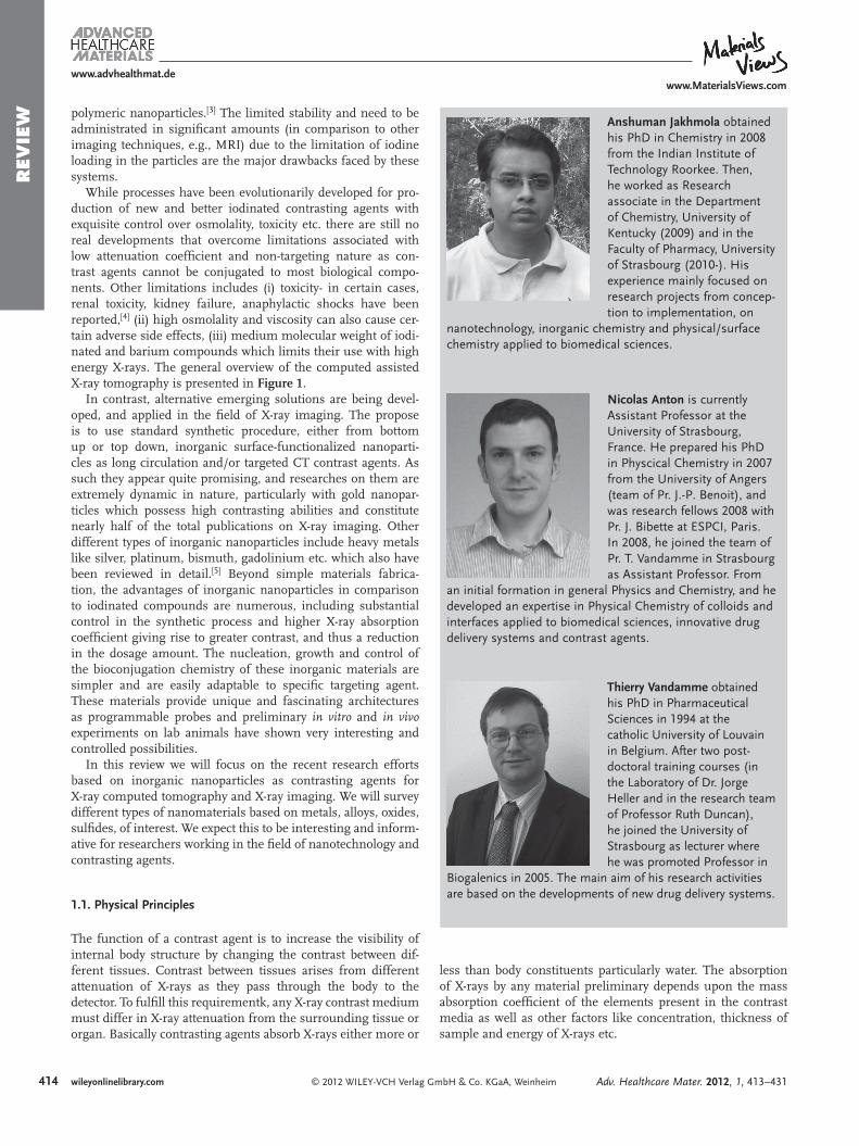
© 2012 WILEY-VCH Verlag GmbH & Co. KGaA, Weinheim www.advhealthmat.de www.MaterialsViews.com wileyonlinelibrary.com 413 REVIEW Adv. Healthcare Mater. 2012, 1, 413–431 Anshuman Jakhmola, Nicolas Anton,* and Thierry F. Vandamme Inorganic Nanoparticles Based Contrast Agents for X-ray Computed Tomography A. Jakhmola, N. Anton, T. F. Vandamme University of Strasbourg Faculté de Pharmacie 74 route du Rhin, BP 60024 F-67401 Illkirch Cedex, France CNRS 7199, Laboratoire de Conception et Application de Molécules Bioactives équipe de Pharmacie Biogalénique E-mail: [email protected] DOI: 10.1002/adhm.201200032 1. Introduction The efficient use of X-rays for imaging and computed tomog- raphy applications not only depends on the advancement of diagnostic equipment but also on the contrasting media neces- sary to generate two and three dimensional images for in vivo biomedical applications. Historically, the importance of X-rays in medical diagnosis and imaging was soon recognized after their discovery in 1895 by Rontgen, and research on contrast media and medical imaging was started soon thereafter. The use of the first positive contrast agent most probably dates back to 1896 when barium sulfate was used for investigation of peri- stalsis movement in animals; [1] later in 1921 iodized oil Lipodol (iodized poppy seed oil) was introduced as first suitable X-ray contrast media in myelography. [2] Although X-ray computed tomography is considered as one of the most important imaging techniques for various medical, scientific and technological applications, its use is quite limited as compared to other imaging techniques like MRI or fluorescence imaging. Major limitations are often associated with con- trasting agents approved for medical diag- nosis. Modern X-ray contrasting agents approved for clinical use are either barium sulfate based solution or poly-iodinated aromatic compounds. Conventional ionic contrast media, which are sodium and meglumine salts of tri-iodinated benzoic acid were introduced more than 50 years ago and were quite hyperosmolar and greatly increased toxic and side effects in patients. With new developments, major improvements were achieved in terms of the chemistry and physiological com- patibility of these solutions to reduce the side effects. However, still to date their processing and synthesis require com- plex methodologies that are performed under stringent and toxic conditions. Besides, these tri-iodo- benzene based agents are small molecular weight hydrophilic molecules, which exhibit a rapid renal clearance and vascular permeation. While traditional clinical CT scanners are quite effective for routine diagnosis due to their small response time ( e.g. five seconds to acquire 15 cm thick volume), MicroCT systems tend to be quite slow in comparison ( e.g. ten minutes to scan a mouse); As a result, these are unsuitable for most non preclinical X-ray imaging experiments and analysis. With time many formulation and chemical strategies were under- taken in order to increase the half-life of iodinated contrasting agents. For instance one such development was the introduc- tion of nanoparticle based iodinated molecules as blood pool contrasting agents (BPCA). BPCA are systems which remain in the blood for a prolonged period of time as compared with conventional contrast agents, which diffuse quickly into the interstitial space. These systems, in the form of nanoparticles with a controlled functionalization and surface coating provide significant stealth and antibiofouling properties towards the immune system. In fact this mixed nature of the structure not only increases their residence time in blood but subsequently enhances the contrast. We have recently reviewed iodinated blood pool contrast agents in detail, [3] covering all their chem- ical and physicochemical properties: surface functionalization, stability, toxicology and pharmacokinetics. These contrasting agents which are mainly in the form of colloidal solutions may contain liposomes (encapsulating small molecular weight iodi- nated molecules), nano-emulsions, micelles, dendrimers and Nanomaterials have gained considerable attention and interest in the devel- opment of new and efficient molecular probes for medical diagnosis and imaging. Heavy metal nanoparticles as such are excellent absorber of X-rays and can offer excellent improvement in medical diagnosis and X-ray imaging. Substantial progress has been made in the synthesis protocol and charac- terization studies of these materials but a major challenge still lies in the toxicological studies, which are rather incomplete. The worst known cases were those associated with Thorotrast (suspension of ThO 2 nanoparticles) which resulted in many deaths over years. Properly protected nanomaterials conjugated or coated with biocompatible materials can be used for the fabri- cation of various functional systems with multimodality, targeting properties, reduced toxicity and proper removal from the body. This review aims mainly to provide the advances in the development of inorganic nanoparticle based X-ray contrasting agents with an overview of methods of their preparation, functionalization and applications in medical diagnosis.
-
Upload
independent -
Category
Documents
-
view
0 -
download
0
Transcript of Inorganic Nanoparticles Based Contrast Agents for X‐ray Computed Tomography

www.advhealthmat.dewww.MaterialsViews.com
REV
IEW
Anshuman Jakhmola, Nicolas Anton,* and Thierry F. Vandamme
Inorganic Nanoparticles Based Contrast Agents for X-ray Computed Tomography
Nanomaterials have gained considerable attention and interest in the devel-opment of new and effi cient molecular probes for medical diagnosis and imaging. Heavy metal nanoparticles as such are excellent absorber of X-rays and can offer excellent improvement in medical diagnosis and X-ray imaging. Substantial progress has been made in the synthesis protocol and charac-terization studies of these materials but a major challenge still lies in the toxicological studies, which are rather incomplete. The worst known cases were those associated with Thorotrast (suspension of ThO 2 nanoparticles) which resulted in many deaths over years. Properly protected nanomaterials conjugated or coated with biocompatible materials can be used for the fabri-cation of various functional systems with multimodality, targeting properties, reduced toxicity and proper removal from the body. This review aims mainly to provide the advances in the development of inorganic nanoparticle based X-ray contrasting agents with an overview of methods of their preparation, functionalization and applications in medical diagnosis.
1. Introduction
The effi cient use of X-rays for imaging and computed tomog-raphy applications not only depends on the advancement of diagnostic equipment but also on the contrasting media neces-sary to generate two and three dimensional images for in vivo biomedical applications. Historically, the importance of X-rays in medical diagnosis and imaging was soon recognized after their discovery in 1895 by Rontgen, and research on contrast media and medical imaging was started soon thereafter. The use of the fi rst positive contrast agent most probably dates back to 1896 when barium sulfate was used for investigation of peri-stalsis movement in animals; [ 1 ] later in 1921 iodized oil Lipodol (iodized poppy seed oil) was introduced as fi rst suitable X-ray contrast media in myelography. [ 2 ] Although X-ray computed tomography is considered as one of the most important imaging techniques for various medical, scientifi c and technological
© 2012 WILEY-VCH Verlag GmbH & Co. KGaA, WeinheimAdv. Healthcare Mater. 2012, 1, 413–431
A. Jakhmola , N. Anton , T. F. Vandamme University of StrasbourgFaculté de Pharmacie74 route du Rhin, BP 60024F-67401 Illkirch Cedex, FranceCNRS 7199, Laboratoire de Conception et Application de Molécules Bioactives équipe de Pharmacie Biogalénique E-mail: [email protected]
DOI: 10.1002/adhm.201200032
applications, its use is quite limited as compared to other imaging techniques like MRI or fl uorescence imaging. Major limitations are often associated with con-trasting agents approved for medical diag-nosis. Modern X-ray contrasting agents approved for clinical use are either barium sulfate based solution or poly-iodinated aromatic compounds. Conventional ionic contrast media, which are sodium and meglumine salts of tri-iodinated benzoic acid were introduced more than 50 years ago and were quite hyperosmolar and greatly increased toxic and side effects in patients. With new developments, major improvements were achieved in terms of the chemistry and physiological com-patibility of these solutions to reduce the side effects. However, still to date their processing and synthesis require com-plex methodologies that are performed
under stringent and toxic conditions. Besides, these tri-iodo-benzene based agents are small molecular weight hydrophilic molecules, which exhibit a rapid renal clearance and vascular permeation. While traditional clinical CT scanners are quite effective for routine diagnosis due to their small response time ( e.g. fi ve seconds to acquire 15 cm thick volume), MicroCT systems tend to be quite slow in comparison ( e.g. ten minutes to scan a mouse); As a result, these are unsuitable for most non preclinical X-ray imaging experiments and analysis. With time many formulation and chemical strategies were under-taken in order to increase the half-life of iodinated contrasting agents. For instance one such development was the introduc-tion of nanoparticle based iodinated molecules as blood pool contrasting agents (BPCA). BPCA are systems which remain in the blood for a prolonged period of time as compared with conventional contrast agents, which diffuse quickly into the interstitial space. These systems, in the form of nanoparticles with a controlled functionalization and surface coating provide signifi cant stealth and antibiofouling properties towards the immune system. In fact this mixed nature of the structure not only increases their residence time in blood but subsequently enhances the contrast. We have recently reviewed iodinated blood pool contrast agents in detail, [ 3 ] covering all their chem-ical and physicochemical properties: surface functionalization, stability, toxicology and pharmacokinetics. These contrasting agents which are mainly in the form of colloidal solutions may contain liposomes (encapsulating small molecular weight iodi-nated molecules), nano-emulsions, micelles, dendrimers and
wileyonlinelibrary.com 413
www.MaterialsViews.com
41
www.advhealthmat.de
REV
IEW Anshuman Jakhmola obtained
his PhD in Chemistry in 2008 from the Indian Institute of Technology Roorkee. Then, he worked as Research associate in the Department of Chemistry, University of Kentucky (2009) and in the Faculty of Pharmacy, University of Strasbourg (2010-). His experience mainly focused on research projects from concep-tion to implementation, on
nanotechnology, inorganic chemistry and physical/surface chemistry applied to biomedical sciences.
Nicolas Anton is currently Assistant Professor at the University of Strasbourg, France. He prepared his PhD in Physcical Chemistry in 2007 from the University of Angers (team of Pr. J.-P. Benoit), and was research fellows 2008 with Pr. J. Bibette at ESPCI, Paris. In 2008, he joined the team of Pr. T. Vandamme in Strasbourg as Assistant Professor. From
an initial formation in general Physics and Chemistry, and he developed an expertise in Physical Chemistry of colloids and interfaces applied to biomedical sciences, innovative drug delivery systems and contrast agents.
Thierry Vandamme obtained his PhD in Pharmaceutical Sciences in 1994 at the catholic University of Louvain in Belgium. After two post-doctoral training courses (in the Laboratory of Dr. Jorge Heller and in the research team of Professor Ruth Duncan), he joined the University of Strasbourg as lecturer where he was promoted Professor in
Biogalenics in 2005. The main aim of his research activities are based on the developments of new drug delivery systems.
polymeric nanoparticles. [ 3 ] The limited stability and need to be administrated in signifi cant amounts (in comparison to other imaging techniques, e.g., MRI) due to the limitation of iodine loading in the particles are the major drawbacks faced by these systems.
While processes have been evolutionarily developed for pro-duction of new and better iodinated contrasting agents with exquisite control over osmolality, toxicity etc. there are still no real developments that overcome limitations associated with low attenuation coeffi cient and non-targeting nature as con-trast agents cannot be conjugated to most biological compo-nents. Other limitations includes (i) toxicity- in certain cases, renal toxicity, kidney failure, anaphylactic shocks have been reported, [ 4 ] (ii) high osmolality and viscosity can also cause cer-tain adverse side effects, (iii) medium molecular weight of iodi-nated and barium compounds which limits their use with high energy X-rays. The general overview of the computed assisted X-ray tomography is presented in Figure 1 .
In contrast, alternative emerging solutions are being devel-oped, and applied in the fi eld of X-ray imaging. The propose is to use standard synthetic procedure, either from bottom up or top down, inorganic surface-functionalized nanoparti-cles as long circulation and/or targeted CT contrast agents. As such they appear quite promising, and researches on them are extremely dynamic in nature, particularly with gold nanopar-ticles which possess high contrasting abilities and constitute nearly half of the total publications on X-ray imaging. Other different types of inorganic nanoparticles include heavy metals like silver, platinum, bismuth, gadolinium etc. which also have been reviewed in detail. [ 5 ] Beyond simple materials fabrica-tion, the advantages of inorganic nanoparticles in comparison to iodinated compounds are numerous, including substantial control in the synthetic process and higher X-ray absorption coeffi cient giving rise to greater contrast, and thus a reduction in the dosage amount. The nucleation, growth and control of the bioconjugation chemistry of these inorganic materials are simpler and are easily adaptable to specifi c targeting agent. These materials provide unique and fascinating architectures as programmable probes and preliminary in vitro and in vivo experiments on lab animals have shown very interesting and controlled possibilities.
In this review we will focus on the recent research efforts based on inorganic nanoparticles as contrasting agents for X-ray computed tomography and X-ray imaging. We will survey different types of nanomaterials based on metals, alloys, oxides, sulfi des, of interest. We expect this to be interesting and inform-ative for researchers working in the fi eld of nanotechnology and contrasting agents.
1.1. Physical Principles
The function of a contrast agent is to increase the visibility of internal body structure by changing the contrast between dif-ferent tissues. Contrast between tissues arises from different attenuation of X-rays as they pass through the body to the detector. To fulfi ll this requirementk, any X-ray contrast medium must differ in X-ray attenuation from the surrounding tissue or organ. Basically contrasting agents absorb X-rays either more or
4 © 2012 WILEY-VCH Verlag Gmwileyonlinelibrary.com
less than body constituents particularly water. The absorption of X-rays by any material preliminary depends upon the mass absorption coeffi cient of the elements present in the contrast media as well as other factors like concentration, thickness of sample and energy of X-rays etc.
bH & Co. KGaA, Weinheim Adv. Healthcare Mater. 2012, 1, 413–431

www.MaterialsViews.comwww.advhealthmat.de
REV
IEW
Figure 1 . Schematic diagram and basic principles of computed assisted tomography.
When a parallel beam of X-rays passes through a layer of matter of thickness X, a part of it passes directly through it without any interaction (primary radiation), while a certain frac-tion of it is absorbed, defl ected or scattered. The intensity of transmitted beam J through this layer is related with the inten-sity of incident beam ( J 0 ) in according to Beer–Lambert law given by equation,
=J0 expµ·mρJ
where μ / ρ is known as the mass attenuation coeffi cient which is directly dependent on the energy of incident X-rays and the mass m of different elements present in the material. Keeping other factors constant, the mass attenuation coeffi cient tends to increase with increase in atomic number of elements in periodic table, and decrease with increase in energy of X-rays. Many theoretical and experimental based studies have also shown that elements with higher atomic number than iodine demonstrate superior X-ray attenuation ability at normal or even higher operating tube voltages due to the higher K-edges of heavy elements. The K-edges of heavy elements lies within the diagnostic X-ray spectrum as such an abrupt increase in attenuation at discrete energies near the K-edge is observed (see below). Thus a contrast media based on elements with higher atomic number will be more advantageous in terms of intrinsic contrast, lower dose requirement and lower radiation exposure to patients.
1.2. Basic Requirements for X-ray Contrast Media
In order to get suffi cient contrast in comparison to the background, a high concentration of contrast media is required,
© 2012 WILEY-VCH Verlag GAdv. Healthcare Mater. 2012, 1, 413–431
as such the most basic requirement for contrast media is its biocompability and inertness with living tissue. Although the currently available iodinated compounds have fulfi lled these basic requirements, their use is largely based on low toxicity and cost effectiveness rather than high radiopacity for X-rays. The main requirements of an X-ray contrast agent are as follows:
• High Solubility: For intravenous administration, any X-ray contrasting agent must have high solubility as very high concentration is needed to compensate for the most relevant drawback of X-rays, which is the low contrast sensitivity.
• Chemical stability: They must be chemically stable and phar-macologically inert after intravenous administration and must not react, coagulate or metabolize once administrated intravenously.
• Opacity: The contrast media must have high effi ciency as X-ray attenuators. Higher X-ray attenuation means higher con-trast enhancement at lower dosage levels.
• Toxicity/tolerability: The osmolality, viscosity and pH must be comparable to that of body fl uids and toxicity (chemotoxic-ity and osmotoxicity) must be as low as possible.
2. Noble Metal Based Nanoparticles
Noble metal nanoparticles are versatile agents with a variety scientifi c and technological applications. The intense interest derives from their unique characteristics: controlled morpholo-gies, spatial charge confi nement, ease of synthesis, and facile
415mbH & Co. KGaA, Weinheim wileyonlinelibrary.com

www.MaterialsViews.com
416
www.advhealthmat.de
REV
IEW
surface chemistry and functionalization. Gold and silver nan-oparticles have been used for centuries in many countries to cure certain diseases, [ 6 ] however their use in the fi eld of X-ray imaging is quite new and one of the most notable applications. Beyond simple fabrication process, recent research efforts points towards their exciting opportunities in the fi eld of CT imaging. In fact gold nanoparticles have shown quite positive results to overcome current limitations and are expected to enter clinical applications and diagnoses in near future.
2.1. Gold Nanoparticles as CT Contrast Agents
Nanogold which is known since ancient times for their meta-physical and healing powers is one of the most extensively studied nanomaterial which has a long history of synthesis and biomedical applications. In 2004 Hainfeld and his team, exploited the high atomic weight of gold in the fi eld of X-ray imaging which has gained considerable momentum world-wide. [ 7 ] It can be clearly seen that since 2004, most of the research investigations on inorganic nanoparticles based X-ray contrasting media are focused on gold nanoparticles or their hybrids. Beside the fact that gold particles displays a strong attenuation in X-ray region, research have also been focused on the substantial control of their physical, chemical and bio-logical properties which makes it one of the most ideal candi-date for X-ray CT and multimodal imaging. [ 8 ] AuNPs are not only stable and inert, but they also present much more fasci-nating properties such as their self-assembly in conjugation with organization motifs and templates, size-related electronic and optical properties (quantum-size effect), and their appli-cations in catalysis and biological process. The applications of AuNPs for medical diagnosis and X-ray CT imaging, can be briefl y summarized into following points: (i) X-ray attenu-ation or radiopacity properties, which have emerged as better and higher than conventional CT contrast agent ( e.g. iodinated), (ii) a highly fl exible facile surface chemistry which allows a wide modularity in the surface functionalization for in vivo stabili-zation or coating (with antibiofouling agents, e.g. polyethylene glycol), and conjugating with specifi c functional molecules for active targeting to specifi ed organs, regions, or cancerous cells (tumor) ( e.g. antibody, peptides, etc. see below), (iii) and in vivo nontoxic and biocompatible properties.
2.1.1. Synthesis of Gold Nanoparticles
While the fi rst formulation of gold nanoparticles dates back around 5 th century BC, in literature it was not until 1847 when Faraday fi rst reported the formation of deep red solutions of colloidal gold by aqueous reduction of gold chloride (AuCl 4 − ) by phosphorus in CS 2 (carbon disulfi de). The modern synthesis protocol are also based on a reduction of gold salt by a variety of reduction agents and involves well-described chemical methods, which can also potentially include their functionaliza-tion, coating or conjugation with active molecules. This section will fi rst focus on a global presentation of the chemical strat-egies commonly undertaken for the formulation of gold col-loids for X-ray imaging, followed by a brief description of their applications in X-ray imaging and fi nally a discussion of the
© 2012 WILEY-VCH Verlag wileyonlinelibrary.com
impact of the formulation parameters, chemistry, and coating/targeting agents on their X-ray attenuation properties, stability and other physical properties. Next, we will discuss the extent in which these AuNPs can offer a plethora of new diagnosis and/or therapeutics tools, where the fi eld of applications ( e.g. the in vivo targeting) will be directly related to the chemical nature of the coating agents. In this part, we will specifi cally focus on the supramolecular chemistry aiming at surface functionalization, by reviewing the results obtained with the combination of CT imaging and active targeting of tumors and specifi c organs.
Citrate reduction: This method pioneered by J. Turkevich in 1951 [ 6 , 9 ] is still one of the most adopted method for the formu-lations of AuNPs for CT imaging. [ 10–16 ] The synthesis protocol is one of the simplest where citrate is used both as a reducing and stabilizing agent to produce more or less monodisperse spher-ical gold nanoparticles with a size range of about 10−20 nm. Further control of particle size and range can easily be carried out by a using a much improved method, [ 17 , 18 ] with size tun-able from 16 to 147 nm. Recent works [ 19 ] have shown that, size can also be controlled by the addition of an amphiphile into the nanoparticle synthesis reaction, and can be accurately con-trolled by manipulating amphiphile-to-gold ratio. The major advantage of this method is that the surface functionalization is not very strong and can be easily replaced by other ligands. As such the citrate molecules can be easily replaced with a variety of materials depending upon aimed applications, for instance, in CT imaging, replacement with a variety of biocompatible materials or functional molecules have shown quite impressive results. [ 10–16 ]
Two-phase synthesis: The Burst-Schiffrin method. This method is based on reduction of gold ions in an organic phase in pres-ence of specifi c thiolated compounds like lipids, lipophilic sur-factants, alkanethiols, for e.g. dodecanethiol. The binding of thiols with gold nanoparticles is particularly strong due to the soft nature of both Au and S. The process, inspired by the Fara-day’s two-phase system, [ 20 ] involves the use of tretraoctylammo-nium bromide to transfer AuCl − ions into the organic phase ( e.g. toluene), which is then reduced instantaneously by NaBH 4 . The AuNPs typically show a size in the range 1–5.2 nm but the size and/or size distribution can easily be modifi ed by manipulating experimental conditions like thiol/gold mole ratios, rate of addi-tion of reducing agent, temperature and the nature of the thiol ligands. [ 21–27 ] The great advantage offered by this technology def-initely lies in the stability of the AuNPs. This synthesis process results in the formulation of thermally and air-stable gold nano-particles, which can be repeatedly isolated and redissolved in common organic solvents without irreversible aggregation or decomposition. This also enables easy particles handling and besides, this method is quite interesting as it allows a simple route to functionalize AuNPs, performed by an “exchange” of the adsorbed alkanethiol for functionalized aklanethiol. [ 27–30 ] Other different sulfur-containing compounds [ 31–40 ] and ligands like phosphine, phosphine oxide, amine and carboxilates were also used in this biphasic formulation method. [ 41–49 ] These sys-tems allowed the fabrication of AuNPs with sizes signifi cantly higher than above, e.g. 12, 28, 42 nm for PPh 3 or 8.59 nm [ 42 ] using octadecylamine. [ 47 ]
In situ synthesis in colloidal template: Microemulsions are ther-modynamically stable systems which are able to develop a large
GmbH & Co. KGaA, Weinheim Adv. Healthcare Mater. 2012, 1, 413–431

www.MaterialsViews.comwww.advhealthmat.de
REV
IEW
oil/water interface through the formation of nano-scale system, e.g. swollen micelles. [ 50 ] In the fabrication of AuNPs, these dis-persed systems have been used as “nano-reservoirs” in the pres-ence or absence of thiol ligands, in which the transfer of the gold ions from the aqueous to the organic phase is enhanced by the large interfacial area. [ 51–58 ]
Other methods: The seeding growth method for generating AuNPs by step-by-step process is also a popular technique which offers the possibility to control the size distribution (5 to 40 nm), [ 59–61 ] and has been widely used for the fabrication of gold nanorods. [ 62 ] Besides the thermodynamical parameters infl uencing the AuNPs synthesis, numerous physical phenom-enon like UV, near-IR irradiations, ultrasounds, γ -irradiation, etc. also displays a signifi cant impact on the physicochemical properties of the nanoparticles (reviewed in detail, along with additional AuNPs fabrication and stabilization techniques, in Ref. 6).
In situ synthesis in dendrimers: The use of dendrimers as tem-plates for entrapping [ 63–65 ] or stabilizing [ 66–70 ] AuNPs is very popular for biomedical applications, and occupies a signifi cant role in the fabrication of gold nanoparticle based contrasting agents for X-ray in vivo imaging applications. [ 63 , 64 , 71–74 ] The syn-thesis protocol is quite similar to the methods described above, as the dendrimer’s cores present sites with affi nities for the gold ions Au 3 + . The fabrication of dendrimer-entrapped gold nano-particles (AuDENPs) is generally performed by the reduction of HAuCl 4 by NaBH 4 in a mixture of water/methanol. [ 63 , 64 , 71 , 72 ] The evident advantage of this method is a simple synthesis route with full control of the surface chemistry, i.e the control of the toxicity, of the long circulation properties and targeting properties. In this respect, the dendrimer chemistry allow to synthesize AuDENPs with terminal amines transformed to acetyl functional groups, signifi cantly avoiding nonspecifi c cell membrane binding and toxicity. [ 68 , 73 , 75–77 ] Likewise, the amine-terminated gold-loaded dendrimers can serve as a platform for conjugation with targeting ligands; [ 73 , 77 ] however pre-modifi ed dendrimers with targeting moieties can be also used for the AuDENPs preparation. [ 68 ]
Typical examples of gold nano-colloids obtained with the methods described above are illustrated in Figure 2 below.
2.1.2. Why are AuNPs Best Suited for X-ray Imaging?
Globally, most work reported in the last decade focuses on the applications of gold nanoparticles for X-ray imaging. There are several basic advantages of these systems in comparision to the traditional iodine-based contrast agents like exceptional stability against oxidation, higher Z-number and absorp-tion coeffi cient, [ 4 ] that is to say, respectively, gold: 79 and 5.16 cm 2 g − 1 , iodine: 53 and 1.94 cm 2 g − 1 (absorption coeffi cient at 100 keV; for comparison, water absorption is 0.171 cm 2 g − 1 ). As a result, gold provides 2.7 times more contrast per unit weight than iodine. Moreover, gold imaging around 80–100 keV also reduces interferences from bone and soft tissues absorp-tion and this subsequently reduces the overall radiation dosage and exposure. Figure 3 reports the comparison of mass attenua-tion coeffi cients plotted against the photon energy for different materials used for in the fabrication of BPCA (iodine, gold and bismuth) compared to blood, soft tissues, bones, and water.
© 2012 WILEY-VCH Verlag GAdv. Healthcare Mater. 2012, 1, 413–431
It can be clearly seen that the greater contrast of inorganic compounds in the measuring zone comes from the sharp increase of the mass attenuation coeffi cients, (photoelectron effect) at metal K-edge energies, i.e. the electronic transitions can only exist above these thresholds energies, resulting in sharp “jumps” in their X-ray attenuation profi les. In this way, it can be clearly seen that in order to maximize the effi ciency, gold must be used at energies higher than ∼ 80 keV. Accord-ingly, numerous publications have compared the in vivo con-trast of inorganic nanoparticles with traditional iodinated com-pounds, highlighting some “equivalent concentrations” which provides the same X-ray attenuation (depending on the experi-mental conditions like photon energy as shown in Figure 3 ), for example the contrast of heparin-coated AuNPs (at 15.7 mg Au · mL − 1 ) was found to be equivalent to eXIA 160 (at 53.3 mg I · mL − 1 ). [ 88 ] Similarly 1.27 M of PEG-coated AuNPs gave Houns-fi eld unit (HU) value (around 6690 HU) which is equivalent to 2.36 M ( ≡ 300 mg I · mL − 1 ) of Ultravist(a conventional iodinated contrast agent). [ 10 ] There are various other examples in litera-ture [ 11 , 64 , 88 ] but such results are not only dependent on concen-tration chosen for the in vivo dose but are also affected by par-ticle size, experimental parameter, and acquisition protocol. At a given energy, the photon absorption is linearly dependent to the concentration, thus exhibiting different slopes in function of the metal (as illustrated in Ref. 64 for instance). At this point, a great challenge is to defi ne a compromise between the best contrast agent loading or concentration, or the higher tolerance and the lower toxicity.
2.1.3. In vitro and in vivo Imaging and/or Targeted-Imaging with AuNPs
Most of the work published on gold nanoparticles for X-ray imaging applications is aimed on the fabrication, coating, tar-geting and biological evaluation, however only a few actu-ally emphasize on in vivo imaging. After the pioneer work by Hainfeld et al. in 2006 [ 4 ] in which they proposed a 2-dimensions projection of mice carried out with a mammography unit (with 8 mAs exposures, 0.4 s at 22 kVp), it was almost after one year when Kim et al. [ 10 ] and Cai et al. [ 11 ] reported the in vivo applica-tions of AuNPs as X-ray contrast agents with micro computed tomography. Both these authors used PEGylted AuNPs as BPCA, which provided signifi cant vascular contrast enhancement with plasma half-time around 12 h in [ 10 ] and at 14.6 h in [ 11 ] . After that, most published works presenting in vivo applications of AuNPs as X-ray contrast agents are quite new, published mostly in 2010 and 2011. It can be seen that after intravenous injec-tion these nanoparticulate systems can exhibit a wide range applications depending on coating agents for example vascular imaging as BPCA, [ 13 , 63 , 64 , 89 ] liver or spleen imaging, [ 12 , 71 , 88 , 90 ] or even tumor-specifi c targeting imaging after i.p. [ 91 ] or i.v. [ 14 ] administration (generally with BPCA properties form enhancing the targeting properties). As emphasized above, the great advan-tages of gold nanoparticles (AuNPs), lies in their highly fl ex-ible surface supramolecular chemistry, enabling the fabrication of hybrid or conjugated nanoparticles, by coating or binding them with specifi c biomolecules or targeting agents, which may include, monoclonal antibodies, aptamers, peptides, and various receptor-specifi c surfaces. [ 92–97 ] Actually, the coating of AuNPs
417mbH & Co. KGaA, Weinheim wileyonlinelibrary.com

www.MaterialsViews.com
418
www.advhealthmat.de
REV
IEW
Figure 2 . Nano gold – Different morphologies ((A) Nanorods. Reproduced with permission. [ 78 ] Copyright 2008, American Chemical Society. (B) Gold Nanowires, Reproduced with permission. [ 79 ] Copyright 2004, American Chemical Society. (C) Monodisperse Gold Nanoparticles. Reproduced with per-mission. [ 80 ] Copyright 2010, American Chemical Society. (D) Star-Shaped Gold Nanoparticles. Reproduced with permission. [ 81 ] Copyright 2006, American Chemical Society. (E) Nanobranches and Nanospheres. Reproduced with permission. [ 82 ] Copyright 2009, American Chemical Society. (F) Nanorods, Nanospheres and Faceted platelets. Reproduced with permission. [ 83 ] Copyright 2008, American Chemical Society (ACS). (G) Satellite type structure. Reproduced with permission. [ 84 ] Copyright 2006. American Chemical Society (ACS). (H) Large polygons. Reproduced with permission. [ 85 ] Copyright 2007, American Chemical Society (ACS), (I) Nanocages. Reproduced with permission. [ 86 ] Copyright 2007, American Chemical Society (ACS)).
involves three main key factors which have to be carefully char-acterized in the development of blood pool and/or targeted con-trast agents: (i) the fi rst one concerns the stabilization of AuNPs suspensions in bulk media (as discussed above in section 2.1.1), and it is generally taken as a part of the AuNPs synthesis step. (ii) The second one involved in the AuNPs coating involves the fabrication of nanoparticles with good in vivo stability and optimum in vivo pharmacokinetics properties, i.e. with reliable stealth properties in bloodstream. This is one of the main objec-tive of the studies carried out for the fabrication of BPCA, and it is generally achieved with PEG-coatings which offer very poor interactions with plasma proteins. [ 10 , 11 ] (iii) Finally, the last factor in the AuNPs coating, which is very closely linked to the latter
© 2012 WILEY-VCH Verlag Gwileyonlinelibrary.com
point (ii), concerns the surface modifi cation in order to promote the site-specifi c targeting like tumor cells targeting, the so-called “active targeting”. Overall, three different markers overexpressed in cancer cells are targeted by these receptor-specifi c function-alized nanoparticles. They are mainly matrix metalloprotease (MMPs), epidermal growth-factor receptor (EGFR), and onco-proteins that are associated with human papillomavirus (HPV) infection. [ 98 ] This AuNPs surface functionalization is sum-marized and illustrated in Figure 4 , which clearly shows their potentials in terms of surface coating as well as nano-encapsula-tion reported in literature.
The in vivo imaging applications of AuNPs, with some inter-esting in vitro results have been summarized in Table 1 and 2 .
mbH & Co. KGaA, Weinheim Adv. Healthcare Mater. 2012, 1, 413–431

www.MaterialsViews.comwww.advhealthmat.de
REV
IEW
Figure 3 . Mass attenuation vs. photon energy, of iodine, gold and bis-muth, compared to blood, soft tissues, bones, and water. The energy range typically chosen for micro computed tomography is emphasized in gray (Constructed using data from Ref [ 87 ] ).
Figure 4 . Schematic of surface functionalization and morphologies of inorganic nanoparticles.
We have mainly emphasized on the synthesis protocol, the coating or targeted agents specifi cally used either for stabi-lizing/surface modifi cation of AuNPs (fundamental for in vivo imaging), or for active targeting of tumor cells, and fi nally the main application(s) and the result(s) obtained. This overview has been presented through Table 1 and there are several key fundamental points that needs to be addressed: (i) fi rst, the cit-rate reduction is still one of the most important and popular methods used for AuNPs core synthesis as this method remains the easiest way to generate a stable dispersion in aqueous media besides surface coating of citrate can also be easily replaced by other functional molecules. However, for dendrimer entrapped gold nanoparticles, the gold synthesis follows a specifi c method, which plays with the particular affi nities of gold ions towards the dendrimers core, followed by reduction by NaBH 4 (described in section 2.1.1). (ii) The second point is that the best in vivo stability in blood stream is given by PEGylation of AuNPs, either for BPCA applications, [ 10 , 11 ] or for active targeting applications (also needing a long vascular circulation). [ 13 , 14 ] In absence of PEG coating, the reticuloendothelial system (RES) uptake results in accumulation of AuNPs in liver and spleen. Actually, in this case (without PEG coating), there are no spe-cifi c rules that can be defi ned to predict the in vivo stability of these systems, besides it is also infl uenced by other specifi c ligands used for targeting. (iii) The last important point is the
© 2012 WILEY-VCH Verlag GAdv. Healthcare Mater. 2012, 1, 413–431
global success of targeted CT contrast agents with AuNPs, from long circulation vascular contrast to targeted tumor cells. These results open new doors and important applications in biomed-ical targeted imaging, along with new possibilities for carrying out personalized cancer therapies, enabling the “online” moni-toring of drug delivery via following-up in real time the nano-carriers imaging. The illustration of the different applications of in vivo imaging applications ( i.e. BPCA, tumor active targeting, lymph nodes targeting, multimodal imaging and liver-specifi c imaging) are also reported from literatures, in Figure 5 .
2.1.4. Gold nanoparticles: summary and perspectives
Over the past 5 years, the number of publications on nano-particulate contrasts agent for CT has increased exponen-tially. [ 109 ] These nanomaterials assembly in function of the chemical nature of the coating layer and/or functionaliza-tion, can behave as blood pool or targeted contrast agent. Actually, the recent works in this field are mostly based on gold nanoparticles or their hybrids due to the strong X-ray attenuation of gold combined with the well-established facile chemistry of the gold nanoparticles. To date, the most significant results on long-circulating or targeted nanopar-ticles CT contrast agents are provided with systems based on gold nanoparticles, and summarized reported in Table 1
419mbH & Co. KGaA, Weinheim wileyonlinelibrary.com

www.MaterialsViews.com
4
www.advhealthmat.de
REV
IEW Table 1. Summary of the main results regarding the use of AuNPs as X-ray contrat agent, emphasizing their applications for in vivo imaging and/or
targeting.
References AuNPs Synthesis Stability coating Active targeting Application Main results
[ 10 ] ( a ) , [ 11 ] ( b ) Citrate reduction PEG coating (incubation of – Blood pool • ( a ) t 1/2 ∼ 12 h; ( b ) t 1/2 ∼ 14.5 h
AuNPs, d = ( a ) 31 nm, AuNPs with ( a ) PEG-SH, or contrast agent
(i.v.)
• X-ray attenuation: ( a ) 1.9 and ( b ) 2.7 times
higher than Ultravist
( b ) 38 nm ( b ) PEG sulfhydryl) • Biocompatible, low toxicity, RES uptake
[ 88 ] Commercial product Heparin coating (conjugating – Liver-specifi c • Liver-specifi c contrast
AuNPs, d = 55 nm Heparin-DOPA + AuNPs) contrast agent
(i.v.)
• Stable, biocompatible
[ 90 ] NaAuCl 4 reduction by THPAL Gum Arabic coating
during the
– Liver/lung-specifi c • Enhanced contrast in
juvenile swine
(phosphino animoacid) AuNPs synthesis,
see also [99, 100]
contrast agent
(i.v.)
• Good tolerance to
administration
AuNPs, d = 20 nm
[ 12 ] • Citrate reduction
(17 to 66 nm)
2-mercaptosuccinic
acid (MSA)
– Spleen-specifi c • Multi-color nanotags
combining
• Seed-growth method (80 to
120 nm)
contrast agent
(i.v.)
surface-enhanced Raman spectroscopy
(SERS) and CT
• Dual in vivo imaging
[ 72 ] Dendrimer in situ HAuCl 4 Neutral acetylated surface – Cancer cell
imaging
• Stable in suspension for months
reduction by NaBH 4 , d =
2.6 nm
(i.t. and i.p.) • AuDENPs accumulation in cancer cells
in vitro and in vivo
[ 13 ] Citrate reduction PEG-COOH coating Antibody: monoclonal Lymph nodes
targeting
• X-ray contrast enhanced in lymph nodes with
targeted AuNPs
AuNPs, d = 28 or 38 nm (incubation with heterofunctional anti-mouse CD4 and imaging (i.v.) • Contrast enhancement stronger
with bigger AuNPs
ligand: dithiol-PEG-COOH)
[ 91 ] NaAuCl 4 reduction by THPAL Starch coating during the Peptide: bombesin
(BBN), to
Human prostate
tumor
• BBN-AuNPs specifi cally targets cancer
receptors sites
(phosphino animoacid) AuNPs synthesis, see also [ 99 ] target the gastrin-
releasing
targeting and
imaging
• Importance of the BBN-AuNP size for
effective tumor penetration
AuNPs, d = 115–150 nm peptides (GRP)
receptors
(i.p.)
[ 14 ] Citrate reduction PEG-COOH coating Antibody: anti-Her2 Human breast
tumor
• Specifi c tumor targeting of herceptin-targeted
AuNPs
AuNPs, d = 15 nm (incubation with heterofunctional (herceptin) targeting and
imaging
• Imaging readily enables detection of
small, 1.5 mm-thick tumors
ligand: thiol-PEG-COOH) (i.v.) • Contrast 22-fold higher in tumors than
surronding muscle
and Table 2 . This simply means that, today, the best com-promise between contrasting properties, physicochemical and surface properties, and low toxicity, is found with gold nanoparticles-based systems. However, these systems can reach certain limitations regarding the different aspects, and mainly the future research challenges should be focused on these points.
The fi rst one lies in the cost of gold, which can become a critical point when the bridge between fundamental research and industrial transposition will be considered. This is actually
20 © 2012 WILEY-VCH Verlag wileyonlinelibrary.com
one of the main reasons that allowed the monopole of iodi-nated molecules for X-ray contrast agents. The solution for getting round this problem can be found on many emerging research works focused on inorganic nanoparticles made of metal cheaper than gold, but still with X-ray attenuation proper-ties comparable to the ones of gold. These works report that the chemistry of inorganic nanoparticles (other than gold nanopar-ticles) can allow realistic alternatives for the fabrication of CT contrast agents. This aspect is as well developed in detail in the second part of the present review.
GmbH & Co. KGaA, Weinheim Adv. Healthcare Mater. 2012, 1, 413–431

www.MaterialsViews.com
421
www.advhealthmat.de
© 2012 WILEY-VCH Verlag GmbH & Co. KGaA, Weinheim wileyonlinelibrary.com
REV
IEW
Adv. Healthcare Mater. 2012, 1, 413–431
Table 2. Summary of the main results regarding the use of AuNPs as X-ray contrat agent, and their applications for in vitro imaging and/or targeting.
References AuNPs Synthesis Stability coating Active targeting Application Main results
[ 8 ] Gold NanoRod
(AuNR)
Poly(acrylic acid) (PAA)
coating
Antibody: UM-A9 (amidation Squamous cell carcinoma • Strong selective X-ray attenuation
of squamous cell carcinoma
by mediated
growth method
(AuNR surface
adsorption)
reaction on EDC/NHS targeting and imaging • CT imaging allows reliable and
sensitive detection of lymph nodes
size: d = 15 nm,
l = 45 nm
activated sites) ( α 6 β 4 integrin) and metastasis, in head and neck
cancer diagnosis and staging
[ 15 ] Citrate reduction – Aptamer: prostate-specifi c Prostate cancer cells
imaging,
• Strong selective X-ray attenuation
of prostate cancer cells
AuNPs, d = 15 nm membrane antigen RNA targeting, and drug delivery • Simultaneous and specifi c pancreas
cancer cell targeting,
aptamer (PSMA) imaging, and drug (doxorubicin) delivery
[ 16 ] Citrate reduction Poly-vynil alcohol
coating
L-glutamic acid Damaged bone tissues • Functionalized AuNPs target
damaged bones in vitro
AuNPs, d = 15 and 40 nm (added while removing
citrate
(primary amine for binding targeting and imaging
ions by ion exchange
resin)
to the gold surface) (-COOH chelates Ca 2 + )
[ 73 ] NaBH 4 reduction Dendrimer G5-NH 2 Folic acid and Cancer cell targeting • Effi ciency in targeting (and thus
imaging) KB cells
AuNPs, d = 3.2 nm FITC and imaging overexpressing folate receptors
[ 101 ] Citrate reduction Citrate Thioglycolic acid, Tracking digestive • AuNPs serve as tracer in
microparticles
AuNPs, d = 10 − 20 nm 6-thioganine, mechanisms in • Imaging delicate insects like
mosquito
2-mercaptoethanol, mosquito
1-propane thiol
[ 102 ] Burst synthesis Alkane thiolate w-carboxylic acid Size related • Attenuation increase with decrease
in size from 12 to 6.6 nm
NaBH 4 reduction attenuation properties • Highly stable, monodisperse, water
soluble
d = 6.6 − 12 nm
[ 103 , 104 ] Citrate redustion citrate 2-deoxy-D-glucose Cancer cell targeting • Labelling AuNPs with
2-deoxy-D-glucose induces
d = 4 nm and imaging specifi c cancer cells uptake
(human alveolar epithelial
cancer cell line, A-549)
[ 105 ] Various methods • 4 − 20 nm: citrate – Size related • Stable in physiological conditions,
nontoxic to Hela S3 cells
d = 4 − 60 nm • 4 − 20 nm: mercapto- attenuation properties • Higher attenuation displayed
by smaller particles
succinic acid
[ 106 ] Commercial product – – Effect of size, concentration • Passive cancer cell uptake depends
on the AuNPs size
d = 5, 10, 20, 30, 40, 50 nm incubation time on the
AuNPs
• Better uptake with
20 nm AuNPs
uptake by pancreas cancer
cells
[ 107 ] Citrate reduction Horse serum protein – Cell imaging • AuNPs is used a permanent marker
for implanted normal and malignant
cells
AuNPs, d = 50 nm • Quantifi cation of cells numbers
within small animals

www.MaterialsViews.com
4
www.advhealthmat.de
REV
IEW
Figure 5 . Schematic of the main applications of in vivo imaging using nano-gold. (A) Reproduced with permission. [ 10 ] Copyright 2007, American Chemical Society (ACS); (B) Reproduced with permission. [ 14 ] Copyright 2011, The British Institure of Radiology Publications; (C) Reproduced with permission. [ 88 ] (D) Reproduced with permission. [ 13 ] Copyright 2010. American Chemical Society (ACS); (E) Reproduced with permission. [ 108 ] Copyright 2011, IOP Publishing.
Gold nanoparticles and more generally of inorganic nano-particles can appear effi cient in imaging and targeted imaging. However, and it is the second limitation of these systems, their intrinsic morphology is not very adapted for their use as drug carrier, ( i.e. for the simultaneous imaging and drug delivery). This point could actually become a predominant research interest in the near future, enabling the so-called personalized therapy. This will allow an “in time” in vivo quantifi cation of the administrated drugs and a precise monitoring and control of the dosages. Earlier works of Kim et al. [ 15 ] (see also Table 1 ) already exist, in which an anticancer molecule (doxorubicin) is entrapped in the gold nanoparticle coating layer. Other poten-tial ideas for exploring this way should lie in the multiple encapsulation of inorganic nanoparticles, for example in nano-emulsions giving possible the co-encapsulation of bioactive molecules in oil along with the contrasting NPs.
2.2. Hybrid Gold Nanoparticles
As discussed in the above section, the employment of gold in CT imaging offers unique advantages over traditional iodine based contrasting agents. Additionally their surface can be modifi ed and functionalized with various functional materials to create
22 © 2012 WILEY-VCH Verlag GmbH & Co. KGaA, Weiwileyonlinelibrary.com
new hybrid systems for use in multimodality diagnostic imaging and therapy. We will see in this section that how various coating and/or synthesis technologies associated with AuNPs allows the fabrication of hybrid gold nanoparticles that can be detected with mul-tiple techniques such as magnetic resonance imaging (MRI), X-ray computed tomography (CT), Fluorescence imaging. Such systems can offer excellent advantages for molecular diagnosis, multimodal imaging and biotech-nology. It is quite interesting to note that most of CT scanners are generally coupled with other instruments as no single imaging modality can address all biological questions. A good example could be coupling the X-ray imaging system with other imaging modali-ties (PET/SPECT, radionuclides etc.) which increases experimental and therapeutic fl exi-bility. Such a combination is often used to vis-ualize the bones with CT, in order to situate the radiolabelled probes in the whole animal body. [ 110–112 ] Such multimodal applications often allow faster detection and increases therapeutic effi cacy to save time and money, but do not actually correspond to the multi-modal imaging in the same line that the one discussed in the present paper, that is using multimodal NPs including CT contrast agent. Recently Wang group successfully used 125 I for radiolabeling of gold nanorods. [ 111 ] These probes could be successfully used as nuclear and optical dual-modality contrast agents for better examination of infl ammation.
2.2.1. Gold/Gadolinium Hybrid Nanoparticles
Gadolinium, occupying central position in the lanthanide series has 7 unpaired electrons and gives excellent signal in MRI. As such coupling of AuNPs with Gd chelates can give rise to nano-systems detectable both by X-Ray CT and MRI. Roux and his group [ 113 ] were the fi rst to develop a method to synthesize such nanosystems. They encapsulate gold nanoparticles with a mul-tilayered organic shell composed of gadolinium chelates bound to each other with disulfi de bonds (illustrated in Figure 6 (A)). This nanoparticle complex could be served both as X-ray and MRI contrasting agent due to presence of radiopaque gold and superparamagnetic gadolinium ions. The synthetic procedure involved the reduction of gold salt in presence of dithiolated derivatives of diethylenetriaminepentaacetic acid followed by addition of Gd 3 + . The two thiol groups played the key role in the formation of multilayered ligand shell through disulfi de linkages. The in vivo experiments revealed free circulation of these particles in blood stream with no undesirable side effects, toxicity or accumulation in liver, lungs and spleen. These par-ticles could be easily be detected and followed up by MRI and X-ray imaging despite of low concentration of gold (10 mg/mL) and gadolinium (5 mg/mL). Various other research groups also reported the functionalization of AuNPs with Gd DTPA
nheim Adv. Healthcare Mater. 2012, 1, 413–431

www.MaterialsViews.comwww.advhealthmat.de
REV
IEW
Figure 6 . Gold-based nanoparticles for multimodal imaging: (A) Gold-gadolinuim chelate for X-ray and MRI imaging (Reproduced with permission. [ 113 ] Copyright 2008. American Chem-ical Society (ACS); (B) Hybrid gold/iron oxide for dual CT/MRI imaging, corresponding to the images reported in Figure 5 (E) (Reproduced with permission. [ 108 ] Copyright 2011, IOP Publishing).
complex using conjugates like penicillamine, [ 114 ] 4-aminothi-ophenol [ 115 ] and cysteine. [ 116 ] These particles exhibited very high r 1 and r 2 relaxivity. The in vivo X-ray attenuation of these con-jugate particles [ 114 ] was found to be almost twice as compared to simple ligand coated gold particles and displayed excellent contrast enhancement of liver, the blood pool and lymph nodes with no visual side effects. In a similar approach Song et al. [ 117 ] used oligonucleotide-modifi ed gold nanoparticle conjugates for covalently attaching GdIII complexes (DNAGdIII@AuNP). This nanoparticle conjugate was highly biocompatible and stable with an extraordinary ability to enter cells, which were about 50 folds higher as compared to the clinically available contrast agent DOTAGdIII. This nanoparticle system was suitable for CT and MRI applications and could also be used in fl uores-
Figure 7 . Lipid coated gold nanoparticle for multimodal CT/MRI/fl uorescence imaging (Repro-duced with permission. [ 120 ] Copyright 2008, American Chemical Society (ACS)).
cence imaging by attachment of fl uoroprobes like Cy3 (cyanine dye) to the DNA strand.
2.2.2. Gold/Iron Oxide Nanoparticles
Similar nanosystems active both in MRI and CT imaging can also be designed by using superparamagnetic iron oxide with AuNPs. Jon and coworkers studied two types of sys-tems containing both iron and gold. [ 108 , 118 ] The fi rst one published in 2009 reported polyethylene glycol (PEG) coated hybrid core-shell type nanoparticles consisting of iron oxide core and gold shell. The synthesis pro-tocol involved deposition of gold layer over
© 2012 WILEY-VCH Verlag GmbH & Co. KGaA, WeinheAdv. Healthcare Mater. 2012, 1, 413–431
superparamagnetic iron oxide core particles coated with oleyamine. The surfaces of the particles were coated with a layer of PEG to ensure biocompatibility and long circula-tion time in blood stream even at high con-centration. The X-ray contrasting effect was found to be higher when compared with iodinated contrasting agents. Recently, they developed hybrid nano-emulsions containing both gold and iron oxide [ 108 ] (illustrated in Figure 6 (B) and Figure 5 (E)). This nanoe-mulsion was synthesized by thermal decom-position of Fe-oleate and Au-oleylamine complexes and were coated with amphiphilic poly(DMA-r-mPEGMA-r-MA) to prevent their aggregation and make them water soluble. The particles displayed high contrast in CT imaging which was even more than pure gold nanoparticles. The in vivo experiments with hepatoma”bearing mice resulted in high contrast difference between hepatoma and normal tissue in liver.
2.2.3. Gold/Silica Nanoparticles
Another approach for the development of multifunctional probes for MRI, CT and fl uorescence imaging was shown by Mulder and co-workers. [ 119 ] In their experiments they deposited a fl uorescent and paramagnetic
lipid coating (contaning Cy5.5 (NIR dye) and a Gd conjugate) over gold/silica core shell-type nanoparticles. This system exhibited magnetic and optical properties with the dense gold core serving as X-ray contrast agent. The PEGylated layer was essential to ensure a water dispersible, stable and biocompat-ible system. The CT or MRI contrast per particle could easily be tuned by increasing or decreasing the surface area simply by manipulating the size of gold core. This system was highly sen-sitive and it was possible to get in vivo signal enhancements in mice liver by 24% and 50% for MRI and CT, with concentration as low as 0.15 nmol · kg − 1 . In their another study, they modifi ed high density lipoproteins (HDL) by replacing their hydrophobic core with gold, iron oxide or quantum dot nanoparticles [ 120 ] (see Figure 7 ). The authors used ordinary phospholipid, gadolinium
423im wileyonlinelibrary.com

www.MaterialsViews.com
424
www.advhealthmat.de
REV
IEW
Figure 8 . Hybrid FePt nanoparticles for multimodal MRI/CT imaging (Reproduced with permission. [ 134 ] Copyright 2010, American Chemical Society (ACS)).
labeled phospholipid and rhodamine labeled phospholipids to synthesize these nano-crystal core HDL. This system was not only multimodal but it was seen that the gold core based nanosystems displayed more contrast than Omnipaque and PEG coated gold nanoparticles. The in vitro experiments demonstrated the uptake of nanoparticles by macrophages. This nanosystem could also be specifi cally targeted for imaging of athero-sclerosis when in vivo experiments were performed with mice fed exclusively with cholesterol rich diet.
2.3. Silver Nanoparticles
Colloidal form of silver compounds like AgI and Ag 2 O [ 121–126 ] were used as contrast mate-rials in the earlier 20th century but their use
were quickly abolished because of the high toxicity and fatalities encountered. [ 127 , 128 ] However, studies on silver iodide colloids way back in the late seventies revealed that silver was retained in the liver long enough to allow CT examination with the con-trast difference of 4 to 5 times that the background. [ 126 ]Silver nanoparticles as such suffer from the same drawback regarding toxicity issues, [ 129–131 ] but with new developments and technological advancements the toxicity of silver nanoparticles has not only been greatly reduced but they could be made more biocompatible with appropriate surface modifi cations. [ 132 ]
In a recent report Lui et al. [ 133 ] reported the X-ray contrasting properties of dendrimer stabilized silver nanoparticles which could also be used for in vivo CT imaging applications. They reported size controlled facile synthesis of silver nanoparticles using G5 PAMAM dendrimer (generation 5 poly(amidoamine)) as template. By simply varying the molar ratio of dendrimer/Ag salt they could easily tune the size of silver nanoparticles from 8.8 to 23.2 nm. The dendrimer templated silver nanoparticles were not only stable in water, PBS buffer, fetal bovine serum but also at different pH (pH 5-8) and temperature (20–50 ° C) conditions. The X-ray absorption coeffi cient of silver nanopar-ticles was found to be both size and concentration dependent. It was found that particles with diameter of 16.1 nm displayed X-ray attenuation properties similar to that of iodine based con-trasting agent (Omnipaque) even though the atomic weight of silver is lower than that of Iodine. In vivo experiments with mice the silver particles displayed prolonged contrast enhance-ment and did not showed any toxic effect.
2.4. Platinum Based Nanoparticles
Though platinum can serve as a good contrasting agent due to its high atomic number, (Z = 78) but till date only little work has been done in this respect. Recently Chen’s lab demon-strated that water dispersible FePt nanoparticles [ 134 ] can poten-tially serve as a dual modal contrast agent for both CT and MRI molecular imaging where the heavy Pt contributed mainly as X-ray contrasting agent. These nanoparticles could easily be
© 2012 WILEY-VCH Verlagwileyonlinelibrary.com
prepared by a simple synthetic route and the size could be tuned between 3, 6 and 12 nm by simply changing the reaction conditions. This example is reported in Figure 8 where the TEM pictures show three different sizes of FePt particles, along with MRI and CT imaging phantoms emphasizing their contrasting properties. The particles presented excellent biocompatibility (cell viability > 75% at 0.1 M) and hemocompatibility ( < 5%) in all imaging contrast experiments. Surface chemistry and size played a crucial role in biodistribution, half-life, X-ray contrast and target specifi city. For example the 12 nm FePt nanoparti-cles revealed highest serum concentration, circulation half life and X-ray contrast while the 3 nm particles presented highest concentration in the brain and can be ideal for brain mapping. The nanoparticles could also be targeted to specifi c sites like tumor cells when conjugated with antibodies and other func-tion ligands.
3. Heavy Metal Based Nanoparticles (Thorium, Bismuth and Tantalum)
Heavy metals are good X-ray absorbers due to their high density and atomic weight. The X-ray attenuation of heavy metals also remains more or less constant with increasing X-ray energies, a phenomenon which arises due to higher K-edge of heavy ele-ments which falls within diagnostic X-ray spectrum. Although living organisms require varying levels of certain heavy metals, their excessive levels can be toxic and dangerous. Certain heavy metals may cause severe side effects and their accumulation over time can cause serious illness or even death.
3.1. Thorium Oxide Nanoparticles (ThO 2 )
Perhaps thorium based nano-emulsions are the fi rst that fall into this category. Colloidal thorium oxide (ThO 2 ) under the name Umbrathor produced by the Heyden Chemical Company was introduced in Germany as a radiological contrast agent in the late 1920s. [ 135 ] Umbrathor was a stable brownish liquid
GmbH & Co. KGaA, Weinheim Adv. Healthcare Mater. 2012, 1, 413–431

www.MaterialsViews.comwww.advhealthmat.de
REV
IEW
Figure 9 . Bismuth-based nanoparticles for CT imaging. (A) BPCA made with bismuth sulphide nanoparticles (Reproduced with permission. [ 146 ] Copyright 2006, Nature Publishing Group); (B) Multimodal and clot imaging. Reproduced with permission. [ 148 ]
containing 25% ThO 2 , which was administrated orally for visu-alization of the gastrointestinal tract. Later thorium oxide solu-tion with particle size of 3 − 10 nm and trade name of Thorotrast was introduced was produced by Testagar and Company. [ 136 , 137 ] This version of colloidal ThO 2 was alkaline-stable and did not aggregate after intravenous administration. Thorotrast was an opalescent and colorless colloidal solution of 25% colloidal ThO 2 (19 − 20 wt.%) and Dextrin (16 − 19 wt.%) with about 0.15 wt.% of p-hydroxy benzoate as a preservative. [ 5 , 137 ] It proved to be an ideal radiographic agent and showed excellent con-trasting properties due to the high atomic weight of thorium. It could be administrated both intravenously and intra-arterially and was mainly used mainly in angiography. Later due to its excellent X-rays absorbing abilities it was used practically in every radiological contrast study and excellent images were obtained of liver, bladder, urethra, spleen, vessels etc. Other uses of Thorotrast included oral administration for imaging of gastric mucosa and upper part of gut.
Intravenous injection of Thorotrast was considered to be safe at that time as it was quite painless with little adverse effects, however after many years of research it was found that almost all of the injected Thorotrast was retained within the body and the particles were deposited lifelong in the reticuloendothelial system of many organs like liver, spleen, bone marrow and lymph nodes. The biologically half-life was also very high which was estimated to be 400 years, [ 138 ] as such once injected tho-rium was retained inside the body throughout the patient’s life. The long term side-effects was due to the radioactive nature of Thorium in ThO 2 (half-life 1.4 × 10 10 years) which caused formation of malignant tumors, liver fi brosis, blood dyscrasias, leukemia etc even after 20 years of application. [ 139–143 ] The use of colloidal ThO 2 was fi nally discontinued because of the high toxicity encountered.
3.2. Bismuth Based Nanoparticles
Bismuth as a heavy metal displays unusually low toxicity which makes it suitable for various medicines, cosmetics and medical procedures. Bismuth salts were one of the earliest X-ray con-trast agents to be used on human patients. The fi rst radio-graphic use of bismuth compounds dates back to 1897–98, when a suspension of bismuth subnitrate was used to image human gastrointestinal tract. [ 5 , 144 , 145 ] Bismuth subnitrate emul-sions were also used for the contrast enhancements in coronary angiogram. However due to high cost and toxicity associated with high doses of bismuth compounds resulted their discon-tinuation and they were replaced by cheaper alternatives. [ 5 ]
In 2006 Weissleder group reported polymer-coated Bi 2 S 3 nanoparticles (BPNPs) as a new injectable contrast agent for CT imaging. [ 146 ] The nanoparticles were initially grown by refi ning an earlier method, followed by their surface modifi -cation by a biocompatible polymer poly(vinylpyrrolidone). The resulting nanoparticles were fl at, rectangular platelet shaped of size range of 10 − 50 nm and 3 − 4 nm in thickness. Coating with PVP was an essential step as it rendered the nanocrystals bio-compatible, highly soluble in water, more inert and increased their half-life when administrated intravenously. The X-ray opacity of these nanoparticles was found to be 5 fold higher
© 2012 WILEY-VCH Verlag GAdv. Healthcare Mater. 2012, 1, 413–431
than that of commercial available iodine based contrasting agent and thus much lower doses can be used to produce same contrast. Preliminary in vivo experiments with mice showed that the nanoparticles were retained in the bloodstream far longer than commercial iodine based contrasting agents. This example is reported in Figure 9 (A), showing (left) the whole mouse body with contrast vasculature, and (right) temporal evolution from the blood (after injection) and accumulation in liver and spleen (24h after injection). An increase in signal in the reticuloendothelial system was also observed when delayed imaging experiments were carried out at 12 − 24 h after intrave-nous administration. Initial cytotoxicity assay showed bismuth sulfi de nanoparticles to be less toxic than even commercial iodine based contrasting agent (Iopromide) and substantial lower toxicity than Bi 3 + salt. In the same year another report was published where radiopaque bismuth and barium sulfate based micro capsules were used for X-ray guided delivery and imaging of cellular therapeutics. [ 147 ] The radiopaque material was added to the inner core layer surrounded with a layer of alginate-poly-L-lysine-alginate (APA) from outside to avoid any toxic effects. The in vivo results with mice and rabbit demon-strated that the microcapsules could even be visualized singly. They also possessed long lasting contrast properties (two weeks), most probably due to low water solubility.
In a recent report also bismuth enriched nanocolloides were used as contrasting agents to detect clots and make them vis-ible by using a rather new technique, spectral CT molecular
425mbH & Co. KGaA, Weinheim wileyonlinelibrary.com

www.MaterialsViews.com
426
www.advhealthmat.de
REV
IEW
Figure 10 . Tantalum based nanoparticles for multimodal CT imaging, BPCA and lymph nodes imaging (Reproduced with permission. [ 157 ] Copy-right 2011, American Chemical Society (ACS)).
imaging [ 148 ] (see Figure 9 (B)). Spectral CT imaging aims to enhance traditional CT diagnostic capability and can quantify certain elements based on their distinctive K-edge energies. Pan and his group designed a special bismuth based nanocolloid which contained a core made up of bismuth atoms attached to n-decanoic acid suspended in sorbitan sesquioleate (a mixture of fatty acids) and encapsulated in a phospholipid membrane. The electrophoretic potential of these nanocolloids ranged from − 20 to − 27 mV and the hydrodynamic diameter was around 180 and 250 nm. These nanoparticles were so designed that they reduced the toxicity of bismuth by releasing the inner bis-muth compound in a safe non-toxic form. These nanoparticles were capable of interacting and combining with the fi brin pro-tein found in blood clot and showed excellent signal enhance-ment of the clot. The heavy metal bismuth was found to be bioeliminated from the body within two weeks of intravenous administration.
3.3. Tantalum Based Nanoparticles
Ongoing and previous researches have placed Tantalum as one of the potential X-ray contrasting agents as it has shown some advantages over traditional iodinated contrasting agents. Tan-talum metal is highly radiopaque and has a high atomic weight with density of 16.6 g/mL, as such only a small amount is suffi -cient enough to product suffi cient contrasting effect. Tantalum powder was fi rst used as contrasting agent for bronchography in the early 1970s. [ 149–152 ] Later Tantalum oxide Ta 2 O 5 powder which is a biocompatible and an inert oxide with high radio-pacity was also tested for similar studies and excellent results were obtained. Other studies showed that tantalum could also be used for gastro-intestinal imaging and angiography. [ 153–155 ]
Water soluble tantalum oxide nanoparticles with particle size less than 6 nm were recently investigated for their contrasting properties by Bonitatibus et al. [ 156 ] The particles were formu-lated in form of core-shell type structure which could be admin-istrated intravenously. The synthesis of nanoparticles was a two-step process, which involved the synthesis of tantalum oxide core by controlled hydrolysis of tantalum ethoxide followed by coating with a silica shell. The silica shell imparted high water solubility and high stability to the cores, and the concentrated aqueous solutions of these particles remained stable for about 6 months without any change in size. The in vitro studies of these nanoparticles indicated superior contrast as compared to iodine. Experiments performed at high X-ray energy showed more or less a constant contrast effect for tantalum as com-pared to that iodine where contrast decreased with increasing X-ray energy. The in vivo studies in mice showed no acute or toxic effects as such and displayed subsequent renal clearance. Recently Hyeon and coworkers used a microemulsion method for the synthesis of uniform size TaO x particles in gram scale quantities [ 157 ] (illustrated in Figure 10 ). The surface of these particles could easily be functionalized with silane derivatives by a simple in situ sol-gel reaction. This step was necessary as these derivatives provided antifouling and biocompatible prop-erties. This surface modifi cation also provided a platform for attachment of functional molecules like PEG and fl uorescent dyes. This induced fl uorescence as well as increased the in vivo
© 2012 WILEY-VCH Verlag wileyonlinelibrary.com
circulation time (see the vascular contrast 1h after injection in Figure 10 ), making them suitable for image-guided lymph node mapping and CT angiography. The in vivo toxicity studies revealed no adverse toxicity on normal function of organs.
The high radiopacity, hardness, inertness and transparency of tantalum oxide makes it a promising material for dental fi lling. [ 158–162 ] Radiopacity is a desired feature for most of the dental materials for diagnostic purposes and X-ray detection of the fi ller. Researches by various groups have shown that surface functionalized tantalum oxide nanoparticles can serve this pur-pose well. For example Rawls research group synthesized and studied the properties of biocompatible phosphate methacrylate functionalized Ta 2 O 5 nanoparticles and coupled it with a matrix monomer. [ 158 ] Their studies showed that these nanoparticles can act as non-associated radiopaque component of the resin composite used for dental fi ller. Similarly Schulz et al. [ 160 ] used fl ame spray pyrolysis method for the controlled synthesis of 6 − 14 nm Ta 2 O 5 /SiO 2 nanocomposites with specifi c properties. The synthesis involved fl ame spray pyrolysis of a precursor solution ( e.g. Ta butoxide and tetraethoxysilane) using hexane as a solvent. They found out that the properties (refractive index, transparency etc.) of the fi nal product could be easily manipu-lated and controlled by changing the ratio of the Ta and Si pre-cursor. The benefi ts of this mixed nanocomposite as a dental fi ller was its high transparency in the visible spectrum, broad range of refractive index and radiopacity.
4. Rare Earth Nanoparticles
4.1. Gadolinium Based Nanoparticles
The high atomic weight and large number of unpaired elec-tron makes Gadolinium an excellent contrasting agent both for MRI and CT imaging. The K-edge (52 keV) of Gd is higher than iodine and thus produces a greater attenuation of X-rays and better contrast than iodine. Gadolinium oxide suspensions were fi rst tested as contrasting agents for X-ray CT imaging in the late 70’s by Havoron et al. [ 126 , 163 ] They found out that
GmbH & Co. KGaA, Weinheim Adv. Healthcare Mater. 2012, 1, 413–431

www.MaterialsViews.com
REV
IEW
polyvinylpyrrolidone stabilized Gd 2 O 3 microparticles displayed excellent contrasting properties with no observable toxic effects in laboratory animals.
Recently Prosser group [ 164 ] developed a two-step route for the synthesis of poly(acrylic acid) stabilized lanthanide fl uo-ride nanoparticle aggregates. The nanoparticle aggregates con-sisting of either NaGdF 4 or a 50/50 mixture of GdF 3 and CeF 3 were tested for their applications in MRI and CT imaging. A low polydispersity was observed for the particles and their size could easily be manipulated by varying ratio of Gd 3 + and Ce 3 + . X-ray diffraction and high resolution STEM studies confi rmed that the aggregates were in fact made up of smaller particles with an average size of 20 − 22 nm in case of NaGdF 4 and 10 − 12 nm in case 50/50 mixture of GdF 3 and CeF 3 . They dis-played high relaxivities at 1.5 and 3 Tesla (35 − 40 s − 1 · mg − 1 · mL) in MRI which was much higher than Gd 3 + -DTPA complex. For CT measurements their X-ray attenuation were compared with Gd 3 + -DTPA and iopromide at different X-ray energies (25, 35, 45 and 49 keV). It was found that the nanoaggregates displayed higher attenuation at energies below 30 keV and above 50 keV when compared with iopromide and also outperformed Gd 3 + -DTPA complex at all X-ray energies. Further the aggre-gates could also be made target specifi c to human cancer cells by functionalizing PAA with folic acid.
In another study Watkin’s group reported the development of Gd 2 O 3 nanoparticles [ 165 , 166 ] of diameter of 20 − 40 nm embedded within protein microspheres which displayed multimodal con-trast for MRI, CT and US imaging. The formulations of these microspheres were rather simple, Gd 2 O 3 nanoparticles were simply dispersed in oil followed by addition of fi ltered aqueous solution of 5% bovine serum albumen in 1:1:10 concentration ratio. The mixture was then heated to 55 degrees C followed by ultrasonic irradiation, washing and resolubilization in distilled water. The CT attenuation of Gd 2 O 3 -Albumin microspheres was found to increase linearly with concentration and was found to be 40 to 100 times higher than iopamidol on equimolar basis. In another similar approach Santra group designed and engineered single-core-multiple-shell Ru(bpy):Gd III / SiO 2 nanoparticles through a one pot synthesis route for use as multipurpose imaging probes (Ru(bpy) = tris(2,2′-bipyridyl) dichlororuthenium(II) hexahydrate). [ 167 ] The nanoparticles were photostable, radiopaque, paramagnetic and were active as con-trasting agent for MRI, CT imaging and diffuse optical tomog-raphy. They consisted of a silica core doped with organometallic photostable Ru(bpy) dye, surrounded by a inner hydrated silica shell consisting of paramagnetic gadolinium (III) ions, attached to silica shell via silane ligands. The core isolated the Ru(bpy) dye molecules and prevented its photo bleaching from outside environment without causing any change in the fl uorescence characteristics. Both the heavy electron dense metal ions con-tributed towards the radiopacity and displayed X-ray contrast. The outermost silica shell consisted of surface reactive groups which could be specially modifi ed by amine groups, ligands or antibodies for specifi c targeting and in vivo experiments. The composite nanoparticles exhibited strong fl uorescence at 600 nm and were found to generate MR contrast for both longitu-dinal (T1) and transverse (T2) proton relaxation rate measure-ments which was higher than the commercially available Gd-HP-DO3A contrasting agent. They also displayed radiopacity,
© 2012 WILEY-VCH Verlag GAdv. Healthcare Mater. 2012, 1, 413–431
www.advhealthmat.de
although they were less contrasting than commercially available Omnipaque®at equal concentration.
4.2. Holmium Nanoparticles
Similar to gadolinium, holmium also shows multimodal con-trasting behavior and serves as excellent contrasting agents both for MRI and CT imaging due to its paramagnetic nature and high atomic number. Recently studies by Nijsen group have shown that holmium displays more CT contrast than com-monly used iodine based contrasting agents. [ 168 , 169 ] The group synthesized both micro and nanosized particles of HoAcAc and studied their contrasting properties for both MRI and CT imaging. In case of holmium microspheres it was seen that they had higher holmium content than the starting material which increased their sensitivity for MRI and CT imaging. [ 168 ] The mass attenuation coeffi cient of these microspheres at 120 keV was found to be 15.6 HU · mg − 1 which was found to be higher than that of iodine based contrasting agent. The group also studied spherical HoAcAc nanoparticles with diameters about 200 folds lesser than microparticles. [ 169 ] The nanoparticles were stabilized by PVA (polyvinyl alcohol) or DMAB (didodecyld-imethylammonium bromide) and synthesized using a solvent evaporation process. They were similar to microspheres and were contrasting in both MRI and CT imaging. The average par-ticle size was 78 nm in diameter and were quite stable in buffer solution. It was seen that nanoparticles had a clear advantage over microparticles as their small size and positive surface zeta potential prevented their aggregation prior to administration.
5. Soft X-ray molecular probes
Soft X-ray molecular probes are materials which are good absorbers of soft X-rays. Elements with mid-range atomic number fall in such category. Two such examples studied are Pd and TiO 2 nanoparticles which were found to be good soft X-ray contrasting agents.
5.1. Palladium Nanoparticles
Palladium can be used as contrast enhancer when used with as X-ray beam of lower energy due to the lower energy of k-edge absorption peak of Pd (24.35 keV) which is situated close to the mean energy of X-rays. The contrasting effect in CT imaging of biomolecule stabilized palladium nanoparticles were studied by Kannan and collaborators. [ 170 ] For biocompatibility Pd nano-particles were synthesized strictly in a non-toxic environment. To achive this, fi rst palladium ions were reduced by non-toxic reducing agents (trishydroxyphosphine alanine “Katti’s Pep-tide”, THPAL) in presence of biocompatible biomolecules like gum arabic and gelatin to stabilize the particles. Here gum arabic or gelatin played the role of stabilizing agent and also to render the nanoparticles suitable for bio environment. They observed that the change of CT number (HU) with concentra-tion in case of palladium was higher as compared to that of gold at low X-ray energies.
427mbH & Co. KGaA, Weinheim wileyonlinelibrary.com

www.MaterialsViews.com
42
www.advhealthmat.de
REV
IEW
Figure 11 . Quantum dots for multimodal CT/fl uorescence imaging (Reproduced with permission. [ 173 ] Copyright 2005, American Chemical Society (ACS)).
5.2. Titanium-dioxide Nanoparticles
TiO 2 is another soft X-ray molecular probe which can be used as a contrasting agent along with other heavy metal contrasting agents. [ 171 ] The two basic advantages of TiO 2 are that fi rstly they are highly biocompatible and secondly they are quite suitable as contrasting agent when used with soft X-ray imaging. Lar-abell group reported [ 171 ] the development of TiO 2 –streptavidin nanoconjugate, a new biological label via a two-step synthesis approach where the fi rst step involved synthesis of TiO 2 nano-particles via a high temperature pyrolysis reaction using TOPO as capping agent followed by ligand exchange of TOPO with polyacrylic acid while the second step involved nano conjuga-tion with streptavidin using a bridging molecule (1-ethyl-3-[3-dimethylaminopropyl]carbodiimide hydrochloride). The author concluded that such molecular probes can be used together with gold nanoprobles to obtain quantitative information and location of proteins using X-ray microscopy.
6. Other types
6.1. Iron oxide Nanoparticles
Iron oxide nanoparticles, best known as an excellent MRI contrast agent can also be used to increase the radiopacity of hydroxyapatite, a material commonly used for a variety of bone fi lling and augmentation applications. Hydroxyapatite based implants such as porous blocks and self-setting cements shows a poor contrast and as such it is not easy to monitor its implant and early diagnosis complications. In a recent publication Var-ma’s group [ 172 ] synthesized iron oxide nanoparticle-hydroxyapa-tite composite by co-precipitating method using iron salt. They reported a considerable increase in contrast due to presence of iron oxide nanoparticles, besides such interaction did not interfere with physical parameters of hydroxyapatite like phase purity, grain size and morphology of hydroxyapatite. Prelimi-nary cytotoxicity experiments were also carried out and the iron oxide-hydroxyapatite composite was found to be non-toxic with good cell activity.
6.2. Quantum Dots
CdS based quantum dots which are commonly used for fl uo-rescence experiments due to their high quantum effi ciency of fl uorescence can be designed as multimodal probes for medical diagnosis. In 2005 Sandra and coworkers developed ultras-mall 3.1 nm surface modifi ed CdS:Mn/ZnS core-shell type of quantum dots which could be used as multimodal contrast agent in MRI, CT and FI imaging. [ 173 ] These quantum dots were highly soluble in water, stable in in vivo environment as well as suitable for conjugation with biomolecules. A single step microemulsion method (water-in-oil) was used for synthesis of quantum dots, which were further coated with a layer of silica with amine groups. The coating of silica layer increased their solubility in water by many folds and also acted as a platform for bioconjuation with biomolecules like DNA, peptides etc.
8 © 2012 WILEY-VCH Verlag wileyonlinelibrary.com
The particles were, however, less radiopaque than iodine based contrasting agent like Omnipaque. This example is illustrated in Figure 11 presenting optical and radio-opaque multimodal properties of such a nanoparticulate system.
7. Conclusions and Outlook
X-ray computed tomography (CT) is one of the most widely used techniques in scientifi c and technological applications. It is one of the oldest diagnostic tools in terms of frequency of use and cost, yet its use is quite limited due to the limitations faced by current clinical contrasting agents based on small iodinated molecules. The design objective of next generation contrasting agents will probably focus on materials with tissue targeting properties, long retention time and multimodal char-acteristics. Nanomaterials as such can play an important role in development of such contrasting agents which can provide a fl exible platform for next generation contrasting agents. New applications of inorganic nanomaterials as BPCA, tumor and lymph node targeting, are being thoroughly studied. How-ever numerous challenges need to be overcome before they are approved for clinical purposes. Besides, there is currently little experimental data on the toxicity of a vast range of avail-able nanoparticles, which is also diffi cult to measure directly. These materials are still in trial phase and observations till date are only limited to in-vitro experiments or preliminary animal studies. Other drawbacks include high cost, longer retention time, and incomplete toxicity and effi cacy studies.
Gold nanoparticles which have been used for centuries in many countries as traditional medicines to cure certain dis-eases have been recently a subject of extensive research due to their contrasting properties in the X-ray region. It is quite likely that they may be used as a commercial contrasting agent in near future. It is one of the most attractive nanomaterials which can be synthesized and purifi ed easily and seems to have several advantages over traditional iodinated contrasting agents which include higher attenuation coeffi cient, inertness and biocompatibility, longer retention time, low osmolality and viscosity, effective tumor targeting (in conjugation with anti-bodies, peptides etc.), rapid removal from kidneys, stability at
GmbH & Co. KGaA, Weinheim Adv. Healthcare Mater. 2012, 1, 413–431

www.MaterialsViews.comwww.advhealthmat.de
REV
IEW
high concentration etc. The other advantages include conjuga-tion with gadolinium complexes, fl uorescent dyes or iron nan-oparticles for multimodal imaging in MRI, X-ray CT imaging and fl uorescence imaging. The size of gold nanoparticles also seems to play an important role as it has been seen that gold particles of different diameters tend to diffuse and accumulate in specifi c organs of the body [ 174 ] besides the attenuation prop-erties of gold nanoparticles is also related to particle diameter and is found to be higher for smaller particles. [ 102 , 105 ]
Other heavy elements based nanoparticles (Z > 53) like Ta 2 O 5 , Bi 2 S 3 , FePt, holmium etc. have also shown interesting results in preliminary in vivo X-ray imaging experiments and can poten-tially better address clinical needs. Bismuth nanoparticles, for example, can serve as an alternative which can also be used for detection of dangerous blood clots. Multimodal nanoparticles like iron-platinum alloy (FePt) nanoparticles and gadolinium or holmium based nanoparticles can potentially be used as dual modal CT/MRI molecular imaging contrast agents in clinical settings.
To shift into an entirely new clinical platform however requires many precautions. Each study will require thorough examinations by preclinical trials. As discussed above, most of the inorganic nanoparticles based X-ray contrast agents are still in trial phase and there are several key issues that need direct attention. The major challenge lies in the toxicological studies which are still incomplete and uncertain as materials in the nano-regime can take on radically different properties not seen in their bulk counterparts. The cost effectiveness is also another major issue which needs to be taken into account. We, however, do believe that surface functionalization and modifi cation will gain more attention and that increased collaboration between experts from biological and chemical sciences, medicine, and nanotechnology will yield new fundamental insights to reach the fi nal goal.
Received: February 3, 2012Published online: May 18, 2012
[ 1 ] H. Pietsch , U. Hbner-Steiner , P. Seidensticker , X-Ray Contrast Media , Wiley-VCH Verlag GmbH & Co. KGaA , 2002 .
[ 2 ] J. A. Sicard , J. Forestier , L. Laplane , Rev. Neurol. 1923 , 6 , 676 . [ 3 ] F. Hallouard , N. Anton , P. Choquet , A. Constantinesco ,
T. Vandamme , Biomaterials 2010 , 31 , 6249 – 6268 . [ 4 ] J. F. Hainfeld , D. N. Slatkin , T. M. Focella , H. M. Smilowitz , Br. J.
Radiol. 2006 , 79 , 248 – 253 . [ 5 ] S. B. Yu , A. D. Watson , Chem. Rev. 1999 , 99 , 2353 – 78 . [ 6 ] M. C. Daniel , D. Astruc , Chem. Rev. 2004 , 104 , 293 – 346 . [ 7 ] J. F. Hainfeld , D. N. Slatkin , H. M. Smilowitz , Phys. Med. Biol.
2004 , 49 , N309 – N315 . [ 8 ] R. Popovtzer , A. Agrawal , N. A. Kotov , A. Popovtzer , J. Balter ,
T. E. Carey , R. Kopelman , Nano Lett. 2008 , 8 , 4593 – 4596 . [ 9 ] J. Turkevitch , P. C. Stevenson , J. Hillier , Discuss. Faraday Soc. 1951 ,
11 , 55 – 75 . [ 10 ] D. Kim , S. Park , J. H. Lee , Y. Y. Jeong , S. Jon , J. Am. Chem. Soc.
2007 , 129 , 7661 – 7665 . [ 11 ] Q. Y. Cai , S. H. Kim , K. S. Choi , S. Y. Kim , S. J. Byun , K. W. Kim ,
S. H. Park , S. K. Juhng , K. H. Yoon , Invest. Radiol. 2007 , 42 , 797 – 806 .
[ 12 ] M. Xiao , J. Nyagilo , V. Arora , P. Kulkarni , D. Xu , X. Sun , D. P. Davé , Nanotechnology 2010 , 21 , 035101 – 035108 .
© 2012 WILEY-VCH Verlag GmAdv. Healthcare Mater. 2012, 1, 413–431
[ 13 ] W. Eck , A. I. Nicholson , H. Zentgraf , W. Semmler , S. Bartling , Nano Lett. 2010 , 10 , 2318 – 2322 .
[ 14 ] J. F. Hainfeld , M. J. O’connor , F. A. Dilmanian , D. N. Slatkin , D. J. Adams , H. M. Smilowitz , Br. J. Radiol. 2011 , 84 , 526 – 533 .
[ 15 ] D. Kim , Y. Y. Jeong , S. Jon , ACS Nano 2010 , 4 , 3689 – 3696 . [ 16 ] Z. Zhang , R. D. Ross , R. K. Roeder , Nanoscale 2010 , 2 , 582 – 586 . [ 17 ] G. Frens , Nature: Phys. Sci. 1973 , 241 , 20 – 22 . [ 18 ] K. J. Watson , J. Zhu , S. B. T. Nguyen , C. A. Mirkin , J. Am. Chem.
Soc. 1999 , 121 , 462 – 463 . [ 19 ] T. Yonezawa , T. Kunitake , Colloids Surf. A: Physicochem. Eng. Asp.
1999 , 143 , 193 – 199 . [ 20 ] M. Faraday , Philos. Trans. 1857 , 147 , 145 – 181 . [ 21 ] M. Brust , M. Walker , D. Bethell , D. J. Schiffrin , R. J. Whyman ,
J. Chem. Soc. Chem. Commun. 1994 , 801 – 802 . [ 22 ] M. Brust , J. Fink , D. Bethell , D. J. Schiffrin , C. J. Kiely , J. Chem. Soc.
Chem. Commun. 1995 , 1655 – 1656 . [ 23 ] S. Chen , Langmuir 1999 , 15 , 7551 – 7557 . [ 24 ] S. Chen , R. W. Murray , Langmuir 1999 , 15 , 682 – 689 . [ 25 ] M. J. Hostetler , S. J. Green , J. J. Stokes , R. W. Murray , J. Am. Chem.
Soc. 1996 , 118 , 4212 – 4213 . [ 26 ] R. S. Ingram , M. J. Hostetler , R. W. Murray , J. Am. Chem. Soc. 1997 ,
119 , 9175 – 9178 . [ 27 ] A. C. Templeton , W. P. Wuelfi ng , R. W. Murray , Acc. Chem. Res.
2000 , 33 , 27 – 36 . [ 28 ] M. J. Hostetler , J. E. Wingate , C. J. Zhong , J. E. Harris , R. W. Vachet ,
M. R. Clark , J. D. Londono , S. J. Green , J. J. Stokes , G. D. Wignall , G. L. Glish , M. D. Porter , N. D. Evans , R. W. Murray , Langmuir 1998 , 14 , 17 – 30 .
[ 29 ] A. C. Templeton , M. J. Hostetler , C. T. Kraft , R. W. Murray , J. Am. Chem. Soc. 1998 , 120 , 1906 – 1911 .
[ 30 ] M. J. Hostetler , A. C. Templeton , R. W. Murray , Langmuir 1999 , 15 , 3782 – 3789 .
[ 31 ] O. Tzhayik , P. Sawant , S. Efrima , E. Kovalev , J. T. Klug , Langmuir 2002 , 18 , 3364 – 3369 .
[ 32 ] L. A. J. Porter , D. Ji , S. L. Westcott , M. Graupe , R. S. Czernuszewicz , N. J. Halas , T. R. Lee , Langmuir 1998 , 14 , 7378 – 7386 .
[ 33 ] T. Yonezawa , K. Yasui , N. Kimizuka , Langmuir 2001 , 17 , 271 – 273 . [ 34 ] A. Manna , P. L. Chen , H. Akiyama , T. X. Wei , K. Tamada , W. Knoll ,
Chem. Mater. 2003 , 15 , 20 – 28 . [ 35 ] K. Torigoe , K. Esumi , J. Phys. Chem. B 1999 , 103 , 2862 – 2866 . [ 36 ] R. Resch , C. Baur , A. Bugacov , B. E. Koel , P. M. Echternach ,
A. Madhukar , N. Montoya , A. A. G. Requicha , P. Will , J. Phys. Chem. B 1999 , 103 , 3647 – 3650 .
[ 37 ] N. Félidj , J. Aubard , G. Lévi , J. R. Krenn , A. Hohenau , G. Schider , A. Leitner , F. R. Aussenegg , Appl. Phys. Lett. 2003 , 82 , 3095 – 3097 .
[ 38 ] Y. Tan , Y. Li , D. Zhu , Langmuir 2002 , 18 , 3392 – 3395 . [ 39 ] R. Balasubramanian , B. Kim , S. L. Tripp , X. Wang , M. Lieberman ,
A. Wei , Langmuir 2002 , 18 , 3676 – 3681 . [ 40 ] X. M. Li , M. R. de Jong , K. Inoue , S. Shinkai , J. Huskens ,
D. N. Reinhoudt , J. Mater. Chem. 2001 , 11 , 1919 – 1923 . [ 41 ] W. W. Weare , S. M. Reed , M. G. Warner , J. E. Hutchison , J. Am.
Chem. Soc. 2000 , 122 , 12890 – 12891 . [ 42 ] M. Yamamoto , M. Nakamoto , Chem. Lett. 2003 , 32 , 452 – 453 . [ 43 ] J. R. Heath , L. Brandt , D. V. Leff , Langmuir 1996 , 12 , 4723 – 4730 . [ 44 ] J. Heath , C. Knobler , D. Leff , J. Phys. Chem. B 1997 , 101 , 189 – 197 . [ 45 ] S. Gomez , K. Philippot , V. Collière , B. Chaudret , F. Senocq ,
P. Lecante , Chem. Commun. 2000 , 1945 – 1946 . [ 46 ] M. Bardaji , P. Uznanski , C. Amiens , B. Chaudret , A. Laguna , Chem.
Commun. 2002 , 598 – 599 . [ 47 ] M. Green , P. A. O’Brien , Chem. Commun. 2000 , 183 – 184 . [ 48 ] P. R. Selvakannan , S. Mandal , S. Phadtare , R. Pasricha , M. Sastry ,
Langmuir 2003 , 19 , 3545 – 3549 . [ 49 ] A. Ahmad , S. Senapati , M. I. Khan , R. Kumar , M. Sastry , Langmuir
2003 , 19 , 3550 – 3553 . [ 50 ] N. Anton , T. F. Vandamme , Pharm. Res. 2011 , 28 , 978 – 985 .
429bH & Co. KGaA, Weinheim wileyonlinelibrary.com

430
REV
IEW
www.advhealthmat.de
[ 51 ] F. Chen , G. Q. Xu , T. S. A. Hor , Mater. Lett. 2003 , 57 , 3282 – 3286 . [ 52 ] H. Otsuka , Y. Nagasaki , K. Kataoka , Adv. Drug Deliv. Rev. 2003 , 55 ,
403 – 419 . [ 53 ] A. Manna , T. Imae , T. Yogo , K. Aoi , M. Okazaki , J. Colloid Interface
Sci. 2002 , 256 , 297 – 303 . [ 54 ] F. Aliotta , V. Arcoleo , S. Buccoleri , G. La Manna , V. T. Liveri ,
Thermochim. Acta 1995 , 265 , 15 – 23 . [ 55 ] V. Arcoleo , V. T. Liveri , Chem. Phys. Lett. 1996 , 258 , 223 – 227 . [ 56 ] F. Porta , L. Prati , M. Rossi , G. Scarì , Colloids Surf. 2002 , 211 , 43 – 48 . [ 57 ] C. L. Chiang , J. Colloid Surf. Sci. 2001 , 239 , 334 – 341 . [ 58 ] C. L. Chiang , J. Colloid Interface Sci. 2000 , 230 , 60 – 66 . [ 59 ] N. R. Jana , L. Gearheart , C. J. Murphy , Chem. Mater. 2001 , 13 ,
2313 – 2322 . [ 60 ] T. K. Sau , A. Pal , N. R. Jana , Z. L. Wang , T. Pal , J. Nanopart. Res.
2001 , 3 , 257 – 261 . [ 61 ] S. Meltzer , R. Resch , B. E. Koel , M. E. Thompson , A. Madhukar ,
A. A. G. Requicha , P. Will , Langmuir 2001 , 17 , 1713 – 1718 . [ 62 ] B. D. Busbee , S. O. Obare , C. J. Murphy , Adv. Mater. 2003 , 15 ,
414 – 416 . [ 63 ] R. Guo , H. Wang , C. Peng , M. Shen , M. Pan , X. Cao , G. Zhang ,
X. Shi , J. Phys. Chem. C 2010 , 114 , 50 – 56 . [ 64 ] C. Peng , H. Wang , R. Guo , M. Shen , X. Cao , M. Zhu , G. Zhang ,
X. Shi , J. Appl. Polym. Sci. 2011 , 119 , 1673 – 1682 . [ 65 ] X. Shi , I. Lee , J. R. J. Baker , J. Mater. Chem. 2008 , 18 , 586 – 593 . [ 66 ] M. Shen , X. Shi , Nanoscale 2010 , 2 , 1027 – 1032 . [ 67 ] M. Shen , K. Sun , X. Shi , Curr. Nanosci. 2010 , 6 , 307 – 314 . [ 68 ] X. Shi , S. H. Wang , M. E. Van Antwerp , X. Chen , J. R. J. Baker ,
Analyst 2009 , 134 , 1373 – 1379 . [ 69 ] X. Shi , T. R. Ganser , K. Sun , L. P. Balogh , J. R. Baker Jr ., Nanotech-
nology 2006 , 17 , 1072 – 1078 . [ 70 ] X. Shi , K. Sun , J. R. J. Baker , J. Phys. Chem. C 2008 , 112 ,
8251 – 8258 . [ 71 ] C. Kojima , Y. Umeda , M. Ogawa , A. Harada , Y. Magata , K. Kono ,
Nanotechnology 2010 , 21 , 245104 – 245109 . [ 72 ] H. Wang , L. Zheng , C. Peng , R. Guo , M. Shen , X. Shi , G. Zhang ,
Biomaterials 2011 , 32 , 2979 – 2988 . [ 73 ] X. Shi , S. Wang , S. Meshinchi , M. Van Antwerp , X. Bi , I. Lee ,
B. J. R. Jr. , Small 2007 , 3 , 1245 – 1252 . [ 74 ] R. Guo , H. Wang , C. Peng , M. Shen , L. Zheng , G. Zhang , X. Shi ,
J. Mater. Chem. 2011 , 21 , 5120 – 5127 . [ 75 ] X. Shi , S. Wang , H. Sun , J. R. J. Baker , Soft Matter 2007 , 3 ,
71 – 74 . [ 76 ] S. Hong , A. U. Bielinska , A. Mecke , B. Keszler , J. L. Beals , X. Shi ,
L. Balogh , B. G. Orr , J. R. J. Baker , M. M. Banaszak Holl , Bioconjug. Chem. 2004 , 15 , 774 – 782 .
[ 77 ] X. Shi , S. H. Wang , I. Lee , M. Shen , J. R. J. Baker , Biopolymers 2009 , 91 , 936 – 942 .
[ 78 ] V. Kozlovskaya , E. Kharlampieva , B. P. Khanal , P. Manna , E. R. Zubarev , V. V. Tsukruk , Chem. Mater. 2008 , 20 , 7474 – 7485 .
[ 79 ] L. Pei , K. Mori , M. Adachi , Langmuir 2004 , 20 , 7837 – 7843 . [ 80 ] H. Xia , S. Bai , J. Hartmann , D. Wang , Langmuir 2010 , 26 ,
3585 – 3589 . [ 81 ] C. L. Nehl , H. Liao , J. H. Hafner , Nano Lett. 2006 , 6 , 683 – 688 . [ 82 ] X. Kou , Z. Sun , Z. Yang , H. Chen , J. Wang , Langmuir 2009 , 25 ,
1692 – 1698 . [ 83 ] B. P. Khanal , E. R. Zubarev , J. Am. Chem. Soc. 2008 , 130 ,
12634 – 12635 . [ 84 ] X. Xu , N. L. Rosi , Y. Wang , F. Huo , C. A. Mirkin , J. Am. Chem. Soc.
2006 , 128 , 9286 – 9287 . [ 85 ] C. C. Chen , C. H. Hsu , P. L. Kuo , Langmuir 2007 , 23 , 6801 – 6806 . [ 86 ] J. Chen , F. Saeki , B. J. Wiley , H. Cang , M. J. Cobb , Z. Y. Li , L. Au ,
H. Zhang , M. B. Kimmey , Li , Y. Xia , Nano Lett. 2005 , 5 , 473 – 477 .
[ 87 ] http://physics.nist.gov/PhysRefData/XrayMassCoef , [Assessed 12 April 2011 ].
© 2012 WILEY-VCH Verlag wileyonlinelibrary.com
www.MaterialsViews.com
[ 88 ] I. C. Sun , D. K. Eun , J. H. Na , S. Lee , I. J. Kim , I. C. Youn , C. Y. Ko , H. S. Kim , D. Lim , K. Choi , P. B. Messersmith , T. G. Park , S. Y. Kim , I. C. Kwon , K. Kim , C. H. Ahn , Chem. Eur. J. 2009 , 15 , 13341 – 13347 .
[ 89 ] D. P. Cormode , E. Roessl , A. Thran , T. Skajaa , R. Gordon , J. P. Schlomka , V. Fuster , E. A. Fisher, W. J. Mulder, R. Proksa, Z. A. Fayad, Radiology 2010 , 256 , 774 – 782 .
[ 90 ] E. Boote , G. Fent , V. Kattumuri , S. Casteel , K. Katti , N. Chanda , R. Kannan , K. Katti , R. Churchill , Acad. Radiol. 2010 , 17 , 410 – 417 .
[ 91 ] N. Chanda , V. Kattumuri , R. Shukla , A. Zambre , K. Katti , A. Upendran , R. R. Kulkarni , P. Kan , G. M. Fent , S. W. Casteel , C. J. Smith , E. Boote , J. D. Robertson , C. Cutler , J. R. Lever , K. V. Katti , R. Kannan , Proc. Natl. Acad. Sci. U. S. A. 2010 , 107 , 8760 – 8765 .
[ 92 ] O. C. Farokhzad , J. J. Cheng , B. A. Teply , I. Sherifi , S. Jon , P. W. Kantoff , J. P. Richie , R. Langer , Proc. Natl. Acad. Sci. U. S. A. 2006 , 103 , 6315 – 6320 .
[ 93 ] H. F. Wang , T. B. Huff , D. A. Zweifel , W. He , P. S. Low , A. Wei , J. X. Cheng , Proc. Natl. Acad. Sci. U. S. A. 2005 , 102 , 15752 – 15756 .
[ 94 ] C. C. Chen , Y. P. Lin , C. W. Wang , H. C. Tzeng , C. H. Wu , Y. C. Chen , C. P. Chen , L. C. Chen , Y. C. Wu , J. Am. Chem. Soc. 2006 , 128 , 3709 – 3715 .
[ 95 ] A. G. Tkachenko , H. Xie , D. Coleman , W. Glomm , J. Ryan , M. F. Anderson , S. Franzen , D. L. Feldheim , J. Am. Chem. Soc. 2003 , 125 , 4700 – 4701 .
[ 96 ] L. Loo , R. H. Guenther , V. R. Basnayake , S. A. Lommel , S. Franzen , J. Am. Chem. Soc. 2006 , 128 , 4502 – 4503 .
[ 97 ] E. Oh , M. Y. Hong , D. Lee , S. H. Nam , H. C. Yoon , H. S. Kim , J. Am. Chem. Soc. 2005 , 127 , 3270 – 3271 .
[ 98 ] K. Sokolov , J. Aaron , B. Hsu , D. Nida , A. Gillenwater , M. Follen , C. MacAulay , K Adler-Storthz , B. Korgel , M. Descour , R. Pasqualini , W. Arap , W. Lam , R Richards-Kortum , Technol. Cancer Res. Treat. 2003 , 2 , 491 – 504 .
[ 99 ] K. K. Katti , V. Kattumuri , S. Bhaskaran , K. V. Katti , R. Kannan , Int. J. Green Nanotechnol. Biomed. 2009 , 1 , B53 – B59 .
[ 100 ] R. Kannan , V. Rahing , C. Cutler , R. Pandrapragada , K. K. Katti , V. Kattumuri , J. D. Robertson , S. J. Casteel , S. Jurisson , C. Smith , E. Boote , K. V. Katti , J. Am. Chem. Soc. 2006 , 128 , 11342 – 11343 .
[ 101 ] S. Ahn , S.-Y. Jung , B. H. Kim , S. J. Lee , Acta Biomater. 2011 , 7 , 2139 – 2147 .
[ 102 ] Z. Wang , L. Wu , W. Cai , Chem. Eur. J. 2010 , 16 , 1459 – 1463 . [ 103 ] B. Aydogan , J. Li , T. Rajh , A. Chaudhary , S. J. Chmura , C. Pelizzari ,
C. Wietholt , M. Kurtoglu , P. Redmond , Mol. Imaging. Biol. 2010 , 12 , 463 – 467 .
[ 104 ] J. Li , A. Chaudhary , S. J. Chmura , C. Pelizzari , T. Rajh , C. Wietholt , M. Kurtoglu , B. Aydogan , Phys. Med. Biol. 2010 , 55 , 4389 – 4397 .
[ 105 ] C. Xu , G. A. Tung , S. Sun , Chem. Mater. 2008 , 20 , 4167 – 4169 . [ 106 ] J. D. Trono , K. Mizuno , N. Yusa , T. Matsukawa , K. Yokoyama ,
M. Uesaka , J. Radiat. Res. 2011 , 52 , 103 – 109 . [ 107 ] R. H. Menk , E. Schültke , C. Hall , F. Arfelli , A. Astolfo , L. Rigon ,
A. Round , K. Ataelmannan , S. R. McDonald , B. H. J. Juurlink , Nanomed.-Nanotechnol. Biol. Med. 2011 , 7 , 647 – 654 .
[ 108 ] D. Kim , M. K. Yu , T. S. Lee , J. J. Park , Y. Y. Jeong , S. Jon , Nano-technology 2011 , 22 , 155101 – 155107 .
[ 109 ] D. Cormode , Z. A. Fayad , Imaging Med. 2011 , 3 , 263 – 266 . [ 110 ] N. K. Devaraj , E. J. Keliher , G. M. Thurber , M. Nahrendorf ,
R. Weissleder , Bioconjug. Chem. 2009 , 20 , 397 – 401 . [ 111 ] X. Shao , H. Zhang , J. R. Rajian , D. L. Chamberland , P. S. Sherman ,
C. A. Quesada , A. E. Koch , N. A. Kotov , X. Wang , ACS Nano 2011 , 5 , 8967 – 8973 .
[ 112 ] Y. Liu , M. Welch , Bioconjug. Chem. 2012 , in press . [ 113 ] C. Alric , J. Taleb , G. L. Duc , C. Mandon , A. L. Billotey ,
C Meur-Herland , T. Brochard , F. Vocanson , M. Janier , P. Perriat , S. Roux , O. Tillement , J. Am. Chem. Soc. 2008 , 130 , 5908 – 15 .
GmbH & Co. KGaA, Weinheim Adv. Healthcare Mater. 2012, 1, 413–431

www.advhealthmat.de
REV
IEW
www.MaterialsViews.com
[ 114 ] H. K. Kim , H. Y. Jung , J. A. Park , M. I. Huh , J. C. Jung , Y. Chang , T. J. Kim , J. Mater. Chem. 2010 , 20 , 5411 – 5417 .
[ 115 ] S. M. Nasiruzzaman , H. K. Kim , J. A. Park , Y. Chang , K. T. J. , Bull. Korean Chem. Soc. 2010 , 31 , 1177 – 1181 .
[ 116 ] J. A. Park , H. K. Kim , J. H. Kim , S. W. Jeong , J. C. Jung , G. H. Lee , J. Lee , Y. Chang , T. J. Kim , Bioorg. Med. Chem. Lett. 2010 , 20 , 2287 – 2291 .
[ 117 ] Y. Song , X. Xu , K. W. MacRenaris , X. Q. Zhang , C. A. Mirkin , T. J. Meade , Angew. Chem. Int. Edit. 2009 , 48 , 9143 – 9147 .
[ 118 ] D. Kim , J. W. Kim , Y. Y. Jeong , S. Jon , Bull. Korean Chem. Soc. 2009 , 30 , 1855 – 1857 .
[ 119 ] M. M. van Schooneveld , D. P. Cormode , R. Koole , J. T. van Wijngaarden, C. Calcagno , T. Skajaa , J. Hilhorst , D. C. ’t Hart , Z. A. Fayad , W. J. M. Mulder , A. Meijerink , Contrast Media Mol Imaging 2010 , 5 , 231 – 236 .
[ 120 ] D. P. Cormode , T. Skajaa , M. M. van Schooneveld , R. Koole , P. Jarzyna , M. E. Lobatto , C. Calcagno , A. Barazza , R. E. Gordon , P. Zanzonico , E. A. Fisher , Z. A. Fayad , W. J. M. Mulder , Nano Lett. 2008 , 8 , 3715 – 3723 .
[ 121 ] F. Voelcker , A. von Lichtenberg , Munch. Med. Wschr. 1906 , 53 , 105 – 106 .
[ 122 ] A. A. Uhle , G. E. Pfahler , W. H. Mackinney , A. G. Miller , Ann. Surg. 1910 , 51 , 546 – 551 .
[ 123 ] W. Braasch , Urography , Philadelphia and London : W. B. Saunders and Co , 1928 .
[ 124 ] S. E. Seltzer , D. F. Adams , M. A. Davis , S. J. Hessel , A. Havron , P. F. Judy , A. J. Paskins-Hurlburt , N. K. Hollenberg , J. Comput. Assist. Tomogr. 1981 , 5 , 370 – 374 .
[ 125 ] M. Swick , Surg. Clin. North Am. 1978 , 58 , 977 – 994 . [ 126 ] S. E. Seltzer , D. F. Adams , M. A. Davis , S. J. Hessel ,
A. J. Paskins-Hurlburt , A. Havron , N. K. Hollenberg , Invest. Radiol. 1979 , 14 , 356 .
[ 127 ] R. Eisenberg , Radiology, An Illustrated History , Morsby-Year Book : St. Louis , 1992 .
[ 128 ] K. Skrepetis , Febu , I. Siafakas , M. Lykourinas , J. Endourology 2001 , 15 , 691 – 696 .
[ 129 ] P. V. Asharani , G. L. K. Mun , M. P. Hande , S. Valiyaveettil , ACS Nano 2009 , 3 , 279 – 90 .
[ 130 ] S. Kittler , C. Greulich , J. Diendorf , M. Koller , M. Epple , Chem. Mater. 2010 , 22 , 4548 – 4554 .
[ 131 ] C. M. Zhao , W. X. Wang , Environ. Toxicol. Chem. 2011 , 30 , 885 – 892 .
[ 132 ] W. Lu , D. Senapati , S. Wang , O. Tovmachenko , A. K. Singh , H. Yu , P. C. Ray , Chem. Phys. Lett. 2010 , 487 , 92 – 96 .
[ 133 ] H. Liu , H. Wang , R. Guo , X. Cao , J. Zhao , Y. Luo , M. Shen , G. Zhang , X. Shi , Polym. Chem. 2010 , 1 , 1677 – 1683 .
[ 134 ] S.-W. Chou , Y.-H. Shau , P.-C. Wu , Y.-S. Yang , D.-B. Shieh , C.-C. Chen , J. Am. Chem. Soc. 2010 , 132 , 13270 – 13278 .
[ 135 ] J. D. Abbatt , Environ. Res. 1979 , 18 , 6 – 12 . [ 136 ] R. H. Carrigan , Ann. N. Y. Acad. Sci. 1967 , 145 , 530 – 32 . [ 137 ] J. S. Ritter , I. N. Rattner , Am. J. Roentgenol. 1932 , 28 , 629 – 633 . [ 138 ] M. L. Janower , O. S. Miettinen , M. J. Flynn , Radiology 1972 , 103 , 13 – 20 . [ 139 ] J. D. Abbatt , Annals of the New York Academy of Sciences 1967 , 145 ,
767 – 775 . [ 140 ] Y. Ito , M. Kojiro , T. Nakashima , T. Mori , Cancer 1988 , 62 ,
1153 – 1162 . [ 141 ] R. J. W. van Kampen , F. L. G. Erdkamp , F. P. J. Peters , Neth. J. Med.
2007 , 65 , 279 – 282 . [ 142 ] D. Kessel , K. F. Robbins , M. L. Wilkinson , P. M. Smith , Br. J. Radiol.
1991 , 64 , 1067 – 1069 . [ 143 ] G. S. Lipshutz , T. V. Brennan , R. S. Warren , J Am Coll Surg 2002 ,
195 , 713 – 18 . [ 144 ] W. B. Cannon , Am. J. Physiol. 1898 , 1 , 359 – 382 . [ 145 ] T. Rumpel , Muenchen Med. Wschr. 1897 , 44 , 421 – 421 .
© 2012 WILEY-VCH Verlag GAdv. Healthcare Mater. 2012, 1, 413–431
[ 146 ] O. Rabin , J. M. Perez , J. Grimm , G. Wojtkiewicz , R. Weissleder , Nat. Mater. 2006 , 5 , 118 – 122 .
[ 147 ] B. P. Barnett , D. L. Kraitchman , C. Lauzon , C. A. Magee , P. Walczak , W. D. Gilson , A. Arepally , J. W. M. Bulte , Mol. Pharm. 2006 , 3 , 531 – 538 .
[ 148 ] D. Pan , E. Roessl , J. P. Schlomka , S. D. Caruthers , A. Senpan , M. J. Scott , J. S. Allen , H. Zhang , G. Hu , P. J. Gaffney , E. T. Choi , V. Rasche , S. A. Wickline , R. Proksa , G. M. Lanza , Angew. Chem. Int. Edit. 2010 , 49 , 9635 – 9639 .
[ 149 ] R. B. Dilley , J. A. Nadel , Ann. Otol. Rhinol. Laryngol. 1970 , 79 , 945 – 952 . [ 150 ] G. Gamsu , J. A. Nadel , Cancer 1972 , 30 , 1353 – 1357 . [ 151 ] G. Gamsu , A. Platzker , G. Gregory , P. Graf , J. A. Nadel , Radiology
1973 , 107 , 151 – 157 . [ 152 ] J. A. Nadel , W. G. Wolfe , P. D. Graf , J. E. Youker , N. Zamel ,
J. H. M. Austin , W. A. Hinchcliffe , R. H. Greenspan , R. R. Wright , N. Engl. J. Med. 1970 , 283 , 281 – 286 .
[ 153 ] W. J. Dodds , H. I. Goldberg , S. Kohatsu , L. J. McCarthy , J. Nadel , F. F. Zboralske , Invest. Radiol. 1970 , 5 , 30 – 34 .
[ 154 ] H. I. Goldberg , W. J. Dodds , E. H. Jenis , Am. J. Roentgenol Radium Ther. Nucl. Med. 1970 , 110 , 288 – 94 .
[ 155 ] J. A. Nadel , W. J. Dodds , H. Goldberg , P. D. Graf , Invest Radiol. 1969 , 4 , 57 – 62 .
[ 156 ] P. J. Bonitatibus , A. S. Torres , G. D. Goddard , P. F. FitzGerald , A. M. Kulkarni , Chem. Commun. 2010 , 46 , 8956 – 8958 .
[ 157 ] M. H. Oh , N. Lee , H. Kim , S. P. Park , Y. Piao , J. Lee , S. W. Jun , W. K. Moon , S. H. Choi , T. Hyeon , J. Am. Chem. Soc. 2011 , 133 , 5508 – 15 .
[ 158 ] D. C. Chan , H. W. Titus , K. H. Chung , H. Dixon , S. T. Wellinghoff , H. R. Rawls , Dent. Mater. 1999 , 15 , 219 – 222 .
[ 159 ] B. Furman , H. R. Rawls , S. Wellinghoff , H. Dixon , J. Lankford , D. Nicolella , Crit. Rev. Biomed. Eng. 2000 , 28 , 439 – 443 .
[ 160 ] H. Schulz , L. Mdler , S. E. Pratsinis , P. Burtscher , N. Moszner , Adv. Func. Mater. 2005 , 15 , 830 – 37 .
[ 161 ] H. Schulz , B. Schimmoeller , S. E. Pratsinis , U. Salz , T. Bock , J Dent. 2008 , 36 , 579 – 87 .
[ 162 ] S. T. Wellinghoff , H. Dixon , D. Nicolella , M. Fan , H. R. Rawls , J. Dent. Res. 1998 , 77 , 639 abstract 58.
[ 163 ] A. Havron , M. A. Davis , S. E. Seltzer , A. J Paskins-Hurlburt , S. J. Hessel , J. Comput. Assist. Tomogr. 1980 , 4 , 642 – 648 .
[ 164 ] E. N. M. Cheung , R. D. A. Alvares , W. Oakden , R. Chaudhary , M. L. Hill , J. Pichaandi , G. C. H. Mo , C. Yip , P. M. MacDonald , G. J. Stanisz , F. C. J. M. van Veggel , R. S. Prosser , Chem. Mater. 2010 , 22 , 4728 – 4739 .
[ 165 ] M. A. McDonald , K. L. Watkin , Invest. Radiol. 2003 , 38 , 305 – 310 . [ 166 ] K. L. Watkin , M. A. McDonald , Acad. Radiol. 2002 , 9 , S285 – 89 . [ 167 ] S. Santra , R. P. Bagwe , D. Dutta , J. T. Stanley , G. A. Walter , W. Tan ,
B. M. Moudgil , R. A. Mericle , Adv. Mater. 2005 , 17 , 2165 – 2169 . [ 168 ] W. Bult , P. R. Seevinck , G. C. Krijger , T. Visser ,
L. M. Kroon-Batenburg , C. J. Bakker , W. E. Hennink , A. D. van het Schip , J. F. W. Nijsen , Pharm. Res. 2009 , 26 , 1371 – 1378 .
[ 169 ] W. Bult , R. Varkevisser , F. Soulimani , P. R. Seevinck , H. de Leeuw , C. J. Bakker , P. R. Luijten , A. D. van het Schip , W. E. Hennink , J. F. Nijsen , Pharm. Res. 2010 , 27 , 2205 – 2212 .
[ 170 ] S. K. Nune , A. Tinsley , E. Boote , K. Katti , R. Kannan , in NSTI Nanotech Santa Clara , CA, USA May 20-24.
[ 171 ] J. M. Ashcroft , W. Gu , T. Zhang , S. M. Hughes , K. B. Hartman , C. Hofmann , A. G. Kanaras , D. A. Kilcoyne , M. Le Gros , Y. Yin , A. P. Alivisatos , Chem. Commun. 2008 , 21 , 2471 – 2473 .
[ 172 ] M. Ajeesh , B. F. Francis , J. Annie , P. R. H. Varma , J. Mater. Sci.-Mater. Med. 2010 , 21 , 1427 – 1434 .
[ 173 ] S. Santra , H. Yang , P. H. Holloway , J. T. Stanley , R. A. Mericle , J. Am. Chem. Soc. 2005 , 127 , 1656 – 1657 .
[ 174 ] C. Schleh , M. Semmler-Behnke , J. Lipka , A. Wenk , S. Hirn , M. Sch Ffl er , G. N. Schmid , U. Simon , W. G. Kreyling , Nano-toxicology 2012 , 6 , 36 – 48 .
431mbH & Co. KGaA, Weinheim wileyonlinelibrary.com